control of swimming in the hydrozoan jellyfish aequorea ... · photographed live, in the recording...
TRANSCRIPT

3467
INTRODUCTIONMedusae of the cnidarian class Hydrozoa show a dramatic variationin bell shape, ranging from prolate forms (thimble-shaped – heightgreater than bell diameter) to oblate forms (disc-shaped – bell heightless than bell diameter). Many of the prolate jellyfish utilize anambush-style feeding strategy, relaxing their tentacles duringextended periods of non-swimming to produce a passive net forensnaring prey (Colin and Costello, 2002). The tentacles aretypically contracted prior to extended swimming bouts, or duringescape swimming. The oblate forms, by contrast, tend to swim withthe marginal tentacles extended and flowing in the contractionwakes. Colin and Costello (Colin and Costello, 2002) examined thebiomechanics of swimming in several hydromedusan species andfound the prolate forms primarily used a jet propulsive form oflocomotion during a swim contraction. The oblate species derivedonly a small proportion of thrust from jet propulsion, rather usinga drag-based rowing of the margin for thrust generation. This lattermechanism produced a series of vortex rings (toroids) that werewell-suited for carrying food particles through the trailing tentacles,suggesting a more active form of feeding in the oblate medusae(Colin and Costello, 2002). Thus, the mechanics of bell contractionseem to be related to feeding strategy, at least for the speciesinvestigated.
Despite this apparent dichotomy in swim mechanics and feedingstrategies, all hydromedusae, including those with both bell shapes,have what can be considered a common, basic organization forneuromuscular control (Satterlie and Spencer, 1983; Satterlie,2002). This consists of an electrically coupled network of largeneurons, found in the inner nerve ring, which acts as a distributed
swim pacemaker system for swim contractions, and whichcommunicates with overlying epithelial cells (post-synaptic cells)via chemical synapses (Anderson and Mackie, 1977; Anderson,1979; Spencer and Satterlie, 1980; Satterlie and Spencer, 1983;Spencer, 1981; Spencer, 1982; Satterlie, 1985a; Satterlie, 1985b;Satterlie, 2002; Mackie and Meech, 2000; Lin et al., 2001; Mackie,2004). As a second common feature found in all investigatedhydromedusae, long duration action potentials in the post-synapticepithelial cells are conducted to subumbrellar circular muscle cellsvia gap junctions, to produce (or help produce) swim contractions(Singla, 1978a; Spencer, 1978; Spencer, 1979; Spencer, 1981;Spencer and Satterlie, 1981; Satterlie, 1985b; Satterlie, 2002;Kerfoot et al., 1985; Satterlie and Spencer, 1983; Mackie, 2004;Mackie, 1975). Superimposed on these two basic features arespecies-specific neuronal and muscular organizations that allowunique behavioural repertoires for each species. For example,although the circular muscle sheets of the anthomedusa Polyorchispenicillatus are aneural, the motor network of coupled neuronsextends from the inner nerve ring, up (orally) within each of thefour radial nerves (which lie over the radial canals), and across thetop of each quadrant, so excitation of the muscle sheet does notoccur solely at the margin, but from all four sides of the quadrant(Lin et al., 2001). The combination of junctional coupling and actionpotential threshold is such that action potentials are conductedthroughout the muscle sheet without significant decrement whengenerated from the motor network (Spencer, 1978; Spencer, 1982;Spencer and Satterlie, 1981).
In the trachymedusa Aglantha digitale, slow swimming iscontrolled by a similar network of inner nerve ring neurons,
The Journal of Experimental Biology 211, 3467-3477Published by The Company of Biologists 2008doi:10.1242/jeb.018952
Control of swimming in the hydrozoan jellyfish Aequorea victoria: subumbrellarorganization and local inhibition
Richard A. SatterlieDepartment of Biology and Marine Biology and Center for Marine Science, University of North Carolina, Wilmington, NC 28409, USA
e-mail: [email protected]
Accepted 4 September 2008
SUMMARYThe subumbrella of the hydrozoan jellyfish Aequorea victoria (previously classified as Aequorea aequorea) is divided bynumerous radial canals and attached gonads, so the subumbrellar musculature is partitioned into subumbrellar segments. Theectoderm of each segment includes two types of muscle: smooth muscle with a radial orientation, used for local (feeding andrighting) and widespread (protective) radial responses, and striated muscle with a circular orientation which produces swimcontractions. Two subumbrellar nerve nets were found, one of which stained with a commercial antibody produced against thebioactive peptide FMRFamide. Circular muscle cells produce a single, long-duration action potential with each swim, triggered bya single junctional potential. In addition, the circular cells are electrically coupled so full contractions require both electrotonicdepolarization from adjacent cells and synaptic input from a subumbrellar nerve net. The radial cells, which form a layersuperficial to the circular cells, are also activated by a subumbrellar nerve net, and produce short-duration action potentials. Theradial muscle cells are electrically coupled to one another. No coupling exists between the two muscle layers. Spread of excitationbetween adjacent segments is decremental, and nerve net-activated junctional potentials disappear during local inhibition ofswimming (such as with a radial response). Variable swim contractions are controlled by a combination of synaptic input from themotor network of the inner nerve ring, synaptic input from a subumbrellar nerve net, and electrotonic depolarization fromadjacent, active muscle cells.
Key words: cnidaria, hydrozoa, jellyfish, locomotion, motor control, neurobiology.
THE JOURNAL OF EXPERIMENTAL BIOLOGY

3468
although activation of the swim musculature is quite different (seeMackie, 2004). The marginal motoneuron network synapticallyactivates eight radially oriented motor giant neurons that overlie theradial canals (Weber et al., 1982; Kerfoot et al., 1985; Mackie andMeech, 2000; Mackie, 2004). The motor giants produce two typesof action potentials, a calcium spike during slow swimming and asodium-based spike during escape swimming (Mackie and Meech,1985; Meech and Mackie, 1993). The motor giants, in turn, activatelateral neurons that help transmit excitation part of the way acrossthe muscle sheets (Singla, 1978b, Weber et al., 1982; Mackie, 2004).Again, muscle electrical events are transmitted from muscle cell tomuscle cell via gap junctions, however, the conduction isdecremental since contractions in the slow swimming mode arerelatively weak, and restricted to the margins of each subumbrellaroctant.
Polyorchis is a prolate medusa, and produces symmetrical,relatively uniform contractions of the subumbrella. Superimposedon this swimming activity is another seemingly common type ofbehavioural response of hydromedusae that involves contraction ofsubumbrellar radial muscles, pulling the margin of the bell inwardand upward toward the manubrium. These radial responses can belocal, as in a feeding response in which a single tentacle or localizedgroup of tentacles can be turned inward into the bell, ultimately tocontact the manubrium (for prey transfer). A widespread radialresponse rolls the entire margin into the bell cavity, as a protective“crumple” response (Spencer, 1975; Spencer, 1978). Swimming isinhibited during a crumple, and sometimes during a feedingresponse. In either case, the radial responses are produced bycontraction of smooth muscle that is either restricted to bandsoverlying with the radial canals (Polyorchis and several otherspecies) or as more widespread subumbrellar sheets that overlie thestriated swim muscle [as in the leptomedusae Aequorea, Phialidiumand Eutonina (Satterlie and Spencer, 1983)]. The subject of thisstudy, Aequorea victoria Murbach and Shearer (previously classifiedas Aequorea aequorea), which exhibits this latter organization ofradial musculature, provides another excellent example of thesuperimposition of unique species-specific behavioural control onthe basic hydromedusan neuromuscular plan.
As a relatively large oblate medusa, Aequorea spends a significantamount of time swimming, and does so with its tentacles extended.It shows both localized (feeding) and widespread (crumple) radialresponses, but during the former, it can ‘turn off’ swimming inrestricted sections of the bell while the rest of the subumbrellaproduces seemingly normal swim contractions (Satterlie, 1985a).This localized inhibition is unusual, and requires a mechanisticexplanation since the ‘basic’ hydromedusan swim system includesthe through-conducting, electrically coupled network of largemotoneurons in the inner nerve ring (Satterlie and Spencer, 1983;Satterlie, 2002). This network has been studied in Aequorea(Satterlie, 1985a; Satterlie, 1985b), where it was found to functionas both the pacemaker network for swimming activity and theprimary motor network for synaptic activation of the overlyingepithelial cells (Satterlie, 1985a). The neurons produce a rapid actionpotential burst that activates a single, broad action potential in thepost-synaptic epithelial cells (Satterlie, 1985a; Satterlie, 1985b). Theepithelial cells are in electrical contact with the subumbrellar swimmuscle via gap junctions. The ‘muscle’ action potentials are variablein amplitude, and after a period of rest, they show a progressiveincrease in amplitude with each successive swim that is suggestiveof a facilitatory mechanism (Satterlie, 1985b). Local inhibitioncaused by mechanical or electrical stimulation of the margin resultsin regional hyperpolarization in the swim motor network, and either
a suppression of synaptic transmission to the epithelial cells, or adelayed burst in the motor network (Satterlie, 1985b).
Here, the method of transmission of muscle action potentialsthrough the subumbrellar circular muscle sheet is investigated, asis the activity of the radial muscle during a radial response. Thesedata are used to provide a physiological explanation of Aequorea’sability to ‘turn off’ swimming in localized parts of the bell duringrestricted radial responses, and demonstrates yet another species-specific method of neuromuscular control of swimming that issuperimposed on the ‘basic’ hydromedusan plan.
MATERIALS AND METHODSMedusae were collected from the breakwater at Friday HarborLaboratories (University of Washington, Friday Harbor, WA, USA)and held in tanks with flowing seawater. For electrophysiologicalrecordings, strip preparations were created by cutting inward fromthe margin up to the level of the stomach, around the stomachmargin, then back down to the margin to form the strip includingthree to seven intact subumbrellar segments (see Fig.1). The stripwas pinned in a Sylgard (Dow Corning, Midland, MI, USA)-coateddish with spines from the fruit of the prickly pear cactus (Opuntiasp.). Conventional intracellular recordings were used, with either3mol l–1 KCl or 2mol l–1 potassium-acetate-filled microelectrodes.In both cases, the best electrodes had resistances of 15–25MΩ. Fordye injections, the electrode tips were filled with 4% Lucifer Yellowand back-filled with 1mol l–1 LiCl (Stewart, 1978). Dye was injectedinto a recorded cell by iontophoresis, with negative current pulsesapplied to the electrode using an amplifier bridge circuit. Thecurrents were approximately 1–2nA, and consisted of repetitive500ms pulses delivered at 1Hz. The dye-fills were viewed andphotographed live, in the recording dish, with a Nikonepifluorescence microscope and film camera. All experiments werecompleted in natural seawater. Swimming was blocked with a 1:1solution of isotonic MgCl2:seawater. This same solution was usedto anesthetize the animals prior to initial dissection. Electrical stimuliwere delivered by polyethylene suction electrodes (internal tipdiameters approximately 50μm) and a Grass S9 stimulator (Astro-Med, Inc., West Warwick, RI, USA).
Tissue for electron microscopy was anesthetized, dissected, andpieces were fixed for 2h in 2% glutaraldehyde in either cacodylateor phosphate buffer (buffer and fixative calculated at1000–1100mOsm). After several washes in buffer, the tissue waspost-fixed in 1% OsO4 (1h) in cacodylate or sodium bicarbonatebuffer. The tissue pieces were washed and further dissected (ifneeded) and dehydrated in an ethanol series to propylene oxide, andembedded in Epon, an Epon substitute or in Spurr’s resin. Thickand thin sections were cut on a Porter-Blum (Sorvall, New York,NY, USA) MT2-B ultramicrotome and mounted on uncoatedcopper grids, stained with uranyl acetate and lead citrate, and viewedin a Philips EM 201 electron microscope.
For immunohistochemical labelling, tissue pieces were fixed in4% paraformaldehyde in phosphate buffer overnight, washed inphosphate buffer with either 0.05% Triton X-100 or 1% Tween 20,and incubated in 5% goat serum for 4h. The tissue was then soakedin primary antibody (rabbit polyclonal anti-FMRFamide; ChemiconInternational Millipore Corp., Billerica, MA, USA; or mousemonoclonal α-tubulin; Developmental Studies Hybridoma Bank,University of Iowa, IA, USA) at 1:500 dilution in phosphate bufferfor 24 to 48h. In double labelling experiments, both primary antibodieswere added together. After washing with buffer, the tissue wasincubated in secondary antibody overnight (goat anti-mouse AlexaFluor 488 for tubulin and goat anti-rabbit Alexa Fluor 594 for
R. A. Satterlie
THE JOURNAL OF EXPERIMENTAL BIOLOGY

3469Control of swimming in jellyfish
FMRFamide; both from Molecular Probes, Invitrogen, Carlsbad, CA,USA). In double labelling experiments, both secondary antibodieswere added together. The tissue was then washed in phosphate bufferand cleared and mounted in a 1:9 mixture of Tris buffer and glycerol.Specimens were mounted on glass slides using the same mountingmedium, and examined with a Nikon epifluorescence microscope andphotographed with a Spot Slider digital camera.
Staining in the subumbrellar neurons was not robust in eitherbrightness and in resistance to fading, in particular, at themagnifications needed to view the neurons, fading was significantin the time for the camera exposure. In fact, if focusing the imagetook too long, most of the neurons in the visual field were too dimto show in the camera images. For this reason, confocal microscopycould not be used on these preparations. Also, in the highermagnification images (e.g. Fig. 7B,C), there is an under-representation of nerve net density.
The dual staining with the FMRFamide and tubulin antibodiesseems to give consistent differential results in several species ofhydromedusae, scyphomedusae and cubomedusae (R.A.S., personalobservations and in preparation). Dual labelling experiments onAurelia (scyphomedusae) and Carybdea (cubomedusae) indicate thatthe tubulin antibody does stain the FMRFamide-immunoreactiveneurons, but the staining is extremely faint, with a low signal-to-noise ratio, so it does not show up in most observations except athigh magnification (if the observer is quick). Because of theseobservations, there is a possibility that some neurons of cnidariansdo not show up with either antibody, and are thus missed with ourimmunohistochemical techniques.
RESULTSSubumbrellar organization
The subumbrella of Aequorea can be divided into four main regions,going radially from the margin up to the manubrium (Fig.1).Outermost is the velum, an annular flap of muscular tissue thatcontracts with the subumbrella during each swim contraction andnarrows the bell opening for more efficient swimming. The velum isattached to the marginal tissue, which includes the nerve rings andmarginal canal, the latter a circumferential conduit of the digestivesystem. Occupying the summit of the subumbrellar cavity is a largesac-like stomach bearing a central manubrium. Between the stomachand the marginal tissue is a wide muscular sheet that includes circularlyoriented swim muscle and radially oriented muscle that producesmarginal curling during radial responses, feeding and rightingbehaviours. This muscular region is divided into numerous interradialsegments by radial canals which, as branches of the digestive system,connect the apical stomach with the marginal canal. Each radial canalbears a single, raised ridge of gonadal tissue that incompletelyinterrupts the muscular layers of the interradial segments. Theinterradial segmental tissue is the subject of this investigation.
The subumbrella of a large medusa (approximately 8cm belldiameter) is typically divided into over 50 interradial segments(Fig.1). The segments are separated by 1mm wide radial canalswhich are naked (no gonadal tissue) for their first 4mm from themargin. The ridge of gonadal tissue extends from this point to thestomach as a 1.5mm wide ridge that forms the roof of the radialcanal. Each interradial muscular segment is thus bounded by tworadial canals (with attached gonads), the marginal canal and thestomach. In an 8cm diameter medusa, the interradial segments arenearly 30mm tall, and up to 10mm wide.
The ectoderm of the interradial segments includes two sheets ofmuscle, the outer one radial, and the inner one circular in orientation(Figs2 and 3). The muscle sheets are totally interrupted in the
gonadal regions of the radii, but are continuous with the musculatureof the adjacent segment where the radial canals are naked. In thenaked region, the radial canal is found beneath the muscular sheets.The subumbrellar side of the velum contains only circularepitheliomuscular cells, and is not divided by radii.
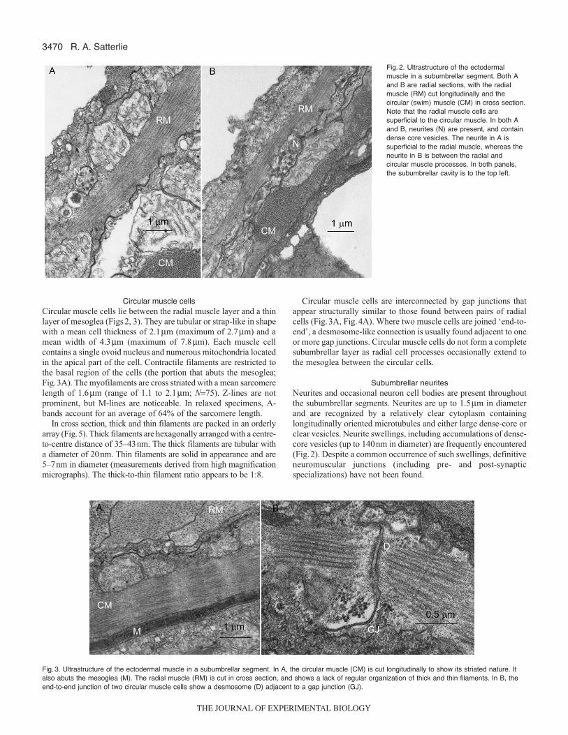
Subumbrellar ectodermTwo types of muscle cells make up the ectoderm of the interradialsegments. The outer layer is composed of surface epithelial cellswith basal muscular processes oriented in a radial direction. Themyofibrillar organization within the processes is of the smooth type(Fig.2). Situated between the radial muscle cells and the mesogleais a layer of circularly oriented, striated muscle cells (swim muscle).The radial muscle lies immediately over the striated muscle (Figs2and 3). Extensions of the circular muscle cells, to the free surfaceof the epithelium, have not been found. The ectoderm thus formsa pseudo-stratified epithelium.
Radial muscle cellsRadial muscle cells have a surface epithelial component and one ormore basal muscle processes (Fig.2). Viewed from the surface, thecells are up to 50μm in diameter (normally around 25μm). Thecells are drawn out basally into one or more muscle ‘feet’ that taperto less than 1μm and interdigitate with similar processes of otherradial muscle cells, and occasionally run between circular musclecells to the mesoglea. Radial muscle cells are very thin (meanthickness, 1.39μm; maximum 5μm) with elongate nuclei andvacuolated apical cytoplasm. The myofibrillar region of the radialcells includes thick and thin filaments that are oriented in the longaxis of the processes but otherwise randomly interspersed. Since noevidence of cross or oblique striations could be found, the radialmuscle is considered to be smooth muscle (Fig.2). In cross section,thick and thin filaments show no orderly or repetitive organization(Fig.3A). Thick filaments are tubular in shape with a diameter of20nm, whereas the thin filaments appear solid with a diameter of5–7nm. Elongate mitochondria are scattered throughout the radialcells and are frequently found among the myofilaments.
Radial muscle cells are interconnected by gap junctions, foundprimarily near the apical surface of the cells, but also occasionallydeeper in the muscle layer (Fig.4). In the junctional area, theintercellular space is 3–6nm as compared with 20–25nm in non-junctional areas. Similar junctions between radial muscle cells andthe underlying circular muscle cells were not found.
S
V
G
RCINR
SS
MC
Fig. 1. Diagram of an intact medusa and a ʻreduced preparationʼ consistingof three adjacent segments (viewed from the subumbrellar side). S,stomach; V, velum; G, gonad; SS, subumbrellar segment; RC, radial canal;MC, marginal canal; INR, inner nerve ring.
THE JOURNAL OF EXPERIMENTAL BIOLOGY
3470
Circular muscle cellsCircular muscle cells lie between the radial muscle layer and a thinlayer of mesoglea (Figs2, 3). They are tubular or strap-like in shapewith a mean cell thickness of 2.1μm (maximum of 2.7μm) and amean width of 4.3μm (maximum of 7.8μm). Each muscle cellcontains a single ovoid nucleus and numerous mitochondria locatedin the apical part of the cell. Contractile filaments are restricted tothe basal region of the cells (the portion that abuts the mesoglea;Fig.3A). The myofilaments are cross striated with a mean sarcomerelength of 1.6μm (range of 1.1 to 2.1μm; N=75). Z-lines are notprominent, but M-lines are noticeable. In relaxed specimens, A-bands account for an average of 64% of the sarcomere length.
In cross section, thick and thin filaments are packed in an orderlyarray (Fig.5). Thick filaments are hexagonally arranged with a centre-to-centre distance of 35–43nm. The thick filaments are tubular witha diameter of 20nm. Thin filaments are solid in appearance and are5–7nm in diameter (measurements derived from high magnificationmicrographs). The thick-to-thin filament ratio appears to be 1:8.
Circular muscle cells are interconnected by gap junctions thatappear structurally similar to those found between pairs of radialcells (Fig.3A, Fig.4A). Where two muscle cells are joined ‘end-to-end’, a desmosome-like connection is usually found adjacent to oneor more gap junctions. Circular muscle cells do not form a completesubumbrellar layer as radial cell processes occasionally extend tothe mesoglea between the circular cells.
Subumbrellar neuritesNeurites and occasional neuron cell bodies are present throughoutthe subumbrellar segments. Neurites are up to 1.5μm in diameterand are recognized by a relatively clear cytoplasm containinglongitudinally oriented microtubules and either large dense-core orclear vesicles. Neurite swellings, including accumulations of dense-core vesicles (up to 140nm in diameter) are frequently encountered(Fig.2). Despite a common occurrence of such swellings, definitiveneuromuscular junctions (including pre- and post-synapticspecializations) have not been found.
R. A. Satterlie
Fig. 2. Ultrastructure of the ectodermalmuscle in a subumbrellar segment. Both Aand B are radial sections, with the radialmuscle (RM) cut longitudinally and thecircular (swim) muscle (CM) in cross section.Note that the radial muscle cells aresuperficial to the circular muscle. In both Aand B, neurites (N) are present, and containdense core vesicles. The neurite in A issuperficial to the radial muscle, whereas theneurite in B is between the radial andcircular muscle processes. In both panels,the subumbrellar cavity is to the top left.
Fig. 3. Ultrastructure of the ectodermal muscle in a subumbrellar segment. In A, the circular muscle (CM) is cut longitudinally to show its striated nature. Italso abuts the mesoglea (M). The radial muscle (RM) is cut in cross section, and shows a lack of regular organization of thick and thin filaments. In B, theend-to-end junction of two circular muscle cells show a desmosome (D) adjacent to a gap junction (GJ).
THE JOURNAL OF EXPERIMENTAL BIOLOGY

3471Control of swimming in jellyfish
Electrophysiology of circular muscle cellsCircular muscle cells were routinely penetrated with microelectrodes,and were recognized by a one-to-one correspondence of depolarizingelectrical events and swim contractions. They were penetrated in allparts of the subumbrellar segments and in the gonad-less regionsoverlying radial canals. The mean resting potential for circularmuscle cells was –62.3mV (N=54).
With each swim contraction, the muscle cells exhibited one (orboth) of two types of depolarizing potentials. During ‘full swims’in the recorded segment, long duration action potentials (up to125mV above resting potential) were recorded (Figs6 and 7). Aswith epithelial cells of the inner nerve ring (Satterlie, 1985b), muscleaction potential duration was related to medusa size, and was up to700ms in some animals. The action potentials were variable inamplitude and duration, particularly when the area of recordingproduced a weak contraction or was inhibited during a radialresponse (Fig.6). The second type of recorded potential was anapparent junctional potential of relatively large size (up to 56mVamplitude) (Fig.6). Junctional potential duration was 160–200ms.Junctional potentials were recorded alone, or immediately preceding(giving rise to) action potentials in actively contracting segments(Fig.6). In partially inhibited preparations, action potentials showedseveral types of variability. In addition to variation in amplitudeand duration, they sometimes exhibited slow rise times with thereduced amplitudes, and either were not preceded by junctionalpotentials or exhibited junctional potentials that were smaller thanthose within the segments. Also, when junctional potentials werepresent with muscle action potentials, slight ‘hitches’ (delays) weresometimes observed between the peak of the junctional potentialand the rise of the action potential (compare the action potential inFig.6A,B). In some cases in which swimming was suppressed inthe recorded segment (via triggered radial responses), the recordedmuscle cell still exhibited a junctional potential, but also showed areduced-amplitude action potential followed by a second actionpotential that lacked a junctional potential and had a slower risetime (Fig.6C). The second action potential in this ‘saw-tooth’ patternis believed to result from electrotonic wash from an adjacent, activesegment, or if close to the margin, from a velar action potential.
In all areas of a subumbrellar segment except immediatelyadjacent to the margin, a single junctional potential was recordedper swim, even when it gave rise to a full action potential (Fig.6).In recordings from the vicinity of the inner nerve ring, multiplejunctional potentials gave rise to action potentials, similar to those
recorded from the post-synaptic epithelial cells that are coupled tothe muscle cells in this region (Satterlie, 1985b). This ‘bursty’junctional activity decreased with distance from the margin so withinapproximately 1mm from the margin and beyond, only a singlejunctional potential was recorded per swim contraction.
Dual intracellular recordings from two circular muscle cells ofthe same segment showed identical activity in terms of the relativeamplitudes of the action potentials (Fig. 7). Injection ofhyperpolarizing currents into one of the recorded muscle cellsshowed time-locked hyperpolarizations in the other recorded cells,confirming electrical coupling between the cells (Fig.7). Similarcoupling was found between a circular muscle cell in a subumbrellarsegment and another over the adjacent radial canal, as well asbetween cells of adjacent segments, although in the latter situation,very large currents had to be injected, and the associatedhyperpolarization was extremely small. In dual recordings of a
Fig. 4. Intercellular junctions between musclecells. (A) Circular muscle cells (CM) havelaterally positioned gap junctions (GJ) withother circular muscle cells. (B) Radial muscle(RM) cells have gap junctions (GJ) near theapical surface of the cells (they are alsopresent deeper in the ectoderm – not shown).
Fig. 5. In cross section, the striated circular muscle cells have an orderedarrangement of thick and thin filaments.
THE JOURNAL OF EXPERIMENTAL BIOLOGY

3472
circular muscle cell and a radial muscle cell, no electrical couplingcould be demonstrated (data not shown).
Injection of Lucifer Yellow into a single circular muscle cellproduced widespread dye movement from the injected cell to othercircular muscle cells (Fig.8A). No dye spread was noted from acircular muscle cell to radial muscle cells.
Intracellular recordings from radial muscle cells were difficult toobtain, compared with those of circular muscle cells, however, stablerecordings could be maintained for several minutes. The mean restingpotential was –61mV (N=21). The cells were silent unless stimulated.In particular, they were not active during swim contractions (Fig.6A),although field effects from the long duration circular muscle actionpotentials were frequently recorded (not believed to be movementartifacts since contractions lagged slightly behind action potentialgeneration). Relatively brief action potentials were recorded followingelectrical stimulation of the muscle sheet, and had a mean amplitudeof 76mV and a mean duration of 65ms (Fig.6A). Dual recordingsfrom pairs of radial muscle cells were not obtained, although dyeinjections with Lucifer Yellow demonstrated spread of dye tosurrounding radial (surface) cells, without spread to underlyingcircular muscle cells (Fig.8B). When electrically stimulated via adistant suction electrode, radial muscle cell action potentials werealways accompanied by junctional potentials in circular muscle cells(Fig.6A). Likewise, electrical stimuli that triggered a circular musclejunctional potential always initiated an action potential in a recordedradial muscle cell. Initiation of a radial muscle action potential viacurrent injection through the recording electrode (only two instances– data not shown) did not produce junctional potentials in circularmuscle cells, however, suggesting that the coincident appearance ofa radial muscle action potential and a circular muscle junctionalpotential is probably a result of their similar threshold to electrical
stimuli and the close apposition of the two cell types or of thesubumbrellar nerve nets.
Pattern of spread of circular muscle activityThe recording of junctional potentials in circular muscle cells fromall parts of a subumbrellar segment following stimulation of thetissue within that segment, and their presence at the beginning ofmuscle cell action potentials, both electrically stimulated andspontaneous, suggest that a subumbrellar nerve net participates inspread of electrical activity throughout each segment. This issupported, in part, by the presence of neurites in these regions(Fig.2). Yet, such a nerve net raises questions about how swimmingcan be ‘turned off’ in a portion of the subumbrella during localizedradial responses. This problem required examination of nerve netconducting properties within the subumbrella.
As shown in Fig.9, a recording electrode was placed in a circularmuscle cell in segment A, and an electrical stimulus was deliveredat a point represented by the letter A. In trace A (stimulating andrecording electrode in same segment), junctional potentials wererecorded in a 1:1 relationship with electrical stimuli, and thejunctional potentials were roughly the same size as those triggeringthe three spontaneous action potentials in the same trace.
When the stimulating electrode was moved to an adjacentsegment (segment B, stimulating position indicated by the letter B,and with the same recording position indicated by the letter R),junctional potentials were still recorded, but not with every stimulus.Trace B shows that only three of the four stimuli triggered ajunctional potential, the potentials occurred with a much longerlatency, suggesting a more circuitous conduction route. Furthermore,if repetitive stimuli were delivered at close to the normal swimfrequency of the intact animal (about 1Hz for the animals used),the faithfulness of junctional potential production in the adjacentsegment became very labile (data not shown).
The delay between the stimulus artifact and junctional potentialappearance was about 200ms in trace B, which is several timesgreater than the expected delay of about 35ms if the circularconduction velocity of swim contractions originating from the nerve
R. A. Satterlie
40 mV1 s
A
B
C
Fig. 6. Intracellular recordings from subumbrellar muscle cellsapproximately 2–2.5 mm from the nerve ring. (A) A double recording from aradial muscle cell (top trace) and a circular muscle cell (bottom trace). Aninitial spontaneous swim produced a full action potential (with initialjunctional potential) in the circular muscle cell, but only a ʻfield effectʼ in theradial cell. When electrical stimuli were delivered to the segment with asuction electrode, both a junctional potential (circular muscle cell) and anaction potential (radial muscle cell) were triggered. The small ʻblipʼ beforeeach event is the stimulus artifact. (B) Variability in the shape andamplitude of circular muscle action potentials. Each event is initiated by asingle junctional potential. All were spontaneously generated in a reducedpreparation that showed varying contractions. This recording was from asmall medusa, 3 cm bell diameter, so the action potentials are of a shorterduration. (C) Complex electrical event from a circular muscle cell in asegment in which a radial response was triggered by a mechanicalstimulus (light touch with a glass micropipette electrode) delivered to thevelum in the region of that segment. The contraction in that segment wasweaker than in the adjacent segments on each side.
25 mV1 s
Fig. 7. Dual recording from a pair of circular muscle cells within 3 mm of theinner nerve ring. All muscle action potentials were spontaneous. The breakin the traces represents a time break in the record. In the first segment, asingle hyperpolarizing current (approximately 1 nA) was injected into thecell represented by the lower trace. The small hyperpolarization in the toptrace (arrow) indicates the cells were electrically coupled. In the secondsegment, two current pulses were injected into the cell represented by thetop trace. Again, small hyperpolarizations in the other cell (arrows)demonstrated similar coupling in the opposite direction.
THE JOURNAL OF EXPERIMENTAL BIOLOGY

3473Control of swimming in jellyfish
ring is considered (43cms–1 conduction velocity). The delay in traceB was calculated as a circular conduction velocity between adjacentsegments of about 7.5 cm s–1, a speed only two-thirds of theconduction speed in a radial direction in a single segment (12cms–1).
When the stimulating electrode was moved over one more segment(Fig.9, segment C), so that it was two segments away from therecording site, electrical stimuli strong enough to trigger contractionsin the stimulated segment did not produce detectable junctionalpotentials or contractions in the recorded segment (Fig.9C). This entireexperimental series was repeated in three preparations from threedifferent animals with identical results, suggesting that the conductingsystem of an individual segment, responsible for production of circularmuscle cell junctional potentials, was not through-conducting in acircular direction around the bell, but was restricted between segments.By contrast, there seemed to be no such restriction in a radial direction,over similar distances, within each segment.
Evidence for participation of a subumbrellar nerve net in thespread of swim contractions
When stimulating a subumbrellar segment (with a suction electrode),junctional potentials in circular muscle cells appeared with a distinctthreshold, and when the stimulus was above that threshold, the sizeof the junctional potential did not vary. Similarly sized junctionalpotentials gave rise to full action potentials in the muscle cells whena spontaneous action potential was triggered by normal inner nervering activity (Fig.6; Fig.9A). Furthermore, identical junctionalpotentials could be stimulated in non-swimming preparations whenthe stimulating electrode was placed well above (more apical to)the recording electrode. When the preparation was bathed in high-magnesium seawater, swimming was inhibited, but so wasproduction of junctional potentials from direct electrical stimulationof subumbrellar segments (via a suction electrode). In theseexperiments, injection of current via the intracellular recordingelectrode showed the muscle cell was capable of electrogenesis.Finally, the presence of neurites detected in electron microscopepreparations was confirmed by immunohistochemical staining usinga commercial antibody to the neuroactive peptide, FMRFamide,which identified a diffuse nerve net within the subumbrellarsegments (Figs10 and 11). The neurites of this network of stainedneurons were preferentially oriented in a radial direction. Stainingwas noted over the radial canals between segments as well as withinthe segments. It also extended to the inner nerve ring but could notbe followed within the nerve ring because of high backgroundstaining in the region. Specifically, connections to immunoreactiveneurons within the inner nerve ring could not be resolved.
A comparative investigation of similar FMRFamide-immunoreactive networks in a number of hydromedusae suggestedthey are associated with radial muscle, including the broad radialmuscle sheets of leptomedusae, and not with the swimming system(Mackie et al., 1985). Mackie et al. noted that the stained neurons
were a subset of the total neuron number found in electronmicroscopical investigations of these same leptomedusae, suggestingthe presence of at least two independent nerve nets in thesubumbrella of these jellyfish. To test this, double-labelimmunohistochemical staining was conducted using the polyclonalantibody to FMRFamide (rabbit primary, Alexa Fluor 594secondary) and a monoclonal α-tubulin antibody (mouse primary,Alexa Fluor 488 secondary). The latter is a fairly non-specificneuronal marker in cnidarians (e.g. Satterlie, 2002), presumablystaining neurotubules.
In preparations using a combination of filters, both green (tubulin)and red (FMRFamide) cell bodies and neurites were visible in thesame focal plane of the subumbrellar segments (Fig.11A). Whenseparate filters were used, both types of neurons were apparent withthe FITC filter, whereas with the TRITC filter, only the
Fig. 8. (A) Lucifer Yellow fill of a circular muscleapproximately one-third of the way from the margin in asubumbrellar segment, showing widespread dyecoupling. The injected cell was in the middle of the brightspot. The small bright dots in the filled cells are nuclei.(B) Lucifer Yellow fill of a radial muscle cell roughly in themiddle of a subumbrellar segment. The dye spread tothe surface cells (radial muscle cells) throughout theepithelial tissue but not the underlying circular musclecells. Again, the nuclei show up as bright dots. The scalebar applies to both A and B.
40 mV
1 s
A
B
C
Fig. 9. Sequential experiment in which a single electrode was placed in acircular muscle cell (at the site marked R in the diagram), and a stimulatingsuction electrode was moved to the sites marked A, B and C. For trace A,the stimulus was delivered in the same segment as the recording site(stimulus site A), and a junctional potential was triggered with a shortlatency. Three spontaneous action potentials were also recorded, and eachwas initiated by a junctional potential of similar size to those electricallystimulated (note the slight inflection on the rising phase of the actionpotentials). When the stimulating electrode was moved over one segment(stimulus site B and trace B), junctional potentials were triggered by threeof the four stimuli, but with a longer latency than in trace A. Following thesecond and fourth stimuli, weak contractions were initiated. The electrodewas then moved over one more segment (stimulus site C and trace C).Electrical stimuli sufficient to produce weak contractions in the stimulatedsegment did not initiate junctional potentials in the recorded segment (twosegments over). A few junctional potentials were recorded during theseexperiments (one is shown in this trace), however, they showed no regularrelationship to imposed stimuli. It appears these are spontaneous events.Delivery of stimuli is marked by the stimulus artifacts (arrows). Therecording site was 2.5 mm from the top of the nerve ring.
THE JOURNAL OF EXPERIMENTAL BIOLOGY

3474
FMRFamide-immunoreactive neurons were visible. For example,in Fig.11B,C, one of the tubulin-immunoreactive neurons is clearlynot stained with the FMRFamide antibody. Similar results wereobtained in all parts of the subumbrellar segments in four differentdouble-labelled preparations (four different animals), indicating thepresence of two distinct nerve nets (at least based onimmunohistochemical staining) in the subumbrellar of Aequorea.
An interesting aspect of junctional potential production is therelationship between action potentials in the swim generatingnetwork of the inner nerve ring, the putative nerve net that innervatesthe swim muscles, and the action potentials in the circular musclecells themselves. In the inner nerve ring, each swim is triggered bya rapid burst of action potentials in the swim-generating neuralnetwork (Satterlie, 1985a). This is translated into a burst of junctionalpotentials and a resulting single action potential in the overlyingepithelial cells (Satterlie, 1985b). These epithelial cells are inelectrical contact with the circular muscle cells of the subumbrellaand velum via gap junctions. Yet, away from the margin, only singlejunctional potentials were recorded in circular muscle cells with eachaction potential (Fig.6), suggesting the action potential burstsobserved in the motoneurons are converted to single action potentialsin the putative subumbrellar motor nerve net.
How does a local radial response turn off swimming in arestricted portion of the bell?
Input from the motor network of the inner nerve ring, to the swimmuscle, includes a direct synaptic activation of epithelial cellsoverlying the neurons and electrotonic spread of activity from theepithelial cells to the circular muscle cells. In addition, synaptic inputfrom a putative subumbrellar nerve net supplements the excitationto the muscle cells. During a localized radial response, the burstproduction in the motor network was partially or totally inhibited(Fig.12), blocking the direct synaptic input to the epithelial cells,and presumably the activation of the subumbrellar nerve net.However, if the region of inhibition was not too broad, muscle cellaction potentials (and contractions) of the subumbrellar muscle cellscould still be recorded, although with lower amplitudes, an absenceof junctional potentials and slower rise times (Fig. 12). Inpreparations in which the area of inhibition involved several
segments on either side of the recorded segment, muscle actionpotentials were not recorded, and swimming in that segment wastotally blocked. This suggests that the electrotonic spread of actionpotentials through the subumbrellar gap junctions is decreasing. Ifso, the putative motor nerve net may play a crucial role in conductionof a full action potential (and contraction) within each segment. Thiswould allow a decrease, or total inhibition, of contractility in themuscle sheet by a localized inhibition of activity in the segmentalregions of the subumbrellar nerve net.
DISCUSSIONIn a comparative sense, the architecture of the hydromedusan nervoussystem has a basal organization upon which specializations aresuperimposed to account for differences in behavioural ecology ofthe individual subgroups and specifically of individual species. Onebasic structural and functional commonality seems to be theorganization of the motor network for swimming in the inner nervering (Satterlie and Spencer, 1983; Satterlie, 2002). The electricallycoupled network of large neurons provides a means for rapid circulardistribution of motor activation around the bell, and also a means ofreceiving symmetrical and asymmetric inputs from sensory structuresthat are distributed around the margin of the animal (Spencer andArkett, 1984; Arkett and Spencer, 1986a; Arkett and Spencer, 1986b;Arkett et al., 1988). This is ultimately translated into locomotoryactivity that is dependent upon coordinated contraction of circularmusculature that lines the inside of the bell – another common featurethat produces a forceful ejection of water from the bell cavity (Singla,1978a; Spencer, 1978; Spencer, 1979; Spencer, 1981; Spencer and
R. A. Satterlie
Fig. 11. Double-label immunohistochemistry using α-tubulin (green) andFMRFamide (red) antibodies. In A, a dual filter shows the two colourstogether. The small arrows indicate FMRFamide cell bodies and neurites,and the large arrows indicate some of the tubulin-stained cell bodies. B andC are from identical areas (no change in focus) with a change from a FITCfilter (B) to a TRITC filter (C). Note the neuron indicated by the arrow in Bis not present in C indicating it was immunoreactive to the tubulin antibody,but not to FMRFamide antibody. This suggests the presence of two distinctneuronal types in the subumbrellar segments of Aequorea.
Fig. 10. FMRFamide immunoreactivity in a subumbrellar segment ofAequorea shows a diffuse nerve net with a definite oral–aboral orientationof neurites (direction indicated by arrow). The region was about one-third ofthe segment length up from the margin.
THE JOURNAL OF EXPERIMENTAL BIOLOGY

3475Control of swimming in jellyfish
Satterlie, 1981; Satterlie, 1985b; Satterlie, 2002; Kerfoot et al., 1985;Satterlie and Spencer, 1983; Mackie, 2004).
In the several medusae studied, the swim motor network of theinner nerve ring is composed of a compressed network of oversizedneurons that activate overlying epithelial cells via chemical synapses(Anderson and Mackie, 1977; Anderson, 1979; Spencer andSatterlie, 1980; Satterlie and Spencer, 1983; Spencer, 1981; Spencer,1982; Satterlie, 1985a; Satterlie, 1985b; Satterlie, 2002; Mackie andMeech, 2000; Lin et al., 2001; Mackie, 2004). Either local ordistributed nerve nets of smaller neurons may further conductexcitation from the motor network to the muscles, as seen in theextreme in Aequorea where a subumbrellar nerve net is foundthroughout the muscle sheets (Fig.13). Other specializations aresuperimposed on the basic organization, as in Aglantha, where motorgiant neurons distribute impulses between the motor network andthe swim musculature (Weber et al., 1982; Kerfoot et al., 1985;Mackie and Meech, 2000; Mackie, 2004). Also, use of the word‘oversized’ to refer to the motor network of Aglantha loses somemeaning since the neurons are dwarfed by the ring giant (in theouter nerve ring) that is involved in escape swimming (see Mackie,2004). Again, however, this illustrates how species-specific neuronalpeculiarities are superimposed on the basic system.
In Aequorea, the pacemaker/motor network of the inner nervering is somewhat unusual in that it produces a burst of actionpotentials to trigger each individual swim contraction (Satterlie,1985a). Other medusae examined produce a single action potentialper swim (Anderson and Mackie, 1977; Anderson, 1979; Spencerand Satterlie, 1980; Satterlie and Spencer, 1983; Spencer, 1981).The action potential burst in Aequorea induces a burst ofjunctional potentials in the postsynaptic epithelial cells thatoverlie the inner nerve ring, and in the immediately adjacentcircular muscle cells, which are electrically coupled to theepithelial cells (Fig. 13) (Satterlie, 1985b). Electrical coupling isfound throughout the circular muscle sheet; this represents anotherbasic feature of hydromedusan locomotory systems that aids in
the spread of excitation between muscle cells. The degree of‘coupling’ varies so aneural conduction is sufficient to transmita muscle action potential throughout the muscle sheet withoutdecrement in Polyorchis (Spencer and Satterlie, 1981; Spencer,1982), whereas the same conduction is apparently decrementalin Aglantha [in slow swimming (Kerfoot et al., 1985; Mackie,2004)] and in Aequorea (this study). This variation in muscle cell‘coupling’ may be due to differences in gap junctionalconductance, to threshold differences for full action potentialproduction, differences in the regenerative properties of theaction potentials, or a combination of these and other properties.As an example, in Polyorchis, the muscle action potential isconducted through the aneural circular muscle sheet withoutdecrement when it is generated via normal inner nerve ringactivity. However, the epithelial cells in the region of the innernerve ring (the neuromuscular synaptic region) form anincrementally conducting region that requires the simultaneousinput of junctional potentials from multiple sites to produce theconducted action potentials in the muscle sheet. A compressednetwork of small neurons is found in this area (Spencer, 1982)that presumably serves as a distributor network for additionalneuromuscular input. In this way, this restricted region may actlike the entire subumbrella of Aequorea, and the distributornetwork may serve the same function as Aequorea’s subumbrellarmotor nerve net. In this case, it is interesting to suggest that themotor system of Aequorea represents a more diffuse organizationof this non-regenerative band of Polyorchis, or that the latter isa compressed version of the former.
Regardless, the nature of subumbrellar conduction holdsimportant consequences for the control of swimming, and inparticular, the effect of other behaviours on swimming, as seen herein Aequorea. In other words, when the two basic properties ofhydromedusan swimming systems are considered (electricallycoupled motor network that synapses onto postsynaptic epithelialcells, and widespread electrical coupling throughout the circular
40 mV
1 s
A
B
Fig.12. Double recordings from a swim motor neuron in the inner nerve ring(top traces of each pair) and a circular muscle cell approximately 0.5mmfrom the neuron recording site (bottom traces). In A, a spontaneous radialresponse occurred between the first and second swim. Note the inhibition ofneuronal action potential burst and the smaller muscle event with a slow risetime. In the third swim, the preparation had partially recovered from theinhibition, and it was fully recovered by the fourth swim. (B)Another radialresponse from the same preparation produced total inhibition of theneuronal burst in the second swim (which occurs after a long delay), asingle neuronal spike in the third swim, and full recovery on the fourth. Notethe changes in the muscle action potentials during the inhibition.
Fig. 13. Schematic of the neuromuscular organization of the subumbrella ofAequorea. The arrows represent excitatory chemical connections and thebars, inhibitory connections. The resistor symbols represent electricalcoupling, as do the dotted lines within the radial and circular muscle boxes.FMRF, FMRFamide-immunoreactive nerve net of the subumbrella; MNN,motor nerve net of the subumbrella; ???, unknown pathway responsible foractivation of the FMRFamide-immunoreactive nerve net followingmechanical stimulation of the margin. The circular and radial muscle sheetsrefer to the musculature from a single subumbrellar segment.
THE JOURNAL OF EXPERIMENTAL BIOLOGY

3476
muscle sheets), the organization and physiology of these two basicfeatures is where we see significant variation that is related to thestructure of the medusa as well as its lifestyle.
In terms of neuromuscular control, perhaps the simplest case isthat of the anthomedusa, Polyorchis. The electrically coupled motornetwork of the inner nerve ring extends up the four radii and acrossthe top of each quadrant (Lin et al., 2001). This provides synapticactivation of the four circular muscle sheets along the entireperiphery of each quadrant. As mentioned above, transmission inthe muscle sheet is strictly via gap junctions. Radial muscle is notpresent in the quadrants, instead it is restricted to smooth musclestrips overlying the radii, so that radial responses in these prolatemedusae, including crumpling, are triggered by radial contractionat the four radii (Singla, 1978a; Spencer, 1978; Spencer, 1979).
In what is possibly the most complex elaboration, Aglantha (atrachyline medusa) has the ‘basic’ organization for slow swimming,however, giant neurons are involved in this and in a faster escaperesponse, with two distinct kinds of action potentials in the motorgiants serving as the muscle input that determines the type ofsubumbrellar contraction [slow or escape (Mackie and Meech, 1985;Meech and Mackie, 1993; Mackie, 2004)].
In many of the disc-shaped leptomedusae, including Aequorea,radial muscle is not restricted to discrete bands, but rather forms sheetsthat overlie the circular swim muscle (Satterlie and Spencer, 1983).Radial responses thus rely on more diffuse radial musculature(Fig.13), possibly because of the challenge of the inward curling ofthe wide, oblate bell. A unique behavioural peculiarity of Aequorea,with its large number of subumbrellar segments, centres on its abilityto invert a variable number of segments, and to inhibit swimming inthose segments only (Satterlie, 1985a). Although the behaviouralsignificance of this is obvious, the mechanisms required to turn offswimming in a variable portion of the subumbrella, with the basicswim system organization mentioned above, is not so simple.
The excitability of the subumbrellar circular muscle sheet ofAequorea appears to be less than that of ‘normally’ triggeredcontractions in Polyorchis. In the former species, muscle activitythat spreads from one subumbrellar segment to another shows adecrease in amplitude and rise time characteristic of decrementaltransmission through gap junctions. For normal swimming, thiselectrotonic current spread could require ‘supplemental’ synapticinput from a peripheral nerve net to bring muscle cell electrogenesisto that of full action potentials. A full contraction in a subumbrellarcircular muscle cell would thus require a combination of electrotoniccurrent spread and synaptic input.
Evidence for a subumbrellar motor nerve net (in addition to theelectron microscopical data) includes both electrophysiological andimmunohistochemical data. Although the motor network in the innernerve ring produces a burst of action potentials per swim, and thisis reflected in the junctional potentials seen in the postsynapticepithelial cells, the synaptic inputs to circular muscle cellsthroughout each subumbrellar segment consist of a single junctionalpotential. This holds for normally initiated swims and forcontractions initiated by direct electrical stimulation of thesubumbrellar tissue. In the latter case, stimulation of any point ofa subumbrellar segment produces a junctional potential andcontraction in the muscle cells of that segment, indicating a non-directional transmission in the putative nerve net.
Immunohistochemical experiments with a commercialFMRFamide antibody show the presence of a subumbrellar nervenet. Neuronal RFamides appear to be ubiquitous within the Cnidaria(Grimmelikhuijzen, 1983; Grimmelikhuijzen et al., 1996), and theyare neuroactive (Spencer, 1988). The presence of an FMRFamide-
immunoreactive nerve net in Aequorea has to be viewed withcaution, however, since data from a number of other hydromedusaesuggest that RFamide-immunoreactive subumbrellar networks areassociated with radial, smooth muscle rather than the striated swimmusculature (Grimmelikhuijzen and Spencer, 1984; Mackie et al.,1985). Perhaps the best data, in this regard, come from theanthomedusa, Podocoryne, where double labelling with an antibodyspecific for cnidarian smooth muscle was coupled with aFMRFamide antibody to directly show the structural correlation(Weber, 1989). However, in their comparative study of severalhydromedusae, Mackie et al. (Mackie et al., 1985) found the densityof subumbrellar neurites exceeded the density of FMRFamide-immunoreactive neurons, suggesting the possibility of multiple,separate nerve nets in the subumbrellar ectoderm. In Aequorea, thispossibility is backed up by data from double labelling experiments,which clearly show the presence of two separate nerve nets in thesubumbrellar segments, only one of which stains with theFMRFamide antibody. This is currently under further investigationin Aequorea using electron microscopical examination ofimmunohistochemical preparations stained for FMRFamide.
The extreme variability in muscle cell action potential size andshape in Aequorea is not always completely ‘predictable’ in eithera normal swimming preparation or in one that is undergoing aspontaneous or triggered radial response. Current evidence suggestsseveral neuromuscular properties of the subumbrellar motor nervenet may contribute to this level of electrophysiological (andcontractile) variability. First, synchrony of inputs to the subumbrellarmuscle cells may be important in production of full muscleelectrogenesis. If an isolated muscle cell is depolarized, thesurrounding, coupled muscle cells may represent a current sink thatcould decrease the amplitude and slow the rise time of the junctionalpotentials, thus decreasing their efficacy. Simultaneousdepolarization of all neighbouring muscle cells would significantlydecrease the current sink effect. Synchrony of activity within theinner nerve ring motor network of Polyorchis was found to influenceaction potential shape (Spencer, 1981), and the same could be truefor the subumbrellar muscle cells of Aequorea. Any inhibitory inputthat decreases the synchrony of synaptic inputs to the muscle sheetcould alter the amplitude and rise time of muscle action potentials.This was found in the postsynaptic epithelial cells of Aequoreaduring radial responses, where a delayed production of motornetwork spike bursts produced smaller postsynaptic action potentialswith slow rise times compared to swims with unaltered synchrony(Satterlie, 1985b). It may also explain the slight delay between thepeak of the junctional potentials and the rise of the action potentialsseen in Fig.6B (compared to the full action potential in Fig.6A),and more clearly in the three full action potentials in Fig.12A.
Second, the subumbrellar nerve nets of Aequorea show a distinctpreferential orientation (radially; Fig.10). This could also reflect apreferred physiological orientation as well, particularly if the overallpathway in one direction is restricted, as seen in the inter-segmentalregions because of the radial canal gonadal tissue. Restriction ofconduction is suggested by the data in Fig.9. As an extreme case,a nerve net could be through-conducting in the preferred direction(radial in this case) and incrementally conducting in theperpendicular (circular) direction. This could show up as a labileconduction of repetitive events in the nerve net, including failuresand re-appearances. An incrementally conducting nerve net has beendescribed in an anthozoan (Anderson, 1976). Also possible,however, are alterations in conduction patterns in a through-conducting nerve net that merely alter the synchrony of activitywithin the nerve net with consequences as previously mentioned.
R. A. Satterlie
THE JOURNAL OF EXPERIMENTAL BIOLOGY

3477Control of swimming in jellyfish
Third, the action potentials of swim muscles of Aequorea arevariable in amplitude even during normal swimming bouts (seeFig.7). In a rested preparation, a swimming bout is initiated byepithelial (and muscle) action potentials that increase in amplitudewith the first several swimming contractions, reminiscent ofneuromuscular facilitation (Satterlie, 1985b). Despite this, nosignificant correlation could be demonstrated between actionpotential amplitude and the preceding inter-spike interval, suggestingthat a strong frequency-dependent facilitation is not in operation(unpublished observations). Yet it appears that muscle cellelectrogenesis is influenced by the electrophysiological history ofthe cell, and this could contribute to some of the observed variability.
Finally, as an unexplored possibility, the swim muscle cells maybe directly sensitive to the neuroactive substance (presumed anRFamide) released from the FMRFamide-immunoreactive neuronsduring a radial response. In this scenario, the nerve net associatedwith the radial muscle would have opposing effects on the twomuscle types. Thus two levels of chemical inhibition could be actingon the swim system during a radial response: direct inhibition ofthe inner nerve ring motor network (Satterlie, 1985a; Satterlie,1985b) and a peripheral modulatory action on the muscle cells. Thusfar, permeability difficulties posed by the tight epithelium of thesubumbrella have prevented direct testing of this possibility.
A combination of these nerve net and neuromuscular properties,in conjunction with the electrical coupling parameters of the swimmuscle sheet, may be required to explain the full range of variabilityseen in the swim muscle cell recordings during normal swimmingand during radial responses. In any event, the ‘turning off’ ofrestricted subumbrellar segments appears to be based on both central(nerve ring) and peripheral (subumbrellar) mechanisms. Add to thisthe ability to totally inhibit activation of the subumbrellar nerve net(as shown in Fig.12), sometimes with action potentials of apparentlyelectronic origin, and it is apparent that a flexible range of muscleforce suppression/inhibition can be achieved in this system.
As a final word on the swimming system of hydromedusae, theorganization of the motor network of the inner nerve ring isinfluenced by parallel sensory networks of the inner and outer nerverings (Spencer and Arkett, 1984; Arkett and Spencer, 1986a; Arkettand Spencer, 1986b; Arkett et al., 1988; Mackie, 2004). Furthermore,fourteen distinct conducting systems have been described inAglantha (reviewed by Mackie, 2004). In Aequorea, multiplestatocysts are found throughout the bell margin (Singla, 1975), andrighting responses to tilt involve asymmetrical swimmingcontractions of the subumbrella, possibly including the localinhibitory mechanisms discussed here. These observations suggestthat parallel and point-source pathways provide excitatory orinhibitory input to the swim motor network in the inner nerve ringof hydromedusae, pointing to an integrative organization that formswhat can be argued to be a true central nervous system in theseradially symmetrical organisms. In these animals, the ‘primitive’condition of diffuse, non-directional nerve nets has been modifiedin some conducting systems into a series of compressed, function-specific nerve networks that interact in the complex neural structuresof the inner and outer nerve rings. As an example of the richnessof function of the hydromedusan nervous system, one has to lookno further than the organization of Aglantha (Mackie, 2004) to beimpressed with the neural complexity of this ‘simple’ animal group.
I thank Dr Dennis Willows, Director Emeritus, and Dr Ken Sebens, Director of theFriday Harbor Laboratories (University of Washington), for providing space andassistance throughout this investigation. Claudia Mills provided valuableinformation on the biology of medusae in the Friday Harbor region. This study wassupported in part by the Frank Hawkins Kenan Professorship Endowment at
UNCW. The α-tubulin antibody developed by J. Frankel and E. M. Nelsen wasobtained from the Developmental Studies Hybridoma Bank developed under theauspices of the NICHD and maintained by The University of Iowa, Department ofBiological Sciences, Iowa City, IA 52242, USA.
REFERENCESAnderson, P. A. V. (1976). An electrophysiological study of mechanisms controlling
polyp retraction in colonies of the scleractinian coral Goniopora lobaba. J. Exp. Biol.65, 381-393.
Anderson, P. A. V. (1979). Ionic basis of action potentials and bursting activity in thehydromedusan jellyfish Polyorchis penicillatus. J. Exp. Biol. 78, 299-302.
Anderson, P. A. V. and Mackie, G. O. (1977). Electrically coupled, photosensitiveneurons control swimming in a jellyfish. Science 197, 186-188.
Arkett, S. A. and Spencer, A. N. (1986a). Neuronal mechanisms of a hydromedusanshadow reflex. I. Identified reflex components and sequence of events. J. Comp.Physiol. A. 159, 201-213.
Arkett, S. A. and Spencer, A. N. (1986b). Neuronal mechanisms of a hydromedusanshadow reflex. II. Graded response of reflex components, possible mechanisms ofphotic integration, and functional significance. J. Comp. Physiol. A. 159, 215-225.
Arkett, S. A., Mackie, G. O. and Meech, R. W. (1988). Hair cell mechanoreception inthe jellyfish Aglantha digitale. J. Exp. Biol. 135, 329-342.
Colin, S. P. and Costello, J. H. (2002). Morphology, swimming performance andpropulsive mode of six co-occurring hydromedusae. J. Exp. Biol. 205, 427-437.
Grimmelikhuijzen, C. J. P. (1983). FMRFamide immmunoreactivity is generallyoccurring in the nervous system of coelenterates. Histochemistry 78, 361-381.
Grimmelikhuijzen, C. J. P. and Spencer, A. N. (1984). FMRFamide immunoreactivityin the nervous system of the medusa Polyorchis penicillatus. J. Comp. Neurol. 230,361-371.
Grimmelikhuijzen, C. J. P., Leviev, I. and Carstensen, K. (1996). Peptides in thenervous systems of cnidarians: structure, function and biosynthesis. Int. Rev. Cytol.167, 37-89.
Kerfoot, P. A. H., Mackie, G. O., Meech, R. W., Roberts, A. and Singla, C. L. (1985).Neuromuscular transmission in the jellyfish Aglantha digitale. J. Exp. Biol. 116, 1-25.
Lin, Y.-C. J., Gallin, W. J. and Spencer, A. N. (2001). The anatomy of the nervoussystem of the hydrozoan jellyfish Polyorchis penicillatus, as revealed by amonoclonal antibody. Invert. Neurosci. 4, 65-75.
Mackie, G. O. (1975). Neurobiology of Stomotoca. II. Pacemakers and conductionpathways. J. Neurobiol. 6, 357-378.
Mackie, G. O. (2004). Central neural circuitry in the jellyfish Aglantha. Neurosignals13, 5-19.
Mackie, G. O. and Meech. R. W. (1985). Separate sodium and calcium spikes fromthe same axon. Science 313, 791-793.
Mackie, G. O. and Meech, R. W. (2000). Central circuitry in the jellyfish Aglanthadigitale. III. The rootlet and pacemaker systems. J. Exp. Biol. 203, 1797-1807.
Mackie, G. O., Singla, C. L. and Stell, W. K. (1985). Distribution of nerve elementsshowing FMRFamide-like immunoreactivity in hydromedusae. Acta Zool. 66, 199-210.
Meech, R. W. and Mackie, G. O. (1993). Ionic currents in giant motor axons of thejellyfish, Aglantha digitale. J. Neurophysiol. 69, 884-893.
Satterlie, R. A. (1985a). Central generation of swimming activity in the hydrozoanjellyfish Aequorea aequorea. J. Neurobiol. 16, 41-55.
Satterlie, R. A. (1985b). Control of swimming in the hydrozoan jellyfish Aequoreaaequorea: direct activation of the subumbrella. J. Neurobiol. 16, 211-226.
Satterlie, R. A. (2002). Neuronal control of swimming in jellyfish: a comparative story.Can. J. Zool. 80, 1654-1669.
Satterlie, R. A. and Spencer, A. N. (1983). Neuronal control of locomotion inhydrozoan medusae: a comparative story. J. Comp. Physiol. 150, 195-206.
Singla, C. L. (1975). Statocysts of hydromedusae. Cell Tissue Res. 158, 391-407.Singla, C. L. (1978a). Fine structure of the neuromuscular system of Polyorchis
penicillatus (Hydromedusae, Cnidaria). Cell Tissue Res. 193, 163-174.Singla, C. L. (1978b). Locomotion and neuromuscular system of Aglantha digitale. Cell
Tissue Res. 188, 317-327.Spencer, A. N. (1975). Behavior and electrical activity in the hydrozoan
Proboscidactyla flavicirrata (Brandt). II. the medusa. Biol. Bull. 148, 236-250.Spencer, A. N. (1978). Neurobiology of Polyorchis. I. Function of effector systems. J.
Neurobiol. 9, 143-157.Spencer, A. N. (1979). Neurobiology of Polyorchis. II. Structure of effector systems. J.
Neurobiol. 10, 95-117.Spencer, A. N. (1981). The parameters and properties of a group of electrically
coupled neurons in the central nervous system of a hydrozoan jellyfish. J. Exp. Biol.93, 33-50.
Spencer, A. N. (1982). The physiology of a coelenterate neuromuscular synapse. J.Comp. Physiol. 148, 353-363.
Spencer, A. N. (1988). Effects of Arg-Phe-amide peptides on identified motor neuronsin the hydromedusa Polyorchis penicillatus. Can. J. Zool. 66, 639-645.
Spencer, A. N. and Arkett, S. A. (1984). Radial symmetry and the organization ofcentral neurons in a hydrozoan jellyfish. J. Exp. Biol. 110, 69-90.
Spencer, A. N. and Satterlie, R. A. (1980). Electrical and dye coupling in an identifiedgroup of neurons in a coelenterate. J. Neurobiol. 11, 13-19.
Spencer, A. N. and Satterlie, R. A. (1981). The action potential and contraction insubumbrellar swimming muscle of Polyorchis penicillatus (Hydromedusae). J. Comp.Physiol. 144, 401-407.
Stewart, W. W. (1978). Functional connections between cells as revealed by dye-coupling with a highly fluorescent naphthalamide tracer. Cell 14, 741-759.
Weber, C. (1989). Smooth muscle fibers of Podocoryne carnea (Hydrozoa)demonstrated by a specific monoclonal antibody and their association with neuronsshowing FMRFamide-like immunoreactivity. Cell Tissue Res. 255, 275-282.
Weber, C., Singla, C. L. and Kerfoot, P. A. H. (1982). Microanatomy of thesubumbrellar motor innervation in Aglantha digitale. (Hydromedusae: Trachylina).Cell Tissue Res. 223, 305-312.
THE JOURNAL OF EXPERIMENTAL BIOLOGY