control of ccnd1 ubiquitylation by the catalytic saga ... · edited by charles j. sherr, hhmi and...
TRANSCRIPT

Control of CCND1 ubiquitylation by the catalytic SAGAsubunit USP22 is essential for cell cycle progressionthrough G1 in cancer cellsVictoria J. Gennaroa, Timothy J. Staneka, Amy R. Peckb, Yunguang Sunb, Feng Wangc, Shuo Qied, Karen E. Knudsene,Hallgeir Ruib, Tauseef Buttc, J. Alan Diehld, and Steven B. McMahona,1
aDepartment of Biochemistry and Molecular Biology, Sidney Kimmel Medical College, Thomas Jefferson University, Philadelphia, PA 19107; bDepartment ofPathology, Medical College of Wisconsin, Milwaukee, WI 53226; cProgenra, Inc., Malvern, PA 19355; dDepartment of Biochemistry and Molecular Biology,Medical University of South Carolina, Charleston, SC 29425; and eDepartment of Cancer Biology, Sidney Kimmel Medical College and Sidney Kimmel CancerCenter, Thomas Jefferson University, Philadelphia, PA 19107
Edited by Charles J. Sherr, HHMI and St. Jude Children’s Research Hospital, Memphis, TN, and approved August 10, 2018 (received for review May 8, 2018)
Overexpression of the deubiquitylase ubiquitin-specific peptidase22 (USP22) is a marker of aggressive cancer phenotypes likemetastasis, therapy resistance, and poor survival. Functionally,this overexpression of USP22 actively contributes to tumorigene-sis, as USP22 depletion blocks cancer cell cycle progression in vitro,and inhibits tumor progression in animal models of lung, breast,bladder, ovarian, and liver cancer, among others. Current modelssuggest that USP22 mediates these biological effects via its role inepigenetic regulation as a subunit of the Spt-Ada-Gcn5-acetyltransferase(SAGA) transcriptional cofactor complex. Challenging the dogma,we report here a nontranscriptional role for USP22 via a directeffect on the core cell cycle machinery: that is, the deubiquitylationof the G1 cyclin D1 (CCND1). Deubiquitylation by USP22 protectsCCND1 from proteasome-mediated degradation and occurs sepa-rately from the canonical phosphorylation/ubiquitylation mechanismpreviously shown to regulate CCND1 stability. We demonstrate thatcontrol of CCND1 is a key mechanism by which USP22 mediates itsknown role in cell cycle progression. Finally, USP22 and CCND1 levelscorrelate in patient lung and colorectal cancer samples and ourpreclinical studies indicate that targeting USP22 in combination withCDK inhibitors may offer an approach for treating cancer patientswhose tumors exhibit elevated CCND1.
USP22 | CCND1 | cell cycle | SAGA | deubiquitylation
Ubiquitin-specific peptidase 22 (USP22) is a deubiquitylatingenzyme that functions as a subunit of the human Spt-Ada-
Gcn5-acetyltransferase (SAGA) transcriptional coactivator com-plex (1–3). Within SAGA, USP22 catalyzes histone H2A and H2Bdeubiquitylation, thereby altering chromatin structure and genetranscription (1, 2, 4). Elevated USP22 expression is tightly cor-related with aggressive behavior in human cancer and it was ini-tially identified as a member of an 11-gene “death-from-cancer”signature linked to poor prognosis (5, 6). Further investigationfound that USP22 is not only a marker of aggressive tumor phe-notypes but also plays a causal role in driving aggressive cancer. Atthe cellular level, USP22 is critical for progression through theG1 phase of the cell cycle (7–13).Biochemically, USP22 deubiquitylates substrates beyond nu-
cleosomal histones, including FBP1 and TRF1 (14, 15). How-ever, there has been limited success in linking direct substrates ofUSP22 to its potent biological phenotypes, including its role inappropriate G1–S transition. To generate a comprehensive un-derstanding of USP22 substrates and how they might contributeto USP22 function, we conducted an unbiased proteome-widescreen. While the screen confirmed known substrates of USP22,including histones H2A and H2B, the sole component of thecore cell cycle machinery identified as a USP22 substrate in thisscreen was the G1 phase cyclin CCND1.Further analysis of the USP22–CCND1 relationship revealed
that USP22 directly reverses polyubiquitylation of CCND1 via a
pathway distinct from protein-destabilizing GSK3β phosphoryla-tion and ultimately protects CCND1 from proteasome-mediateddegradation (16, 17). D-type cyclins are rate-limiting regulators ofG1–S progression in mammalian cells, primarily via their ability tobind and activate the kinases CDK4 and CDK6 (18). Consistentwith the control of CCND1 levels by USP22, Food and DrugAdministration (FDA)-approved CDK inhibitors are epistaticwith USP22 depletion in the growth suppression of cancer cells.Collectively, these findings define a previously unknown pathwayof CCND1 regulation and establish a mechanistic link betweenUSP22, CCND1, and cancer cell cycle progression that expandspotential therapeutic strategies for cancers with elevated CCND1.
ResultsLoss of USP22 in Cancer Cells Results in Defective G1/S Cell CycleTransition. The ubiquitin hydrolase USP22 has been causallylinked to aggressive growth in human tumor cells in vitro and invivo (1, 8–13, 19–25). Consistent with this, depletion of USP22 inthe nonsmall-cell lung cancer line H1299 results in a markeddecrease in cell number (Fig. 1 A and B). This observation was
Significance
The deubiquitylase USP22 is frequently overexpressed in can-cer and contributes to tumorigenesis by driving cell cycle pro-gression. Current models define USP22 as functional mediatorof gene regulation and chromatin modification, workingwithin the SAGA transcriptional cofactor complex. Here wereport a catalytic role for USP22 distinct from its well-characterized transcription regulatory capacity. USP22 directlystabilizes the essential G1-cyclin, CCND1, protecting it fromproteasome-mediated degradation via deubiquitylation. Ourfindings reveal a pathway that regulates CCND1, while alsoraising the possibility that simulteously targeting USP22 mayallow the use of less toxic doses of the new wave of cancertherapies that target the cyclin/CDK complex. Finally, theseresults provide a mechanistic explanation for the effects ofUSP22 in cancer cell cycle control.
Author contributions: V.J.G., T.J.S., J.A.D., and S.B.M. designed research; V.J.G., T.J.S.,A.R.P., Y.S., F.W., and S.Q. performed research; K.E.K., H.R., T.B., and J.A.D. contributednew reagents/analytic tools; V.J.G., T.J.S., and S.B.M. analyzed data; and V.J.G. and S.B.M.wrote the paper.
Conflict of interest statement: The authors declare no conflict of interest.
This article is a PNAS Direct Submission.
This open access article is distributed under Creative Commons Attribution-NonCommercial-NoDerivatives License 4.0 (CC BY-NC-ND).1To whom correspondence should be addressed. Email: [email protected].
This article contains supporting information online at www.pnas.org/lookup/suppl/doi:10.1073/pnas.1807704115/-/DCSupplemental.
Published online September 17, 2018.
E9298–E9307 | PNAS | vol. 115 | no. 40 www.pnas.org/cgi/doi/10.1073/pnas.1807704115
Dow
nloa
ded
by g
uest
on
Dec
embe
r 31
, 201
9

consistent using USP22 shRNA vectors efficiently targeting dis-tinct regions of the transcript (Fig. 1C). A priori, the decrease incell number caused by USP22 depletion might result from eithera defect in cell cycle progression or enhanced cell death. How-ever, directly assessing cell viability and apoptosis revealed nosignificant changes upon USP22 depletion (Fig. 1 D–H), sug-gesting that the defect in cell number was unlikely due strictly tocell death. In contrast, cell cycle analysis revealed that USP22knockdown elicited a substantial increase in population of cellsin the G1 phase and a concomitant reduction of cells in S phase(Fig. 1 I and J and SI Appendix, Fig. S1 F and G). G1 arrest in theabsence of increased cell death was also observed followingUSP22 depletion in the human breast cancer cell line MCF7,human prostate cancer cell line PC3, and human colon cancercell line HCT116 (SI Appendix, Figs. S1–S3).
Proteomic Analysis (UbiScan) Identifies CCND1 as a Candidate Substrateof USP22. To understand the mechanistic basis of this cell cyclephenotype, a proteome-wide screen for USP22-dependent ubiq-uitylation was conducted. This screen analyzed the USP22-dependent accumulation of ubiquitylated proteins in HCT116 cells,where protein degradation was blocked by inhibition of the protea-some. This analysis relies on affinity capture of ubiquitylated peptidesusing an antibody specific for the di-glycine tag that remains linked toubiquitylated lysine residues following proteolysis by trypsin (26).Enriched ubiquitylated peptides were subject to LC-MS/MS analysisfor quantitative profiling of nonredundant ubiquitylated sequencessearched against National Center for Biotechnology (NCBI) Homo
sapiens protein database. Significant hits were defined with a 2.5-foldcut-off between compared samples, a minimum peptide intensity of2 × 105, and a maximum percent coefficient of variation of 50%.Withthe stringency parameters outlined above, 145 peptides were identi-fied whose ubiquitylation levels increased upon USP22 depletion. Inaddition, 203 peptides were identified whose ubiquitylation levelsdecreased. As a ubiquitin hydrolase, USP22 depletion results in anincrease in the ubiquitylation status of its direct substrates. Con-versely, proteins containing ubiquitylation sites that decrease uponUSP22 depletion are presumably indirect targets.Relative to the goal of understanding the mechanism by which
USP22 impacts cell cycle regulation, proteomic analysis revealedthe D-type cyclin CCND1 as a potential USP22 substrate in thisscreen. Of the 348 high-confidence proteins detected with al-tered ubiquitylation status in the absence of USP22, CCND1 wasthe only cyclin, CDK, or CDK inhibitor identified. Upon USP22depletion, five distinct lysines within CCND1 were detected aspotential sites of elevated ubiquitylation (K33, K46, K50, K112,and K114) with an increase relative to control ranging from 2.5-to 3.5-fold (Fig. 2 A–C and SI Appendix, Table S1). Classifyingubiquitylation patterns relied on the identification of single ormultiple di-glycine remnants on specific CCND1 peptides. Dualdi-glycine remnants at both K33 and K46 were observed on asingle peptide, indicating simultaneous ubiquitylation events onthe same CCND1 molecule. As K112 and K114 can reside in thesame peptide after partial cleavage by trypsin, this analysis didnot provide an unambiguous identification of which lysine is thenatural acceptor site.
Fig. 1. Loss of USP22 in cancer cells results in G1-phase cell cycle arrest. H1299 human lung cancercells depleted of USP22 via infection with an shRNA-encoding lentivirus (or a luciferase shRNA as a con-trol). After selection, cells counted and plated at80,000 cells/mL on day 2 postinfection. (A) Cellnumber determined by direct counting of triplicatewells in a six-well plate via hemocytometer at theindicated time points. (B) Colony growth assessed viafixation and methylene blue staining of foci at day7 postinfection. (C) Efficient knockdown of USP22confirmed by both qRT-PCR and immunoblot (IB). (D)Cell viability quantified via flow cytometry. (E)Quantification of three experimental replicates rep-resenting average and SD of percent cell death,based on permeability. (F) Apoptosis measured byAnnexin V-PE and 7AAD DNA staining, quantified byflow cytometry. (G) Quantification of three experi-mental replicates representing average and SD ofpercent apoptosis based on population of AnnexinV-PE+ cells. NS, not significant. (H) PARP and CASPASE-3(CAS-3) cleavage demonstrated by IB. The blackarrowhead indicates cleaved species. (I) Progressionthrough the cell cycle determined by EdU incorpo-ration and PI staining followed by flow cytometry;the percent of cells in G1 phase is represented inblue. (J) Quantification of three experimental rep-licates of cell cycle phase distribution. Error barsindicate SD based on three independent experi-ments. *P < 0.05, **P < 0.02, ***P < 0.005.
Gennaro et al. PNAS | vol. 115 | no. 40 | E9299
CELL
BIOLO
GY
Dow
nloa
ded
by g
uest
on
Dec
embe
r 31
, 201
9

The functional relevance of USP22 regulated ubiquitylationwas assessed by examining steady-state CCND1 levels uponUSP22 depletion. In the colon cancer cell line HCT116 used inthe original screen and in the cancer lines H1299, MCF7, andPC3 (Fig. 2D), USP22 depletion resulted in a decrease inCCND1 protein levels. This effect on steady-state CCND1 wasalso observed in the human dermal fibroblast cell line 2091,linking USP22 to the regulation of CCND1 in both normal andmalignant cells (Fig. 2E). Furthermore, USP22 regulation ofCCND1 occurred independently of p53 activity (SI Appendix,
Fig. S4E). The elevated ubiquitylation and decreased steady-state levels of CCND1 seen after USP22 depletion suggest thatdeubiquitylation of CCND1 by USP22 may protect it fromproteasomal degradation.Levels of both USP22 and CCND1 are elevated in human
cancer, raising the possibility that elevated USP22 might pro-tect CCND1 from degradation. To assess this, a conditionalallele of USP22 was ectopically expressed. Over a 5-d course ofUSP22 induction, CCND1 levels increased (Fig. 2F). Levels ofthe CCND1 partners CDK4 and CDK6 were simultaneously
Fig. 2. Proteomic analysis (UbiScan) identifies CCND1 as a candidate substrate of USP22. (A) USP22-dependent regulation of CCND1 ubiquitylation statusidentified by UbiScan. This unbiased proteomic analysis was performed in HCT116 cells following USP22 depletion and proteasome inhibition. Ubiquitylatedlysine residues and their reported functional roles in CCND1 are listed, along with potential links to cancer mutations. (B) Efficient knockdown ofUSP22 shown by IB for samples subjected to UbiScan analysis. (C) Schematic representation of CCND1 indicating known posttranslational modifications andubiquitylated residues identified by UbiScan. (D) Levels of CCND1 protein following USP22 depletion determined by IB in HCT116, H1299, MCF7, and PC3 cells.(E) CCND1 protein levels following loss of USP22 evaluated in nonmalignant, human fibroblast 2091 cells. (F) FLAG-tagged USP22 was ectopically expressed inHCT116 cells via a stably integrated tetracycline-inducible vector. CCND1 and CDK4/6 protein levels assessed following USP22 induction for the indicated timepoints. (G) Protein levels of phosphorylated pRB, RB, CCND1, CCND2, CCND3 CDK4, and CDK6 assessed by IB in H1299 cells. (H) CCND1 mRNA levels measuredby qRT-PCR following USP22 depletion in H1299, MCF7, and PC3 cells.
E9300 | www.pnas.org/cgi/doi/10.1073/pnas.1807704115 Gennaro et al.
Dow
nloa
ded
by g
uest
on
Dec
embe
r 31
, 201
9
examined, with no apparent impact of USP22 induction on levelsof either kinase.D-type cyclins are the rate-limiting partners of CDK4/6, which
together form an active kinase complex whose canonical sub-strates include the retinoblastoma (RB) tumor suppressor pro-tein (27, 28). Phosphorylation of RB by CCND-CDK4/6 isessential for efficient progression through the G1 to S transitionof the cell cycle (29–31). To probe the functional consequencesof USP22-mediated regulation of CCND1 protein levels, thephosphorylation status of RB was assessed. Depletion of USP22from H1299 cells resulted in the expected decrease in CCND1levels and this was accompanied by a defect in RB phosphory-lation (Fig. 2G). Consistent with results from conditional USP22expression, depletion of USP22 had no substantive impact onCDK4 or CDK6 protein levels. Similarly, levels of the CCND1family members CCND2 and CCND3 were not impacted byUSP22 depletion, suggesting separate regulatory mechanisms forthese otherwise closely related proteins (Fig. 2G and SI Appendix,Fig. S5). USP22 is a critical component of the SAGA transcriptionregulatory complex, and therefore might exert its impact onCCND1 protein levels indirectly via changes in transcription of theCCND1 locus. However, quantitative analysis of CCND1 tran-script levels in cells revealed no decrease after USP22 depletion,suggesting that USP22 affects CCND1 protein stability (Fig. 2H).In addition to USP22, the SAGA complex contains ∼20 proteins,including the GCN5/KAT2A acetyltransferase and ATXN7L3, asubunit tightly bound to USP22 and critical for its catalytic activity(32). Depletion of either of these SAGA subunits resulted in de-creased CCND1 protein, which phenocopied the decrease inCCND1 observed upon USP22 depletion. As with USP22, theeffects of GCN5 and ATXN7L3 on CCND1 protein could not beexplained by an effect on CCND1 mRNA (SI Appendix, Fig. S6).
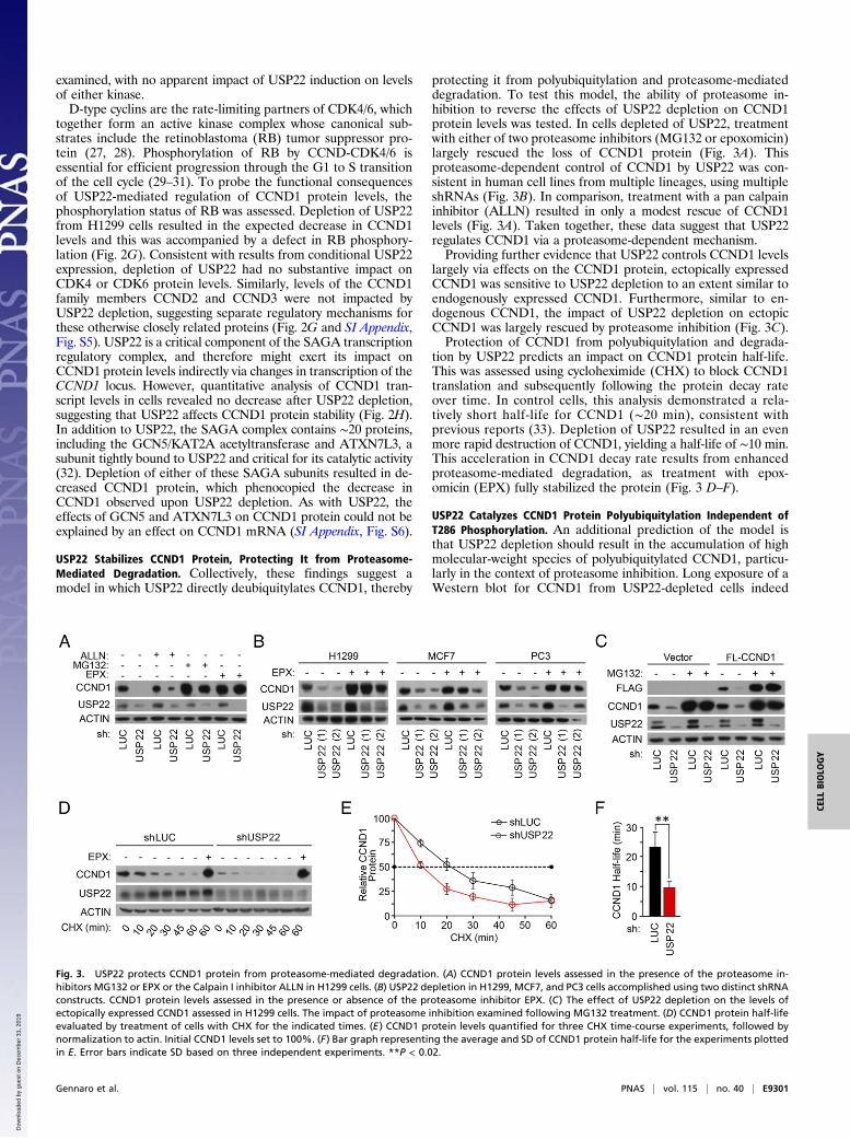
USP22 Stabilizes CCND1 Protein, Protecting It from Proteasome-Mediated Degradation. Collectively, these findings suggest amodel in which USP22 directly deubiquitylates CCND1, thereby
protecting it from polyubiquitylation and proteasome-mediateddegradation. To test this model, the ability of proteasome in-hibition to reverse the effects of USP22 depletion on CCND1protein levels was tested. In cells depleted of USP22, treatmentwith either of two proteasome inhibitors (MG132 or epoxomicin)largely rescued the loss of CCND1 protein (Fig. 3A). Thisproteasome-dependent control of CCND1 by USP22 was con-sistent in human cell lines from multiple lineages, using multipleshRNAs (Fig. 3B). In comparison, treatment with a pan calpaininhibitor (ALLN) resulted in only a modest rescue of CCND1levels (Fig. 3A). Taken together, these data suggest that USP22regulates CCND1 via a proteasome-dependent mechanism.Providing further evidence that USP22 controls CCND1 levels
largely via effects on the CCND1 protein, ectopically expressedCCND1 was sensitive to USP22 depletion to an extent similar toendogenously expressed CCND1. Furthermore, similar to en-dogenous CCND1, the impact of USP22 depletion on ectopicCCND1 was largely rescued by proteasome inhibition (Fig. 3C).Protection of CCND1 from polyubiquitylation and degrada-
tion by USP22 predicts an impact on CCND1 protein half-life.This was assessed using cycloheximide (CHX) to block CCND1translation and subsequently following the protein decay rateover time. In control cells, this analysis demonstrated a rela-tively short half-life for CCND1 (∼20 min), consistent withprevious reports (33). Depletion of USP22 resulted in an evenmore rapid destruction of CCND1, yielding a half-life of ∼10 min.This acceleration in CCND1 decay rate results from enhancedproteasome-mediated degradation, as treatment with epox-omicin (EPX) fully stabilized the protein (Fig. 3 D–F).
USP22 Catalyzes CCND1 Protein Polyubiquitylation Independent ofT286 Phosphorylation. An additional prediction of the model isthat USP22 depletion should result in the accumulation of highmolecular-weight species of polyubiquitylated CCND1, particu-larly in the context of proteasome inhibition. Long exposure of aWestern blot for CCND1 from USP22-depleted cells indeed
Fig. 3. USP22 protects CCND1 protein from proteasome-mediated degradation. (A) CCND1 protein levels assessed in the presence of the proteasome in-hibitors MG132 or EPX or the Calpain I inhibitor ALLN in H1299 cells. (B) USP22 depletion in H1299, MCF7, and PC3 cells accomplished using two distinct shRNAconstructs. CCND1 protein levels assessed in the presence or absence of the proteasome inhibitor EPX. (C) The effect of USP22 depletion on the levels ofectopically expressed CCND1 assessed in H1299 cells. The impact of proteasome inhibition examined following MG132 treatment. (D) CCND1 protein half-lifeevaluated by treatment of cells with CHX for the indicated times. (E) CCND1 protein levels quantified for three CHX time-course experiments, followed bynormalization to actin. Initial CCND1 levels set to 100%. (F) Bar graph representing the average and SD of CCND1 protein half-life for the experiments plottedin E. Error bars indicate SD based on three independent experiments. **P < 0.02.
Gennaro et al. PNAS | vol. 115 | no. 40 | E9301
CELL
BIOLO
GY
Dow
nloa
ded
by g
uest
on
Dec
embe
r 31
, 201
9
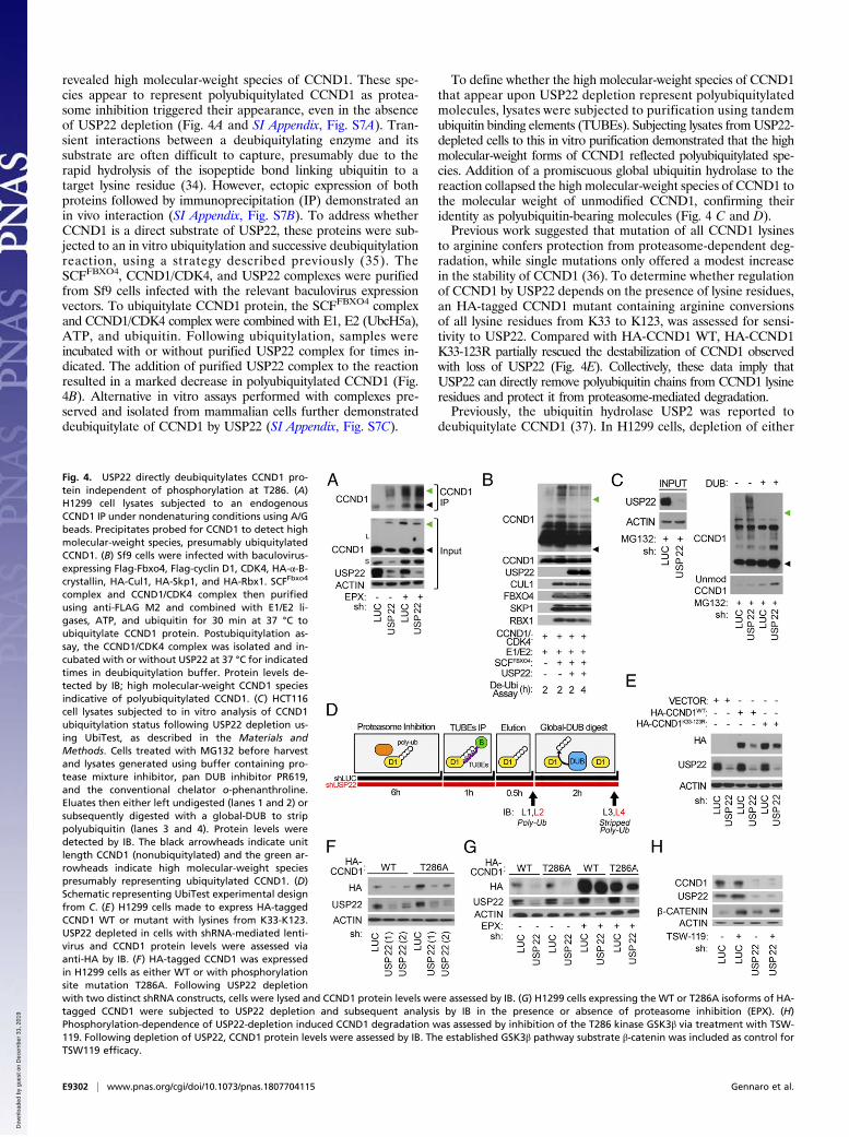
revealed high molecular-weight species of CCND1. These spe-cies appear to represent polyubiquitylated CCND1 as protea-some inhibition triggered their appearance, even in the absenceof USP22 depletion (Fig. 4A and SI Appendix, Fig. S7A). Tran-sient interactions between a deubiquitylating enzyme and itssubstrate are often difficult to capture, presumably due to therapid hydrolysis of the isopeptide bond linking ubiquitin to atarget lysine residue (34). However, ectopic expression of bothproteins followed by immunoprecipitation (IP) demonstrated anin vivo interaction (SI Appendix, Fig. S7B). To address whetherCCND1 is a direct substrate of USP22, these proteins were sub-jected to an in vitro ubiquitylation and successive deubiquitylationreaction, using a strategy described previously (35). TheSCFFBXO4, CCND1/CDK4, and USP22 complexes were purifiedfrom Sf9 cells infected with the relevant baculovirus expressionvectors. To ubiquitylate CCND1 protein, the SCFFBXO4 complexand CCND1/CDK4 complex were combined with E1, E2 (UbcH5a),ATP, and ubiquitin. Following ubiquitylation, samples wereincubated with or without purified USP22 complex for times in-dicated. The addition of purified USP22 complex to the reactionresulted in a marked decrease in polyubiquitylated CCND1 (Fig.4B). Alternative in vitro assays performed with complexes pre-served and isolated from mammalian cells further demonstrateddeubiquitylate of CCND1 by USP22 (SI Appendix, Fig. S7C).
To define whether the high molecular-weight species of CCND1that appear upon USP22 depletion represent polyubiquitylatedmolecules, lysates were subjected to purification using tandemubiquitin binding elements (TUBEs). Subjecting lysates fromUSP22-depleted cells to this in vitro purification demonstrated that the highmolecular-weight forms of CCND1 reflected polyubiquitylated spe-cies. Addition of a promiscuous global ubiquitin hydrolase to thereaction collapsed the high molecular-weight species of CCND1 tothe molecular weight of unmodified CCND1, confirming theiridentity as polyubiquitin-bearing molecules (Fig. 4 C and D).Previous work suggested that mutation of all CCND1 lysines
to arginine confers protection from proteasome-dependent deg-radation, while single mutations only offered a modest increasein the stability of CCND1 (36). To determine whether regulationof CCND1 by USP22 depends on the presence of lysine residues,an HA-tagged CCND1 mutant containing arginine conversionsof all lysine residues from K33 to K123, was assessed for sensi-tivity to USP22. Compared with HA-CCND1 WT, HA-CCND1K33-123R partially rescued the destabilization of CCND1 observedwith loss of USP22 (Fig. 4E). Collectively, these data imply thatUSP22 can directly remove polyubiquitin chains from CCND1 lysineresidues and protect it from proteasome-mediated degradation.Previously, the ubiquitin hydrolase USP2 was reported to
deubiquitylate CCND1 (37). In H1299 cells, depletion of either
Fig. 4. USP22 directly deubiquitylates CCND1 pro-tein independent of phosphorylation at T286. (A)H1299 cell lysates subjected to an endogenousCCND1 IP under nondenaturing conditions using A/Gbeads. Precipitates probed for CCND1 to detect highmolecular-weight species, presumably ubiquitylatedCCND1. (B) Sf9 cells were infected with baculovirus-expressing Flag-Fbxo4, Flag-cyclin D1, CDK4, HA-α-B-crystallin, HA-Cul1, HA-Skp1, and HA-Rbx1. SCFFbxo4
complex and CCND1/CDK4 complex then purifiedusing anti-FLAG M2 and combined with E1/E2 li-gases, ATP, and ubiquitin for 30 min at 37 °C toubiquitylate CCND1 protein. Postubiquitylation as-say, the CCND1/CDK4 complex was isolated and in-cubated with or without USP22 at 37 °C for indicatedtimes in deubiquitylation buffer. Protein levels de-tected by IB; high molecular-weight CCND1 speciesindicative of polyubiquitylated CCND1. (C) HCT116cell lysates subjected to in vitro analysis of CCND1ubiquitylation status following USP22 depletion us-ing UbiTest, as described in the Materials andMethods. Cells treated with MG132 before harvestand lysates generated using buffer containing pro-tease mixture inhibitor, pan DUB inhibitor PR619,and the conventional chelator o-phenanthroline.Eluates then either left undigested (lanes 1 and 2) orsubsequently digested with a global-DUB to strippolyubiquitin (lanes 3 and 4). Protein levels weredetected by IB. The black arrowheads indicate unitlength CCND1 (nonubiquitylated) and the green ar-rowheads indicate high molecular-weight speciespresumably representing ubiquitylated CCND1. (D)Schematic representing UbiTest experimental designfrom C. (E) H1299 cells made to express HA-taggedCCND1 WT or mutant with lysines from K33-K123.USP22 depleted in cells with shRNA-mediated lenti-virus and CCND1 protein levels were assessed viaanti-HA by IB. (F) HA-tagged CCND1 was expressedin H1299 cells as either WT or with phosphorylationsite mutation T286A. Following USP22 depletionwith two distinct shRNA constructs, cells were lysed and CCND1 protein levels were assessed by IB. (G) H1299 cells expressing the WT or T286A isoforms of HA-tagged CCND1 were subjected to USP22 depletion and subsequent analysis by IB in the presence or absence of proteasome inhibition (EPX). (H)Phosphorylation-dependence of USP22-depletion induced CCND1 degradation was assessed by inhibition of the T286 kinase GSK3β via treatment with TSW-119. Following depletion of USP22, CCND1 protein levels were assessed by IB. The established GSK3β pathway substrate β-catenin was included as control forTSW119 efficacy.
E9302 | www.pnas.org/cgi/doi/10.1073/pnas.1807704115 Gennaro et al.
Dow
nloa
ded
by g
uest
on
Dec
embe
r 31
, 201
9

USP2 or USP22 resulted in similar decreases in steady-stateCCND1, and effects of both deubiquitinating enzymes (DUBs)were partially rescued by proteasome inhibition (SI Appendix,Fig. S8A). No interdependent changes in protein levels weredetected in USP2 and USP22 upon knockdown, suggesting thatUSP22 depletion does not result in decreased steady-state CCND1via an indirect effect on USP2 levels. Moreover, loss of CCND1 upondepletion of either USP2 or USP22 had similar functional con-sequences and similarly increased high molecular-weight poly-ubiquitylated CCND1 species (SI Appendix, Fig. S8 B and C).The best-characterized pathway of CCND1 degradation relies
on its phosphorylation-dependent export from the nucleus fol-lowed by polyubiquitylation and proteasome-mediated degrada-tion in the cytoplasm (17, 38). To directly test the phosphorylationdependence of the effect of USP22 on CCND1, CCND1 mutantscontaining alanine conversions at either T286 or T288 were assessedfor sensitivity to USP22. For both of these phosphorylation-defectivemutants, depletion of USP22 had effects similar to its effect onWT CCND1 (Fig. 4F and SI Appendix, Fig. S9A). Further analysisdemonstrated that inhibition of proteolysis rescued the USP22effect on mutant T286A CCND1, as it did for WT CCND1 (Fig.4G). Previous studies have identified GSK3β as a primary kinaseresponsible for CCND1 phosphorylation at T286 (39). To confirmthat USP22 affects endogenous CCND1 stability independent ofphosphorylation at this site, USP22-depleted cells were treatedwith the GSK3β inhibitor TSW-119. Consistent with the obser-vation that USP22 depletion affects mutant T286A CCND1 sta-bility, inhibiting phosphorylation at T286 with TSW-119 resultedin no alteration of endogenous CCND1 protein or phosphorylatedCCND1 (Fig. 4H and SI Appendix, Fig. S9B). Because phos-phorylation at T286 acts as a precursor to nuclear export, CCND1levels in separate cellular fractions were assessed upon depletionof known CCND1-dependent USP2 and USP22. CCND1 inwhole-cell lysates and in the cytoplasmic fraction were both de-creased upon depletion of USP22 or USP2. A long exposure of theWestern blots suggests that the nuclear pool of CCND1 decreasedfollowing USP22 depletion, while USP2 depletion had little effect (SIAppendix, Fig. S10A). To gain a more precise understanding of thecellular CCND1 pools that are targeted by USP22, nuclear fractionswere further subdivided into chromatin-bound and chromatin-freeprotein pools (SI Appendix, Fig. S10B). These findings suggestthat USP22 and USP2 may have distinct mechanisms of actionon CCND1, with USP22 controlling a CCND1 degradationpathway that is at least partially distinct from the canonical,phosphorylation-dependent pathway.
Ectopic CCND1 Protein Partially Rescues the Aberrant Cell ProliferationPhenotype Observed with USP22 Depletion. As reported here andelsewhere, USP22 plays a significant role in cell cycle progression,with the greatest impact on the G1 phase (Fig. 1). BecauseCCND1 is among the central regulators of G1 progression inmammalian cells, we tested the hypothesis that CCND1 deficiencycontributes to the effect of USP22 depletion on G1 progression.Despite data shown in Fig. 3 demonstrating USP22 regulatingexogenous CCND1, a modest level of CCND1 was established bytransient expression (Fig. 5A). Ectopic expression of CCND1 incells depleted of USP22 caused an increase in proliferation rela-tive to control cells (Fig. 5 B and C). Further analysis confirmedthat this rescue occurred primarily via a reduction in transitthrough G1 (Fig. 5D). That the aberrant cell proliferation ob-served with USP22 depletion can be partially rescued by ectopicCCND1 protein suggests that stabilization of CCND1 is a keyevent in the USP22-mediated regulation of the cell cycle.
USP22 Regulation of CCND1 Has Clinical Consequences as CDK4/6iTreatment Rescues the G1 Phenotype Associated with USP22Overexpression. Overexpression of CCND1 is a well-establishedhallmark of human cancer (40, 41). Similarly, USP22 over-expression is conserved among many aggressive forms of cancer(5, 6, 42). To assess any potential correlation between USP22elevation and CCND1 elevation, human tumor tissue microarrayswere quantified for levels of each protein. Among 110 colorectalcarcinoma samples and 110 lung carcinoma samples (Fig. 6 A andB and SI Appendix, Fig. S11), a significant correlation betweenCCND1 and USP22 protein levels was observed. ElevatedCCND1 activity and expression in cancer can be targeted clinicallyusing FDA-approved CDK inhibitors. The findings presentedhere suggest that tight regulation of USP22 is essential for G1–Stransition via CCND1 regulation, raising the possibility thatUSP22 might represent a therapeutic target. To assess the ther-apeutic potential of this pathway, HCT116 cells engineered tooverexpress USP22 were treated with the CDK inhibitor PD-0332991. As expected, induction of ectopic USP22 resulted inan enhanced G1 exit (Fig. 6 C and D) and increased CCND1protein levels (Fig. 6E). Strikingly, inhibiting downstream CCND1/CDK activity with PD-0332991 in USP22-overexpressing cells wassufficient to reverse the effect of USP22 on cell-cycle progression.Furthermore, HCT116 cells treated with PD-0332991 demonstrateda similar defect in G1–S transition to that of cells with reducedUSP22 expression. Concomitant depletion of USP22 and inhibitionof CDK4/6 resulted in no additive cell cycle or death response (SI
Fig. 5. Ectopic expression of CCND1 provides apartial genetic rescue of the cell cycle phenotypeobserved with USP22 depletion. H1299 cells weretransfected with a vector encoding 3XFLAG-taggedCCND1, followed by USP22 depletion. (A) Immuno-blot demonstrating efficient knockdown of USP22,exogenous 3XFLAG-CCND1, and endogenous CCND1expression. (B) Cell number was determined bymanual counting of experimental triplicates at eachof the postdepletion time points indicated. Errorbars indicate SD based on three independent ex-periments. (C) Methylene blue-stained foci of rep-resentative plates from B. (D) At day 7 followingUSP22 depletion, cells were harvested and cell cycleprofile was determined by PI staining and flowcytometry. G1-phase cells were gated (in blue) aspercent of the total population. **P < 0.02.
Gennaro et al. PNAS | vol. 115 | no. 40 | E9303
CELL
BIOLO
GY
Dow
nloa
ded
by g
uest
on
Dec
embe
r 31
, 201
9

Appendix, Fig. S12), suggesting that USP22 and CDK4/6 may regu-late the same pathway in an epistatic manner. Collectively, thesefindings implicate the DUB USP22 as a key regulator of CCND1,and thereby a potential complement to current therapeutic strategiestargeting CCND1/CDK4/6 in cancer. For example, the often severeside effects experienced by patients receiving CDK4/6, might be re-duced by use of lower doses in combination with USP22 inhibition.
DiscussionThe role of USP22 in transcription is presumed to result in largepart from its ability to remove ubiquitin from nucleosomal histonesH2A and H2B (1–3). Empirical studies have demonstrated thatUSP22 can deubiquitylate nonhistone substrates as well (e.g., TRF1and FBP) (14, 15). USP22 plays a critical role in progressionthrough the G1–S phase transition of the cell cycle and its activity is
Fig. 6. CCND1 and USP22 protein levels correlate in patient samples from lung and colon adenocarcinoma and CDK4/6i treatment rescues the G1 phenotypeassociated with USP22 overexpression. (A) Representative images of serial sections of colon adenocarcinoma or lung adenocarcinoma BioMax tissue microarraysstained with DAPI and either USP22 or CCND1 antibodies. (B) Graphical representation of USP22(log) expression against CCND1(log) expression in each of the120 cases with representative cases is indicated on the graph. [Pearson’s correlation coefficient (PCC) of 0.338 for colon adenocarcinoma or 0.357 for lung ad-enocarcinoma.] (C) FLAG-tagged USP22 was ectopically expressed in HCT116 cells via a stably integrated tetracycline-inducible vector. Following USP22 inductionand treatment with selective CDK4/6 inhibitor PD-0332991 (PD), cells were harvested and cell cycle profile was determined by assessment of DNA content with PIstaining and flow cytometry. G1-phase cells were gated (in blue) as percent of the total population. (D) Quantification of cell cycle phase distribution. Error barsindicate SD based on three independent experiments. (E) Induction of ectopic USP22 and increased CCND1 protein levels were assessed by IB. **P < 0.02, ***P <0.005. (F) A schematic representing the proposed model of CCND1 stabilization by deubiquitylase USP22. CCND1–CDK4/6 complex advances cell cycling viahyperphosphorylating RB, which in turn releases E2F transcription factor from an inhibitory constraint and enables the expression of genes required for G1–Sphase transition. To regulate rapid turnover, CCND1–CDK4/6 can incur phosphorylation at Thr286 by GSK3β, a precursor to nuclear export and subsequentpolyubiquitylation by distinct E3 ligases (e.g., FBX4, PARK2, SKP2, FBXW8. . .) (66–71). Polyubiquitylated CCND1–CDK4/6 is then targeted for proteasomal deg-radation. USP22 promotes CCND1 stabilization via removing CCND1 ubiquitin within the nucleus and/or cytoplasm and blocking degradation by the proteasome.
E9304 | www.pnas.org/cgi/doi/10.1073/pnas.1807704115 Gennaro et al.
Dow
nloa
ded
by g
uest
on
Dec
embe
r 31
, 201
9
both elevated and required for the aggressive growth of numerouscancer lineages (7–13). [Somewhat paradoxically, a role for USP22in M phase has been proposed based on evidence that CCNB1 isdeubiquitylated by USP22 (43)]. Nonetheless, no known substrateof USP22 explains its central biological phenotype, progressionthrough the G1–S cell cycle transition. These studies were un-dertaken with the expectation that elucidating the role of USP22 inorchestrating the G1–S progression might advance our under-standing of aggressive growth phenotypes in cancer and inform thedevelopment of novel therapeutics.What remains lacking is a mechanistic understanding of which
substrates of USP22 mediate these potent effects on prolifera-tion. Here, a proteome-wide analysis in human cells identifiedproteins whose ubiquitylation is regulated by USP22. This screenidentified several high-confidence hits within proteins not pre-viously described as USP22 substrates. As reported above, theG1 cyclin CCND1 was validated experimentally as a directUSP22 substrate. USP22 hydrolyzes ubiquitin at a set of specificlysine residues on CCND1 (K33, K46, 50, K112/114). Func-tionally, K33 ubiquitylation is implicated in nuclear localizationof CCND1 (44), and K112 and K114 ubiquitylation are linked tothe interaction of CCND1 with partners, including CDK4/6 (45–47). The ubiquitylation of CCND1 at K46 and K50 has not beenpreviously reported. Notably, several of these ubiquitylation sitesare mutated in cancer, with missense mutations at K46 andK112 reported in lymphoma and breast cancer, respectively (36).While the detection of these lysine residues indicates preferredsites affected by USP22, ubiquitylation demonstrates notablepromiscuity at the level of individual lysine acceptor sites (48).Previous attempts to uncover specific residues that regulateCCND1 proteasomal degradation have demonstrated single anddouble lysine residue mutations have only modest effects onCCND1 stability (36). This capacity of USP22 to remove poly-ubiquitin chains protects CCND1 from proteosomal degradation,thereby doubling its half-life and significantly elevating steady-stateCCND1 protein levels. Four central observations highlight the bi-ological relevance of CCND1 as a substrate of USP22. First, the G1-phase cell cycle arrest observed upon depletion of USP22 fromhuman cells is genetically rescued by ectopic expression of CCND1.Second, mimicking the scenario in cancer by modest overexpressionof USP22 in cells results in a concomitant increase in steady-stateCCND1 protein levels. Third, USP22 protein levels directly cor-relate with CCND1 levels in tumor tissue samples from both colonand lung cancer patients. Finally, experimentally elevatingUSP22 levels both increases CCND1 and drives more rapid transitthrough the G1 phase of the cell cycle, an effect blocked by treat-ment with the FDA-approved CDK4/6 inhibitor PD-0332991. Thesefindings appear not to be linked to the known effect of USP22 onthe SIRT1/p53 pathway (49), because they were observed in bothp53-proficient and -deficient cell lines. Collectively these findingssupport a model in which elevated expression of USP22 contributesto the aggressive growth of cancer cells in part via its ability todeubiquitylate and stabilize the rate-limiting cyclin CCND1, therebypromoting the G1–S transition.The identification of the USP22-CCND1 enzyme–substrate
relationship may explain the previously reported phosphorylation-independent ubiquitylation and degradation of CCND1 (50). Ourfindings demonstrate that USP22 deubiquitylates CCND1 regard-less of phosphorylation events at T286 or T288. Genetic evidencethat USP2 and USP22 control distinct CCND1 degradation path-ways comes from the observation that these enzymes are not re-dundant, with depletion of either one sufficient to destabilizeCCND1. Independent of CDK interaction, CCND1 binds nuclearreceptors (ERα, THR, PPARγ, and AR) to regulate cell pro-liferation, growth, and differentiation (51). Furthermore, CCND1regulates transcriptional activation by binding histone acetylases,deacetylases, and coactivators (52). Levels of CCND1 in the cyto-plasm decreased following depletion of either USP22 or USP2;
however, both the chromatin-bound and chromatin-free pools ofCCND1 were sensitive to USP22 depletion, while USP2 depletionhad little effect on either pool. These findings suggest that USP22 andUSP2 may regulate CCND1 via distinct pathways/mechanisms.A number of the observations reported here suggest that
USP22 may target the unbound form of CCND1 rather than theCDK-bound form. For example, the K112 and K114 sites onCCND1 identified as USP22 targets reside within the CDK in-teraction domain. Furthermore, the phosphorylation-independentnature of CCND1 targeting by USP22 may allow new therapeuticstrategies for targeting this pathway. For example, the clinical effi-cacy of CDK4/6 inhibitors in cancer might be complemented bycombinatorial treatment with a USP22 inhibitor, thus simulta-neously targeting both the CDK-bound and the -free forms ofCCND1. More broadly, the identification of the critical cell cycleregulator CCND1 as a substrate of the USP22 subunit of SAGAbroadens our understanding of this complex, which has previouslybeen linked primarily to direct effects on transcription.
Materials and MethodsCell Culture, Viral Infection, Transfection, and Treatment. The human cell linesHCT116, H1299, MCF7, PC3, and 2091 were obtained from American TypeCulture Collection (ATCC). The ectopic USP22-inducible HCT116 tetracyclinehydrochloride (TET) operable cell line was generated by cloning pcDNA3.1FLAG-USP22 into the pLenti6.3/V5/TO-DEST vector using the Virapower T-Rexsystem (ThermoFisher). All cell lines were cultured in DMEM (Mediatech)supplemented with 10% FCS (FBS; Gemini Bio-Products) at 37 °C in 5% CO2.
As indicated, cells were infected with lentiviral shRNA plasmids corre-sponding to USP22 (XM_042698.6-914s1c1, NM_015276.1-545s21c1), USP2(NM_004205.3-1266s1c1, NM_004205.3-1554s1c1), ATXN7L3 (NM_001098833.1-1008s21c1), GCN5 (NM_021078.1-2484s1c1), and control luciferase shRNA(SHC007) that were obtained from the TRC collection (Sigma-Aldrich). Cells wereselected with 8 μg/mL puromycin 24 h after infection.
Transfection was performed using Continuum reagents according to themanufacturer’s protocol (Gemini Bio-Products). HA-CCND1 WT, HA-CCND1K33-123R, and HA-CCND1 T286A expression plasmids were kindly providedby E. Dmitrovsky, MD Anderson Cancer Center, Houston, TX. HA-CCND1T288A mutant was constructed by site-directed mutagenesis using Quik-Change (Agilent Technologies). The previously described 3XFLAG-CCND1 WTexpression plasmid was kindly provided by R. Pestell, Baruch S. BlumbergInstitute, Doylestown, PA (53).
For indicated treatments, MG132 was used at 10 μM for 6 h (SelleckChem),EPX at 10 μM for 6 h (SelleckChem), ALLN at 100 μM for 6 h (Abcam), CHX at10 μg/mL as indicated (Sigma-Aldrich), TET at 2.25 μM for 5 d (Sigma-Aldrich), PR619 at 50 μM for 30 min (Life Sensors), o-phenanthroline at5 μM for 15 min (Life Sensors), TSW-119 at 1 μM for 12 h (Santa Cruz), andPD-0332991 (PD) at 100 nM for 3 d (Sigma-Aldrich).
Cell Proliferation, Viability, and Cell Cycle. For growth assays, cells were seededat equal densities and harvested at the indicated time points. Media and treat-ments were refreshed every 72 h. Cell number was determined quantitativelyby triplicate experiments using Trypan blue exclusion and counting by hemo-cytometer. Cell density was visualized by methylene blue staining.
Levels of necrotic cells were assessed by Muse Count and Viability Reagentand analyzed using the Muse Cell Analyzer as described by the manufacturer(Millipore). To quantify apoptotic cell death, cells were collected by trypsi-nization and stained using the Annexin V PE-7AAD apoptosis detection kit(BD Pharmingen). Fluorescence was detected by flow cytometry CytoFlex LXFlow Cytometer (Beckman Coulter).
Cell cycle analysis was conducted using the Click-iT EdU flow cytometry cellproliferation assay where cells were labeled with EdU for 2 h, harvested, andstained according to the manufacterer’s instructions (Thermo Fisher). DNA con-tent was measured using propidium iodide (PI) staining for 30 min on cells fixedwith 70% ethanol. Cell cycle analysis was processed using the CytoFlex LX.
Immunoblotting, Cellular Fractionation, Co-IP, and mRNA Analysis. Cells wereharvested and lysed in E1A whole-cell lysate buffer supplemented withprotease inhibitor mixture (Sigma-Aldrich) and PR619. Western blottingconcentration in lysates was determined using the bicinchoninic acid (BCA)assay and analyzed by SDS/PAGE using antibodies against CCND1 (#2978; CellSignaling), USP22 (#ab195289; Abcam), USP2 (#8036S; Cell Signaling), CDK4(#sc-260; Santa Cruz), CDK6 (#3136S; Cell Signaling), FLAG (#F3165; Sigma-Aldrich), HA (#sc-805; Santa Cruz), Ubiquitin (#sc-7905; Santa Cruz), RB (#sc-
Gennaro et al. PNAS | vol. 115 | no. 40 | E9305
CELL
BIOLO
GY
Dow
nloa
ded
by g
uest
on
Dec
embe
r 31
, 201
9
50; Santa Cruz), pRB (#sc-271930; Santa Cruz), ATXN7L3 (#A302-800A;Bethyl), GCN5 (#sc-365321; Santa Cruz), CCND2 (#3741T; Cell Signaling),CCND3 (#2936S; Cell Signaling), phospho-CCND1 (#3300T; Cell Signaling),FBXO4 (#YZ1779; YenZym Antibodies), CUL1 (#sc-11384; Santa Cruz), SKP1(#2156; Cell Signaling), RBX1 (#4397; Cell Signaling), ACTIN (#sc-47778; SantaCruz), β-CATENIN (#8480; Cell Signaling), GAPDH (#ab9485; Abcam), ORC2(#559266; Pharmingen), and TUBULIN (#T9026; Sigma-Aldrich).
Cellular Fractionations Were Performed as Described Previously. Cellularfractionations were performed as described previously (54). For protein–protein interaction studies, ∼750 μg of whole-cell lysates was used for IP.FLAG IPs were performed by incubating lysates with 20 μL Anti-FLAGM2 AffinityBeads (Sigma-Aldrich) for 16 h at 4 °C before Western blot analysis. EndogenousCCND1 IPs were conducted using 5 μg of CCND1 antibody (TA329665; OriGene)for 16 h at 4 °C and precipitates captured using protein A/G beads (Santa Cruz).
Total RNA was extracted using TRIzol (Thermo Fisher) and reverse-transcribed using the High-Capactiy cDNA Reverse Transcription Kit (ThermoFisher) according to the manufacturer’s instructions. Real-time PCR was per-formed using Fast SYBR Green (Thermo Fisher), as described previously (55),using primer sequences are listed in SI Appendix, Table S1. In all cases, mRNAlevels between samples were normalized to actin levels.
UbiScan. Samples were analyzed using the PTMScan method as previouslydescribed (56–58). Cellular extracts were prepared in urea lysis buffer, son-icated, centrifuged, reduced with DTT, and alkylated with iodoacetamide.Total protein (15 mg) for each sample was digested with trypsin and purifiedover C18 columns for enrichment with the Ubiquitin K-GG Remnant Motifantibody (#5562). Enriched peptides were purified over C18 STAGE tips (59).Replicate injections of each sample were run nonsequentially on the instrument.Peptides were eluted using a 72-min linear gradient of acetonitrile in 0.125%formic acid delivered at 280 nL/min. MS/MS spectra were collected in a data-dependent manner with an Orbitrap Elite Hybrid Ion Trap-Orbitrap Mass Spec-trometer running XCalibur 2.0.7 SP1 using a top-20 MS/MS method, a dynamicrepeat count of one, and a repeat duration of 30 s. Real-time recalibration ofmass error was performed using lock mass (60), with a singly charged poly-siloxane ionm/z = 371.101237. MS/MS spectra were evaluated using the Sorcererplatform (61, 62). Files were searched against the NCBI Homo sapeins FASTAdatabase. Amass accuracy of ±50 ppmwas used for precursor ions and 1.0 Da forproduct ions. Enzyme specificity was limited to trypsin, with at least one tryptic(K- or R-containing) terminus required per peptide and up to four miscleavagesallowed. Cysteine carboxamidomethylation was specified as a static modification;oxidation of methionine and a di-glycine remnant on lysine residues wereallowed as variable modifications. Reverse decoy databases were included for allsearches to estimate false discovery rates, and filtered using a 5% false-discoveryrate in Sorcerer. Peptides were also manually filtered using a ±5 ppm mass errorrange and the presence of at least one K-GG on each peptide. All quantitativeresults were generated using Progenesis v4.1 (Waters Corporation) to extract theintegrated peak area of the corresponding peptide assignments. Accuracy ofquantitative data was ensured by manual review in Progenesis or in the ionchromatogram files. A 2.5-fold cut-off was used to denote changes betweensamples and analytical percent coefficient of variation values were calculated foreach peptide to determine reproducibility across runs.
UbiTest. HCT116 cells were harvested after 6 d of knockdown and 4 h afterproteasome inhibitionwithMG132. Cellswere lysed in radioimmunoprecipitationbuffer supplementedwith proteasemixture inhibitor, PR619, ando-phenanthroline.Protein lysate was incubated with α-Ub TUBE1 agarose resin, eluted, and digestedwith global DUB USP2 for 2 h to remove polyubiquitylation, as recommended bythemanufacturer (LifeSensors). Undigested and digested samples were analyzed bySDS/PAGE and Western blotting.
In Vitro Ubiquitylation–Deubiquitylation Assay. Sf9 cells were infected withbaculovirus expressing FLAG-FBXO4, FLAG-CCND1, CDK4, HA-α-B-CRYSTALLIN,HA-CUL1, HA-SKP1, and HA-RBX1. Seventy-two hours postinfection, cells werelysed in Tween 20 buffer [50 mM Hepes (pH 8.0), 150 mM NaCl, 2.5 mM EGTA,1 mM EDTA, and 0.1% Tween 20 with protease and phosphatase inhibitors]. Togenerate the DUB module with active USP22, Sf9 cells were infected withbaculovirus expressing FLAG-USP22N, ATXN7, ATXN7L3, and HA-ENY2. Forty-eight hours postinfection, cells were lysed in Tween 20 buffer. Cell lysateswere subjected to anti-FLAG immunoaffinity purification to isolate USP22 withinan active DUB complex. The SCFFBXO4 complex and CCND1/CDK4 complex werepurified using anti-FLAG M2 affinity gel.
Beads with SCFFBXO4 complex and CCND1/CDK4 complex were combinedwith E1, E2 (UbcH5a), ATP, and ubiquitin for 30 min at 37 °C to achieveubiquitylated-CCND1 protein. After reaction, beads were washed withdeubiquitylation buffer [100 mM Tris (pH 8.0), 1 mM EDTA, 1 mM DTT, and5% glycerol]. Thereafter, the beads were incubated with or without hDUBUSP22 in deubiquitylation buffer for indicated times at 37 °C, as previouslydescribed (63). Beads were subsequently boiled in 2× loading buffer. Proteinswere resolved in 10% SDS/PAGE gel and detected by relative antibodies.
In Vitro Deubiquitylation Assay. H1299 cells were transfected with 3XFLAG-CCND1 and HA-Ub, or separately with FLAG-USP22. Cell lysates were sub-jected toanti-FLAG immunoaffinitypurification to isolateFLAG-USP22and3XFLAG-CCND1with conjugatedHA-Ub. The in vitro enzymatic assaywas performed for 2 hat 37 °C, as previously described (63), and subjected to SDS/PAGE analysis.
Tissue Microarray and Quantitative Immunofluorescence. Tissue microarraysconsisting of formalin-fixed, paraffin-embedded colon or lung cancer spec-imens were obtained from US Biomax, Inc. The colon array (Cat# BC051110b)consisted of 110 cases of colon adenocarcinoma and 10 normal colon cases.The age range of patients represented on the array was 22–86 y (median55 y). The lung array (Cat# BC041115d) consisted of 110 cases of mixed-pathology lung cancer and 10 normal lung cases. The age range of pa-tients represented on the array was 15–76 y (median 57 y).
CCND1 and USP22 were detected in clinical colon and lung cancer specimensby immunofluorescence-immunohistochemistry (IF-IHC) performed on DakoOmnis autostainer using the TSA Plus Fluorescence Kit (Perkin-Elmer), as pre-viously described (64). Antigen retrieval was performed using citrate buffer (pH6.1; Dako). Rabbit monoclonal CCND1 (1:400, M3635; Dako) and rabbit poly-clonal USP22 (1:400, HPA044980; Sigma) were individually diluted 1:400 andcoincubated withmouse monoclonal antipancytokeratin (clone AE1/AE3, M35151:100; Dako) for 45 min. High-resolution digital images of immuno-stained slideswere captured using the Pannoramic 250 Flash scanner (3DHistech). Quantita-tive biomarker analysis was performed as previously described (65) using TissueStudio image analysis software (Definiens). Briefly, user-guided machine learn-ing was used to generate an analysis solution to specifically quantify meannuclear signal intensity of each biomarker within epithelial DAPI-stained cellnuclei of each tissue core. Statistical analyses were performed in SPSS.
Statistical Analysis. Data collected from at least three independent experi-ments are presented as mean ± SD. Statistical testing was performed using SPSSwith differences between two groups determined by a Student’s t test. Signifi-cance is denoted in the figures as: ***P < 0.005, **P < 0.02, and *P < 0.05.
ACKNOWLEDGMENTS. We thank Drs. E. Dmitrovsky (MD Anderson CancerCenter) and R. Pestell (Baruch S. Blumberg Institute) for providing reagentsor advice. These studies were supported in part via NIH Grant R01CA182569(to K.E.K. and S.B.M.). F.W. and T.B. are employed by Progenra Inc., a biotechcompany engaged in targeting deubiquitinating enzymes for therapeuticbenefit. The National Cancer Institute-supported Sloan-Kettering CancerCenter Cancer Genomics facility was used in these studies.
1. Zhang XY, et al. (2008) The putative cancer stem cell marker USP22 is a subunit of the
human SAGA complex required for activated transcription and cell-cycle progression.
Mol Cell 29:102–111.2. Zhao Y, et al. (2008) A TFTC/STAGA module mediates histone H2A and H2B deubi-
quitination, coactivates nuclear receptors, and counteracts heterochromatin silenc-
ing. Mol Cell 29:92–101.3. Weake VM, et al. (2008) SAGA-mediated H2B deubiquitination controls the de-
velopment of neuronal connectivity in the Drosophila visual system. EMBO J 27:
394–405.4. Zhang XY, Pfeiffer HK, Thorne AW, McMahon SB (2008) USP22, an hSAGA subunit
and potential cancer stem cell marker, reverses the polycomb-catalyzed ubiq-
uitylation of histone H2A. Cell Cycle 7:1522–1524.5. Glinsky GV (2005) Death-from-cancer signatures and stem cell contribution to meta-
static cancer. Cell Cycle 4:1171–1175.
6. Glinsky GV (2007) Stem cell origin of death-from-cancer phenotypes of human
prostate and breast cancers. Stem Cell Rev 3:79–93.7. Ding F, et al. (2014) USP22 promotes NSCLC tumorigenesis via MDMX up-regulation
and subsequent p53 inhibition. Int J Mol Sci 16:307–320.8. Ning Z, et al. (2014) USP22 promotes epithelial-mesenchymal transition via the FAK
pathway in pancreatic cancer cells. Oncol Rep 32:1451–1458.9. Schrecengost RS, et al. (2014) USP22 regulates oncogenic signaling pathways to drive
lethal cancer progression. Cancer Res 74:272–286.10. Hu J, et al. (2015) USP22 promotes tumor progression and induces epithelial-
mesenchymal transition in lung adenocarcinoma. Lung Cancer 88:239–245.11. Xiao H, et al. (2015) USP22 acts as an oncogene by regulating the stability of cyclo-
oxygenase-2 in non-small cell lung cancer. Biochem Biophys Res Commun 460:703–708.12. Li Y, et al. (2017) USP22 drives colorectal cancer invasion and metastasis via epithelial-
mesenchymal transition by activating AP4. Oncotarget 8:32683–32695.
E9306 | www.pnas.org/cgi/doi/10.1073/pnas.1807704115 Gennaro et al.
Dow
nloa
ded
by g
uest
on
Dec
embe
r 31
, 201
9
13. Wang A, et al. (2017) USP22 induces cisplatin resistance in lung adenocarcinoma byregulating γH2AX-mediated DNA damage repair and Ku70/bax-mediated apoptosis.Front Pharmacol 8:274.
14. Atanassov BS, et al. (2009) Gcn5 and SAGA regulate shelterin protein turnover andtelomere maintenance. Mol Cell 35:352–364.
15. Atanassov BS, Dent SY (2011) USP22 regulates cell proliferation by deubiquitinatingthe transcriptional regulator FBP1. EMBO Rep 12:924–930.
16. Diehl JA, Cheng M, Roussel MF, Sherr CJ (1998) Glycogen synthase kinase-3betaregulates cyclin D1 proteolysis and subcellular localization. Genes Dev 12:3499–3511.
17. Alt JR, Cleveland JL, Hannink M, Diehl JA (2000) Phosphorylation-dependent regu-lation of cyclin D1 nuclear export and cyclin D1-dependent cellular transformation.Genes Dev 14:3102–3114.
18. Diehl JA (2002) Cycling to cancer with cyclin D1. Cancer Biol Ther 1:226–231.19. Kim D, et al. (2017) Deubiquitinating enzyme USP22 positively regulates c-Myc sta-
bility and tumorigenic activity in mammalian and breast cancer cells. J Cell Physiol232:3664–3676.
20. Ling S, et al. (2017) USP22 mediates the multidrug resistance of hepatocellular car-cinoma via the SIRT1/AKT/MRP1 signaling pathway. Mol Oncol 11:682–695.
21. Ling SB, et al. (2012) Knock-down of USP22 by small interfering RNA interferenceinhibits HepG2 cell proliferation and induces cell cycle arrest. Cell Mol Biol 58:OL1803–OL1808.
22. Liu YL, et al. (2012) USP22 acts as an oncogene by the activation of BMI-1-mediatedINK4a/ARF pathway and Akt pathway. Cell Biochem Biophys 62:229–235.
23. Liu YL, et al. (2015) The deubiquitinating enzyme activity of USP22 is necessary forregulating HeLa cell growth. Gene 572:49–56.
24. Lv L, et al. (2011) Silencing USP22 by asymmetric structure of interfering RNA inhibitsproliferation and induces cell cycle arrest in bladder cancer cells. Mol Cell Biochem346:11–21.
25. Ma Y, et al. (2017) USP22 maintains gastric cancer stem cell stemness and promotesgastric cancer progression by stabilizing BMI1 protein. Oncotarget 8:33329–33342.
26. KimW, et al. (2011) Systematic and quantitative assessment of the ubiquitin-modifiedproteome. Mol Cell 44:325–340.
27. Kato J, Matsushime H, Hiebert SW, Ewen ME, Sherr CJ (1993) Direct binding of cyclinD to the retinoblastoma gene product (pRb) and pRb phosphorylation by the cyclin D-dependent kinase CDK4. Genes Dev 7:331–342.
28. Ewen ME, et al. (1993) Functional interactions of the retinoblastoma protein withmammalian D-type cyclins. Cell 73:487–497.
29. Buchkovich K, Duffy LA, Harlow E (1989) The retinoblastoma protein is phosphory-lated during specific phases of the cell cycle. Cell 58:1097–1105.
30. Chen PL, Scully P, Shew JY, Wang JY, Lee WH (1989) Phosphorylation of the retino-blastoma gene product is modulated during the cell cycle and cellular differentiation.Cell 58:1193–1198.
31. Mihara K, et al. (1989) Cell cycle-dependent regulation of phosphorylation of thehuman retinoblastoma gene product. Science 246:1300–1303.
32. Lang G, et al. (2011) The tightly controlled deubiquitination activity of the humanSAGA complex differentially modifies distinct gene regulatory elements.Mol Cell Biol31:3734–3744.
33. Sewing A, et al. (1993) Human cyclin D1 encodes a labile nuclear protein whosesynthesis is directly induced by growth factors and suppressed by cyclic AMP. J Cell Sci104:545–555.
34. Hanpude P, Bhattacharya S, Kumar Singh A, Kanti Maiti T (2017) Ubiquitin recogni-tion of BAP1: Understanding its enzymatic function. Biosci Rep 37:BSR20171099.
35. Barbash O, Lee EK, Diehl JA (2011) Phosphorylation-dependent regulation ofSCF(Fbx4) dimerization and activity involves a novel component, 14-3-3E.Oncogene 30:1995–2002.
36. Feng Q, Sekula D, Müller R, Freemantle SJ, Dmitrovsky E (2007) Uncovering residuesthat regulate cyclin D1 proteasomal degradation. Oncogene 26:5098–5106.
37. Shan J, Zhao W, Gu W (2009) Suppression of cancer cell growth by promoting cyclinD1 degradation. Mol Cell 36:469–476.
38. Diehl JA, Zindy F, Sherr CJ (1997) Inhibition of cyclin D1 phosphorylation on threo-nine-286 prevents its rapid degradation via the ubiquitin-proteasome pathway.Genes Dev 11:957–972.
39. Mukherji A, Janbandhu VC, Kumar V (2008) GSK-3beta-dependent destabilization ofcyclin D1 mediates replicational stress-induced arrest of cell cycle. FEBS Lett 582:1111–1116.
40. Bates S, Peters G (1995) Cyclin D1 as a cellular proto-oncogene. Semin Cancer Biol 6:73–82.
41. Harris AW, et al. (1995) Cyclin D1 as the putative bcl-1 oncogene. Curr Top MicrobiolImmunol 194:347–353.
42. Glinsky GV, Berezovska O, Glinskii AB (2005) Microarray analysis identifies a death-from-cancer signature predicting therapy failure in patients with multiple types ofcancer. J Clin Invest 115:1503–1521.
43. Lin Z, et al. (2015) Ubiquitin-specific protease 22 is a deubiquitinase of CCNB1. CellDiscov 1:15028.
44. Wang XD, et al. (2011) SUMO-modified nuclear cyclin D1 bypasses Ras-induced se-nescence. Cell Death Differ 18:304–314.
45. Day PJ, et al. (2009) Crystal structure of human CDK4 in complex with a D-type cyclin.Proc Natl Acad Sci USA 106:4166–4170.
46. Benzeno S, et al. (2006) Identification of mutations that disrupt phosphorylation-dependent nuclear export of cyclin D1. Oncogene 25:6291–6303.
47. Hinds PW, Dowdy SF, Eaton EN, Arnold A, Weinberg RA (1994) Function of a humancyclin gene as an oncogene. Proc Natl Acad Sci USA 91:709–713.
48. Danielsen JM, et al. (2011) Mass spectrometric analysis of lysine ubiquitylation revealspromiscuity at site level. Mol Cell Proteomics 10:M110.003590.
49. Lin Z, et al. (2012) USP22 antagonizes p53 transcriptional activation by deubiquiti-nating Sirt1 to suppress cell apoptosis and is required for mouse embryonic devel-opment. Mol Cell 46:484–494.
50. Germain D, Russell A, Thompson A, Hendley J (2000) Ubiquitination of free cyclin D1 isindependent of phosphorylation on threonine 286. J Biol Chem 275:12074–12079.
51. Zwijsen RM, Buckle RS, Hijmans EM, Loomans CJ, Bernards R (1998) Ligand-independent recruitment of steroid receptor coactivators to estrogen receptor bycyclin D1. Genes Dev 12:3488–3498.
52. Pestell RG (2013) New roles of cyclin D1. Am J Pathol 183:3–9.53. Casimiro MC, et al. (2012) ChIP sequencing of cyclin D1 reveals a transcriptional role in
chromosomal instability in mice. J Clin Invest 122:833–843.54. Carey MF, Peterson CL, Smale ST (2009) Dignam and Roeder nuclear extract prepa-
ration. Cold Spring Harb Protoc 2009:pdb.prot5330.55. Zhang XY, et al. (2005) Metastasis-associated protein 1 (MTA1) is an essential
downstream effector of the c-MYC oncoprotein. Proc Natl Acad Sci USA 102:13968–13973.
56. Rush J, et al. (2005) Immunoaffinity profiling of tyrosine phosphorylation in cancercells. Nat Biotechnol 23:94–101.
57. Stokes MP, et al. (2015) Complementary PTM profiling of drug response in humangastric carcinoma by immunoaffinity and IMAC methods with total proteome anal-ysis. Proteomes 3:160–183.
58. Stokes MP, et al. (2012) PTMScan direct: Identification and quantification of peptidesfrom critical signaling proteins by immunoaffinity enrichment coupled with LC-MS/MS. Mol Cell Proteomics 11:187–201.
59. Rappsilber J, Ishihama Y, Mann M (2003) Stop and go extraction tips for matrix-assisted laser desorption/ionization, nanoelectrospray, and LC/MS sample pre-treatment in proteomics. Anal Chem 75:663–670.
60. Olsen JV, et al. (2005) Parts per million mass accuracy on an orbitrap mass spec-trometer via lock mass injection into a C-trap. Mol Cell Proteomics 4:2010–2021.
61. Eng JK, McCormack AL, Yates JR (1994) An approach to correlate tandem massspectral data of peptides with amino acid sequences in a protein database. J Am SocMass Spectrom 5:976–989.
62. Lundgren DH, Martinez H, Wright ME, Han DK (2009) Protein identification usingSorcerer 2 and SEQUEST. Curr Protoc Bioinformatics Chapter 13:Unit 13.13.
63. Sussman RT, Zhang XY, McMahon SB (2011) Enzymatic assays for assessing histonedeubiquitylation activity. Methods 54:339–347.
64. Peck AR, et al. (2011) Loss of nuclear localized and tyrosine phosphorylated Stat5 inbreast cancer predicts poor clinical outcome and increased risk of antiestrogentherapy failure. J Clin Oncol 29:2448–2458.
65. Peck AR, et al. (2016) Validation of tumor protein marker quantification by two in-dependent automated immunofluorescence image analysis platforms. Mod Pathol29:1143–1154.
66. Lin DI, et al. (2006) Phosphorylation-dependent ubiquitination of cyclin D1 by theSCF(FBX4-alphaB crystallin) complex. Mol Cell 24:355–366.
67. Gong Y, et al. (2014) Pan-cancer genetic analysis identifies PARK2 as a master regu-lator of G1/S cyclins. Nat Genet 46:588–594.
68. Ganiatsas S, Dow R, Thompson A, Schulman B, Germain D (2001) A splice variant ofSkp2 is retained in the cytoplasm and fails to direct cyclin D1 ubiquitination in theuterine cancer cell line SK-UT. Oncogene 20:3641–3650.
69. Spruck C, et al. (2001) A CDK-independent function of mammalian Cks1: Targeting ofSCF(Skp2) to the CDK inhibitor p27Kip1. Mol Cell 7:639–650.
70. Yu ZK, Gervais JL, Zhang H (1998) Human CUL-1 associates with the SKP1/SKP2 complex and regulates p21(CIP1/WAF1) and cyclin D proteins. Proc Natl Acad SciUSA 95:11324–11329.
71. Okabe H, et al. (2006) A critical role for FBXW8 and MAPK in cyclin D1 degradationand cancer cell proliferation. PLoS One 1:e128.
Gennaro et al. PNAS | vol. 115 | no. 40 | E9307
CELL
BIOLO
GY
Dow
nloa
ded
by g
uest
on
Dec
embe
r 31
, 201
9