contributions of cardiac pet/ct to assess cardiovascular risk filereview article 83 a promising tool...
TRANSCRIPT
REVIEW ARTICLE 83
A promising tool for the identification of “vulnerable” coronary plaque burden
Contributions of cardiac PET/CT to assess cardiovascular riskInes Valentaa, Alessandra Querciolib, Thomas H. Schindlera
a Department of Radiology SOM, Division of Nuclear Medicine, Johns Hopkins University School of Medicine, Baltimore, Maryland, USAb Division of Cardiology, Department of Specialities in Medicine, University Hospitals of Geneva, Switzerland
An article form the series “Atherosclerosis and inflammation”
Summary
Imaging of myocardial perfusion with SPECT, SPECT/CT and PET/CT is
widely used for the detection of flow-limiting epicardial lesions and risk
stratification of patients with suspected or known CAD. While regional re-
ductions in radiotracer uptake during stress as compared to rest identify
flow-limiting effects of the most advanced focal epicardial lesions, the
haemodynamic significance of less severe obstructive CAD lesions in mul-
tivessel disease or the presence of subclinical and nonobstructive CAD
may be missed. The concurrent ability of PET/CT to determine regional
myocardial blood flow (MBF) in ml/g/min at rest and during pharmacolog-
ically induced hyperaemic flows allows the calculation of the myocardial
flow reserve (MFR). Adding the hyperaemic MBF and MFR to the conven-
tional visual analysis of myocardial perfusion (1.) signifies reductions in
coronary vasodilator capacity, as functional precursor of the CAD process,
and determines its response to preventive medical intervention, (2.) pro-
vides important prognostic information in subclinical – and clinically man-
ifest CAD, as well as in cardiomyopathy, (3.) improves the identification
and characterisation of the extent and severity of CAD burden; and (4.)
contributes to denote the flow-limiting effect of single lesions in multives-
sel CAD. The diagnostic scope of PET/CT, however, extends beyond myo-
cardial flow to the identification of hibernating stunning myocardium in is-
chaemic cardiomyopathy, cardiac sarcoid involvement, and inflammatory
coronary plaque burden. It is anticipated that with the advent of PET/MRI
(magnetic resonance imaging) further advances and refinement in the
comprehensive assessment of cardiovascular pathology will ensue.
Key words: atherosclerosis; CAD; coronary circulatory function; inflammation; myocardial blood flow;
myocardial perfusion; PET/CT; prognosis; viability; cardiac sarcoidosis; vulnerable plaque imaging
Introduction
Myocardial perfusion scintigraphic imaging (MPS) with SPECT, SPECT/CT and PET/CT is the mainstay for the detection of flow-limiting epicardial lesions and risk stratification of patients with suspected or known CAD in clinical practice [1–3]. Regional reduc-tions in radiotracer uptake during stress as com-pared to rest commonly signify flow-limiting effects of the most advanced focal epicardial lesions, while
the haemodynamic significance of less severe ob-structive CAD lesions in multivessel disease and/or the presence of subclinical and non-obstructive CAD may not be detected. The concurrent ability of PET/CT to determine regional myocardial blood flow (MBF) in ml/g/min during different forms of vasomo-tor stress and at rest enables the calculation of the myocardial flow reserve (MFR). Adding the hyperae-mic MBF and MFR to the conventional visual analysis of myocardial perfusion (1.) signifies reductions in coronary vasodilator capacity, as functional precur-sor of the CAD process, and determines its response to preventive medical intervention, (2.) provides im-portant prognostic information in subclinical and clinically manifest CAD as well as in cardiomyopa-thy, (3.) improves the identification and characterisa-tion of the extent and severity of CAD burden, and (4.) contributes to denote the flow-limiting effect of single lesions in multivessel CAD [1, 4, 5]. The diag-nostic scope of PET/CT, however, expands beyond myocardial flow to the identification of hibernating stunning myocardium in ischaemic cardiomyopathy as described in depth more recently [6], cardiac sar-coid involvement, and inflammatory coronary plaque burden. This review aims to summarise the contributions of cardiac PET and PET/CT in the identi-fication and characterisation of subclinical or clini-cally manifest CAD with its diagnostic and prognos-tic implications, its potential influence on the clinical decision-making process in CAD patients, evolving role in the detection of cardiac sarcoid involvement as well as “vulnerable” coronary plaque burden.
Perfusion and flow in CAD detection
Although contrast CT coronary angiography with 128 slice or dual source CT is commonly used for the non-invasive detection of CAD [7], invasive coronary angiography with quantitative evaluation of luminal narrowing is still regarded as the “gold standard” for the evaluation of the severity of obstructive luminal narrowing [8]. Seminal investigations by Gould et al.
CARDIOVASCULAR MEDICINE – KARDIOVASKULÄRE MEDIZIN – MÉDECINE CARDIOVASCULAIRE 2015;18(3):83–95
REVIEW ARTICLE 84
[9–11] have demonstrated that coronary flow at rest remains normal and is related to cardiac workload [12] unless the epicardial luminal diameter is reduced by 90%. A compensatory vasodilation of the coro-nary microcirculation, aiming to compensate the in-crease in vascular resistance at the site of an ad-vanced epicardial stenosis, provides an explanation for this. Conversely, there is an inverse relationship between increasing severity of epicardial lesions with ≥50% luminal obstruction and decreases of hy-peraemic myocardial blood flow (MBF) during phar-macologic vasodilation, myocardial flow reserve (MFR), and stress-induced regional myocardial perfu-sion defects [13–16]. This inverse relationship be-tween epicardial stenosis severity and hyperaemic flows, however, may be confounded by collateral flow supply, relatively preserved MFR owing to physical exercise and/or beneficial effects of preventive medi-cal care with statins and/or ACE inhibitors on vasodi-lator effects of coronary arteriolar vessels, resulting in a marked variability in hyperaemic flow increases [1, 13, 15, 17]. In clinical practice, therefore, no simple association between the severity of obstructive CAD lesions and hyperaemic MBFs exists [13–15, 18–21]. The clinical decision-making process regarding interven-tional or surgical procedures to restore myocardial perfusion of blood flow is generally based on the identification of stress induced myocardial ischae-mia, or invasively measured fractional myocardial flow reserve [1, 22]. The ability of PET/CT in concert with tracer kinetic modelling to concurrently assess regional myocar-dial blood flow (MBF) in ml/g/min at rest and during pharmacologically stimulated hyperaemia enables the quantification of the myocardial flow reserve (MFR) that expands the scope of conventional myo-cardial perfusion imaging from the detection of ad-vanced, obstructive epicardial CAD to the identifica-tion and description of CAD burden in multivessel disease as well as to early functional stages of the atherosclerotic process or microvascular dysfunc-tion [1, 3, 4] (table 1).
Positron emitting myocardial flow tracers
In clinical practice, PET/CT quantification of regional MBF in ml/g/min is commonly performed with the positron emitting perfusion tracers, such as 13N-am-monia, 82rubidium, or 15O-water [1, 3]. 13N-ammonia myocardial perfusion images afford high contrast resolution that relates to its high first pass myocar-dial extraction fraction (≈80%) and its relatively long physical half-life (9.8 minutes). These properties of 13N-ammonia lead to a statistically high count images of the myocardium with PET/CT, which permits the evaluation of high quality myocardial perfusion im-ages and accurate MBF quantification [1, 3]. In centres with a relatively high patient volume and without ac-cess to a cyclotron, 82rubidium is commonly the pre-ferred perfusion radiotracer owing to its ultrashort 75-second physical half-life and its independency of an onsite cyclotron through the availability of a strontium-82 / rubidium-82 generator system with a 4- to 5-week shelf life. As compared to 13N-ammonia, the lower first-pass extraction (≈60%) of 82rubidium and the more prominent nonlinear myocardial up-take with increasing blood flow, termed “roll-off phe-nomenon,” manifest in a relatively lower myocardial contrast resolution on PET perfusion images. In clin-ical practice, however, this does not appear to affect the diagnostic accuracy of 82rubidium for the identifi-cation of CAD as it was reported comparable to the one of 13N-ammonia [1, 23]. As regards the flow tracer 15O-labelled water, the static images of the myocar-dium commonly demonstrate low count density due to its short physical and biological half-life (≈2.4 min) in the myocardium, which hampers its clinical use. Conversely, the high first pass myocardial extraction fraction of 15O-labelled water (≈95%) widely avoids the “roll-off phenomenon” at higher hyperaemic flows and therefore yields a high accuracy in MBF quantifi-cation, which renders it as radiotracer of choice for flow research investigations [24, 25]. More recently, flurpiridaz a 18F-labelled positron emitting myocar-dial flow tracer has been developed for clinical appli-cation [26, 27]. The radiotracer has a first pass myo-
Table 1: Clinical use of PET-determined MFR.
1. Identification and characterisation of subclinical CAD and its response to preventive medical intervention
2. Prognostic information in subclinical and clinically manifest CAD, as well as in cardiomyopathy
3. Characterisation of the extent and severity of CAD burden in multivessel disease
4. Unravelling of “balanced” reduction in myocardial blood flow owing to 3-vessel or main stem CAD.*
* Effects of diffuse myocardial ischaemia need to be confirmed by a peak stress transient cavity dilation of the left ventricle during maximal vasomotor stress on gated PET images. (CAD = coronary artery disease; MFR = myocardial flow reserve.)
CARDIOVASCULAR MEDICINE – KARDIOVASKULÄRE MEDIZIN – MÉDECINE CARDIOVASCULAIRE 2015;18(3):83–95

REVIEW ARTICLE 85
cardial extraction fraction higher than 90%, which is also maintained at very high flow rates. In addition, the uptake ratios of the heart and lungs, liver, or blood appear to be more than three times higher than those of 13N-ammonia [28]. These favourable physical and physiological features of flurpiridaz yield high diagnostic a quality of PET/CT myocardial perfusion images [27]. In addition, the long half-life of flurpiridaz ≈110 minutes in conjunction with good image quality, stable kinetics, high extraction over a wide flow range and potential use in centres without a cyclotron may trigger its clinical application, given that flurpiridaz performs well in phase 3 clinical in-vestigations [3, 26].
Enhanced identification of CAD burden
The visual or semi-quantitative evaluation of re-gional radiotracer uptake of the left ventricle of rest and stress myocardial perfusion scintigraphic im-ages on SPECT, SPECT/CT or PET/CT is in “relative” terms. A reduction in regional radiotracer uptake during stress as compared to rest commonly signifies flow-limiting effects of advanced or severe CAD le-sions. Conversely, the myocardial region with the with the highest radiotracer uptake is deemed as the “normal” reference region as compared with stress induced regional perfusion defect. In patients with multivessel disease, however, the defined normal ref-erence region on conventional scintigraphic images may be abnormally perfused as well. In comparison to the other myocardial territories, it may be the re-gion with the least impaired perfusion or hyperae-mic flow increases [17]. Combining conventional anal-ysis of scintigraphic myocardial perfusion images with the PET determined hyperaemic MBF and MFR
Figure 1: 13N-ammonia PET in the evaluation of multivessel CAD. (A) Myocardial perfu-
sion study with 13N-ammonia PET/CT during dipyridamole stimulation and at rest in a
61-year-old patient with arterial hypertension and type 2 diabetes mellitus. On stress
images, there is a moderately decreased perfusion defect involving the mid-to-distal
anterior, anteroseptal, and apical regions of the left ventricle, which becomes reversible
on the rest images. Uptake is preserved in the lateral and inferior regions. (B) Regional
myocardial blood flow quantification (MBF) and myocardial flow reserve (MFR) calcula-
tion with 13N-ammonia PET/CT and tracer kinetic modelling. The summarised quantita-
tive data suggest a distinct impairment of the MFR not only in the (LAD) territory, but
also in the RCA and LCX vascular territories (regional MFR <2.0). (C) Invasive coronary
angiography in this patient demonstrated a proximal occlusion of the LAD, 80% stenosis
in the proximal segments of the LCX (left panel), and sequential 50% to 60% lesions in
the RCA (right panel). Corresponding regional MFRs are demonstrated for each vascular
territory. Reprinted from: Schindler TH, Schelbert HR, Quercioli A, Dilsizian V. Cardiac
PET imaging for the detection and monitoring of coronary artery disease and microvas-
cular health. JACC Cardiovasc Imaging. 2010;3:623–40, with permission from Elsevier.
(MBF = myocardial blood flow; MFR = myocardial flow reserve; LAD = left anterior de-
scending artery; LCx = left circumflex artery; RCA = right coronary artery.)
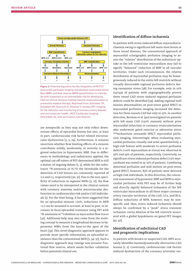
may unravel not only the most advanced or “culprit” lesion but also the flow-limiting effects of the other CAD stenoses of less severity in regions without stress induced perfusion defects on visual perfusion analysis (fig. 1). The concurrent assessment of “rela-tive” perfusion and “absolute” MBF, therefore, may afford not only the detection of the culprit CAD le-sion but also the flow-limiting downstream effects of less severe, intermediate or sequential CAD lesions or both in the remaining myocardial regions (fig. 2) [1, 4]. It is important to keep in mind, however, that re-ductions in hyperaemic MBF and MFR, respectively,
A
B
C
CARDIOVASCULAR MEDICINE – KARDIOVASKULÄRE MEDIZIN – MÉDECINE CARDIOVASCULAIRE 2015;18(3):83–95
REVIEW ARTICLE 86
are nonspecific as they may not only reflect down-stream effects of epicardial lesions but also, at least in part, cardiovascular risk factor related microvas-cular dysfunction [3, 5, 29]. Furthermore, it remains uncertain whether flow limiting effects of a stenosis contributes mildly, moderately, or severely to a re-gional reduction in hyperaemic MBF. Due to differ-ences in methodology and radiotracers applied, the optimal cut-off values of PET-determined MFR is still a matter of ongoing debate [1, 5], while for the radio-tracer 13N-ammonia or H2
15O the thresholds for the detection of CAD lesions are commonly reported of 2.0 and 2.5, respectively [30, 31]. Due to the non-speci-ficity of reductions in regional MFRs [5, 17], the flow values need to be interpreted in the clinical context with coronary anatomy and/or microvascular dys-function in cardiovascular risk and/or CAD individu-als [1]. For the time being, it has been suggested that for an epicardial stenosis ≥70%, reductions in MFR <1.7 can be assumed to account, at least in part, to in-creases in focal epicardial resistance using PET with 13N-ammonia or 82rubidium as myocardial flow tracer [32]. Additional help may also come from the evolv-ing concept to measure a longitudinal decrease in hy-peraemic MBFs from the base-to-the apex of the heart [33]. This novel diagnostic approach appears to provide more specific information on epicardial re-sistance than the conventional MFR [5, 34–37]. Such a diagnostic approach may emerge non-invasive frac-tional flow reserve, which needs further validation before potential clinical use.
Identification of diffuse ischaemia
In patients with stress induced diffuse myocardial is-chaemia owing to significant left main stem lesion or three vessel diseases, the conventional approach of myocardial scintigraphic perfusion imaging to as-sess the “relative” distribution of the radiotracer up-take in the left ventricular myocardium may fail to signify “balanced” reduction of MBF in all vascular territories. Under such circumstances, the relative distribution of myocardial perfusion may be homo-geneously reduced in the entire left ventricle without visually discernable regional perfusion defects dur-ing vasomotor stress [38]. For example, only in 10% (14/143) of patients with angiographically proven three vessel CAD stress induced regional perfusion defects could be identified [39]. Adding regional wall motion abnormalities on post-stress gated SPECT to myocardial perfusion imaging increased the detec-tion for three vessels CAD but only to 25%. In another direction, Berman et al. [40] investigated 101 patients with left main CAD (≥50% stenosis) without prior myo cardial infarction or coronary revascularisation who underwent gated exercise or adenosine stress 99mtechnetium sestamibi SPECT myocardial perfu-sion imaging. Interestingly, when evaluating myo-cardial perfusion visually and semi-quantitatively, a high-risk feature with moderate to severe perfusion defects (>10% myocardium at stress) was observed in 56% and 59% of patients, respectively. Conversely, no significant stress induced perfusion defect (≥5% myo-cardium) was noted in 13–15% of patients. Combining abnormal perfusion and wall motion on post-stress gated SPECT, however, 83% of patients were detected as high-risk individuals. In this direction, the concur-rent assessment of hyperaemic MBF and MFR to myo-cardial perfusion with PET may be of further help and directly signify balanced ischaemia of the left ventricular myocardium in all three major coronary artery vascular territories of the LAD, LCx, and RCA. Diffuse reductions of MFR, however, may be non- specific and, thus, stress induced ischaemia should always be confirmed by a “peak” stress transient isch aemic cavity dilation of the left ventricle associ-ated with a global hypokinesis on gated PET images [41, 42].
Identification of subclinical CAD and prognostic implications
In patients with known or suspected CAD, MPS accu-rately identifies haemodynamically obstructive CAD lesions [1, 2]. Conversely, cardiovascular risk factors induced dysfunction of the coronary arteriolar ves-
Figure 2: Potential algorithm for the integration of PET/CT
myocardial perfusion imaging and absolute myocardial blood
flow (MBF) and flow reserve (MFR) quantification in individu-
als with suspected or an intermediate risk for developing
CAD for clinical decision making towards revascularisation or
preventive medical therapy. Reprinted from: Schindler TH,
Schelbert HR, Quercioli A, Dilsizian V. Cardiac PET imaging
for the detection and monitoring of coronary artery disease
and microvascular health. JACC Cardiovasc Imaging.
2010;3:623–40, with permission from Elsevier.
CARDIOVASCULAR MEDICINE – KARDIOVASKULÄRE MEDIZIN – MÉDECINE CARDIOVASCULAIRE 2015;18(3):83–95

REVIEW ARTICLE 87
sels, commonly regarded as functional precursor of the CAD process [43, 44], remain undetected with the conventional myocardial perfusion SPECT/CT or PET/CT approach [45]. Individuals with subclinical stages of CAD may exhibit either subtle heterogeneity in relative myocardial uptake of the radiotracer or ho-mogeneously impaired hyperaemic blood flow of the left ventricle [33]. By assessing also hyperaemic MBF and MFR, PET/CT can identify such early functional disturbances of the coronary circulation, which may favour the development of structural CAD [36, 44, 46, 47]. For example, in insulin-resistant population without other traditional cardiovascular risk factors and with normal stress-rest myocardial perfusion on PET images, concurrent MBF quantification has un-ravelled disturbances in endothelium-related MBF responses to cold pressor testing (CPT), while phar-macologically induced hyperaemic flow increase were still normal [48]. These observations outline that initial stages of the vascular damage may in-volve only the endothelium [49–51], while in more advanced stages of cardiovascular risk factors states, commonly associated with increases in oxidative
stress burden, may also manifest in an impairment in smooth muscle cell vasodilator function of the coronary arteriolar vessels [47, 52, 53]. Notably, a dys-function of the coronary endothelium in cardiovas-cular risk individuals but with normal coronary an-giograms, as determined with PET-measured flow responses to sympathetic stimulation with CPT and its MFR, was associated with a higher risk for cardio-vascular events when compared to those individuals with normal flow increases [43]. In particular, it was observed that the incidence of cardiovascular events increased with progressive worsening of coronary endothelial dysfunction (fig. 3). These observations were followed by more recent investigations with PET-measured hyperaemic MBFs during pharmaco-logic vasodilation [44, 54–57]. Herzog et al. [44] ob-served that adding the MFR information to the stress 13N-ammonia perfusion PET with normal perfusion enabled a further risk stratification, while a “war-ranty” period of event-free survival of three years was observed when the MFR was normal. On the other hand, when patients had stress-induced re-gional myocardial perfusion defects and abnormally reduced MFR, impaired MFR provided incremental information to the stress 13N-ammonia perfusion im-aging for the prediction of adverse cardiovascular outcome (fig. 4). This was also confirmed by Ziadi et
Figure 3: PET determined coronary endothelial vasoreactivity
and prognosis. Kaplan-Meier analyses in patients with
cardiovascular risk factors and normal coronary angiograms
undergoing assessment of myocardial blood flow (MBF)
response to cold pressor test (CPT) with positron emission
tomography (PET). Attenuation of PET-measured and en-
dothelium related MBF responses to sympathetic stimulation
with cold pressor testing are associated with a higher risk
for cardiac events (during long-term follow-up) as compared
with those with normal flow increases; normal (%ΔMBF
≥40%), impaired (%ΔMBF >0% and <40%), and decreased
(%ΔMBF ≤0%). Reprinted from: Schindler TH, Nitzsche EU,
Schelbert HR, Olschewski M, Sayre J, Mix M, et al. Positron
emission tomography-measured abnormal responses of my-
ocardial blood flow to sympathetic stimulation are associated
with the risk of developing cardiovascular events. J Am Coll
Cardiol. 2005;45:1505–12, with permission from Elsevier.
Figure 4: Myocardial perfusion, coronary flow reserve
and prognosis. Coronary flow reserve (CFR) predicts major
cardiovascular events (MACE) such as cardiac death, nonfatal
myocardial infarction, and hospitalisation for any cardiac
reasons including late percutaneous coronary intervention
(PCI) or late coronary artery bypass grafting (CABG) in the
entire study population. Reprinted from: Herzog BA, Hus-
mann L, Valenta I, Gaemperli O, Siegrist PT, Tay FM, et al.
Long-term prognostic value of 13N-ammonia myocardial
perfusion positron emission tomography added value of
coronary flow reserve. J Am Coll Cardiol. 2009;54:150–6,
with permission from Elsevier.
CARDIOVASCULAR MEDICINE – KARDIOVASKULÄRE MEDIZIN – MÉDECINE CARDIOVASCULAIRE 2015;18(3):83–95
REVIEW ARTICLE 88
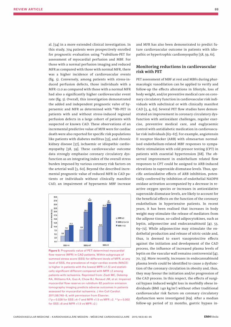
al. [54] in a more extended clinical investigation. In this study, 704 patients were prospectively enrolled for prognostic evaluation using 82rubidium-PET for assessment of myocardial perfusion and MBF. For those with a normal perfusion imaging and reduced MFR as compared with those with normal MFR, there was a higher incidence of cardiovascular events (fig. 5). Conversely, among patients with stress-in-duced perfusion defects, those individuals with a MFR <2.0 as compared with those with a normal MFR had also a significantly higher cardiovascular event rate (fig. 5). Overall, this investigation demonstrated the added and independent prognostic valve of hy-peraemic and MFR as determined with 82Rb-PET in patients with and without stress-induced regional perfusion defects in a large cohort of patients with suspected or known CAD. These observations of an incremental predictive value of MFR were for cardiac death were also reported for specific risk populations like patients with diabetes mellitus [55], and chronic kidney disease [57], ischaemic or idiopathic cardio-myopathy [58, 59]. These cardiovascular outcome data strongly emphasise coronary circulatory dys-function as an integrating index of the overall stress burden imposed by various coronary risk factors on the arterial wall [3, 60]. Beyond the described incre-mental prognostic value of reduced MFR in CAD pa-tients or individuals without clinically manifest CAD, an impairment of hyperaemic MBF increase
and MFR has also been demonstrated to predict fu-ture cardiovascular outcome in patients with idio-pathic or hypertrophic cardiomyopathy [58, 59, 61].
Monitoring reductions in cardiovascular risk with PET
PET assessment of MBF at rest and MBFs during phar-macologic vasodilation can be applied to verify and follow-up the effects alterations in lifestyle, loss of body weight, and/or preventive medical care on coro-nary circulatory function in cardiovascular risk indi-viduals with subclinical or with clinically manifest CAD [3, 4, 62]. Several PET flow studies have demon-strated an improvement in coronary circulatory dys-function with antioxidant challenges, regular exer-cise, preventive medical care, and euglycaemic control with antidiabetic medication in cardiovascu-lar risk individuals [63–67]. For example, angiotensin II receptor blocker (ARB) with olmesartan normal-ised endothelium-related MBF responses to sympa-thetic stimulation with cold pressor testing (CPT) in patients with essential hypertension [68]. The ob-served improvement in endothelium related flow responses to CPT could be assigned to ARB-induced elevations in superoxide dismutase levels. Thus, spe-cific antioxidative effects of ARB inhibition, poten-tially conferred by inhibition of endothelial NADPH oxidase activation accompanied by a decrease in re-active oxygen species or increases in antioxidative superoxide dismutase levels, are likely to account for the beneficial effects on the function of the coronary endothelium in hypertensive patients. In recent years, it has been realised that increases in body weight may stimulate the release of mediators from the adipose tissue, so-called adipocytokines, such as leptin, adiponectine and endocannabinoid [47, 53, 69–72]. While adiponectine may stimulate the en-dothelial production and release of nitric-oxide and, thus, is deemed to exert vasoprotective effects against the initiation and development of the CAD process, the influence of increased plasma levels of leptin on the vascular wall remains controversial [47, 70, 73]. More recently, increases in endocannabinoid plasma levels could be identified to cause a dysfunc-tion of the coronary circulation in obesity and, thus, they may favour the initiation and/or progression of the CAD process. In this respect, the effects of surgi-cal bypass induced weight loss in morbidly obese in-dividuals (BMI ≥40 kg/m2) without other traditional cardiovascular risk factors on coronary circulatory dysfunction were investigated [69]. After a median follow-up period of 22 months, gastric bypass in-
Figure 5: Prognostic value of PET-determined myocardial
flow reserve (MFR) in CAD patients. Within subgroups of
summed stress score (SSS) for different levels of MFR, at any
level of SSS, the prevalence of major cardiac events (MACE)
is higher in patients with the lowest MFR (<1.5) and statisti-
cally significant different compared with MFR ≥2 among
patients with ischaemia. Reprinted from: Ziadi MC, Dekemp
RA, Williams KA, Guo A, Chow BJ, Renaud JM, et al. Impaired
myocardial flow reserve on rubidium-82 positron emission
tomography imaging predicts adverse outcomes in patients
assessed for myocardial ischaemia. J Am Coll Cardiol.
2011;58:740–8, with permission from Elsevier.
(*p = 0.028 for SSS ≥4–7 and MFR <1.5 vs MFR ≥2. **p = 0.002
for SSS ≥8 and MFR <1.5 vs MFR ≥2.)
CARDIOVASCULAR MEDICINE – KARDIOVASKULÄRE MEDIZIN – MÉDECINE CARDIOVASCULAIRE 2015;18(3):83–95
REVIEW ARTICLE 89
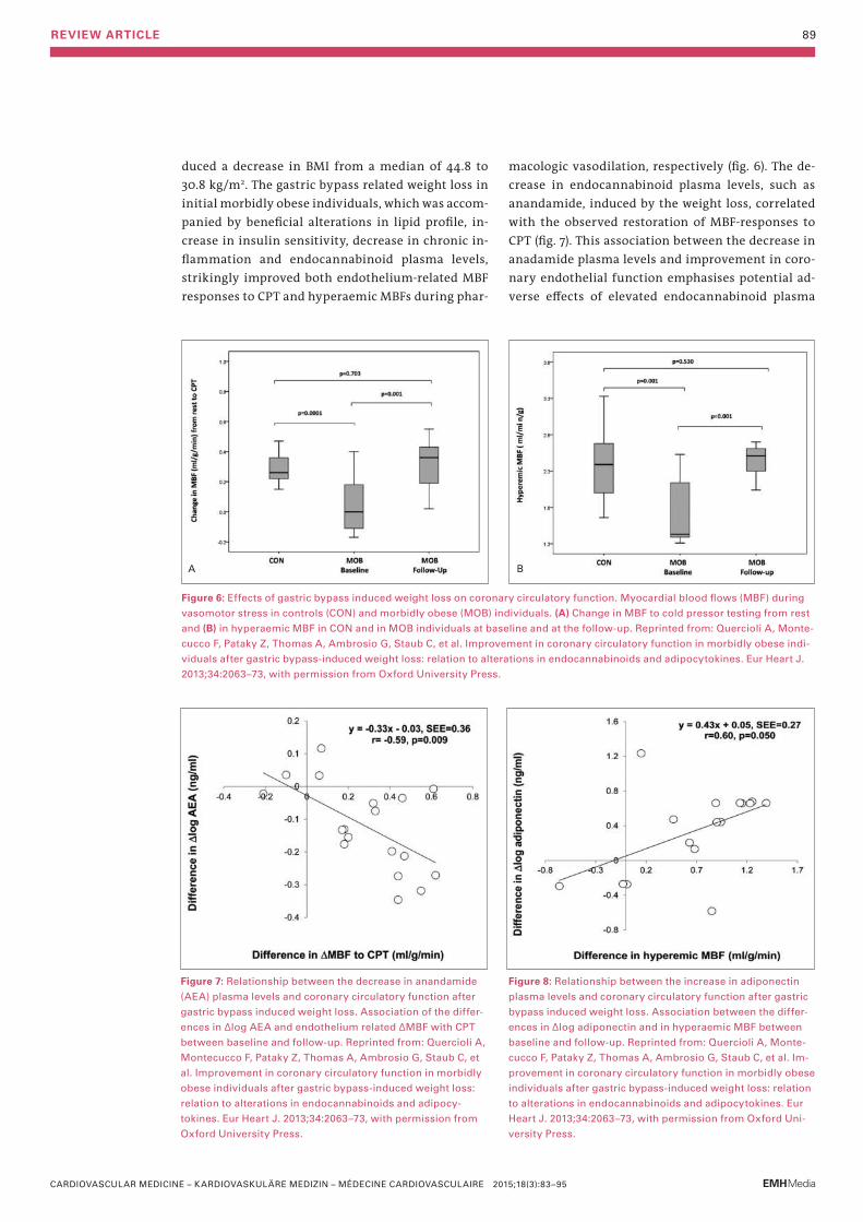
duced a decrease in BMI from a median of 44.8 to 30.8 kg/m2. The gastric bypass related weight loss in initial morbidly obese individuals, which was accom-panied by beneficial alterations in lipid profile, in-crease in insulin sensitivity, decrease in chronic in-flammation and endocannabinoid plasma levels, strikingly improved both endothelium-related MBF responses to CPT and hyperaemic MBFs during phar-
macologic vasodilation, respectively (fig. 6). The de-crease in endocannabinoid plasma levels, such as anandamide, induced by the weight loss, correlated with the observed restoration of MBF-responses to CPT (fig. 7). This association between the decrease in anadamide plasma levels and improvement in coro-nary endothelial function emphasises potential ad-verse effects of elevated endocannabinoid plasma
Figure 6: Effects of gastric bypass induced weight loss on coronary circulatory function. Myocardial blood flows (MBF) during
vasomotor stress in controls (CON) and morbidly obese (MOB) individuals. (A) Change in MBF to cold pressor testing from rest
and (B) in hyperaemic MBF in CON and in MOB individuals at baseline and at the follow-up. Reprinted from: Quercioli A, Monte-
cucco F, Pataky Z, Thomas A, Ambrosio G, Staub C, et al. Improvement in coronary circulatory function in morbidly obese indi-
viduals after gastric bypass-induced weight loss: relation to alterations in endocannabinoids and adipocytokines. Eur Heart J.
2013;34:2063–73, with permission from Oxford University Press.
A B
Figure 7: Relationship between the decrease in anandamide
(AEA) plasma levels and coronary circulatory function after
gastric bypass induced weight loss. Association of the differ-
ences in Δlog AEA and endothelium related ΔMBF with CPT
between baseline and follow-up. Reprinted from: Quercioli A,
Montecucco F, Pataky Z, Thomas A, Ambrosio G, Staub C, et
al. Improvement in coronary circulatory function in morbidly
obese individuals after gastric bypass-induced weight loss:
relation to alterations in endocannabinoids and adipocy-
tokines. Eur Heart J. 2013;34:2063–73, with permission from
Oxford University Press.
Figure 8: Relationship between the increase in adiponectin
plasma levels and coronary circulatory function after gastric
bypass induced weight loss. Association between the differ-
ences in Δlog adiponectin and in hyperaemic MBF between
baseline and follow-up. Reprinted from: Quercioli A, Monte-
cucco F, Pataky Z, Thomas A, Ambrosio G, Staub C, et al. Im-
provement in coronary circulatory function in morbidly obese
individuals after gastric bypass-induced weight loss: relation
to alterations in endocannabinoids and adipocytokines. Eur
Heart J. 2013;34:2063–73, with permission from Oxford Uni-
versity Press.
CARDIOVASCULAR MEDICINE – KARDIOVASKULÄRE MEDIZIN – MÉDECINE CARDIOVASCULAIRE 2015;18(3):83–95
REVIEW ARTICLE 90
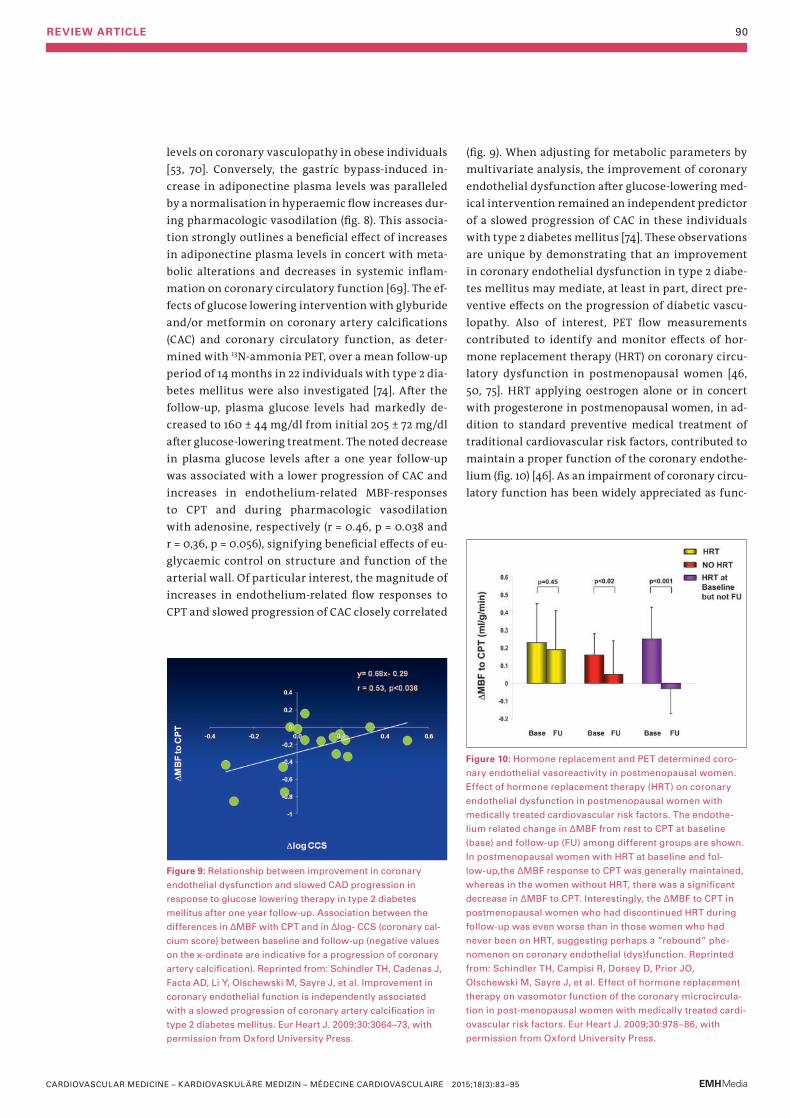
levels on coronary vasculopathy in obese individuals [53, 70]. Conversely, the gastric bypass-induced in-crease in adiponectine plasma levels was paralleled by a normalisation in hyperaemic flow increases dur-ing pharmacologic vasodilation (fig. 8). This associa-tion strongly outlines a beneficial effect of increases in adiponectine plasma levels in concert with meta-bolic alterations and decreases in systemic inflam-mation on coronary circulatory function [69]. The ef-fects of glucose lowering intervention with glyburide and/or metformin on coronary artery calcifications (CAC) and coronary circulatory function, as deter-mined with 13N-ammonia PET, over a mean follow-up period of 14 months in 22 individuals with type 2 dia-betes mellitus were also investigated [74]. After the follow-up, plasma glucose levels had markedly de-creased to 160 ± 44 mg/dl from initial 205 ± 72 mg/dl after glucose-lowering treatment. The noted decrease in plasma glucose levels after a one year follow-up was associated with a lower progression of CAC and increases in endothelium-related MBF-responses to CPT and during pharmacologic vasodilation with adenosine, respectively (r = 0.46, p = 0.038 and r = 0,36, p = 0.056), signifying beneficial effects of eu-glycaemic control on structure and function of the arterial wall. Of particular interest, the magnitude of increases in endothelium-related flow responses to CPT and slowed progression of CAC closely correlated
(fig. 9). When adjusting for metabolic parameters by multivariate analysis, the improvement of coronary endothelial dysfunction after glucose-lowering med-ical intervention remained an independent predictor of a slowed progression of CAC in these individuals with type 2 diabetes mellitus [74]. These observations are unique by demonstrating that an improvement in coronary endothelial dysfunction in type 2 diabe-tes mellitus may mediate, at least in part, direct pre-ventive effects on the progression of diabetic vascu-lopathy. Also of interest, PET flow measurements contributed to identify and monitor effects of hor-mone replacement therapy (HRT) on coronary circu-latory dysfunction in postmenopausal women [46, 50, 75]. HRT applying oestrogen alone or in concert with progesterone in postmenopausal women, in ad-dition to standard preventive medical treatment of traditional cardiovascular risk factors, contributed to maintain a proper function of the coronary endothe-lium (fig. 10) [46]. As an impairment of coronary circu-latory function has been widely appreciated as func-
Figure 9: Relationship between improvement in coronary
endothelial dysfunction and slowed CAD progression in
response to glucose lowering therapy in type 2 diabetes
mellitus after one year follow-up. Association between the
differences in ΔMBF with CPT and in Δlog- CCS (coronary cal-
cium score) between baseline and follow-up (negative values
on the x-ordinate are indicative for a progression of coronary
artery calcification). Reprinted from: Schindler TH, Cadenas J,
Facta AD, Li Y, Olschewski M, Sayre J, et al. Improvement in
coronary endothelial function is independently associated
with a slowed progression of coronary artery calcification in
type 2 diabetes mellitus. Eur Heart J. 2009;30:3064–73, with
permission from Oxford University Press.
Figure 10: Hormone replacement and PET determined coro-
nary endothelial vasoreactivity in postmenopausal women.
Effect of hormone replacement therapy (HRT) on coronary
endothelial dysfunction in postmenopausal women with
medically treated cardiovascular risk factors. The endothe-
lium related change in ΔMBF from rest to CPT at baseline
(base) and follow-up (FU) among different groups are shown.
In postmenopausal women with HRT at baseline and fol-
low-up,the ΔMBF response to CPT was generally maintained,
whereas in the women without HRT, there was a significant
decrease in ΔMBF to CPT. Interestingly, the ΔMBF to CPT in
postmenopausal women who had discontinued HRT during
follow-up was even worse than in those women who had
never been on HRT, suggesting perhaps a “rebound” phe-
nomenon on coronary endothelial (dys)function. Reprinted
from: Schindler TH, Campisi R, Dorsey D, Prior JO,
Olschewski M, Sayre J, et al. Effect of hormone replacement
therapy on vasomotor function of the coronary microcircula-
tion in post-menopausal women with medically treated cardi-
ovascular risk factors. Eur Heart J. 2009;30:978–86, with
permission from Oxford University Press.
CARDIOVASCULAR MEDICINE – KARDIOVASKULÄRE MEDIZIN – MÉDECINE CARDIOVASCULAIRE 2015;18(3):83–95
REVIEW ARTICLE 91
tional precursor of the CAD process, its normalisation after life-style intervention and/or preventive medical care, for example by statin and/or ACE inhibitors, is likely to result into an improved cardiovascular out-come, which, however, needs to be tested clinically.
Cardiac sarcoid involvement
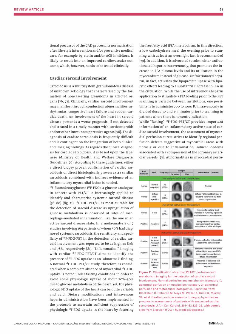
Sarcoidosis is a multisystem granulomatous disease of unknown aetiology that characterised by the for-mation of noncaseating granuloma in affected or-gans [76, 77]. Clinically, cardiac sarcoid involvement may manifest through conduction abnormalities, ar-rhythmias, congestive heart failure and sudden car-diac death. An involvement of the heart in sarcoid disease portends a worse prognosis, if not detected and treated in a timely manner with corticosteroids and/or other immunosuppressive agents [78]. The di-agnosis of cardiac sarcoidosis is frequently difficult and is contingent on the integration of both clinical and imaging findings. As regards the clinical diagno-sis for cardiac sarcoidosis, it is based upon the Japa-nese Ministry of Health and Welfare Diagnostic Guidelines [79]. According to these guidelines, either a direct biopsy proven confirmation of cardiac sar-coidosis or direct histologically proven extra cardiac sarcoidosis combined with indirect evidence of an inflammatory myocardial lesion is needed. 18F-fluorodeoxyglucose (18F-FDG), a glucose analogue, in concert with PET/CT is increasingly applied to identify and characterise systemic sarcoid disease [78–80] (fig. 11). 18F-FDG-PET/CT is most suitable for the detection of sarcoid disease as upregulation of glucose metabolism is observed at sites of mac-rophage-mediated inflammation, like the one in an active sarcoid disease state. In a meta-analysis of 7 studies involving 164 patients of whom 50% had diag-nosed systemic sarcoidosis, the sensitivity and speci-ficity of 18F-FDG-PET in the detection of cardiac sar-coid involvement was reported to be as high as 89% and 78%, respectively [81]. “Inflammation” imaging with cardiac 18F-FDG-PET/CT aims to identify the presence of 18F-FDG uptake as an “abnormal” finding. A normal 18F-FDG-PET/CT study, therefore, is consid-ered when a complete absence of myocardial 18F-FDG uptake is noted under fasting conditions in order to avoid some physiologic uptake of about 20%–30% due to glucose metabolism of the heart. Yet, the phys-iologic FDG uptake of the heart can be quite variable and avid. Dietary modifications and intravenous heparin administration have been implemented in the protocols to ascertain sufficient suppression of physiologic 18F-FDG uptake in the heart by fostering
the free fatty acid (FFA) metabolism. In this direction, a low carbohydrate meal the evening prior to scan-ning with at least an overnight fast is recommended [79]. In addition, it is advocated to administer unfrac-tionated heparin intravenously, that promotes the in-crease in FFA plasma levels and its utilisation in the myocardium instead of glucose. Unfractionated hepa-rin, in fact, activates the lipoprotein lipase with lipo-lytic effects leading to a substantial increase in FFA in the circulation. While the use of intravenous heparin application to stimulate a FFA loading prior to the PET scanning is variable between institutions, one possi-bility is to administer 700 to 1000 IU intravenously in divided doses 30 and 15 minutes prior to scanning in patients where there is no contraindication.While “fasting” 18F-FDG-PET/CT provides important information of an inflammatory active state of car-diac sarcoid involvement, the assessment of myocar-dial perfusion at rest strives to identify regional per-fusion defects suggestive of myocardial areas with fibrosis or due to inflammation induced oedema associated with a compression of the coronary arteri-olar vessels [78]. Abnormalities in myocardial perfu-
Figure 11: Classification of cardiac PET/CT perfusion and
metabolism imaging for the detection of cardiac sarcoid
involvement. Normal perfusion and metabolism (category 1),
abnormal perfusion or metabolism (category 2), abnormal
perfusion and metabolism (category 3). Reprinted from:
Blankstein R, Osborne M, Naya M, Waller A, Kim CK, Murthy
VL, et al. Cardiac positron emission tomography enhances
prognostic assessments of patients with suspected cardiac
sarcoidosis. J Am Coll Cardiol. 2014;63:329–36, with permis-
sion from Elsevier. (FDG = fluorodeoxyglucose.)
CARDIOVASCULAR MEDICINE – KARDIOVASKULÄRE MEDIZIN – MÉDECINE CARDIOVASCULAIRE 2015;18(3):83–95
REVIEW ARTICLE 92
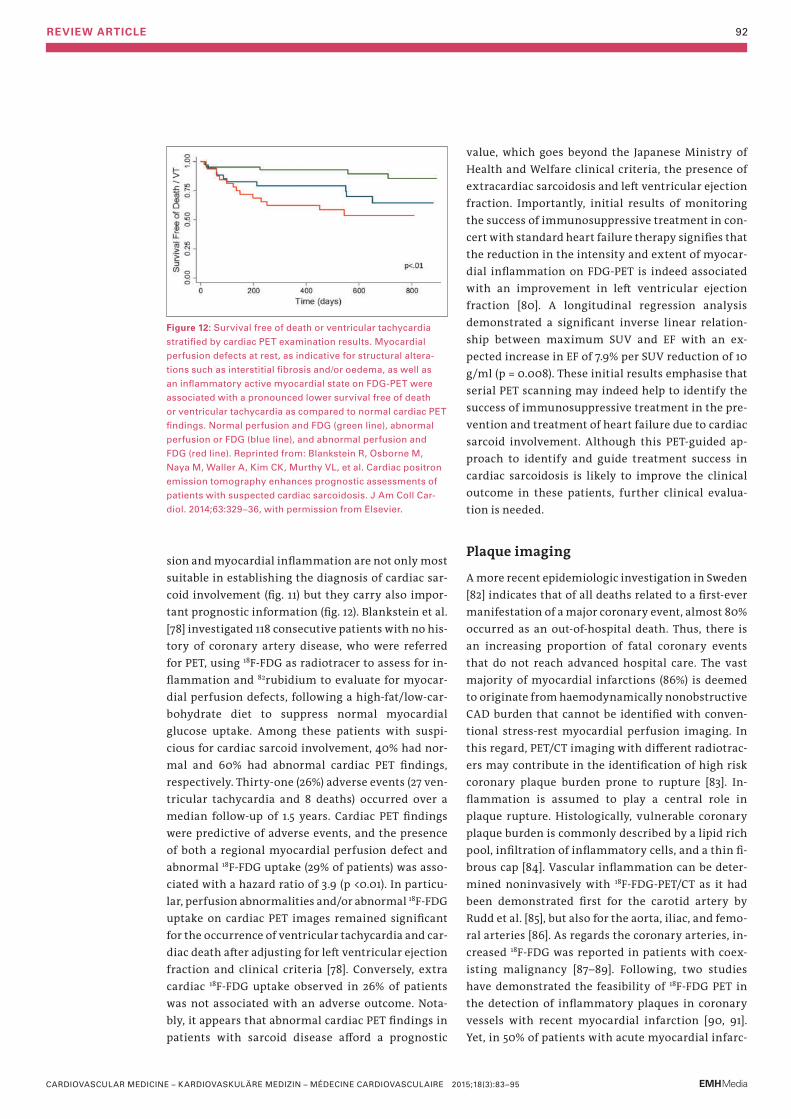
sion and myocardial inflammation are not only most suitable in establishing the diagnosis of cardiac sar-coid involvement (fig. 11) but they carry also impor-tant prognostic information (fig. 12). Blankstein et al. [78] investigated 118 consecutive patients with no his-tory of coronary artery disease, who were referred for PET, using 18F-FDG as radiotracer to assess for in-flammation and 82rubidium to evaluate for myocar-dial perfusion defects, following a high-fat/low-car-bohydrate diet to suppress normal myocardial glucose uptake. Among these patients with suspi-cious for cardiac sarcoid involvement, 40% had nor-mal and 60% had abnormal cardiac PET findings, respectively. Thirty-one (26%) adverse events (27 ven-tricular tachycardia and 8 deaths) occurred over a median follow-up of 1.5 years. Cardiac PET findings were predictive of adverse events, and the presence of both a regional myocardial perfusion defect and abnormal 18F-FDG uptake (29% of patients) was asso-ciated with a hazard ratio of 3.9 (p <0.01). In particu-lar, perfusion abnormalities and/or abnormal 18F-FDG uptake on cardiac PET images remained significant for the occurrence of ventricular tachycardia and car-diac death after adjusting for left ventricular ejection fraction and clinical criteria [78]. Conversely, extra cardiac 18F-FDG uptake observed in 26% of patients was not associated with an adverse outcome. Nota-bly, it appears that abnormal cardiac PET findings in patients with sarcoid disease afford a prognostic
value, which goes beyond the Japanese Ministry of Health and Welfare clinical criteria, the presence of extracardiac sarcoidosis and left ventricular ejection fraction. Importantly, initial results of monitoring the success of immunosuppressive treatment in con-cert with standard heart failure therapy signifies that the reduction in the intensity and extent of myocar-dial inflammation on FDG-PET is indeed associated with an improvement in left ventricular ejection fraction [80]. A longitudinal regression analysis demonstrated a significant inverse linear relation-ship between maximum SUV and EF with an ex-pected increase in EF of 7.9% per SUV reduction of 10 g/ml (p = 0.008). These initial results emphasise that serial PET scanning may indeed help to identify the success of immunosuppressive treatment in the pre-vention and treatment of heart failure due to cardiac sarcoid involvement. Although this PET-guided ap-proach to identify and guide treatment success in cardiac sarcoidosis is likely to improve the clinical outcome in these patients, further clinical evalua-tion is needed.
Plaque imaging
A more recent epidemiologic investigation in Sweden [82] indicates that of all deaths related to a first-ever manifestation of a major coronary event, almost 80% occurred as an out-of-hospital death. Thus, there is an increasing proportion of fatal coronary events that do not reach advanced hospital care. The vast majority of myocardial infarctions (86%) is deemed to originate from haemodynamically nonobstructive CAD burden that cannot be identified with conven-tional stress-rest myocardial perfusion imaging. In this regard, PET/CT imaging with different radiotrac-ers may contribute in the identification of high risk coronary plaque burden prone to rupture [83]. In-flammation is assumed to play a central role in plaque rupture. Histologically, vulnerable coronary plaque burden is commonly described by a lipid rich pool, infiltration of inflammatory cells, and a thin fi-brous cap [84]. Vascular inflammation can be deter-mined noninvasively with 18F-FDG-PET/CT as it had been demonstrated first for the carotid artery by Rudd et al. [85], but also for the aorta, iliac, and femo-ral arteries [86]. As regards the coronary arteries, in-creased 18F-FDG was reported in patients with coex-isting malignancy [87–89]. Following, two studies have demonstrated the feasibility of 18F-FDG PET in the detection of inflammatory plaques in coronary vessels with recent myocardial infarction [90, 91]. Yet, in 50% of patients with acute myocardial infarc-
Figure 12: Survival free of death or ventricular tachycardia
stratified by cardiac PET examination results. Myocardial
perfusion defects at rest, as indicative for structural altera-
tions such as interstitial fibrosis and/or oedema, as well as
an inflammatory active myocardial state on FDG-PET were
associated with a pronounced lower survival free of death
or ventricular tachycardia as compared to normal cardiac PET
findings. Normal perfusion and FDG (green line), abnormal
perfusion or FDG (blue line), and abnormal perfusion and
FDG (red line). Reprinted from: Blankstein R, Osborne M,
Naya M, Waller A, Kim CK, Murthy VL, et al. Cardiac positron
emission tomography enhances prognostic assessments of
patients with suspected cardiac sarcoidosis. J Am Coll Car-
diol. 2014;63:329–36, with permission from Elsevier.
CARDIOVASCULAR MEDICINE – KARDIOVASKULÄRE MEDIZIN – MÉDECINE CARDIOVASCULAIRE 2015;18(3):83–95

REVIEW ARTICLE 93
tion no increased 18F-FDG was noted, which might suggest that non-inflammatory plaques may also be prone to rupture [90], potentially by increased activ-ity of metalloproteinases leading to soft coronary plaque burden [92]. Such observations also empha-sise the complexity of coronary plaque composition, susceptibility to rupture, and plaque metabolic imag-ing for risk assessment. The 18F-FDG uptake in the ar-terial wall as non-invasive surrogate marker for in-flammation has been demonstrated to correlate with macrophage burden in the plaque [93], symptoms [85], and conventional Framingham Risk Score [94]. In particular, increases in arterial 18F-FDG-uptake in the noncoronary arteries can be attenuated with sta-tin and dalcetrapib medication, respectively [95, 96]. As regards the assessment of 18F-FDG uptake in the coronary arteries with PET/CT, it remains an ongoing challenge as cardiac and respiratory motion and con-current 18F-FDG in the myocardium hamper an accu-rate visualisation of any plaque signal [91, 97]. 64- or 128-slice CT from the cardiac PET/CT systems is com-monly used to assess coronary artery calcifications (CAC) or, with intravenous contrast CT coronary mor-phology, respectively [7, 98]. CAC scoring, as a surro-gate marker of coronary atherosclerotic burden, has been established as a powerful cardiovascular risk predictor, which is increasingly applied to enhance primary risk stratification and justification of pre-ventive medical care of the CAD process [99, 100]. CAC may be combined with measurements of high-sensitive CRP, as a marker of systemic micro-in-flammation, to improve and refine the prediction of cardiovascular risk [100]. Although measure of CAC and CT-coronary angiography provide important in-formation on coronary morphology and risk predic-tion, they cannot identify active inflammation of coronary plaque burden, commonly seen as vulnera-ble plaque prone to rupture with its atherothrom-botic sequelae. As mentioned before, measurements of 18F-FDG uptake in coronary arteries with PET/CT is still challenging [91]. Another interesting approach has been proposed more recently [101, 102] with the use of 18F-sodium flu-oride (18F-NaF) as PET radiotracer, which identifies novel regions of bone formation and remodelling. Such a diagnostic approach is assumed to identify ac-tive calcification and/or micro-calcifications as po-tential source of microfractures and acute thrombo-sis [103–105]. Dweck MR et al. [102] investigated the potential of 18F-NaF and FDG uptake in the coronary arterial wall as marker for active calcification and in-flammation, respectively. In 119 volunteers with and without aortic valve disease, coronary CAC score and
18F-NaF and FDG were determined with PET/CT. In in-dividuals with a CAC score of 0 served as control in-dividuals as compared to those with CAC (calcium score>0). As it turned out, the 18F-NaF strongly corre-lated with CAC, while in 41% of individuals with CAC >1000 had no significant 18F-NaF uptake in the arte-rial wall. This outlines that 18F-NaF uptake signifies different information reflecting metabolically active calcific plaque and developing microcalcification. 18F-NaF uptake, therefore, may differentiate between individuals with “dormant” coronary atherosclerotic plaque burden, developed many months or years pre-viously, and individuals with metabolically active CAD and ongoing calcification process. This is sub-stantiated by the observation that individual with in-creased coronary 18F-NaF activity (n = 40) had higher rates of prior cardiovascular events (p = 0.016), an-gina pectoris (p = 0.023) and higher Framingham risk scores (p = 0.011) (fig. 13). Somehow surprising, how-ever, 18F-FDG activity was not increased in CAD indi-viduals as compared to controls without CAC. This might suggest that 18F-FDG uptake in the coronary ar-terial wall is of little use in individuals with stable CAD. Someone could argue that inflammation, as re-flected by increased arterial 18F-FDG uptake, is more prevalent in acute coronary syndrome than in stable CAD. The clinical observations of increases in 18F-NaF activity in the coronary arterial wall in CAD patients associated with angina symptoms, prior cardiovas-cular events, and cardiovascular risk scores may give
Figure 13: 10-year Framingham Risk Scores for control indi-
viduals and patients with atherosclerosis who did and did not
have increased 18F-NaF uptake. Reprinted from: Dweck MR,
Chow MW, Joshi NV, Williams MC, Jones C, Fletcher AM, et
al. Coronary arterial 18F-sodium fluoride uptake: a novel
marker of plaque biology. J Am Coll Cardiol. 2012;59:1539–
48, with permission from Elsevier. (CHD = coronary heart
disease; CVD = cardiovascular disease).
CARDIOVASCULAR MEDICINE – KARDIOVASKULÄRE MEDIZIN – MÉDECINE CARDIOVASCULAIRE 2015;18(3):83–95
REVIEW ARTICLE 94
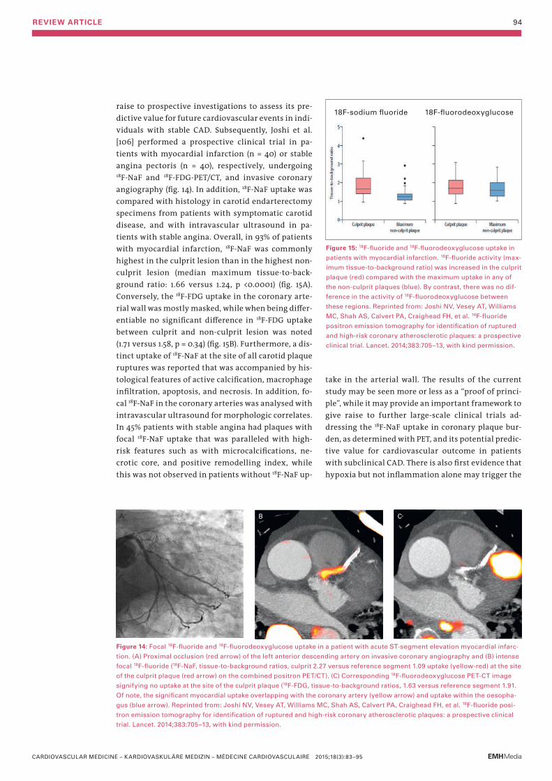
raise to prospective investigations to assess its pre-dictive value for future cardiovascular events in indi-viduals with stable CAD. Subsequently, Joshi et al. [106] performed a prospective clinical trial in pa-tients with myocardial infarction (n = 40) or stable angina pectoris (n = 40), respectively, undergoing 18F-NaF and 18F-FDG-PET/CT, and invasive coronary angiography (fig. 14). In addition, 18F-NaF uptake was compared with histology in carotid endarterectomy specimens from patients with symptomatic carotid disease, and with intravascular ultrasound in pa-tients with stable angina. Overall, in 93% of patients with myocardial infarction, 18F-NaF was commonly highest in the culprit lesion than in the highest non-culprit lesion (median maximum tissue-to-back-ground ratio: 1.66 versus 1.24, p <0.0001) (fig. 15A). Conversely, the 18F-FDG uptake in the coronary arte-rial wall was mostly masked, while when being differ-entiable no significant difference in 18F-FDG uptake between culprit and non-culprit lesion was noted (1.71 versus 1.58, p = 0.34) (fig. 15B). Furthermore, a dis-tinct uptake of 18F-NaF at the site of all carotid plaque ruptures was reported that was accompanied by his-tological features of active calcification, macrophage infiltration, apoptosis, and necrosis. In addition, fo-cal 18F-NaF in the coronary arteries was analysed with intravascular ultrasound for morphologic correlates. In 45% patients with stable angina had plaques with focal 18F-NaF uptake that was paralleled with high-risk features such as with microcalcifications, ne-crotic core, and positive remodelling index, while this was not observed in patients without 18F-NaF up-
18F-sodium fluoride 18F-fluorodeoxyglucose
Figure 14: Focal 18F-fluoride and 18F-fluorodeoxyglucose uptake in a patient with acute ST-segment elevation myocardial infarc-
tion. (A) Proximal occlusion (red arrow) of the left anterior descending artery on invasive coronary angiography and (B) intense
focal 18F-fluoride (18F-NaF, tissue-to-background ratios, culprit 2.27 versus reference segment 1.09 uptake (yellow-red) at the site
of the culprit plaque (red arrow) on the combined positron PET/CT). (C) Corresponding 18F-fluorodeoxyglucose PET-CT image
signifying no uptake at the site of the culprit plaque (18F-FDG, tissue-to-background ratios, 1.63 versus reference segment 1.91.
Of note, the significant myocardial uptake overlapping with the coronary artery (yellow arrow) and uptake within the oesopha-
gus (blue arrow). Reprinted from: Joshi NV, Vesey AT, Williams MC, Shah AS, Calvert PA, Craighead FH, et al. 18F-fluoride posi-
tron emission tomography for identification of ruptured and high-risk coronary atherosclerotic plaques: a prospective clinical
trial. Lancet. 2014;383:705–13, with kind permission.
Figure 15: 18F-fluoride and 18F-fluorodeoxyglucose uptake in
patients with myocardial infarction. 18F-fluoride activity (max-
imum tissue-to-background ratio) was increased in the culprit
plaque (red) compared with the maximum uptake in any of
the non-culprit plaques (blue). By contrast, there was no dif-
ference in the activity of 18F-fluorodeoxyglucose between
these regions. Reprinted from: Joshi NV, Vesey AT, Williams
MC, Shah AS, Calvert PA, Craighead FH, et al. 18F-fluoride
positron emission tomography for identification of ruptured
and high-risk coronary atherosclerotic plaques: a prospective
clinical trial. Lancet. 2014;383:705–13, with kind permission.
take in the arterial wall. The results of the current study may be seen more or less as a “proof of princi-ple”, while it may provide an important framework to give raise to further large-scale clinical trials ad-dressing the 18F-NaF uptake in coronary plaque bur-den, as determined with PET, and its potential predic-tive value for cardiovascular outcome in patients with subclinical CAD. There is also first evidence that hypoxia but not inflammation alone may trigger the
A B C
CARDIOVASCULAR MEDICINE – KARDIOVASKULÄRE MEDIZIN – MÉDECINE CARDIOVASCULAIRE 2015;18(3):83–95

REVIEW ARTICLE 95
18F-FDG uptake in human macrophages within the ar-terial plaque burden [107], which would also accord, at least in part, with previous report that could not find increases in 18F-FDG in culprit plaque burden in 50% of patients with acute myocardial infarction [90]. Hypoxia is known to play a central role in stim-ulating atherosclerotic burden progression by stimu-lation foam cell formation, metabolic adaption of in-filtrated macrophages, and plaque neovascularisation [108]. Hypoxaemic conditions may activate plaque growth by signalling of the hypoxia-inducible factor (HIF-1) leading to the formation of lipid droplets, the activation of a metabolic switch to anaerobic glycoly-sis, and increasing the secretion of proinflammatory and angiogenic mediators [109–111]. Imaging of hy-poxia has been performed with PET and the radio-tracer 18F-fluoromisonidazole (18F-FMISO) in patients with ischaemic stroke [112], myocardial ischaemia [113], and various malignancies [114]. 18F-FMISO is a cell-permeable 2-nitroimidazole derivative that is re-duced in vivo by nitroreductases independent of the levels of intracellular oxygen. When there is a nor-mal oxygenation environment, 18F-FMISO is rapidly reoxidised and diffuses out of the cells. Conversely, in hypoxic viable cells, 18F-FMISO is further reduced to a more reactive form that binds covalently to in-tracellular macromolecules and remains in the cells. In this direction, recent study evaluated the feasibil-ity of 18F-FMISO PET imaging for in vivo detection of hypoxia in advanced lesions in a rabbit atherosclero-sis model [115]. A significant 18F-FMISO accumulation in the aortas of atherosclerotic animals compared with healthy controls, and the uptake increased over time with atherogenic diet, suggesting hypoxia as a good biomarker of disease progression. In addition, the in-vivo detection of hypoxia was translated by ex-vivo PET imaging of the excised aorta that demon-strated regions of strong 18F-FMISO accumulation in atherosclerotic aortas when compared nearly absent uptake in the normal aorta. Thus, hypoxia in athero-sclerotic plaque increases with disease progression and is present in macrophage-rich areas associated with neovascularisation, which can be determined
noninvasively with 18F-FMISO. This novel imaging ap-proach holds promise to further improve the cardio-vascular risk prediction, which should be further tested in clinical investigations in CAD patients.
Recapitulation
In the last decade, the assessment of hibernating stun-ning myocardium in ischaemic cardiomyopathy with PET/CT has evolved as mainstay in clinical practice for the decision making process for coronary revasculari-sation procedures. In addition, the assessment of myo-cardial perfusion and vasodilator capacity of the coro-nary circulation with PET/CT imaging is increasingly applied clinically for the detection and characterisa-tion of CAD burden in subclinical and clinically mani-fest CAD, respectively, and risk stratification in pa-tients with hypertrophic cardiomyopathy. An important role of PET/CT imaging for the detection of cardiac sarcoid involvement and the monitoring of successful immunosuppressive treatment has also been appreciated more recently. While the role of PET/CT imaging with various radiotracers emerges as a promising tool for the identification of “vulnerable” coronary plaque burden, its clinical value remains to be tested. It is anticipated that with the advent of PET/MRI (magnetic resonance imaging) further advances and refinement in the comprehensive assessment of cardiovascular pathology will ensue.
AcknowledgmentSome sections of the manuscript are similar to sections of extensive reviews of cardiac PET by Schindler et al. [1, 3].
Funding / potential competing interestsNo potential conflict of interest exists and all sources of funding for the work are acknowledged as follows. This work was supported by a departemental fund of Johns Hopkins University, Baltimore, MD, USA; and a Research Grant of the Swiss National Science Foundation (SNF: 3200B0–122237, Dr Schindler), with contributions of the Clinical Research Centre, University Hospital and Faculty of Medicine, Geneva, and the Louis-Jeantet Foundation, Gustave and Simone Prevot, and Swiss Heart Foundation.
References– The full list of references is attached to the online version at
www.cardiovascmed.ch.
Correspondence: Thomas Hellmut Schindler, MD Johns Hopkins University, School of Medicine, Division of Nuclear Medicine, Cardio-vascular Nuclear Medicine Department of Radiology and Radiological Science SOM, JHOC 3225, 601 N. Caroline Street USA-Baltimore, MD, 21287 tschind3[at]jhmi.edu
CARDIOVASCULAR MEDICINE – KARDIOVASKULÄRE MEDIZIN – MÉDECINE CARDIOVASCULAIRE 2015;18(3):83–95