contribution of e3-ubiquitin ligase activity to hiv-1...
TRANSCRIPT

JOURNAL OF VIROLOGY, Sept. 2011, p. 8725–8737 Vol. 85, No. 170022-538X/11/$12.00 doi:10.1128/JVI.00497-11Copyright © 2011, American Society for Microbiology. All Rights Reserved.
Contribution of E3-Ubiquitin Ligase Activity to HIV-1 Restriction byTRIM5�rh: Structure of the RING Domain of TRIM5��†
Maritza Lienlaf,1 Fumiaki Hayashi,2 Francesca Di Nunzio,5 Naoya Tochio,2 Takanori Kigawa,2,3
Shigeyuki Yokoyama,2,4 and Felipe Diaz-Griffero1*Department of Microbiology and Immunology, Albert Einstein College of Medicine, Bronx, New York 104611; Systems and
Structural Biology Center, Yokohama Institute, RIKEN, 1-7-22 Suehiro-cho, Tsurumi, Yokohama 230-0045, Japan2;Department of Computational Intelligence and Systems Science, Interdisciplinary Graduate School of Science and
Engineering, Tokyo Institute of Technology, 2-12-1 Ookayama Meguro-ku, Tokyo 152-8550, Japan3;UT-RIKEN Cooperation Laboratory of Structural Biology, Graduate School of Science, University of
Tokyo, 2-11-16 Yayoi, Bunkyo-ku, Tokyo 113-0032, Japan4; and Institut Pasteur, Laboratoire deVirologie Moleculaire et de Vaccinologie, 28 rue du Docteur Roux, 75015 Paris, France5
Received 10 March 2011/Accepted 23 June 2011
TRIM5�rh is a cytosolic protein that potently restricts HIV-1 before reverse transcription. TRIM5�rh iscomposed of four different domains: RING, B-box 2, coiled coil, and B30.2(SPRY). The contribution of each ofthese domains to restriction has been extensively studied, with the exception of the RING domain. The RINGdomain of TRIM5� exhibits E3-ubiquitin ligase activity, but the contribution of this activity to the restrictionof HIV-1 is not known. To test the hypothesis that the E3-ubiquitin ligase activity of the RING domainmodulates TRIM5�rh restriction of HIV-1, we correlated the E3-ubiquitin ligase activity of a panel ofTRIM5�rh RING domain variants with the ability of these mutant proteins to restrict HIV-1. For this purpose,we first solved the nuclear magnetic resonance structure of the RING domain of TRIM5� and defined potentialfunctional regions of the RING domain by homology to other RING domains. With this structural information,we performed a systematic mutagenesis of the RING domain regions and tested the TRIM5� RING domainvariants for the ability to undergo self-ubiquitylation. Several residues, particularly the ones on the E2-bindingregion of the RING domain, were defective in their self-ubiquitylation ability. To correlate HIV-1 restriction toself-ubiquitylation, we used RING domain mutant proteins that were defective in self-ubiquitylation but preserveimportant properties required for potent restriction by TRIM5�rh, such as capsid binding and higher-orderself-association. From these investigations, we found a set of residues that when mutated results in TRIM5�molecules that lost both the ability to potently restrict HIV-1 and their self-ubiquitylation activity. Remarkably, allof these changes were in residues located in the E2-binding region of the RING domain. Overall, these resultsdemonstrate a role for TRIM5� self-ubiquitylation in the ability of TRIM5� to restrict HIV-1.
Several newly discovered proteins that are endogenouslyexpressed in primates show the ability to dominantly blockretroviral infection and cross-species transmission by interfer-ing with the early phase of viral replication (3, 33, 57, 63). Ofparticular interest are members of the tripartite motif (TRIM)family of proteins. Splicing variant alpha of TRIM5 from rhe-sus macaques (TRIM5�rh) is an �53-kDa cytosolic proteinthat potently restricts HIV-1 (28, 61). TRIM5�rh blocks HIV-1and certain other retroviruses soon after viral entry but prior toreverse transcription (28, 63). The retroviral capsid protein isthe viral determinant for susceptibility to restriction byTRIM5� (48). Studies on the fate of the HIV-1 capsid in thecytosol of infected cells have correlated restriction with a de-crease amount of cytosolic particulate capsid (11, 51, 64).
TRIM5�rh is composed of four different domains: RING,B-box 2, coiled coil, and B30.2(SPRY) (53). The RING do-main of TRIM5�rh is an E3-ubiquitin ligase (13, 27, 37, 43, 68);
however, a role for the really interesting new gene (RING)domain’s E3-ubiquitin ligase activity in HIV-1 restriction byTRIM5�rh has not been established. The B-box 2 domain ofTRIM5� and other TRIM proteins, such as TRIM63, self-associates, forming dimeric complexes that are important forTRIM5� higher-order self-association and capsid bindingavidity; these B-box 2 domain functions are essential for fulland potent restriction of HIV-1 (12, 15, 19, 25, 44, 49). Thecoiled-coil domain enables TRIM5�rh dimerization (27, 37),which is critical for the interaction of the B30.2(SPRY) domainwith the HIV-1 capsid (58, 64). The B30.2(SPRY) domain,which provides the capsid recognition motif, dictates the spec-ificity of restriction (45, 56, 62, 65, 70).
The specific interaction of substrates with other TRIM pro-teins, such as TRIM8 and TRIM11, results in RING domain-dependent ubiquitylation and proteasomal degradation of thetarget protein (24, 46, 66). The RING domain, originallytermed the A-box domain, is involved in protein-protein inter-actions (17). This domain binds two Zn2� atoms tetrahedrallyin a cross-brace conformation (5, 21). RING domains in otherproteins play the role of molecular scaffolds, allowing the for-mation of supramolecular complexes by self-association of theRING domain, which in some cases improves E3-ubiquitin ligaseactivity (4, 29, 30, 36, 52). Interestingly, RING-RING interactions
* Corresponding author. Mailing address: Albert Einstein Collegeof Medicine, 1301 Morris Park-Price Center 501, New York, NY10461. Phone: (718) 678-1191. Fax: (718) 632-4338. E-mail: [email protected].
† Supplemental material for this article may be found at http://jvi.asm.org/.
� Published ahead of print on 6 July 2011.
8725
on May 17, 2018 by guest
http://jvi.asm.org/
Dow
nloaded from

have been reported to be functionally important for genomicstability, as in the heterodimer formed by the RING domain ofBRCA1 with the RING domain of BARD-1 (6).
TRIM5�rh exhibits an intrinsic rapid turnover of 50 to 60min that is dependent on an intact RING domain, but thisproperty is apparently not important for restriction (11, 13,68). However, TRIM5�rh degrades at a higher rate than itsnormal turnover when in the presence of the HIV-1 capsid(54). Interestingly, this capsid-dependent degradation is inhib-ited by the use of proteasome inhibitors. Disruption of protea-some function alters TRIM5�rh localization and allows thecompletion of HIV-1 late reverse transcription during infec-tion (1, 13, 67); however, inhibitors of the proteasome do notalleviate restriction (1, 50, 54, 64, 67). Altogether these resultssuggest a role for the RING domain and proteasome in re-striction.
We present a nuclear magnetic resonance (NMR) structureof the TRIM5 RING domain. Alteration of the different func-tional regions of the RING domain revealed structures impor-tant for TRIM5 self-ubiquitylation, HIV-1 restriction, higher-order self-association, and capsid binding. To understandwhich RING functions contribute to retroviral restriction, therelationship among these TRIM5 properties was investigated.We found that alteration of the RING domain self-ubiquity-lation activity correlated with a loss of restriction potency.These results suggested a contribution of the RING domainself-ubiquitylation activity to the restriction of HIV-1 byTRIM5.
MATERIALS AND METHODS
Sample preparation of the TRIM5 RING domain. The DNA fragment encod-ing the RING domain of TRIM5�hu (amino acid residues 1 to 78, Swiss-Protaccession no. Q9C035) was amplified via PCR from Invitrogen Japan K. K. cloneIOH 14670 and cloned into the plasmid vector pCR2.1 (Invitrogen, Carlsbad,CA) as a fusion with an N-terminal His tag and a tobacco etch virus proteasecleavage site. The 13C- and 15N-labeled protein was synthesized by a cell-freeprotein expression system (32). Purification was performed by a standard pro-cedure (39). For structure determination, a single 1.25 mM uniformly 13C- and15N-labeled sample was prepared in a mixture of 20 mM Tris-HCl buffer at pH7.0, 100 mM NaCl, 1 mM dithiothreitol (DTT), 0.02% NaN3, 0.05 mM ZnCl2,and 1 mM iminodiacetic acid, with the addition of D2O to 10% (vol/vol). Theengineered protein sample used for the NMR measurements includes sevenadditional residues (GSSGSSG) as a tag linker.
NMR spectroscopy, structure determination, and analysis. All of the NMRspectra for structure determination were recorded at 23°C on Bruker AVANCE600 and 800 spectrometers equipped with a pulse-field gradient triple-resonanceprobe. Sequence-specific resonance assignments were made using the standardtriple-resonance techniques. The backbone assignment was achieved by the com-bined analysis of HNCO, HN(CA)CO, HNCA, HN(CO)CA, HNCACB, andCBCA(CO)NH spectra. The side chain resonances were identified by the com-bined use of HBHA(CO)NH, (H)CC(CO)NH, HCCH correlation spectroscopy,HCCH total correlation spectroscopy (TOCSY), (H)CCH TOCSY, and two-dimensional 1H and15N heteronuclear single quantum coherence (HSQC) and1H-13C-HSQC spectra. Nuclear Overhauser effect (NOE) data for structuredetermination were extracted from three-dimensional 15N- and 13C-edited NOEspectra recorded with a mixing time of 150 ms. The stereospecific assignments forprochiral b-methylene protons were carried out with HN(CO)HB and HNHB.The 1H-15N-HSQC spectra for concentration-dependent experiments were re-corded at 25°C on a Varian INOVA 800 spectrometer equipped with a pulse-field gradient triple-resonance probe. The NMRpipe software package (8) andthe program KUJIRA (34), created on the basis of NMRView (26), was em-ployed for optimal visualization and spectral analysis. Automated NOE cross-peak assignments (22) and structure calculations with torsion angle dynamicswere performed using the software package CYANA 2.0.17. Dihedral anglerestrains were derived using the program TALOS (7). A total of 100 conformerswere calculated independently. The 20 conformers with the lowest final CYANA
target function values were finally selected. The structures were validated usingPROCHECK-NMR (38). The program MOLMOL (35) was used to analyze theresulting 20 conformers and to prepare drawings of the structures, unless notedotherwise in the figure legends. The 20 selected conformers have been depositedin the Protein Data Bank (PDB; entry 2ECV).
Creation of cells stably expressing TRIM5� variants. Retroviral vectors en-coding wild-type or mutant rhesus monkey TRIM5�rh proteins were createdusing the pLPCX vector. The TRIM5�rh proteins contained an influenza hem-agglutinin (HA) epitope tag at the C terminus or a FLAG epitope tag at the Nterminus. Recombinant viruses were produced in 293T cells by cotransfecting thepLPCX plasmids with the pVPack-GP and pVPack-VSV-G packaging plasmids(Stratagene). The pVPack-VSV-G plasmid encodes the vesicular stomatitis virus(VSV) G envelope glycoprotein, which allows efficient entry into a wide range ofvertebrate cells. Cf2Th canine thymocytes were transduced and selected in 5�g/ml puromycin (Sigma).
Infection with viruses expressing green fluorescent protein (GFP). Recombi-nant HIV-1 and equine infectious anemia virus (EIAV) expressing GFP wereprepared as described previously (14). All recombinant viruses were pseudotypedwith the VSV G glycoprotein. For infections, 3 � 104 Cf2Th cells seeded in24-well plates were incubated at 37°C with virus for 24 h. Cells were washed andreturned to culture for 48 h and then subjected to fluorescence-activated cellsorter (FACS) analysis with a FACScan (BD). HIV-1 and EIAV stocks weretitrated by serial dilution on Cf2Th cells to determine the concentration ofinfectious viruses.
Protein analysis. Cellular proteins were extracted with radioimmunoprecipi-tation assay buffer (10 mM Tris [pH 7.4], 100 mM NaCl, 1% sodium deoxy-cholate, 0.1% sodium dodecyl sulfate [SDS], 1% NP-40, 2 mg/ml aprotinin, 2mg/ml leupeptin, 1 mg/ml pepstatin A, 100 mg/ml phenylmethylsulfonyl fluo-ride). The cell lysates were analyzed by SDS-PAGE (10% acrylamide), followedby blotting onto nitrocellulose membranes (Amersham Pharmacia Biotech).Detection of protein by Western blotting utilized monoclonal antibodies directedagainst the HA epitope tags (Roche) and FLAG epitope tags (Sigma) andmonoclonal antibodies to �-actin (Sigma) directly conjugated to horseradishperoxidase (HRP). Proteins were detected by enhanced chemiluminescence(NEN Life Science Products) using the FluorChem FC2 detection system (AlphaInnotech). Signals were acquired as an image (TIFF) file and quantified by theQuantity One software (Bio-Rad Laboratories).
TRIM5� self-ubiquitylation. Human 293T cells were transfected with plas-mids encoding FLAG-tagged mutant and wild-type TRIM5�rh proteins. Forty-eight hours later, the cells expressing each TRIM5�rh variant were lysed in 1 mlof whole-cell extract buffer (50 mM Tris [pH 8.0], 280 mM NaCl, 0.5% octyl-phenoxypolyethoxyethanol [IGEPAL]–10% glycerol, 1 mM DTT, protease in-hibitor cocktail [Roche]). Lysates were centrifuged at 14,000 rpm for 1 h at 4°C.Postspin lysates were then precleared using protein A-agarose (Sigma) for 1 h at4°C. Precleared lysates were incubated with anti-FLAG-agarose beads (Sigma)for 2 h at 4°C to precipitate the FLAG-tagged proteins. Beads containing theimmunoprecipitate were washed four times in whole-cell extract buffer. Subse-quently, immune complexes were eluted using 200 �g/ml FLAG tripeptide inwhole-cell extract buffer. The eluted samples were separated by SDS-PAGE andanalyzed by Western blotting using anti-FLAG antibodies conjugated to HRP.Subsequently, similar amounts of mutant and wild-type TRIM5� were supple-mented with 5 �M ubiquitin aldehyde, a potent inhibitor of all ubiquitin C-ter-minal hydrolases, ubiquitin-specific proteases, and deubiquitylating enzymes(BostonBiochem). The inhibitor-treated fractions containing mutant and wild-type TRIM5�rh were incubated in a final reaction mixture containing 200 nM E1(human recombinant UBE1; BostonBiochem), 100 nM E2 (human recombinantUbcH5b; BostonBiochem), 200 �M ubiquitin tagged with a myc epitope (humanrecombinant ubiquitin), and ATP (energy regeneration solution containingMgCl2, ATP, and ATP-regenerating enzymes to recycle hydrolyzed ATP;BostonBiochem). The reaction mixture was incubated at 37°C for 1 h, andcollected fractions were analyzed by Western blotting using HRP-conjugatedantibodies against FLAG and myc. Similar reactions were performed in theabsence of recombinant E1 and E2 enzymes to determine the contribution ofendogenous E1 and E2 enzymes to TRIM5�rh ubiquitylation.
Higher-order self-association of TRIM5�. Human 293T cells were indepen-dently transfected with plasmids encoding FLAG-tagged and HA-tagged mutantor wild-type TRIM5�rh proteins. Forty-eight hours later, the cells expressingeach TRIM5�rh variant were lysed in 1 ml of whole-cell extract buffer (50 mMTris [pH 8.0], 280 mM NaCl, 0.5% octylphenoxypolyethoxyethanol–10% glyc-erol, 1 mM DTT, protease inhibitor cocktail [Roche]). Lysates were centrifugedat 14,000 rpm for 1 h at 4°C. Postspin lysates were then precleared using proteinA-agarose (Sigma) for 1 h at 4°C; a small aliquot of each of these lysates wasstored as an input sample. Precleared lysates containing the differently tagged
8726 LIENLAF ET AL. J. VIROL.
on May 17, 2018 by guest
http://jvi.asm.org/
Dow
nloaded from

proteins were mixed at a 1:1 ratio and incubated with anti-FLAG–agarose beads(Sigma) for 2 h at 4°C to precipitate the FLAG-tagged proteins. Beads contain-ing the immunoprecipitate were washed four times in whole-cell extract buffer.Subsequently, immune complexes were eluted using 200 �g/ml FLAG tripeptidein whole-cell extract buffer. The eluted samples were separated by SDS-PAGEand analyzed by Western blotting using anti-HA or anti-FLAG antibodies con-jugated to HRP.
HIV-1 capsid-nucleocapsid (CA-NC) expression and purification. The HIV-1CA-NC protein was expressed, purified, and assembled as previously described(18, 20). The pET11a expression vector (Novagen) expressing the CA-NC pro-tein of HIV-1 was used to transform Escherichia coli BL-21(DE3). CA-NCexpression was induced with 1 mM isopropyl-�-D-thiogalactopyranoside (IPTG)when the culture reached an optical density at 600 nm of 0.6. After 4 h ofinduction, the cells were harvested and resuspended in a mixture of 20 mMTris-HCl (pH 7.5), 1 �M ZnCl2, 10 mM 2-mercaptoethanol, and protease in-hibitors (Roche). Lysis was performed by sonication, and debris was pelleted for30 min at 35,000 � g. Nucleic acids were stripped from the solution by using 0.11equivalent of 2 M (NH4)2SO4 and the same volume of 10% polyethylenimine.Nucleic acids were removed by stirring and centrifugation at 29,500 � g for 15min. The protein was recovered by addition of 0.35 equivalent of saturated(NH4)2SO4. The protein was centrifuged at 9,820 � g for 15 min and resus-pended in a mixture of 100 mM NaCl, 20 mM Tris-HCl (pH 7.5), 1 �M ZnCl2,and 10 mM 2-mercaptoethanol. The CA-NC protein was dialyzed against amixture of 50 mM NaCl, 20 mM Tris-HCl (pH 7.5), 1 �M ZnCl2, and 10 mM2-mercaptoethanol and stored at �80°C.
In vitro assembly of CA-NC complexes. HIV-1 CA-NC particles were assem-bled in vitro by diluting the CA-NC protein to a concentration of 0.3 mM in amixture of 50 mM Tris-HCl (pH 8.0), 0.5 M NaCl, and 2 mg/ml DNA oligo-(TG)50. The mixture was incubated at 4°C overnight and centrifuged at 8,600 �g for 5 min. The pellet was resuspended in assembly buffer (50 mM Tris-HCl [pH8.0], 0.5 M NaCl) at a final protein concentration of 0.15 mM (18, 20) and storedat 4°C until needed.
Binding of TRIM5�rh variants to HIV-1 capsid complexes. 293T cells weretransfected with plasmids expressing wild-type or mutant TRIM5�rh proteins.Forty-eight hours after transfection, cell lysates were prepared as follows. Pre-viously washed cells were resuspended in hypotonic lysis buffer (10 mM Tris [pH7.4], 1.5 mM MgCl2, 10 mM KCl, 0.5 mM DTT). The cell suspension was frozen,thawed, and incubated on ice for 10 min. Afterwards, the lysate was centrifugedat full speed in a refrigerated Eppendorf microcentrifuge (�14,000 � g) for 5min. The supernatant was supplemented with 1/10 volume of 10� phosphate-buffered saline (PBS) and then used in the binding assay. In some cases, samplescontaining the TRIM5�rh variants were diluted with extracts prepared in parallelfrom untransfected cells. To test binding, 5 �l of CA-NC particles assembled invitro was incubated with 200 �l of cell lysate at room temperature for 1 h. Afraction of this mixture was stored (input). The mixture was spun through a 70%sucrose cushion (70% sucrose, 1� PBS, 0.5 mM DTT) at 100,000 � g in an SW55rotor (Beckman) for 1 h at 4°C. After centrifugation, the supernatant was care-fully removed and the pellet was resuspended in 1� SDS-PAGE loading buffer(pellet). The level of TRIM5�rh proteins was determined by Western blottingwith an anti-HA antibody as described above. The level of HIV-1 CA-NC proteinin the pellet was assessed by Western blotting with an anti-p24 CA antibody.
Quantitative real-time PCR. Cf2Th cells expressing wild-type and mutantTRIM5�rh proteins were challenged with HIV-1–GFP at a multiplicity of infec-tion (MOI) of 0.2. Viruses were pretreated with DNase to prevent contaminationfrom carryover plasmid DNA. An infection using a virus that was heat inactivated(60°C for 30 min) was performed as a control for carryover plasmid DNA in thePCR. After 6 h, the cells were lysed and DNA was extracted using a QiagenBlood Tissue DNA extraction kit. PCRs were prepared using the QuantiTectprobe PCR kit. Each sample contained 100 ng of total cellular DNA. PCR wascarried out using two primers that amplify a 263-bp fragment of GFP (GFP-fwd,5�-GAC GTA AAC GGC CAC AAG-3�; GFP-rev, 5�-GGT CTT GTA GTTGCC GTC GT-3�; GFP-Probe, 5�-56-FAM-CCT ACG GCA AGC TGA CCCTGA-36-TAMRA-3�). The calibration curve was prepared using an HIV-1–GFPplasmid.
Protein structure accession number. The coordinates and structure factors forthe RING domain of human TRIM5� have been deposit in the PDB underaccession number 2ECV.
RESULTS
Solution structure of the TRIM5� RING domain. A frag-ment of human TRIM5� that encompasses the RING domain
(residues 1 to 78) was expressed and labeled in a cell-freesystem. The solution structure of the RING domain was solvedusing multidimensional NMR spectroscopy (PDB entry2ECV). The solution structures were well defined from resi-dues 10 to 70 (Fig. 1). Residues 10 to 61 form a core structurethat interacts with the C-terminal region composed of residues62 to 70; however, the interaction between the core and C-ter-minal regions did not form a single conformation. The rootmean square deviations (RMSD) from the mean structurewere 0.70 0.18 Šfor backbone atoms and 1.05 0.15 Šforheavy atoms in the defined region of the domain. The struc-tural statistics are summarized in Table 1. The RING domainof TRIM5� adopts a ��� RING fold; however, it containsshorter �-strands and a longer �-helix than typical RING do-main folds (9). Most hydrophobic residues are packed in thecore region of the RING domain between the �-hairpin and�-helix. The hydrophobic core is partly exposed to solvent andforms two hydrophobic patches (Fig. 1).
Mutational analysis of the TRIM5�rh RING surface. Sev-eral studies suggest that RING domains exhibit two functionalsurfaces (9). One surface is destined to interact with an E2enzyme, as has been shown for a large number of RINGdomains (9). The ability of the RING domain (E3) to interactwith E2 facilitates the transfer of ubiquitin from E2 to thetarget substrate, which might be a protein interacting with theE3-containing protein or the E3-containing protein itself (9).In the case of the RING domain of TRIM5�rh, we named thissurface the E2-binding region (Fig. 2A). Opposite to the E2-binding region, a second functional surface is destined to eitherself-associate, as shown for the RING domain of RAG1 (2), orassociate with a related RING domain, as in the case ofBRCA1-BARD1 interaction (6). We named this surface theRING-RING interaction region of the RING domain ofTRIM5�rh because of the structural similarity to the RING-RING interaction region between BRCA-1 and BARD1 (Fig.2B). We created an extensive panel of mutant proteins thatincluded both the E2-binding and RING-RING interactionregions based on the NMR structure of the RING domain(Table 2). Additionally, we mutagenized residues that werelocated neither in the E2-binding region nor in the RING-RING interaction region.
Assay of the E3-ubiquitin ligase activity of TRIM5�rh RINGdomain variants. The RING domain of TRIM5�rh presentsself-ubiquitylation activity and could use UbcH5b as an E2-conjugating enzyme (13, 27, 37, 43, 68). Self-ubiquitylationassays are widely used as a sensitive indicator of potential E3activity of RING domain proteins, particularly in vitro, andwhen the nature of the substrate is not established (69), asin the case of TRIM5�rh. To measure the ability of theTRIM5�rh RING domain variants to undergo RING domain-dependent self-ubiquitylation, we established an in vitro assayusing purified, FLAG-tagged TRIM5�rh by immunoprecipita-tion. Purified RING domain variants and wild-type TRIM5�rh
proteins were incubated with recombinant UBE-1 (E1), re-combinant UbcH5b (E2), an energy regeneration system, andmyc-tagged ubiquitin. TRIM5�rh self-ubiquitylation is ob-served only when E1 and E2 enzymes are added to the reactionmixture (Fig. 3). The amount of ubiquitylated TRIM5� proteinwas determined by subtracting the amount of nonubiquitylatedTRIM5� protein remaining in the reaction mixture that was
VOL. 85, 2011 ROLE OF TRIM5�’s E3-LIGASE ACTIVITY IN RESTRICTION 8727
on May 17, 2018 by guest
http://jvi.asm.org/
Dow
nloaded from

incubated with E1 and E2 enzymes from the amount ofTRIM5� protein in the control reaction mixture, which wasnot incubated with E1 and E2 enzymes. The value of nonubiq-uitylated TRIM5 in the reaction mixtures was quantified byusing the Quantity-One software from Bio-Rad. TRIM5� self-ubiquitylation was expressed as the percentage of the totalTRIM5� variant input protein (Table 2).
On the E2-binding region of the RING domain, we identi-fied residues that when mutated led to a loss of self-ubiquity-lation activity: I17, L19, E20, L21, A41/N42, S46, L48, Y49/K50, S55, P57, V58, and R60 (Fig. 3; see Fig. S1 in thesupplemental material). Among all of the mutations that af-fected self-ubiquitylation (Table 2), we found several residuesin the E2-binding region that when mutated dramatically af-fected self-ubiquitylation, such as I17, L19, E20, L21, A41/N42,L48, V58, and R60, as shown by the purple filled circles in Fig.3. These results were in agreement with those of other studieswhere mutation of the E2-binding region of the RING domainaffected the self-ubiquitylation or ubiquitylation of a specificsubstrate (23, 42, 59, 71).
Several residues located in the RING-RING interaction re-gion of the RING domain also affected the self-ubiquitylationactivity of TRIM5�rh, i.e., N67, I68, Q69, P70, N71, R72, andV74 (Fig. 3; see Fig. S1 in the supplemental material). Inter-estingly, residues I68 and N71, represented by green filledcircles in Fig. 3, were affected the most in their self-ubiquity-lation ability. These results suggested that the RING-RINGinteraction region might also be important for self-ubiquityla-
FIG. 1. Solution structure of the human TRIM5� RING domain. (A) Superposition of 20 NMR structures showing the �-helix in red, �-strandsin blue, and Zn2� in green. (B) Ribbon diagram of the NMR structure shown in panel A from the same perspective. The Zn2� coordinatingresidues are also shown in green. (C) Electrostatic mapping of the RING domain surface highlighting the positions of positive (blue), negative(red), and neutral (white) charges. Dotted circles indicate the location of the putative E2-binding site for the RING domain of TRIM5�. (D) ACorey-Pauling-Koltun model of the RING domain of human TRIM5� is shown with labels of the visualized residues. Acidic and basic residuesare shown in red and blue, respectively. Hydrophobic residues are shown in green.
TABLE 1. Summary of conformational constraints and statistics ofthe final 20 structures of the RING domain of TRIM5�
Parameter Value
NOE upper distance restraintsIntraresiduea ......................................................................... 345Medium rangeb..................................................................... 613Long rangec .......................................................................... 349Total ...................................................................................... 1,307
Dihedral angle restraints ( and �) ...................................... 26
CYANA target function value (Å2) ......................................0.20 0.01
No. of violationsDistance violations (�0.30 Å)............................................ 0Dihedral angle violations (�5.0°) ...................................... 0
RMSD from averaged coordinates (Å)d
Backbone atoms ...................................................................0.70 0.18Heavy atom...........................................................................1.05 0.15
Ramachandran plot (%)d........................................................Residues in most-favored regions...................................... 71.7Residues in additional allowed regions............................. 28.2Residues in generously allowed regions ........................... 0.1Residues in disallowed regions .......................................... 0.0
a �i � j� 0.b 1 � �i � j� � 4.c �i � j� � 4.d Values calculated for the region encompassing residues 8 to 63.
8728 LIENLAF ET AL. J. VIROL.
on May 17, 2018 by guest
http://jvi.asm.org/
Dow
nloaded from

FIG. 2. The putative E2-binding and RING-RING interaction regions of the TRIM5�rh RING domain. The structure of the TRIM5�rh RINGdomain is based on the human TRIM5� RING domain (PDB 2ECV) and was assembled by using the SWISS-MODEL protein homology modelingprogram. (A) The putative E2-binding region (magenta) was identified by fitting the model structure to the Cbl-UBCH7 complex structure andby modeling the interaction between the RING domain and UBCH7. (B) The putative RING-RING interaction region (green) was identified inthe same way by fitting it to the BRCA1-BARD1 RING structure for the RING domain region. Since conformational prediction of the N-terminaland C-terminal regions was difficult due to poor sequence homology, these regions were not included. Residues that interact with Zn2� atoms areindicated by black asterisks above the sequence alignment.
VOL. 85, 2011 ROLE OF TRIM5�’s E3-LIGASE ACTIVITY IN RESTRICTION 8729
on May 17, 2018 by guest
http://jvi.asm.org/
Dow
nloaded from

TABLE 2. Phenotypes of TRIM5� RING variantsa
TRIM5�rhvariant
RINGdomainsurfaceb
Mean restrictionpotency SDc against:
Mean % TRIM5self-ubiquitylation
SDd
Mean binding toHIV-1 CA-NC
complexes SDe
Mean % reversetranscription
SDf
Mean % higher-orderself-association
SDgHIV-1 EIAV
Wild type NA 100.00 100.00 100 1 0 100I61E RING-RING 101.00 0.24 101.44 2.10 99.56 3.24 1.1 0.03 2.63 0.35 105.32 3.56P65E Other 100.98 0.02 102.66 0.87 96.79 4.77 0.98 0.08 4.95 3.79 103.09 5.61E66R Other 101.23 0.45 103.55 0.45 98.22 9.82 0.81 0.20 0.33 0.05 40.23 3.29Q64E RING-RING 102.20 0.33 99.75 1.70 97.37 9.34 1.02 0.30 5.89 1.33 99.81 4.77Q69E RING-RING 93.00 3.70 99.34 2.30 100.32 6.55 0.69 0.20 0 0.15 45.21 7.51K45E E2 binding 91.23 1.73 101.34 5.67 105.26 4.24 0.91 0.32 1.96 0.19 102.26 2.91M47D Other 89.79 7.38 104.45 0.27 97.05 5.99 1.1 0.05 5.67 0.64 100.33 3.59Y49A/K50A E2 binding 73.23 6.75 22.45 2.34 102.41 11.12 1.15 0.09 2.92 0.45 75.32 2.56K45A E2 binding 73.18 2.57 50.39 3.70 105.26 5.78 0.5 0.15 8.74 3.05 78.36 7.8P70E RING-RING 68.99 5.64 95.00 0.56 40.33 7.65 0.65 0.10 0.74 0.15 100.19 5.6A41E E2 binding 67.71 3.83 49.92 0.98 101.23 3.15 0.72 0.06 1.96 2.49 101.25 6.72Y49A E2 binding 63.08 9.79 21.12 1.78 111.58 9.73 0.91 0.35 0.41 0.23 55.04 9.11S62E E2 binding 62.99 2.84 37.86 2.51 114.63 5.99 0.67 0.07 4.5 1.59 100.04 3.11H43A RING-RING 58.10 7.21 66.52 3.67 102.26 4.32 0.57 0.14 2.98 0.55 30.37 4.62V74E RING-RING 54.18 4.50 40.00 0.97 32.42 2.39 0.62 0.15 2.84 1.43 48.98 7.21E51A Other 53.17 2.60 92.70 3.48 101.33 8.4 0.81 0.11 4.62 2.72 55 5.71H29A RING-RING 53.04 5.71 43.43 1.52 74.32 6.39 0.94 0.10 2.35 0.16 97.51 5.17R54A Other 49.11 8.33 74.24 1.33 99.39 4.51 0.89 0.08 6.24 0.08 70.16 2.57P57A E2 binding 47.48 4.51 35.20 4.71 27.05 8.22 0.54 0.07 5.89 0.07 101.11 3.18R72E RING-RING 45.50 6.36 66.75 2.89 45.79 5.99 0.4 0.03 0.43 0.07 51.38 7.15M47A Other 45.08 5.55 93.00 3.21 105.26 5.11 0.71 0.19 5.86 0.061 56.74 0.85S55E Other 43.35 5.17 29.69 1.76 49.63 3.65 0.67 0.05 0 0.09 59.73 3.19N42E E2 binding 40.81 9.63 30.62 1.44 44.26 4.21 0.87 0.27 1.98 1.74 9.32 4.73I68E RING-RING 40.51 3.54 59.50 2.59 21.02 4.88 0.89 0.04 1.96 0.66 11.3 4.55S46A Other 39.67 3.30 83.69 2.33 71.25 2.84 0.98 0.10 3.23 1.19 89.56 2.5K50E Other 39.52 3.51 31.95 3.72 103.47 6.22 1.15 0.20 15.72 4.93 62.11 0.34R60K E2 binding 38.94 9.81 22.60 0.79 44.74 8.7 0.97 0.05 14.82 2.87 91.46 3.77L48A E2 binding 34.99 4.08 48.49 3.21 49.13 5.33 1.03 0.10 6.46 0.11 100.09 3.87N67E RING-RING 32.18 5.30 58.75 2.19 48.69 4.59 1.1 0.02 19.65 1.12 98.51 6.33I17A/L19A E2 binding 29.94 4.16 36.50 3.66 0 0.56 0.20 18.73 12.68 7.83 1.86K50A Other 29.29 3.23 63.09 1.76 107.88 9.41 0.84 0.18 2.98 0.23 60.82 4.75V58A E2 binding 26.23 7.39 22.74 1.79 0 1.05 0.25 1.96 0.53 55.29 7.19A41E/N42E E2 binding 25.54 5.00 20.11 1.21 49.63 6.16 0.94 0.30 3.25 1.3 90.41 3.11L21K E2 binding 23.54 3.59 71.67 3.66 14.77 3.98 0.95 0.09 19.73 0.58 10.95 5.29V58E E2 binding 23.13 7.25 20.68 1.22 0 0.65 0.30 3.93 2.02 5.18 2.77E20K E2 binding 21.31 4.67 50.39 1.99 2.11 1.45 1.02 0.14 2.81 0.1 30.42 3.69L48D E2 binding 19.21 5.36 61.80 2.51 31.99 3.45 0.89 0.04 19.38 0.91 0h
R60E E2 binding 15.65 5.16 14.92 1.24 0 0.96 0.11 4.95 2.59 90.48 7.92I17E E2 binding 14.71 3.83 15.98 0.83 0 1.1 0.06 8.09 2.53 88.23 4.93N71E RING-RING 14.70 3.81 45.33 3.88 23.94 3.11 0.32 0.12 3.93 0.05 5.7 2.11K44A E2 binding 14.03 7.03 46.25 2.13 102.34 6.71 0.62 0.02 15.56 0.57 97 6.49E24A Other 12.24 2.49 31.20 2.45 101.45 8.16 0.51 0.05 5.68 0.55 101.25 4.93L19K E2 binding 10.47 4.43 35.30 1.66 0 1.03 0.03 14.97 1.35 74.23 0.45R60A E2 binding 10.46 2.18 33.25 3.11 0 1.12 0.04 4.11 0.53 98.26 3.72H32A Other 8.27 1.80 15.40 2.61 0 0.36 0.09 1.96 0.05 0
a Shaded variants retain wild-type binding to HIV-1 capsid and higher-order self-association. These variants were used to establish a correlation between TRIM5�rhself-ubiquitylation and anti-HIV-1 activity (see Fig. 7).
b Location of each TRIM5�rh variant on the NMR structure of the RING domain. E2 binding means that the residue is located in the E2-binding region of the RINGdomain. Similarly, RING-RING represents a variant located in the RING-RING interaction region. NA means not applicable. Other means location on a surfacedifferent from the E2-binding and RING-RING interaction regions.
c Restriction was measured by infecting cells expressing the indicated TRIM5�rh variants with HIV-1 and EIAV expressing GFP. After 48 h, the percentage of GFP-positivecells (infected cells) was determined by flow cytometry. Restriction potency was defined here as the fraction of TRIM5�rh’s RING domain variant restriction fold relative tothe wild-type’s restriction fold when 50% of the control cells are infected. Experiments were performed at least three times; typical results are shown.
d To assay the E3-ubiquitin ligase activity of TRIM5�rh RING domain variants, the ability of these TRIM5� variants to undergo self-ubiquitylation was assayed.FLAG-tagged TRIM5� variants from transfected 293T cells semipurified by immunoprecipitation were incubated with E1, E2, myc-tagged ubiquitin, and ATP for 1 hat 37°C. Samples resulting from the incubation were analyzed by Western blotting using antibodies against FLAG and myc for the detection of TRIM5� and ubiquitin,respectively. The amount of ubiquitylated TRIM5� protein was determined by subtracting the amount of nonubiquitylated TRIM5� protein remaining in the reactionmixture that was incubated with E1 and E2 from the TRIM5� protein in the control reaction mixture, which was not incubated with E1 and E2 enzymes. The amountof nonubiquitylated TRIM5� in the reaction mixtures was quantified by using the Quantity-One software from Bio-Rad. TRIM5� self-ubiquitylation was expressed asthe percentage of the total TRIM5� variant input protein. Experiments were performed at least three times.
e Binding to the HIV-1 capsid complexes was determined for each TRIM5�rh variant as described in Materials and Methods. Binding is expressed as the amountof the variant TRIM5�rh bound to HIV-1 capsid complexes divided by the amount of bound wild-type TRIM5�rh at a similar input level. Experiments were repeatedat least three times. Note that because the binding ratios are calculated at input levels at which some binding of the mutant TRIM5�rh protein to the HIV-1 capsidcomplexes can be detected, these ratios overestimate the relative capsid-binding affinities of the mutant proteins.
f HIV-1 reverse transcription was measured by real-time PCR 7 h after infection as described in Materials and Methods. The value shown represents the percentageof late reverse transcripts observed in cells expressing the indicated ����5�rh variant relative to the level of late reverse transcripts in control HIV-1-infected cellstransduced with the empty LPCX vector. Experiments were performed at least two times.
g Each TRIM5�rh variant was assayed for higher-order self-association as described in Materials and Methods. The percentage represents the fraction of theTRIM5�rh variant coprecipitated with itself relative to the coprecipitation of wild-type TRIM5�rh with itself.
h A high background level was observed for the TRIM5�rh variant in the control sample without a TRIM5� target protein; the reported values may be less reliableas a result of this background level.
8730 LIENLAF ET AL. J. VIROL.
on May 17, 2018 by guest
http://jvi.asm.org/
Dow
nloaded from

tion activity, as has been shown for other RING domains (4,30, 36, 52). Altogether, these results demonstrated that theself-ubiquitylation activity of TRIM5�rh could be eliminatedby mutating single residues in the E2-binding or the RING-RING interaction region.
Effect of TRIM5�rh RING changes on HIV-1 capsid binding.The capacity of TRIM5�rh to bind the HIV-1 capsid is aproperty essential to its ability to block HIV-1 infection (16,64). To identify RING domain variants that lost self-ubiquity-lation but bind the HIV-1 capsid, we measured the abilities ofthese mutant proteins to bind in vitro-assembled HIV-1CA-NC complexes. To measure the binding of the differentTRIM5�rh RING domain variants to in vitro-assembled HIV-1CA-NC complexes, we used our previously described quanti-tative binding assay (15), which adjusts the input levels ofTRIM5� variants to compare capsid-binding abilities moreaccurately. Of all of the TRIM5�rh RING domain variants thatwere defective in self-ubiquitylation, those that retained wild-type binding to HIV-1 CA-NC (Fig. 4) were the I17E, L19K,
E20K, L21K, A41E/N42E, S46A, L48A, L48D, Y49A/K50A,V58A, R60A, R60K, and R60E mutant proteins. The normalbinding of these RING domain variants to capsid implied thatthe defect in self-ubiquitylation is likely to be a defect in theE2-binding region. The majority of the mutations in the E2-binding regions of different RING domains result in a defect inthe recruitment of the corresponding E2 to the RING domain(9). Remarkably, most of the TRIM5�rh RING domain vari-ants that lost self-ubiquitylation but bound HIV-1 CA-NCcomplexes at wild-type levels were located in the E2-bindingregion of the RING domain (Fig. 2A). However, a few muta-tions in the RING-RING interaction region, including H29A,N67E, and I68E, retained wild-type binding to capsid but weredeficient in self-ubiquitylation (Fig. 4; see Fig. S2 in the sup-plemental material).
Higher-order association of TRIM5�rh RING mutant pro-teins. Higher-order self-association is important for the abilityof TRIM5�rh to restrict HIV-1 (12, 15, 19, 40). The B-box 2domain is critical to the formation of higher-order complexes,
FIG. 3. E3-ubiquitin ligase activities of TRIM5�rh RING domain variants. Human 293T cells were transfected with plasmids encodingFLAG-tagged mutant and wild-type TRIM5�rh proteins. Forty-eight hours later, the cells expressing each TRIM5�rh variant were lysed inwhole-cell extract and immunoprecipitated using anti-FLAG–agarose beads as described in Materials and Methods. Beads containing theimmunoprecipitated TRIM5�rh variants were washed and eluted using 200 �g/ml FLAG tripeptide in whole-cell extract buffer as described inMaterials and Methods. Samples were supplemented with 5 �M ubiquitin aldehyde, a potent inhibitor of all ubiquitin C-terminal hydrolases,ubiquitin-specific proteases, and deubiquitinating enzymes. Similar amounts of inhibitor-treated samples containing mutant and wild-typeTRIM5�rh were incubated with or without 200 nM enzyme E1 (human recombinant UBE1) and 100 nM enzyme E2 (human recombinant UbcH5a)as indicated. Reaction mixtures were supplemented with 200 �M ubiquitin tagged with a myc epitope (human recombinant ubiquitin) and anenergy regeneration solution containing MgCl2, ATP, and ATP-regenerating enzymes to recycle hydrolyzed ATP. The reaction mixture wasincubated at 37°C for 1 h, and collected fractions were analyzed by Western blotting using HRP-conjugated antibodies against FLAG to detectthe levels of TRIM5�rh variants. To detect ubiquitylated forms of TRIM5�rh variants, membranes were blotted using HRP-conjugated antibodiesagainst myc. Purple circles and green circles indicate TRIM5�rh variants with defective self-ubiquitylation activity located on the E2-binding andRING-RING interaction region, respectively. The results of three independent experiments were similar; the result of a single experiment is shown.
VOL. 85, 2011 ROLE OF TRIM5�’s E3-LIGASE ACTIVITY IN RESTRICTION 8731
on May 17, 2018 by guest
http://jvi.asm.org/
Dow
nloaded from
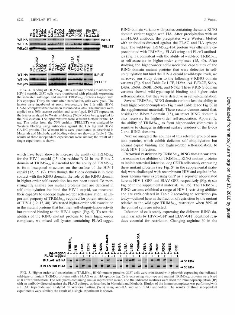
which have been shown to increase the avidity of TRIM5�rh
for the HIV-1 capsid (15, 40); residue R121 in the B-box 2domain of TRIM5�rh is essential for the ability of TRIM5�rh
to form hexagonal structures on the surface of the HIV-1capsid (12, 15, 19). Even though the B-box domain is in closecontact with the RING domain, the role of the RING domainin higher-order self-association has not been tested. To morestringently analyze our mutant proteins that are deficient inself-ubiquitylation but bind the HIV-1 capsid, we measuredtheir capacity to undergo higher-order self-association, an im-portant property of TRIM5�rh required for potent restrictionof HIV-1 (12, 15, 40). We tested higher-order self-associationin the mutant proteins that lost their self-ubiquitylation activitybut retained binding to the HIV-1 capsid (Fig. 5). To test theabilities of the RING mutant proteins to form higher-ordercomplexes, we mixed cell lysates containing FLAG-tagged
RING domain variants with lysates containing the same RINGdomain variant tagged with HA. After precipitation with ananti-FLAG antibody, the precipitates were Western blottedwith antibodies directed against the FLAG and HA epitopetags. The wild-type TRIM5�rh-HA protein was efficiently co-precipitated with TRIM5�rh-FLAG using anti-FLAG antibod-ies (Fig. 5), consistent with the ability of wild-type TRIM5�rh
to self-associate in higher-order complexes (15, 40). Afterstudying the higher-order self-association capabilities of theRING domain mutant proteins that were defective in self-ubiquitylation but bind the HIV-1 capsid at wild-type levels, wenarrowed our study down to the following 9 RING domainvariants (Fig. 5 and Table 2): I17E, H29A, A41E/E42E, S46A,L48A, R60A, R60K, R60E, and N67E. These 9 RING domainvariants showed wild-type capsid binding and higher-orderself-association, but they were defective in self-ubiquitylation.
Several TRIM5�rh RING domain variants lost the ability toform higher-order complexes (Fig. 5 and Table 2; see Fig. S3 inthe supplemental material). These results demonstrated that,besides the B-box 2 domain (15), an intact RING domain isalso necessary for higher-order self-association. Apparently,the ability of TRIM5�rh to form higher-order complexes issensitive to changes in different surface residues of the B-box2 and RING domains.
Next we analyzed the abilities of this selected group of mu-tant proteins, which exhibit deficient self-ubiquitylation butnormal capsid binding and higher-order self-association, toblock HIV-1 infection.
Retroviral restriction by TRIM5�rh RING domain variants.To examine the abilities of TRIM5�rh RING mutant proteinsto inhibit retroviral infection, dog Cf2Th cells stably expressingthese mutant proteins (see Fig. S4 in the supplemental mate-rial) were challenged with recombinant HIV and equine infec-tious anemia virus expressing GFP as a reporter abbreviatedhere as HIV-1–GFP and EIAV-GFP, respectively (Fig. 6; seeFig. S5 in the supplemental material) (47, 55). The TRIM5�rh
RING variants exhibited a range of HIV-1-restricting abilitiesand are rank ordered in Table 2 according to restriction po-tency—defined here as the fraction of restriction by the mutantrelative to the wild-type TRIM5�rh restriction when 50% ofthe control cells are infected.
Infection of cells stably expressing the different RING do-main variants by HIV-1–GFP and EIAV-GFP identified resi-dues essential for restriction. Changing arginine 60 in the
FIG. 4. Binding of TRIM5�rh RING mutant proteins to assembledHIV-1 capsids. 293T cells were transfected with plasmids expressingthe indicated wild-type and mutant TRIM5�rh proteins tagged withHA epitopes. Thirty-six hours after transfection, cells were lysed. Thelysates were incubated at room temperature for 1 h with HIV-1CA-NC complexes that had been assembled in vitro. The mixtures wereapplied to a 70% sucrose cushion and centrifuged. INPUT representsthe lysates analyzed by Western blotting (WB) before being applied tothe 70% cushion. The input mixtures were Western blotted for the HAtag. The pellet from the 70% cushion (PELLET) was analyzed byWestern blotting using antibodies against the HA tag and HIV-1CA-NC protein. The Western blots were quantitated as described inMaterials and Methods, and binding values are shown in Table 2. Theresults of three independent experiments were similar; the result of asingle experiment is shown.
FIG. 5. Higher-order self-association of TRIM5�rh RING mutant proteins. 293T cells were transfected with plasmids expressing the indicatedwild-type or mutant TRIM5� proteins with a FLAG or an HA epitope tag. Cells expressing wild-type and mutant TRIM5�rh proteins were lysed48 h after transfection. The cell lysates containing similar inputs were mixed, and the indicated mixtures were used for immunoprecipitation (IP)with an antibody directed against the FLAG epitope, as described in Materials and Methods. Elution of the immunocomplexes was performed witha FLAG tripeptide and analyzed by Western blotting (WB) using anti-HA and anti-FLAG antibodies. The results of three independentexperiments were similar; the result of a single experiment is shown.
8732 LIENLAF ET AL. J. VIROL.
on May 17, 2018 by guest
http://jvi.asm.org/
Dow
nloaded from

RING domain of TRIM5�rh to lysine, glutamic acid, or alaninedrastically reduced the potency of TRIM5�rh against HIV-1 by5- to 10-fold (Fig. 6A). Similarly, the I17E, A41E/N42E, N67E,and L48A variants showed reduced potency against HIV-1(Fig. 6B). Remarkably, all of these residues were located in theE2-binding region of the RING domain, with the exception ofN67E, which is located in the RING-RING interaction region.When cells expressing these RING domain variants were chal-lenged with EIAV, we observed a consistent decrease in re-striction (Fig. 6C and D). These results suggested a role forubiquitylation in HIV-1 restriction by TRIM5�rh.
TRIM5�rh self-ubiquitylation correlates with HIV-1 restric-tion. Using this selected group of variants that exhibit deficient
self-ubiquitylation but normal capsid binding and higher-orderself-association (shaded residues in Table 2), we tested thehypothesis that TRIM5� self-ubiquitylation correlates with re-striction. To do so, we graphically represented HIV-1 restric-tion versus self-ubiquitylation for this selected group of vari-ants. Remarkably, a strong correlation (rs 0.9090, P � 0.001)between TRIM5� self-ubiquitylation and HIV-1 restriction bythis panel of mutant proteins was observed (Fig. 7). Theseresults support the hypothesis that the E3-ligase activity of theRING domain represents a major contribution of the RINGdomain to HIV-1 restriction by TRIM5�.
Inhibition of HIV-1 reverse transcription by TRIM5�rh mu-tant proteins. HIV-1 restriction by TRIM5�rh occurs prior to
FIG. 6. Restriction of HIV-1 and EIAV infection by TRIM5�rh mutant proteins. Cf2Th cells were transduced with the LPCX vectorexpressing HA-tagged wild-type and mutant TRIM5�rh proteins. Stable cell lines were selected with 5 �g/ml puromycin, and the expressionlevels of mutant and wild-type TRIM5�rh proteins were assayed by Western blotting using HRP-conjugated antibodies against HA (see Fig.S4 in the supplemental material). The cells were challenged with different amounts of HIV-1–GFP (A, B) or EIAV-GFP (C, D). Thepercentage of GFP-positive cells was measured 48 h later by FACS. The results of three independent experiments were similar; the resultsof a single experiment are shown.
VOL. 85, 2011 ROLE OF TRIM5�’s E3-LIGASE ACTIVITY IN RESTRICTION 8733
on May 17, 2018 by guest
http://jvi.asm.org/
Dow
nloaded from

the initiation of reverse transcription (28, 63). However, theuse of proteasome inhibitors during restriction allows the oc-currence of reverse transcription without affecting the block-age of infection (1, 64, 67). To examine the ability of the
TRIM5�rh RING domain variants to block HIV-1 reversetranscription, we assayed the level of late reverse transcripts inmutant-expressing cells challenged with HIV-1 (Fig. 8; see Fig.S6 in the supplemental material). HIV-1 reverse transcriptlevels were low in cells expressing potently restrictingTRIM5�rh RING domain variants (Fig. 8 and Table 2). Incontrast, in cells expressing RING domain variants that did notrestrict HIV-1 infection potently, the levels of HIV-1 reversetranscripts were higher (Fig. 8 and Table 2).
Half-lives of TRIM5�rh RING domain variants. We mea-sured the half-lives of the TRIM5�rh RING domain variantsthat lost self-ubiquitylation but preserved capsid binding andhigher-order self-association (HOSA) (see Fig. S7 in the sup-plemental material), as previously described (12, 15). Some ofthe RING domain variants exhibited a longer half-life thanwild-type TRIM5�rh, which is �50 min (13). For example, theA41E/N42E, N67E, and S46A variants exhibited a half-lifeslightly longer than the wild-type half-life of �70 min; the I17Evariant exhibited a half-life of �105 min. Other variants, suchas the R60E variant, exhibit a half-life similar to that of thewild-type protein of �55 min. Interestingly, the R60A andR60K variants exhibited half-lives shorter than that of thewild-type protein at �15 min and �45 min, respectively. Theseresults did not correlate self-ubiquitylation with degradationsince we found mutant proteins that did not self-ubiquitylatebut have shorter half-lives than wild-type TRIM5�rh. Theseresults are in agreement with previous observations suggestingthat the degradation of TRIM5�rh is modestly affected by the
FIG. 7. TRIM5� self-ubiquitylation activity correlates with anti-HIV-1 activity. The abilities of RING domain variants to self-ubiquitylate (TRIM5� self-ubiquitylation) and restrict HIV-1 wereassessed as described in the footnotes to Table 2 and in Materialsand Methods. TRIM5� RING domain variants that were not de-fective in binding to the HIV-1 capsid and higher-order self-asso-ciation were analyzed. The Spearman rank correlation coefficient,rs, is 0.9090, with a 95% confidence interval of 0.8623 to 0.9847(two-sided P value of � 0.0001).
FIG. 8. Blockade of HIV-1 reverse transcription by TRIM5�rh RING mutant proteins. Cf2Th cells expressing the indicated wild-type andmutant TRIM5�rh proteins or containing the empty LPCX vector were challenged at an MOI of 0.4 with DNase-pretreated HIV-1–GFP. After7 h, cells were lysed and total DNA was extracted. The levels of viral DNA were measured by quantitative real-time PCR using a probe againstGFP as described in Materials and Methods. Similar results were obtained in three independent experiments.
8734 LIENLAF ET AL. J. VIROL.
on May 17, 2018 by guest
http://jvi.asm.org/
Dow
nloaded from

use of proteasome inhibitors in the absence of restricted vi-ruses (12).
DISCUSSION
The RING domain of TRIM5� exhibits E3-ubiquitin ligaseactivity, but the contribution of this activity to the restriction ofHIV-1 is not understood. Here we present the structure of theRING domain of human TRIM5� and use this information todirect a mutational analysis of the functional surfaces of theRING domain of TRIM5�rh. To explore the role of the E3-ubiquitin ligase in restriction, we correlated the E3-ubiquitinligase activity of TRIM5� with the different properties of therestriction factor TRIM5�, including HIV-1 restriction, bind-ing to the HIV-1 capsid, inhibition of reverse transcription, andthe ability to form higher-order complexes. We found a distinctset of TRIM5� variants located on the E2-binding surface ofthe RING domain, where the loss of E3-ubiquitin ligase activ-ity correlated with a defect in HIV-1 restriction ability. Ourresults demonstrate that E3-ubiquitin ligase activity has a rolein HIV-1 restriction by TRIM5�, as has been previously sug-gested by others (1, 50, 54, 64, 67).
The RING domain of TRIM5� adopts a ��� RING foldcontaining shorter �-strands and a longer �-helix than thetypical fold observed in RING domains. Comparison of theRING domain of TRIM5� with other RING domains revealedthat this structure has two regions that are potentially impor-tant for RING domain function (Fig. 2). Similar to the RINGdomains of Cbl, CHIP, and cIAP2 (9), the RING domain ofTRIM5� presents a distinct E2-binding region composed ofsimilar amino acids (Fig. 2A). Opposite to the E2-bindingregion is the RING-RING interaction region, which is similarto the interaction region that allows BRCA1 and BARD1RING domains to form a heterodimer (6) (Fig. 2B). Theconstruct used to solve the NMR structure of the RING do-main of TRIM5� did not include the last 10 amino acids; theseresidues are part of the association helix used by BRCA1 andBARD1 RING domains to heterodimerize. Longer constructsof the TRIM5� RING domain resulted in a poor HSQC spec-trum.
The E2-binding region of RING domains is essential fordocking of the E2 protein and allows the transfer of ubiquitinfrom E2 to the target protein. In order to disrupt this dockingsite in the RING domain of TRIM5�, we generated a series ofmutations on the different surfaces of the RING domain (Ta-ble 2); these variants were tested for the different properties ofthe restriction factor TRIM5�. These experiments revealedthat TRIM5� self-ubiquitylation requires an intact E2-bindingregion, which suggested that an intact E2 docking site in theRING domain is required for the self-ubiquitylation propertyof TRIM5�. Mutations in all of the residues of the E2-bindingsite of the RING domain affected self-ubiquitylation to a cer-tain extent; in some cases, complete loss of self-ubiquitylationwas observed. Mutations in the E2-binding region that resultedin a partial effect on self-ubiquitylation could be explained bythe existence of complementation by a different RING do-main. Hetero-oligomerization with a related RING domaincould rescue ubiquitylation activity in a defective RING do-main, as has been shown for ubiquitylation-deficient mutant
proteins of the Mdm2 RING domain that can be rescued byhetero-oligomerization with the RING domain of MdmX (60).
To exclude RING mutations that had effects on other prop-erties that are important for HIV-1 restriction by TRIM5�, wequantitatively measure the binding of these variants to theHIV-1 capsid, as previously shown (15). Similarly, we alsomeasured the ability of the RING variants to undergo higher-order self-association, which is also required for potent restric-tion of HIV-1. Remarkably, TRIM5� self-ubiquitylation activ-ity correlates with restriction activity on mutant proteins wherebinding to the HIV-1 capsid and higher-order self-associationwere not affected. This correlation supports the hypothesis thatthe E3-ubiquitin ligase activity of the RING domain is re-quired for potent restriction of HIV-1 by TRIM5�.
Several observations have linked the restriction of HIV-1 byTRIM5� with the proteasome. The observation that protea-somal inhibitors allow the occurrence of reverse transcriptionwithout affecting restriction suggests that TRIM5� blocks re-striction before and after reverse transcription (1, 64, 67). Theuse of proteasome inhibitors in the fate of the capsid assayshowed an increase in particulate capsid during infection in thepresence of TRIM5�, which also suggests a role for the pro-teasome in restriction and uncoating (11, 14). The Aiken lab-oratory has demonstrated that TRIM5� is degraded in a pro-teasome-dependent manner in the presence of a restrictedcapsid, which links the proteasome with the HIV-1 restrictionby TRIM5� (54). The present work attempted to connectubiquitylation, a process preceding proteasomal degradation,with the ability of TRIM5� to block HIV-1. Similar to what weobserved for a panel of B-box 2 mutant proteins (15), the levelsof HIV-1 late reverse transcripts for this panel of RING mu-tant proteins inversely correlated with the degree of restriction.
Mutations in the RING-RING interaction region that re-moved the ability to undergo self-ubiquitylation might cause adefect in RING oligomerization, which is different from affect-ing the docking site of the E2 enzyme. Several mutations in theRING-RING interaction region also affected the self-ubiqui-tylation activity of TRIM5� without affecting folding measuredby HIV-1 capsid binding and higher-order self-association.This is in agreement with findings suggesting that RING do-main oligomerization enhances ubiquitylation (30, 31, 52).Loss of RING domain oligomerization could account for thepartial defect in self-ubiquitylation observed in some of theRING-RING interaction region variants. In some cases, loss ofRING dimerization could result in complete loss of E3-ubiq-uitin ligase activity, as has been shown for the RING domain ofRNF4 (41). Even though concentration dependence experi-ments to test RING domain dimerization failed to prove ho-modimerization (data not shown), these experiments did notexclude the possibility that the RING domain hetero-oli-gomerizes with a different RING domain, as shown forBRCA-1 and BARD (6).
Our results demonstrated that potent restriction of HIV-1by TRIM5�rh requires intact self-ubiquitylation activity. Onecould conceive a model in which the self-ubiquitylation ofTRIM5� is required to remove TRIM5� when it is forminghexagonal structures on the surface of the capsid (19, 54);removal of TRIM5� from the surface of the capsid will allowa decrease on the amount of particulate capsid during infec-tion, assisting a rapid uncoating process (10). Further analysis
VOL. 85, 2011 ROLE OF TRIM5�’s E3-LIGASE ACTIVITY IN RESTRICTION 8735
on May 17, 2018 by guest
http://jvi.asm.org/
Dow
nloaded from

destined to understand the nature of the endogenous E2 en-zyme and the ubiquitylation substrate of TRIM5� will give newmechanistic insights into restriction.
ACKNOWLEDGMENTS
We thank Steve Porcelli for critical reading of the manuscript. Wethank Joe Sodroski for the initial support of this project. We thankTakashi Umehara for analytical ultracentrifugation measurements andXu-rong Qin for concentration dependency measurements in 1H-15N-HSQC experiments. We also thank Satoru Watanabe, TakushiHarada, Takeshi Nagira, Yasuko Tomo, Masaomi Ikari, KazuharuHanada, Yukiko Fujikura, and Akiko Tanaka for sample preparationand help with the screening data of the human TRIM5 RING domain.
The work of the structure determination was supported by theRIKEN Structural Genomics/Proteomics Initiative (RSGI) of theNational Project on Protein Structural and Functional Analyses,Ministry of Education, Culture, Sports, Science and Technology ofJapan. This work has also been supported by a K99/R00 Pathway toIndependence Award to F.D.-G. (4R00MH086162-02) and grantR01AI7930231 from the National Institutes of Health, an AmericanFoundation for AIDS Research Mathilde Krim fellowship phase IIin basic biomedical research (amfAR research grant 107787-47-RKHF), and a Claudia Adams Barr award from the Dana-FarberCancer Institute to F.D.-G.
REFERENCES
1. Anderson, J. L., et al. 2006. Proteasome inhibition reveals that a functionalpreintegration complex intermediate can be generated during restriction bydiverse TRIM5 proteins. J. Virol. 80:9754–9760.
2. Bellon, S. F., K. K. Rodgers, D. G. Schatz, J. E. Coleman, and T. A. Steitz.1997. Crystal structure of the RAG1 dimerization domain reveals multiplezinc-binding motifs including a novel zinc binuclear cluster. Nat. Struct. Biol.4:586–591.
3. Best, S., P. Le Tissier, G. Towers, and J. P. Stoye. 1996. Positional cloning ofthe mouse retrovirus restriction gene Fv1. Nature 382:826–829.
4. Borden, K. L. 2000. RING domains: master builders of molecular scaffolds?J. Mol. Biol. 295:1103–1112.
5. Borden, K. L. 1998. RING fingers and B-boxes: zinc-binding protein-proteininteraction domains. Biochem. Cell Biol. 76:351–358.
6. Brzovic, P. S., P. Rajagopal, D. W. Hoyt, M. C. King, and R. E. Klevit. 2001.Structure of a BRCA1-BARD1 heterodimeric RING-RING complex. Nat.Struct. Biol. 8:833–837.
7. Cornilescu, G., F. Delaglio, and A. Bax. 1999. Protein backbone angle re-straints from searching a database for chemical shift and sequence homol-ogy. J. Biomol. NMR 13:289–302.
8. Delaglio, F., et al. 1995. NMRPipe: a multidimensional spectral processingsystem based on UNIX pipes. J. Biomol. NMR 6:277–293.
9. Deshaies, R. J., and C. A. Joazeiro. 2009. RING domain E3 ubiquitin ligases.Annu. Rev. Biochem. 78:399–434.
10. Diaz-Griffero, F. 2011. Caging the beast: TRIM5� binding to the HIV-1core. Viruses 3:423–428.
11. Diaz-Griffero, F., et al. 2007. Comparative requirements for the restriction ofretrovirus infection by TRIM5alpha and TRIMCyp. Virology 369:400–410.
12. Diaz-Griffero, F., et al. 2007. Modulation of retroviral restriction and pro-teasome inhibitor-resistant turnover by changes in the TRIM5alpha B-box 2domain. J. Virol. 81:10362–10378.
13. Diaz-Griffero, F., et al. 2006. Rapid turnover and polyubiquitylation of theretroviral restriction factor TRIM5. Virology 349:300–315.
14. Diaz-Griffero, F., et al. 2008. A human TRIM5alpha B30.2/SPRY domainmutant gains the ability to restrict and prematurely uncoat B-tropic murineleukemia virus. Virology 378:233–242.
15. Diaz-Griffero, F., et al. 2009. A B-box 2 surface patch important forTRIM5alpha self-association, capsid binding avidity, and retrovirus restric-tion. J. Virol. 83:10737–10751.
16. Diaz-Griffero, F., et al. 2006. Requirements for capsid-binding and an effec-tor function in TRIMCyp-mediated restriction of HIV-1. Virology 351:404–419.
17. Freemont, P. S., I. M. Hanson, and J. Trowsdale. 1991. A novel cysteine-richsequence motif. Cell 64:483–484.
18. Ganser, B. K., S. Li, V. Y. Klishko, J. T. Finch, and W. I. Sundquist. 1999.Assembly and analysis of conical models for the HIV-1 core. Science 283:80–83.
19. Ganser-Pornillos, B. K., et al. 2011. Hexagonal assembly of a restrictingTRIM5alpha protein. Proc. Natl. Acad. Sci. U. S. A. 108:534–539.
20. Ganser-Pornillos, B. K., U. K. von Schwedler, K. M. Stray, C. Aiken, andW. I. Sundquist. 2004. Assembly properties of the human immunodeficiencyvirus type 1 CA protein. J. Virol. 78:2545–2552.
21. Hennig, J., et al. 2008. The fellowship of the RING: the RING-B-box linkerregion interacts with the RING in TRIM21/Ro52, contains a native autoan-tigenic epitope in Sjogren syndrome, and is an integral and conserved regionin TRIM proteins. J. Mol. Biol. 377:431–449.
22. Herrmann, T., P. Guntert, and K. Wuthrich. 2002. Protein NMR structuredetermination with automated NOE-identification in the NOESY spectrausing the new software ATNOS. J. Biomol. NMR 24:171–189.
23. Huang, A., et al. 2009. E2-c-Cbl recognition is necessary but not sufficient forubiquitination activity. J. Mol. Biol. 385:507–519.
24. Ishikawa, H., H. Tachikawa, Y. Miura, and N. Takahashi. 2006. TRIM11binds to and destabilizes a key component of the activator-mediated cofactorcomplex (ARC105) through the ubiquitin-proteasome system. FEBS Lett.580:4784–4792.
25. Javanbakht, H., F. Diaz-Griffero, M. Stremlau, Z. Si, and J. Sodroski. 2005.The contribution of RING and B-box 2 domains to retroviral restrictionmediated by monkey TRIM5alpha. J. Biol. Chem. 280:26933–26940.
26. Johnson, B. A. 2004. Using NMRView to visualize and analyze the NMRspectra of macromolecules. Methods Mol. Biol. 278:313–352.
27. Kar, A. K., F. Diaz-Griffero, Y. Li, X. Li, and J. Sodroski. 2008. Biochemicaland biophysical characterization of a chimeric TRIM21-TRIM5alpha pro-tein. J. Virol. 82:11669–11681.
28. Keckesova, Z., L. M. Ylinen, and G. J. Towers. 2004. The human and Africangreen monkey TRIM5alpha genes encode Ref1 and Lv1 retroviral restrictionfactor activities. Proc. Natl. Acad. Sci. U. S. A. 101:10780–10785.
29. Kentsis, A., and K. L. Borden. 2004. Physical mechanisms and biologicalsignificance of supramolecular protein self-assembly. Curr. Protein Pept. Sci.5:125–134.
30. Kentsis, A., R. E. Gordon, and K. L. Borden. 2002. Control of biochemicalreactions through supramolecular RING domain self-assembly. Proc. Natl.Acad. Sci. U. S. A. 99:15404–15409.
31. Kentsis, A., R. E. Gordon, and K. L. Borden. 2002. Self-assembly propertiesof a model RING domain. Proc. Natl. Acad. Sci. U. S. A. 99:667–672.
32. Kigawa, T., et al. 1999. Cell-free production and stable-isotope labeling ofmilligram quantities of proteins. FEBS Lett. 442:15–19.
33. Kirmaier, A., et al. 2010. TRIM5 suppresses cross-species transmission of aprimate immunodeficiency virus and selects for emergence of resistant vari-ants in the new species. PLoS Biol. 8:e1000462.
34. Kobayashi, N., et al. 2007. KUJIRA, a package of integrated modules forsystematic and interactive analysis of NMR data directed to high-throughputNMR structure studies. J. Biomol. NMR 39:31–52.
35. Koradi, R., M. Billeter, and K. Wuthrich. 1996. MOLMOL: a program fordisplay and analysis of macromolecular structures. J. Mol. Graph. 14:51–55.
36. Kostic, M., T. Matt, M. A. Martinez-Yamout, H. J. Dyson, and P. E. Wright.2006. Solution structure of the Hdm2 C2H2C4 RING, a domain critical forubiquitination of p53. J. Mol. Biol. 363:433–450.
37. Langelier, C. R., et al. 2008. Biochemical characterization of a recombinantTRIM5alpha protein that restricts human immunodeficiency virus type 1replication. J. Virol. 82:11682–11694.
38. Laskowski, R. A., J. A. Rullmannn, M. W. MacArthur, R. Kaptein, and J. M.Thornton. 1996. AQUA and PROCHECK-NMR: programs for checking thequality of protein structures solved by NMR. J. Biomol. NMR 8:477–486.
39. Li, H., et al. 2008. Structure of the C-terminal phosphotyrosine interactiondomain of Fe65L1 complexed with the cytoplasmic tail of amyloid precursorprotein reveals a novel peptide binding mode. J. Biol. Chem. 283:27165–27178.
40. Li, X., and J. Sodroski. 2008. The TRIM5alpha B-box 2 domain promotescooperative binding to the retroviral capsid by mediating higher-order self-association. J. Virol. 82:11495–11502.
41. Liew, C. W., H. Sun, T. Hunter, and C. L. Day. 2010. Ring domain dimeriza-tion is essential for RNF4 function. Biochem. J. 431:23–29.
42. Mace, P. D., et al. 2008. Structures of the cIAP2 RING domain revealconformational changes associated with ubiquitin-conjugating enzyme (E2)recruitment. J. Biol. Chem. 283:31633–31640.
43. Maegawa, H., T. Miyamoto, J. Sakuragi, T. Shioda, and E. E. Nakayama.2010. Contribution of RING domain to retrovirus restriction byTRIM5alpha depends on combination of host and virus. Virology 399:212–220.
44. Mrosek, M., et al. 2008. Structural analysis of B-Box 2 from MuRF1: iden-tification of a novel self-association pattern in a RING-like fold. Biochem-istry 47:10722–10730.
45. Nakayama, E. E., H. Miyoshi, Y. Nagai, and T. Shioda. 2005. A specificregion of 37 amino acid residues in the SPRY (B30.2) domain of Africangreen monkey TRIM5alpha determines species-specific restriction of simianimmunodeficiency virus SIVmac infection. J. Virol. 79:8870–8877.
46. Niikura, T., et al. 2003. A tripartite motif protein TRIM11 binds and desta-bilizes Humanin, a neuroprotective peptide against Alzheimer’s disease-relevant insults. Eur. J. Neurosci. 17:1150–1158.
47. Olsen, J. C. 1998. Gene transfer vectors derived from equine infectiousanemia virus. Gene Ther. 5:1481–1487.
48. Owens, C. M., P. C. Yang, H. Gottlinger, and J. Sodroski. 2003. Human andsimian immunodeficiency virus capsid proteins are major viral determinantsof early, postentry replication blocks in simian cells. J. Virol. 77:726–731.
8736 LIENLAF ET AL. J. VIROL.
on May 17, 2018 by guest
http://jvi.asm.org/
Dow
nloaded from

49. Perez-Caballero, D., T. Hatziioannou, A. Yang, S. Cowan, and P. D. Bien-iasz. 2005. Human tripartite motif 5alpha domains responsible for retrovirusrestriction activity and specificity. J. Virol. 79:8969–8978.
50. Perez-Caballero, D., T. Hatziioannou, F. Zhang, S. Cowan, and P. D. Bien-iasz. 2005. Restriction of human immunodeficiency virus type 1 by TRIM-CypA occurs with rapid kinetics and independently of cytoplasmic bodies,ubiquitin, and proteasome activity. J. Virol. 79:15567–15572.
51. Perron, M. J., et al. 2007. The human TRIM5alpha restriction factor medi-ates accelerated uncoating of the N-tropic murine leukemia virus capsid.J. Virol. 81:2138–2148.
52. Poyurovsky, M. V., et al. 2007. The Mdm2 RING domain C-terminus isrequired for supramolecular assembly and ubiquitin ligase activity. EMBO J.26:90–101.
53. Reymond, A., et al. 2001. The tripartite motif family identifies cell compart-ments. EMBO J. 20:2140–2151.
54. Rold, C. J., and C. Aiken. 2008. Proteasomal degradation of TRIM5alphaduring retrovirus restriction. PLoS Pathog. 4:e1000074.
55. Saenz, D. T., W. Teo, J. C. Olsen, and E. M. Poeschla. 2005. Restriction offeline immunodeficiency virus by Ref1, Lv1, and primate TRIM5alpha pro-teins. J. Virol. 79:15175–15188.
56. Sawyer, S. L., L. I. Wu, M. Emerman, and H. S. Malik. 2005. Positiveselection of primate TRIM5alpha identifies a critical species-specific retro-viral restriction domain. Proc. Natl. Acad. Sci. U. S. A. 102:2832–2837.
57. Sayah, D. M., E. Sokolskaja, L. Berthoux, and J. Luban. 2004. Cyclophilin Aretrotransposition into TRIM5 explains owl monkey resistance to HIV-1.Nature 430:569–573.
58. Sebastian, S., and J. Luban. 2005. TRIM5alpha selectively binds a restric-tion-sensitive retroviral capsid. Retrovirology 2:40.
59. Shloush, J., et al. 2011. Structural and functional comparison of the RINGdomains of two p53 E3 ligases, Mdm2 and Pirh2. J. Biol. Chem. 286:4796–4808.
60. Singh, R. K., S. Iyappan, and M. Scheffner. 2007. Hetero-oligomerization
with MdmX rescues the ubiquitin/Nedd8 ligase activity of RING fingermutants of Mdm2. J. Biol. Chem. 282:10901–10907.
61. Song, B., et al. 2005. TRIM5alpha association with cytoplasmic bodies is notrequired for antiretroviral activity. Virology 343:201–211.
62. Song, B., et al. 2005. The B30.2(SPRY) domain of the retroviral restrictionfactor TRIM5alpha exhibits lineage-specific length and sequence variation inprimates. J. Virol. 79:6111–6121.
63. Stremlau, M., et al. 2004. The cytoplasmic body component TRIM5alpharestricts HIV-1 infection in Old World monkeys. Nature 427:848–853.
64. Stremlau, M., et al. 2006. Specific recognition and accelerated uncoating ofretroviral capsids by the TRIM5alpha restriction factor. Proc. Natl. Acad.Sci. U. S. A. 103:5514–5519.
65. Stremlau, M., M. Perron, S. Welikala, and J. Sodroski. 2005. Species-specificvariation in the B30.2(SPRY) domain of TRIM5alpha determines the po-tency of human immunodeficiency virus restriction. J. Virol. 79:3139–3145.
66. Toniato, E., et al. 2002. TRIM8/GERP RING finger protein interacts withSOCS-1. J. Biol. Chem. 277:37315–37322.
67. Wu, X., J. L. Anderson, E. M. Campbell, A. M. Joseph, and T. J. Hope. 2006.Proteasome inhibitors uncouple rhesus TRIM5alpha restriction of HIV-1reverse transcription and infection. Proc. Natl. Acad. Sci. U. S. A. 103:7465–7470.
68. Yamauchi, K., K. Wada, K. Tanji, M. Tanaka, and T. Kamitani. 2008.Ubiquitination of E3 ubiquitin ligase TRIM5 alpha and its potential role.FEBS J. 275:1540–1555.
69. Yang, Y., K. L. Lorick, J. P. Jensen, and A. M. Weissman. 2005. Expressionand evaluation of RING finger proteins. Methods Enzymol. 398:103–112.
70. Yap, M. W., S. Nisole, and J. P. Stoye. 2005. A single amino acid change inthe SPRY domain of human Trim5alpha leads to HIV-1 restriction. Curr.Biol. 15:73–78.
71. Yin, Q., B. Lamothe, B. G. Darnay, and H. Wu. 2009. Structural basis for thelack of E2 interaction in the RING domain of TRAF2. Biochemistry 48:10558–10567.
VOL. 85, 2011 ROLE OF TRIM5�’s E3-LIGASE ACTIVITY IN RESTRICTION 8737
on May 17, 2018 by guest
http://jvi.asm.org/
Dow
nloaded from