contents - hacettepeyunus.hacettepe.edu.tr/~selis/teaching/webkmu396/ppt/presentations... ·...
TRANSCRIPT
Contents
What is AFM?HistoryBasic principles and devicesOperating modesApplication areasAdvantages and disadvantages

Figure1: 2004 Seth Copen Goldstein
What is AFM?
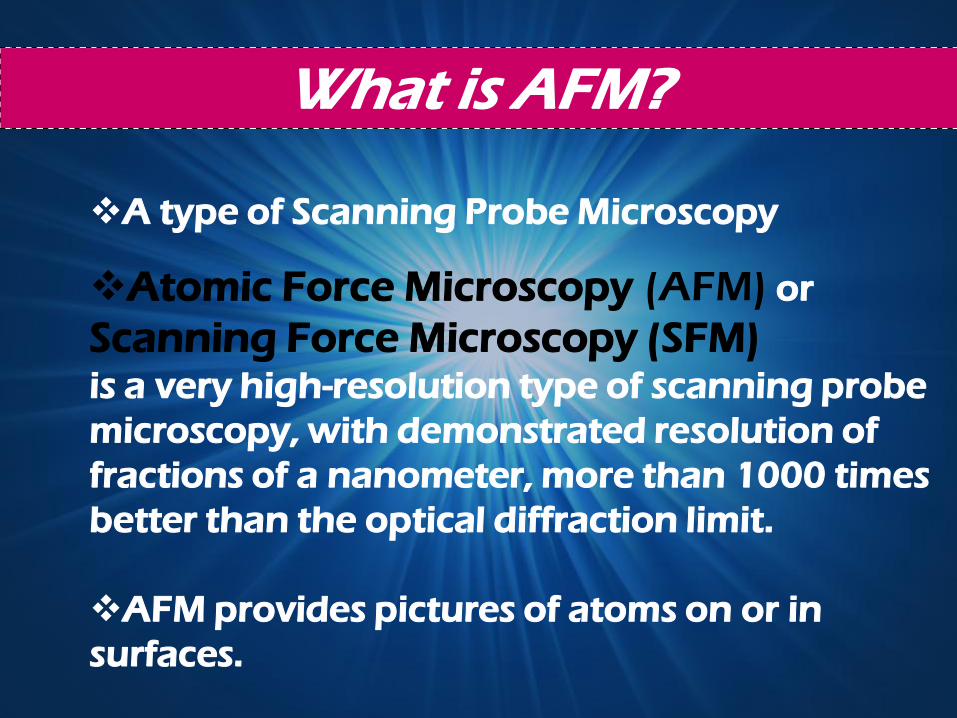
Atomic Force Microscopy (AFM) or
Scanning Force Microscopy (SFM)is a very high-resolution type of scanning probe
microscopy, with demonstrated resolution of
fractions of a nanometer, more than 1000 times
better than the optical diffraction limit.
AFM provides pictures of atoms on or in
surfaces.
A type of Scanning Probe Microscopy
History of AFM
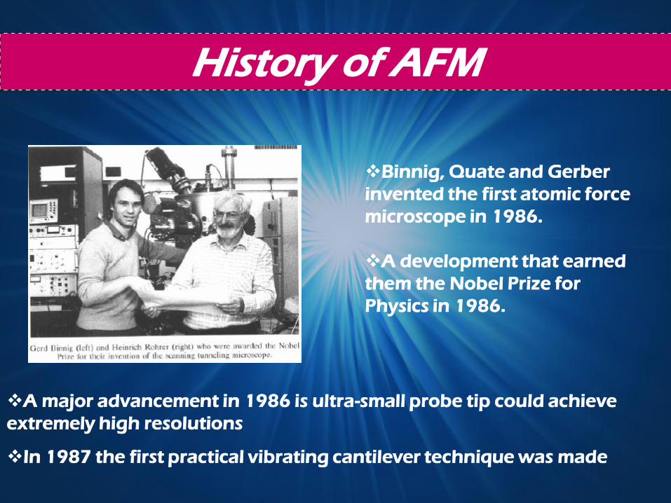
Binnig, Quate and Gerber
invented the first atomic force
microscope in 1986.
A development that earned
them the Nobel Prize for
Physics in 1986.
In 1987 the first practical vibrating cantilever technique was made
A major advancement in 1986 is ultra-small probe tip could achieve
extremely high resolutions
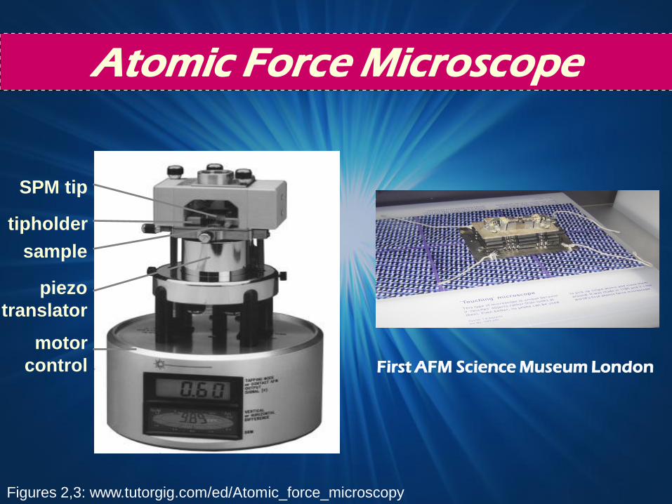
First AFM Science Museum London
tipholder
motor
control
SPM tip
sample
piezo
translator
Atomic Force Microscope
Figures 2,3: www.tutorgig.com/ed/Atomic_force_microscopy
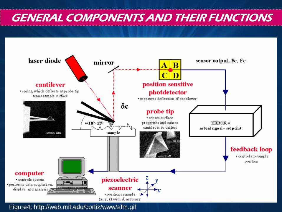
GENERAL COMPONENTS AND THEIR FUNCTIONS
Figure4: http://web.mit.edu/cortiz/www/afm.gif
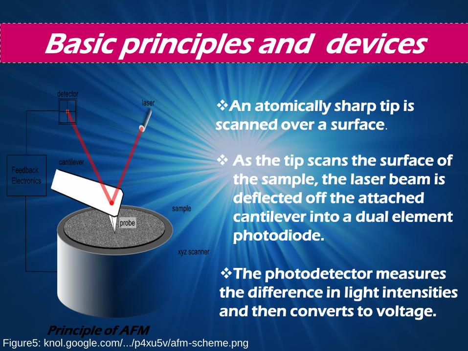
Basic principles and devices
Principle of AFM
An atomically sharp tip is
scanned over a surface.
As the tip scans the surface of
the sample, the laser beam is
deflected off the attached
cantilever into a dual element
photodiode.
The photodetector measures
the difference in light intensities
and then converts to voltage.
Figure5: knol.google.com/.../p4xu5v/afm-scheme.png
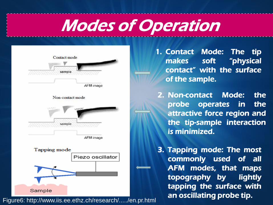
Modes of Operation
1. Contact Mode: The tip
makes soft “physical
contact” with the surface
of the sample.
2. Non-contact Mode: the
probe operates in the
attractive force region and
the tip-sample interaction
is minimized.
3. Tapping mode: The most
commonly used of all
AFM modes, that maps
topography by lightly
tapping the surface with
an oscillating probe tip.Figure6: http://www.iis.ee.ethz.ch/research/...../en.pr.html
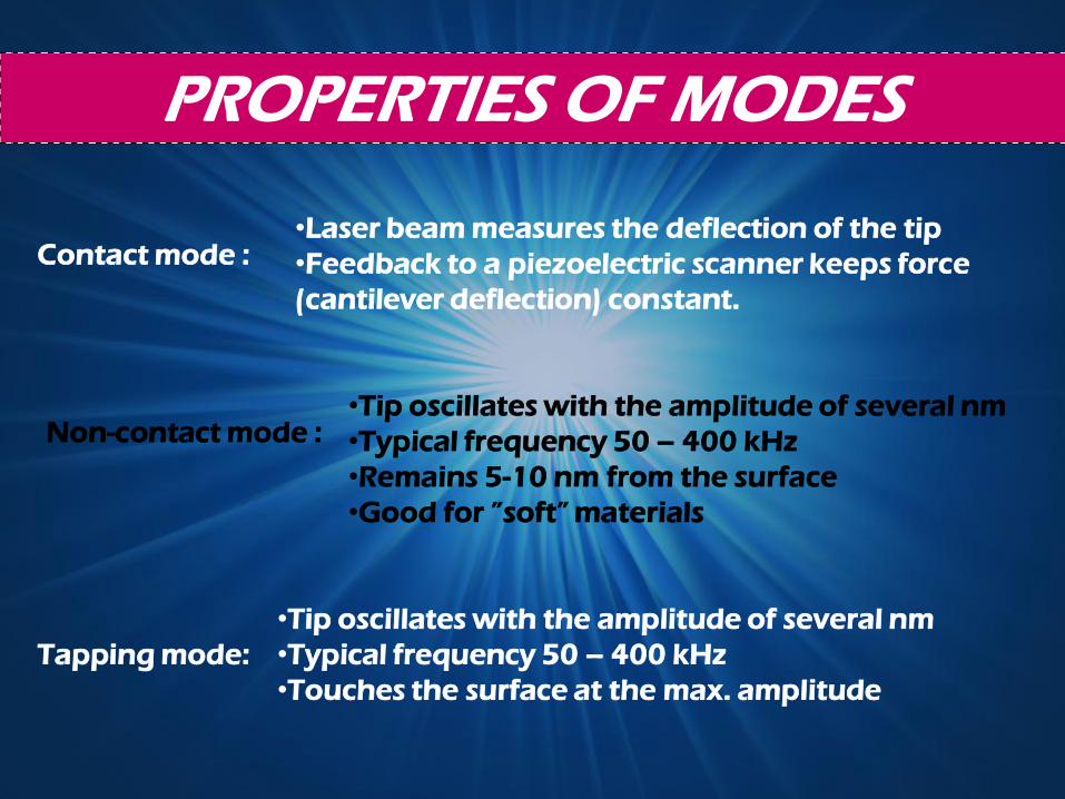
PROPERTIES OF MODES
Contact mode :
Non-contact mode :
Tapping mode:
•Laser beam measures the deflection of the tip
•Feedback to a piezoelectric scanner keeps force
(cantilever deflection) constant.
•Tip oscillates with the amplitude of several nm
•Typical frequency 50 – 400 kHz
•Remains 5-10 nm from the surface
•Good for ”soft” materials
•Tip oscillates with the amplitude of several nm
•Typical frequency 50 – 400 kHz
•Touches the surface at the max. amplitude
0APPLICATION AREAS
Qualitative macromolecule and polymer
imaging
Complicated or qualitative structure
analysis
Molecular interaction, molecular
manipulation, surface topography,
nanofood characterization

Physical Science
Friction,
Contact electrification,
Elasticity
Wetting to be studied on smaller scale
than previously possible.
•The AFM will contribute new knowledge:
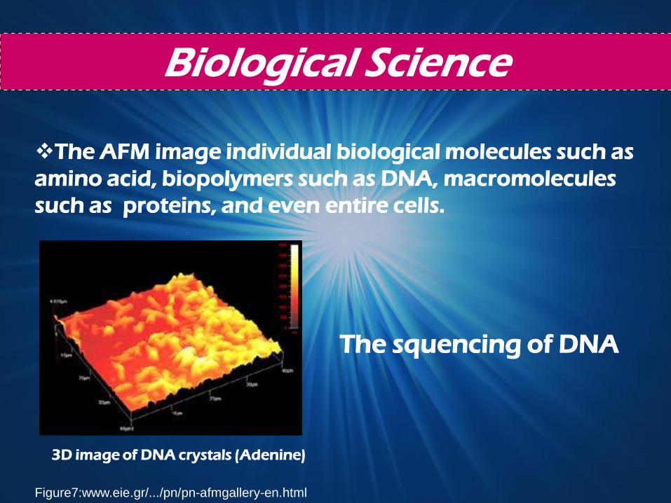
Biological Science
The AFM image individual biological molecules such as
amino acid, biopolymers such as DNA, macromolecules
such as proteins, and even entire cells.
The squencing of DNA
3D image of DNA crystals (Adenine)
Figure7:www.eie.gr/.../pn/pn-afmgallery-en.html
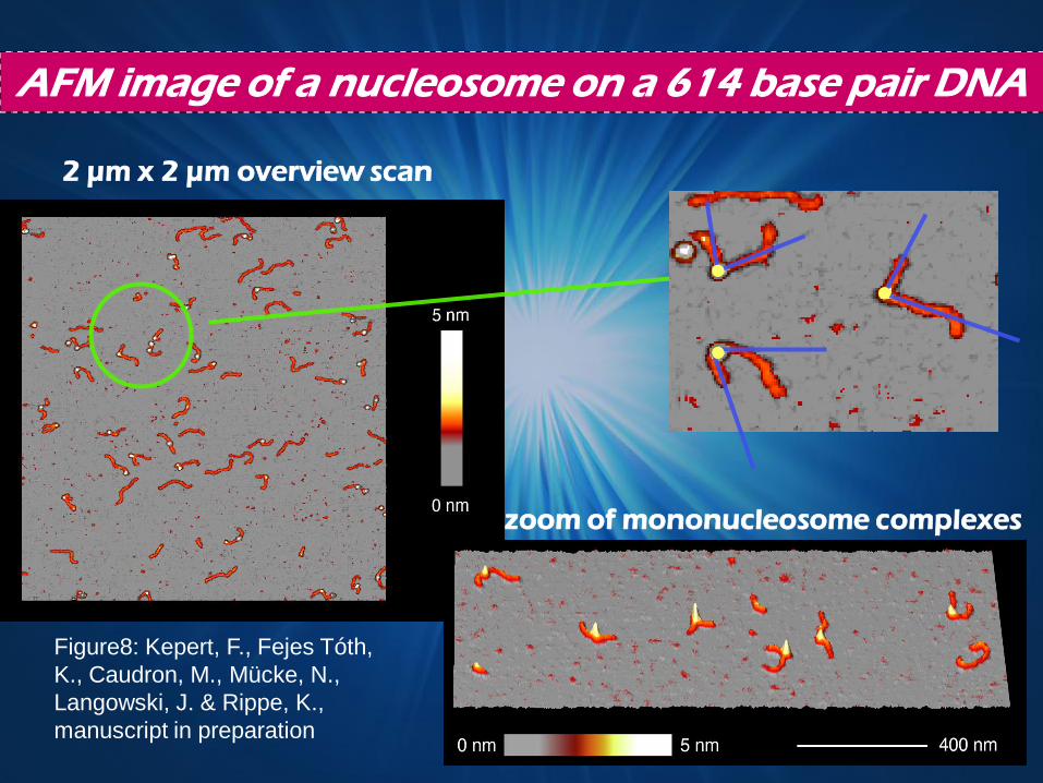
AFM image of a nucleosome on a 614 base pair DNA
2 µm x 2 µm overview scan
zoom of mononucleosome complexes
Figure8: Kepert, F., Fejes Tóth,
K., Caudron, M., Mücke, N.,
Langowski, J. & Rippe, K.,
manuscript in preparation

Application Areas
Biology
Chemistry
Electronic
Telecommunication
Automotive
Aerospace
Energy
Industries that AFM used;
In the Future
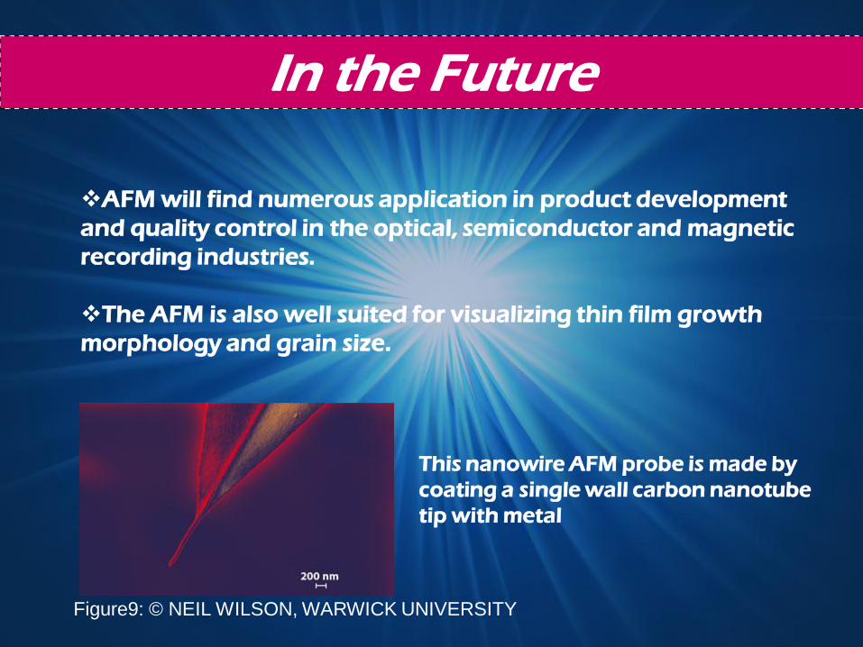
AFM will find numerous application in product development
and quality control in the optical, semiconductor and magnetic
recording industries.
The AFM is also well suited for visualizing thin film growth
morphology and grain size.
This nanowire AFM probe is made by
coating a single wall carbon nanotube
tip with metal
Figure9: © NEIL WILSON, WARWICK UNIVERSITY
Advantages
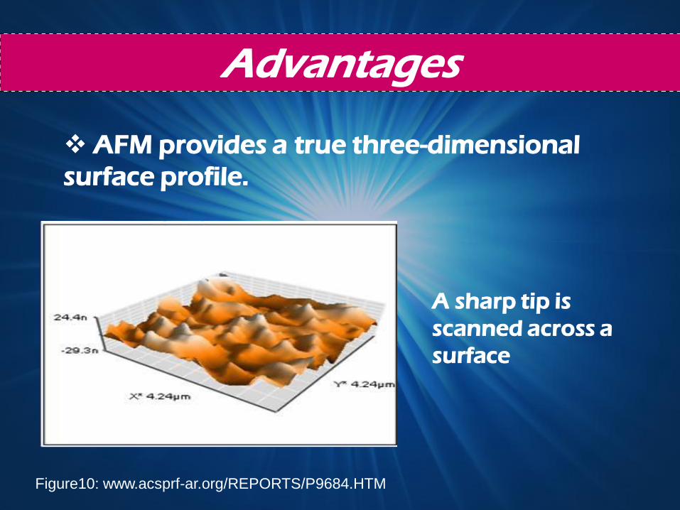
AFM provides a true three-dimensional
surface profile.
A sharp tip is
scanned across a
surface
Figure10: www.acsprf-ar.org/REPORTS/P9684.HTM
Advantages
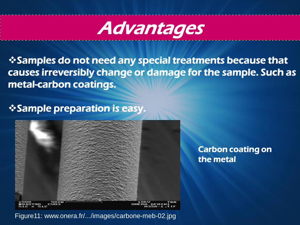
Samples do not need any special treatments because that
causes irreversibly change or damage for the sample. Such as
metal-carbon coatings.
Sample preparation is easy.
Carbon coating on
the metal
Figure11: www.onera.fr/.../images/carbone-meb-02.jpg
Advantages
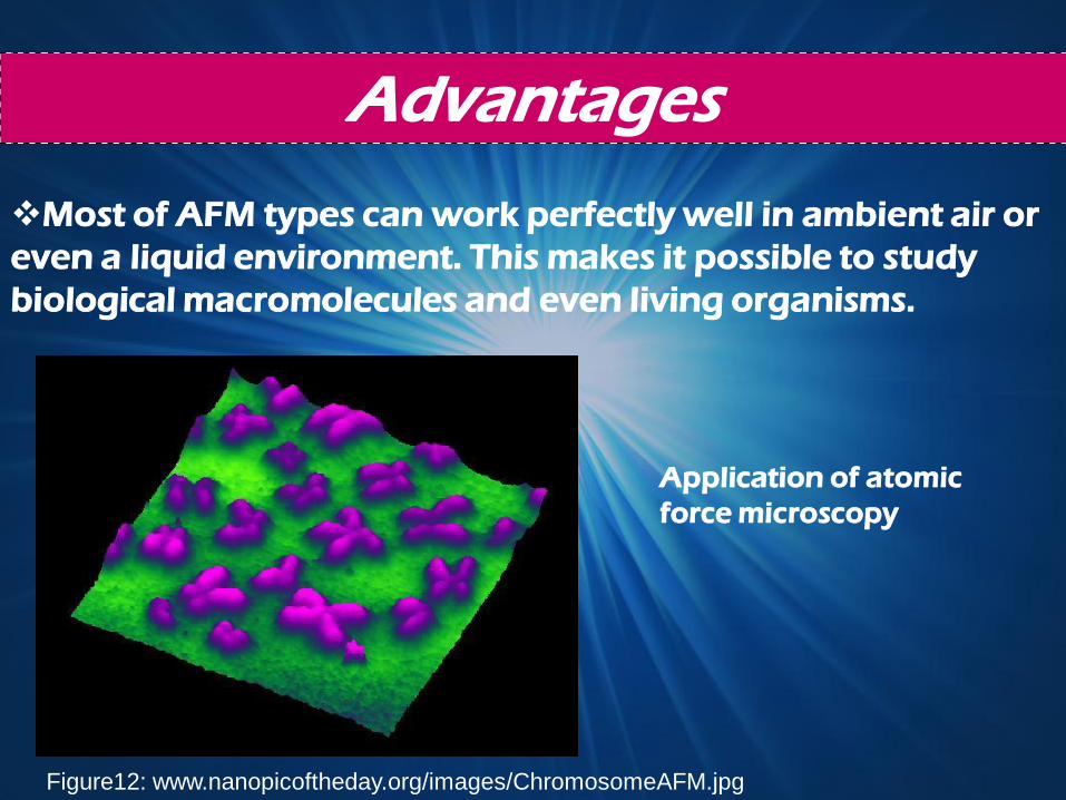
Most of AFM types can work perfectly well in ambient air or
even a liquid environment. This makes it possible to study
biological macromolecules and even living organisms.
Application of atomic
force microscopy
Figure12: www.nanopicoftheday.org/images/ChromosomeAFM.jpg
Advantages
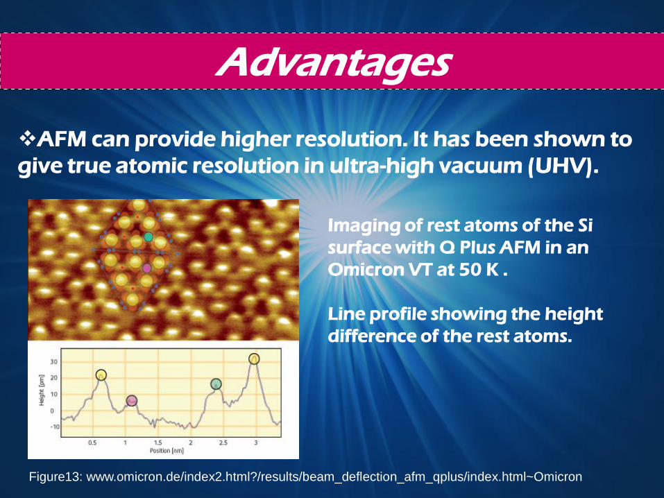
AFM can provide higher resolution. It has been shown to
give true atomic resolution in ultra-high vacuum (UHV).
Imaging of rest atoms of the Si
surface with Q Plus AFM in an
Omicron VT at 50 K .
Line profile showing the height
difference of the rest atoms.
Figure13: www.omicron.de/index2.html?/results/beam_deflection_afm_qplus/index.html~Omicron
Disadvantages
AFM has a limited vertical range
Also it has a limited magnification range
AFM probes cannot normally measure too high walls
or overhangs.
Datas are not independent of tip. Incorrect choice of
tip for the required resolution can lead to image
artifacts.
Traditionally the AFM could not scan images as fast as
the SEM.
Disadvantages
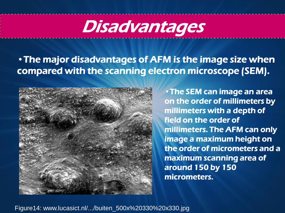
•The SEM can image an area
on the order of millimeters by
millimeters with a depth of
field on the order of
millimeters. The AFM can only
image a maximum height on
the order of micrometers and a
maximum scanning area of
around 150 by 150
micrometers.
Figure14: www.lucasict.nl/.../buiten_500x%20330%20x330.jpg
•The major disadvantages of AFM is the image size when
compared with the scanning electron microscope (SEM).
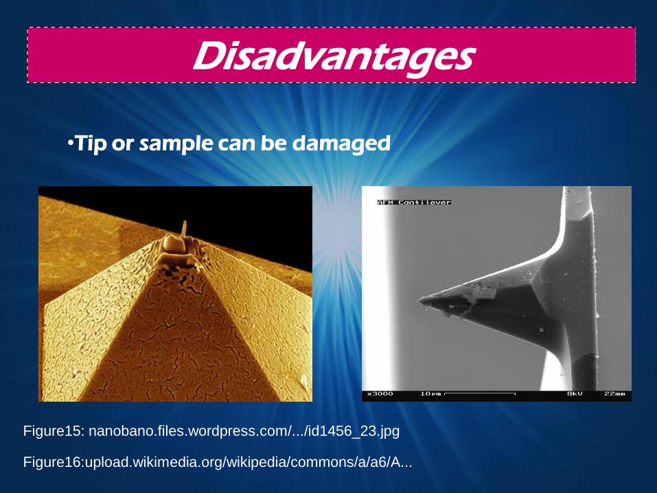
•Tip or sample can be damaged
Disadvantages
Figure15: nanobano.files.wordpress.com/.../id1456_23.jpg
Figure16:upload.wikimedia.org/wikipedia/commons/a/a6/A...
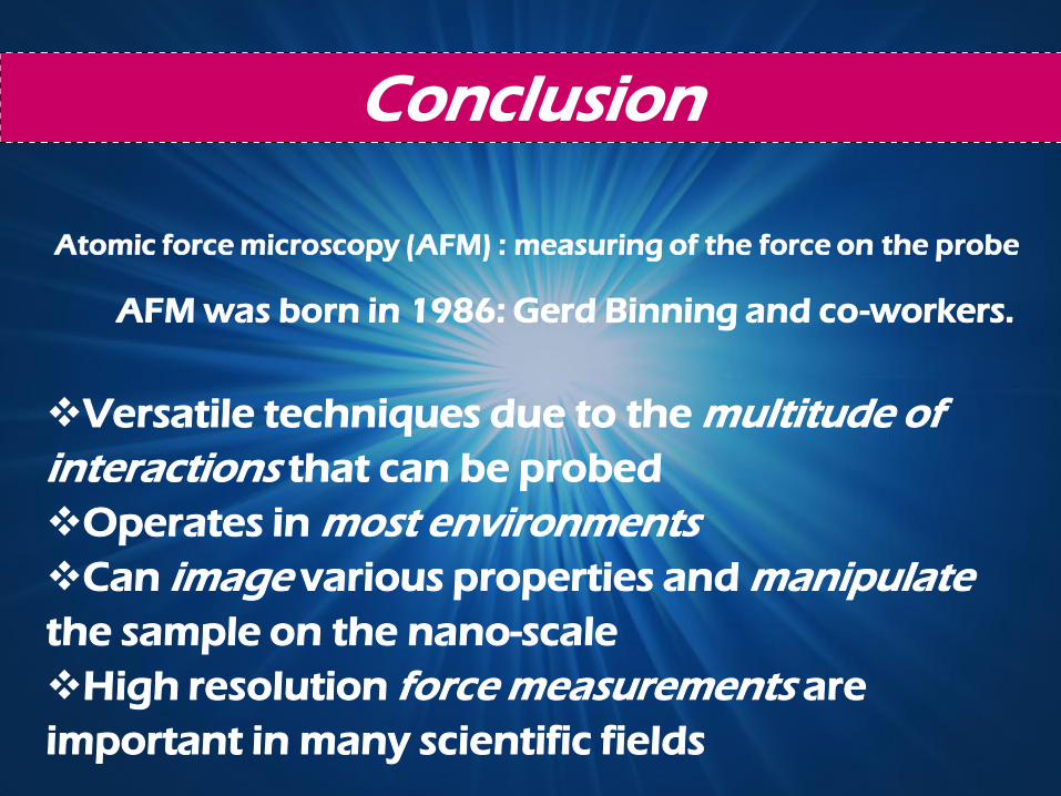
Conclusion
Atomic force microscopy (AFM) : measuring of the force on the probe
Versatile techniques due to the multitude of
interactions that can be probed
Operates in most environments
Can image various properties and manipulate
the sample on the nano-scale
High resolution force measurements are
important in many scientific fields
AFM was born in 1986: Gerd Binning and co-workers.
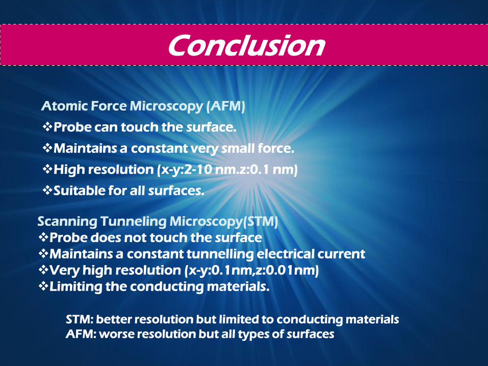
Conclusion
Atomic Force Microscopy (AFM)
Probe can touch the surface.
Maintains a constant very small force.
High resolution (x-y:2-10 nm.z:0.1 nm)
Suitable for all surfaces.
Scanning Tunneling Microscopy(STM)
Probe does not touch the surface
Maintains a constant tunnelling electrical current
Very high resolution (x-y:0.1nm,z:0.01nm)
Limiting the conducting materials.
STM: better resolution but limited to conducting materials
AFM: worse resolution but all types of surfaces
References
http://people.web.psi.ch/nolting/afm.pdf
http://en.wikipedia.org/wiki/Atomic_force_microscopy
http://www.ifm.liu.se/courses/TFFM12/AFM%20lecture.pdf
http://people.web.psi.ch/nolting/afm.pdf
http://www.lot-oriel.com/site/site_down/pn_afmhistory_deen.pdf
http://www.chembio.uoguelph.ca/educmat/chm729/afm/details.ht
http://webcache.googleusercontent.com/search?q=cache:L4xMPdRfMf
AJ:www.mansic.eu/documents/PAM1/Frangis.pdf+atomic+force+microsc
opy+advantages+and+disadvantages&cd=3&hl=tr&ct=clnk&gl=tr
www.acsprf-ar.org/REPORTS/P9684.HTM
References
www.tutorgig.com/ed/Atomic_force_microscopy
http://web.mit.edu/cortiz/www/afm.gif
http://www.iis.ee.ethz.ch/research/...../en.pr.html
www.eie.gr/.../pn/pn-afmgallery-en.html
www.acsprf-ar.org/REPORTS/P9684.HT
www.nanopicoftheday.org/images/ChromosomeAFM.jpg
www.lucasict.nl/.../buiten_500x%20330%20x330.jpg
www.nanobano.files.wordpress.com/.../id1456_23.jpg

~THANK YOU ~

NEŞE KAYNAK 20622737
DUYGU GÖKÇE 20622665
YUDUM YARAL 20622995
Z.ÇAĞLA MERAL 20622756