construction of a human fab phage display library from ... · construction of a human fab phage...
TRANSCRIPT

C. Dantas-Barbosa et al. 126
Genetics and Molecular Research 4 (2): 126-140 (2005) www.funpecrp.com.br
Construction of a human Fab phage displaylibrary from antibody repertoires ofosteosarcoma patients
Carmela Dantas-Barbosa1,4, Marcelo M. Brígido2,3 andAndréa Q. Maranhão2,3
1Laboratório de Genética Molecular, Patologia,Rede Sarah de Hospitais de Reabilitação, 70330-150 Brasília, DF, Brasil2Laboratório de Biologia Molecular, Universidade de Brasília,Campus Universitário, Asa Norte, 70910-900 Brasília, DF, Brasil3III - Immunological Investigation Institute, CNPq4Pós-graduação em Biologia Molecular, Departamento de Biologia Celular,Universidade de Brasília, Campus Universitário, Asa Norte,70910-900 Brasília, DF, BrasilCorresponding author: A.Q. MaranhãoE-mail: [email protected]
Genet. Mol. Res. 4 (2): 126-140 (2005)Received August 2, 2004Accepted January 7, 2005Published April 19, 2005
ABSTRACT. Osteosarcoma is the commonest type of primary malig-nant bone tumor, frequently found in adolescents at sites of rapid bonegrowth. Despite current management protocols, up to half of the pa-tients succumb to this disease. Moreover, there is no well-characterizedmolecular marker for diagnosis and prognosis. Since phage display meth-odology allows the selection of human antibody fragments with potentialuse in clinical applications, we applied this procedure to construct a re-combinant Fab (antigen binding fragment) library from patients with os-teosarcoma. We used peripheral blood lymphocyte total RNA from 11osteosarcoma patients and cloned recombinant Fab representing the µ, γand κ chain antibody repertoires of these individuals. The resulting li-brary was cloned in the pComb3X vector and attained 1.45 x 10⁸ differ-ent functional forms. BstO I fingerprinting and DNA sequencing analy-sis of randomly selected clones revealed the diversity of the library, dem-onstrating that Fab harbors Vκ chains from subgroups I to V, biased
Genetics and Molecular Research 4 (2): 126-140 (2005) FUNPEC-RP www.funpecrp.com.br

Human Fab phage display library 127
Genetics and Molecular Research 4 (2): 126-140 (2005) www.funpecrp.com.br
towards the A27 fragment, as normally reported for the human reper-toire. Analysis of the VH repertoire revealed that our library has a slightbias towards the VH4 family, instead of the usually reported VH3. Thisis the first description of a phage display library from osteosarcoma pa-tients. We believe these human Fab fragments will provide a valuabletool for the study of this neoplasia and could also contribute to improve-ments in the diagnosis of this disease.
Key words: Phage display, Fab, Osteosarcoma, Recombinant antibody,Human antibody repertoire
INTRODUCTION
Antibodies have been used for antigen detection and therapeutics, and their specificitycombined with low toxicity make them a promising pharmaceutical commodity (Berger et al.,2002). Currently, they comprise the second-largest category of biological medicines in clinicaldevelopment, after vaccines (Chester et al., 2004). Successful examples include rituximab, ap-proved by the FDA since 1997, an anti-CD20 antibody that is now an integral component ofmany treatment strategies for non-Hodgkin’s lymphoma (Seymour, 2004), and OKT3, an anti-CD3 that is widely used to reduce graft rejection (Cosimi et al., 1981). Therapeutic use ofantibodies is limited by methodological constraints. They are usually obtained by immunizationof experimental animals with target antigens; after screening, a specific antibody-producinghybridoma is identified (Kohler and Milstein, 1975). Although well established, this technology islaborious, and it is biased by the experimental model immune system, making it difficult to obtaina high-affinity antibody against conserved mammal proteins. Additionally, the heterologous pro-teins are often immunogenic for humans, preventing their therapeutic use (Maranhão and Brigido,2000). Several examples of clinical antibody humanization to bypass this bottleneck have beendescribed (Morrison and Oi, 1989); they minimize the human anti-murine antibody response byreplacing murine sequences with human framework homologous sequences. The constructionand selection of antibody combinatorial libraries displayed on filamentous phage surfaces be-came an alternative to this approach (McCafferty et al., 1990). In this technique, the repertoireof V genes of one or more individuals is amplified with primers covering all V gene families,giving rise to human antibodies. The library is generated by a random combination of variablelight (VL) and variable heavy (VH) chain genes produced as antigen binding (Fab) or singlechain variable (scFv) antibody fragments (Brigido and Maranhão, 2001). Theoretically, eachclone codes for a specific antigen-binding site, corresponding to a natural repertoire, increasedby an artificial domain combination that extrapolates the individual repertoire. This panningprocedure mimics the B-cell clonal selection system in vitro by specifically enriching phageparticles that display antibodies with a desired specificity (Barbas III et al., 1991). Severalhuman antibody combinatorial libraries displayed on filamentous phage surface have been builtby various groups, from either naive (Griffiths et al., 1993; de Haard et al., 1999; Lu et al., 2002)or immunized/infected repertoires (Portolano et al., 1993; Wu et al., 2001); this system has beenapplied to select antibodies against different antigens, including melanoma (Cai and Garen, 1995),colorectal (Somers et al., 2002) and prostate (Mintz et al., 2003) cancer proteins.

C. Dantas-Barbosa et al. 128
Genetics and Molecular Research 4 (2): 126-140 (2005) www.funpecrp.com.br
Osteosarcoma is the most common primary malignant bone tumor, predominantly af-fecting children and adolescents; it is mainly found in areas with a high rate of skeletal growth.These tumors typically arise in the metaphyseal regions of long bones. The distal femur, proxi-mal tibia and proximal humerus are the most common sites (Dahlin, 1975). Almost all osteosar-comas are high grade and have a poor prognosis. Usually, when the initial osteogenic sarcomadiagnosis occurs, tumors are large, and numerous lung micrometastases are already present(Jesus-Garcia Filho, 1992). Once metastases are diagnosed, the potential for cure is markedlydecreased, and only 10% of the patients with this malignancy achieve a long-term disease-freeinterval (Meyers et al., 1993). In recent years, the immunocytochemical subclassification ofosteosarcoma has significantly enhanced the accuracy of pathological diagnosis (Ueda et al.,1993); however, a specific marker has not been found and several studies have been made onthe delineation of carcinogenesis molecular aspects. We developed a combinatorial Fab phagedisplay library from randomly combined variable (V) region antibody genes of 11 osteosarcomapatients. This library could help us find new markers for diagnosis and even for the developmentof new treatment strategies.
MATERIAL AND METHODS
Patients
We obtained peripheral blood lymphocytes (PBL) from 11 osteosarcoma patients at-tended at the Sarah Hospital for Rehabilitation in Brasília from 2000 to 2001. This study waspreviously approved by the hospital Ethics Committee. Patients’ features are shown in Table 1.
ND: not determined, either because the patients had not received chemotherapy before tumor resection (patients 7 and8) or died before surgery (patient 6).
Patient Histological Age Sex Anatomic site Extent of necrosis after Lymphocytes/µlnumber subtype (year) chemotherapy in %
1 Chondroblastic 31 F femur 51 5.6 x 10³
2 Osteoblastic 22 M femur 70 2.3 x 10³
3 Chondroblastic 12 M femur 41 5.6 x 10³
4 Osteoblastic 23 M fibula >95 6.6 x 10³
5 Epithelioid 11 M tibia >95 1.6 x 10⁴
6 Telangiectasic 16 M tibia ND 1.2 x 10³
7 Fibroblastic 24 F femur ND 0.9 x 10³
8 Osteoblastic 6 M radius ND 8.8 x 10³
9 Osteoblastic 6 F femur 31 1.5 x 10⁴
10 Telangiectasic 38 M femur 58 8.7 x 10³
11 Osteoblastic 10 M tibia 85 8.3 x 10³
Table 1. Clinical features of osteosarcoma patients.
Library construction
Total RNA from osteosarcoma patients was used to prepare a Fab combinatorial li-

Human Fab phage display library 129
Genetics and Molecular Research 4 (2): 126-140 (2005) www.funpecrp.com.br
brary displayed on the phage surface. The protocol involves four consecutive steps, as follows:i) synthesis of specific first-strand cDNAs; ii) synthesis by polymerase chain reaction (PCR) ofVH genes for IgG and IgM classes and of the kappa light chain genes and their respectiveconstant regions; iii) overlapping PCR to join each variable chain coding fragment with therespective constant region and a second PCR to obtain the complete Fab with the introductionof cloning sites at the extremities; iv) ligation of Fab into the pComb3X phagemid vector (Scottand Barbas III, 2000).
A total of 1 x 10⁷ lymphocytes from each patient was used to prepare the RNA usingthe QIAamp® RNA Blood Mini Kit (Qiagen). RNAs were pooled together and used to synthe-size three different cDNAs (SuperScript System, Invitrogen), using specific primers to heavyand light chain immunoglobulins (Marks et al., 1991). Library construction was done as de-scribed by Andris-Widhopf et al. (2000). Briefly, we performed two sets of six amplifications,using sense primers for VH fragments, combined with gamma (γ) or mi (µ) anti-sense primers.Similarly, VL gene fragments were obtained, using four sense primers, covering the wholekappa repertoire. These primers were used with a single 3’ oligonucleotide targeting kappaconstant region (Cκ). Primers used in this first round PCR were designed for the assembly of aheavy chain (Fd) fragment after a second round PCR. For this purpose, γ and µ VH fragmentswere fused to a γ constant region (CH1). The complete light chains were made by fusing VLsto a Cκ fragment in another second round PCR. Fd fragments and kappa light chains wereultimately shuffled in a final overlap PCR. The final Fab coding fragments were digested withSfiI and cloned into the pComb3X, precipitated with ethanol, resuspended in 15 µl water andused to transform XL1-Blue E. coli (Stratagene) by electroporation, as described by Rader etal., 2000. After transformation, 5 ml SOC medium was added, and the culture was incubated for1 h at 37°C at 250 rpm. At this point, culture aliquots were plated on LB agar/carbenicillin (50µg/ml) to titer library size, which was calculated by counting the number of carbenicillin-resis-tant colonies per microgram DNA. SB medium (10 ml), containing carbenicillin (20 µg/ml) andtetracycline (10 µg/ml), was added to the transformed culture. After additional incubation underthe same conditions, 100 ml SB medium, supplemented with carbenicillin (50 µg/ml) and tetracy-cline (10 µg/ml), was added. After 1 h of incubation, as above, 1012 plaque-forming units ofVCSM13 helper phage (Stratagene) were added and the culture was shaken for 1 h at 37°Cand 250 rpm. Kanamycin (70 µg/ml) was then added, and the culture was incubated overnightat 37°C and 250 rpm. Phages were obtained from the culture medium by polyethylene glycol/NaCl precipitation and were resuspended in TBS containing 1% BSA.
Sequence analysis
In order to prepare phagemid DNA, 96 different clones were cultured in a deep wellplate containing 1 ml 2x YT medium with carbenicillin (100 µg/ml) and glucose (0.5% p/v) ineach well. The cultures were incubated for 20 h at 37°C/320 rpm. Cells were recovered bycentrifugation (3,000 g for 6 min), and the pellets were resuspended in 240 µl GET (0.92%glucose, 10 mM EDTA, pH 8.0, and 26 mM Tris-HCl) and harvested; an additional 80 µl GETbuffer was then added. Sixty microliters of this suspension was transferred to a polypropyleneplate (round bottom), treated with 2.5 µl RNAseA (10 mg/ml), and then 60 µl 1% 0.2 M NaOHSDS was added. Complete homogenization was obtained by inverting the sealed plate 30 times.To complete cell lysis, the plates were incubated at room temperature for 10 min. After this, 80

C. Dantas-Barbosa et al. 130
Genetics and Molecular Research 4 (2): 126-140 (2005) www.funpecrp.com.br
µl 3 M KOAc was added; the material was homogenized and incubated for an additional 10 minand then centrifuged at 3000 g for 8 min. The supernatants were transferred to a new 96-wellV-bottom plate and filtered through a Millipore membrane plate by centrifuging at 3000 g for 5min. The DNA was precipitated by adding isopropanol (100 µl), followed by centrifugation at3000 g for 45 min. The pellet was washed with 70% ethanol, dried and resuspended in 30 µlmilliQ water. Library quality was assessed by digestion of carbenicillin resistant clones DNAwith SfiI, and its diversity was evaluated by BstO I restriction pattern and DNA sequencing.
The sequencing reactions were prepared with specific reverse primers CH1 (5’CGCCTGAGTTCCACGACACC 3’) or MMB5 (5’ CGTTTGCCATCTTTTCATAATC 3’)for VH and primers Cκ (5’ AGAGGAGTCCAGATTTCA 3’) or MMB4 (5’ GCTTCCGGCTCGTATGTTGTGT 3’) for Vκ genes, using the ET Terminator kit (Amershan-Pharmacia), fol-lowing manufacturers’ instructions. Sequencing reactions were resolved in a MegaBACE 1000automatic sequencer (Molecular Dynamics). V gene sequence determinations were based onPhred basecalling (Ewing and Green, 1998) and chromatograms were used for manual check-ing of sequence ambiguities. Sequence alignments and translations were made with the pro-gram BioEdit (Hall, 1999). V gene families were assigned using the Ig-Blast server at NCBI(http://www.ncbi.nlm.nih.gov). The Kabat numbering and CDR definition were adopted fromAndrew’s web site (www.bioinf.org.uk/abs/).
RESULTS
Human Fab phage display library construction
Eleven patients with a confirmed diagnosis of osteosarcoma, attended at the SarahHospitals for Rehabilitation from September 2000 to April 2001, were the immunoglobulin genedonors. All of them had conventional high-grade intramedular osteosarcoma, with various histo-logical features (Table 1). The different behaviors after chemotherapy treatment may reflecttheir individual oncogenic characteristics. This may be attributed to very diverse immunologicalresponses to the tumors, which contribute to the generation of a very diverse Fab library. In fact,the pathological and clinical heterogeneity of osteosarcoma has long been recognized by thoseregularly involved in the management of these tumors (Whelan, 1997).
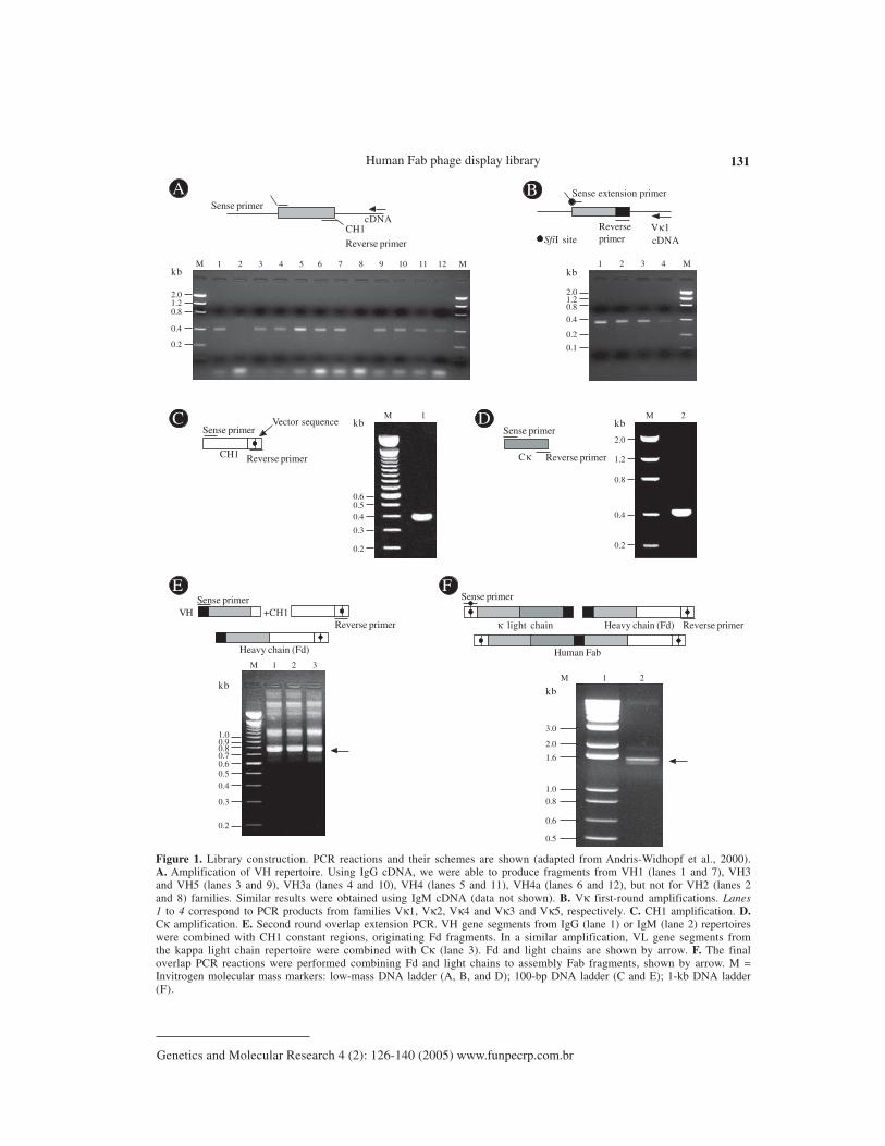
The Fab heavy chain (Fd) and κ light chain genes were obtained using cDNA synthe-sized from the total RNA of PBL of the 11 osteosarcoma patients. Twelve separate VH andfour Vκ PCR reactions were performed to optimize the amplifications of V gene rearrange-ments. The resulting PCR products, corresponding to VH (Figure 1A) and Vκ (Figure 1B) genefragments, account for a large fraction of the rearranged variable region repertoire of thesepatients. All the primary products of PCR amplification of the variable and constant regions(Figure 1C for CH1 and 1D for Cκ) ranged from 350 to 400 bp in size. In the overlappingsecond round of PCR, the resulting variable genes were pooled and joined together, along withthe constant region, generating PCR products ranging from 750 to 800 bp (Figure 1E). The thirdPCR round consisted of a final overlap amplification of the complete human dicistronic Fabgene, generating fragments of 1.5 to 1.6 kb (Figure 1F). The generated Fabs were cloned intothe pComb3X phage display vector, after digestion with the restriction enzyme SfiI. Since thePCR primers incorporated two asymmetric SfiI sites, this strategy produced a directional liga-tion of the SfiI digested Fab, in frame with ompA and pelB leader sequences, to direct expres-
Human Fab phage display library 131
Genetics and Molecular Research 4 (2): 126-140 (2005) www.funpecrp.com.br
Sense primer
Reverse primer
cDNACH1
Sense extension primer
Reverseprimer cDNA
Vκ1SfiI site
Sense primer
Reverse primer
Vector sequence
CH1
Sense primer
Reverse primer
Heavy chain (Fd)
+CH1VH
Sense primer
Reverse primerCκ
Sense primer
Reverse primerκ light chain Heavy chain (Fd)
Human Fab
2.01.20.8
0.4
0.2
kbM M1 2 3 4 5 6 7 8 9 10 11 12
2.01.20.8
0.4
0.2
kbM1 2 3 4
0.1
0.60.50.4
0.3
kbM 1
0.2
2.0
1.2
0.8
0.4
kbM 2
0.2
1.00.90.80.7
kb
M 1
0.60.5
0.4
0.3
0.2
2 3
3.0
2.0
1.6
kbM 1
0.6
1.0
0.8
0.5
2
A B
C D
E F
Figure 1. Library construction. PCR reactions and their schemes are shown (adapted from Andris-Widhopf et al., 2000).A. Amplification of VH repertoire. Using IgG cDNA, we were able to produce fragments from VH1 (lanes 1 and 7), VH3and VH5 (lanes 3 and 9), VH3a (lanes 4 and 10), VH4 (lanes 5 and 11), VH4a (lanes 6 and 12), but not for VH2 (lanes 2and 8) families. Similar results were obtained using IgM cDNA (data not shown). B. Vκ first-round amplifications. Lanes1 to 4 correspond to PCR products from families Vκ1, Vκ2, Vκ4 and Vκ3 and Vκ5, respectively. C. CH1 amplification. D.Cκ amplification. E. Second round overlap extension PCR. VH gene segments from IgG (lane 1) or IgM (lane 2) repertoireswere combined with CH1 constant regions, originating Fd fragments. In a similar amplification, VL gene segments fromthe kappa light chain repertoire were combined with Cκ (lane 3). Fd and light chains are shown by arrow. F. The finaloverlap PCR reactions were performed combining Fd and light chains to assembly Fab fragments, shown by arrow. M =Invitrogen molecular mass markers: low-mass DNA ladder (A, B, and D); 100-bp DNA ladder (C and E); 1-kb DNA ladder(F).

C. Dantas-Barbosa et al. 132
Genetics and Molecular Research 4 (2): 126-140 (2005) www.funpecrp.com.br
sion of antibody light and heavy chain-pIII fusion proteins, under the control of the lac promoter.The gene III Fd fusions included His and influenza hemagglutinin epitope tags. In addition, anamber stop codon, TAG, was inserted after the hemagglutinin tag, allowing the production ofsoluble Fab in a nonsuppressor E. coli strain (Scott and Barbas III, 2000). OmpA and pelBsignal sequences directed the Fab to periplasmatic cell compartment and the resulting Fab-coatprotein fusion was incorporated onto the surface of M13 Fab-phage fusion particles after helperphage infection. The resulting combinatorial library reached 1.8 x 10⁸ Fabs, forming potentialparatopes able to recognize an equivalent number of epitopes.
Library diversity
V gene usage
To assess the quality and diversity of the library, 10 randomly selected Fab clones weredigested with SfiI and BstO I restriction enzymes. The SfiI digestion yielded 1.5- to 1.6-kbfragments, corresponding to the expected Fab insert. The BstO I fingerprint profiles confirmedthe diversity of the library (Figure 2). All the analyzed clones presented the correct Fab size, andall of them had distinct BstO I restriction fragment patterns. Since all clones had the same CH1and Cκ region genes and vector backbones, differences in the restriction patterns reflecteddifferences in the VH or Vκ rearranged genes. Thus, the library shows diversity at the V genelevel (Figure 2).
A further investigation included the sequencing of several V gene clones. Sequencecomparison was made using the Ig Blast defined patients’ V gene family repertoire usage. Wesequenced either VH or Vκ in 125 V regions (51 VH and 74 Vκ). Although all the clones in ourlibrary bear a full length insert, 80.8% of them had a functional VH/VL fusion Fab, leading to afunctional library size of 1.45 x 10⁸.
The VH repertoire, evaluated among 51 randomly chosen clones, showed 20 differentVH subfamilies (Figure 3A). No significant bias was observed, but there was a slight predomi-nance of clones using genes from the VH4 family (35.4%), which differs from the reportedpredominance of the VH3 family (33.3% of our clones) in the normal human repertoire (Cookand Tomlinson, 1995). No VH2 amplification was observed. The failure in detecting VH2 mem-bers, notwithstanding the use of specific primers, can be explained by the low frequency ofusage of this family (Knappik et al., 2000), though polymorphism or even primer design defectscannot be discarded. Concerning Vκ genes (Figure 3B), among 74 sequenced clones, 11 differ-ent families were found, with 33% predominance of A27, similar to what was found in a previ-ous report (Tomlinson et al., 1995). In our sequences, the second most frequent family was O2(18.9%), followed by A17 (14.8%) (Figure 3B). The resulting library contained VH genes fromfour different families (VH families 1, 3, 4, and 5), and κ light chain genes from subgroups I, II,III, IV, and V. Complementary determining (CDR) and framework region assignments werebased on Kabat definition. Only high quality sequences were used for translation (Figures 4 and5). The analysis was successfully accomplished in 43 VH and 51 Vκ sequences, respectively.
Random Fab assembly was evident in the analysis of VH and VL pairs of 47 individualclones (Table 2). Most of the clones were assembled with different variable genes; only threepairs of clones (D12 and E03, G04 and G05 and H8 and H9) shared the same families of bothVH and VL genes even though they accumulated other differences in framework or CDR

Human Fab phage display library 133
Genetics and Molecular Research 4 (2): 126-140 (2005) www.funpecrp.com.br
sequences. Three pairs of clones presented the same VH sequence, H08-C04, F07-H02 andG10-H10, but they had different VL genes (Figure 4 and Table 2).
VH diversity
Heavy chain gene analysis revealed a great variability of family usage (Table 2, Figure3). Most of the clones presented the canonical residues at clue positions. One of the exceptionsis clone E04, which had a serine instead of a glycine at position 26 (G26S). Position 27 isexpected to show F, Y, S, D, G, or T. Some of our clones presented different residues: H (cloneB05), A (clones E09 and D09) and E (clone F04). We also found differences in residue 94: T, S,K, A, and L, instead of the canonical R, G or N (Chothia et al., 1989).
The heterogeneity of CDRH3 was remarkable. Its length varied from 6 to 20 aminoacid residues. In our clones, the most frequent CDR sizes were 8 (23%), 9, 11, and 13 (11.6%each) residue lengths. Classifying canonical loop 1 structures, our clones were mainly type 1(90%), followed by some with type 2 (5%) and 3 (5%) loops. These frequencies are similar to
2.01.6
0.8
0.4
0.6
kbM 1 2 3 4 5 6 7 8 9 10
B
A
3.0
0.5
1.0
M 1 2 3 4 5 6 7 8 9 10
0.7
0.5
0.2
0.4
kb
0.8
0.3
0.6
0.9
Figure 2. Variability of phagemid clones. A. SfiI digests of 10 randomly chosen clones presenting fragments correspond-ing to the Fab size (arrows). B. DNA fingerprint analysis by BstO I digestion. M = Invitrogen molecular mass markers: 1-kb DNA ladder (A) and 100-bp DNA ladder (B).

C. Dantas-Barbosa et al. 134
Genetics and Molecular Research 4 (2): 126-140 (2005) www.funpecrp.com.br
those known for human antibody canonical H1 conformations (Almagro et al., 1997). Assign-ment of H2 canonical conformation was not possible for nearly half of the sequenced clones(Figure 4). The recurrence of histidine and tyrosine residues at position 52a has not been previ-ously reported, but we found it to be frequent. This observation may reflect a polymorphism ofthe Brazilian population or a donor patient repertoire bias. It is noteworthy that these non-usualresidues are found in the VH4-derived Fabs, which were 48% of our clones. The H2 loops hadtype 1 (30%), 2 (12%), 3 (9%), or 4 (7%) canonical conformations.
VL diversity
Key residues at positions 2, 25, 29, 33, and 71 contribute to the L1 structure. The 51
Figure 3. VH and VL segment usage. The graph shows the frequency of VH (A) and VL (B) gene segment family usage (%).The sequences were aligned to their closest germline, using the Ig-Blast to identify the V family.
VH4.59 12%
A VH families
VH4.39 2%
VH4.34 10%
VH5.51 12% VH1.2 10% VH1.3 2% VH1.69 4%
VH3.7 4%
VH3.15 2%
VH1.46 4%
VH3.21 4%
VH3.23 4%
VH3.30 4%VH3.43 6%
VH3.49 2%
VH4.316% VH4.28
4%VH4.4
2%VH3.74
6%VH3.73
2%
B
O2 19%O8 3% A3 5%
A17 15%
B3 5%B2 7% A30 1% A27 34%
L12 4%
L6 4%
L2 3%
VL families

Human Fab phage display library 135
Genetics and Molecular Research 4 (2): 126-140 (2005) www.funpecrp.com.br
Fig
ure
4. D
educ
ed a
min
o ac
id s
eque
nces
of
func
tion
al V
H c
lone
s. F
ram
ewor
k (F
R)
and
com
plem
enta
ry d
eter
min
ing
regi
ons
(CD
R)
are
show
n. U
nusu
al V
H4
resi
dues
at
posi
tion
52A
are
und
erli
ned.

C. Dantas-Barbosa et al. 136
Genetics and Molecular Research 4 (2): 126-140 (2005) www.funpecrp.com.br
Fig
ure
5. D
educ
ed a
min
o ac
id s
eque
nces
of
func
tion
al V
L c
lone
s. F
ram
ewor
k (F
R)
and
com
plem
enta
ry d
eter
min
ing
regi
ons
(CD
R)
are
show
n. R
esid
ue p
osit
ions
use
d fo
rca
noni
cal
stru
ctur
e as
sign
men
ts a
re l
abel
ed (
*) i
n th
e co
rres
pond
ing
colu
mn.

Human Fab phage display library 137
Genetics and Molecular Research 4 (2): 126-140 (2005) www.funpecrp.com.br
sequences listed in Figure 4 are according to these key presentations or have previously de-scribed mutations (three clones from the O2 family had changes at position 33: clone H05:L33V, and clones F04 and H02: L33I, and one clone, A04, with F71Y).
All functional germ line Vκ segments had a single canonical L2 structure, which con-tained an isoleucine at position 48, a glycine at site 64 and one residue between residues 50 and52. Among our sequences, we observed five clones with rare changes at this position. ClonesG11 and H11 (from the VLO2 family) and clone F02, belonging to VLB3, had a conservativechange from isoleucine to valine (I48V) and both A04 and B03 clones (VLB3) presented a non-conservative substitution from isoleucine to phenylalanine (I48F), which is reported to occur inless than 1% of the Vκ sequences (Tomlinson et al., 1995). The B03 clone also presented aconservative change from glycine to alanine (G64A) at position 64, while clone B11 had anaspartic acid (G64D) at this position.
The most variable region was CDRL3, due to V-J gene segment rearrangements. Thejoining process involved trimming and repair of the V and J segments, and there was also Naddition. CDR3 can vary in length from 1 to 11 residues, with most of the sequences presentingbetween 6 and 8 amino acid residues (Knappik et al., 2000). Among our patients, there was apredominance of CDRL3s with 9 (44.5%), followed by 8 (24.0%), 10 (20.4%) and 11 (11.1%)residues. Tomlinson and co-workers (1995) studied 634 VL genes and reported 67% with 6residues, 19% with 7 and only 0.15% of the CDRL3s with 9 residues. In a study of 382 VKgenes, Knappik and co-workers (2000) found 72.3% of all CDR3s with 8 amino acid residues,7.3% with 7, 17.3% with 9, 1.3% with 10, and 1.8% with less than 7 residues in the CDRL3.There are five canonical structures for the L3, which vary according to the number of residues.Most of them have a glutamine residue at position 90 and a proline at position 95. Some of our cloneshad differences at those positions. One clone had a tyrosine at position 90 (Q90Y) and a proline atposition 94 (C04 clone from the L12 family). The presence of a proline at positions 94 has been
Clone VH VL Clone VH VL Clone VH VL
E04 4.34 A17 A11 5.51 O2 B05 1.46 A27F05 4.39 A17 D08 1.46 O2 B11 3.23 A27F11 1.2 A17 F04 4.34 O2 C01 4.59 A27E12 3.30 A17 F07 3.43 O2 C05 4.4 A27G08 5.51 A17 F08 4.59 O2 C09 3.43 A27D09 4.39 B2 G11 4.34 O2 D03 3.74 A27E06 1.3 B2 H02 3.43 O2 D05 4.34 A27F12 1.2 B2 H05 3.15 O2 D12 5.51 A27B03 3.21 B3 H11 3.73 O2 E03 5.51 A27F02 3.30 B3 D11 3.7 O8 E08 3.21 A27B10 3.23 L12 H03 3.74 O8 G04 4.59 A27C04 1.2 L12 C12 5.51 A3 G05 4.59 A27C06 3.74 L12 D02 4.31 A3 H08 1.2 A27E09 4.31 L12 F05 3.7 L2 H09 1.2 A27F03 1.69 L6 F06 4.59 L2 H10 4.28 A27G10 4.28 L6 F10 3.30 L2
Table 2. V gene usage of randomly selected Fab clones.

C. Dantas-Barbosa et al. 138
Genetics and Molecular Research 4 (2): 126-140 (2005) www.funpecrp.com.br
described in very few sequences (Chothia et al., 1989). Three of our clones had a proline atposition 95A: C06, E11, G09. Three clones from the B2 family (D09, E06 and A01) had aleucine at this position (P95L) and one clone (B10, from the L12 family) had a P95F.
DISCUSSION
We cloned genes coding for the human antibody repertoire of osteosarcoma patients,for future use for the isolation of immunoglobulins against these tumor antigens. We attemptedto maximize the library diversity by using primers optimized for VH (IgG and IgM) and Vκ genefamily mRNAs. Osteosarcoma antibody variable gene repertoires were cloned into a phagedisplay vector, allowing combinatorial Fab assembly. Library size was compatible to the re-ported size for naive and immunized human phage displayed libraries, which range from 106 to109 (Vaughan et al., 1996; Itoh et al., 2003). It has long been recognized that actual library sizemust correspond to the functional antibody forms. Thus, the real size is smaller than that foundby counting recombinant colonies, since frameshifts, stop codons, or deletions can be generatedby PCR or can be a product of non-productive immunoglobulin rearrangements. Our real libraryis formed of 81% functional clones, originating around 108 Fabs. This order of magnitude shouldpermit successful antigen-binding antibody selection. Moreover, due to its origin, it is expectedthat this library is tumor specific, along with environmental and self-antigen-reacting antibodies.Even new forms, resulting from in vitro shuffling of VH and VL, enrich the library’s naturalrepertoire.
Analysis of VH/VL pairs also revealed that none of these 47 randomly selected clonesshared the same VH and VL sequences, being unique and hypothetically accounting for differ-ent specificities. Thus, the library size and its variability will certainly allow for the selection ofuseful anti-osteosarcoma antibodies and also immunoglobulins that recognize a great variety ofepitopes, since non-immune libraries are being used to isolate antibodies against different anti-gens (de Haard et al., 1999). Our library will be used to select antibodies against bone tumor celltotal protein preparations and also against their cell surface, targeting molecular markers.
Ours is the first report of an osteosarcoma-patient antibody repertoire-derived library.The considerable variability of our library means that it should be useful for isolating Fabscapable of recognizing osteosarcoma antigens. These new molecules are potentially valuabletools in the detection of these tumor malignant cells and could be helpful for improved diagnosisand treatment.
ACKNOWLEDGMENTS
We are grateful to Isabella Simões and Fabricio Arraes for sequencing, Dr. MarcioPoças-Fonseca for English revision, Cid Alexandre Pereira for figure preparation, and the SarahNetwork of Hospitals for Rehabilitation for financial support.
REFERENCES
Almagro, J.C., Hernandez, I., Ramirez, M.D.C. and Vargas-Madrazo, E. (1997). The difference betweenthe structural repertoire of VH germline gene segments of mice and human: implication for themolecular mechanism of the immune response. Mol. Immunol. 34: 1199-1214.
Andris-Widhopf, J., Steinberger, P., Fuller, R., Rader, C. and Barbas III, C.F. (2000). Generation of

Human Fab phage display library 139
Genetics and Molecular Research 4 (2): 126-140 (2005) www.funpecrp.com.br
antibody libraries: PCR amplification and assembly of light- and heavy-chain coding sequences In:Phage Display Laboratory Manual (Barbas III, C.F., Burton, D.R., Scott, J.K. and Silverman, G.J.,eds.). 1st edn. Cold Spring Harbor Laboratory Press, Cold Spring Harbor, NY, USA, pp. 9.1-9.22.
Barbas III, C.F., Kang, A.S., Lerner, R.A. and Benkovic, S.J. (1991). Assembly of combinatorial antibodylibraries on phage surfaces: The gene III site. Proc. Natl. Acad. Sci. USA 88: 7978-7982.
Berger, M., Shankar, V. and Abbas, V. (2002). Therapeutic applications of monoclonal antibodies. Am. J.Med. Sci. 324: 14-30.
Brigido, M.M. and Maranhão, A.Q. (2001). Bibliotecas apresentadas em fagos. Biotecnologia - Ciênciae Desenvolvimento 26: 44-51.
Cai, X. and Garen, A. (1995). Anti-melanoma antibodies from melanoma patients immunized with geneti-cally modified autologous tumor cells: Selection of specific antibodies from single-chain Fv fusionphage libraries. Proc. Natl. Acad. Sci. USA 92: 6537-6541.
Chester, K., Pedley, B., Tolner, B., Violet, J., Mayer, A., Sharma, S., Boxer, G., Green, A., Nagl, S. andBegent, R. (2004). Engineering antibodies for clinical applications in cancer. Tumor Biol. 25: 91-98.
Chothia, C., Lesk, A.M., Tramontano, A., Levitt, M., Smith-Gill, S.J., Air, G., Sheriff, S., Padlan, E.A.,Davies, D., Tulip, W.R., Colman, P.M., Spinelli, S., Alzari, P.M. and Poljak, R.J. (1989). Conforma-tions of immunoglobulin hypervariable regions. Nature 342: 877-883.
CooK, G.P. and Tomlinson, I.M. (1995). The human immunoglobulin VH repertoire. Immunol. Today 16:237-242.
Cosimi, A., Burton, R., Colvin, R., Goldstein, G., Delmonico, F., LaQuaglia, M., Tolkoff-Rubin, N., Rubin,R., Herrin, J. and Russell, P. (1981). Treatment of acute renal allograft rejection with OKT3 mono-clonal antibody. Transplantation 32: 535-539.
Dahlin, D.C. (1975). Pathology of osteosarcoma. Clin. Orthop. 111: 23-32.de Haard, H.J., van Neer, N., Reurs, A., Hufton, S.E., Roovers, R.C., Henderikx, P., Bruïne, A.P., Arends,
J.W. and Hoogenboom, H. (1999). A large non-immunized human Fab fragment phage display thatpermits rapid isolation and kinetics analysis of high affinity antibodies. J. Biol. Chem. 274: 18218-18230.
Ewing, B. and Green, P. (1998). Base-calling of automated sequencer traces using phred. II. Error prob-abilities. Genome Res. 8: 186-194.
Griffiths, A.D., Malmqvist, M., Marks, J.D., Bye, J.M., Embleton, M.J., McCafferty, J., Baier, M., Holliger,K.P., Gorick, B.D., Hughes-Jones, N.C., Hoogenboom, H.R. and Winter, G. (1993). Human anti-selfantibodies with high specificity from phage display libraries. EMBO J. 12: 725-734.
Hall, T.A. (1999). BioEdit: a user-friendly biological sequence alignment editor and analysis. NucleicAcids Symp. 41: 95-98.
Itoh, K., Inoue, K., Tezuka, T., Tada, H., Hashimoto, Y., Masuko, T. and Suzuki, T. (2003). Molecularstructural and functional characterization of tumor suppressive anti-erbB-2 monoclonal antibody byphage display system. J. Biochem. 133: 239-245.
Jesus-Garcia Filho, R. (1992). Tumores produtores de tecido ósseo. In: Manual de Tumores Músculo-Esqueléticos (Jesus-Garcia Filho, R. and Nery, C.A.S., eds.). Escola Paulista de Medicina, São Paulo,SP, Brasil, pp. 18-27.
Knappik, A., Ge, L., Honegger, A., Pack, P., Fischer, M., Wellnhofer, G., Hoess, A., Wölle, J., Plünckthun,A. and Virnekas, B. (2000). Fully synthetic human combinatorial antibody libraries (HuCAL) basedon modular consensus frameworks and CRDs randomized with trinucleotides. J. Mol. Biol. 296: 57-86.
Kohler, G. and Milstein, C. (1975). Continuous cultures of fused cells secreting antibody of predefinedspecificity. Nature 256: 495-497.
Lu, D., Jimenez, X., Zhang, H., Boleen, P., Witte, L. and Zhu, Z. (2002). Selection of high affinity humanneutralizing antibodies to VEGFR2 from a large antibody phage display for antiangiogenesis therapy.Int. J. Cancer 97: 393-399.
Maranhão, A.Q. and Brigido, M.M. (2000). Anticorpos humanizados. Biotecnologia - Ciência e Desen-volvimento 23: 38-43.
Marks, J.D., Hoogenboom, H.R., Bonnert, T.P., McCafferty, J., Griffiths, A.D. and Winter, G. (1991). By-passing immunization of human antibodies from V-gene libraries displayed on phage. J. Mol. Biol.222: 581-597.
McCafferty, J., Griffiths, A.D., Winter, G. and Chiswell, D.J. (1990). Phage antibodies: filamentous phagedisplaying antibody variable domains. Nature 348: 552-554.
Meyers, P.A., Heller, G., Huvos, A., Applewhite, A., Sun, M. and LaQuaglia, M. (1993). Osteogenic sar-coma with clinically detectable metastasis at initial presentation. J. Clin. Oncol. 11: 449-453.

C. Dantas-Barbosa et al. 140
Genetics and Molecular Research 4 (2): 126-140 (2005) www.funpecrp.com.br
Mintz, P.J., Kim, J., Do, K.-A., Wang, X., Zinner, R.G., Cristofanilli, M., Arap, M.A., Hong, W.K., Troncoso,P., Logothetis, C.J., Pasqualini, R. and Arap, W. (2003). Fingerprinting the circulating repertoire ofantibodies from cancer patients. Nat. Biotechnol. 21: 57-63.
Morrison, S.L. and Oi, V.T. (1989). Genetically engineered antibody molecules. Adv. Immunol. 44: 65-92.Portolano, S., McLachlan, S.M. and Rapoport, B. (1993). High affinity, thyroid-specific human autoanti-
bodies displayed on the surface of filamentous phage use V genes similar to other autoantibodies. J.Immunol. 151: 2839-2851.
Rader, C., Steinberger, P. and Barbas III, C.F. (2000). Selection from antibody libraries In: Phage DisplayLaboratory Manual (Barbas III, C.F., Burton, D.R., Scott, J.K. and Silverman, G.J., eds.). 1st edn. ColdSpring Harbor Laboratory Press, Cold Spring Harbor, NY, USA, pp. 10.2-10.20.
Scott, J.K. and Barbas III, C.F. (2000). Phage display vectors. In: Phage Display Laboratory Manual(Barbas III, C.F., Burton, D.R., Scott, J.K. and Silverman, G.J., eds.). 1st edn. Cold Spring HarborLaboratory Press, Cold Spring Harbor, NY, USA, pp. 2.1-2.19.
Seymour, J.F. (2004). New treatment approaches to indolent non-Hodgkin’s lymphoma. Semin. Oncol. 31:27-32.
Somers, V.A., Brandwijk, R.J., Joosten, B., Moerkerk, P.T., Arends, J.W., Menheere, P., Pieterse, W.O.,Claessen, A., Scheper, R.J., Hoogenboom, H.R. and Hufton, S.E. (2002). A panel of candidate tumorantigens in colorectal cancer revealed by serological selection of a phage displayed cDNA expres-sion library. J. Immunol. 169: 2772-2780.
Tomlinson, I.M., Cox, J.P.L., Gherardi, E., Lesk, A.M. and Chothia, C. (1995). The structural repertoire ofthe human Vk domain. EMBO J. 14: 4628-4638.
Ueda, Y., Roessner, A. and Grundmann, E. (1993). Pathological diagnosis of osteosarcoma: the validity ofthe subclassification and some new diagnostic approaches using immunohistochemistry. CancerTreat. Res. 62: 109-124.
Vaughan, T.J., Williams, AJ., Pritchard, K., Osbourn, J.K., Pope, A.R., Earnshaw, J.C., McCafferty, J.,Hodits, R.A., Wilton, J. and Johnson, K.S. (1996). Human antibodies with sub-nanomolar affinitiesisolated from a large non-immunized Phage Display Library. Nat. Biotechnol. 14: 309-314.
Whelan, J.S. (1997). Osteosarcoma. Eur. J. Cancer 33: 1611-1619.Wu, B.P., Xiao, B., Wan, T.M., Zhang, Y.L., Zhang, Z.S., Zhou, D.Y., Lai, Z.S. and Gao, C.F. (2001).
Construction and selection of the natural immune Fab antibody phage display library from patientswith colorectal cancer. World J. Gastroenterol. 7: 811-815.