constraint induced movement therapy : a longitudinal case
TRANSCRIPT
University of New MexicoUNM Digital Repository
Occupational Therapy ETDs Electronic Theses and Dissertations
2-9-2010
Constraint induced movement therapy : alongitudinal case studySarah Renee McMillan
Follow this and additional works at: https://digitalrepository.unm.edu/octh_etds
This Thesis is brought to you for free and open access by the Electronic Theses and Dissertations at UNM Digital Repository. It has been accepted forinclusion in Occupational Therapy ETDs by an authorized administrator of UNM Digital Repository. For more information, please [email protected].
Recommended CitationMcMillan, Sarah Renee. "Constraint induced movement therapy : a longitudinal case study." (2010).https://digitalrepository.unm.edu/octh_etds/2

i

ii
CONSTRAINT INDUCED MOVEMENT THERAPY:
A LONGITUDINAL CASE STUDY
BY
SARAH R. MCMILLAN
B.A., PYSCHOLOGY, NEW COLLEGE OF FLORIDA, 2007
THESIS
Submitted in Partial Fulfillment of the Requirements for the Degree of
Master's Of Occupational Therapy
The University of New Mexico Albuquerque, New Mexico
December, 2009

iii
CONSTRAINT INDUCED MOVEMENT THERAPY:
A LONGITUDINAL CASE STUDY
BY
SARAH R. MCMILLAN
ABSTRACT OF THESIS
Submitted in Partial Fulfillment of the Requirements for the Degree of
Master's Of Occupational Therapy
The University of New Mexico Albuquerque, New Mexico
December, 2009

iv
CONSTRAINT INDUCED MOVEMENT THERAPY:
A LONGITUDINAL CASE STUDY
By
Sarah R. McMillan
B.A., Psychology, New College of Florida, 2007
ABSTRACT
Purpose. Constraint-induced movement therapy (CIMT) involves use of a
constraint on the non-affected extremity and intensive therapy comprised of shaping and
repetition during functional activities to improve quality and quantity of use of the
affected extremity. The literature has shown that CIMT is effective and two articles
support the use of 2 doses of CIMT. The current study investigated if 3 doses of CIMT
over 5 years continued to produce improvements in functional use for a boy with
hemiplegic cerebral palsy (CP).
Methods. One child, with spastic hemiplegic CP, met inclusion criteria of a
minimum of 20° of active wrist extension and 10° of active finger extension in the
hemiplegic hand. Grip strength, pinch strength, modified Nine Hole Peg Test, Box and
Blocks, Canadian Occupational Performance Measure, Pediatric Evaluation of Disability
Inventory, Melbourne Assessment of Unilateral Upper Limb Function, and the modified
House Functional Classification System were conducted pre-,during, post-, and 3 months
post-intervention. The child wore a bivalve cast during waking hours and participated in
4 hours of therapy 5 days/week for 2 weeks.
Results. Improvements in strength, functional use, and participation were seen
across all three doses. There were no significant changes in mobility or social skills
following any of the doses.
Conclusions. This study supports the use of CIMT for up to 3 doses during
childhood. More research needs to be conducted to determine the optimal length,
intensity, and frequency of doses; optimal time in the child's development; optimal
impairment levels; and if CIMT is best used alone or in conjunction with other protocols.

v
TABLE OF CONTENTS
LIST OF FIGURES ........................................................................................................ vii
CHAPTER 1 INTRODUCTION......................................................................................1
Constraint Induced Movement Therapy ........................................................................2
Constraint Induced Movement Therapy in Children with Cerebral Palsy.....................4
Purpose of Current Study...............................................................................................6
CHAPTER 2 METHODS .................................................................................................7
Participant ......................................................................................................................7
Measures ........................................................................................................................7
Procedures....................................................................................................................11
Intervention ..................................................................................................................11
Data Analysis……….……………………………………………………………….12
CHAPTER 3 RESULTS………………………………………………………………..13
Grip Strength................................................................................................................13
Pinch Strength………………………………………………………………………..15
Modified Nine Hole Peg Test ......................................................................................17
Box and Block..............................................................................................................19
Canadian Occupational Performance Measure.......................................................….21
Melbourne Assessment of Unilateral Upper Limb Function.......................................24
Pediatric Evaluation of Disability Index .....................................................................26
Modified House Classification System……….……………………………………...29
CHAPTER 4 DISCUSSION ...........................................................................................35
APPENDIX: Extended Literature Review ....................................................................39
Introduction..................................................................................................................39

vi
Constraint Induced Movement Therapy (CIMT).........................................................40
CIMT in Primates ........................................................................................................41
CIMT in Adults Post Stroke ........................................................................................42
CIMT versus Conventional Therapy for Adults ..........................................................46
Brain Imaging Studies in Adults Receiving CIMT......................................................47
CIMT in Children with Cerebral Palsy........................................................................49
Brain Imaging Studies in Children Receiving CIMT ................................................. 57
Effects of Repeated Doses of CIMT in Children………………………………….....59
REFERENCES.................................................................................................................63

vii
LIST OF FIGURES Figure 1. Grip Strength Across Doses ...............................................................................14
Figure 2. Pinch Strength Across Doses………………………………………..…………16
Figure 3. Modified Nine Hole Peg Test Scores Across Doses…………………….…….18
Figure 4. Box and Blocks Scores Across Doses…………………………………...…….20
Figure 5. COPM Performance Scores Across Doses…………………………………… 23
Figure 6. COPM Satisfaction Scores Across Doses……………………………………..23
Figure 7.Melbourne Unilateral Upper Limb Function Scores Across Doses……………25
Figure 8. PEDI Self-Care Scores Across Doses…..……………………………………..28
Figure 9. Modified House Classification System for a dressing activity………………..33
Figure 10. Modified House Classification System for a play activity…………………...34
\

1
Chapter 1 Introduction
Cerebral palsy (CP) is a neurodevelopmental clinical diagnosis in the developing
child based on observations of decreased motor control; no test alone can define its
presence (Bax, Goldstein, Rosenbaum, Leviton, & Paneth, 2005; Paneth, Hong, &
Korzeniewski, 2006). Bax et al. (2005) stated that “Cerebral palsy (CP) describes a group
of disorders of the development of movement and posture, causing activity limitation,
that are attributed to non-progressive disturbances that occurred in the developing fetal or
infant brain. The motor disorders of individuals with cerebral palsy are often
accompanied by disturbances of sensation, cognition, communication, perception, and/or
behavior, and/or by a seizure disorder” (p. 572). Many subtypes of children with CP have
been established to help categorize the disorder’s differing manifestations. These include
spastic, dyskinetic, ataxic, and mixed (NINDS, 2006).
Spastic cerebral palsy is the most common form, comprising up to 78% of the
population of those diagnosed (Yeargin-Allsopp, Braun, Doernberg, Benedict, Kirby, &
Durkin, 2008). Tight or stiff muscles and hyperreflexia generally characterize spastic CP
(NINDS, 2006). This subtype is further divided into individuals with diplegia,
hemiplegia, or tetra/quadriplegia depending on which of their extremities that are
affected. According to Yeargin-Allsopp et al. (2008), approximately 25-34% of
individuals with spastic cerebral palsy have hemiplegia with impairments primarily on
one side of their body.
Regardless of the type of cerebral palsy present, therapy can often help to improve
the individual’s capabilities (NINDS, 2006). Individuals with CP often receive physical

2
therapy (PT), occupational therapy (OT), and/or speech therapy. However, according to a
review by Antitila, Suoranta, Malmivaara, Mäkelä, and Autti-Rämö (2008) there is
limited evidence supporting comprehensive PT or OT services for individuals with CP.
These authors state that “well-conducted studies on current treatment options as well as
new treatment approaches using valid outcomes are obviously needed” (p. 490). In 2001,
Charles, Lavinder, and Gordon reported that one intervention approach, constraint-
induced movement therapy (CIMT), could potentially increase arm function in children
with hemiplegic CP.
Constraint Induced Movement Therapy
Constraint-induced movement therapy has two main components: 1) constraint of
the non-affected extremity and 2) intensive training using shaping and repetition
(Gordon, Charles, & Wolf, 2005). Constraint of the non-affected extremity has been
achieved through use of bivalve casts (DeLuca, Echols, Ramey, & Taub, 2003; Martin,
Burtner, Poole, & Phillips, 2008; Stearns, Burtner, Keenan, Qualls, & Phillips, 2009;
Sutcliffe, Gaetz, Logan, Cheyne, & Fehlings, 2007; Taub, Ramey, DeLuca, Echols;
2004), splints (Dickerson & Brown, 2007; Hamzei, Liepert, Weiller, & Rijntjes, 2006;
Kunkel, Kopp, Müller, Villringer, Villringer, Taub, & Flor, 1999; Liepert, Uhde, Graf,
Leidner, & Weiller, 2001; Miltner, Bauder, Sommer, Dettmers, & Taub, 1999; Taub,
Miller, Novack, Cook, Fleming, Nepomuceno, Connell, & Crago, 1993; van der Lee,
Wagenaar, Lankhorst, Vogelaar, Deville, & Bouter, 1999), slings ( Charles & Gordon,
2007; Charles, Lavinder, & Gordon, 2001; Charles, Wolf, Schneider, & Gordon, 2006;
Gordon, Charles, & Wolf, 2006; Wolf, S.L., Lecraw, D.E., Barton, L.A., & Jann, B.B.,
1989), gloves/mitts ( Dromerick, Edwards, & Hahn, 2000; Juenger, Linder-Lucht,
3
Walther, Berweck, Mall, & Staudt, 2007; Levy, Nichols, Schmalbrock, Keller, &
Chakeres, 2001; Pierce, Daly, Gallagher, Gershkoff, & Schaumburg, 2002; Richards,
Rothi, Davis, Wu, Nadeau, 2006 ), or holding the individual’s hand in a constrained
position (Naylor & Bower, 2005 ). Shaping during intensive practice is a method of
continually increasing the demands of the activity to match and slightly challenge the
individuals’ current capabilities (Charles et al., 2005). Repetition is deemed an important
component of CIMT and involves practicing a functional task to illicit a targeted
movement repeatedly over 15-20 minutes. Other than these two components, there is no
agreed upon CIMT protocol. Current research is being conducted to determine the
optimal frequency (hours in a day) and duration (number of days), constraint type, age,
and other variables (Damiano, 2005).
The hypothesis that CIMT could help individuals with hemiplegia arose from
studies with non-human primates (see Knapp, Taub, & Berman, 1963;. Taub & Uswatte,
2003 for comprehensive review of of this research). Taub (2003) proposed that this
protocol could be used following any neurological injury that resulted in a period of
central nervous system shock, including a stroke. Since then, many studies have been
conducted to investigate the use of CIMT with individuals with hemiplegia following a
stroke.
This literature has shown that CIMT produces improvement in adults post CVA
for neuromuscular factors such as grip strength and dexterity, and improves their ability
to perform activities of daily living such as basic self-care (Dromerick et al., 2000;
Hemzei et al., 2006; Levy et al., 2001; Kunkel et al., 1999; Liepert et al., 2001; Miltner et
al., 1999). Studies also show that CIMT produces changes within the adult brain post

4
CVA that traditional therapies may not (Dromerick et al., 2000; Hamzei et al., 2006;
Liepert et al., 2001; van der Lee et al., 1999). Research points to CIMT as being
applicable to individuals in all stages post-stroke and can be implemented in different
time frames and with different constraints (Dromerick et al., 2000; Richards et al., 2006).
More recently CIMT has been used with children with hemiplegic cerebral palsy.
Constraint Induced Movement in Children with Cerebral Palsy
Similar results to those found with adults post-stroke have been found when
CIMT is used with children with cerebral palsy. CIMT increases factors such as grip
strength, dexterity, and functional use of the child's affected extremity (e.g. DeLuca,
Echols, Ramey, & Taub, 2003; Sung et al., 2005; Taub et al., 2003; Taub, Ramey,
DeLuca, & Echols, 2004). This remains true for protocols with modified schedules (e.g.
Charles et al., 2006; Naylor & Bower, 2005; Pierce et al., 2002). In addition, CIMT
produces cortical reorganization in the brains of children and adults with congenital
hemiparesis as shown by fMRI studies (Juenger et al., 2007; Sutcliffe et al., 2007).
Despite numerous studies, research still needs to address the optimal conditions of
CIMT. Particularly, what is the optimal duration, frequency, length of sessions, time to
wear the constraint, and number of doses. Recently, two studies have began the
investigation into the latter. Deluca and colleagues (2003) conducted a case study with a
child with hemiplegic cerebral palsy to investigate their constraint induced movement
therapy (CIMT) protocol, and the effects of multiple CIMT sessions. The participant was
a 15 month old girl with no active use of her affected extremity. For the intervention, she
wore a bivalved, fiberglass cast 24 hours a day for 3 weeks and participated in therapy for
6 hours per day for 15 consecutive week days. The therapy consisted of play based
5
activities that focused on sensorimotor and gross motor skills. Pre and post tests were
conducted using the Peabody Developmental Motor Skills (PDMS) with a focus on the
fine motor portion, the Denver Developmental Screening Tool (DDST), the Pediatric
Motor Activity Log (PMAL), and the Toddler Arm Use Test (TAUT). By the end of the
first period of intervention, she demonstrated increased attention to her right extremity,
her PDMS score increased from a 43 to a 62, her DDST increased in all subgroups, the
PMAL showed an increase in attempts to use her arm and in the quality of movements
produced, and the TAUT showed that spontaneous use of her affected extremity
increased by 50%.
A second period of intervention was conducted when she was 21 months old. She
had retained most of the skills she had learned previously, however her spontaneous
affected upper extremity use had decreased slightly. The intervention was similar to the
first intervention, except that she participated in the therapy for 6 hours a day for 21
consecutive days. After the intervention, her PMAL scores indicated that she had
increased both in the quality of use of her affected extremity and increased frequency in
her extremity use. Her TAUT scores showed that she used her affected extremity during
100% of the chosen activities.
Although Deluca et al.’s (2003) study supported the hypothesis that multiple
doses of CIMT could be beneficial to children with CP, Charles and Gordon (2007)
expanded on the Deluca et al. study by using an increased number of participants and
more objective measures. The purpose of the study was to determine if gains were
maintained for 12 months after the first dose and if a second dose resulted in continued
improvement. Eight children ages 5-11 years old participated. CIMT was given 6 hours a

6
day, 10 out of 12 consecutive days in a group format (2-4 kids). Children wore a sling
only during the intervention sessions. Shaping and massed practice of repetitive tasks
were used during play activities. Measures were the Jebsen-Taylor Test of Hand
Function, subtest number 8 of the Bruininks-Oseretsky Test of Motor Proficiency, and a
Caregiver Functional Use Survery. The children were assessed pre-intervention and 1
week-, 1 month-, and 6 months- post intervention. At 12 months after the initial
intervention children were assessed again, as both a follow up to the initial dose and a pre
test for the second dose. The children were reassessed 1 week following the second dose.
During the initial dose, children spent 55% of the session in structured activities, 61% in
repetitive tasks and 39% in shaping. During the second dose children spent 71% of the
time in structured activities with 81% in repetitive tasks and 19% in shaping. On the
Jebsen-Taylor, Buininks-Oseretsky, and CFUS there were significant improvements from
pretest 1 to posttest 6 months, with no decreases between 6 months and 12 months. The
authors concluded that CIMT provided more sustained results post intervention and that
children could benefit from multiple doses.
Purpose of Current Study
The previous studies that have investigated the effects of multiple doses of CIMT
have focused on two doses that were administered within 1 year of each other. The aim of
the current study is to determine whether a young boy with spastic hemiplegic cerebral
palsy will continue to make gains with a third dose of CIMT over a 6 year window of
time. He has previously made gains in two CIMT doses, one when he was 3 years old
(Martin et al., 2008) and the other when he was 5 years old (Stearns et al., 2009); the time
periods between doses was greater in this study than previous studies.

7
Chapter 2
Methods
Participant
One 8.5 year old child with right hemiplegic cerebral palsy participated in this
single subject study. He participated in two previous 2 week intensive trials of CIMT, one
when he was 3 years and the other when he was 5 years of age. The participant currently
is enrolled in a regular education classroom, as well as the gifted program. The study was
approved by the Internal Review Board/ Human Research Review Committee at the
University of New Mexico Health Sciences Center. To qualify for the study, he met
inclusion criteria of a minimum of 20° of active wrist extension and 10° of active finger
extension in the hemiplegic hand and exclusion criteria of: no other neurologic
impairments and no orthopedic surgery or neuropharmacologic interventions such as
botulinum toxin within the past 6 months.
Measures
Measures were selected to assess the child’s functional changes at multiple ICF
levels outlined in the World Health Organization (WHO) International Classification of
Function (ICF) (World Health Organization, 2001). Measures of the child’s function at
the ICF leves of Body Stucture and Function were measured by grip strength, pinch
strength, box and blocks, nine hole peg test, and Melbourne Assessment of Unilateral
Upper Limb Function; and ICF level of Activities and Participation were measured by the
Canadian Occupational Performance Measure, Pediatric Evaluation of Disability Index,

8
and analysis of dressing and play tasks with the Modified House Functional
Classification System.
Grip Strength. Grip strength was assessed using a calibrated Jamar Dynamometer
following the modified protocol for children between the ages of 6-19 (Mathiowetz,
Weimer, & Federman, 1986). The participant was seated with the dynamometer resting
on the table, his arm adducted and neutrally rotated, shoulder flexed between 50-70°,
elbow flexed to 90°, wrist in slight extension (0-30°), wrist in slight ulnar deviation (0-
15°), and his forearm in neutral. Grip strength was recorded three times, with 30 seconds
resting time between each trial, and then averaged together. Good to excellent test-retest
reliability (r = 0.88 - 0.93) has been found for grip strength, as well as good validity when
the standardized protocol is followed (Mathiowetz, Weber, Volland, & Kashman, 1984).
Pinch Strength. A calibrated pinch meter was used to determine lateral pinch
strength. The participant was seated, his arm adducted and neutrally rotated, shoulder
flexed between 50-70°, elbow flexed to 90°, wrist in slight extension (0-30°), wrist in
slight ulnar deviation (0-15°), and his forearm in neutral. Pinch strength was recorded
three times for each finger position with a 30-second rest break in between each trial.
Test-retest reliability after one week was found to be greater than r= 0.81 and inter-rater
reliability was found to be r = 0.98 (Mathiowetz et al., 1984).
Modified Nine-Hole Peg Test. The Modified Nine-Hole Peg Test was used to
assess dexterity and grasp and release. The modified board is 5.5 by 5.5 inches, with
holes in 3 rows by 3 columns, and uses ½ inch diameter pegs. This modification allows
the pegs to have a wider diameter in order to make picking them up a little easier. The

9
participant was timed while he unimanually removed all of the pegs and put them on the
table. The original Nine-Hole Peg Test was been found to be reliable (inter-rater,
r = 0.97-0.99; test-retest, r = 0.43-0.69) when used with adults (Mathiowetz, Weber,
Kashman, & Volland, 1985b).
Box and Block Test. This test was chosen to measure gross manual dexterity. The
test has specific dimensions and procedures as published in an article by Mathiowetz and
colleagues (Mathiowetz, Volland, Kashman, & Weber, 1985a). The participant was
instructed to move the blocks from one side of the container, over the partition, and then
to drop them on the other side of the container. The participant was stopped after 1
minute and the blocks moved were counted. Inter-rater reliability is extremely high,
r = 0.999-1.00 (Mathiowetz et al., 1985a).
Canadian Occupational Performance Measure (COPM). The COPM is a semi-
structured interview designed to elicit the client’s occupational performance concerns
(Law et al., 2005). It also provides quantitative information regarding the client’s current
performance level and satisfaction level in relation to each occupational performance
issue (OPI). These scores can then be compared pre- and post- intervention to determine
the client’s perspective on any change in his performance and satisfaction levels. The
COPM has high test-retest reliability, with scores ranging from r = 0.84-0.92 across
different populations (Law et al., 2005). Validity has also been established by comparing
scores on the COPM to scores on the Reintegration to Normal Living Scale
( r = 0.72-0.93).
Melbourne Assessment of Unilateral Upper Limb Function. This assessment was
developed to test upper extremity function in children ages 5-15 with neurological

10
impairments (Randall, Carlin, Chondros, & Reddihough, 2001). The test measures the
quality of motor function in tasks that involve reach, grasp, release, and manipulation and
involves video taping the child performing 16 standardized tasks. Reliability has been
found to be high with inter-rater reliability at r = 0.95, and test-retest reliability at
r = 0.97-0.98 (Randall et al., 2001).
Pediatric Evaluation of Disability Inventory (PEDI). The PEDI assesses the
functional ability of children ages 6 months to 7.5 years through an inventory of
functional performance and caregiver assistance in a) self-care, b) mobility, and c) social
function (Haley, Coster, Ludlow, Haltiwanger, & Adrellos, 1992; Nichols and Case-
Smith, 1996). The assessment can be filled out by a familiar practitioner, or as in this
study, by a primary caregiver. Test-retest reliability for the standardized scores was high,
ranging from r = 0.70-0.98, and content validity, as compared with the Peabody
Developmental Motor Scales (PDMS) test, was high ranging from r = 0.82-0.95. REF
The Modified House Functional Classification System (MHC). The MHC is used
to describe and classify upper extremity function in children with CP (Koman et al.,
2008). There are nine categories with a total of 32 descriptors that clinicians can use to
determine the child's current functional ability; either a score derived from the summed
descriptors present, or a category determined by the highest category in which all
descriptors are marked. According to a study by Koman and colleagues (2008) interrater
reliability was r = 0.94 and intrarater reliability was r = 0.96. Concurrent validity was
established through correlation with the Melbourne, r = 0.84. This system was applied to
a series of videos that showed the participant dressing and pulling apart and putting
together a ring of pop beads, for five days during each phase of the study. Average

11
functional scores and categories were then determined for each phase of the study and
each component of the activities to determine any changes following the intervention.
Procedure
Design. This study used an A1 (Baseline of 2 weeks) – B (Intervention of 2
weeks) – A2 (Post 2 weeks)- A3 (3 months Post) design. Grip strength, pinch strength,
the Nine Hole Peg Test, and Box ‘n’ Blocks were administered with standard procedures
five times during each phase of the study. The COPM, the Melbourne Assessment of
Unilateral Limb Function, and the PEDI were administered once during each assessment
period.
Intervention. The participant wore a bivalved plaster cast that extended from his
upper arm to his fingertips all day during the 2-week intervention period, except for
sleeping, bathing, and weekly skin checks. The intervention was conducted by a student
research assistant supervised by a registered occupational therapist. The therapy was
scheduled for 4 hours per day/6 days a week/ for two weeks. (Two hours were spent in
the morning practicing morning activities of daily living (ADLs) and 2 hours spent in the
afternoon focusing on play activities.) All of the therapy was conducted in the
participant's home utilizing mass blocked practice of repetitive tasks and shaping to
attain: complete active range of motion at the shoulder and elbow, active supination of
the wrist, active mass grasp and release patterns, radial grasp with two fingers and the
thumb, fine prehension with the index finger and thumb, increased force production of
the hand and individual digits, and isolated use of the fingers and thumb in extension and
flexion. The activities chosen were those that the participant enjoyed and were
developmentally appropriate for the participant’s age and cognitive status. As the

12
participant’s skills and abilities improved, the activities were graded using the CFOG
(Poole, Burtner, & Stockman, 1997) to ensure that the activity was both challenging and
successful. Activities included, but not limited to, bathing, dressing, brushing teeth,
eating breakfast, playing card games, playing board games, bowling, blowing up balloons
with a manual hand pump, and play doh fun kits.
Data Analysis
Data from each dose was compiled in Microsoft Excel and SPSS. Data from
measures with five data points per phase (grip strength, pinch strength, mNHPT, and Box
and Blocks) were graphed in SPSS using a two standard deviation band method; a type of
control chart. According to Portney and Watkins (2009) two consecutive points above or
below the band indicate a significant change from baseline for that phase. Means were
computed for the other measures, bar graphs were created, and visual analysis of trends
were conducted.

13
Chapter 3
Results
Grip Strength
See Figure 1 for a comparison of grip strength across doses.
Dose 1. The participant's grip strength increased during the course of this dose,
with a significant difference between baseline and follow up. During the baseline phase
(A1), the participant's average grip strength was 0.33 lbs. This increased to 0.75 lbs
immediately following the intervention (A2), and 4.17 lbs at the 3 month follow up (A3).
Dose 2.The participant's grip strength increased during the course of this dose,
approaching significance at immediately following the intervention, and reaching
significant levels at the 3 month follow up. During the baseline phase (A1), the
participant's average grip strength was 9.36 lbs, dropping slightly to 8.8 lbs during the
intervention (B), most likely due to fatigue. Immediately following the intervention (A2)
the average grip strength was 13.13 lbs, increasing to 14.46 at the 3 month follow up
(A3).
Dose 3. The participant's grip strength increased during the course of this dose,
with significant differences between baseline and immediately post-intervention, and
baseline and follow up. During the baseline phase (A1), the participant's average grip
strength was 9.37 lbs. This increased to 10.87 lbs during the study (B), 12.59 lbs
immediately following the intervention (A2), and 14.13 lbs after 3 months (A3).

14
Figure 1 . Grip Strength Across Doses. The dotted lines represent the two standard deviation band around the baseline mean. Two consecutive data points above the top dotted line show significant change during that phase.

15
Pinch Strength
See Figure 2 for the comparison of pinch strength across doses.
Dose 1. The participant's average pinch strength remained at 0 during all phases
of this dose, except for an average strength of 0.20 lbs immediately following the
intervention, which reached a level of significance.
Dose 2. The participant's pinch strength increased during this dose, with the
largest gains demonstrated between baseline and immediately post intervention. At
baseline his average pinch strength was 4.33 lbs, increasing slightly to 4.97 lbs during the
intervention, raising to 6.31 lbs immediately post-intervention, and remaining at 6.33 lbs
at the 3 month follow up. The differences between pinch strength at baseline and post-
intervention, and baseline and follow up were both statistically significant.
Dose 3. The participant's pinch strength increased slightly, though statistically
non-significantly due to a large standard deviation during baseline measures. Through the
posttest increases were noted and then decreased minimally by the 3 month follow up.
During the baseline phase (A1) the average pinch strength was 6.8 lbs. This increased to
7.4 lbs during the intervention phase (B), then to 8.03 lbs after the intervention (A2), and
decreased slightly to 7.87 lbs by the 3 month follow up.

16
Figure 2. Pinch Strength Across Doses. The dotted lines represent the two standard deviation band around the baseline mean. Two consecutive data points above the top dotted line show significant change during that phase.

17
Modified Nine Hole Peg Test
See Figure 3 for the comparison of time across doses.
Dose 1. This measure was not conducted during this dose.
Dose 2. The participant's time on the NHPT decreased through the posttest and
then increased slightly at the 3 month follow up, demonstrating an statistically significant
increase in manual dexterity between baseline and all other phases . At baseline the
participant's average score was 19.33 seconds, decreasing to 15.2 seconds and 13.86
seconds during the intervention and at the posttest, respectively. At the three month
follow up the participant's time had increased to 15.36 seconds, which indicated
coordination patterns that remained faster than recorded at baseline.
Dose 3. The participant's time on the nine hole peg test decreased slightly
throughout the course of this dose, indicating a slight, though statistically non-significant,
improvement in manual dexterity. At baseline (A1) the participant's average time was
10.4 seconds decreasing to 9.4 seconds during the intervention (B), 9.1 seconds following
the intervention (A2) and finally 7.87 seconds at the 3 month follow up (A3).

18
Figure 3. Modified Nine Hole Peg Test Scores Across Doses. The dotted lines represent the two standard deviation band around the baseline mean. Two consecutive data points below the bottom dotted line show significant change during that phase.

19
Box and Blocks
See Figure 4 for the comparison of number of blocks across doses.
Dose 1. This measure was not conducted during this dose.
Dose 2. The participant's gross manual dexterity statistically improved between
baseline and A2, and baseline and A3, as shown by an increase in the number of blocks
he was able to successfully manipulate. During the baseline phase the participant
successfully manipulated an average of 12.17 blocks, the number remained virtually the
same during the intervention, 11.8 blocks, increasing to 16 blocks and 20.4 blocks
respectively.
Dose 3. The participant's gross manual dexterity statistically improved between
baseline and A2, and baseline and A3, as shown by an increase in the number of blocks
he was able to successfully manipulate. At baseline (A1) the participant's average number
of blocks was 28.6, increasing to 31.2 blocks during the intervention (B), to 33 blocks
following the intervention (A2), and finally to 34.2 blocks at the 3 month follow up (A3).

20
Figure 4. Box and Blocks Scores Across Doses. The dotted lines represent the two standard deviation band around the baseline mean. Two consecutive data points above the top dotted line show significant change during that phase.
21
Canadian Occupational Performance Measure
See Figures 5 and 6 for the comparison of COPM scores across doses.
Dose 1. The participant's mother identified two goals: a) to increase coordination
when compared with other children his age, particularly on the playground and (b) to use both
of his hands equally when playing. During the baseline phase the mother’s mean rating of the
participant's performance was 4/10 and the mean rating of the mother's satisfaction was
5/10. Immediately following the intervention the mean performance and mean
satisfaction scores increased to 6.5/10. At the three month follow up the mean rating of
the participant's performance reached 7.5/10 and the mean rating of the mother's
satisfaction reached 10/10.
Dose 2. The participant’s mother identified 5 goals: a) writing/coloring b) ability
to snap pants c) cutting d) ability to manipulate buttons e) ability to don shoes. During the
baseline phase the participant’s mother’s average rating of his performance was 3.2/10
and his mother’s satisfaction with that performance was 3.4/10. Immediately following
the intervention the mother’s mean rating of performance was 7.4/10 and the mean
satisfaction level was 7.4/10. Both the performance and satisfaction levels dropped
slightly by the three month follow up with mean scores of 5.8/10 and 6.8/10 in
performance and satisfaction respectively.
Dose 3. The participant’s mother identified 4 goals: a) thoroughly washing his
hair while showering b) using both arms to pull himself in/out of the truck and pool c)
manipulating objects without overflow d) and legibility of handwriting. During the
baseline phase the mother’s mean rating of the participant’s performance across the goals
was 5/10 and mean satisfaction score was 5/10. These scores increased slightly
immediately after the intervention to 6.6/10 for both performance and satisfaction. At the

22
three month follow-up the participant’s mean scores were 9.4/10 for performance and
9.2/10 for satisfaction.

23
0
2
4
6
8
10
Dose1 Dose 2 Dose3
A1
A2
A3
Figure 5. Canadian Occupational Performance Measure Performance Scores Across Doses. A change of two points or more indicates a significant change between phases.
0
2
4
6
8
10
Dose1 Dose 2 Dose3
A1
A2
A3
Figure 6. Canadian Occupational Performance Measure Satisfaction Scores Across Doses. A change of two points or more indicates a significant change between phases.
Sco
re o
ut
of 1
0 S
core
Ou
t of
10

24
Melbourne Assessment of Unilateral Upper Limb Function
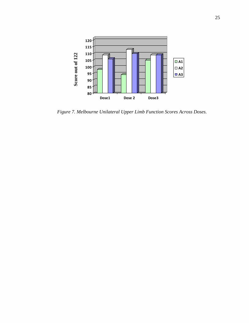
See Figure 7 for the comparison of Melbourne scores across doses.
Dose 1. The participant’s scores reflected an initial increase in functional use of
his impaired upper extremity that decreased slightly at the three month follow-up. The
baseline score was 98/122, increasing to 109/122 following the intervention, and
decreasing slightly to 106/122 at the three month follow up. In particular, the participant
showed increases in his ability to bring his hand to his head.
Dose 2. The participant’s scores demonstrated a substantial increase in functional
upper extremity use followed by a slight decrease. The baseline score was 94/122, which
increased substantially immediately following the intervention to 113/122. His scores
decreased slightly by the three month follow-up to 110/122. He improved in aspects of
reaching forward and sideways, grasp and release of objects, internal and external
rotation, grasp, manipulation, pronation/supination, hand to hand transfer, hand to mouth
and down, and reaching to opposite shoulder.
Dose 3. The participant's scores reflected a slight increase in functional use of the
upper extremity that was maintained at the follow-up. The baseline (A1) score was
105/122 and both the post test (A2) and follow up scores (A3) were 109/122. At A2 the
participant demonstrated less wrist flexion than at A1, greater fluency in manipulation,
and greater range and fluency in reaching a brush from forehead to back of neck. At A3
the participant maintained most improvements and demonstrated an increase in quality of
release of a crayon and pointing to squares. His fluency in reaching a brush from
forehead to neck decreased to baseline levels.
25
80
85
90
95
100
105
110
115
120
Dose1 Dose 2 Dose3
A1
A2
A3
Figure 7. Melbourne Unilateral Upper Limb Function Scores Across Doses.
S
core
ou
t of
122

26
Pediatric Evaluation of Disability Inventory
See Figure 8 for the comparison of PEDI- Self-Care scores across doses.
Dose 1. During Dose 1 only the self-care portion of the PEDI was administered.
The participant’s scores showed increases in self-care skills across the study phases. At
baseline he had a score of 47/73, which increased to 53/73 immediately following the
intervention, and continued to increase to 63/73 at the 3 month follow-up. Between
baseline and A2 the participant demonstrated the greatest increase in skills in the toileting
tasks category. Between A2 and follow-up he had large increases in managing his shoes,
socks, and increases in managing fasteners, pants, and washing his body and face.
Dose 2. During this dose all three domains of the PEDI were administered: self-
care, mobility, and social. The participant showed increases in his self-care skills
following the intervention, maintenance of his social skills, and the highest score possible
on the mobility domain. At baseline the participant obtained a score of 67/73 on self-care,
improving to a 70/73 immediately following the intervention and remaining at that level
at follow-up. The increase included the skills of snapping and unsnapping fasteners,
manipulating zippers, and putting on pants including fasteners. The participant scored
highly on the social domain from baseline, 60/65, and retained that score throughout the
study. The participant’s scores on the mobility domain remained at 59/59 from the
baseline phase throughout the study.
Dose 3. The participant showed slight improvements throughout the course of the
study on self-care and social function domains with no changes in the mobility domain
due to attaining the highest score upon baseline. At baseline the participant obtained a
raw score of 72/73 on the self-care domain, improving to a 73/73 at the 3 month follow

27
up with the additional skill of fastening his pants. At baseline the participant's social
function domain raw score was 62/65, increasing to 63/65 at the 3 month follow up with
the addition of being able to make a transaction in a store without assistance.

28
80
85
90
95
100
105
110
115
120
Dose1 Dose 2 Dose3
A1
A2
A3
Figure 8. Pediatric Evaluation of Disability Index Self-Care Scores Across Doses.
Sco
re o
ut
of 7
3

29
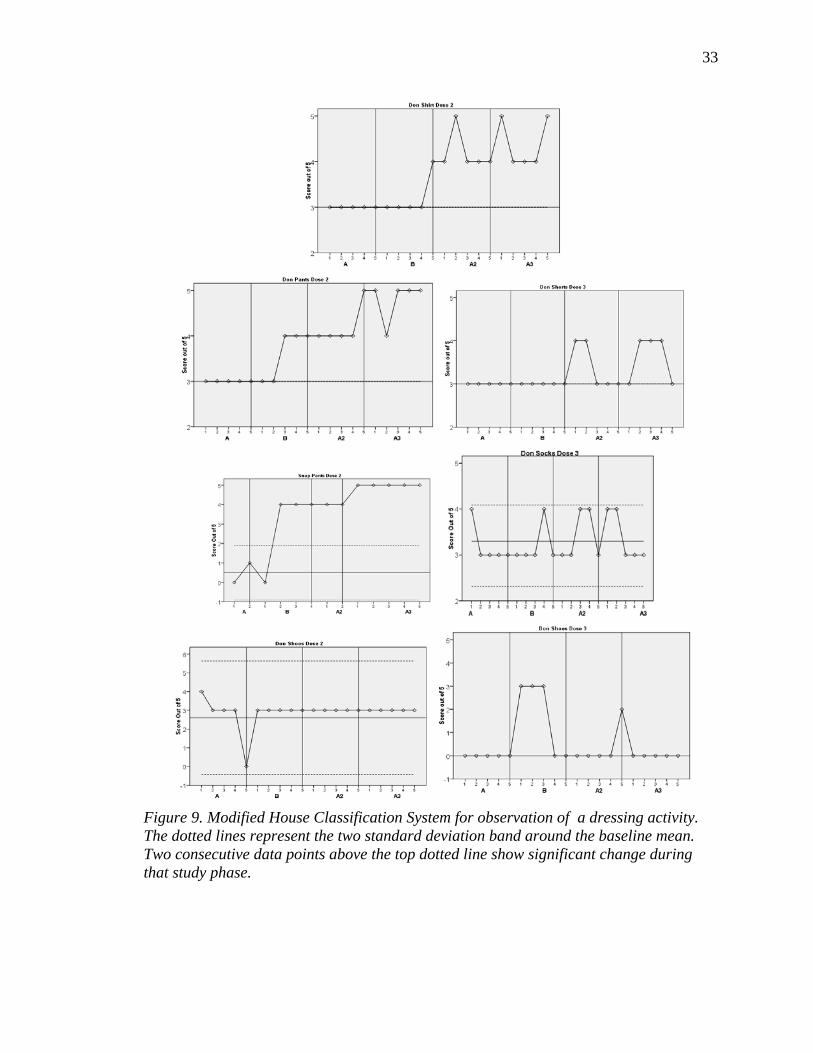
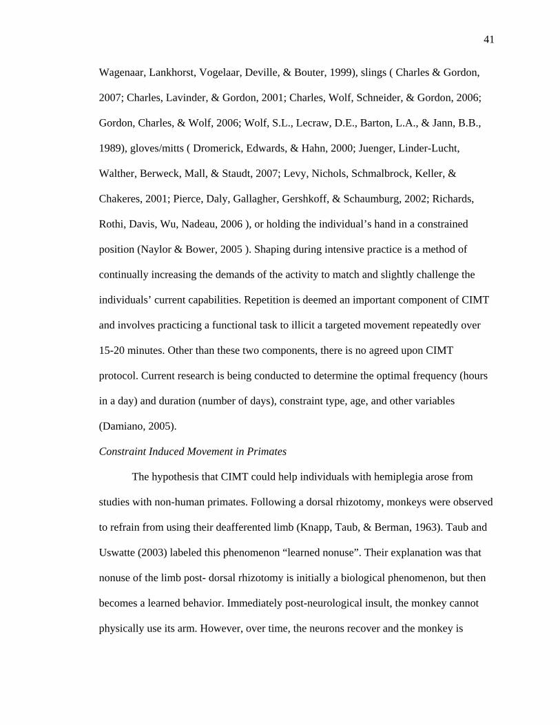
The Modified House Functional Classification System
See Figures 9 and 10 for the comparison of MHC scores across doses.
Dose 1. The participant was not video taped and analyzed using the MHC during
the first dose.
Dose 2. The participant was video taped and analyzed while performing a
dressing task and a play task. During the dressing task he showed improvements in
functional use of his affected extremity while donning shirt, donning his pants, snapping
his pants, and donning his shoes. The participant made significant gains in donning his
shirt immediately following the intervention and at follow up. At baseline the participant
received a 3/5 for donning his shirt, which increased slightly to 3.2/5 during the
intervention. Immediately following the intervention the participant’s ability improved to
4.2/5, which continued to increase slightly to a 4.4/5 at the three month follow-up. He
also made significant gains from baseline in all phases when donning his pants; he
improved from 3/5 at baseline to 3.6/5 during the intervention phase, 4.2/5 immediately
following the intervention, and 4.8/5 at the three month follow-up, ending with an almost
typical ability to perform this task. At baseline the participant was unable to snap his
pants with his affected hand receiving a 1/5, his ability improved to 4/5 during and
immediately following the intervention, increasing to an effortless ability, 5/5, at the
three month follow-up; all statistically significant gains. The participant made no
significant gains for donning his shoes. At baseline the participant demonstrated a score
of 2.6/5 when donning his shoes, which increased to a 3/5 during the intervention and
remained at that level through the three month follow-up.
30
During the play task he showed improvements in use of his affected extremity
while unpopping beads, attaching beads, and forming a circle with the beads. His ability
to unpop the beads increased slightly from a score of 3/5 at baseline to 3.2/5 during the
intervention. Immediately following the intervention his scores increased substantially to
4.4/5 and then decreased slightly to a 4/5 at the three month follow-up, both of these
phases were significant improvements from baseline. His ability to attach the beads
increased from scores of 2/5 at baseline, to 2.2/5 during the intervention, to 3/5
immediately following the intervention, to 3.4/5 at the three month follow-up; these
changes were significant at post-intervention and the follow-up. His ability to form the
string of beads into a circle significantly increased at all phases from 2.4/5 at baseline, to
3.6/5 during the intervention, to 4.8/5 immediately following the intervention and through
the 3 month follow-up. These scores show functional increases in fine motor skills from
baseline through the three month follow-up.
Dose 3. The participant was video-taped and analyzed while performing a
dressing task and a play task. During the dressing task he showed no improvements while
donning his shirt, modest improvements while donning his shorts, modest improvements
while donning his socks, and moderate improvements that weren’t maintained while
donning his shoes. The participant’s use of his affected extremity while donning his shirt
remained at a 3/5 throughout the course of the dose. While donning his shorts, the
participant’s baseline score was a 3/5, this score increased significantly following the
intervention to 3.4/5 and continued to improve slightly to 3.6/5 at the three month follow-
up. The participant’s score while donning his socks started at 3.2/5 during the baseline,
improving significantly to a 3.4/5 following the intervention and was maintained at this

31
level at the three month follow-up; none of these changes were statistically significant.
The participant donned slip on shoes with a score of 0/5 at baseline, which improved
significantly to 1.8/5 during the intervention, decreased to a non-significant level of 0.4/5
immediately following the intervention, and reverted back to a 0/5 at the three month
follow-up. In general, the participant’s scores were low because the slip-on nature of the
shoes did not require him to use both hands, so he often used his non-affected and
dominant upper extremity to slip them on. During the intervention the participant was
used to only being able to use his affected extremity and automatically used it
approximately 50% of the time, at a level of 3/5, while donning his shoes. Immediately
following the intervention he spontaneously used his non-affected extremity the majority
of the time, though when he did use his affected extremity it remained at a level of 3/5.
During all of the dressing tasks he was able to get dressed quickly primarily using his
non-affected extremity with his affected extremity as an assist when needed.
During the play task he showed improvements in use of his affected extremity
while unpopping beads, attaching beads, and forming a circle with the beads, which
required more bilateral use and precise use than the dressing activity. While unpopping
the beads during baseline the participant scored a 3.2/5, which increased to a 3.8/5 during
the intervention and then remained at 3.6/5 following the intervention through follow-up;
none of the increases were statistically significant. While attaching the beads the
participant scored a 2.2/5 at baseline, improving to a 2.4/5 during the intervention, to a
2.6/5 following the intervention, and continued to improve to a 3/5 at the three month
follow-up; though at statistically non-significant levels. His greatest improvements
occurred when he was creating a circle with the beads. At baseline his score was 2.6/5,

32
which increased to 3.2/5 during the intervention, to 3.8/5 following the intervention, and
ended at a 4/5 at the three month follow-up; the changes from baseline to post-
intervention and follow-up were statistically significant. His scores on the Modified
House Classification System showed an increase in active use and quality of use of his
affected upper extremities, particularly when force production was needed bilaterally
while forming the circle. Although he demonstrated the ability to use more active control
while unpopping and attaching the beads, he often used his leg or trunk to help stabilize
the beads.
33
Figure 9. Modified House Classification System for observation of a dressing activity. The dotted lines represent the two standard deviation band around the baseline mean. Two consecutive data points above the top dotted line show significant change during that study phase.

34
Figure 10. Modified House Classification System for observation of a play activity. The dotted lines represent the two standard deviation band around the baseline mean. Two consecutive data points above the top dotted line show significant change during that study phase.

35
Chapter 4
Discussion
The previous studies outlined in the literature review have shown support for the
efficacy of CIMT protocols with varying constraints, durations, and intensities. However,
there has been a lack of evidence of the clinical merit of multiple CIMT doses. This study
investigated the efficacy of three doses of CIMT, across 5 years, with a child with
hemiplegic spastic cerebral palsy. Considering changes at the WHO ICF Body Structure
and Function Level, the participant had significant changes in grip strength from baseline
at follow-up during all three doses and significant increases 3 months post-intervention
during dose 3. Although not as robust as grip changes, the participant also showed
significant increases in pinch strength at follow-up for doses 1 and 2, with additional
significant gains post-intervention in dose 2. The participant showed a significant
increase in manual dexterity from baseline during all phases of dose 2. Gross manual
dexterity improved significantly from baseline to post-intervention and follow-up in
doses 2 and 3. Upper extremity function increased in each dose on the Melbourne, with
the greatest improvement occurring during dose 2, which also had the lowest initial
ability.
Changes at the WHO ICF Activities and Participation Level were noted in self-
care skills as measured by the PEDI which increased in dose 1 and 2, as a result of the
intervention, but were constrained by a ceiling effect in dose 3. Neither mobility nor
social skills were significantly affected by any dose. The mother’s satisfaction of her
son’s performance on goals she identified on the COPM increased with each intervention.
The second dose demonstrated increases after the intervention with less retention at three

36
months and the first and third doses increasing throughout the follow-up. Video analyses
of activities performed by the child showed slight improvements on dressing and play
performance following doses 2 and 3.
Overall, all three doses produced improvements in ICF Body Structural/
Functional and Activity/Participation levels. The third dose showed greater improvement
in between A2 and A3 than the previous doses, which the participant’s mother attributed
to his participation in community football. The community football practices included
intensive bimanual use, indicating that this child could potentially have benefitted from
bimanual training such as the hand-arm bimanual intensive therapy (HABIT) program
following the CIMT. For example, the Cincinnati Children's Medical Center uses a model
where the child receives 8 or 4 weeks of CIMT followed by 4 weeks of weekly bimanual
intervention (Garcia, Coker, Echols, Allgier, Chamudot, & Little-Hays, 2008). Like
CIMT, HABIT uses intensive training, motor control and plasticity principles, and
meaningful and functional activities to improve arm use (Gordon, Schneider, Chinnan, &
Charles, 2007). Unlike CIMT, it does not use a constraint and is focused on improving
bimanual coordination.
In the literature, there are currently two studies that have investigated multiple
doses of CIMT with children with Cerebral Palsy. The first study, conducted by DeLuca
et al. (2003), was a case report of a 15 month old girl with no functional use of her right
upper extremity that participated in two doses of CIMT 5 months apart. The second
study, conducted by Charles and Gordon (2007), used an ABABA design to investigate
the effects of small group CIMT intervention with 8 children with CP across two doses,
12 months apart. All three studies used different measures, preventing direct comparisons

37
of scores. The Deluca et al. study showed improvements in fine motor scores following
the first dose (these assessments were not conducted following the second dose), and
improvements in quantity and quality of upper extremity use following both doses. The
greatest gains in upper extremity use were seen during the first dose, when the participant
had the lowest baseline scores. These results are similar to the finding in the current study
that the participant made more gains during the second dose than the third, when his
baseline scores were higher. Scores for speed and dexterity, quality of movement,
showed a similar trend in the Charles and Gordon study, with both doses showing
improvements, but a greater improvement seen in the first dose. These studies support
that CIMT is effective with up to three doses. They also seem to indicate that the
children’s improvements were affected by the level of their baseline scores.
This study had the following limitations. There was only one participant and there
were not enough data points to run statistics for every measure, instead descriptive
statistics were used for those measures. Although the participant demonstrated
improvements in each of the areas assessed, there was no way to determine if the changes
were statistically significant. Each dose had slight differences in the measures, duration,
and locations used, which may account for some of the differences in amount of change
across doses. In addition, the participant experienced ceiling effects on the PEDI, which
was designed to be used with children younger than him. Measures designed to be used
with children over 8 years of age and thus more sensitive to changes may have shown
greater progress. The student-researchers conducting the intervention also collected the
data, allowing for a potential researcher bias.

38
Future studies should investigate the optimal time between doses, since currently
each study has used a different time frame: 5 months (Deluca et al., 2003), 12 months
(Charles and Gordon, 2007), and 24-36 months in this study. This study also points to the
importance of investigating the level of impairment best served, so that an optimal time
in the child’s development to use CIMT can be determined. There continues to be a need
to determine the optimal length of doses and intensity of doses as well. Finally, the use of
CIMT alone versus CIMT followed by bimanual training should be investigated across
impairment levels.
Overall, CIMT has been shown to be an effective intervention for improving
functional use in children with hemiplegic CP. This study supports the previous findings
and adds support for up to three doses over a 5 year period. Improvements in grip and
pinch strength, dexterity, and upper extremity function were demonstrated following each
of the three doses. CIMT may be more or less effective depending on the child’s current
functioning. In addition, CIMT shouldn’t be viewed as the only treatment, but a
complementary intervention to others such as bimanual training. Further work with larger
sample sizes and direct comparisons of duration, intensity, and use of CIMT with other
protocols will increase the benefit to children with hemiplegic CP.

39
APPENDIX
EXTENDED REVIEW OF LITERATURE
Cerebral palsy (CP) is a neurodevelopmental clinical diagnosis in the developing
child based on observations of decreased motor control; no test alone can define its
presence (Bax, Goldstein, Rosenbaum, Leviton, & Paneth, 2005; Paneth, Hong, &
Korzeniewski, 2006). Bax et al. (2005) stated that “Cerebral palsy (CP) describes a group
of disorders of the development of movement and posture, causing activity limitation,
that are attributed to non-progressive disturbances that occurred in the developing fetal or
infant brain. The motor disorders of individuals with cerebral palsy are often
accompanied by disturbances of sensation, cognition, communication, perception, and/or
behavior, and/or by a seizure disorder” (p. 572). Many subtypes of CP have been
established to help categorize the disorder’s differing manifestations. These include
spastic, dyskinetic, ataxic, and mixed (NINDS, 2006).
Spastic cerebral palsy is the most common form, comprising up to 78% of the
population of those diagnosed (Yeargin-Allsopp, Braun, Doernberg, Benedict, Kirby, &
Durkin, 2008). Tight or stiff muscles and hyperreflexia generally characterize spastic CP
(NINDS, 2006). This subtype is further divided into individuals with diplegia,
hemiplegia, or tetra/quadriplegia depending on which of their extremities that are
affected. According to Yeargin-Allsopp et al. (2008), approximately 25-34% of

40
individuals with spastic cerebral palsy have hemiplegia with impairments primarily on
one side of their body.
Regardless of the type of cerebral palsy present, therapy can often help to improve
the individual’s capabilities (NINDS, 2006). Individuals with CP often receive physical
therapy (PT), occupational therapy (OT), and/or speech therapy. However, according to a
review by Antitila, Suoranta, Malmivaara, Mäkelä, and Autti-Rämö (2008) there is
limited evidence supporting comprehensive PT or OT services for individuals with CP.
These authors state that “well-conducted studies on current treatment options as well as
new treatment approaches using valid outcomes are obviously needed” (p. 490). In 2001,
Charles, Lavinder, and Gordon reported that one intervention approach, constraint-
induced movement therapy (CIMT), could potentially increase arm function in children
with hemiplegic CP.
Constraint Induced Movement Therapy
Constraint-induced movement therapy has two main components: 1) constraint of
the non-affected extremity and 2) intensive training using shaping and repetition
(Gordon, Charles, & Wolf, 2005). Constraint of the non-affected extremity has been
achieved through use of bivalve casts (DeLuca, Echols, Ramey, & Taub, 2003; Martin,
Burtner, Poole, & Phillips, 2008; Stearns, Burtner, Keenan, Qualls, & Phillips, 2009;
Sutcliffe, Gaetz, Logan, Cheyne, & Fehlings, 2007; Taub, Ramey, DeLuca, Echols;
2004), splints (Dickerson & Brown, 2007; Hamzei, Liepert, Weiller, & Rijntjes, 2006;
Kunkel, Kopp, Müller, Villringer, Villringer, Taub, & Flor, 1999; Liepert, Uhde, Graf,
Leidner, & Weiller, 2001; Miltner, Bauder, Sommer, Dettmers, & Taub, 1999; Taub,
Miller, Novack, Cook, Fleming, Nepomuceno, Connell, & Crago, 1993; van der Lee,
41
Wagenaar, Lankhorst, Vogelaar, Deville, & Bouter, 1999), slings ( Charles & Gordon,
2007; Charles, Lavinder, & Gordon, 2001; Charles, Wolf, Schneider, & Gordon, 2006;
Gordon, Charles, & Wolf, 2006; Wolf, S.L., Lecraw, D.E., Barton, L.A., & Jann, B.B.,
1989), gloves/mitts ( Dromerick, Edwards, & Hahn, 2000; Juenger, Linder-Lucht,
Walther, Berweck, Mall, & Staudt, 2007; Levy, Nichols, Schmalbrock, Keller, &
Chakeres, 2001; Pierce, Daly, Gallagher, Gershkoff, & Schaumburg, 2002; Richards,
Rothi, Davis, Wu, Nadeau, 2006 ), or holding the individual’s hand in a constrained
position (Naylor & Bower, 2005 ). Shaping during intensive practice is a method of
continually increasing the demands of the activity to match and slightly challenge the
individuals’ current capabilities. Repetition is deemed an important component of CIMT
and involves practicing a functional task to illicit a targeted movement repeatedly over
15-20 minutes. Other than these two components, there is no agreed upon CIMT
protocol. Current research is being conducted to determine the optimal frequency (hours
in a day) and duration (number of days), constraint type, age, and other variables
(Damiano, 2005).
Constraint Induced Movement in Primates
The hypothesis that CIMT could help individuals with hemiplegia arose from
studies with non-human primates. Following a dorsal rhizotomy, monkeys were observed
to refrain from using their deafferented limb (Knapp, Taub, & Berman, 1963). Taub and
Uswatte (2003) labeled this phenomenon “learned nonuse”. Their explanation was that
nonuse of the limb post- dorsal rhizotomy is initially a biological phenomenon, but then
becomes a learned behavior. Immediately post-neurological insult, the monkey cannot
physically use its arm. However, over time, the neurons recover and the monkey is

42
physically capable of moving the arm. Because of failed and sometimes painful attempts
to use the arm during the initial period, the monkey learns not to use it. In addition, it is
presumed that with the insult and disuse, the cortical representation of that extremity
decreases in size. The combination of the punishment incurred when using the affected
extremity, the reinforcement of the use of the intact extremity, and the decrease in
cortical representation interact to produce the permanent learned nonuse.
To intervene in the nonuse of the involved extremity, the monkeys were
“conditioned”, which involved restraining their non-surgical limbs and receiving an
electrical shock that could be prevented by pressing a lever with their deafferented
forelimb. After conditioning, several of the monkeys would use their deafferented limb to
attempt to eat fruit. Thus, these researchers demonstrated that if constraint is used shortly
after the biological use-restriction ends, then the monkey is forced to use the extremity
for functional tasks. This creates increased motivation and reinforcement for the use of
the affected extremity resulting in the ability to functionally use the affected arm after
removing the constraint. Taub et al. (1993) felt that learned nonuse applied to “ … any
neurological injury that results in central nervous system shock and an initial inability to
use an extremity” (p. 347), including stroke. However, there are more factors involved in
recovery of function with humans, such as psychosocial support, co-morbidities, and the
areas damaged by the stroke.
Constraint Induced Movement in Adults Post Stroke
Many studies have investigated the efficacy of CIMT with individuals post-stroke
since it was first hypothesized that CIMT could prove beneficial. Studies have not only
looked at the efficacy of CIMT in general, but whether CIMT was more effective than

43
forced use or neurodevelopmental treatment, what durations and what type of constraints
are most effective, and more recently how CIMT affects the brain.
Wolf et al. (1989) conducted the initial study involving humans. They decided
what they called “forced use”, i.e. use of a restraint without therapy, would be worth
investigating based on results from the monkey studies. Rather than using a traditional
control group, the researchers used an extensive baseline period with subjects serving as
their own controls, followed by 14 days of intervention, and four post-intervention
assessments. Sixteen individuals 1 year or more post-stroke wore a sling on their affected
extremities during waking hours. Improvements on the functional tasks used for
assessment were visible by the second week and the individuals continued to improve
through the 1-year assessment period. The authors believed that their results showed that
aversive stimuli such as electric shock was not necessary to motivate individuals to use
their affected extremity and that forced use could help them overcome their learned
nonuse.
Taub et al. (1993) wanted to improve upon the work of Wolf et al. (1989) by
conducting a single-blind randomized control trial (RCT) that would also investigate if
the gains extended to the individuals’ lives. Nine participants were randomly assigned to
a control group and experimental group. The control group were told, in four different
10-minute sessions, that they had more motor ability in their affected extremity than they
were showing; were instructed to try to use their affected arm in as many new activities
as possible; and were given instructions during two sessions labeled “physical therapy”
for passive range of motion exercises to perform 15 minutes per day. The experimental
group received therapy for 6 hours each weekday and wore a resting hand splint and sling

44
for 14 days. The participants were assessed using the Emory Motor Function Test
(EMFT), Arm Motor Activity Test (AMAT), and Motor Activity Log (MAL). The
experimental group’s scores increased significantly from the pre- to the post-tests and
were significantly higher than the control group’s scores, showing that the intervention
was causing the improvement, not just the attention. The participants’ improvements on
the timed sections of the EMFT and the AMAT were much greater than the
improvements of the participants in the Wolf et al (1989) study, which the authors
interpreted as showing that training the limb in conjunction with constraint use greatly
increased the efficacy of the intervention. This combination of intensive therapy and
constraint is now known as CIMT.
Miltner et al. (1999) attempted to replicate these results in a different setting, with
different researchers, and in a different country that provides more conventional therapy
following a stroke. Fifteen adults ranging from 6 months to 17 years post-stroke
participated in the study, wearing a resting hand splint and sling for approximately 90%
of their waking hours for 12 days. They also received intensive “training” of their
impaired arm for 7 hours per day for 8 weekdays. Although there was no control group in
this study, the researchers used an extensive research design that included an initial
assessment, two separate baseline assessments, two separate post-intervention
assessments (post 1 at 1 week and post 4 at 4 weeks), and a follow up assessment at 6
months. They found significant improvements between the initial assessment and post 4
assessment on the MAL for both amount and quality of arm movement and on the Wolf
Motor Function Test (WMFT) for functional ability and quality, which did not diminish
by the 6 month follow up. The study had a large effect size of 1.59, and found that neither

45
chronicity, amount of prior therapy, nor did initial level of motor ability have an impact
on the outcomes.
Kunkel, et al. (1999) also sought to replicate previous studies, but their
participants had received less conventional therapy immediately post-stroke than those
used by Miltner et al (1999). This study also excluded individuals who were less than 1-
year post-stroke. Kunkel et al.’s (1999) intervention consisted of the patient wearing a
sling for 90% of their waking hours for 14 days. The participants received therapy for 6
hours on each weekday and were asked to practice certain tasks for 1 hour per day on the
weekends. Overall, from pretest to posttest the participants scores: doubled for use and
quality increased by 124% on the Actual Amount of Use Test (AAUT), increased 166%
for quality and 136% for amount of use on the MAL, and increased their movement
speed by 20% and movement quality improved by 24% on the WMFT. Scores decreased
slightly on the 6 month follow up, but remained higher than at pretest.
These early studies showed that not only does CIMT result in improvements for
individuals post-stroke, but that these improvements do affect their daily lives. More
recent studies have examined different variables such as intensity and chronicity.
Richards et al. (2006) found that individuals who received therapy for 1 hour per day
with instructions to complete 5 more hours at home scored similarly as individuals with
received a full 6 hours of therapy a day on the WFMT after two weeks of CIMT.
However, these results need to be replicated, because the two different groups were from
two previous studies that were examining the effects of pharmaceuticals, not the CIMT
protocols. A study by Dromerick et al. (2000) also showed that less intense CIMT could
be beneficial for individuals in the acute stage of recovery by replacing typical inpatient

46
OT (2 hours per day/ 5 days per week). Overall studies have provided evidence that
CIMT is applicable to all stages of stroke recovery: acute (Dromerick et al., 2000), sub
acute (Levy et al., 2001; Liepert et al., 2001), and chronic (e.g. Hemzei et al., 2006;
Kunkel et al., 1999; Miltner et al., 1999).
CIMT versus Conventional Therapy for Adults.
While the aforementioned studies showed that CIMT was effective, other
researchers questioned if CIMT was more or less effective than more traditional or
comprehensive therapies. van der Lee and colleagues (1999) conducted a single-blinded
RCT with 58 participants in an effort to determine if CIMT was more effective than
intensive bimanual therapy based on NDT for chronic stroke patients The participants
were treated in groups of four, for 6 hours a day 5 days a week for 2 weeks. The CIMT
group wore a resting splint during the entire 2 weeks and a sling during therapy hours.
Both groups were involved in group activities, exercises, housekeeping activities, crafts,
and games. The bimanual group focused on supporting the affected arm with the non-
affected arm, symmetry of posture, and inhibition of inappropriate movements. After
treatment, the CIMT group had higher scores on all measures, though most were not at a
statistically significant level. The authors hypothesized that the lack of statistical
significance could be due to the use of measures that were developed for the sub acute
population and not a chronic population (e.g. the Fugl Meyer Assessment and the
Rehabilitation Activities Profile).
Another study compared CIMT to conventional OT during the acute phase post-
stroke (Dromerick et al., 2000). Individuals in the CIMT group (n=11) received CIMT
during the normally scheduled OT, 2 hours a day 5 days a week for 2 weeks. The

47
conventional OT (n= 9) group received therapy focusing on compensatory techniques for
activities of daily living, upper extremity strength, range of motion, and traditional
positioning. Mean scores on the Action Research Arm Test (ARA) were significantly
higher for the CIMT group, and scores on the Bathel Index and Functional Independence
Measure (FIM) were also higher for the CIMT group, but only dressing reached statistical
significance.
Finally, Liepert et al. (2001) compared physical therapy with the use of a
constraint to traditional physical therapy. Nine individuals participated in the study,
which included a pretest, one week of typical physical therapy, a post-test, one week of
physical therapy paired with constraint, and a second post-test. Transcranial magnetic
stimulation (TMS) showed no change in the motor cortex after the week of tradition PT,
but by the end of the week of therapy with constraint, there was a 50% increase in the
area of the motor cortex. Grip strength and Nine Hole Peg Tests both showed
improvement after the week of therapy paired with constraint, but similar results were not
present after the traditional PT. It is important to note that the study did not specify the
intensity of the therapies, or exactly what was done, but the results seem to indicate that
PT with use of a constraint is more effective for individuals 4-8 weeks post-stroke.
Brain Imaging Studies in Adults Receiving CIMT
The study by Liepert and his colleagues (2001) was one of the first studies to
determine if CIMT had any cortical reorganization effects within the brain after a stroke.
In an effort to verify previous findings regarding cortical reorganization through
functional magnetic resonance imaging (fMRI), Levy and colleagues (2001) conducted a
pilot study with two patients that had plateaued in their typical therapies and did not, at

48
that time, use their affected arms. They received CIMT for 6 hours per day, 5 days per
week, for 2 weeks and wore a mitt that prevented use of their non-affected fingers during
their waking hours. Following therapy, the fMRI studies of these participants showed
activity bordering the region of their lesions, where prior to CIMT therapy, cortical
activity was present only in the contralateral hemisphere. Both participants also
demonstrated increased scores on the MFT and the MAL after CIMT.
A more recent study conducted by Hamzei et al. (2006) included six participants
at least 1.5 years post-stroke with no noted progress within the past 3 months. The
individuals wore a splint during their waking hours and participated in therapy for 6
hours a day, but the authors did not report the duration of the intervention. Participants
improved on both the MAL and the Wolf Motor Function Test post intervention. fMRI
results were mixed, with some participants exhibiting increased activation in the
sensorimotor cortex and others exhibiting decreased activation after the intervention. The
authors hypothesized that the decreased activation could be due to improved performance
and/or better intersynaptic communication, in support of a use-dependent functional
reorganization. The individuals with an increased activation in the sensorimotor cortex
may have expanded the area of intersynaptic communication to compensate for the
decreased efficiency in intersynaptic communication caused by the stroke. The limited
number of participants may have been responsible for the lack of a difference in
functional gain between the two groups (i.e. the participants that had increased activation
and those with decreased activation).
In summary, not only is there evidence that CIMT produces improvement in
adults post CVA for neuromuscular factors such as grip strength and dexterity, but also

49
improves their ability to perform activities of daily living such as basic self-care. Studies
also show that CIMT produces changes within the adult brain post CVA that traditional
therapies may not. Research points to CIMT as being applicable to individuals in all
stages post-stroke and can be implemented in different time frames and with different
constraints. But what about different populations?
Constraint Induced Movement in Children with Cerebral Palsy
Because hemiplegic cerebral palsy is also due to a neurological insult and results
in a decreased ability to use one upper extremity, a similar outcome to stroke, researchers
began to wonder if CIMT was applicable to some individuals with CP (Charles et al.,
2001) One of the earliest studies on CIMT’s effectiveness within the cerebral palsy
population was conducted by Charles et al. (2001). The authors focused on function,
strength, sensation, and fingertip force coordination changes in three boys from ages 8-
13. Two of the boys regularly used their affected arms to assist their unaffected arms,
whereas the other boy showed no use of affected arm. Two pretests, a post-test, 2 weeks
post-test, 4 weeks post-test, and 6 months post test were conducted using a) a custom
made grip instrument to measure fingertip forces, b) the Jebsen-Taylor Test of Hand
Function (Jebsen-Taylor), c) two point discrimination tactile testing, d) Semmes-
Weinstein Monofilament Test, e) precision pinch, and f) lateral pinch. The children wore
a cotton sling for 6 hours per day for 14 days and participated in therapy focusing on
activities of daily living and play that was instructor-led for 2 hours and child-led for 4
hours per day. Two of the participants had improved scores on the Jebsen-Taylor by the
post-test and continued to improve through the 6 month follow up. All three participants
improved on the two point discrimination test and no participants improved on pinch,

50
grip, or fingertip force. The families reported more functional use of the affected arms
during the therapy, but after the sling was removed the children discontinued spontaneous
use of their affected arms.
Although Charles et al.’s (2001) study didn’t support increased long-term
functional use of the child’s extremity, another case study conducted by Pierce et al.
(2002) showed that increased use of the affected arm at home could follow therapy even
with an altered schedule. A 12 year old that was independent in all functional activities,
but had decreased strength and problems with grasp and release participated in CIMT that
replaced his normal outpatient PT (2x/1 hour) and OT (2x/1 hour) each week for 3 weeks.
He wore a mitt during the CIMT, which focused on exercise, play, and fine motor in the
PT sessions, and neuromuscular reeducation and functional activities during the OT
sessions. He was also asked to wear the mitt whenever possible at home and was given a
home exercise program to reinforce therapeutic goals. Assessments were conducted prior
to the therapy, immediately after the 3 week intervention, and 8 months later. Although
these assessments were not standardized for children, the participant improved by 37%
on the WMFT by the post-assessment and continued to improve through the 8 months.
His grip strength increased by approximately 4 pounds per square inch. He improved in 8
of the 16 motor skills, and 5 of the 20 process skills as measured in the Assessment of
Motor Process Skills (AMPS). Finally, at follow up the participant reported using his
affected arm more often than he had prior to the study.
Pierce at al.’s (2002) study provided beginning evidence that CIMT was
beneficial, but as a case study without a control group better evidence was needed to
confirm CIMT’s effectiveness in children with CP. In 2004, Taub et al. conducted a

51
randomized control trial (RCT) to determine the effectiveness. The intervention group
was composed of 9 children (2 females), with a mean age of 39 months, and 2 exhibiting
flaccidity in their affected extremity and 7 exhibiting spasticity. The control group also
had 9 participants (3 females), but had a mean age of 43 months, all with spasticity. The
intervention protocol involved placing a bivalve fiberglass cast on the child’s less
affected arm from the upper arm to the fingers for 21 days, with removal weekly for skin
checks. Intensive therapy involved shaping and repetitive training of functional tasks for
6 hours a day throughout the 21 day period. Immediate and frequent verbal feedback and
rewards were used. The control group continued with their normal therapies (an average
of 2.2 hours a week in occupational and physical therapies). Results for the intervention
group indicated an increase in new motor patterns and functional activities, an increase in
the amount and quality of use of the affected extremity, and improvements in using the
affected arm to perform activities when compared to the control group. Within the
experimental group children with higher pre-test scores on the Developmental Activities
Screening Inventory (DASI) had greater improvements in arm use, although those with
lower DASI pre-test scores did improve. These results remained at the 3 week and 6
month follow up assessments. The Pediatric Motor Activity Log (PMAL) results showed
slightly decreased scores for the children (but not significantly), however it is important
to note that this measure was created for this study and modeled after the MAL. The
parents also reported an increase in social and communication skills. These results,
though still limited by a small sample size, indicated that CIMT was more effective than
typical OT and/or PT. However, the CIMT was much more intensive than the typical
OT/PT that these children received.

52
Naylor and Bower (2005) conducted a pilot study to determine if a less invasive
constraint and a less intensive dose of CIMT would still improve arm function in children
with hemiplegic CP. The study design was a single case ABA design where the children
were their own controls and each phase lasted 4 weeks. During the “A” periods, the 9
participants (ages 18 months- 5 years) had no therapy. During the “B” period they
received CIMT for 1 hour per day for 2 days per week that focused on improving fine
motor skills through play. An adult held the child’s unaffected hand during the activities
and there was no constraint between activities. The parents were also given a detailed
home program provided 1 hour per day on days without CIMT therapy. To measure fine
motor function the Quality of Upper Extremity Skills Test (QUEST) was conducted at
the beginning of the study, between the first “A” phase and “B” phase, after the “B”
phase, and again follow the final “A” phase, for a total of 4 measures over the 12 week
study. Significant improvements in scores on the QUEST were found following the
intervention (“B”) phase as compared with the control (“A”) phases, even when factoring
in the small changes that occurred during the control periods. Although this study does
not provide as of strong evidence as an RCT, it shows that CIMT can be effective when
used on a reduced schedule (1 hour, twice a week, plus a home program).
Charles et al. (2006) investigated how intensity and the child’s compliance affect
results when using what they called a “child-friendly” CIMT protocol. This single-blind,
randomized control trial consisted of a control group (n =11) and an intervention group (n
= 11). Both groups were allowed to continue their regular therapies during the study. The
intervention group participated in the child-friendly constraint induced movement therapy
(CIMT). The therapy consisted of a 6 hour session per day for 10 out of 12 consecutive
53
days. The children would participate in groups of 2-4 during the session and only wear a
sling to constrain their less affected arm during the therapy. This differs from other CIMT
protocols that involve a cast and require the child to wear it 24 hours a day (e.g. Taub,
Ramey, DeLuca, & Echols, 2004; Sung et al., 2005; Sutcliffe, Gaetz, Logan, Cheyne, &
Fehlings, 2007; DeLuca, Echols, Ramey, & Taub, 2003). “Shaping and repetitive task
practice” (p. 367) were emphasized during the 6 hours of therapy, as is typical for CIMT.
The families were also asked to do one hour of activities at home using the affected hand
each day, without the sling. The investigators conducted a pretest, and three post tests (1
week, 1 month, and 6 months post treatment) using the Jebsen-Taylor, Bruinink-
Oseretsky Test of Motor Proficiency (Bruinink-Oseretsky) - subtest 8, Caregiver
Functional Use Survey (CFUS), two point discrimination, hand grip, the modified
Ashworth Scale, and also recorded the intensity of the treatment (time spent on task) and
any behavioral issues. The results showed improvement in both groups on the Jebsen-
Taylor, but the CIMT group improved more and maintained the improvement after 6
months, whereas the control group’s scores fluctuated. The CIMT group also improved
more on the Bruinink-Oseretsky, and despite a decrease between the 1 and 6 month
posttests the final score was still higher than their initial score. Caregivers reported more
use of the affected extremity after the CIMT treatment on the CFUS. Finally, severity of
hand function and the child’s compliance-related behavior influenced the child’s score on
the Jebsen-Taylor more than the intensity of the treatment received.
Dickerson and Brown (2007) found that even a child with minimal active arm
movement could improve after CIMT. Consistent with Charles and colleagues (2006)
finding, the improvements were not as robust as children in other studies with children

54
having less impairments. The child was 24 months old and had no observed active range
of motion (but full passive range of motion) in his shoulder, elbow, or wrist, which would
have excluded him from most other CIMT studies. An ABA design was conducted with
one follow up at 2 weeks post. During the “A” periods he was videotaped for 15 minutes
each day for consecutive 5 days while performing reach, grasp, release, sustained grasp,
push, pull, and finger feeding with his affected arm. During the “B”, or intervention,
period he wore a custom-molded splint with sock cover to prevent use of his fingers,
during most waking hours. He received 6 hours of CIMT, conducted by an OT that
focused on fine motor, gross motor, and ADLs, for 21 consecutive days. Two weeks
following the second “A” period the participant received a “follow up” period that
consisted of 5, 1 hour sessions of CIMT over two weeks. Frequency, rate, and duration of
arm use by the participant was measured to document changes. He was also scored
between 0-3 for quality of specific activities: a) spontaneously hitting an inflated balloon,
b) successful attempts to grasp a deflated balloon, c) successful attempts to release the
deflated balloon without dropping it, d) successful attempts to pull a toy car to activate it,
e) successful attempts to push a button on a toy to activate a noise, f) sustained grasp on a
marker, g) grasp small, round pieces of cereal to feed himself, and h) using two hands to
catch a ball. During the pretest, he scored a 0 on every activity. Increases were seen
following in all of the activities, except for feeding himself, after the intervention. These
scores decreased during the second “A” phase, and increased again with the follow up
intervention. Computations of the C statistic showed that the increases were significant
following the intervention, but were no longer significant after 6 months. Observed arm
use that remained included use of his arm for simple play activities, and for weight

55
shifting during walking following the intervention. Although the results were small and
did not last through the 6 month assessment, this study showed that with intensive CIMT
it may be possible to increase arm function in children with severe impairments.
Many of these earlier studies focused on children ages 5 and younger, possibly
due to an idea that there was a critical period of development during which CIMT needed
to be administered. In 2006, Gordon and colleagues conducted a study to determine if
there was an interaction between CIMT effectiveness and age. The participants were
divided into a younger group (ages 4-8, n = 12) and an older group ( ages 9-13, n = 8).
The participants were assessed with the Jebsen-Taylor Test of Hand Function, Bruninks-
Oseretsky Test of Motor Proficiency, Two Point Discrimination, grip strength, the
Modified Ashworth Scale, Caregiver Functional Use Survey (CFUS) at four points in
time: pretest, 1 week posttest, 1 month post, and 6 months post. In addition, their
behaviors during the session were recorded through observation. The protocol involved
wearing a sling during the 6 hour daily sessions for 10 out of 12 consecutive days with
repetitive motor task training and shaping used during the therapy sessions. No difference
was found in the rate of improvement between the older and younger participants on the
Jebsen-Taylor or the Bruninks-Oseretsky, with both groups improving during the
intervention period and maintaining most of the improvements through the 6 month
period. The caregivers reported improvement in the frequency and quality of movements
for both groups through the CFUS. Neither group had gains in grip strength, Two Point
Discrimination, or on the Modified Ashworth Scale. Finally, for the younger group,
severity of impairment and negative behaviors during the sessions were negatively

56
correlated with the outcomes, such that increased severity and behaviors translated to less
improvement. This correlation was not found with the older children.
Many of these studies tend to focus on one or two levels of the World Health
Organizations: International Classification of Function (ICF) Disability and Health (see
World Health Organization, 2001 for a full description). Martin et al (2008) conducted an
ABA single subject study that measured all 4 levels. During the intervention phase, the
35 month old boy participated in CIMT for 4 hours per day (2 hours for self-care and 2
hours for play), 6 days per week, for 2 week. He spent approximately 7 hours a day in a
bivalve cast. After the intervention his Participation as measure by scores on the
Canadian Occupational Performance Measure (COPM) for performance increased from a
4 to a 6.5 and from a 5 to a 6.5 for satisfaction. On the Pediatric Evaluation of Disability
Index (PEDI) he improved from a 27 to a 53; and his scores on the Melbourne
Assessment of Unilateral Upper Limb Function (the Melbourne) increased from 98 to
108 supporting changes at the Activity ICF Level. His also increased from 0.3lbs to
0.8lbs grip strength, but made no improvements with pinch strength. All of his scores
continued to increase, except for his scores on the Melbourne, which decreased slightly
by 2 points, and pinch strength, which remained at 0. These results showed that CIMT
can improve function at every level of the ICF.
Since other studies had shown that after CIMT children demonstrated a higher
quality of movement with their affected extremities, Stearns and colleagues (2009)
wanted to know if 2 weeks of CIMT would also result in a change in muscle activation
patterns. Six children, ages 5-8 yrs, without sensory deficits participated in CIMT 4 hours
a day, 5 days a week, for two weeks. Electromyographic (EMG) recordings were

57
obtained during grip, pinch, the Nine Hole Peg Test, and Box and Blocks for the
pectoralis major, long head of the triceps, upper trapezius, anterior deltoid, middle
deltoid, long head of the biceps, flexor carpi ulnaris, and extensor carpi radialis muscles.
A two way repeated ANOVA (joint and phase) was conducted for each activity. The
children’s grip strengths increased at all phases, but there were no significant changes on
the EMG. Pinch strength also increased from the initial assessment to the follow up, with
an increase in the percentage of maximum voluntary contraction (% MVC) approaching
significance for the children’s shoulders and elbows. Children also achieved decreased
times on the NHPT, with decreased %MVC scores for their shoulders and slightly
decreased scores for their shoulders and wrists. The authors hypothesized that this
decrease was due to a more efficient use of their arms. This study showed that some
changes were being made at functional and structural levels, not just participatory levels
of the ICF.
Brain Imagining Studies in Children receiving CIMT
Previous studies showed that CIMT was effective at improving functional use of
an impaired extremity for children with hemiplegic cerebral palsy. But researchers
wanted to know if changes were being made at a neurological level. Sutcliffe and
colleagues (2007) conducted a case study that analyzed fMRI and
magnetoencephalography results following 3 weeks of modified CIMT with an eight year
old boy. He wore a cast for 3 weeks and received 1 hour of OT a week. After the
treatment period his scores on the PMAL, both amount and quality of use, and the
Assisting Hand Assessment increased, but decreased slightly by the 6 month follow up,
remaining higher than baseline. His scores on the Quality of Upper Extremity Skills Test

58
decreased slightly following the protocol, but increased substantially by the 6 month
follow up. Sutcliffe et al. hypothesized that the follow up scores were due to increased
use and practice during the period between the intervention and follow-up. Both
performance and satisfaction increased as measured by the COPM. Following the
intervention, the fMRI showed increases in activity in the sensorimotor cortex, that were
maintained at the follow up. The magnetoencephalography showed increased amplitudes
in both the motor and somatosensory cortices. The authors stated that the results
supported an increased sensory input to the affected hand as use increased, which allowed
for further recruitment and use. The authors did not state whether or not the child had any
sensory deficits in the affected extremity prior to the study, so it is unclear if these gains
would be made for individuals with intact sensation.
Juenger and colleagues (2007) conducted a study to determine if CIMT would
result in changes in cortical activation when the child’s affected arm was used. This study
was different from others in that it included ten participants with congenital hemiparesis
from ages 10-30 years old, an older sample than other CIMT studies with this population.
Participants had to have had a “unilateral cortico-subcortical infarction in the MCA
territory… the presence of crossed corticospinal projections from the affected hemisphere
to target muscles of the paretic hand with a short-latency (<30ms) Motor Evoked
Potential (MEP), as documented by a focal Transcranial Magnetic Stimulation” (p. 5),
some active grasp, significantly impaired motor function of one arm, and be 10 years of
age or older. Participants were excluded if they had a bilateral lesion, brain malformation,
periventricular lesion, any contraindications for MRI or TMS, seizures within the past 2
years, botox injections, or any continuous medication. The participants wore a custom fit

59
glove on their less affected hand that blocked finger flexion from 9 am to 7pm, with a 30
minute dinner break. They also wore a sling on the less affected arm that secured their
arm to their trunk. The individuals received individual therapy for two hours per day and
group therapy for 12 days. Shaping and repetition were used to guide the therapy process.
Pre and post tests included the Wolf Motor Function Test, fMRI. Participants showed
increased function of the affected extremity after the intervention period. Unlike Hamzei,
Liepert, Dettmers, Weiller, and Rijntjes (2006) who found that stroke survivors exhibited
either increased or decreased activation in the sensorimotor cortex, these researchers
found that individuals with congenital hemiparesis only exhibited increased activation in
the sensorimotor cortex following CIMT. These results show that when the injury occurs
in an immature central nervous system that reorganization is still possible. The authors
did choose a very specific subset of individuals with congenital hemiparesis, so other
studies are needed to determine the effects CIMT has on cortical representation for a
wider portion of the population.
Effects of Repeated Doses of CIMT in Children
Although more research is needed, the previously mentioned studies provide
support that CIMT is both effective in producing arm use changes in children with CP
and also contributes to changes within the brain. Since individuals can benefit from one
dose of CIMT, researchers questioned the effects of multiple doses of CIMT. Deluca and
colleagues (2003) conducted a case study with a child with hemiplegic cerebral palsy to
investigate their constraint induced movement therapy (CIMT) protocol, and the effects
of multiple CIMT sessions. The girl’s first session occurred when she was 15 months old.
At that time she was unable to sit independently, her right elbow, wrist, and fingers were

60
in flexion with her thumb adducted across her palm. She showed no active use of her
affected extremity for any unilateral or bilateral tasks. For the intervention, she wore a
bivalved, fiberglass cast 24 hours a day for 3 weeks and participated in therapy for 6
hours per day for 15 consecutive week days. The therapy consisted of play based
activities that focused on sensorimotor and gross motor skills. The activities were broken
into steps that were practiced separately and then chained together. Pre and post tests
were conducted using the Peabody Developmental Motor Skills (PDMS) with a focus on
the fine motor portion, the Denver Developmental Screening Tool (DDST), the Pediatric
Motor Activity Log (PMAL), and the Toddler Arm Use Test (TAUT). The child
demonstrated new functional behaviors during the first day, and by day 3 she
spontaneously used her affected extremity to pop a bubble, by day 4 she grasped the
bubble wand in her affected hand. By the end of the first period of intervention, she
demonstrated increased attention to her right extremity, her PDMS score increased from a
43 to a 62, her DDST increased in all subgroups, the PMAL showed an increase in
attempts to use her arm and in the quality of movements produced, and the TAUT
showed that spontaneous use of her affected extremity increased by 50%.
A second period of intervention was conducted when she was 21 months old. At
the initial assessment conducted during this second period, she had retained most of the
skills she had learned previously, however her spontaneous affect upper extremity use
had decreased slightly. She was able to sit independently and belly crawl. The
intervention was similar to the first intervention, except that she participated in the
therapy for 6 hours a day for 21 consecutive days. By day two of the second period of
intervention she had more refined fine motor skills, by day five she was able to pick up a

61
cookie from a flat counter. By the end of the intervention period she was playing more
with her siblings and was able to independently grasp her walker for 10-20 steps. Her
PMAL scores indicated that she had increased both in the quality of use of her affected
extremity and increased frequency in her extremity use. Her TAUT scores showed that
she used her affected extremity during 100% of the chosen activities.
Although Deluca et al.’s (2003) study supported the hypothesis that multiple
doses of CIMT could be beneficial to children with CP, Charles and Gordon (2007)
expanded on the Deluca et al. study by using increased number of participants and more
objective measures. The purpose of the study was to determine if gains were maintained
for 12 months after the first dose and if a second dose resulted in continued improvement.
Eight children ages 5-11 years old participated. CIMT was given 6 hours a day, 10 out of
12 consecutive days in a group format (2-4 kids). Children wore a sling only during the
intervention sessions. Shaping and massed practice of repetitive tasks were used during
play activities. Measures were the Jebsen-Taylor Test of Hand Function, subtest number
8 of the Bruininks-Oseretsky Test of Motor Proficiency, and a Caregiver Functional Use
Survery. The children were assessed pre-intervention and 1 week-, 1 month-, and 6
months- post intervention. At 12 months after the initial intervention children were
assessed again, as both a follow up to the initial dose and a pre test for the second dose.
The children were reassessed 1 week following the second dose. During the initial dose,
children spent 55% of the session in structured activities, 61% in repetitive tasks and 39%
in shaping. During the second dose children spent 71% of the time in structured activities
with 81% in repetitive tasks and 19% in shaping. On the Jebsen-Taylor, Buininks-
Oseretsky, and CFUS there were significant improvements from pretest 1 to posttest 6
62
months, with no decreases between 6 months and 12 months. The authors concluded that
CIMT provided long term results and that children could benefit from multiple doses.

63
References
Antitila, H., Suoranta, J., Malmivaara, A., Mäkelä, M., and Autti-Rämö, I. (2008). Effectiveness of physiotherapy and conductive education interventions in children with cerebral palsy: A focused review. American Journal of Physical Medicine & Rehabilitation, 87, 478-501.
Bax, M., Goldstein, M., Rosenbaum, P., Leviton, A., & Paneth, N. (2005). Proposed
definition and classification of cerebral palsy. Developmental Medicine & Child Neurology, 47, 571-576.
Charles, J.R. & Gordon, A.M. (2007). A repeated course of constraint-induced movement
therapy results in further improvements. Developmental Medicine and Child Neurology, 49, 770-773.
Charles, J., Lavinder, G., & Gordon, A. M. (2001). Effects of constraint-induced therapy
on hand function in children with hemiplegic cerebral palsy. Pediatric Physical Therapy, 13, 68-76.
Charles, J.R., Wolf, S.J., Schneider, J.A., & Gordon, A.M. (2006). Efficacy of a child-
friendly form of constraint-induced movement therapy in hemiplegic cerebral palsy: A randomized control trial. Developmental Medicine and Child Neurology, 48, 635-642.
DeLuca, S., Echols, K., Ramey, S.L., & Taub, E. (2003). Pediatric constraint-induced movement therapy for a young child with cerebral palsy: Two episodes of care. Physical Therapy, 83, 1003-1013.
Dickerson, A.E. & Brown, L. E. (2007). Pediatric constraint-induced movement therapy
in a young child with minimal active arm movement. American Journal of Occupational Therapy, 61, 563-573.
Dromerick, A. W., Edwards, D. F., Hahn, M. (2000). Does the application of constraint-
induced movement therapy during acute rehabilitation reduced arm impairment after ischemic stroke? Stroke, 31, 2984-2988.
Garcia, T., Coker, P., Echols, K., Allgier, A., Chamudot, R., & Little-Hays, P. (2008). Pediatric constraint-induced therapy: A panel presentation of current models in the U.S. and Israel. AOTA 2008 Annual Conference. Gordon, A.M., Charles, J., & Wolf, S.L. (2005). Methods of constraint-induced
movement therapy for children with hemiplegic cerebral palsy: Development of a child-friendly intervention for improving upper-extremity function. Archives of Physical Medicine and Rehabilitation, 86, 837-844.
Gordon, A.M., Charles, J., & Wolf, S.L. (2006). Efficacy of constraint-induced

64
movement therapy on involved upper-extremity use in children with hemiplegic cerebral palsy is not age-dependent. Pediatrics, 117, 363-373.
Gordon, A.M., Schneider, J.A., Chinnan, A., Charles, J.R. (2007). Efficacy of a hand-arm bimanual intensive therapy (HABIT) in children with hemiplegic cerebral palsy: A randomized control trial. Developmental Medicine and Child Neurology, 49, 830-838. Haley, S.M., Coster, W.J., Ludlow, L.J., Haltowanger, J., & Andrellos, P. (1992).
Pediatric Evaluation of Disability Inventory (PEDI). San Antonio, Tx: Psychological Corp.
Hamzei, F., Liepert, J., Dettmers, C., Weiller, C., & Rijntjes, M. (2006). Two different reorganization patterns after rehabilitative therapy: An exploratory study with fMRI and TMS. Neuroimage, 31, 710-720.
Juenger, H., Linder-Lucht, M., Walther, M., Berweck, S., Mall, V., & Staudt, M. (2007) Cortical neuromodulation by constraint-induced movement therapy in congenital hemiparesis- An fMRI study. Neuropediatrics, 38, 130-136.
Knapp, H.D., Taub, E., Berman, A.J. (1963). Movements in monkeys with deafferented
forelimbs. Experimental Neurology, 7, 305-315. Koman, L.A., Williams, R.M.M., Evans, P.J., Richardson, R., Naughton, M.J., Passmore, et al. (2008). Quantification of upper extremity function and range of motion in children with cerebral palsy. Developmental Medicine and Child Neurology, 50, 910-917. Kunkel, A., Kopp, B., Müller, G., Villringer, K., Villringer, A., Taub, E., & Flor, H.
(1999). Constraint-induced movement therapy for motor recovery in chronic stroke patients. Archives of Physical Medicine and Rehabilitation, 80, 624-628.
Law, M., Baptiste, S., Carswell, A., McColl, M.A., Polatajko, H., & Pollock, N. (2005).
Canadian Occupational Performance Measure (4th ed.). CAOT Publications ACE; Ottawa.
Levy, C.E., Nichols, D.S., Schmalbrock, P.M., Keller, P., & Chakeres, D.W. (2001).
Functional MRI evidence of cortical reorganization in upper-limb stroke hemiplegia treated with constraint-induced movement therapy. American Journal of Physical Medicine Rehabilitation, 80, 4-12.
Liepert, J., Uhde, I., Graf, S., Leidner, O., & Weiller, C. (2001). Motor cortex plasticity
during forced-use therapy in stroke patients: A preliminary study. Journal of Neurology, 248, 315-321.
Martin, A., Burtner, P., Poole, J., & Phillips, J. (2008). Case report: ICF-level changes in

65
a preschooler after constraint-induced movement therapy. American Journal of Occupational Therapy, 62, 282-288.
Mathiowetz, V., Volland, G. Kashman, N., & Weber, K.(1985a). Adult norms for the
Box and Block Test of manual dexterity. American Journal of Occupational Therapy, 39, 386-391.
Mathiowetz, V., Weber, K., Kashman, N. & Volland, G. (1985b). Adult norms for the
Nine Hole Peg Test of finger dexterity. Occupational Therapy Journal of Research, 5, 24-38.
Mathiowetz, V., Weber, K., Volland, G., & Kashman, N. (1984). Reliability and validity
of grip and pinch strength evaluations, Journal of Hand Surgery, 9A, 222-226. Mathiowetz, V., Wiemer, M.D., and Federman, S.M. (1986). Grip and pinch strength:
Norms for 6- to 19-year-olds. American Journal of Occupational Therapy, 40, 705-711.
Miltner, W.H.R., Bauder, H., Sommer, M., Dettmers, C., & Taub, E. (1999). Effects of
constraint-induced movement therapy on patients with chronic motor deficits after stroke: A replication. Stroke, 30, 586-592.
Nichols, D.S. & Case-Smith, J. (1996). Reliability and validity of the pediatric evaluation
of disability inventory. Pediatric Physical Therapy, 8, 15-24. NINDS (2006). Cerebral Palsy, Hope through Research. Retrieved on July 8 2008 from
http://www.ninds.nih.gov/disorders/cerebral_palsy/detail_cerebral_palsy.htm Naylor, C.E. & Bower, E. (2005). Modified constraint-induced movement therapy for
young children with hemiplegic cerebral palsy: A pilot study. Developmental Medicine & Child Neurology, 27, 365-369.
Paneth, N., Hong, T., & Korzeniewski, S. (2006). The descriptive epidemiology of
cerebral palsy. Clinics in Perinatology, 33, 251-267. Pierce, S.R., Daly, K., Gallagher, K.G., Gershkoff, A.M., & Schaumburg, S.W. (2002).
Constraint-induced therapy for a child with hemiplegic cerebral palsy: A case report. Archives of Physical Medicine and Rehabilitation, 83, 1462- 1463.
Poole, J.L., Burtner, P.A., & Stockman, G. (1997). Functional tasks for upgrading upper
extremity motor control. Conference Abstracts and Resources. American Occupational Therapy Association; Bethesda, MD, pp. 275-276.
Portney, L.G. & Watkins, M.P. (2009). Foundations of clinical research: Applications to practice. 3rd ed. NJ: Prentice Hall.

66
Randall, M., Carlin, J.B., Chondros, P., & Reddihough, D. (2001). Reliability of the Melbourne assessment of unilateral upper limb function. Developmental Medicine and Child Neurology, 43, 761-767.
Richards, L., Rothi, L.J.G., Davis, S., Wu, S.S., & Nadeau, S.E. (2006). Limited dose
response to constraint-induced movement therapy in patients with chronic stroke. Clinical Rehabilitation, 20, 1066-1074.
Stearns, G.E., Burtner, P., Keenan, K.M., Qualls, C., & Phillips, J. (2009). Effects of
constraint-induced movement therapy on hand skills and muscle recruitment of children with spastic hemiplegic cerebral palsy. Neurorehabilitation 24, 95-108.
Sung, I., Ryu, J., Pyun, S., Yoo, S., Spng, W., & Park, M. (2005). Efficacy of forced-use
Therapy in hemiplegic cerebral palsy. Arch Phys Med Rehabil, 86, 2195-2198. Sutcliffe, T.L., Gaetz, W.C., Logan, W.J., Cheyne, D.O., & Fehlings, D.L. (2007).
Cortical reorganization after modified constraint-induced movement therapy in pediatric hemiplegic cerebral palsy. Journal of Child Neurology, 22, 1281-1287.
Taub, E., Miller, N.E., Novack, T.A., Cook, E.W., Fleming, W.C., Nepomuceno, C.S.,
Connell, J.S., & Crago, J.E. (1993). Technique to improve chronic motor deficit after stroke. Arch Phys Med Rehabil, 74, 347-354.
Taub, E., Ramey, S.L., DeLuca, S., & Echols, K. (2004). Efficacy of constraint-induced
movement therapy for children with cerebral palsy with asymmetric motor impairment. Pediatrics, 113, 305-312.
Taub, E. & Uswatte, G. (2003). Constraint-induced movement therapy: Bridging from the
primate laboratory to the stroke rehabilitation laboratory. Journal of Rehabilitation Medicine, 41, 34-40.
Van der Lee, J.H., Wagenaar, R.C., Lankhorst, G.J., Vogelaar, T.W., Deville, W.L.,
Bouter, L.M. (1999). Forced use of the upper extremity in chronic stroke patients: Results from a single-blind randomized clinical trial. Stroke, 30, 2369-2375.
Wolf, S.L., Lecraw, D.E., Barton, L.A., & Jann, B.B. (1989). Forced use of hemiplegic
upper extremities to reverse the effect of learned nonuse among chronic stroke and head-injured patients. Experimental Neurology, 104, 125-132.
Yeargin-Allsopp, M., Braun, K.v.N., Doernberg, N.S., Benedict, R.E., Kirby, R.S., &
Durkin, M.S. (2008). Prevalence of cerebral palsy in 8-year-old children in three areas of the United States in 2002: A multisite collaboration. Pediatrics, 121, 547-554.

67
World Health Organization. (2001). International classification of functioning, disability and health (ICF). Retrieved April 8, 2008, from http://www.who.int/classifications/icf/en/