considerations for bioequivalence evaluation of nano ... · considerations for bioequivalence...
TRANSCRIPT
Considerations for bioequivalence evaluation of
nano-particulate/molecular medicine
Jessie L.-S. Au, Pharm.D., Ph.D. Institute of Quantitative Systems Pharmacology
University of Oklahoma College of Pharmacy Taipei Medical University College of Pharmacy
Ohio State University Optimum Therapeutics LLC
• Definition of nanotechnology and molecular medicines • FDA paradigm for equivalence recommendation • Dimension-dependent and nanomaterial-dependent
issues Transport: whole organism, organ, extracellular matrix Biointerfaces (interactions with biological materials) Internalization, intracellular trafficking, recycling/exocytosis
• Quantitative multiscale modeling to address Systemic/blood BE vs. target sites BE Product-specific critical quality attributes
Outline
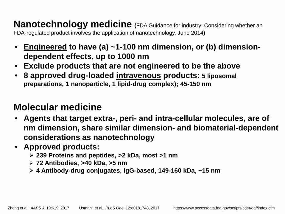
Molecular medicine • Agents that target extra-, peri- and intra-cellular molecules, are of
nm dimension, share similar dimension- and biomaterial-dependent considerations as nanotechnology
• Approved products: 239 Proteins and peptides, >2 kDa, most >1 nm 72 Antibodies, >40 kDa, >5 nm 4 Antibody-drug conjugates, IgG-based, 149-160 kDa, ~15 nm
• Engineered to have (a) ~1-100 nm dimension, or (b) dimension-dependent effects, up to 1000 nm
• Exclude products that are not engineered to be the above • 8 approved drug-loaded intravenous products: 5 liposomal
preparations, 1 nanoparticle, 1 lipid-drug complex); 45-150 nm
Nanotechnology medicine (FDA Guidance for industry: Considering whether an FDA-regulated product involves the application of nanotechnology, June 2014)
Zheng et al., AAPS J. 19:619, 2017 Usmani et al., PLoS One. 12:e0181748, 2017 https://www.accessdata.fda.gov/scripts/cder/daf/index.cfm
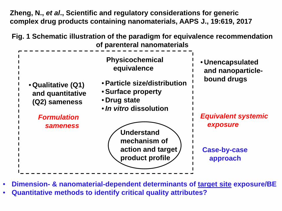
• Dimension- & nanomaterial-dependent determinants of target site exposure/BE • Quantitative methods to identify critical quality attributes?
Zheng, N., et al., Scientific and regulatory considerations for generic complex drug products containing nanomaterials, AAPS J., 19:619, 2017
• Qualitative (Q1) and quantitative (Q2) sameness
• Particle size/distribution • Surface property • Drug state • In vitro dissolution
Physicochemical equivalence
Formulation sameness
Understand mechanism of action and target product profile
Equivalent systemic exposure
• Unencapsulated and nanoparticle-bound drugs
Case-by-case approach
Fig. 1 Schematic illustration of the paradigm for equivalence recommendation of parenteral nanomaterials
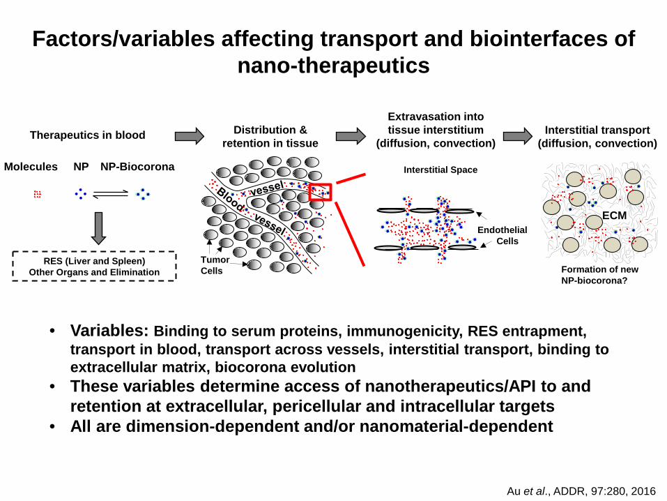
Interstitial transport (diffusion, convection)
Extravasation into tissue interstitium
(diffusion, convection) Distribution &
retention in tissue Therapeutics in blood
Tumor Cells
Endothelial Cells
RES (Liver and Spleen) Other Organs and Elimination
Molecules NP NP-Biocorona
ECM
Formation of new NP-biocorona?
Interstitial Space
Factors/variables affecting transport and biointerfaces of nano-therapeutics
• Variables: Binding to serum proteins, immunogenicity, RES entrapment, transport in blood, transport across vessels, interstitial transport, binding to extracellular matrix, biocorona evolution
• These variables determine access of nanotherapeutics/API to and retention at extracellular, pericellular and intracellular targets
• All are dimension-dependent and/or nanomaterial-dependent
Au et al., ADDR, 97:280, 2016
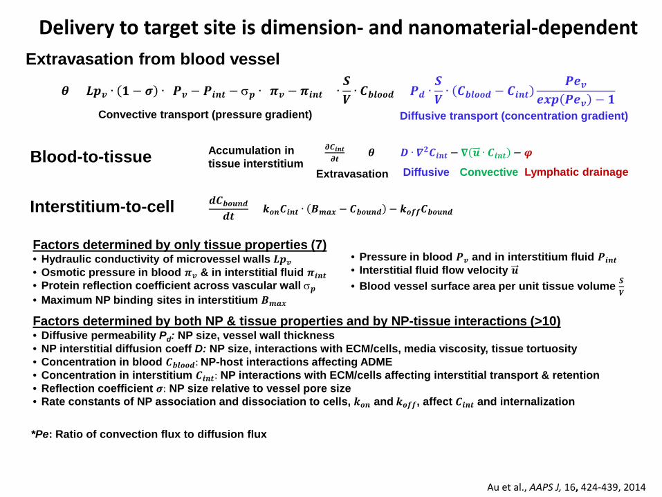
Factors determined by only tissue properties (7) • Hydraulic conductivity of microvessel walls 𝑳𝑳𝑳𝑳𝒗𝒗 • Osmotic pressure in blood 𝝅𝝅𝒗𝒗 & in interstitial fluid 𝝅𝝅𝒊𝒊𝒊𝒊𝒊𝒊 • Protein reflection coefficient across vascular wall σ𝑳𝑳 • Maximum NP binding sites in interstitium 𝑩𝑩𝒎𝒎𝒎𝒎𝒎𝒎
• Pressure in blood 𝑷𝑷𝒗𝒗 and in interstitium fluid 𝑷𝑷𝒊𝒊𝒊𝒊𝒊𝒊 • Interstitial fluid flow velocity 𝒖𝒖 • Blood vessel surface area per unit tissue volume 𝑺𝑺
𝑽𝑽
Factors determined by both NP & tissue properties and by NP-tissue interactions (>10) • Diffusive permeability Pd: NP size, vessel wall thickness • NP interstitial diffusion coeff D: NP size, interactions with ECM/cells, media viscosity, tissue tortuosity • Concentration in blood 𝑪𝑪𝒃𝒃𝒃𝒃𝒃𝒃𝒃𝒃𝒃𝒃: NP-host interactions affecting ADME • Concentration in interstitium 𝑪𝑪𝒊𝒊𝒊𝒊𝒊𝒊: NP interactions with ECM/cells affecting interstitial transport & retention • Reflection coefficient 𝝈𝝈: NP size relative to vessel pore size • Rate constants of NP association and dissociation to cells, 𝒌𝒌𝒃𝒃𝒊𝒊 and 𝒌𝒌𝒃𝒃𝒐𝒐𝒐𝒐, affect 𝑪𝑪𝒊𝒊𝒊𝒊𝒊𝒊 and internalization
Au et al., AAPS J, 16, 424-439, 2014
Interstitium-to-cell
Delivery to target site is dimension- and nanomaterial-dependent
Diffusive transport (concentration gradient) Convective transport (pressure gradient)
Extravasation from blood vessel
𝜽𝜽 = 𝑳𝑳𝑳𝑳𝒗𝒗 ∙ 𝟏𝟏 − 𝝈𝝈 ∙ (𝑷𝑷𝒗𝒗 − 𝑷𝑷𝒊𝒊𝒊𝒊𝒊𝒊 − σ𝑳𝑳 ∙ (𝝅𝝅𝒗𝒗 − 𝝅𝝅𝒊𝒊𝒊𝒊𝒊𝒊)) ∙𝑺𝑺𝑽𝑽 ∙ 𝑪𝑪𝒃𝒃𝒃𝒃𝒃𝒃𝒃𝒃𝒃𝒃 + 𝑷𝑷𝒃𝒃 ∙
𝑺𝑺𝑽𝑽 ∙ 𝑪𝑪𝒃𝒃𝒃𝒃𝒃𝒃𝒃𝒃𝒃𝒃 − 𝑪𝑪𝒊𝒊𝒊𝒊𝒊𝒊
𝑷𝑷𝑷𝑷𝒗𝒗𝑷𝑷𝒎𝒎𝑳𝑳 𝑷𝑷𝑷𝑷𝒗𝒗 − 𝟏𝟏
Blood-to-tissue Accumulation in tissue interstitium
Diffusive Convective Lymphatic drainage
𝝏𝝏𝑪𝑪𝒊𝒊𝒊𝒊𝒊𝒊𝝏𝝏𝒊𝒊
= 𝜽𝜽 + 𝑫𝑫 ∙ 𝜵𝜵𝟐𝟐𝑪𝑪𝒊𝒊𝒊𝒊𝒊𝒊 − 𝛁𝛁 𝒖𝒖 ∙ 𝑪𝑪𝒊𝒊𝒊𝒊𝒊𝒊 − 𝝋𝝋
Extravasation
𝒃𝒃𝑪𝑪𝒃𝒃𝒃𝒃𝒖𝒖𝒊𝒊𝒃𝒃𝒃𝒃𝒊𝒊
= 𝒌𝒌𝒃𝒃𝒊𝒊𝑪𝑪𝒊𝒊𝒊𝒊𝒊𝒊 ∙ 𝑩𝑩𝒎𝒎𝒎𝒎𝒎𝒎 − 𝑪𝑪𝒃𝒃𝒃𝒃𝒖𝒖𝒊𝒊𝒃𝒃 − 𝒌𝒌𝒃𝒃𝒐𝒐𝒐𝒐𝑪𝑪𝒃𝒃𝒃𝒃𝒖𝒖𝒊𝒊𝒃𝒃
*Pe: Ratio of convection flux to diffusion flux
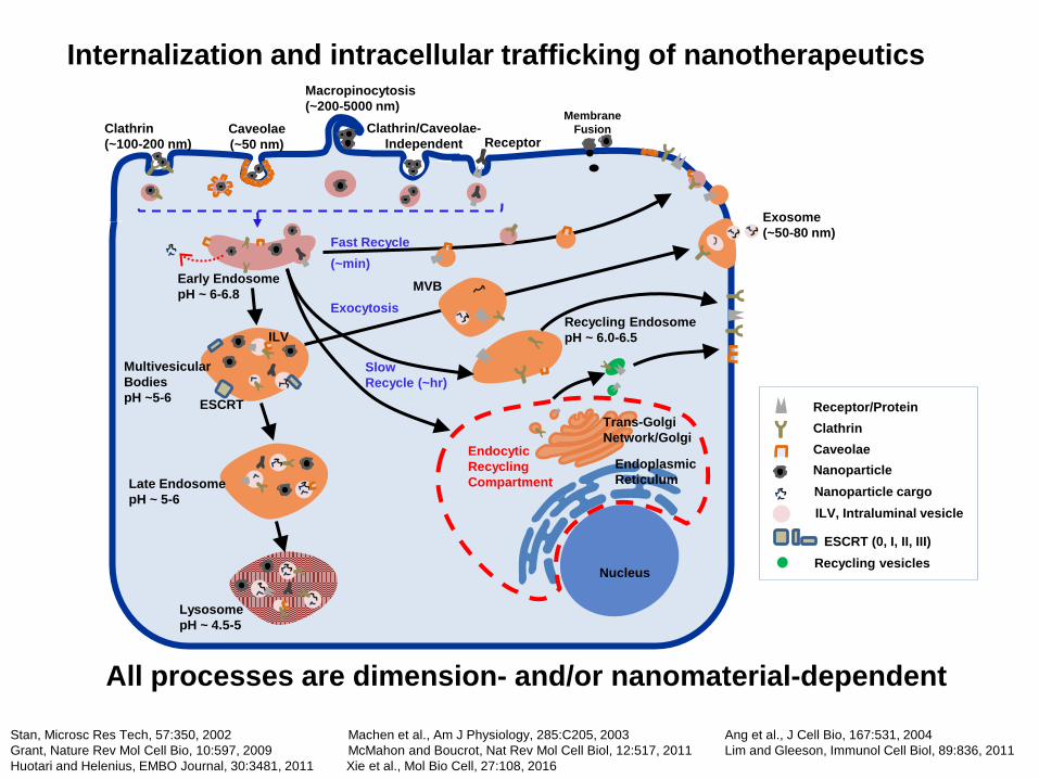
Fast Recycle (~min)
Multivesicular Bodies pH ~5-6 ESCRT
ILV
Late Endosome pH ~ 5-6
Lysosome pH ~ 4.5-5
Stan, Microsc Res Tech, 57:350, 2002 Machen et al., Am J Physiology, 285:C205, 2003 Ang et al., J Cell Bio, 167:531, 2004 Grant, Nature Rev Mol Cell Bio, 10:597, 2009 McMahon and Boucrot, Nat Rev Mol Cell Biol, 12:517, 2011 Lim and Gleeson, Immunol Cell Biol, 89:836, 2011 Huotari and Helenius, EMBO Journal, 30:3481, 2011 Xie et al., Mol Bio Cell, 27:108, 2016
All processes are dimension- and/or nanomaterial-dependent
Exosome (~50-80 nm)
MVB Exocytosis
Slow Recycle (~hr)
Recycling Endosome pH ~ 6.0-6.5
Nucleus
Trans-Golgi Network/Golgi
Endoplasmic Reticulum
Endocytic Recycling Compartment
Clathrin (~100-200 nm)
Caveolae (~50 nm)
Macropinocytosis (~200-5000 nm)
Clathrin/Caveolae-Independent
Internalization and intracellular trafficking of nanotherapeutics
Receptor
Membrane Fusion
ILV, Intraluminal vesicle
Receptor/Protein Clathrin Caveolae Nanoparticle Nanoparticle cargo
ESCRT (0, I, II, III) Recycling vesicles
Early Endosome pH ~ 6-6.8
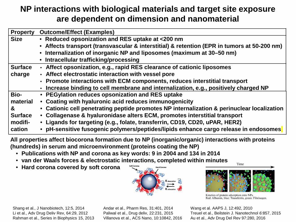
NP interactions with biological materials and target site exposure are dependent on dimension and nanomaterial
Shang et al., J Nanobiotech, 12:5, 2014 Andar et al., Pharm Res, 31:401, 2014 Wang et al. AAPS J, 12:492, 2010 Li et al., Adv Drug Deliv Rev, 64:29, 2012 Paliwal et al., Drug deliv, 22:231, 2015 Treuel et al., Beilstein J. Nanotechnol 6:857, 2015 Rahman et al., Series in Biophysics 15, 2013 Villanova et al., ACS Nano, 10:10842, 2016 Au et al., Adv Drug Del Rev 97:280, 2016
Property Outcome/Effect (Examples) Size
• Reduced opsonization and RES uptake at <200 nm • Affects transport (transvascular & interstitial) & retention (EPR in tumors at 50-200 nm) • Internalization of inorganic NP and liposomes (maximum at 30–50 nm) • Intracellular trafficking/processing
Surface charge
• Affect opsonization, e.g., rapid RES clearance of cationic liposomes • Affect electrostatic interaction with vessel pore • Promote interactions with ECM components, reduces interstitial transport • Increase binding to cell membrane and internalization, e.g., positively charged NP
Bio-material & Surface modifi- cation
• PEGylation reduces opsonization and RES uptake • Coating with hyaluronic acid reduces immunogenicity • Cationic cell penetrating peptide promotes NP internalization & perinuclear localization • Collagenase & hyaluronidase alters ECM, promotes interstitial transport • Ligands for targeting (e.g., folate, transferrin, CD19, CD20, uPAR, HER2) • pH-sensitive fusogenic polymers/peptides/lipids enhance cargo release in endosomes
All properties affect biocorona formation due to NP (inorganic/organic) interactions with proteins (hundreds) in serum and microenvironment (proteins coating the NP)
• Publications with NP and corona as key words: 9 in 2004 and 134 in 2014 • van der Waals forces & electrostatic interactions, completed within minutes • Hard corona covered by soft corona
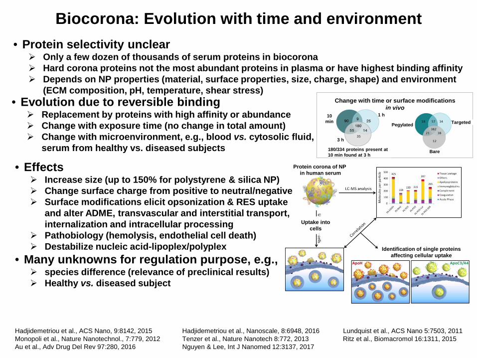
Biocorona: Evolution with time and environment
Hadjidemetriou et al., ACS Nano, 9:8142, 2015 Hadjidemetriou et al., Nanoscale, 8:6948, 2016 Lundquist et al., ACS Nano 5:7503, 2011 Monopoli et al., Nature Nanotechnol., 7:779, 2012 Tenzer et al., Nature Nanotech 8:772, 2013 Ritz et al., Biomacromol 16:1311, 2015 Au et al., Adv Drug Del Rev 97:280, 2016 Nguyen & Lee, Int J Nanomed 12:3137, 2017
• Protein selectivity unclear Only a few dozen of thousands of serum proteins in biocorona Hard corona proteins not the most abundant proteins in plasma or have highest binding affinity Depends on NP properties (material, surface properties, size, charge, shape) and environment
(ECM composition, pH, temperature, shear stress)
• Effects Increase size (up to 150% for polystyrene & silica NP) Change surface charge from positive to neutral/negative Surface modifications elicit opsonization & RES uptake
and alter ADME, transvascular and interstitial transport, internalization and intracellular processing
Pathobiology (hemolysis, endothelial cell death) Destabilize nucleic acid-lipoplex/polyplex
• Many unknowns for regulation purpose, e.g., species difference (relevance of preclinical results) Healthy vs. diseased subject
Uptake into cells
Identification of single proteins affecting cellular uptake
Protein corona of NP in human serum
Change with time or surface modifications in vivo • Evolution due to reversible binding
Replacement by proteins with high affinity or abundance Change with exposure time (no change in total amount) Change with microenvironment, e.g., blood vs. cytosolic fluid,
serum from healthy vs. diseased subjects
10 min
1 h
3 h
Pegylated Targeted
Bare 180/334 proteins present at 10 min found at 3 h
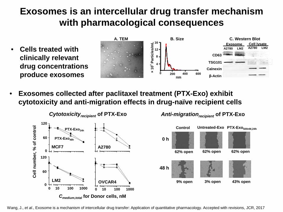
Exosomes is an intercellular drug transfer mechanism with pharmacological consequences
C. Western Blot
CD63
TSG101
Calnexin
β-Actin
ExosomeA2780 LM2
Cell lysateA2780 LM2
A. TEM B. Size
nm0 200 400 600×
107
Part
icle
s/m
L
4
8
12
16
• Cells treated with clinically relevant drug concentrations produce exosomes
Control
9% open
PTX-Exo500nM,24h
2%
43% open 3% open
Untreated-Exo
Anti-migrationrecipient of PTX-Exo
0 h
48 h
62% open 62% open 62% open
Wang, J., et al., Exosome is a mechanism of intercellular drug transfer: Application of quantitative pharmacology. Accepted with revisions, JCR, 2017
• Exosomes collected after paclitaxel treatment (PTX-Exo) exhibit cytotoxicity and anti-migration effects in drug-naïve recipient cells
Cytotoxicityrecipient of PTX-Exo
Cmedium,total for Donor cells, nM
Cel
l num
ber,
% o
f con
trol
MCF7 A2780
LM2 OVCAR4
120
60
0
120
60
00 10 100 1000 0 10 100 1000
PTX-Exo48h
PTX-Exo24h
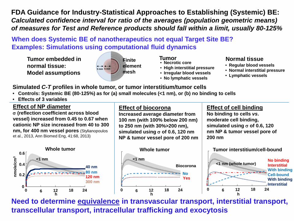
FDA Guidance for Industry-Statistical Approaches to Establishing (Systemic) BE: Calculated confidence interval for ratio of the averages (population geometric means) of measures for Test and Reference products should fall within a limit, usually 80-125% When does Systemic BE of nanotherapeutics not equal Target Site BE? Examples: Simulations using computational fluid dynamics
Need to determine equivalence in transvascular transport, interstitial transport, transcellular transport, intracellular trafficking and exocytosis
Tumor embedded in normal tissue: Model assumptions
Tumor • Regular blood vessels • Normal interstitial pressure • Lymphatic vessels
• Necrotic core • High interstitial pressure • Irregular blood vessels • No lymphatic vessels
Normal tissue 5 mm 5 mm
Finite element mesh
Whole tumor Whole tumor Tumor interstitium/cell-bound
Simulated C-T profiles in whole tumor, or tumor interstitium/tumor cells • Controls: Systemic BE (80-125%) as for (a) small molecules (<1 nm), or (b) no binding to cells • Effects of 3 variables
<1 nm
0
0.2
0.4
0.6
0 6 12 18 24
nmol
e/cm
3
h
<1 nm
0 6 12 18 24 h
<1 nm (whole tumor)
0 6 12 18 24 h
Effect of NP diameter σ (reflection coefficient across blood vessel) increased from 0.45 to 0.67 when cationic NP size increased from 40 to 300 nm, for 400 nm vessel pores (Stylianopoulos et al., 2013, Ann Biomed Eng, 41:68, 2013)
40 nm 80 nm 120 nm 300 nm
Effect of biocorona Increased average diameter from 100 nm (with 100% below 200 nm) to 250 nm (with 30%>200 nm), simulated using σ of 0.6, 120 nm NP & tumor vessel pore of 200 nm
Biocorona
No Yes
Effect of cell binding No binding to cells vs. moderate cell binding, simulated using σ of 0.6, 120 nm NP & tumor vessel pore of 200 nm
With binding Interstitial
With binding Cell-bound
No binding Interstitial
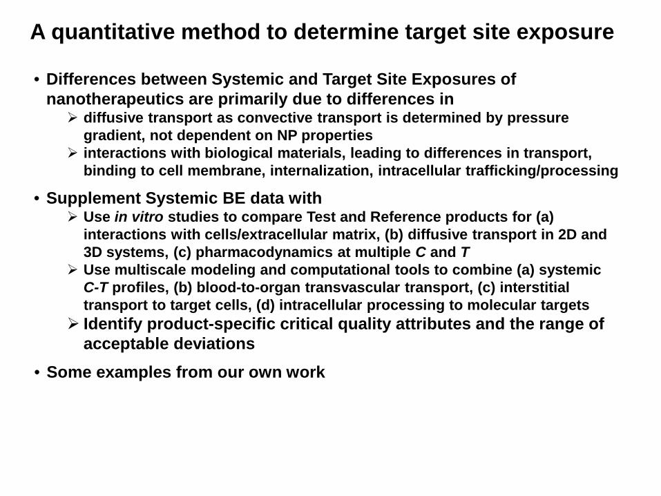
• Some examples from our own work
• Differences between Systemic and Target Site Exposures of nanotherapeutics are primarily due to differences in diffusive transport as convective transport is determined by pressure
gradient, not dependent on NP properties interactions with biological materials, leading to differences in transport,
binding to cell membrane, internalization, intracellular trafficking/processing
A quantitative method to determine target site exposure
• Supplement Systemic BE data with Use in vitro studies to compare Test and Reference products for (a)
interactions with cells/extracellular matrix, (b) diffusive transport in 2D and 3D systems, (c) pharmacodynamics at multiple C and T
Use multiscale modeling and computational tools to combine (a) systemic C-T profiles, (b) blood-to-organ transvascular transport, (c) interstitial transport to target cells, (d) intracellular processing to molecular targets
Identify product-specific critical quality attributes and the range of acceptable deviations
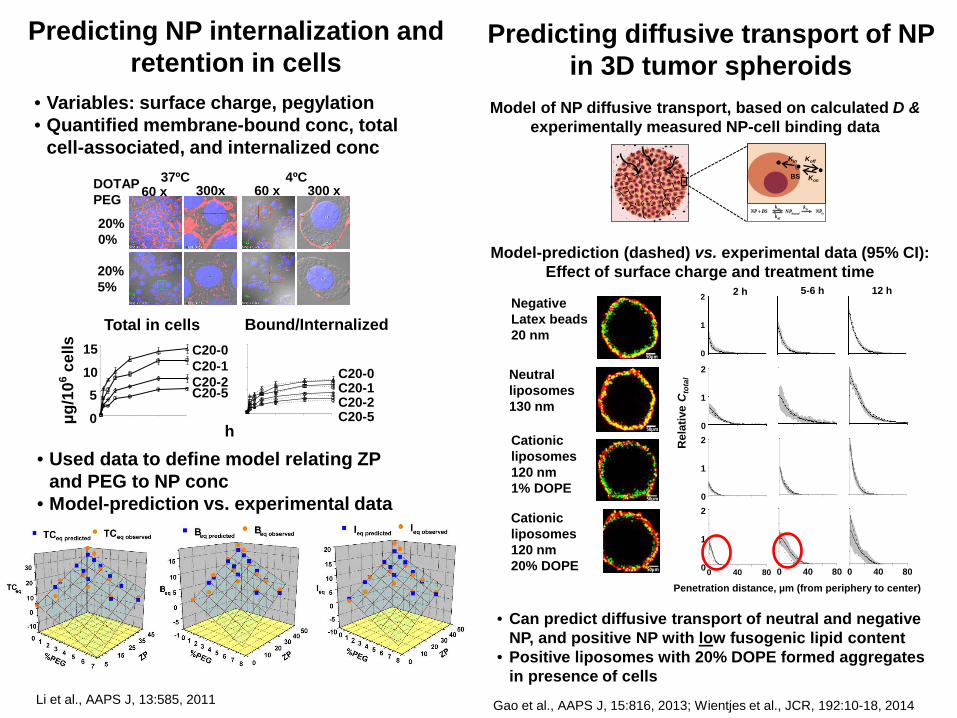
Predicting NP internalization and retention in cells
• Variables: surface charge, pegylation • Quantified membrane-bound conc, total
cell-associated, and internalized conc 37ºC 4ºC
60 x 300x 60 x 300 x DOTAP PEG
20% 0%
20% 5%
Bound/Internalized Total in cells
C20-0 C20-1 C20-2 C20-5
C20-5
C20-0 C20-1 C20-2
0
5 10
15
μg/1
06 cel
ls
h
Li et al., AAPS J, 13:585, 2011
• Used data to define model relating ZP and PEG to NP conc
• Model-prediction vs. experimental data
Predicting diffusive transport of NP in 3D tumor spheroids
Model of NP diffusive transport, based on calculated D & experimentally measured NP-cell binding data
Kon
KoffKin
offkBSNP + onk
boundNP inkinNP
BS
Gao et al., AAPS J, 15:816, 2013; Wientjes et al., JCR, 192:10-18, 2014
Negative Latex beads 20 nm
• Can predict diffusive transport of neutral and negative NP, and positive NP with low fusogenic lipid content
• Positive liposomes with 20% DOPE formed aggregates in presence of cells
Model-prediction (dashed) vs. experimental data (95% CI): Effect of surface charge and treatment time
Rel
ativ
e C
tota
l
50µm
50µm
0 8040
5-6 h 2 h 12 h
50µm
50µm
0
2
1
0
2
1
0
2
1
0
2
1
0 8040 0 8040
Penetration distance, µm (from periphery to center)
Neutral liposomes 130 nm
Cationic liposomes 120 nm 1% DOPE
Cationic liposomes 120 nm 20% DOPE
Tubulin bound PTXCcell,tubulin
Saturable bindingBtubulin,max, ktubulin,on , ktubulin,off
PTX-Exo
PTX-Exo
Releasekrel, exo
Intracellular free PTX Ccell,free
DiffusionDfd
P-gp effluxJmaxPgp, KdPgp
Medium free PTXCmedium,free
n, IC50,initial
γIC50, kkill
InternalizationJmaxinter,exo, Kdinter,exo
Cell Growth
kg
Non-saturable bindingNSB
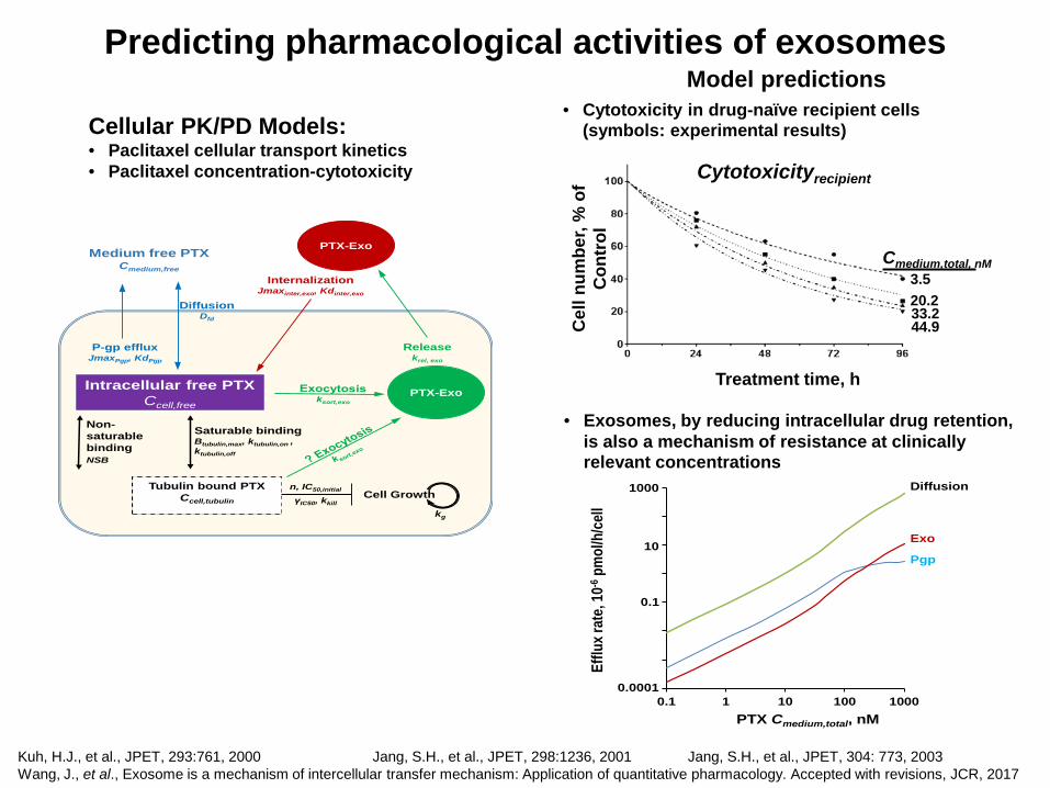
Predicting pharmacological activities of exosomes
Kuh, H.J., et al., JPET, 293:761, 2000 Jang, S.H., et al., JPET, 298:1236, 2001 Jang, S.H., et al., JPET, 304: 773, 2003 Wang, J., et al., Exosome is a mechanism of intercellular transfer mechanism: Application of quantitative pharmacology. Accepted with revisions, JCR, 2017
Cellular PK/PD Models: • Paclitaxel cellular transport kinetics • Paclitaxel concentration-cytotoxicity
Cmedium,total, nM
Cel
l num
ber,
% o
f C
ontr
ol
Treatment time, h
3.5 20.2 33.2 44.9
Cytotoxicityrecipient
Model predictions • Cytotoxicity in drug-naïve recipient cells
(symbols: experimental results)
0.0001
0.001
0.01
0.1
1
10
100
1000
0.1 1 10 100 1000PTX Cmedium,total, nM
Efflu
x rat
e, 10
-6pm
ol/h
/cell
Diffusion
Exo
Pgp
1000
10
0.1
0.0001
• Exosomes, by reducing intracellular drug retention, is also a mechanism of resistance at clinically relevant concentrations
• Nanoparticulate and molecular medicines are subjected to dimension- and material-dependent effects on transport and residence, and biointerfaces
• These properties can result in differences in target site PK/PD that can be predicted by systemic BE
Conclusions
• Therapeutic equivalence (TE) for nanotherapeutics requires additional considerations, such as equivalence in transvascular transport (blood-to-organ) interstitial transport (organ-to-extra-/peri-cellular targets) transcellular transport, intracellular trafficking, exocytosis (from
interstitium to intracellular targets)
• Potential use of in vitro studies & computational multiscale modeling tools to supplement Systemic BE results, to demonstrate Target Site BE and TE
Collaborators OUHSC (Oklahoma) NCI NCI Sukyung Woo, Cody Peer, William Figg Sr., PhD PhD PharmD
Acknowledgements
Supported in part by research grants RO1EB015253 from NIBIB, RO1GM100487 from NIGMS, and RO1CA158300 & RO1CA163015 from NCI, NIH, DHHS
Jie Wang, PhD Yiquan Li, PhD Yue Gao, PhD Bertrand Yeung, PhD Jin Wang, PhD Roberto Abbiati, PhD Minjian Cui Michael Wientjes Ze Lu, PhD Guill Wientjes, PhD