conquering the steep cornea contact lenses in keratoconus conquering the steep cornea... ·...
TRANSCRIPT
Conquering the Steep Cornea
Contact Lenses in Keratoconus
Melissa Barnett, OD, FAAO Department of Ophthalmology & Vision Science
University of California, Davis

Acculens Alcon
Allergan B + L Nidek
SynergEyes Vistakon
Disclosures
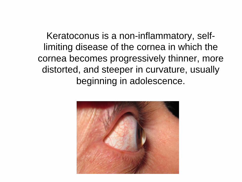
Keratoconus is a non-inflammatory, self-limiting disease of the cornea in which the
cornea becomes progressively thinner, more distorted, and steeper in curvature, usually
beginning in adolescence.

• Bilateral, asymmetric,
non-inflammatory
corneal ectasia
• Abnormal curvature
causes changes in
cornea’s refractive
power in myopia and
astigmatism


• Prevalence ~ 50 - 230 per 100,000
• Incidence ~ 2 per 100,000
• Onset at puberty
• Progressive until the 3rd or 4th decades of
life
• No racial or gender predilection

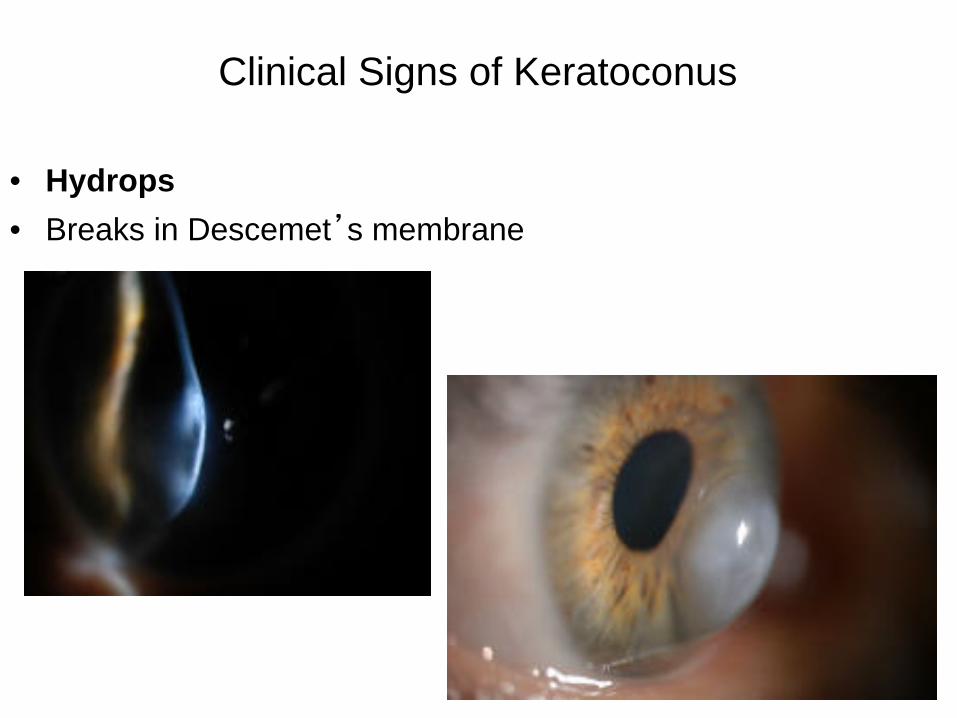
Clinical Signs of Keratoconus
• Munson’s sign • Stromal thinning • Conical protrusion • Vogt’s striae • Fleischer ring • Hydrops • Subepithelial or
anterior stromal scars

Clinical Signs of Keratoconus
• Munson’s sign • V-shaped conformation of the lower lid by the ectatic cornea
in downgaze

Clinical Signs of Keratoconus
• Stromal thinning • Thinning of the stroma • Most commonly inferiorly or inferotemporally

Clinical Signs of Keratoconus
• Conical protrusion

Clinical Signs of Keratoconus
• Vogt’s striae • Fine vertical lines in deep stroma and Descemet’s
membrane

Clinical Signs of Keratoconus
• Fleischer ring • Iron line surrounding the cone partially or completely
Clinical Signs of Keratoconus
• Hydrops • Breaks in Descemet’s membrane

Clinical Signs of Keratoconus
• Subepithelial or anterior stromal scars

Hydrops and Mitral Valve Prolapse
• Hydrops affects 5% patients with KCN • Prevalence of mitral valve prolapse in patients
with corneal hydrops due to KCN is 65%
Mindy Toabe, OD Rabbanikhah, Z. Zahra, M. “Association Between Acute Corneal Hydrops in Patients with Keratoconus and Mitral Valve Prolapse.” Cornea 2011; 30(2): 154-157.
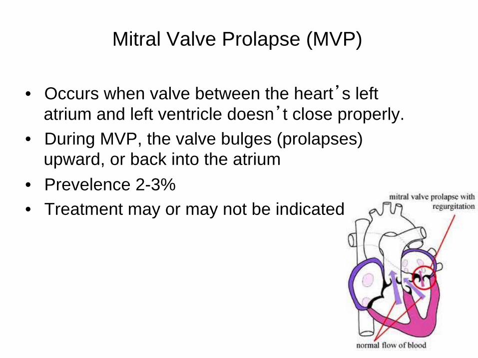
Mitral Valve Prolapse (MVP)
• Occurs when valve between the heart’s left atrium and left ventricle doesn’t close properly.
• During MVP, the valve bulges (prolapses) upward, or back into the atrium
• Prevelence 2-3% • Treatment may or may not be indicated.

Keratoconus • Variation in the Lysyl Oxidase (LOX) Gene is associated with
keratoconus in family-based and case-control studies.
• Bykhovskaya Y, Li X, Epifantseva I, et al.
• Invest Ophthalmol Vis Sci 2012
• Genome-wide linkage scan in keratoconus families
• Identified a locus at 5q23.2, overlapping the gene coding for
the Lysyl Oxidase (LOX)

Keratoconus
• Variation in the Lysyl Oxidase (LOX) Gene is associated with keratoconus in family-based and case-control studies.
• LOX encodes an enzyme responsible for collagen cross-linking in
a variety of tissues including the cornea.
• Conclusion
• LOX variants lead to increased susceptibility to develop keratoconus.

What is the pathogenesis of Keratoconus?
Mechanical trauma? Eye rubbing
KCN patients eye rubbing 80% normal patients eye rubbing 58% (p = 0.001)
Abnormal structure of Bowmans?
Abnormal structure of corneal stroma Fewer collagen lamellae? Fewer collagen fibrils per lamella? Abnormal cross-linking of collagen fibrils?
What is the pathogenesis of Keratoconus?
May be a genetic predisposition that requires a “second hit” or environmental event to elicit progressive disease All play a role in KCN
genetic factors environmental factors inflammatory mediators

Pathogenesis continued Increased digestion of corneal stroma
Normal collagen composition
Increased levels of proteases and catabolic enzymes / decreased
levels of proteinase inhibitors

Pathogenesis continued Role of interleukin-1 receptors
IL-1 induces keratocyte death and negative keratocyte chemotaxis
4-fold increase in IL-1 receptors in keratoconus corneas

Pathogenesis continued Increased expression of matrix-metalloproteinase-1
(MMP-1) in KCN tears
MMP-1
Enzyme that breaks down corneal collagens type
I and III
May be intermittently expressed, leading to
variations in findings

Pathogenesis continued Increased expression of tissue inhibitor of
metalloprotinase 1 (TIMP-1) in KCN tears
TIMP-1 and TIMP-2 are underexpressed in clear
corneas of early KCN
TIMP-1 and TIMP-2 may cause scarring in KCN
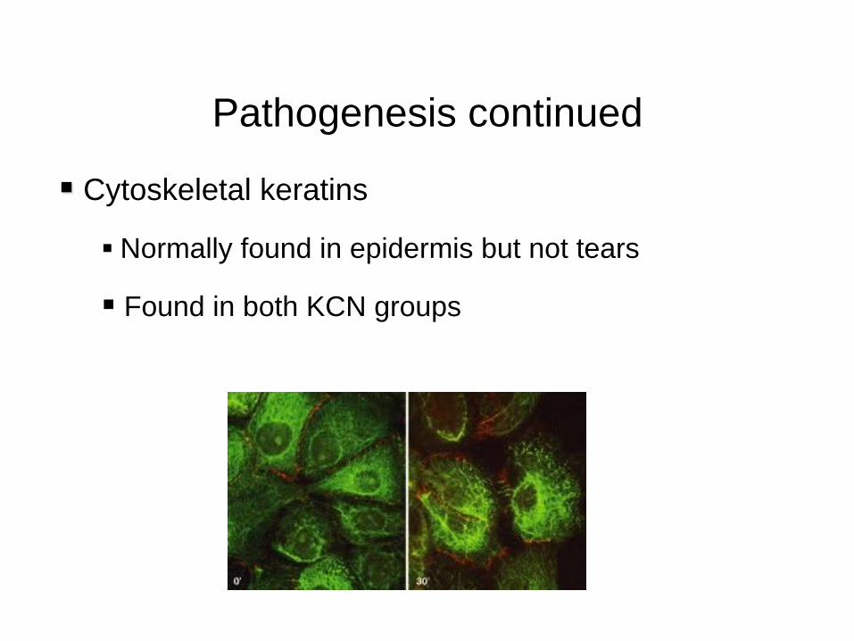
Pathogenesis continued Cytoskeletal keratins
Normally found in epidermis but not tears
Found in both KCN groups

Factors that Reduce risk of KCN
Smoking
Reduced prevalence in KCN patients
Smoking may increase corneal collagen cross-linking
Diabetes
Diabetic hyperglycemia may increase corneal collagen
cross-linking
Diabetic patients with KCN have less severe disease

Treatments for Keratoconus
Contact lenses
Penetrating Keratoplasty
Deep Anterior Lamellar Keratoplasty (DALK)
Intacs - Intrastromal Corneal Rings
Collagen Cross Linking

• Three-fourths of patients with keratoconus in the developed world will be successfully treated with contact lenses

Evaluation Prior to Contact Lens Fitting
• Verify current lens parameters – Base curve – Diameter – Power
• Evaluate fit of current lenses • Obtain history of prior lens wear

Evaluation Prior to Contact Lens Fitting
• Keratometry • Corneal topography • Subjective refraction
• Anterior segment evaluation • Ocular surface quality
Carrie • 31 year old female • Resident physician • Entering VA (with CLs) • OD 20/30 • OS 20/25 • OD - faint Fleisher ring inferior, central corneal thinning • OS - very faint Fleisher ring inferior, central corneal thinning
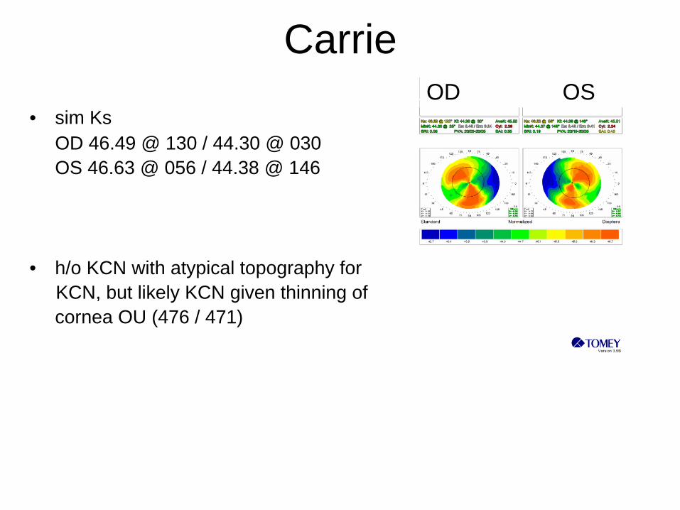
Carrie • sim Ks
OD 46.49 @ 130 / 44.30 @ 030 OS 46.63 @ 056 / 44.38 @ 146
• h/o KCN with atypical topography for KCN, but likely KCN given thinning of
cornea OU (476 / 471)
OD OS

Carrie
new contact lens fitting Kone design • OD 45.62 (7.40) / -3.25 / 9.5 G • OS 46.00 (7.34) / -3.25 / 9.2 B • Follow up
– Good vision and comfort – Able to wear lenses all day,
not all night
• OD 20/25+2 • OS 20/20
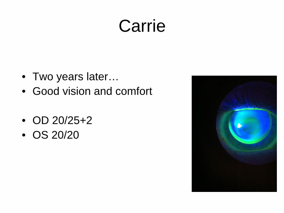
Carrie
• Two years later… • Good vision and comfort
• OD 20/25+2 • OS 20/20

Indications for a Small Diameter Gas Permeable Contact Lens
• Normal corneas
• Regular astigmatism • Irregular astigmatism that is focal,
symmetric or centered – Small, central cones – Mild cones

Small diameter GP lenses (8.0 – 10.0 mm diameter)
• Custom parameters • CLEK diagnostic set • Rose K (Blanchard) GP lenses
(aspheric base curve) • Reverse Geometry designs

Fitting goals • Minimally vault over corneal apex • Mid-peripheral bearing • Moderate peripheral clearance • Lens centration over the cone
Rose K ideal fit

Advantages
• Provide smooth, regular surface that
masks underlying corneal irregularity • Good tear exchange
Disadvantages
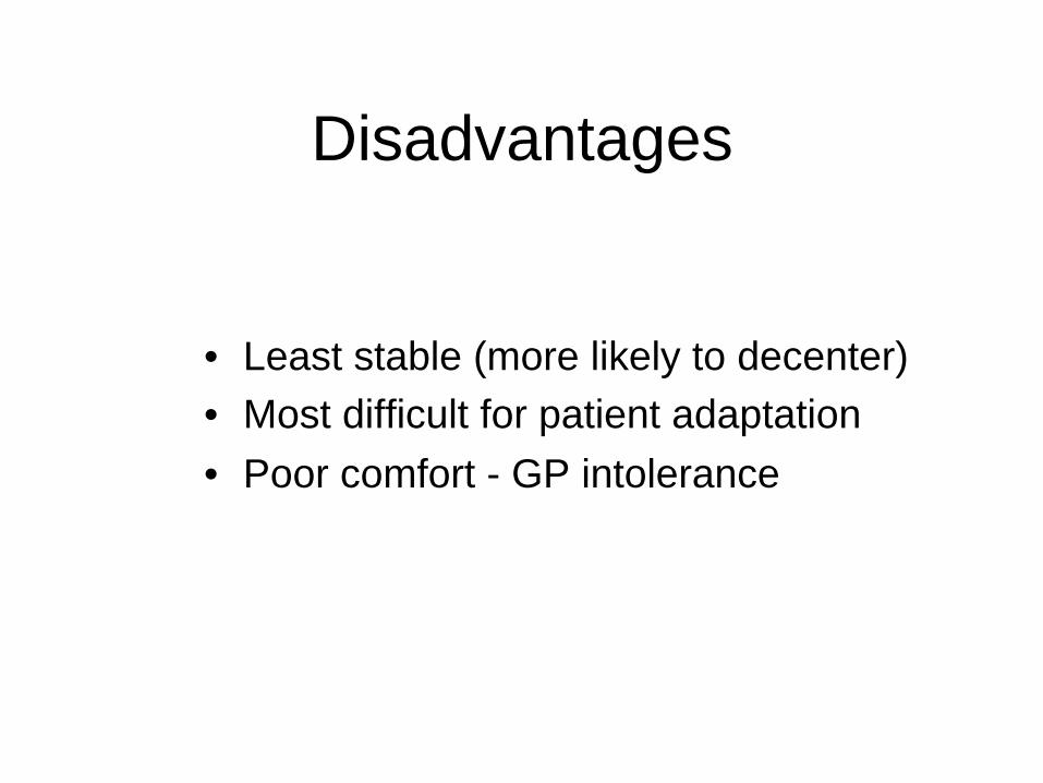
• Least stable (more likely to decenter) • Most difficult for patient adaptation • Poor comfort - GP intolerance

Rose K2: Central Fit
• Start steep
• Go flat pictures courtesy of Lee Buffalo, Blanchard Contact Lens, Inc.

Rose K2
• Ideal fit – Light feather
touch
pictures courtesy of Lee Buffalo, Blanchard Contact Lens, Inc.

Too tight, go flatter Too loose, go tighter
Peripheral curves

Steve • 57 year old Caucasian male • Entering VA (with CLs) • OD 20/50 SOR +2.50 20/25+2 • OS 20/25 SOR +0.50 20/25 • Cornea
OD - inferior central apical scarring, central thinning OS - inferior central scarring
• Lens 2+ nuclear sclerosis, 1+ cortical cataract OU

Steve
• Medical history – diabetes, hypercholesterolemia, erectile dysfunction, keratoconus, glaucoma suspect
• Family history – no significant history • Social history – office worker • Ocular Medications – none • Systemic Medications – glucosamine HCL, sildenafil,
fosinopril, metformin, metoprolol, pravastatin, warfarin

Steve • Optic nerves
OD 0.65 / 0.65 OS 0.50 / 0.45
• Macula OD normal OS trace hard drusen

Steve • sim Ks
OD 50.15 @ 134 / 47.87 @ 044 OS 44.53 @ 047 / 43.05 @ 137
OD central and superior steepening OS inferior steepening

Steve Current gas permeable lenses • OD 7.00 / -10.75 / 8.7 • OS 7.60 / -5.75 / 9.0
• Vision could be better • Good comfort
• Fit OD OS Interpalpebral Interpalpebral Inferior decentered Centered Alignment Alignment Thin peripheral systems Good peripheral systems 1+ scratches on lens surface 1+ scratches on lens surface

Steve
Impresssion • No evidence of diabetic retinopathy in either eye. • Keratoconus with scarring OD > OS. • Glaucoma suspect. • Fit and vision could be improved with gas permeable contact
lenses.
• Plan • Good diabetic control. • Corneal topography done. • Visual field and optic nerve photographs scheduled. • Gas permeable contact lens refit.

Steve New gas permeable lenses ordered • OD 49.00 / -9.12 / 9.0 / 8.50x.4 / 10.8x.4 / 7.0 OZ blue • OS 49.00 / -5.25 / 9.0 / 8.50x.4 / 10.8x.4 / 7.0 OZ blue

Steve • With new lenses
• VA OD 20/20 SOR pl OS 20/20-2 SOR pl
• Fit OU Interpalpebral Centered Alignment Good peripheral systems Clean lens surface

Steve
• Two years later… • Retains good vision and comfort
• OD 20/20+2 • OS 20/20-2
• Stable fit of lenses

Intra-Limbal Lenses
• Slightly smaller than the cornea • 10.5 mm - 12 mm diameter
• Adult cornea – Horizontal diameter 11.5 - 12.6 mm – Vertical diameter 10.5 - 11.7 mm

The Intra-limbal Lens: Indications
• Irregular corneas • Poor centration, stability and / or comfort
with smaller RGP lens diameters • RGP lens intolerance with smaller RGP
lens diameters • Soft lens intolerance due to large amounts
of astigmatism or neovascularization

Intra-limbal Lenses: Indications
• Large cones • Decentered keratoconus • Pellucid marginal degeneration

Intra-limbal Lenses (10.5 mm - 12 mm diameter)
• DynaZ Intralimbal 11.2mm – Lens Dynamics
• KBA 10.2mm – Precision Technology Services
• Rose-K2 IC 11.2mm – Blanchard CL

• XL-T 11.0 - 12.0mm
– Visionary Optics Innovations
• GBL 11.2 mm – ABB - Concise
• I Kone 10.4 mm – Visionary Optics and Valley Contax
Intra-limbal Lenses (10.5 mm - 12 mm diameter)

Fitting Goals • Central corneal vault or light feather
touch • Mid-peripheral bearing with moderate
peripheral clearance • Movement (less than traditional
RGPs) and should provide adequate tear exchange

I Kone
• Bi-surface aspheric design • Diameter 9.6mm
– Also available in 8.8mm and 10.4mm
• Aspheric anterior surface – Reduce spherical aberrations

I Kone • Four conic zones on posterior surface
– Central area vaults cornea to reduce corneal scarring
– First and second zones distribute pressure over larger area to manage corneal ectasia
– Peripheral curve, composed of third and fourth
zones, provides an alignment fit over non-ectatic area of cornea


DynaZ Intralimbal alignment fit

DynaZ Intralimbal flat fit

DynaZ Intralimbal steep fit
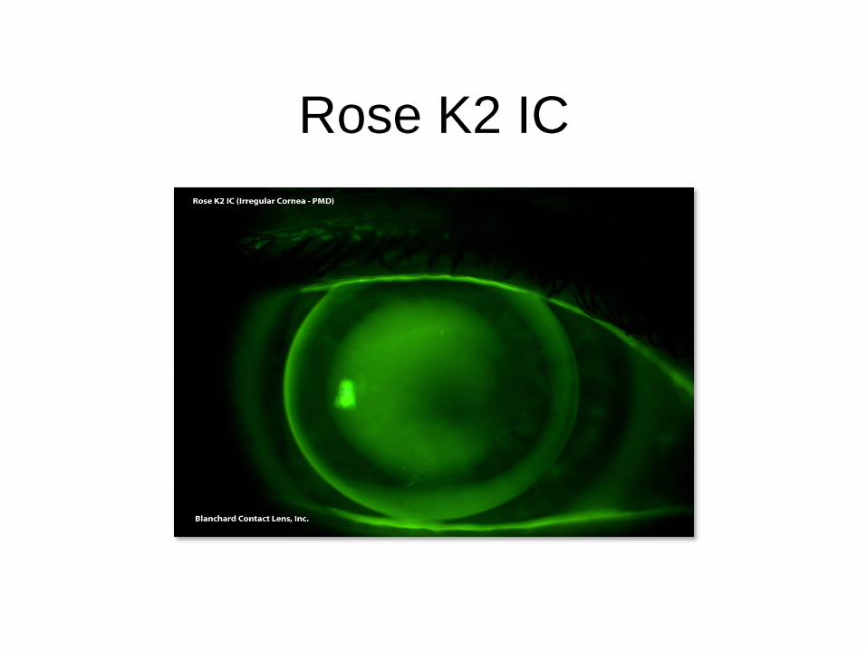
Rose K2 IC

Advantages of Intra-Limbal Lenses
• Better centration and stability • Good vision • Better initial comfort

Disadvantages • Patients may have more difficulty with
insertion and removal

Kenneth • 49 year old African American male • Office worker
• h/o KCN with atypical topography for KCN with irregular astigmatism
• sim Ks OD 47.25 @ 029 / 37.25 @ 119 OS 45.75 @ 174 / 48.25 @ 084

Kenneth • Entering VA (with GP CLs) • OD 20/40+2 • OS 20/40 • OD - 2+ temporal PEK, no Fleisher ring, no striae or thinning • OS - 1+ temporal PEK, no Fleisher ring, no striae or thinning

Kenneth
Contact lenses • OD F60 45.25 / -7.25 / 8.8 • OS F60 45.50 / -5.75 / 8.8 • Fit could be improved • Interpalpebral with inferior decentration • Excessive movement OD
• Patient interested in monovision

Kenneth New contact lenes • OD Oxy HDS 44.50 / -4.50 / 11.0 (N) • OS Oxy HDS 44.50 / -6.00 / 11.0
• OD 20/40-2 with -2.00 20/25-2 • OS 20/25+2 SOR +0.25 NI
Fit • Lid attachment, centered • Alignment • Good peripheral systems

La Ser Eye Jewelry Dr. Chandrashekahr Chawan
Scleral Lenses

Scleral Lens Classification • Classification designed by Dr. Rob Breece • Corneo-Scleral
– Corneal bearing and scleral touch • 12.9 - 13.5mm • Limited tear reservoir capacity
• Semi-Scleral – Corneal and scleral bearing
• 13.6 - 14.9mm
• Mini-Scleral – Scleral bearing and minimal corneal clearance
• 15.0 - 18.0mm • Somewhat limited tear reservoir capacity

Scleral Lens Classification • Full Scleral
– Scleral bearing and maximal corneal clearance • 18.1 - 24.0mm • Almost unlimited tear reservoir capacity

Scleral Lens Indications • Advanced (notably decentered) cones • Pellucid marginal degeneration • Failure with piggyback lenses • Poor comfort with traditional gas permeable designs • Severe dry eyes, GVHD, stem cell deficiency, post graft…

Scleral lens Contraindications
• Corneas with significant edema from
reduced endothelial cell count

Are Scleral Lenses comfortable? They are so big!

Corneoscleral Lenses
• Corneal bearing and scleral touch • 12.9mm to 13.5mm

Corneoscleral Lenses:
Indications for Use
• Decentered irregular astigmatism
• Pellucid marginal degeneration • Oval or globus Cones

Corneoscleral Lenses:
• Do not use with focal steep cone • Do not fit if corneal epithelium cannot
tolerate bearing (lens puts some pressure on the cornea)

Corneoscleral Lenses (12.9mm to 13.5mm)
• Semi-Scleral – Abba
• 13.5mm
• SoClear Lens – Dakota Sciences / Art Optical
• 13.5 - 15.0mm

SoClear Contact Lens fitting • Equally distribute pressure along corneal
and scleral surfaces • Central and peripheral portions of lens may
be independently adjusted • Too flat • Too steep

• SoClear Contact Lens • Ideal fit
– light feather touch at the central cornea – moderate mid-peripheral clearance – even amount of scleral bearing
Picture courtesy of Russell R. Franques, CEO Dakota Sciences

Corneoscleral Lens Fitting
• Different lenses fit differently • Lens movement desirable for all
lenses

Semi-Scleral (13.6 mm to 14.9 mm)
• Corneal and scleral bearing • Jupiter lens
– Visionary Optics (formerly Medlens) / Essilor / ABB-Concise
• 13.5 -16.6mm
• So2Clear – Art Optical
• 14.0 mm

Mini-Scleral (15.0 mm to 18.0 mm)
• Scleral bearing and minimal corneal clearance

Mini-Scleral (15.0 mm to 18.0 mm)
• Msd – Blanchard
• 15.8mm
• Maxim – Acculens
• 16.0mm
• Jupiter – Visionary Optics / Essilor / ABB-Concise
• 15.0-18.8mm
• Boston MiniScleral – Foundation for Sight
• 15.0-15.5mm

Jupiter Lens Fitting • Completely vault cornea and limbus and
rest on sclera • Three zones
– Corneal zone - includes central corneal curve and aspheric peripheral corneal curve
– Limbal zone – Scleral zone - aspheric scleral curve and
aspheric edge curve

Jupiter Lens Fitting • Fit on principle of sagittal depth
– Sagittal depth too high, leads to central bubbles
– Sagittal depth too low,
excessive central touch and bubbles in the sclera


• Jupiter 18.8mm 60 diopter lens with a 2 mm flatter scleral curves
• Reverse geometry design
Pictures courtesy of Dennis Neifert, Essilor, USA

Don, 55 year old Caucasian male
• Date of examination - 11/16/10 • History of KCN • History of discomfort with gas
permeable contact lenses, especially on windy days
• Glaucoma suspect, monitored by glaucoma service

Don
VA with GP CLs OD 20/30-2 OS 20/30-2
Manifest Refraction OD -6.00+1.75x180 20/25 OS -6.00+2.00x180 20/30-2
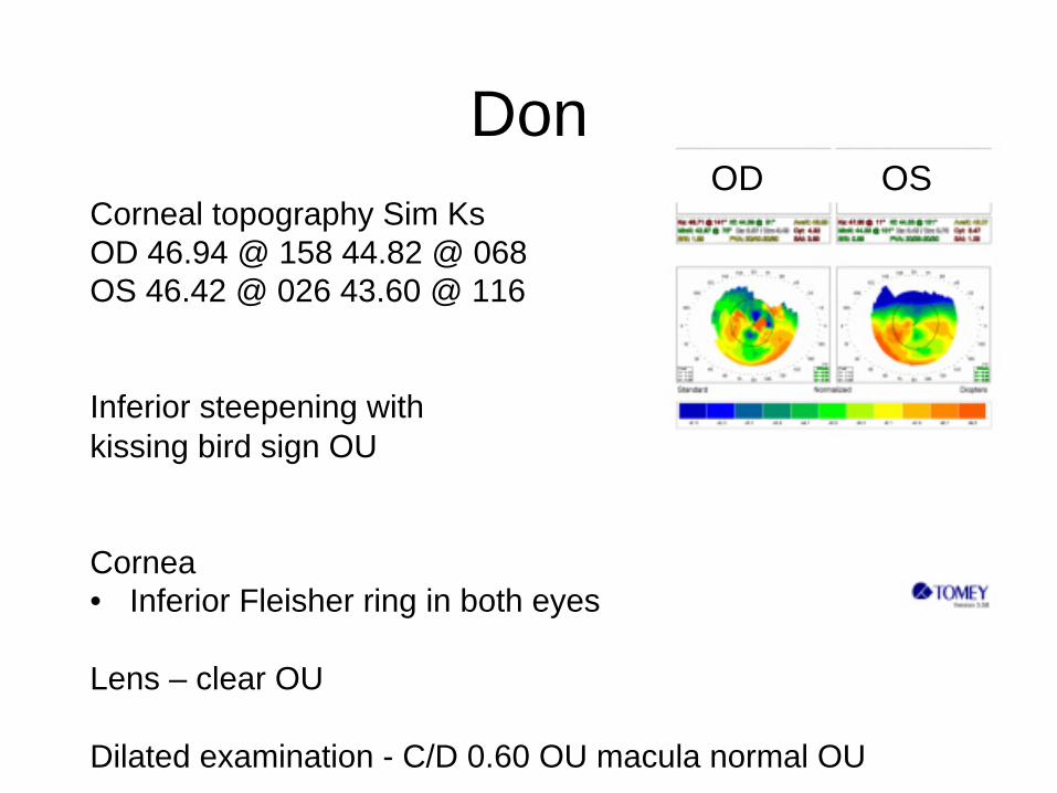
Don Corneal topography Sim Ks OD 46.94 @ 158 44.82 @ 068 OS 46.42 @ 026 43.60 @ 116 Inferior steepening with kissing bird sign OU Cornea • Inferior Fleisher ring in both eyes Lens – clear OU Dilated examination - C/D 0.60 OU macula normal OU
OD OS

Don • Impression • Keratoconus OU. • Fit could be improved with GP contact lenses,
however patient interested in scleral lenses for improved comfort.

Don Scleral Lens Fitting • Initial best fitting Jupiter scleral lenses • OD Jupiter 49.00 / - 9.00 / 15.6
With +1.00DS 20/25-2
• OS Jupiter 49.00 / - 9.00 / 15.6 With +0.75 20/25

Don Scleral Lens Fitting • Ordered lenses with larger diameter and chamber size due to fit
(not enough clearance superior nasal)
• OD Jupiter 48.00 / - 7.00 / 16.0 / 13.25 / 14.75 Ice blue • OS Jupiter 48.00 / - 7.25 / 16.0 / 13.25 / 14.75 Ice blue • Advised need for reading glasses over contact lenses.


John, 51 year old Caucasian Male
• Date of examination - 4/13/09 • History of KCN x 20 years since 30 years old • Sister also has KCN • Tried soft, hard and hybrid contact lenses (most
recently 10 years ago) without success. • Rigid lens improved vision OD, however unable to
tolerate lens. • Right eye vision is deteriorating. Left eye vision is
very poor. • Lights have rings around them like halos.

John
• Medical history – seasonal allergies • Family history – no significant history • Social history – state office worker, lots of computer
work. • Hobby - reading • Ocular Medications – none • Systemic Medications – Claritin, steroid nasal spray,
MVI

John VA corrected with glasses OD 20/30+2 OS 20/150 -1 PH 20/60+2 Manifest Refraction OD -4.75+3.00x170 20/25 OS -9.25+4.50x120 20/60

John Cornea OD - inferior Fleischer ring, paracentral inferior thinning OS - Vogt’s striae centrally, inferior Fleisher ring, paracentral inferior thinning

OD OS
Corneal topography Sim Ks OD 48.43 @ 036 / 40.23 @ 125 OS 60.39 @ 069 / 52.82 @ 159

John Pachymetry OD 491µm OS 446 µm Dilated examination – normal OU Diagnosis - Keratoconus OU

• Clear Kone • OD Vault 200 / -2.00 / medium skirt
20/20-2 • OS Vault 350 / -5.50 /
medium skirt 20/25+2
John - Contact Lens Fitting

• Follow up, limited CL wear due to irritation OS • New lens fit OS • Clear Kone Vault 300 / -2.75 / steep • 20/25
John - Contact Lens Fitting

• Follow up #2 • Left eye poor comfort, only able to wear lenses 2
hours • Refit to Jupiter scleral lenses

John Scleral Contact Lens Fitting Best fitting lenses OD Jupiter 49.00 / -9.00 / 18.2 With +1.00DS 20/25+2 OS Jupiter 50.00 / -9.00 / 18.2 With +0.25DS 20/30+2 Fit OU good central apical clearance, good peripheral fit, well centered, good movement, no blanching

John Scleral Lens Dispense OD Jupiter 49.00 / -8.00 / 18.2 VA 20/25+1 SOR -0.50 NI OS Jupiter 50.00 / -8.75 / 18.2 VA 20/25-2 SOR +0.25 NI Binocular VA without SOR 20/20-2
Fit OU good central apical clearance, well centered, good movement, no blanching
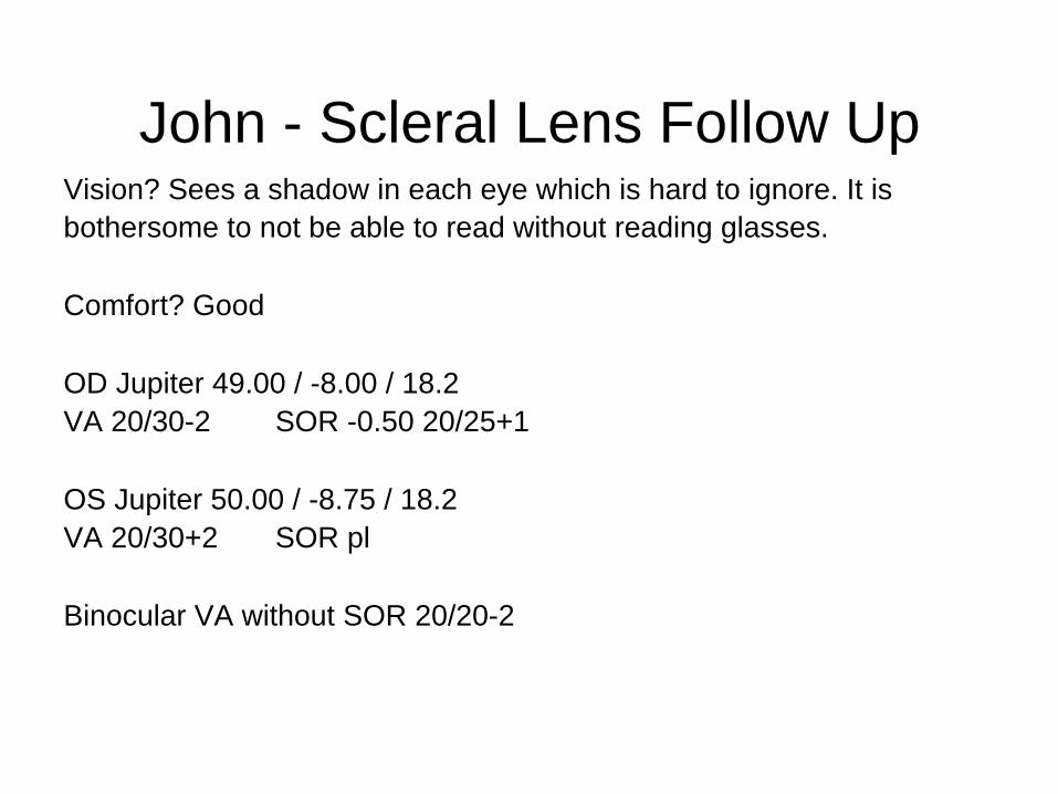
John - Scleral Lens Follow Up Vision? Sees a shadow in each eye which is hard to ignore. It is bothersome to not be able to read without reading glasses. Comfort? Good
OD Jupiter 49.00 / -8.00 / 18.2 VA 20/30-2 SOR -0.50 20/25+1 OS Jupiter 50.00 / -8.75 / 18.2 VA 20/30+2 SOR pl Binocular VA without SOR 20/20-2

John Impression Good overall fit OD, more clearance needed OS Patient interested in monovision Plan Ordered OD Jupiter 49.00 / -8.50 / 18.2 OS Jupiter 50.50 / -7.25 / 18.2 (near) Advised non-preserved artificial tears PRN

John New Scleral Lenses Vision? Much improved except sees shadows when reading very small letters up close. Happy with monovision. Comfort? Very good.
OD Jupiter 49.00 / -8.50 / 18.2 VA 20/25+2 SOR -0.25 20/20-2 OS Jupiter 50.50 / 7.25 / 18.2 (near) VA 20/70 J1+ with -1.50DS 20/25+2

• Impression • Good overall fit, vision and comfort with Jupiter scleral lenses • Good adaptation to monovision • Plan • Continue contact lens wear for
daily wear. • Reviewed solutions - Lobob cleaner,
Boston conditioner and Unisol for insertion.
• Non-preserved artificial tears as needed. • Follow up in 4 months for a
scleral lens check / PRN.

Msd Fitting
• 15.8 mm • Central optic zone - apical clearance or feather
touch • Mid-peripheral limbal zone - vaults the limbus
and aligns with the sclera • Posterior surface incorporates reverse
geometry • Sagittal depth is adjusted independently of
central optic zone profile

• Sagittal depth is the measurement from the flat plane to the highest point of a concave surface
• If sagittal depth is too high, leads to central bubbles
• If sagittal depth is too low, leads to excessive central touch and bubbles in sclera
Sagittal Depth

Msd Fitting • With each sagittal depth value, there is the option
of three Mid-Peripheral / Limbal Clearance Values • Standard, increased and decreased

4.20 S
Excessive Mid-peripheral clearance - bubbles in mid-peripheral / limbal zone
Excessive sagittal depth - bubbles centrally

Full Scleral Lenses (18.1 mm to 24.0mm)
• Scleral bearing and maximum corneal
clearance • First used in late 1800s and early 1900s • Manufacturing process now more
reproducible • Modern scleral lenses
– Don Ezekiel, O.D. – Perry Rosenthal, M.D. Boston Scleral Lens

Advantages
• Good centration • Good stability • Stable vision and optics • Good initial comfort

Disadvantages • Insertion / removal difficult with larger
diameters • Worse tear exchange


Handling - Lens Insertion
• Goal “bubble free” insertion • Patient bends over so that patient’s
face is parallel to the horizontal plane • May use target for patient to look at
(such as Amsler grid) when training • Fill scleral lens fully with fluid

Handling - Scleral Lens Insertion

Handling - Lens Insertion
• Use plunger or three finger approach to hold the lens
• Three finger method • Three fingers are thumb, index, and
middle fingers (may use ring finger also) • Hold eyelids open • Place the lens on the eye

Handling - Lens Insertion
• Use plunger or three finger approach to hold the lens
• Plunger method • Hold eyelids open • Place the lens on the eye • Release plunger if plunger is used • Prefer large plunger for insertion

Handling - Lens Removal
• Manual two finger method • Have patient look down • Move lower eyelid outward while
applying mild pressure to eyeball • Then gently push lower eyelid with
index finger underneath the lower edge of the lens
• Remove the lens

Handling - Lens Removal
• Plunger method • Squeeze plunger to induce suction • Apply plunger to periphery of lens
(not to center of lens) • Twist and pull away from eye • Remove the lens

Soft Lenses and Soft Toric Lenses: Indications
• Early keratoconus • Decentered keratoconus • Globus-like keratoconus • Poor comfort / wearing time / lens
tolerance with RGP lenses

Soft Lenses and Soft Toric Lenses
• Fit centered over cornea
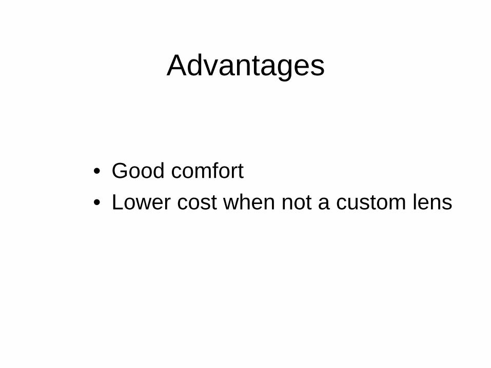
Advantages
• Good comfort • Lower cost when not a custom lens
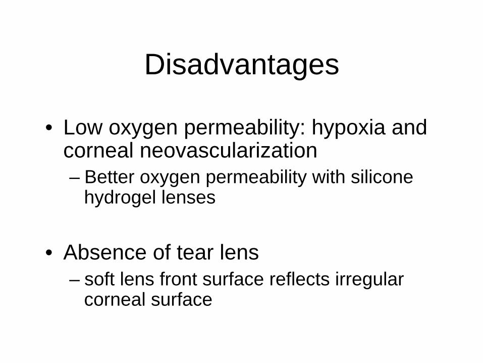
Disadvantages
• Low oxygen permeability: hypoxia and corneal neovascularization – Better oxygen permeability with silicone
hydrogel lenses
• Absence of tear lens – soft lens front surface reflects irregular
corneal surface

Non-Custom Toric Contact Lenses
• Proclear Toric (XR) (Cooper Vision) – Omafilcon A / 59% water – BC 8.4 , 8.8 – Sphere pl to +/- 10.00 – Cylinder -0.75 to -5.75 in 0.50 D steps
(axis full circle in 5 degree steps)
• Biofinity Toric – Biofinity Toric (Cooper Vision) – Comfilcon A / 48% water – BC 8.7 – Sphere +8.00 to -10.00D – Cylinder -0.75 to -2.25 in 0.50D steps
(axis full circle in 10 degree steps)

Non-Custom Toric Soft Contact Lenses Continued
• Metrosoft Toric (Metro Optics) – BC 8.4, 8.7, 9.0 – Sphere +/- 5.25 to +/- 10.00 – Cylinder -0.75 to -8.00 (axis full circle 5 degree steps)
• Preference Toric (XR) (Cooper Vision) – Tetrafilcon A / 43% water content – BC 8.4 , 8.7 – Sphere +6.00 to -9.50 – Cylinder -0.75 to -9.75
(axis full circle in 5 degree steps)

Custom Soft Contact Lenses • SpecialEyes 59 / 54 Toric (SpecialEyes, LLC) • HydroKone (Visionary Optics) • Soft K (Advanced Vision Technologies) • Solus Soft K (Strategic Lens Innovations) • Ocu-Flex Toric (Ocu-Ease) • KeraSoft IC (Bausch + Lomb) • NovaKone (Alden Optical)
• Base Curves 6.0 -7.0mm • Hydrocone 4.1mm BC • Relatively flatter paracentral curve ~ 8mm to match
high eccentricity of the eye

NovaKone
• Soft lens for keratoconus • May be used for pellucid marginal
degeneration

NovaKone Design A. Base Curve to match average
central Ks B. Independent Fitting Curve to optimize lens position and movement C. Aspheric Optical Zone D. Multiple “IT” factors to neutralize
irregularity
NovaKone Design
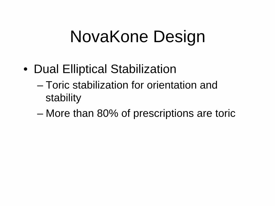
• Dual Elliptical Stabilization – Toric stabilization for orientation and
stability – More than 80% of prescriptions are toric

How does NovaKone work?
1. NovaKone uses lens thickness to neutralize corneal irregularity
2. The NovaKone optical design is then employed to correct for normal spherical and astigmatic refractive errors
3. Dual Elliptical Stabilization™ and precision Rx manufacturing ensure a stable precise Rx lens

Step 1 Select Initial Base Curve
Average K for CENTRAL 3 to 4 mm ONLY

An Ideal base curve should yield light central touch and stable opCcal findings
Base Curve Verification
Photo courtesy of Mark Andre, FCLSA, Pacific University
Photo courtesy of Mark Andre, FCLSA, Pacific University

High Molecular NaFl

Step 2 The IT Factor
• IT = “Index of Thickness”, ranges from 0 to 4.
• Use the lowest IT factor possible
• The more irregular the cornea the higher the IT Factor should be to optimize visual acuity
• Verify IT factor with Keratometry or Topography over the lens. If any irregularities are observed, increase the IT Factor to improve optical stability.

Mire Evaluation
Keratometric mires over the NovaKone lens will be crisp and clear with the proper IT factor
Photo courtesy of Mark Andre, FCLSA, Pacific University Photo courtesy of Mark Andre, FCLSA, Pacific University

Step 3 Determine Lens Power
• Over refract and calculate the power of the Rx lens
• Compensate for rotation – All Dx lenses have Dual Elliptical Stabilization to
assess rotation – Dx lenses have no actual cylinder power

Step 4 The Fitting Curve
• Able to select the base curve, IT factor, and lens power. Given any base curve, the Dx lens will only have a single fitting curve from the fitting set.
• Evaluate initial lens in order to determine if the fitting curve on the diagnostic lens is appropriate or needs to be altered on the prescription lens order.

Step 4 The Fitting Curve
The fitting curve should demonstrate typical fitting characteristics of a standard soft lens fit.
• If the fitting curve is too flat there will be excessive movement
and / or edge lift (order steeper fitting curve) • Little or no movement and / or edge impingement would indicate
the fit curve is too steep (order flatter fitting curve) • Alden labels the fitting curve with the actual radius in millimeter,
practitioners should be comfortable with these values in relationship to a “good” lens fit.
• The fitting curve should be adjusted in a minimum of 0.2mm increments

Glasses
• Early keratoconus • Patient convenience • Use for cylinder correction in
conjunction with contact lenses correcting the sphere

Piggyback Lenses: Indications
• Soft lens under RGP lens • Poor comfort / movement with RGP
lenses • Epithelial defects with RGP lenses • Apical nodules • Epithelial Basement Membrane
Dystrophy
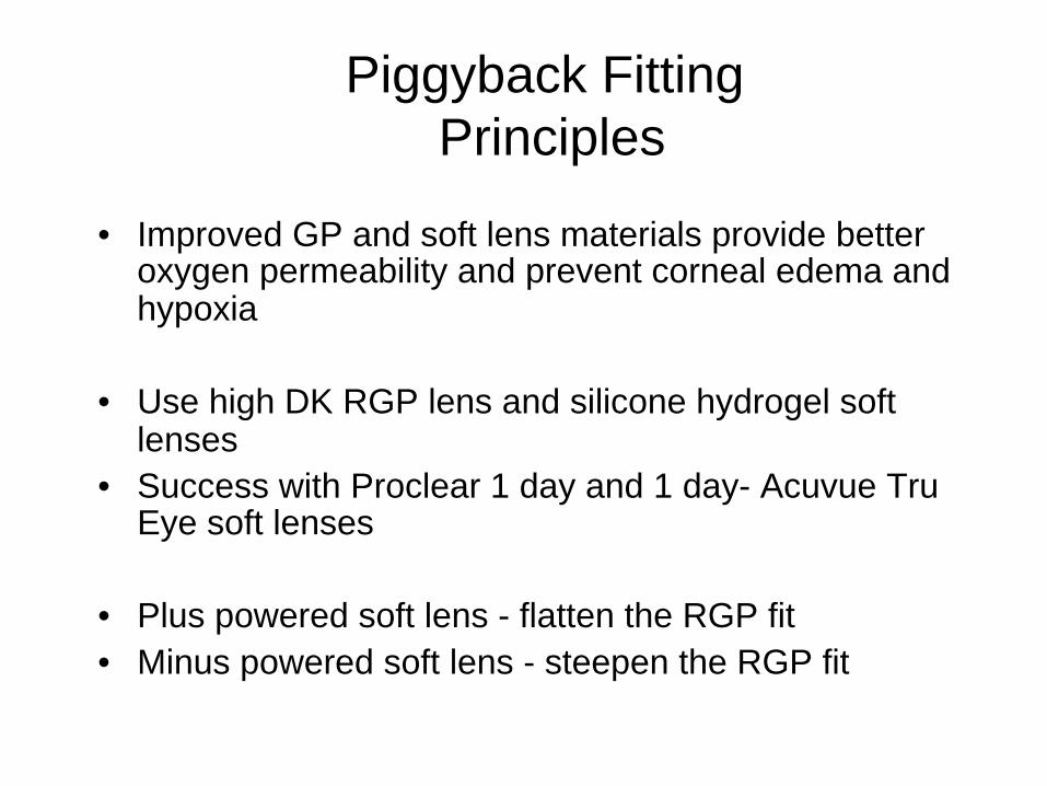
• Improved GP and soft lens materials provide better oxygen permeability and prevent corneal edema and hypoxia
• Use high DK RGP lens and silicone hydrogel soft lenses
• Success with Proclear 1 day and 1 day- Acuvue Tru Eye soft lenses
• Plus powered soft lens - flatten the RGP fit • Minus powered soft lens - steepen the RGP fit
Piggyback Fitting Principles

Advantages • Better comfort than standard RGP CL • No corneal compromise or complications • No hypoxia • Improved comfort compared with RGP lens alone • Same or increased wearing time vs. the RGP lens
worn alone • Same or better visual acuity • Study by Jill J. Rodio-Vivadelli, OD, FAAO, & Ralph Gundel, OD,
FAAO Sept 2006

Disadvantages
• More difficulty and inconvenience with piggyback lens system
• Loss of GP lens • Damage to soft lens • Multiple lens care systems

Piggyback lens

Piggyback Lens - flat fit

Piggyback Lens - optimal fit

Hybrid Lenses - SynergEyes
• Rigid center • Soft skirt • Adjustable central base curve and
skirt curvature

Hybrid lenses - SynergEyes • SynergEyes A lens design
– Early or moderate keratoconus – Normal corneas
• SynergEyes KC lens design – More advanced keratoconus
• Poor centration, stability and / or wearing time with RGP lenses
• RGP lens intolerance
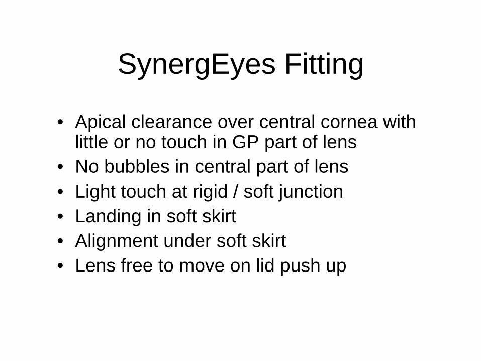
SynergEyes Fitting
• Apical clearance over central cornea with little or no touch in GP part of lens
• No bubbles in central part of lens • Light touch at rigid / soft junction • Landing in soft skirt • Alignment under soft skirt • Lens free to move on lid push up

Apical clearance Insufficient clearance (note landing)

Advantages • Good vision • Good lens centration • Higher oxygen permeability centrally
(and soon in periphery) • Increased fitting parameters for base
curve and power • Various peripheral curve systems

Disadvantages
• Lens tightening • Need high-molecular-weight
fluorescein to evaluate the fit

SynergEyes A • Emerging or moderate
keratoconus
• Acceptable fit • Central clearance with
minimal touch
Picture courtesy of Erin Clark, SynergEyes

SynergEyes A
• Unacceptable fit • Central bubble,
inferior touch • Consider SynergEyes
KC lens design
Picture courtesy of Erin Clark, SynergEyes
SynergEyes A
7.3 BC

SynergEyes A
Minimal apical clearance Apical touch
Ideal apical clearance

SynergEyes KC
• Ideal fit • Apical clearance • Soft landing where base
curve meets skirt curve • Minimal touch in rigid
portion
Picture courtesy of Erin Clark, SynergEyes
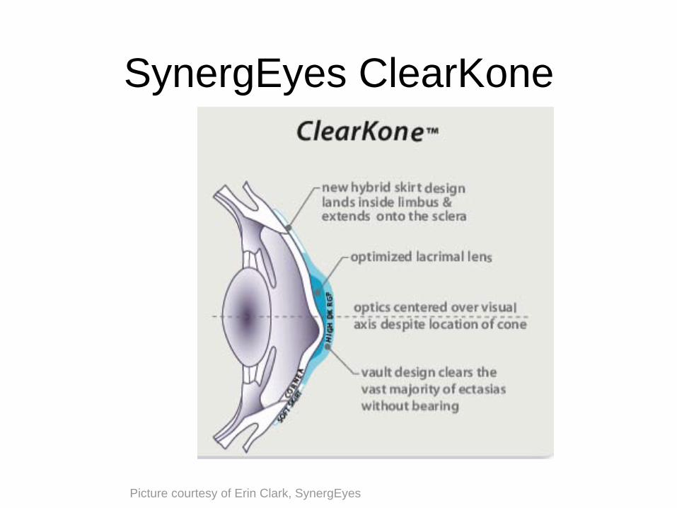
SynergEyes ClearKone
Picture courtesy of Erin Clark, SynergEyes

• The vault value describes the overall relative depth of the lens on the cornea.
• The end point of the fitting is the least amount of vault needed to clear the cone.
• Design gives the ability to “vault” over the vast majority of ectasias without bearing

Unlike RGPs, hybrid platform centers optics independent of the location of the cone

• Vault – The vault value describes the overall relative
depth of the lens
• Outer Landing Zone (OLZ) – Portion of the lens that lands on the soft
material
• Inner Landing Zone (ILZ) – Portion of the lens that lands on the RGP
material

SynergEyes ClearKone • Oval / nipple KCN (moderate to advanced) • Central and the majority of decentered cones • Post RK, PRK, Lasik induced ectasia • May be able to fit Globus, PMD and irregular corneas
SynergEyes ClearKone • Disadvantages • May not be able to fit the following: • Ectasia that extends beyond landing zone • Highly irregular or asymmetric landing pattern (seen
with advanced PMD)

OLZ Bearing Thinning Centrally
RGP/SCL Junction
ILZ Thinning
Ideal ClearKone Fit

Barbara, 77 year old Caucasian Female
• S/P PK, AK, AC IOL OU • S/P Lasik OS • H/o KCN OU 1/2008 fit with bitoric lens OD VA: 20/25 poor comfort High astigmatism OU (6D on topography)

• 7/2009 refit to Clear Kone OD • Clear Kone Vault 250 / -1.25 / medium skirt • 20/20-1 • Great comfort, can not feel lens at all
Barbara

Barbara fit continued
• 3/2010 Post multiple retinal surgeries and now ready for contact lens OS
• Uncorrected VA OS 20/400 • Fit with Clear Kone VLT 350 / -7.75 / medium • VA improved to 20/150+1 • Good vision, helps a lot with driving and
reading

Surgical treatment options for keratoconus

Surgical treatment options for keratoconus
• Penetrating keratoplasty
– ~10-20% of KCN patients will require a PKP in their lifetime
– Long-term complications
of PKP

Surgical treatment options for keratoconus
• Penetrating keratoplasty
– 20-30% of patients develop immunologic rejection – ~ 14-29% long-term graft failure rate

Surgical treatment options for keratoconus
• Deep Anterior Lamellar Keratoplasty (DALK) – The concept of lamellar keratoplasty to remove the risk of
endothelial rejection has been around for over 150 years
– Dissecting near Descemet’s membrane was first attempted in the late 1950s
• Pathological stroma is completely removed
• Less interface opacity than prior LKP
• Visual acuity comparable to PKP

Deep Anterior Lamellar Keratoplasty (DALK)
• Direct Open Dissection
– Stroma removed layer by layer until clear stroma remains or Descemet’s Membrane is exposed
– HOWEVER, rate of intraoperative perforation is high >30-35% and frequently, the surgeon fails to bare Descemet’s
– Difficult to visualize depth of dissection relative to corneal thickness during surgery
– Difficult to tell small differences in refractive index between the corneal tissue and aqueous

Deep Anterior Lamellar Keratoplasty (DALK)
• Hydrodelamination – Cornea is trephined to ~ 75% depth. – Hydrodelamination. Small pocket made in central stroma
and BSS injected with a blunt 27-guage cannula. The solution penetrates between collagen fibers which whiten and
swell.

Deep Anterior Lamellar Keratoplasty (DALK)
• Hydrodelamination continued – Spatula delamination.
• A fine spatula is inserted into the hydrodelaminated stroma to remove the stroma layer by layer.
– Finally, Descemet’s membrane is exposed.
• Study by Sugita et al. • Descemet’s punctured intraoperatively 39.2% • However, no significant differences in VA when Descemet’s
is punctured.
DALK: Advantages and Disadvantages
• Advantages of DALK over LKP – Smooth donor-to-recipient interface – Reduced risk of interface scarring – Identical level of dissection depth between donor and
recipient tissues

DALK: Advantages and Disadvantages
• Advantages of DALK over PKP – Fewer intraocular complications:
• Endophthalmitis, glaucoma, anterior synechia, injury to lens and vitreous
– Less risk of endothelial rejection (main cause of graft failure post PKP) – Faster rehabilitation; no long-term immunosuppression / steroids
• Decreased risk of infection, glaucoma, and cataract – Superior wound strength – Fewer rigid criteria for donor corneal tissue selection
DALK: Advantages and Disadvantages
• Disadvantages of DALK – Technically more difficult and time consuming
– Cannot be performed in patients with prior disruption of
Descemet’s
– Relatively high rate of intraoperative perforation of
Descemet’s
– Still may have interface haze and night vision problems
– Donor tissue may not be adequate should the need to
convert to PKP arise intraoperatively

Keratoprosthetics

Keratoprosthetics • Synthetic or partially synthetic device to replace an
opaque human cornea in order to provide a clear view through the front of the eye.
• Surgical procedure where a severely damaged or
diseased cornea is replaced with an artificial cornea.

Keratoprosthetics • Used for severe corneal opacities.
• Failed corneal transplants.
• Used when standard corneal transplants are unlikely to succeed.
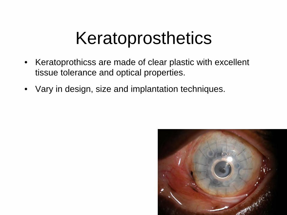
Keratoprosthetics • Keratoprothicss are made of clear plastic with excellent
tissue tolerance and optical properties.
• Vary in design, size and implantation techniques.

Keratoprosthetics • Keratoprothetics consist of three parts and when fully
assembled and has the shape of a collar-button.

Keratoprosthetics • Two devices are approved for use in the United States.
• AlphaCorTM
• Developed in Australia.

Boston Keratoprosthesis (Kpro)
• Developed by Dr. Claes H. Dohlman, corneal specialist
• Under development since the 1960s.
• Received FDA clearance in 1992.

AlphaCor • Made of a plastic-type material known as pHEMA. • Consists of two parts
1. A transparent low water content central core 2. A cloudy high water content outer porous skirt

AlphaCor • AlphaCor procedure performed in two stages carried out
approximately three months apart.
• First, a 180 degree incision is used to place the implant within the central portion of the diseased cornea.
• Then the outer conjunctiva is placed over the implant in order for the cornea to heal.

AlphaCor • Three months later, the outer half of the cornea is
removed in order to provide a clear view into the eye.

AlphaCor Design DPk & AlphaCorAlphaCor Design DPk & AlphaCor

Kpro
• The most commonly used artificial cornea in the United States and in the world.
• It consists of three parts and when fully assembled, has the shape of a collar-button.

Kpro
• Consists of a central PMMA plastic button with a surrounding human donor corneal skirt.
• A back plate with porous holes sandwiches the inner human cornea.

Kpro
• Donor cornea is placed on the front collar button and a titanium screw locks the KPro device into proper alignment.
• Entire KPro procedure is done in a single procedure.
• If the eye is otherwise healthy, vision should return more rapidly than with the AlphaCor procedure.

Potential complications with Keratoprosthetics
• Infection • Melting of the device • Hemorrhage during surgery • Worsening glaucoma • Acute retinal necrosis • Chronic hypotony • Poor visual potential if the retina and optic nerve are
unhealthy.

Intacs

Intrastromal Corneal Rings (ISCR)
• Arclike PMMA segments inserted into deep corneal
stroma (~75% deep) • Separate corneal lamella • Shortens the arc length of
the anterior corneal surface • Flattens the central cornea • Provides biomechanical support for thin ectatic corneas • Increased flattening with thicker segments

Intrastromal Corneal Rings • Two different rings available
– In US since 2004, only INTACS are FDA approved to treat keratoconus in humans under surveillance of an IRB
• 7 mm optical zone
• Available sizes in the U.S.: – 0.25, 0.275, 0.30, 0.325, 0.35 mm
• In Europe: 0.40 and 0.45 mm
– Ferrara rings available outside U.S. • 4.5 to 5 mm optical zone

After INTACS insertion
Before INTACS insertion
INTACS

Indications for INTACS • Best indications for INTACS • Mild to moderate keratoconus • Clear optical zone
• Contact lens intolerant
• Maximum steepest K reading: 55 to 57 Diopters • Corneal thickness at least 450 µm over area where
INTACS will be placed

Indications for INTACS • Other uses for INTACS • Low myopia • Post-LASIK ectasia • Pellucid marginal degeneration • Corneas too thin for additional
enhancements after prior myopic LASIK

Goals of ISCR?
• Patient or clinician based? • To eliminate need for glasses and contact
lenses? • To delay or avoid corneal grafts?
• To create a cornea more receptive to contact lenses?

INTACS studies • Boxer-Wachler (2003) • Ophthalmology. 2003; 110:1031-1040. • 74 eyes • Mean spherical equivalent decreased from -3.98D to -1.46D
• Ibrahim (2006) • Cataract & Refractive Surgery Today, Europe. 2006; 1:45-48. • 186 Eyes • 5 year follow-up • Minimum simK readings decreased ~ 4.00D

INTACS studies • Ertan (2006) • Journal of Cataract and Refractive Surgery. 2006; 32:2039-2042. • 118 Eyes • 1 year follow-up • Mean spherical equivalent decreased from -7.57D to -3.72D • Mean keratometry decreased from 51.56D to 47.66D
• Colin (2007) • Journal of Cataract and Refractive Surgery. 2007; 33:69-74. • 100 Eyes • 2 year follow-up • Mean spherical equivalent decreased from -6.93D to -3.80D • Mean keratometry decreased from 50.1D to 46.8D

INTACS - changes in UCVA
72%
81% 81%85%
19%15% 13%15%
9%3% 6%
0%
20%
40%
60%
80%
100%
Gain of More Than 1Line
No Gain +/- 1 Line Loss of More Than 1Line
Boxer Wachler Ertan Colin Ibrahim

INTACS - changes in BCVA
48%
65%68%
86%
51%
17%17%14%4%
9%15%
0%
20%
40%
60%
80%
100%
Gain of More Than1 Line
No Gain +/- 1 Line Loss of More Than1 Line
Boxer Wachler Ertan Colin Ibrahim

INTACS: Segment Choice and Location
• Placement of rings – Multiple options: – Incisions on the steepest axis
to reduce astigmatism – Incisions temporally and
asymmetric sizes of segments – Incisions made to bisect the segments
at the thinnest area of the cornea in order to thicken a thin area
– Segments centered over the cone and not the center of the cornea
– Vertical Intacs implantation based on the ease of manipulation from a 12 o’clock incision

INTACS: Single Segment vs. Double Segment
• Patients with KCN tend to have an inferior cone with steepening and flattening superiorly.
• Double segments flatten both inferiorly and superiorly which
does not address the issue of asymmetric astigmatism
• Single segments flatten inferiorly and steepen superiorly
• Consider single-segment INTACS for peripheral cones and double-segments for centrally located cones for improved visual and topographic outcomes

Intrastomal Corneal Ring Complications
• European multicenter study of intrastromal corneal ring segments (2001)
– 1 year data; 163 eyes of 110 patients for myopia – Intraoperative complications 2% of eyes (4/163)
• 3 eyes with anterior surface perforations • 1 eye with posterior microperforation

Intrastomal Corneal Ring Complications
• European multicenter study of intrastromal corneal ring segments (2001)
– 2 incisional gapes:
• 1 healed without complications • 1 ISCR removed at 3 months post-op due to non-
healing incision – 4 eyes required repositioning of ISCR – 1 eye with a channel infection 3 weeks post-op – Stromal thinning 2% of eyes at 1 month

Intrastomal Corneal Ring Complications continued
– Diffuse haze under stromal
tunnel medial and lateral to the segments
• Gradually decreased with time; did not spread beyond the edge of the tunnel
• No affect on visual outcome
– Epithelial cysts at the incision site in 7% of eyes (11/156)
• Lasted 7 days to 3 months; 1 eye at 12 months
Epithelial Cysts
Residual stromal tunnel haze post-Intacs removal

Intrastomal Corneal Ring Complications continued
– Lamellar channel deposits along the inner or outer curvatures of the ISCR
• Developed within the 1st months post-op
• No clinical impact – Intraepithelial iron line occurred
in most eyes 6-9 months post-op
Intraepithelial Iron Line
Lamellar Channel Deposits
Intraepithelial Iron Line

Intrastomal Corneal Ring Complications continued
– Visual symptoms:
• Mild-moderate post-op pain – FBS, photophobia in first 24-48 hours – Typically, no visual symptoms by 12 months
• Severe glare occurred within the first 2 months – < 4% of eyes – By 12 months, 96% had no or mild glare

INTACS: Complications • Zare et al, 2007 – 30 eyes
with KCN: – 3 cases of ISCR movement and
exposure, 3-5 months post-op – 2 cases of repeated exposure
and significant corneal thinning over the ring segments
– 1 case of severe FBS / discomfort – 1 case of corneal melting and
severe corneal infiltration required segment removal and fortified antibiotic drops

PKP vs. INTACS • Rodriguez et al, 2007
– Nonrandomized, retrospective comparison – 17 pts with PKP in one eye and Intacs in other
• Uncorrected vision – Less time to reach
potential visual acuity – Statistically significant
improvement in UCVA with both INTACS and PKP

PKP vs. INTACS continued • Astigmatism
– BCVA better at 3 months with INTACS, but not statistically significant at 10 months
– Astigmatism lower at 10 months with INTACS, but not statistically significant

Collagen Crosslinking (CXL)

Collagen Crosslinking (CXL)
• Studies of keratoconic corneas have demonstrated lower corneal elasticity and ocular rigidity in keratoconic eyes compared to normal corneas
• Decreased stiffness and elasticity of the cornea in keratoconus is thought to be related to a reduction in collagen cross-linking
CXL
• Improves the biomechanical properties of the cornea by strengthening the corneal tissue in the anterior stroma.
• The only procedure available to specifically stop the progression of keratoconus and strengthen the individual collagen fibers in the cornea.
CXL • Corneal stromal crosslinking investigations began in mid
1990s as a conservative treatment for keratoconus
– The biomechanical behavior of the cornea could be altered by irradiation using ultraviolet light with photosensitizers and by aldehyde reactions (Spoerl and Seiler).
– Porcine corneas treated with either glutaraldehyde, Karnovsky’s solution (glutaraldehayde and paraformaldehyde) or riboflavin and UV-irradiation.
– Compared to untreated corneas, these treatments caused
an increase in corneal stiffness.


CXL
• UV-radiation alone did not induce mechanical changes in the cornea, but required a photosensitizer
• Riboflavin is a non-toxic photosensitizer
– Vitamin B2 – Soluble in water – Non-mutagenic – Penetrates easily into the corneal
stroma in the absence of epithelium

CXL • Riboflavin is activated by UV-A radiation which generates
singlet oxygen and superoxide free radicals that results in crosslinking of the collagen fibers
windsoreyeclinic.com

Collagen Crosslinking: Biomechanical testing
• Significant increase in corneal rigidity by ~70% in porcine corneas treated with riboflavin + UVA – Wollenski, Spoerl and Seiler

CXL • Wollensak et al were the first to develop and
introduce this new technique of collagen crosslinking.
• Pilot study 2003 with 23 eyes of 22 patients – Prospective, non-randomized study – Inclusion criteria:
• Clinical diagnosis of keratoconus based on corneal topography and signs such as stromal thinning, Fleischer ring, Vogt striae, apical stromal scar
• All patients showed preoperative progression of keratoconus

CXL • Treatment Procedure
– 7mm central corneal epithelium removed
– Riboflavin 0.1 % applied 5 minutes prior to irradiation and every 5 minutes during irradiation treatment
– 2 UVA-light diodes (370nm) used to irradiate the cornea at a distance of 1 cm for 30 minutes
– Antibiotic ointment applied post-treatment


Guildlines for CXL • Patients with progressive Keratoconus • Minimum corneal thickness of 300 microns to protect
the epithelium • Maximal keratometry readings < 60 D • No other corneal disease • Patients over the age of 16 years but under 35 years
old

Guidelines for CXL • Corneal epithelium should be removed to facilitate
diffusion of riboflavin through the stroma • Pinelli, 2008 evaluated whether or not this step is
necessary – 10 eyes (5 with intact epithelium, 5 depithelized) – At 6 and 9 months post-op, no significant difference
between 2 groups – De-epithelized group showed demarcation lines in stroma – Post-operatively, non-depithelized group had significantly
less discomfort and did not require topical steroids
• 0.1% riboflavin solution should be applied 30 minutes prior to UV exposure
• Homogenous UV irradiance of 3 mW/cm2 and wavelength of 370 nm
• Serves as both a photosensitizer and a UV blocker

CXL • Postoperative healing unremarkable with slight
transient stromal edema until reepitheliazation
• No significant side effects – No corneal scarring
– No persistent epithelial defects
– No change in corneal and lens transparency, no cataract formation
– No change in endothelial cell density
– No effect on postoperative contact lens use
CXL • Stromal haze has been reported after CXL treatment
– In a series of 40 eyes of 39 patients, two cases of stromal haze developed in patients with stage III keratoconus
• Occurred between the 2nd and 3rd post-operative months
• Resistant to topical steroids; unchanged at post-op month 6
• Pre-operative confocal analysis showed reticular hypo-reflective microstriae in these two patients
• Post-operative confocal analysis showed an increase in keratocyte population at a 170-200 µm depth
• Stromal haze did not impair BCVA post-operatively

Collagen Crosslinking • Stromal demarcation line has
been reported after C3-R treatment
– Thin stromal demarcation line over the whole cornea at a depth of ~ 300 µm
– Visible beginning 2 weeks after treatment
– No other side effects were noted to the corneal endothelium, the lens, or IOP
– ? Change in refractive index between untreated and treated cornea vs. reflection properties of treated and untreated cornea

CXL - US clinical trial • First U.S clinical trial to study collagen cross-linking with
riboflavin from December 2007 to April 2011 • Data has been collected and the results are pending
• R. Doyle Stulting, MD, PhD - principal investigator for the clinical trial

CXL - US clinical trial • Two prospective, randomized, parallel-group, open-label, sham-controlled,
12-month trials
• Goal - to determine the safety and effectiveness of performing CXL with progressive keratoconus or corneal ectasia following refractive surgery.
• A single application of riboflavin ophthalmic solution / UVA irradiation used.
• Two multicenter studies
– progressive keratoconus – corneal ectasia

CXL - US clinical trial • Two multicenter studies
– Progressive keratoconus – Corneal ectasia
• Planned Sample Size – 160 eyes with progressive keratoconus – 160 eyes with corneal ectasia – 10 sites randomized in 1:1 ratio of active : control
• Sponsored by the Swiss-based company, Peschke Meditrade GmbH and then the US-based company, Avedro

Avedro’s Cross-Linking Products
The VibeX™ / KXL™ System is not approved for sale in the United States MA-000178 Rev. A
• CE Marked • FDA Orphan Drug Designation
RFID Card & Riboflavin KXLTM System
© 2012 Avedro

Avedro’s KXL System
The VibeX™ / KXL™ System is not approved for sale in the United States MA-000178 Rev. A © 2012 Avedro

CXL - US clinical trial • Primary Efficacy Criteria
• Mean change in Kmax of ≥1 diopter (D) between the CXL treatment group and the control group from baseline to 12 months.
• Schedule of Assessments
• Screening / baseline • Day 0 (randomization / treatment day) • 1 day • 1 week • 1, 3, 6 and 12 months after treatment

Treatment Groups
Active CXL Group N = 80
for each indication
Epithelial removal
0.1% riboflavin 1 gtt/2 mins
30 mins
Irradiated at 3 mW/cm2
for 30 minutes (5.4 J/cm2)
Control (Sham) Group N = 80
for each indication
No Epithelial removal
0.1% riboflavin 1 gtt/2 mins for 30 mins
No irradiation
© 2012 Avedro

© 2012 Avedro
Keratoconus and Corneal Ectasia Total Eyes Enrolled & Treated

Safety and Efficacy Analyses

KCN CXL Clinical Time course – Randomized Eyes Only (LOCF)
*LOCF = Last Observation Carried Forward
1 Yr
Topography indices – Keratoconus Index (KI)Patient with significant improvement in the keratoconus idnex
Preop 1 Year
© 2012 Avedro

KCN CXL Clinical Time course – All CXL Eyes
*LOCF = Last Observation Carried Forward © 2012 Avedro

• The difference between CXL and control groups in the mean change from baseline Kmax progressively improved, in favor of CXL
• Improvement met the definition of success (i.e. a difference between treatment groups of ≥ 1D in the mean change in Kmax from baseline) at Months 3, 6, and 12
• The difference between treatment groups in mean change from baseline Kmax was statistically significant at month 12 (p < 0.0001)
LOCF imputation was used © 2012 Avedro
KCN: Total Difference between Active and Control Groups

CXL - US clinical trial • At 3 to 6 months, subjects given the option to perform CXL on
untreated fellow eyes and eyes that were randomized to the control group.
• Only if no contraindications with the CXL treatment.
• All eyes were followed for 12 months after the CXL procedure.

Conclusions • CXL treatment decreases the progression of keratoconus and
corneal ectasia
• CXL impedes the progressive loss of vision that naturally occurs in KCN and ectasia and which may necessitate corneal transplantation
© 2012 Avedro

Conclusions • CXL procedure with riboflavin provided statistically significant
and clinically meaningful effects
• CXL treatment was safe and well tolerated in subjects
• CXL offers a safe and clinically meaningful treatment for these corneal disorders that currently have no FDA-approved therapeutic treatment
© 2012 Avedro

Thank You!
Please feel free to contact me with any questions Melissa Barnett, OD, FAAO
[email protected] 916-734-7851