conjugation of polycarbophil: preparation and evaluation ... · pdf filethe effect of...
TRANSCRIPT

216 Indian Journal of Pharmaceutical Education and Research | Vol 49 | Issue 3 | Jul-Sep, 2015
Pharmaceutical Research
www.ijper.org
Conjugation of Polycarbophil: Preparation and Evaluation of Bilayered Buccoadhesive Tablet form Polycarbophil Conjugate
Satheeshababu BK* and Patel Sathis A
Government college of pharmacy, #02 P Kalinga Rao Road Subbaiah Circle, Bengalur - 560027, INDIA.
ABSTRACT
The purpose of the present research envisaged was synthesize, characterize polycarbophil conjugate, to study the effect of conjugation on bioadhesion and drug release from the buccoadhesive tablet. The polycarbophil conjugate was synthesised by covalent attachment of thiol group of L-cysteine with the carboxylic acid group of polycarbophil. The synthesised conjugate was characterised by charring point determination, fourier transmission infra-red spectroscopic, differential scanning calorimetric analysis and measurement of gel strength. The bilayered buccoadhesive tablets provide the unidirectional diffusion. The drug core layer was prepared by various proportions of polycarbophil and polycarbophil conjugate with diltiazem hydrochloride. The backing layer was prepared by hydrophobic polymer Ethylcellulose. The buccoadhesive drug core was subjected to following evaluation tests such as weight uniformity, hardness, thickness, drug content, swelling index, moisture uptake, ex vivo bioadhesion strength, ex vivo bioadhesion time; in vitro drug release and in vitro drug permeation; ex vivo drug permeation was carried out in modified Franz diffusion cell. The study concluded that as the proportion of polycarbophil conjugate increased, increased drug release and drug permeation with enhanced ex vivo bioadhesive properties.
Key words: Bioadhesion, Buccoadhesive tablets, Impermeable Cap, Polycarbophil Conjugate, Diltiazem hydrochloride.
Key Messages: The polycarbophil L-cystine conjugate enhances the bioadhesion and drug release from the polycarbophil buccoadhesive core tablet. The improved bioadhesion is due to inclusion of sulphahydril group in the conjugate and improved drug release due to conjugate was acting as pore forming and channelling agent.
DOI: 10.5530/ijper.49.3.7Correspondence AddressDr. BK BabuGovernment college of pharmacy, # 02 P Kalinga Rao Road Subbaiah Circle, Bengalur-INDIA.E mail: [email protected]
INTRODUCTION
The present study was done to the study the effect of conjugation of polycarbophil with L-cysteine on bioadhesive property and drug release from the buccoadhesive core tablets. The aims and objectives of the present research envisged were to synthe-sis and chacterise polycarbophil L-cysteine conjugate, to study the effect of conjuga-tion on bioadhesion and drug release from the buccoadhesive tablet. The oral route has been a potential route for administration of systemically active moieties. But most of the therapeutic moieties were undergo extensive presystemic elimination in gastro-intestinal track and first pass hepatic metab-olism, low bioavailability, shorter period of action and forming therapeutically inactive
Submission Date :11-09-2014Revision Date :30-09-2014Accepted Date :19-01-2015
or toxic metabolites when administered orally.1 To overcome the above problems the novel routes of drug administrations were employed to improve the systemic bioavail-ability by preventing first pass hepatic metab-olism. Administration of drugs through via buccal route provides such novel route of drug administration.2
In this route, drug permeated through the mucosal membrane lining of the oral cav-ity. It offers some advantages like high vascularity, easy accessible for application and self-removal of dosage form and high acceptability compared to other non-oral route. This route of administration very suit-able for drugs undergoes extensive first pass metabolism and degradation in critical gas-
Satheeshababu et al., effect of conjugation on drug release
Indian Journal of Pharmaceutical Education and Research | Vol 49 | Issue 3 | Jul-Sep, 2015 217
tric environment.3 For administration of typically large, hydrophilic and unstable proteins, oligonucleotides, polysaccharides and conventional small size drug mol-ecules buccal route is one of the potential choice.4 The polycarbophil conjugate consists of a carbodiimide-mediated thiol bond; show much enhanced bioadhesive property as compared with polycarbophil.5 Diltiazem hydrochloride is drug of choice for the treating the complications such as angina pectoris and hypertension. It has low oral bioavailability since it undergo extensive first-pass metabolism, shorter half-life, optimum parti-tion coefficient and low molecular weight, makes it a makes it a very suitable drug candidate for incorporating it into the buccal mucoadhesive drug delivery system.6
MATERIAL AND METHODS
Diltiazem hydrochloride was received as a gift sample from Sun Advance Research Centre; (Baroda, India), Polycarbophil was procured from B. F. Goodrich Chemicals Co., (USA), L-Cysteine was procured from Loba Chemie Pvt. Ltd, (Mumbai, India), Carbodiimide hydrochloride was procured from Spectro chem. Pvt. Ltd., (Mumbai, India), Ethyl cellulose was procured from S.D. Fine-chem Limited, (Mumbai, India).
Synthesis of polycarbophil-L-cysteine conjugates
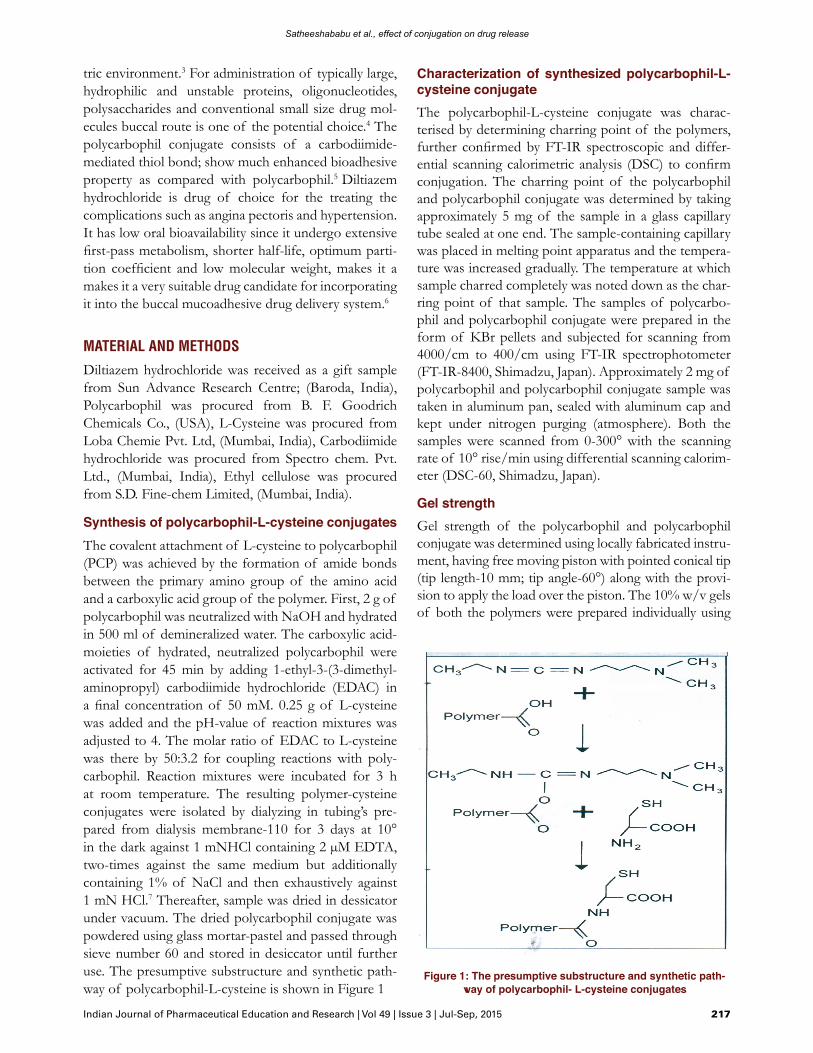
The covalent attachment of L-cysteine to polycarbophil (PCP) was achieved by the formation of amide bonds between the primary amino group of the amino acid and a carboxylic acid group of the polymer. First, 2 g of polycarbophil was neutralized with NaOH and hydrated in 500 ml of demineralized water. The carboxylic acid-moieties of hydrated, neutralized polycarbophil were activated for 45 min by adding 1-ethyl-3-(3-dimethyl-aminopropyl) carbodiimide hydrochloride (EDAC) in a final concentration of 50 mM. 0.25 g of L-cysteine was added and the pH-value of reaction mixtures was adjusted to 4. The molar ratio of EDAC to L-cysteine was there by 50:3.2 for coupling reactions with poly-carbophil. Reaction mixtures were incubated for 3 h at room temperature. The resulting polymer-cysteine conjugates were isolated by dialyzing in tubing’s pre-pared from dialysis membrane-110 for 3 days at 10° in the dark against 1 mNHCl containing 2 µM EDTA, two-times against the same medium but additionally containing 1% of NaCl and then exhaustively against 1 mN HCl.7 Thereafter, sample was dried in dessicator under vacuum. The dried polycarbophil conjugate was powdered using glass mortar-pastel and passed through sieve number 60 and stored in desiccator until further use. The presumptive substructure and synthetic path-way of polycarbophil-L-cysteine is shown in Figure 1
Characterization of synthesized polycarbophil-L-cysteine conjugate
The polycarbophil-L-cysteine conjugate was charac-terised by determining charring point of the polymers, further confirmed by FT-IR spectroscopic and differ-ential scanning calorimetric analysis (DSC) to confirm conjugation. The charring point of the polycarbophil and polycarbophil conjugate was determined by taking approximately 5 mg of the sample in a glass capillary tube sealed at one end. The sample-containing capillary was placed in melting point apparatus and the tempera-ture was increased gradually. The temperature at which sample charred completely was noted down as the char-ring point of that sample. The samples of polycarbo-phil and polycarbophil conjugate were prepared in the form of KBr pellets and subjected for scanning from 4000/cm to 400/cm using FT-IR spectrophotometer (FT-IR-8400, Shimadzu, Japan). Approximately 2 mg of polycarbophil and polycarbophil conjugate sample was taken in aluminum pan, sealed with aluminum cap and kept under nitrogen purging (atmosphere). Both the samples were scanned from 0-300° with the scanning rate of 10° rise/min using differential scanning calorim-eter (DSC-60, Shimadzu, Japan).
Gel strength
Gel strength of the polycarbophil and polycarbophil conjugate was determined using locally fabricated instru-ment, having free moving piston with pointed conical tip (tip length-10 mm; tip angle-60°) along with the provi-sion to apply the load over the piston. The 10% w/v gels of both the polymers were prepared individually using
Figure 1: The presumptive substructure and synthetic path-way of polycarbophil- L-cysteine conjugates

Satheeshababu et al., effect of conjugation on drug release
218 Indian Journal of Pharmaceutical Education and Research | Vol 49 | Issue 3 | Jul-Sep, 2015
demineralise water as a solvent. The homogenized gel was filled in sample holder and stored below 10° in a refrigerator for 24 h. The gel strength of the polymer was determined by placing the piston tip over gel surface and the load was applied over the piston at a constant rate by adding the water using i.v. infusion set at a con-stant flow rate (100 ml/min). The load required to pierce the piston tip up to 4 mm in the gel was taken as the gel strength of that polymer. The temperature of the gel was maintained below 10° throughout the study. 7
Drug-polymers compatibility study
The compatibility of drug with polymers was determined by subjecting the drug, polymers and physical mixture of drug and polymers (1:1) for DSC. Approximately 2 mg of each sample was taken in aluminum pan, sealed with aluminum cap and kept under nitrogen purging (atmo-sphere). The samples were scanned from 0-300° with the scanning rate of 10° rise/min using differential scanning calorimeter (DSC-60, Shimadzu, Japan).
Preparation of bilayered buccoadhesive tablets
The bilayered buccoadhesive tablets were prepared by a direct compression procedure which involved two con-secutive steps. In the first step the buccoadhesive drug core tablet was compressed using various proportions of polycarbophil and polycarbophil-L-cysteine conju-gate with drug composition of buccoadhesive drug core tablet as given in Table 1. The 10.05 mm, round-shaped flat punch in a single station tablet compression machine (Cadmach, Ahmedabad, India) was used. The subse-quently the buccoadhesive drug core tablet was placed in the center of a 12.60 mm diameter die cavity and ethyl celluose was poured on the in the sides and top of the buccoadhesive drug core tablet. The content of the die cavity was subjected to compression with suitable punch to get a bilayered buccoadhesive drug core tablet consist of impermeable cap in all the sides except one side through which a unidirectional drug permeation take place. Design of the tablet is given in Figure 2.8
Weight uniformity, hardness and thickness
The compressed buccoadhesive drug core tablets were characterised for weight uniformity by weighing 20 tablets of each formulation using an electronic balance (Citizen Balance CY 220). The hardness of the tablets of each for-mulation was determined using Pfizer hardness tester. The thickness of the tablets of each formulation was measured using a dial thickness apparatus (Mitutoyo 2046F, Japan).
Drug content
For determination of drug content, buccoadhesive drug core tablets were crushed in glass mortar-pastel and the powder was shaken with 100 ml of distilled water for 3 h, the solution was filtered using Whatman filter paper and analyzed after appropriate dilution by UV spectro-photometer (1601, Shimadzu, Kyoto, Japan) at 237 nm.
Swelling study
The buccoadhesive drug core tablets were weighed indi-vidually (W1) and placed separately in 2% agar gel (pH 6.8) plates with the core facing the gel surface and incu-bated at 37 ± 1°. At regular 1 h time intervals until 8 h, the tablet was removed from the petri dish. The swollen tablet was then reweighed (W2) and the swelling index (SI) of each batch was calculated using the following Eqn, percent SI = (W2 - W1)/W1× 100.9
Moisture uptake study
The buccoadhesive drug core tablets were weighed indi-vidually (W1) and exposed at temperature 40 ± 2° and relative humidity 75 ± 5% in programmable environ-mental test chamber (CHM-10S, Remi Instruments Ltd., Mumbai, India) till the weight of the tablet remained constant (W2). The percent moisture uptake was calcu-lated using the following Eqn. Percent moisture uptake = (W2- W1)/W1× 100.
Ex vivo bioadhesion strength
A modified balance method was used for determina-tion of the ex vivo bioadhesion strength. The balance
Table 1: Formulation variables of various bilayerd bioadhesive tablets
Formulation codeF1 F2 F3 F4 F5 F6
Adhesive layer - - - - - -
Diltiazemhydrochloride (mg)
30 30 30 30 30 30
Polycarbophil conjugate l(mg)
- 60 54 48 42 30
Polycarbophil (mg) 60 - 6 12 18 30
Impermeable cap - - - - - -
Ethylcellulose (mg) 75 75 75 75 75 75Figure 2: A schematic illustration of bilayered bioadhesive
tablet

Satheeshababu et al., effect of conjugation on drug release
Indian Journal of Pharmaceutical Education and Research | Vol 49 | Issue 3 | Jul-Sep, 2015 219
was modified by replacement of one pan with the metal shaft 5 g heavier in weight than pan. Fresh porcine buc-cal mucosa obtained from local slaughterhouse was cut into pieces, washed with distilled water followed by phosphate buffer pH 6.8. A piece of buccal mucosa was fixed in a Petridish with instant adhesive, which was filled with phosphate buffer pH 6.8 so that it just touched the mucosal surface. The tablet was stuck to the lower side of a shaft with instant adhesive. The two sides of the balance were made equal before the study by keeping a 5 g weight on the right hand pan. A weight of 5 g was removed from the right hand pan, which lowered the shaft along with the tablet over the mucosa. The balance was kept in this position for 3 min contact time. The weight was added slowly to the right hand pan until the tablet detached from the mucosal surface. This detachment force gave the bioadhesion strength of the buccoadhesive tablet in g (total weight on right hand pan minus 5 g). The following parameters were also cal-culated using following Equ. Force of adhesion (N) = Bioadhesion strength × 9.81/1000 and Bond strength (N/m2) = Force of adhesion (N)/Surface area (m2).10
Ex vivo bioadhesion time
The ex vivo bioadhesion time was determined using freshly cut porcine buccal mucosa. The fresh porcine buccal mucosa was obtained from a local slaughter-house and used within 3 h of slaughter. The porcine buccal mucosa was cut into pieces, washed with distilled water and then with phosphate buffer pH 6.8, and fixed on the inner wall of a 250 ml beaker with instant adhe-sive and a mucoadhesive core side of tablet was wetted with 1 drop of phosphate buffer pH 6.8 and pasted on porcine buccal mucosa by applying a force of 5 g for 30 seconds. The beaker was filled with 200 ml of phos-phate buffer pH 6.8 and was kept at 37 ± 1°. After 2 min, a 50 rpm stirring rate was applied by a magnetic stirrer to simulate the buccal cavity environment, and tablet adhesion was monitored. The time for the tablet to detach from the porcine buccal mucosa was recorded as the bioadhesion time.10
In vitro drug release study
In vitro drug release was performed by fixing the imper-meable layer of the tablet with a glass slide using instant adhesive, and placed in a beaker containing 200 ml phosphate buffer pH 6.8 as dissolution medium. The temperature was maintained at 37 ± 0.5° and the hydrodynamics was maintained by stirring on a mag-netic stirrer at 50 rpm.11 5 ml aliquots were withdrawn at predetermined time intervals and replaced with fresh medium. The aliquots were analyzed after appropriate dilution by UV spectrophotometer (1601, Shimadzu, Kyoto, Japan) at 237 nm.
In vitro drug permeation
In vitro drug permeation through dialysis membrane-110 was performed using modified Franz diffusion cell at 37 ± 0.5°. The dialysis membrane-110 was mounted between the donor and receptor compartments. The tablet was placed with the core facing the membrane and a 5 g weight was placed over the tablet, the receptor compartment (16 ml capacity) was filled with phosphate buffer pH 7.4 and the hydrodynamics in the receptor compartment was maintained by stirring on a mag-netic stirrer at 50 rpm. A 1 ml aliquot was withdrawn at predetermined time intervals and replaced with fresh medium. The aliquots were analyzed after appropriate dilution by UV spectrophotometer (1601, Shimadzu, Kyoto, Japan) at 237 nm.
Ex vivo drug permeation
Ex vivo drug permeation through the porcine buccal mucosa was performed using modified Franz diffu-sion cell at 37 ± 0.5°. The freshly cut porcine buccal mucosa after removing underlying fat and loose tissues and washing with phosphate buffer pH 6.8 and dis-tilled water was mounted between the donor and recep-tor compartments. The receptor compartment (16 ml capacity) was filled with phosphate buffer pH 7.4, and the buccal mucosa was allowed to stabilize for 30 min by hydrodynamics in the receptor compartment was main-tained by stirring on a magnetic stirrer at 50 rpm and was maintained for the entire study. A 1 ml aliquot was withdrawn at predetermined time intervals and replaced with fresh medium. The aliquots were analyzed after appropriate dilution by UV spectrophotometer (1601, Shimadzu, Kyoto, Japan) at 237 nm.
RESULTS
The charring point of polycarbophil and polycarbophil conjugate were found to be 230° and 275° respectively. This might be due to conjugation of polycarbophil with L-cysteine. The FT-IR spectra of polycarbophil conju-gate, showed the bands representing the -C=O stretch-ing of amide bond (at 1560/cm), -NH stretching (at 3759.39/cm) and -SH stretching (at 2364.81/cm) which were absent in the FT-IR spectra of the polycarbophil. The FT-IR spectra of polycarbophil conjugate, showed the band at 3759.39/cm due to the conjugation through the amide bond and another additional peak at 2364.81/cm due to entering of thiol group after conjugation. In polycarbophil the peak at 1741.77/cm represent the -C=O group of carboxylic acid but in polycarbophil conjugate this peak was shifted to 1560/cm due to the electronic effect of the amino group. In polycarbophil conjugate, other additional peaks were seen at 1417.73/

Satheeshababu et al., effect of conjugation on drug release
220 Indian Journal of Pharmaceutical Education and Research | Vol 49 | Issue 3 | Jul-Sep, 2015
cm due to C-N stretching and at 773.48/cm due to N-H bending which were also absent in the FT-IR spectra of polycarbophil because of the absence of amino group in the polymer structure. The additional peaks in the FT-IR spectra of the polycarbophil conjugate have confirmed the conjugation of polycarbophil with L-cysteine. The FT-IR spectra of polycarbophil and polycarbophil conjugate are shown in Figure 3. The DSC thermogram of polycarbophil conjugate, have shown one extra exothermic peak at 79.95° represented the amino group, which was absent in the DSC ther-mogram of polycarbophil. This has further confirmed the conjugation of polycarbophil with L-cysteine. The DSC thermograms of polycarbophil and polycarbophil conjugate are shown in Figure 4. Gel strength of the polycarbophil was found to be 254.01 ± 1.32 g whereas
the polycarbophil conjugate showed gel strength of less than 10 g (n=3).DSC thermogram of the drug showed the sharp endo-thermic peak at 215.73° has suggested the purity of the drug. Polycarbophil showed broad endothermic peak at 92.99° and at 267.71° whereas polycarbophil conjugate showed one extra exothermic peak at 79.95° along with the peaks showed by polycarbophil which represented the presence of amino group. The physical mixture of drug, polycarbophil and polycarbophil conjugate showed peaks which represented polycarbophil and polycarbophil conjugate with an endothermic peak at 212.94° which represented the drug. The comparative DSC thermograms are shown in Figure 5.The prepared tablets of each formulation showed acceptable uniformity of weight.12 The hardness, thick-
Figure 4: DSC thermograms of (A) polycarbophil and (B) polycarbophil conjugate
Figure 5: DSC thermograms of pure drug (A), polymers (B, C) and mixture of drug and polymers (D)
Figure 3: FT-IR spectra of (A) polycarbophil and (B) polycarbophil conjugate

Satheeshababu et al., effect of conjugation on drug release
Indian Journal of Pharmaceutical Education and Research | Vol 49 | Issue 3 | Jul-Sep, 2015 221
ness and drug content for the tablets of each formula-tion are shown in Table 2. The swelling index for the tablets of F1 to F6 varied from 19.68 ± 0.67% to 49.91 ± 1.17%. The swelling profile of the tablets is shown in figure 6. The percent moisture uptake for the tablets of F1 to F6 varied from 5.45 ± 0.31% to 27.36 ± 0.43%. The results of moisture uptake study are shown in Table 2 and Figure 7. The ex vivo bioadhesion strength for the tablets of F1 to F6 varied from 5.83 ± 0.18 g to 12.33 ± 0.31 g. The results of the ex vivo bioadhesion strength, the force of adhesion and the bond strength for the
tablets are shown in Table 2. The ex vivo bioadhesion time for formulation F1 was 6 h and all other formula-tion were more than 24 h. The tablets were evaluated for in vitro drug release and the cumulative percent drug released was calculated. The tablets contained only poly-carbophil (F1) have shown 29 ± 0.24% drug release in 3 h whereas tablets contained only polycarbophil conju-gate (F2) have shown 91.26 ± 1.81% drug release in 3 h. The tablets contained polycarbophil conjugate to poly-carbophil in ratios of 9:1 (F3) and 8:2 (F4) have shown 99.87 ± 0.69% and 99.76 ± 0.45% drug release in 3 h and 2 h respectively. The tablets contained polycarbo-
Table 2: Physical evaluation parameters of various bilayerd buccoadhesive tablets
Code Hardness (kg/cm2)
Thickness (mm)
Drug content (%)
Moisture uptake (%)
Bioadhesion strength*(g)
Force of adhesion (N)
Bond strength(N/
m2)F1 4.06±0.18 1.35±0.01 99.47±0.73 5.45±0.31 5.83±0.18 0.0571 72.01
F2 4.11±0.20 1.34±0.02 98.87±1.06 27.36±0.43 12.33±0.31 0.1209 152.47
F3 3.96±0.27 1.33±0.02 99.91±0.42 24.09±0.98 10.83±0.92 0.1062 133.93
F4 4.12±0.16 1.30±0.01 99.38±0.67 22.89±0.91 10.50±0.89 0.1030 129.90
F5 4.05±0.14 1.31±0.01 97.81±1.53 20.48±0.83 9.66±0.27 0.0947 119.43
F6 4.03±0.22 1.33±0.03 97.58±1.65 16.76±0.92 8.50±0.12 0.0833 113.38Values are mean±SD, n=3.
Figure 6: Swelling profile of various bilayered buccoadhesive tablets
F1 (♦), F2 (■), F3 (▲), F4 (◊), F5 (×) and F6 (●)Figure 7: Moisture uptake of various bilayered buccoadhesive
tablets
Figure 8: In vitro drug release profile of various bilayered buccoadhesive tablets
F1 (♦), F2 (■), F3 (▲), F4 (◊), F5 (×) and F6 (●)Figure 9: In vitro drug permeation profile of various bilayered
buccoadhesive tablets

Satheeshababu et al., effect of conjugation on drug release
222 Indian Journal of Pharmaceutical Education and Research | Vol 49 | Issue 3 | Jul-Sep, 2015
phil conjugate to polycarbophil in ratios of 7:3 (F5) and 5:5 (F6) have shown 85.98 ± 1.04% and 47.84 ± 1.3% drug release in 3 h. The in vitro drug release profile is shown in Figure 8.The tablets were also evaluated for in vitro drug perme-ation and the cumulative percent drug permeated was calculated. The tablets contained only polycarbophil have shown (F1) 20.65 ± 1.02% drug permeation in 8 h whereas the tablets contained only polycarbophil conju-gate have shown (F2) 35.85 ± 0.96% drug permeation in 8 h. The tablets contained polycarbophil conjugate to poly-carbophil in ratios of 9:1 (F3), 8:2 (F4), 7:3 (F5) and 5:5 (F6) have shown 46.31 ± 1.32%, 60 ± 1.74%, 32 ± 0.96% and 27.53 ± 1% drug permeation in 8 h respectively. The in vitro drug permeation profile is shown in Figure 9.The tablets contained polycarbophil conjugate and polycarbophil in proportion of 8:2 (F4) was selected for ex vivo drug permeation study on the basis of bio-adhesive properties, in vitro drug release and in vitro drug permeation study and it showed 55 ± 1.16% drug permeation through the porcine buccal mucosa in 8 h. The comparison of in vitro and ex vivo drug perme-ation profile is shown in Figure 10. The drug perme-ation data were analyzed for the rate and mechanism of drug permeation using zero order, first order, Higuchi and Korsemeyer-Peppas models. The r2 values for zero order, first order and Higuchi’s equations and n values for Korsemeyer-Peppas equation are shown in Table 3.
DISCUSSION
The significant difference in charring point of poly-carbophil and polycarbophil conjugate has suggested that there might be the conjugation of polycarbophil with L-cysteine. The FT-IR spectra have shown bands represented -NH stretching (at 3759.39/cm) and -SH stretching (at 2364.81/cm) confirmed the conjugation of polycarbophil with L-cysteine. The FT-IR spectra of polycarbophil conjugate showed the band repre-sented -C=O stretching of amide bond (at 1560/cm) which has confirmed that the conjugation has occurred
through the amide linkage only. The DSC thermogram of polycarbophil conjugate, have shown one extra exo-thermic peak at 79.95° represented the amino group, which was absent in the DSC thermogram of polycar-bophil. This has further confirmed the conjugation of polycarbophil with L-cysteine. DSC thermogram of the drug showed the sharp endothermic peak at 215.73°, (melting point range 207-213°) has suggested the purity of the drug, which retained at 212.94° in the physical mixture of drug, polycarbophil conjugate and polycar-bophil. The study suggested that the drug and polymers were compatible with each other.The tablets of each formulation have shown acceptable uniformity of weight and drug content with the optimum hardness and thickness. Appropriate swelling behavior of a buccoadhesive tablet is an essential property for uniform and prolonged release of drug and effective bioadhesion. The tablets of (F2) contained only poly-carbophil conjugate have shown highest swelling index (49.91 ± 1.17%). The swelling study suggested that the swelling index was increased with increased proportion of polycarbophil conjugate in the tablet. This might be due to the presence of thiol moieties (hydrophilic in nature) in the polycarbophil conjugate, which enhanced the rate of moisture uptake and thus the swelling index. The conjugation also causes the opening of the poly-mer chains which facilitates the polymer to hold more amount of water in its matrix, whereas the polycarbo-phil forms a rigid matrix and thus reduces the swelling index as it will not allow to take up and to hold more amount of water within the matrix. The tablets of (F2) contained only polycarbophil conjugate have shown highest moisture uptake (27.36 ± 0.43%). The mois-ture uptake study suggested that the moisture uptake was increased with increased proportion of polycarbo-phil conjugate in the tablet. This might be due to the presence of thiol moieties (hydrophilic in nature) in the polycarbophil conjugate, which enhanced the rate of moisture uptake. The conjugation also causes the open-ing of the polymer chains which facilitates the polymer to hold more amount of water in its matrix, whereas
Table 3: Kinetics of drug permeation from bilayered bioadhesive tablets
Formulation code Zero order equation (r2 value)
First order equation (r2 value)
Higuchi’s equation (r2 value)
Korsemeyer-Peppas equation (n value)
F1 0.9886 0.9909 0.9388 1.1062
F2 0.9887 0.9781 0.893 1.1027
F3 0.9954 0.9805 0.9087 1.0486
F4 0.9894 0.9535 0.8897 1.0839
F5 0.9928 0.9941 0.9357 1.0982
F6 0.9924 0.9927 0.9293 1.0648
F4 (ex vivo) 0.9819 0.9482 0.8707 1.1808Values are mean±SD, n=3.
Satheeshababu et al., effect of conjugation on drug release
Indian Journal of Pharmaceutical Education and Research | Vol 49 | Issue 3 | Jul-Sep, 2015 223
the polycarbophil forms a rigid matrix and thus it will not allow to take up and to hold more amount of water within the matrix. The tablets contained polycarbophil conjugate in different proportions with polycarbophil have shown good ex vivo bioadhesion strength, ex vivo bioadhesion time, force of adhesion and bond strength than the formulation contained polycarbophil only. The improved bioadhesive properties exhibited by the polycarbophil conjugate may be explained by the pres-ence of thiol groups in polycarbophil conjugate which supposed to interact with cysteine rich subdomains of mucus glycoprotein’s via disulfide exchange reactions and this resulted in the formation of stronger cova-lent bonds between the polymer and the mucus layer.13
Whereas in case of polycarbophil, due to the absence of thiol groups there would be formation of weak non-covalent bonds only. A very significant change in the gel strength of polycarbophil was observed after the conju-gation with L-cysteine. This might be due to opening of the polymer chains after conjugation which resulted in the formation of a very soft gel as it retained more amount of moisture. Whereas in the polycarbophil formed a hard gel as the amount of moisture retained was less due to the rigidity of the matrix. The reduction in the gel strength of polycarbophil after the conjuga-tion has further supported the results of swelling index, moisture uptake, in vitro drug release and in vitro drug permeation of the tablets contained different propor-tion of polycarbophil conjugate.The tablets prepared with polycarbophil conjugate in different proportions with polycarbophil (F2 to F6) have shown faster rate of in vitro drug release than the tablets prepared with only polycarbophil (F1). This might be due to the conjugation, which introduced thiol groups in the polycarbophil, which were able to take up more amount of moisture at a faster rate and thus allowed the polymer matrix to swell at a faster rate due to its hydro-philic nature. The conjugation also causes opening of the polymer chains which will not resist the drug diffu-sion and consequently, give rise to more rapid release of drug. This would be the reason for the amount of drug released with increased proportion of polycarbophil conjugate in the tablets. In vitro drug permeation was found to be more for the tablets contained polycarbophil conjugate in different proportions with polycarbophil (F2 to F6) than the tablets contained only polycarbophil (F1). The reason for higher drug permeation from the tablets contained polycarbophil conjugate in different proportions is same as discussed for in vitro drug release the drug permeation data were analyzed for the rate and mechanism of drug permeation using zero order, first order, Higuchi and Korsemeyer-Peppas models. The in vitro drug permeation of the selected tablets (F4) fol-
lowed zero order kinetics (r2=0.9894) and the mecha-nism of drug permeation was found to be super case II (n=1.0839). The selected tablets (F4) were subjected for ex vivo drug permeation study, showed good drug permeation (54.90 ± 1.16%) through the porcine buc-cal mucosa in 8 h. The ex vivo drug permeation of F4 followed zero order kinetics (r2=0.9819) and the mecha-nism of drug permeation was found to be super case II (n=1.1808). The comparative in vitro and ex vivo drug permeation profile suggested the same drug release pat-tern from this formulation. The r2 value (0.8897) of Higuchi’s equation has suggested that the drug release from the tablet matrix is diffusion controlled.
CONCLUSION
The conjugation of the polycarbophil with L-cyste-ine was primarily confirmed on the basis of charring point of the polycarbophil and polycarbophil conjugate which was further confirmed by FT-IR and DSC. The drug and polymers were subjected for the compatibility study using DSC, which suggested that there was no sig-nificant interaction between the drug and polymers. The various formulations of bilayered buccoadhesive tablets of diltiazem hydrochloride were prepared using polycar-bophil and polycarbophil conjugate in different propor-tions. The bilayered design of the tablet was modified from the conventional bilayered tablet design to achieve perfect unidirectional drug release by incorporating the impermeable cap of ethyl cellulose over the polymeric core containing the drug by leaving only one side of the core to release drug. This design was able to provide perfect unidirectional drug release directly towards the buccal mucosal lining and by preventing the drug loss in the saliva. The gel strength of the polycarbophil conju-gate have supported the results obtained from swelling study, moisture uptake study, in vitro drug release and in vitro drug permeation.The tablets were evaluated by different parameters such as weight uniformity, hardness, thickness, drug content, swelling index, moisture uptake, ex vivo bioadhesion strength, ex vivo bioadhesion time; in vitro drug release and in vitro drug permeation; ex vivo drug permeation study was carried out using porcine buccal mucosa in modified Franz diffusion cell. The tablets contained different proportion of polycarbophil conjugate have shown improved and promising ex vivo bioadhesive properties with improved in vitro and ex vivo drug per-meation. Results of in vitro permeation study have sug-gested that the selected tablets (F4) followed zero order drug permeation rates and the drug transport mecha-nism was found to be super case-II. The tablets (F4) were selected for the ex vivo drug permeation study

Satheeshababu et al., effect of conjugation on drug release
224 Indian Journal of Pharmaceutical Education and Research | Vol 49 | Issue 3 | Jul-Sep, 2015
on the basis of swelling study, moisture uptake study, bioadhesive parameters, in vitro drug release and drug permeation studies. It has shown a good ex vivo drug permeation through the porcine buccal mucosa.
ACKNOWLEDGEMENTThe authors expressed gratitude towards Principal National college of pharmacy Shimoga for providing facility to carryout the research work.
REFERENCES1. Chein YW. Novel drug delivery systems. 2nd ed. New York: Marcel Dekker
Inc; 1992.
2. Patel VM, Prajapati BG, Patel MM. Formulation evaluation and comparison
of bilayered and multilayeredmucoadhesivebuccal devices of propranolol
hydrochloride. AAPS PharmSciTech 2007;8:E1-8.
3. Miller NS, Chittchang M, Johnston TP. The use of mucoadhesive polymers in
buccal drug delivery. Adv Drug Deliv Rev 2005;57:1666-91.
4. Shojaei AH. Buccal mucosa as a route for systemic drug delivery: A review. J
Pharm PharmSci 1998;1:15-30.
5. Langoth N, Kalbe J, Bernkop-Schnürch A. Development of buccal drug
delivery systems based on a thiolated polymer. Int J Pharm 2003;252:141-48.
6. Singh B, Ahuja N. Development of controlled-release buccoadhesive
hydrophilic matrices of diltiazem hydrochloride: optimization of bioadhesion,
dissolution, and diffusion parameters. Drug DevInd Pharm 2002; 28:431-42.
7. Satheeshababu BK and shivakumar KL. Synthesis of conjugated chitosan and its effect on drug permeation from transdermal patches. Indian J Pharm Sci 2013; 75:162-70.
8. Bernkop-Schnürch A, Steininger S. Synthesis and characterization of mucoadhesivethiolated polymers. Int J Pharm 2000;194:239-247
9. Parodi B, Russo E, Caviglioli G, Cafaggi S, Bignardi G. Development and characterization of a buccoadhesive dosage form of oxycodone hydrochloride. Drug DevInd Pharm 1996;22:445-50.
10. Satishbabu BK, Srinivasan BP. Preparation and evaluation of buccoadhesive films of atenolol. Indian J Pharm Sci 2008; 70:175-79.
11. Borodkin S, Tucker FE. Drug release from hydroxypropylcellulose-polyvinyl acetate films. J Pharm Sci 1974; 63:1359-64.
12. Indian Pharmacopoeia. Delhi: The Controller of Publications; 1996. vol 1.13. Kast CE, Bernkop-Schnürch A. Thiolated polymers-thiomers: development
and in vitro evaluation of chitosan-thioglycolic acid conjugates. Biomaterials 2001; 22:2345-52.