congenital deformity of the spine-classification ... · pdf filecongenital deformity of the...
TRANSCRIPT

1140 Wien, Penzingerstrasse 63
Tel: +43 1 894 61 46
Stand: 18.1.2012
Congenital deformity of the spine-classification, diagnosis and therapy Author: Univ. Doz. Dr. Werner Lack
Classification of congenital scoliosis failure of formation-wedge vertebrae (2 pedicles), half vertebrae… unsegmented half vertebra: fused to the vertebral body above and below partially segmented half vertebra fully segmented half vertebra-separated above and below by disc space failure of segmentation-block vertebrae, unilateral bar… combination Special combinations hemimetameric shift-hemivertebra counterbalanced by another one on the contralateral side in the same region unilateral bar and contralateral hemivertebra-“worst case scenario”, progression per year up to 10° Cobb!
Picture: worst case scenario: unilateral bar and contralateral half-vertebra Severe progression
hemivertebra thoracolumbar 2-3,5°/year 2 hemivertebrae 5° unilateral unsegmented bar 6-9° unilateral unsegmented bar with contralateral hemivertebra >10° most severe progression in thoracolumbar region! most severe progression up to 5th year and in adolescence/puberty
Classification of congenital kyphosis (Mc Master) failure of formation posterolateral quadrant hemivertebra butterfly vertebra posterior hemivertebra wedge vertebra failure of segmentation anterior unsegmented bar block vertebra mixed anormalies unclassifiable anomalies

1140 Wien, Penzingerstrasse 63
Tel: +43 1 894 61 46
Stand: 18.1.2012
Congenital vertebral displacement-sudden sagittal kyphotic displacement, most severe of congenital kyphosis
Picture: there exist no known genetic abnormalities associated with the development of congenital kyphosis
Picture: posterior half vertebra Klippel-Feil-syndrome
segmentation defects of the cervical spine plagiocephaly reduced cervical spine mobility
anomalies of urinary system in one third cong. Heart anomalies in 15%
Sprengel-deformity in 20-30% problems in hearing in one third Sprengel deformity
congenital failure of descendence of one or both shoulders; mostly left shoulder elevated „omovertebral bone“ (connection scapula-C5 or C6) often Klippel-Feil, rib anomalies… 75% girls scapula small and rotated

1140 Wien, Penzingerstrasse 63
Tel: +43 1 894 61 46
Stand: 18.1.2012
Picture: Klippel-Feil with Sprengel deformity
Congenital sacral problems lumbosacral abnormalities (partial, total, asymmetric, symmetric) sacral obliquity sacral dysplasias up to sacral agenesis sacral dysraphism (MMC)
Congenital sacral obliquity angular deformation of the sacrum from a horizontal line drawn parallel from a line across the femoral heads from congenital origin! it is not a pelvic obliquity! mostly sacral endplate elevated at the right side max.20°, but can cause lumbar scoliosis up to 50°-effect like a lumbar hemivertebra
Congenital sacral agenesia (Renshaw 1978)
1) unilateral agenesis, partial or total 2) symmetrical partial with stable articulation iliac bone-S1 3) variable lumbar and total sacral agenesis with articulation iliac bone-lowest lumbar vertebra 4) variable lumbar and total sacral agenesis with either fused iliac bones or amphiarthrosis
Symptoms of severe forms often maternal diabetes
in severe cases bowel and bladder dysfunction deformities of hips (flexion, abduction, outward rotation), knees (flexion contracture), feet, atrophy of the legs, motoric palsy („Buddha-like-position“)
Congenital rib deformities mostly in combination with congenital scoliosis, rarely with congenital kyphosis 70% had thoracic or thoracolumbar concomitant congenital scoliosis

1140 Wien, Penzingerstrasse 63
Tel: +43 1 894 61 46
Stand: 18.1.2012
mostly on concave side of unilateral failure of segmentation 40% with Sprengel deformity prognosis without difference to scoliosis without rib fusions
Associated anomalies syringomyelia (abnormal fluid collection in the medullary canal of the spine, caused by Arnold-Chiari-syndrome, basilar invagination, cord compression, trauma, arachnoiditis, can cause scoliosis, kyphosis, motor weakness, dyscoordination, neuropathic arthropathy, pain)
Picture: Chiari malformation (caudal dislocation of cerebellar tonsils below the foramen magnum), diastematomyelia, diplomeylia-fibrous ligament or osseous bar, mostly in lumbar spine
Picture: diastematomyelia tethered cord (filum terminale pulls cord down to L4)…MRI brain stem and complete spine; release always before spine surgery, better not at the same operation; seldom spontaneous release of symptoms in scoliosis >40° congenital heart disease (25%)-septum defects, hypoplastic left heart, transposition… echocardiogram genitourinary anomalies (20%)-renal aplasia, duplicate ureters, hypospadia…renal ultrasound anamnesis for neurologic infantile symptoms is the child toilet trained ? bed wetting problem ? bowel or bladder „accidents“ ? limping ? shoe size difference ? clinical evaluation
spinal dysraphm ? asymmetric calves, cavus feet, clubfeet, vertical tali ? truncal or pelvic imbalance ?

1140 Wien, Penzingerstrasse 63
Tel: +43 1 894 61 46
Stand: 18.1.2012
spinal balance frontal and sagittal rib cage deformity ? in-and exspiration capacity neurologic deficit ?
standing and sitting size rib hump asymmetry of lumbar height height of shoulders height of pelvic rim vertical centre line radiologic parameters ap Cobb - angle (main-curve, secondary - curve) rotation (Nash and Moe 0 - 4) deviation of perpendicular line pelvic inclination Risser-sign (0-5) radiologic parameters sagittal evaluation of perpendicular line S1 evaluation of L4- (should be horizontal) measurements Cobb-angle thoracic kyphosis Cobb-angle lumbar lordosis Cobb-angle Th11-L1 (should be 0°) CT in congenital deformities bony details good details of apical hemivertebra (for hemivertebra excision) myelo-CT for complex dysraphic problems CT-models for complex deformities MRI in congenital deformities for detection of neural axis abnormalities necessary MRI from brain stem to sacrum evaluation of disks and growth potential evaluation of kidneys and lungs… Lung function „of all spinal deformity patients having early death due to cor pulmonale , congenital scoliosis patients probably have the highest frequency“ (Winter 1983) lung function analysis (total capacity, vital capacity, Tiffeneau-test…) blood gas analysis Conservative therapy
in clear cases of progression (2 hemivertebrae, unilateral bar etc no waiting ! watching and control brace therapy only in cases of compensatory curves, very infrequent indicated!
Cperative therapy-general rules better a short and straight than a long and curved spine at optimal time in most cases small operations are sufficient; in late cases frequently long operations with high complication risk are necessary waiting up to the end of growth in progressive congenital deformities is a severe mistake!
Preoperative therapy in case of severe deformity, especially with severely reduced lung function Halo-extension (Halo-gravity, Halo-wheelchair, Halo-pelvic)

1140 Wien, Penzingerstrasse 63
Tel: +43 1 894 61 46
Stand: 18.1.2012
exact neurologic examination during Halo-extension: eye muscles – looking at moving finger n. facialis – closing eyes, showing the teeth n. accessorius – lifting of shoulders n. hypoglossus – showing the tongue, speech! motor and sensoric testing of upper and lower extremities pyramid signs - Babinsky, abdominal reflex Severe complications in Halo-Pelvic-extension peritonitis by perforation of iliac screws cervikal problems >50%, degenerationen of cervikal spine, avascular necrosis of dens, spontaneous fusion (Dove et al. 1980)
Picture: Halo-wheelchair-extension Complications of Halo-ring pin-infection – change of screw position, oral antibiotics pin-loosening nerve irritation (n.supraorbitalis) intracerebral pin penetration-bleeding, pneumencephalus, brain abscess.. Halo-extension is contraindicated in rigid kyphosis apex-by extension of the proximal and distal spine in a rigid apex the myelon can be bent over the apex with following paraplegia!
1140 Wien, Penzingerstrasse 63
Tel: +43 1 894 61 46
Stand: 18.1.2012
Operation techniques for congenital deformity posterior fusion in situ convex hemiepiphyseodesis resection of half vertebra (anterior and posterior, from posterior alone) instrumented correction and fusion concavesided osteotomy and distraction (D.Jesensky) growing rods (single rod concavesided, Luque Trolley…) VEPTR (vertical expandible prosthetic vertebra rib) combined operations Fusion in situ
in segmentation defects (unilateral bar) in fully segmented hemivertebrae (as soon as diagnosis is clear!) use of implants is recommended already in toddlers posterior fusion alone mainly in kyphotic deformities
additional anterior fusion depends on the quality of disks-cave Crankshaft-phenomenon, mainly in lordotic deformities!
Convexsided hemiepiphyseodesis in cases of expected growth at the concave side (hemivertebra) optimal in children <5 years correction av.<15° anterior and posterior approach anterior: disc resection cranial and caudal of hemivertebra only to midline with bone grafting posterior only approach with use of pedicle screws to obtain anterior growth arrest by transpedicular convexsided disc resection and bone transplantation
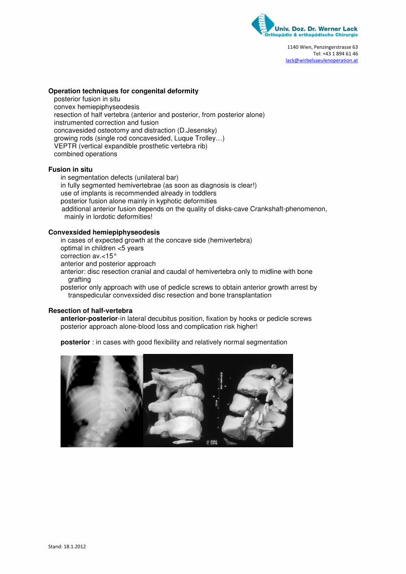
Resection of half-vertebra anterior-posterior-in lateral decubitus position, fixation by hooks or pedicle screws posterior approach alone-blood loss and complication risk higher! posterior : in cases with good flexibility and relatively normal segmentation

1140 Wien, Penzingerstrasse 63
Tel: +43 1 894 61 46
Stand: 18.1.2012
Picture: half vetebra resection
Correction and instrumented fusion
anterior-posterior: 1)in cases of less mobility in bending films 2)at risk of Crankshaft in combination with osteotomies-eventually with intermittant Halo traction (cave-no traction in rigid apex of kyphosis!) as posterior correction alone by pedicle substraction osteotomy
Anterior support in remaining kyphosis Bradford-technique of vascularized rib graft preparation of elected rib under remaining intercostal muscles cranial and caudal; anterior ligation of intercostals vessels, posterior cautious deperiostation and cut of rib under care of vessels; preparation of intercostal artery and vein to the foramen, then creation of holes into the end-vertebral bodies of kyphosis and implantation of vascularized rib; osseous integration within of 2 months!
Pictures: nonon-fusion-techniques growing rods-subcutaneous or submuscular rods
1140 Wien, Penzingerstrasse 63
Tel: +43 1 894 61 46
Stand: 18.1.2012
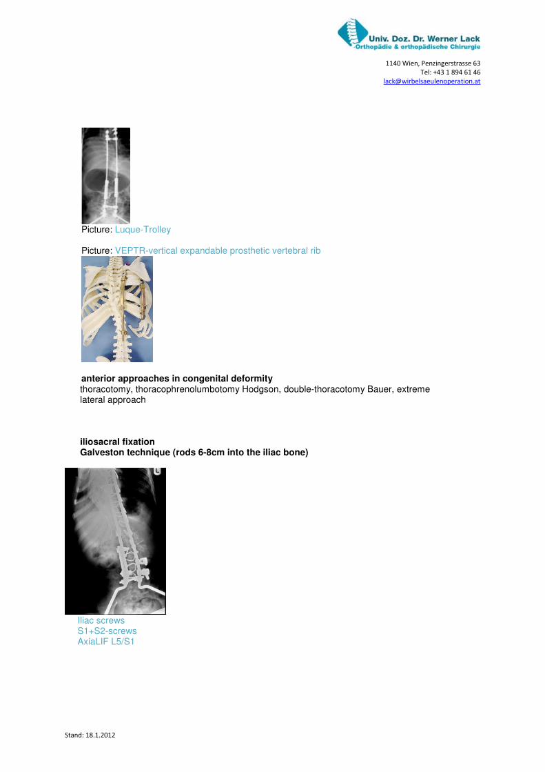
Picture: Luque-Trolley Picture: VEPTR-vertical expandable prosthetic vertebral rib
anterior approaches in congenital deformity
thoracotomy, thoracophrenolumbotomy Hodgson, double-thoracotomy Bauer, extreme lateral approach iliosacral fixation Galveston technique (rods 6-8cm into the iliac bone)
Iliac screws S1+S2-screws AxiaLIF L5/S1

1140 Wien, Penzingerstrasse 63
Tel: +43 1 894 61 46
Stand: 18.1.2012
Picture: AxiaLIF als anterior lumbosacral support in long-distance fusion