congenital brain abnormalities: pictorial essay
TRANSCRIPT

Eastern Journal of Medicine 20 (2015) 11-19
Original Article
11
Congenital brain abnormalities: Pictorial essay
Abdussamet Batura,* and M. Emin Sakaryab
aDepartment of Radiology, Yüzüncü Yıl University, Dursun Odabaş Medical Center, Van, Turkey bDepartment of Radiology, Necmettin Erbakan University, Meram School of Medicine, Konya Abstract. Among all fetal anomalies, the central nervous system anomalies represent one of the most frequently involved structures with an estimated incidence of 1 per 100 births. It is mostly difficult to make an accurate diagnosis of congenital brain malformation, based on clinical findings; thus use of computed tomography (CT) or magnetic resonance imaging (MRI) is essential in these cases. The aim of this essay is to state the imaging findings of the main, most prominent congenital brain abnormalities and to present a practical classification of the entity.
Key words: Congenital abnormality, central nervous system, Imaging
1. Introduction Among all fetal anomalies, the central nervous
system anomalies (CNS) represent one of the most frequently involved structures with an estimated incidence of 1 per 100 births (1). Several studies have shown that malformations of cerebral cortical development are the cause of 23% to 26% of intractable epilepsies in children and young adults (2) and according to the Burdenko Institute of Neurosurgery of Russian Academy of Medical Sciences data, 22.6% children with hydrocephalus have additional brain malformations, and 11.8% have multiple malformations (3). This number emphasizes the point that cortical malformations must be ruled out in essentially every pediatric patient with developmental delay or epilepsy. It is very difficult to make a diagnosis of congenital brain malformation, based on clinical findings, and use of CT or MRI is essential in these cases.
The aim of this essay is to demonstrate a simplified classification of congenital anomalies affecting brain, and imaging findings of the common, prominent congenital anomalies.
*Correspondence: Abdussamet Batur M.D. Yüzüncü Yıl University, Dursun Odabaş Medical Center, Department of Radiology, Van, Turkey Telephone number: 00905067928305 Fax number: 00904322167519 E-mail: [email protected] Received: 24.09.2013 Accepted: 11.02.2014
2. Materials and Methods
2.1. Imaging Technique A retrospective analysis of 16 congenital brain
abnormalities through 152 cases, from November 2009 to May 2013, was conducted. The examinations were performed on a 1.5 T equipment (SIEMENS Medical Systems) with sequences SE T1WI (TR/TE, 468/8.7 ms), FSE T2WI (TR/TE, 4000/92 ms) with a flip-angle of 30 and a field of view of 230 mm. 2.2. Anatomy
The human nervous system consists of the central nervous system (CNS) and peripheral nervous system. The brain consists of soft, delicate, non-replaceable neural tissue. It is supported and protected by the surrounding skin, skull, meninges and cerebrospinal fluid (CSF). The skin and skull constitute a protective barrier against to physical damage of underlying tissues, invasion of hazardous chemical and bacterial substances. Three meninges are connective tissue membranes enclosing the brain and the spinal cord. Their functions are to protect the CNS and blood vessels, enclose the venous sinuses, retain the cerebrospinal fluid, and form partitions within the skull. The outermost meninx is the dura mater, which encloses the arachnoid mater and the innermost pia mater. CSF is a watery liquid similar in composition to blood plasma. It is formed in the choroid plexuses and circulates through the ventricles into the subarachnoid space, where it is returned to the dural venous sinuses and absorbed by the arachnoid villi. The main purpose of the CSF is to support and cushion of the brain and helping to nourish it.

A. Batur et al / Congenital brain abnormalities
12
3. Results and Discussion A number of classification systems have been
proposed for congenital brain abnormalities, but none is universally accepted. We categorized the congenital malformations of brain into disorders of: • Hindbrain malformations (Posterior fossa
malformations and cysts) • Hindbrain Herniations and Miscellaneous
Malformations • Malformations Of Cortical Development • Disorders of Diverticulation and Cleavage
3.1. Hindbrain Cystic Malformations 3.1.1. Dandy-Walker Malformation Classic Dandy-Walker malformation comprises
complete or partial vermian agenesis, cystic dilatation of the fourth ventricle, and enlargement of the posterior fossa, with elevation of the transverse sinus, tentorium, and torcula (Fig. 1) (4). Nearly 70% of patients with DWM have associated supratentorial malformations, and up to 50% have extracranial anomalies.
Fig. 1. Sagittal T1 weighted image. The floor of fourth ventricle (arrow) is normal, but dorsally fourth ventricle opens into a large CSF filled cyst (arrowheads). Vermis is not visualized.
3.1.2. Dandy-Walker Variant The most common form of this anomaly
demonstrates partial dysgenesis of the vermis (mild vermian hypoplasia) and remnant fourth ventricle that communicates with retrocerebellar cyst (Fig. 2). Posterior fossa is of normal size (5).
3.1.3. Mega Cisterna Magna In this variant, fourth ventricle, vermis, and cerebellar hemispheres are normal. A large cisterna magna is present and may extend above
the vermis to the straight sinus (Fig. 3). Occasionally scalloping of occipital bone is seen (6,7).
Fig. 2. Sagittal T1 weighted image. There is communication (arrowheads) between fourth ventricle and cisterna magna through enlarged vallecula, with a posterior fossa cyst (arrow).
Fig. 3. Axial T2 weighted image showing mega cisterna magna (arrows). Fourth ventricle, vermis and cerebellar hemispheres are normal.
3.1.4. Arachnoid Cyst An arachnoid cyst is a congenital extracerebral
mass that contains cerebrospinal fluid (CSF) encircled by walls composed of arachnoid membrane. Internal and external walls of the cyst consist of thin layers of arachnoid cells and join
Eastern Journal of Medicine 20 (2015) 11-19
Original Article
13
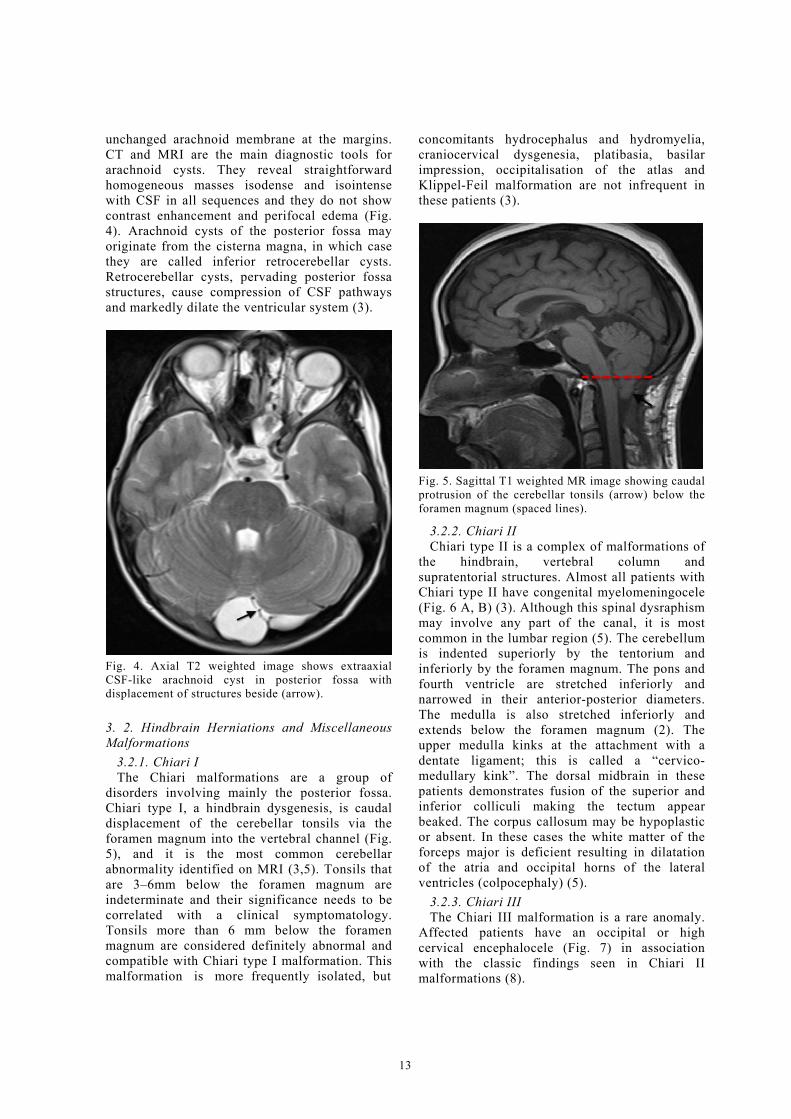
unchanged arachnoid membrane at the margins. CT and MRI are the main diagnostic tools for arachnoid cysts. They reveal straightforward homogeneous masses isodense and isointense with CSF in all sequences and they do not show contrast enhancement and perifocal edema (Fig. 4). Arachnoid cysts of the posterior fossa may originate from the cisterna magna, in which case they are called inferior retrocerebellar cysts. Retrocerebellar cysts, pervading posterior fossa structures, cause compression of CSF pathways and markedly dilate the ventricular system (3).
Fig. 4. Axial T2 weighted image shows extraaxial CSF-like arachnoid cyst in posterior fossa with displacement of structures beside (arrow). 3. 2. Hindbrain Herniations and Miscellaneous Malformations
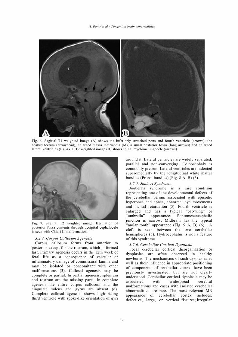
3.2.1. Chiari I The Chiari malformations are a group of
disorders involving mainly the posterior fossa. Chiari type I, a hindbrain dysgenesis, is caudal displacement of the cerebellar tonsils via the foramen magnum into the vertebral channel (Fig. 5), and it is the most common cerebellar abnormality identified on MRI (3,5). Tonsils that are 3–6mm below the foramen magnum are indeterminate and their significance needs to be correlated with a clinical symptomatology. Tonsils more than 6 mm below the foramen magnum are considered definitely abnormal and compatible with Chiari type I malformation. This malformation is more frequently isolated, but
concomitants hydrocephalus and hydromyelia, craniocervical dysgenesia, platibasia, basilar impression, occipitalisation of the atlas and Klippel-Feil malformation are not infrequent in these patients (3).
Fig. 5. Sagittal T1 weighted MR image showing caudal protrusion of the cerebellar tonsils (arrow) below the foramen magnum (spaced lines).
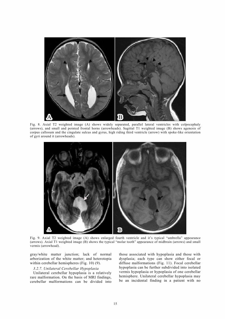
3.2.2. Chiari II Chiari type II is a complex of malformations of
the hindbrain, vertebral column and supratentorial structures. Almost all patients with Chiari type II have congenital myelomeningocele (Fig. 6 A, B) (3). Although this spinal dysraphism may involve any part of the canal, it is most common in the lumbar region (5). The cerebellum is indented superiorly by the tentorium and inferiorly by the foramen magnum. The pons and fourth ventricle are stretched inferiorly and narrowed in their anterior-posterior diameters. The medulla is also stretched inferiorly and extends below the foramen magnum (2). The upper medulla kinks at the attachment with a dentate ligament; this is called a “cervico-medullary kink”. The dorsal midbrain in these patients demonstrates fusion of the superior and inferior colliculi making the tectum appear beaked. The corpus callosum may be hypoplastic or absent. In these cases the white matter of the forceps major is deficient resulting in dilatation of the atria and occipital horns of the lateral ventricles (colpocephaly) (5).
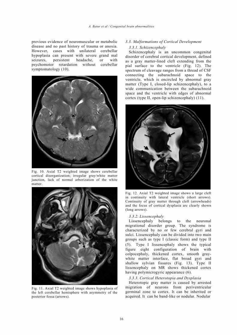
3.2.3. Chiari III The Chiari III malformation is a rare anomaly.
Affected patients have an occipital or high cervical encephalocele (Fig. 7) in association with the classic findings seen in Chiari II malformations (8).
A. Batur et al / Congenital brain abnormalities
14
Fig. 6. Sagittal T1 weighted image (A) shows the inferiorly stretched pons and fourth ventricle (arrows), the beaked tectum (arrowhead), enlarged massa intermedia (M), a small posterior fossa (long arrows) and enlarged lateral ventricles (L). Axial T2 weighted image (B) shows spinal myelomeningocele (arrows).
Fig. 7. Sagittal T2 weighted image. Herniation of posterior fossa contents through occipital cephalocele is seen with Chiari II malformation.
3.2.4. Corpus Callosum Agenesis Corpus callosum forms from anterior to
posterior except for the rostrum, which is formed last. Primary agenesia occurs in the 12th week of fetal life as a consequence of vascular or inflammatory damage of commissural lamina and may be isolated or concomitant with other malformations (3). Callosal agenesis may be complete or partial. In partial agenesis, splenium and rostrum are the missing parts. In complete agenesis the entire corpus callosum and the cingulate sulcus and gyrus are absent (6). Complete callosal agenesis shows high riding third ventricle with spoke-like orientation of gyri
around it. Lateral ventricles are widely separated, parallel and non-converging. Colpocephaly is commonly present. Lateral ventricles are indented superomedially by the longitudinal white matter bundles (Probst bundles) (Fig. 8 A, B) (6).
3.2.5. Joubert Syndrome Joubert’s syndrome is a rare condition
representing one of the developmental defects of the cerebellar vermis associated with episodic hyperpnea and apnea, abnormal eye movements and mental retardation (5). Fourth ventricle is enlarged and has a typical “bat-wing” or “umbrella” appearance. Pontomesencephalic junction is narrow. Midbrain has the typical “molar tooth” appearance (Fig. 9 A, B) (6). A cleft is seen between the two cerebellar hemispheres (5). Hydrocephalus is not a feature of this syndrome.
3.2.6. Cerebellar Cortical Dysplasia Focal cerebellar cortical disorganization or
dysplasias are often observed in healthy newborns. The mechanisms of such dysplasias as well as their influence in appropriate positioning of components of cerebellar cortex, have been previously investigated, but are not clearly understood. Cerebellar cortical dysplasia may be associated with widespread cerebral malformations and cases with isolated cerebellar abnormalities are rare. The most relevant MR appearance of cerebellar cortex includes: defective, large, or vertical fissures; irregular
Eastern Journal of Medicine 20 (2015) 11-19
Original Article
15
Fig. 8. Axial T2 weighted image (A) shows widely separated, parallel lateral ventricles with colpocephaly (arrows), and small and pointed frontal horns (arrowheads). Sagittal T1 weighted image (B) shows agenesis of corpus callosum and the cingulate sulcus and gyrus, high riding third ventricle (arrow) with spoke-like orientation of gyri around it (arrowheads).
Fig. 9. Axial T2 weighted image (A) shows enlarged fourth ventricle and it’s typical “umbrella” appearance (arrows). Axial T1 weighted image (B) shows the typical “molar tooth” appearance of midbrain (arrows) and small vermis (arrowhead). gray/white matter junction; lack of normal arborization of the white matter; and heterotopia within cerebellar hemispheres (Fig. 10) (9).
3.2.7. Unilateral Cerebellar Hypoplasia Unilateral cerebellar hypoplasia is a relatively
rare malformation. On the basis of MRI findings, cerebellar malformations can be divided into
those associated with hypoplasia and those with dysplasia; each type can show either focal or diffuse malformations (Fig. 11). Focal cerebellar hypoplasia can be further subdivided into isolated vermis hypoplasia or hypoplasia of one cerebellar hemisphere. Unilateral cerebellar hypoplasia may be an incidental finding in a patient with no
A. Batur et al / Congenital brain abnormalities
16
previous evidence of neuromuscular or metabolic disease and no past history of trauma or anoxia. However, cases with unilateral cerebellar hypoplasia can present with severe grand mal seizures, persistent headache, or with psychomotor retardation without cerebellar symptomatology (10).
Fig. 10. Axial T2 weighted image shows cerebellar cortical disorganization; irregular gray/white matter junction, lack of normal arborization of the white matter.
Fig. 11. Axial T2 weighted image shows hypoplasia of the left cerebellar hemisphere with asymmetry of the posterior fossa (arrows).
3.3. Malformations of Cortical Development 3.3.1. Schizencephaly Schizencephaly is an uncommon congenital
disorder of cerebral cortical development, defined as a gray matter-lined cleft extending from the pial surface to the ventricle (Fig. 12). The spectrum of cleavage ranges from a thread of CSF connecting the subarachnoid space to the ventricle, which is encircled by abnormal gray matter (Type I, closed-lip schizencephaly), to a wide communication between the subarachnoid space and the ventricle with edges of abnormal cortex (type II, open-lip schizencephaly) (11).
Fig. 12. Axial T2 weighted image shows a large cleft in continuity with lateral ventricle (short arrows). Continuity of gray matter through cleft (arrowheads) and the focus of cortical dysplasia are clearly shown (long arrows).
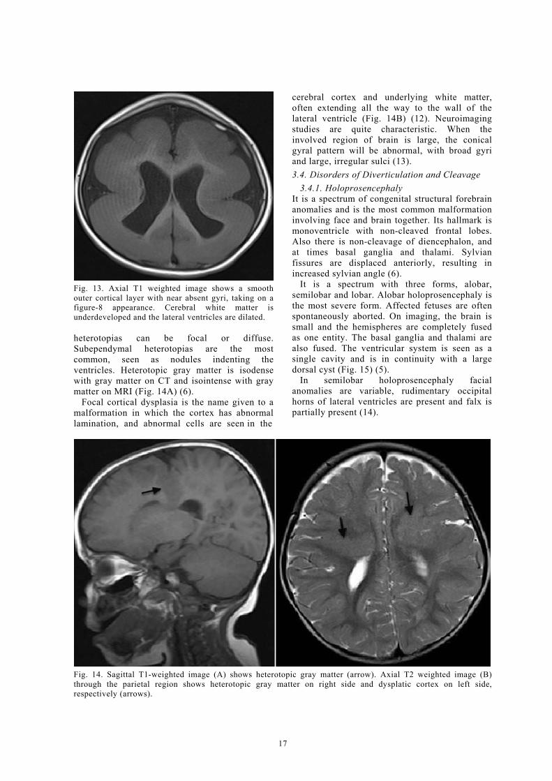
3.3.2. Lissencephaly Lissencephaly belongs to the neuronal
migrational disorder group. The syndrome is characterized by no or few cerebral gyri and sulci. Lissencephaly can be divided into two main groups such as type I (classic form) and type II (5). Type I lissencephaly shows the typical figure eight configuration of brain with colpocephaly, thickened cortex, smooth gray-white matter interface, flat broad gyri and shallow sylvian fissures (Fig. 13). Type II lissencephaly on MR shows thickened cortex having polymicrogyric appearance (6).
3.3.3. Cortical Heterotopia and Dysplasia Heterotopic gray matter is caused by arrested
migration of neurons from periventricular germinal zone to cortex. It can be inherited or acquired. It can be band-like or nodular. Nodular
Eastern Journal of Medicine 20 (2015) 11-19
Original Article
17
Fig. 13. Axial T1 weighted image shows a smooth outer cortical layer with near absent gyri, taking on a figure-8 appearance. Cerebral white matter is underdeveloped and the lateral ventricles are dilated. heterotopias can be focal or diffuse. Subependymal heterotopias are the most common, seen as nodules indenting the ventricles. Heterotopic gray matter is isodense with gray matter on CT and isointense with gray matter on MRI (Fig. 14A) (6).
Focal cortical dysplasia is the name given to a malformation in which the cortex has abnormal lamination, and abnormal cells are seen in the
cerebral cortex and underlying white matter, often extending all the way to the wall of the lateral ventricle (Fig. 14B) (12). Neuroimaging studies are quite characteristic. When the involved region of brain is large, the conical gyral pattern will be abnormal, with broad gyri and large, irregular sulci (13). 3.4. Disorders of Diverticulation and Cleavage
3.4.1. Holoprosencephaly It is a spectrum of congenital structural forebrain anomalies and is the most common malformation involving face and brain together. Its hallmark is monoventricle with non-cleaved frontal lobes. Also there is non-cleavage of diencephalon, and at times basal ganglia and thalami. Sylvian fissures are displaced anteriorly, resulting in increased sylvian angle (6).
It is a spectrum with three forms, alobar, semilobar and lobar. Alobar holoprosencephaly is the most severe form. Affected fetuses are often spontaneously aborted. On imaging, the brain is small and the hemispheres are completely fused as one entity. The basal ganglia and thalami are also fused. The ventricular system is seen as a single cavity and is in continuity with a large dorsal cyst (Fig. 15) (5).
In semilobar holoprosencephaly facial anomalies are variable, rudimentary occipital horns of lateral ventricles are present and falx is partially present (14).
Fig. 14. Sagittal T1-weighted image (A) shows heterotopic gray matter (arrow). Axial T2 weighted image (B) through the parietal region shows heterotopic gray matter on right side and dysplatic cortex on left side, respectively (arrows).

A. Batur et al / Congenital brain abnormalities
18
Fig. 15. Axial T1-weighted image shows a central monoventricle (V) with minimal residual frontal brain tissue with the fused thalami (arrow).
In lobar holoprosencephaly, the brain is generally of normal volume and shows almost complete separation into two hemispheres. The falx is often dysplastic anteriorly but otherwise normal. The ventricular system is well defined but may be dysmorphic. The septum pellucidum is absent. The corpus callosum is absent or dysmorphic (Fig. 16A, B) (15).
3.4.2. Septooptic Dysplasia Septo-optic dysplasia is a hypoplasia of optic
nerves, combined with hypoplasia or absence of the septum pellucidum. It is thought that septo-optic dysplasia is a result of different genetic abnormalities and intrauterine ischemic events during the first two trimesters of pregnancy (3). It is characterized by hypoplastic optic nerves/ tract, absence of septum pellucidum and hypothalamic-pituitary dysfunction (16). Diagnosis is confirmed by optic discs hypoplasia, absence of the septum pellucidum and optic nerves atrophy detection on CT and MRI (Fig. 17) (3).
Fig. 16. Axial T2 (A) and coronal T1 (B) weighted images show fusion of the frontal lobes (short arrows), separated thalami (arrowhead) and presence of the falx (long arrows).
Fig. 17. Coronal T2 weighted image shows absent septum pellucidum with squared-off appearance of frontal horns with inferior pointing (A). Optic nerves (long arrows) and ocular muscles (short arrows) are hypoplastic (B).

Eastern Journal of Medicine 20 (2015) 11-19
Original Article
19
References 1. Huisman TAGM, Wisser J, Martin E, Huch RK,
Marincek B. Fetal magnetic resonance imaging of the central nervous system. Eur Radiol 2002; 12: 1952-1961.
2. Barkovich AJ. Pediatric Neuroimaging (4th ed). Philadelphia: Lippincott Williams & Wilkins, 2005, pp 291-422.
3. Kornienko VN, Pronin IN. Congenital Malformations of the Brain and Skull. Diagnostic Neuroradiology 2009: 29-86.
4. Fath BMK, Garcia MAC. Prenatal Imaging of Congenital Malformations of the Brain. Semin Ultrasound CT MRI 2011; 32: 167-188.
5. Altman NR, Naidich TP, Braffman BH. Posterior fossamalformations. AJNR Am J Neuroradiol 1992; 13: 691-724.
6. Shinagare AB, Patil NK. Imaging of congenital malformations of brain: a pictorial essay. The Internet Journal of Radiology 2008; 9: 4.
7. Osborne AG. Brain development and congenital malformation. In diagnostic neuroradiology. Mosby, 1994, pp 59-70.
8. Kannegieter LS, Dietrich RB, Pais MJ, Goldenberg TM. Pediatric case of the day. RadioGraphics 1994; 14: 452-454.
9. Ares GS, Delmaire C, Deries B, Vallee L, Pruvo JP. Cerebellar Cortical Dysplasia: MR Findings in a Complex Entity. AJNR Am J Neuroradiol 2000; 21: 1511-1519.
10. Vagh JD, Gadekar A, Agrawal A, Deshmukh K. Unilateral cerebellar hypoplasia. Indian J Radiol Imaging 2009; 19: 146-147.
11. Packard AM, Miller VS, Delgado MR. Schizencephaly: Correlations of clinical and radiologic features. Neurology 1997; 48: 1427-1434.
12. Barkovich AL, Kuzniecky RI, Jackson GD, Guerrini R, Dobyns WB. Classification system for malformations of cortical development: Neurology 2001; 57: 2168-2178.
13. Barkovich AL, Kuzniecky RI, Bollen AW, Grant PE. Focal transmantle dysplasia: a specific malformation of cortical development. Neurology 1997; 49: 1148-1152.
14. Altman NR, Altman DH, Sheldon JJ, Leborgne J. Holoprosencephaly classified by computed tomography. Am J Neuroradiol. 1984; 5: 433-437.
15. Simon EM, Barkovich AJ. Holoprosencephaly: new concepts. Magn Reson Imaging Clin North Am 2001; 9: 149-164.
16. Campbell CL. Septo-optic dysplasia: a literature review. Optometry. 2003; 74: 417-426.