congenital anomalies of superior vena cava and … · failure and/or complications of cvc...
TRANSCRIPT

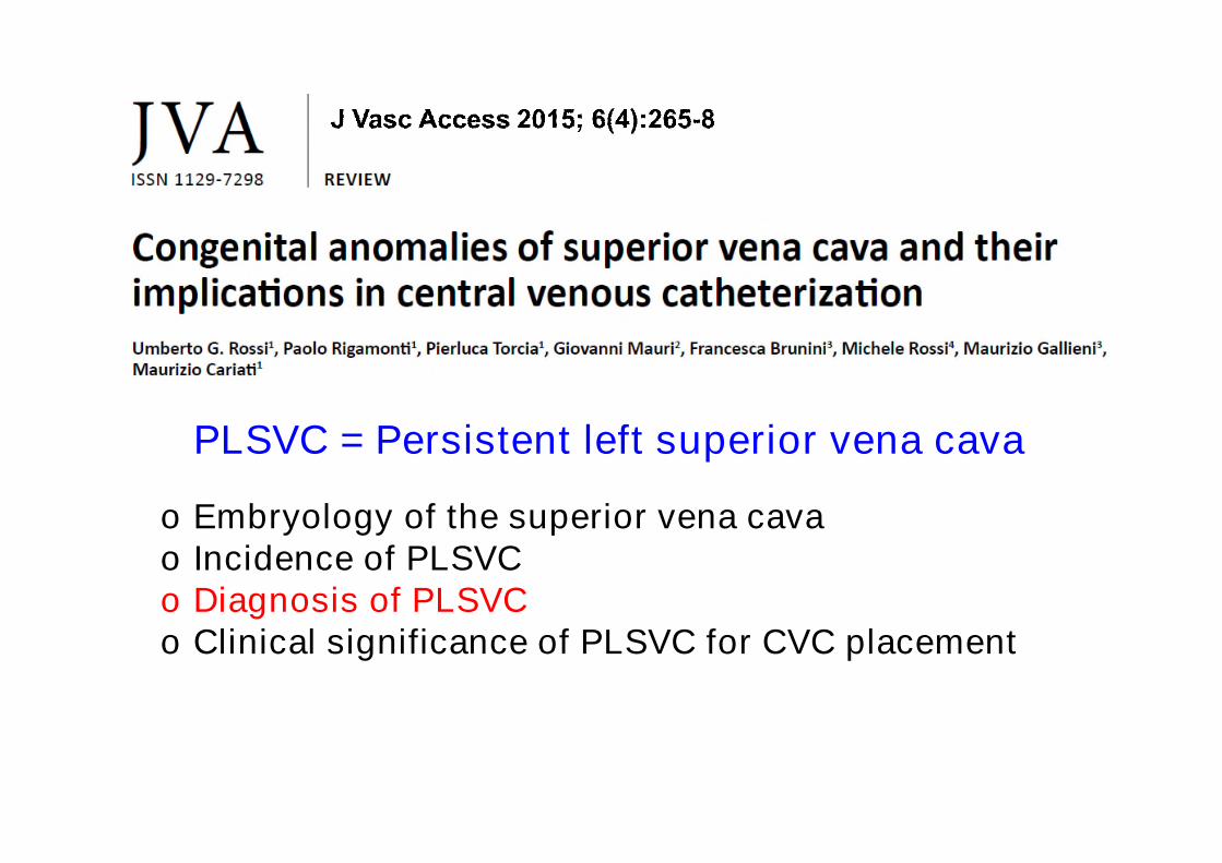
Congenital Anomalies of Superior Vena Cavaand Central Venous Catheterization
Maurizio Gallieni
Nephrology and Dialysis Unit
Ospedale S. Carlo Borromeo, ASST Santi Paolo e Carlo,
University of Milano, Milano, Italy

Congenital Anomalies of Superior Vena Cavaand Central Venous Catheterization
o Although rare, the presence of congenitalanomalies of the SVC may determinefailure and/or complications of CVCimplantation.
o Thus, knowledge of normal vascularanatomy and its possible variants iscrucial for the success of the procedure

Br Heart J 1954 16: 423-439

Semin Dial 2006; 19: 540–542
o A PLSVC is the most common variant ofanomalous venous entry into the heart.
o It should be considered in the setting of:• Difficult central vein catheterization• When a central venous catheter takes
an unexpected left-mediastinal course

o Embryology of the superior vena cavao Incidence of PLSVCo Diagnosis of PLSVCo Clinical significance of PLSVC for CVC placement
PLSVC = Persistent left superior vena cava

https://meducation.net/resources/1750959-Cardiovascular-System-Heart-Embryology

5th week of gestation
Embryology of the superior vena cavaPosterior view of the embryological vein system
Rossi U et al. J Vasc Access 2015; 6(4):265-8
o During the fourth weekof gestation, two majorsymmetrical veinsystems, the right andthe left precardinalveins, drain the upperportion of the embryo.
o Each precardinal veindrains into a commoncardinal vein beforeentering theembryological heart.

o During the fourth week of gestation, two majorsymmetrical vein systems, the right and the leftprecardinal veins, drain the upper portion of theembryo.
o Each precardinal vein drains into a common cardinalvein before entering the embryological heart.
o By the eighth week of gestation, a vein anastomosisconnects the two precardinal veins and the leftcommon cardinal vein atrophies progressively untilcomplete regression.
Embryology of the superior vena cava

5th week of gestation
Embryology of the superior vena cavaPosterior view of the embryological vein system
8th week of gestation
Rossi U et al. J Vasc Access 2015; 6(4):265-8

o During the fourth week of gestation, two majorsymmetrical vein systems, the right and the leftprecardinal veins, drain the upper portion of theembryo.
o Each precardinal vein drains into a common cardinalvein before entering the embryological heart.
o By the eighth week of gestation, a vein anastomosisconnects the two precardinal veins and the leftcommon cardinal vein atrophies progressively untilcomplete regression.
o With this regression, the anastomotic vein betweenthe two precardinal veins becomes the left innominate(brachio-cefalic) vein and the right precardinal veinand the right common cardinal vein form the SVC.
Embryology of the superior vena cava

5th week
Embryology of the superior vena cavaPosterior view of the embryological vein system
8th week Normal
Rossi U et al. J Vasc Access 2015; 6(4):265-8

o The failure of this normal regression can lead to theformation of a PLSVC, determining the clinicalsituation of double SVC.
o This PLSVC runs on the back of the left atrium,entering the right atrium through the orifice of anenlarged coronary sinus, although in about 10% ofcases it drains in the left atrium.
o If the normally persistent right cardinal veinundergoes regression, then there is only an LSVC.This anatomical variant is the rarest, with a rightinnominate (brachio-cefalic) vein draining blood fromthe right to the left
Embryology of the superior vena cava

Schematic drawing of variations of PLSVC

a) LSVC drains via the coronary sinus (CS) into the right atrium (RA);b) Infrequently, the right superior vena cava (RSVC) may be absent.
In this case, the CS is large because it receives blood from boththe right and left upper parts of the body
Granata A et al. J Vasc Access 2009; 10: 207-211

c) The CS is absent, the LSVC drains directly into the left atrium (LA),and the atrial septum has no defect.d) The LSVC connects to the LA, and there is a posterior atrial septaldefect, which allows a predominant left-to-right atrial (RA) shunt.
Granata A et al. J Vasc Access 2009; 10: 207-211

o Embryology of the superior vena cavao Incidence of PLSVCo Diagnosis of PLSVCo Clinical significance of PLSVC for CVC placement
PLSVC = Persistent left superior vena cava

o The persistence of LSVC is described asthe most common central venousanomaly.
o Its prevalence in the general population isestimated at 0.1-0.3%.
o In patients with congenital heart disease,the reported prevalence ranges between2.1 and 5%.
Congenital Anomalies of Superior Vena CavaIncidence
o Embryology of the superior vena cavao Incidence of PLSVCo Diagnosis of PLSVCo Clinical significance of PLSVC for CVC placement
PLSVC = Persistent left superior vena cava

Congenital Anomalies of Superior Vena CavaDiagnosis
Radiologic signs suggestive of the presence of aPLSVC are:o Widening of the mediastinumo An enlarged aortic shadowo A paramediastinal bulge below the aortic arch
on a posterior–anterior plain filmo Marked dilatation of the coronary sinus on
echocardiographyo A leftward P axis with normal PR interval on
electrocardiogram.Clinically, a left jugular vein distention may benoted.
Wasse H. Semin Dial 2006; 19: 540–542
Rossi U et al. J Vasc Access 2015; 6: 265-8

o In about 90% of cases, the LSVC drains into theright atrium via the coronary sinus.
o In 10% of cases, the LSVC drains into the leftatrium, causing right-to-left shunting, which maydetermine cyanosis and may be associated withcardiac malformations.
o Importantly, draining into the left atrium can beassociated with hemodynamic instability, syncope,systemic emboli (thrombotic and infectious)
o More often, these anomalies are detectedincidentally during imaging studies performed forother reasons
Congenital Anomalies of Superior Vena CavaDiagnosis
Rossi U et al. J Vasc Access 2015; 6(4):265-8

A dilated coronary sinus (CS) seen on echocardiography shouldraise the suspicion of PLSVC
Congenital Anomalies of Superior Vena CavaDiagnosis
Goyal S. Cardiovasc Ultrasound. 2008; 6: 50

o Contrast echocardiography is the maindiagnostic test of PLSVC
Congenital Anomalies of Superior Vena CavaDiagnosis
Gupta S. N Am J Med Sci. 2013; 5: 496–497
Echocardiogramshowing a bolus ofbubble into the leftatrium (LA) andventricle (LV) afterperipheral injection(Tip: inject in the leftarm).

Gupta S. N Am J Med Sci. 2013; 5: 496–497
RSVC
Left arm venous angiogram showing LSVCdraining into the left atrium (LA)

*°
Rossi U et al. J Vasc Access 2015
PLSVC (doublesuperior vena cava)
Right side CVC (arrowheads)and left side pacemaker
(arrows).
Chest venography can clarify PLSVC blood flow patterns
Rossi U et al. J Vasc Access 2015

Rossi U et al. J Vasc Access 2015
CT scan: coronal multiplanar reconstruction

o Embryology of the superior vena cavao Incidence of PLSVCo Diagnosis of PLSVCo Clinical significance of PLSVC for CVC placement
PLSVC = Persistent left superior vena cava

Two different settings should be considered:1. Patients with known congenital anomalies of the SVC
• Accurate preoperative planning with imagingdescription of veins anomalies
• Careful selection of the most adequate vascularaccess (avoid CVCs whenever possible, considerfemoral access)
• Use of ultrasound and fluoroscopic guidance byskilled operators.
2. Patients with no prior diagnosis of congenital anomaliesof the SVC• Recognize the possibility of PLSVC during a
procedure with unusual CVC positioning• Act consequently
Wasse H. Semin Dial 2006; 19: 540–542
Clinical significance of PLSVC for CVCplacement
Rossi U et al. J Vasc Access 2015; 6(4):265-8

o In patients with no prior diagnosis of PLSVC, the mostcommon situation is noticing, during an insertionprocedure done under fluoroscopy, that the catheterfollows an anomalous left-sided course.
o This may not only represent the presence of a PLSVCbut also an intra-arterial catheter placement, as wellas other possible tip displacements (pericardium,mediastinum pleural space, minor thoracic veins)
o In the absence of a right SVC, perforation of the rightbrachiocephalic (innominate) vein may be observed,while catheter tip manipulation in the coronary sinusmay cause angina, arrhythmias, cardiac arrest, orcoronary sinus thrombosis.
Wasse H. Semin Dial 2006; 19: 540–542
Clinical significance of PLSVC for CVCplacement
Rossi U et al. J Vasc Access 2015; 6(4):265-8

Goyal S. Cardiovasc Ultrasound. 2008; 6: 50
Unusual course of Swan-Ganz catheter (arrows)

Goyal S. Cardiovasc Ultrasound. 2008; 6: 50
Chest CT scan showing left sided SVC

o The presence of PLSVC may cause difficulties or possiblecomplications during access to the right atrium, especiallywith a left internal jugular and a left subclavian approach,which are common sites of access for hemodialysiscatheters or when placing pacemakers and Swan-Ganzcatheters.
o Tip position: Wasse suggested that the tip should belocated cranial to the PLSVC–coronary sinus junction inorder to avoid coronary sinus thrombosis. This is incontrast with the guideline suggesting tip position oftunneled dialysis CVC to be positioned in the right atrium.PLSVC thrombosis may occur with both options. An AVaccess should be an absolute priority in this patientcategory
Wasse H. Semin Dial 2006; 19: 540–542
Clinical significance of PLSVC for CVCplacement
Rossi U et al. J Vasc Access 2015; 6(4):265-8

Conclusions
• A persistent lefts superior vena cava (PLSVC) is arelatively rare but clinically relevant variant of venousreturn to the heart, which should be well known by theinterventional nephrologist/radiologist
• Recognition during an interventional procedure of anunexpected course of the guidewire will indicate thepossibility of performing a phlebography to confirm thepossible diagnosis of congenital anomaly of SVC
• In patients with known anomalies of the SVC, pre-procedure imaging is warranted, in order to define thepattern of PSLVC
• Fluoroscopy is needed to adequately approach thisclinical problem
