conformational, concomitant polymorphs of 4,4-diphenyl-2,5-cyclohexadienone: conformation and...
TRANSCRIPT

DOI: 10.1002/chem.200501417
Conformational, Concomitant Polymorphs of 4,4-Diphenyl-2,5-cyclohexadienone: Conformation and Lattice Energy Compensation in theKinetic and Thermodynamic Forms
Saikat Roy,[a] Rahul Banerjee,[a] Ashwini Nangia,*[a] and Gert J. Kruger*[b]
Introduction
McCrone�s[1] definition of a polymorph as “a solid crystal-line phase of a given compound resulting from the possibili-
ty of at least two different arrangements of the molecules ofthat compound in the solid state” is widely accepted today.[2]
The existence of polymorphism implies that free-energy dif-ferences between various forms are small (<3 kcalmol�1)and that kinetic factors are important during crystal nuclea-tion and growth. Molecular conformations, hydrogen bond-ing, packing arrangements, and lattice energies of the samemolecule in different supramolecular environments may becompared in polymorphic structures.[3] Polymorphs are idealsystems to study molecular structure–crystal structure–crys-tal energy relationships with a minimum number of varia-bles because differences arise due to different intermolecu-lar interactions (supramolecular synthons)[4] and crystalpacking effects and not because they are different chemicalspecies. There is increasing interest in understanding poly-morphism, growing new crystal forms, controlling the selec-tive growth of one form, transformations between poly-morphs, and the high-throughput crystallization of drugs.[5]
Polymorphism is more widespread in pharmaceutical
Abstract: 4,4-Diphenyl-2,5-cyclohexa-dienone (1) crystallized as four confor-mational polymorphs and a recordnumber of 19 crystallographically inde-pendent molecules have been charac-terized by low-temperature X-ray dif-fraction: form A (P21, Z’=1), form B(P1, Z’=4), form C (P1, Z’=12), andform D (Pbca, Z’=2). We have nowconfirmed by variable-temperaturepowder X-ray diffraction that form Ais the thermodynamic polymorph andB is the kinetic form of the enantio-tropic system A–D. Differences in thepacking of the molecules in these poly-morphs result from different acidic C�H donors approaching the C=O accept-or in C�H···O chains and in synthonsI–III, depending on the molecular con-
formation. The strength of the C�H···Ointeraction in a particular structure cor-relates with the number of symmetry-independent conformations (Z’) in thatpolymorph, that is, a short C�H···O in-teraction leads to a high Z’ value. Mo-lecular conformation (Econf) and latticeenergy (Ulatt) contributions compensateeach other in crystal structures A, B,and D resulting in very similar total en-ergies: Etotal of the stable form A=
1.22 kcalmol�1, the metastable form
B=1.49 kcalmol�1, and form D=
1.98 kcalmol�1. Disappeared poly-morph C is postulated as a high-Z’,high-energy precursor of kinetic formB. Thermodynamic form A matcheswith the third lowest energy framebased on the value of Ulatt determinedin the crystal structure prediction(Cerius2, COMPASS) by full-body min-imization. Re-ranking the calculatedframes on consideration of both Econf
(Spartan 04) and Ulatt energies gives aperfect match of frame #1 with stablestructure A. Diphenylquinone 1 is anexperimental benchmark used to vali-date accurate crystal structure energiesof the kinetic and thermodynamic poly-morphs separated by <0.3 kcalmol�1
(~1.3 kJmol�1).
Keywords: conformation analysis ·crystal structure prediction ·polymorphism · supramolecularchemistry · symmetry-independentmolecule · X-ray diffraction
[a] S. Roy, R. Banerjee, Prof. A. NangiaSchool of Chemistry, University of HyderabadHyderabad 500 046 (India)Fax: (+91)40-2301-1338E-mail : [email protected]
[b] Prof. G. J. KrugerDepartment of Chemistry, University of JohannesburgPO Box 524, Auckland Park, Johannesburg 2006 (South Africa)Fax: (+27)11-489-2819E-mail : [email protected]
Supporting information for this article is available on the WWWunder http://www.chemeurj.org/ or from the author. Simulatedpowder XRD plots of forms A–D, DSC of diphenylquinone 1, confor-mational overlay diagram, Rietveld fit of Polymorph Predictor framewith form A, and tables containing crystal structure predictionframes, energies, and cell parameters.
Chem. Eur. J. 2006, 12, 3777 – 3788 L 2006 Wiley-VCH Verlag GmbH&Co. KGaA, Weinheim 3777
FULL PAPER

solids[6] than the estimates of 4–5% polymorphic crystals[7]
in the Cambridge Structural Database (CSD) suggest.[8]
Among organic crystal structures, there is one example of acompound with seven polymorphs (5-methyl-2-[(2-nitrophe-nyl)amino]-3-thiophenecarbonitrile, common name ROY),[9]
sulfathiazole has five forms, there are 14 clusters of tetra-morphs, and over a 100 trimorphic systems.[9b,10a] Polymor-phism is of great current interest because different solid-state forms can have different physical, chemical, and func-tional properties, for example, melting point, stability, color,bioavailability, pharmacological activity, and a nonlinear op-tical response.
Concomitant polymorphs[11] crystallize simultaneouslyfrom the same solvent and crystallization flask under identi-cal crystal growth conditions. Bernstein[12] carried out earlystudies on conformational polymorphs (different conforma-tions of the same molecule in different crystal structures)and conformational isomorphs (different conformers of thesame molecule in the same crystal structure). Herein we de-scribe a tetramorphic cluster of conformational polymorphsin which molecular- and lattice-energy compensation resultsin very small differences in the total energy of the concomi-tant polymorphs A–D of 4,4-diphenyl-2,5-cyclohexadienone(1). Experimental conditions are described for the prepara-tion of thermodynamic form A and kinetic form B in a rea-sonably pure state. The implication of a molecular and crys-tal-packing balance is to rank crystal structure predictionframes derived from metastable rotamers by considerationof both conformation- and lattice-energy contributions,which gives an excellent match of the global minimum struc-ture with stable form A.
Results and Discussion
Crystal packing and multiple Z’ in polymorphs : Crystallo-graphic data for the polymorphs A–D of diphenylbenzoqui-none 1 are listed in Table 1.[13] The molecule has severalacidic, activated donor hydrogen atoms of sp2- and phenylC�H-type whereas there is a single carbonyl acceptor. De-pending on the molecular conformation one or more of theseveral possible C�H···O interactions[14] are optimized in thecrystal structure (Figure 1). For example, form A has zigzagchains of C�H···O interactions between screw-axis-relatedAi molecules of graph-set notation[15] C(8). Form B has C�
H···O quinone dimer synthon Iand p-phenyl C�H···O synthonII [graph set R2
2(8) and R22(20)]
between Bi, Bii and Biii, Biv mol-ecules, respectively. Form C hasthe same synthons and overallpacking as B but has 12 mole-cules (Ci–Cxii) in the asymmetricunit. Form D has C(10) chainsthat connect to form the cyclicR4
3(32) pattern and o-phenylC�H···O synthon III with a
R22(16) ring through Di and Dii molecules. The parameters
of the C�H···O geometries are listed in the legend toFigure 1. We have not found polymorphs of bis(biphenylyl)ketone 2[16] or its substituted phenyl derivatives 3 (4-Cl/Br/Me)[10b] so far.
The number of symmetry-independent or crystallographi-cally unique molecules/ions in a crystal lattice is Z’. Alterna-tively, Z’ may be defined as the number of formula units (Z)divided by the number of independent general positions forthat space group. Z’ is typically 1 or 0.5 in crystal structures(87%). A high Z’ of 12 for form C is a record for poly-morph clusters[9b] (Table 2) and as such rare in the CSD[8]
(only five hits).Structures with high Z’ values continue to interest crystal-
lographers but it is still not properly understood why somecategories of structures exhibit a higher frequency of Z’>1.Steed[17] has critically reviewed the reasons for high-Z’ crys-tal structures. 1) The molecule has a packing problem be-cause of its awkward shape, which is reconciled by havingtwo or more molecules in different conformations.[18] 2) Themolecules organize in stable clusters prior to reaching thehighest symmetry arrangement in strong O�H···O hydrogen-bonded structures because of the enthalpic advantage de-rived from s-cooperative chains,[19] for example, as in alco-hols, phenols, steroids, nucleotides, and nucleosides. 3) Sev-eral low-lying molecular conformations interconvert in solu-tion and more than one molecule may crystallize simultane-ously for kinetic reasons. The last of these situations occursin the conformational polymorphs of 1, which provides aunique opportunity to study polymorphic structures withmultiple values of Z’.
Cholesterol (Z’=16) is a prototype example of strong O�H···O hydrogen bonds being associated with unusually highZ’ values. An exceptional case in the weak hydrogen bondcategory is the crystal structure [ReCl2ACHTUNGTRENNUNG(NCMe)(NO)-ACHTUNGTRENNUNG(PMe3)2] (CSD refcode WODCOH, Z’=11),[20] which has adense network of C�H···O and C�H···Cl interactions. Weobserved an interesting trend in the polymorphs of 1,namely that the number of conformations in a particularstructure (Z’) correlates with C�H···O bond strength/dis-tance. Figure 2 is a distance–angle scatter plot of C�H···Ointeractions in polymorphic forms A–D. C�H···O contacts inform C are, in general, shorter than those in forms B and D
Table 1. Crystallographic data for polymorphs A–D of diphenylquinone1.[13]
Form A Form B Form C Form D
CSD refcode[a] HEYHUO HEYHUO01 HEYHUO02 HEYHUO03space group P21 P1 P1 PbcaZ’, Z 1, 2 4, 8 12, 24 2, 16a [Q] 7.9170(6) 10.0939(2) 18.3788(4) 10.7921(6)b [Q] 8.4455(6) 16.2592(3) 19.9701(4) 17.4749(12)c [Q] 10.3086(9) 16.2921(4) 24.4423(5) 27.9344(19)a [8] 90 88.2570(10) 95.008(1) 90b [8] 105.758(2) 85.3380(10) 111.688(1) 90g [8] 90 83.6450(10) 105.218(1) 90V [Q3] 663.36(9) 2648.00(10) 7871.8(3) 5268.2(6)R factor 0.050 0.068 0.112 0.059
[a] See ref. [8].
www.chemeurj.org L 2006 Wiley-VCH Verlag GmbH&Co. KGaA, Weinheim Chem. Eur. J. 2006, 12, 3777 – 37883778

Figure 1. a) Helices of C�H···O hydrogen bonds (2.55 Q, 163.68 ; 2.60 Q, 129.98) between 21 related molecules in form A [conformer Ai, graph set C(8)].b) Centrosymmetric C�H···O synthon I between Bi, Bii molecules of R2
2(8) pattern (2.33 Q, 169.28 ; 2.49 Q, 123.58) and synthon II between Biii, Biv mole-cules of R2
2(20) pattern (2.64 Q, 138.08 ; 2.74 Q, 121.68) in form B. The crystal structure of form C is similar to B. Twelve symmetry independent mole-cules (Ci–Cxii) engage in similar synthons instead of four molecules in B. c) C�H···O interactions of graph set R4
3(32) between translation and screw-axis-related Di molecules (2.36 Q, 137.98 ; 2.54 Q, 166.98) in form D. d) Centrosymmetric C�H···O synthon III between Dii molecules [R2
2(16) pattern] and C�H···O interaction (2.57 Q, 144.58 ; 2.47 Q, 165.18). Neutron-normalized distances are quoted. Note that different C�H donors participate in C�H···O inter-actions in different crystal forms. Cyclic C�H···O synthons I–III are labeled.
Table 2. Data for polymorphs (�3 forms)[a] in organic crystal structureswith multiple molecules in the asymmetric unit.
Entry CSD refcode[b] No. of polymorphs Highest Z’
conformational polymorphs (�4 forms)[a]
1 QAXMEH 7 12 SUTHAZ 5 23 BEWKUJ 4 24 BIXGIY 4 15 HEYHUO[c] 4 126 KAXHAS 4 17 MABZNA 4 48 RUWYIR 4 2
multiple molecules in asymmetric unit (Z’>4)[a]
9 PUBMUU[c] 3 1610 IFULUQ 4 811 DUVZOJ 3 612 ZZZVTY 3 513 THIOUR 3 4.5
[a] Cut-offs were made to limit the number of structures analyzed.[b] See ref. [8]. [c] Compound has a high number of polymorphs and ahigh Z’ value.
Figure 2. H···O distance (2.2–3.0 Q) versus C�H···O angle (120–1808)scatter plot of interactions in tetramorphs A–D. A=* (Z’=1), B=&
(Z’=4), C=~ (Z’=12), D=^ (Z’=2). The shortest H···O distance(marked with an arrow in the linear band) is inversely related to Z’ (thenumber of symmetry-independent conformations).
Chem. Eur. J. 2006, 12, 3777 – 3788 L 2006 Wiley-VCH Verlag GmbH&Co. KGaA, Weinheim www.chemeurj.org 3779
FULL PAPERPolymorphs of 4,4-Diphenyl-2,5-cyclohexadienone

and longer in form A. Interestingly, the shortest linear inter-action (q=160–1758) in a particular form is inversely relatedto the Z’ value of that structure: for example, form C hasthe shortest H···O distance of 2.30 Q and the highest Z’value of 12, form B has H···O=2.33 Q, Z’=4, and forms Dand A have even longer H···O distances of 2.47 and 2.55 Qand smaller Z’ values of 2 and 1, respectively. Our findingthat the strength of C�H···O interactions can promote Z’>1among flexible molecules means that the well-known exam-ples of structures with values of Z’>1, that is, alcohols andphenols, can be expanded to new categories of crystal struc-tures stabilized by weak hydrogen bonds. We show that therelative strengths of directional C�H···O interactions are im-portant in promoting high Z’ structures[21] particularly astheir energies are comparable to favorable close-packingforces. The inverse relationship between Z’ and H···O dis-tance is observable in polymorphic cluster 1 because de-tailed supramolecular effects can be clearly discernedagainst a background of a constant molecular structure.
The occurrence of high-Z’ polymorphs and the relation-ship between Z’ and C�H···O strength alludes to the impor-tance of kinetic factors during crystallization. We thereforewanted to identify which of the concomitant polymorphs A–D is the kinetic form and which is the thermodynamic oneand determine the nature of the phase transitions betweenthese forms.
Variable-temperature powder X-ray diffraction : We ob-tained single crystals of all four forms in preliminary batch-es[13] but subsequent experiments gave mostly forms A andB, as determined by unit-cell checking of several crystals.However, powder X-ray diffraction shows all four forms inthe concomitant mixture at room temperature (Figure 3). Atypical solid upon crystallization from EtOAc/n-hexane con-tains form A (~40%), forms B+C (~50%), and form D
(~10%). The ratios were determined by the Rietveld refine-ment[22] of observed powder XRD plots with simulatedpeaks for each crystal structure (Powder Cell 2.3). Triclinicforms B and C are taken together because it is not possibleto distinguish between these closely related forms from theiroverlapping diffraction patterns (Figure S1, Supporting in-formation). The mixture of forms at room temperature washeated to study phase transformations. The peak profile isrelatively stable between 30–60 8C, however, we noticedchanges as the sample was heated to 70 8C (Figure 4): Cer-tain peaks disappeared and the overall pattern became sig-nificantly sharper with fewer but more intense lines. ThePXRD profile is relatively unchanged between 70–100 8C,after which the material became mostly amorphous andthen gradually turned to a semi-solid/melt mass at 105–115 8C. There are no reflections from the sample at T>
105 8C except for the peak from the sample holder at 25.28.VT-PXRD shows that heating polymorphs A–D to a pre-melt temperature of 70 8C transforms the mixture to form A(Figure 5) with good polymorph purity (>95%), based on amatch with simulated peaks of the crystal structure. Mono-clinic polymorph A therefore is a thermodynamic modifica-tion of the enantiotropic system of polymorphs 1 between30–80 8C.
Chiral form A was prepared in high purity and shown tohave a nonlinear optical signal equal to that of urea when ir-radiated with a Nd3+–YAG laser (1.06 mm). However, themixture of polymorphs obtained from a typical solutioncrystallization does not emit light at 532 nm. The prepara-tion of form A by the above heating method is preferredover controlled crystallization at �5 8C because of contami-nation from other polymorphs over a period of time, pre-sumably due to accidental seeding of laboratory space,[23] aterm used to describe difficulties in isolating an early poly-morph after the appearance of other forms of the samecompound. Polymorphs of 1 do not follow Ostwald�s rule ofstages,[24] with stable form A appearing first from solutioncrystallization followed by metastable forms B and C.
Kinetic form B was prepared by heating the polymorphicmixture to a melt phase in the powder X-ray diffractometerpan at ~115 8C. Cooling the sample to room temperature af-forded reasonably pure polymorph B (Figure 6). This wasconfirmed by unit-cell checking of a few randomly pickedcrystals. Although polymorphs A–D have quite different ar-rangements of molecules and unit cells, they melt at thesame temperature (Tm=120.45 8C) and there is no apparentphase transition other than the melting endotherm in differ-ential scanning calorimetry (Figure S2, Supporting informa-tion).
There are alternative explanations for the occurrence ofconcomitant polymorphs:[11] Simultaneous nucleation ofmore than one form from solution, interconversion betweenpolymorphs, their appearance in order of stability, and het-erogeneous cross nucleation.[5e] The simultaneous crystalliza-tion of all four forms A–D from the homogeneous mediumis the most likely reason for the concomitant cluster 1. Inter-conversion in solution is minimal at room temperature be-
Figure 3. Powder X-ray diffraction of solid 1 at room temperature:black=experimentally observed powder pattern; red, blue, and green=calculated powder pattern of form A (37.5%), B+C (52.0%), and D(10.5%), respectively. Rietveld refinement in Powder Cell 2.3: Rp=14.82,Rwp=19.32. Percentages of polymorphic forms in different batches varywithin 5%.
www.chemeurj.org L 2006 Wiley-VCH Verlag GmbH&Co. KGaA, Weinheim Chem. Eur. J. 2006, 12, 3777 – 37883780
A. Nangia, G. J. Kruger et al.

cause the percentages of various forms in different batchesare within the experimental limit of 5%. As mentioned, thesystem does not obey Ostwald�s law of stages. Heterogene-ous cross nucleation in the heated/melt phase is ruled outbecause if this were happening then cooling the samplefrom 70 and 115 8C would not only give form A and B, re-spectively, but also other polymorphs from seeded nuclea-tion and growth.
Conformation and latticeenergy compensation : Com-pound 1 is an excellent systemfor studying how interconvert-ing molecular conformations insolution lead to different crystalpacking arrangements involvingseveral conformers in the solidstate. The energies of the mo-lecular conformations and thecrystal lattice were calculatedto obtain a quantitative pictureof polymorphism in 1 and aclue as to why these conforma-tional polymorphs appear con-comitantly. The conformationenergies (Econf) and dipole mo-ments (m) were calculated usingthe Spartan 04 package[25]
(Table 3). Each molecule wasextracted from the crystal struc-ture and energy-minimized(HF/6-31G**) keeping the con-formation fixed (heavy carbonand oxygen atoms invariant)while the hydrogen atoms wereallowed to relax to reasonablegeometries following themethod of Yu et al.[9a] The tor-sion angles t1 and t2 that definethe rotation about the Cquinone�Cphenyl single bonds lie in therange of 8–22 and 12–388, re-spectively, along the scatter plotdiagonal (Figure 7). In general,in the 19 rotamers t1¼6 t2 saveconformer Ai and Cxi (see Fig-ure S3, Supporting information,for the overlay diagram). Mole-cule Bi has the most stable con-formation (Econf=�479813.50kcalmol�1, a value that is arbi-trarily fixed to 0); the energiesof the conformers of forms A,B, and D (Econf) are within1.3 kcalmol�1 of Bi (Table 3).Conformers Ci–Cxii are higherin energy (Econf=2–9 kcalmol�1) but this could be due to
an error in the experimental X-ray geometry as the R factorof form C is high (11.1%). Therefore we focus on structuresA, B, and D in this discussion. The conformers of forms A,B, and D readily interconvert in solution through geared(correlated) rotation of the phenyl rings[13] about t1 and t2because the energy barrier should be accessible through thethermal motion of atoms (RT~0.5 kcalmol�1 at 298 K) inthe crystallization regime between �5 and 100 8C.
Figure 4. a) Powder X-ray diffraction patterns of 1 recorded at different temperatures. The sample is a mixtureof forms A–D at room temperature, it transforms to forms A at around 70 8C, and becomes amorphous uponfurther heating to 105 8C. The wide peak at 25.28 is from the sample holder. b) Powder XRD of 1 at 28 (top)and 69 8C (bottom). Peaks that disappear upon heating are marked with an arrow. Note the increase in intensi-ty of the peaks and the overall simplification of the profile at a higher temperature.
Chem. Eur. J. 2006, 12, 3777 – 3788 L 2006 Wiley-VCH Verlag GmbH&Co. KGaA, Weinheim www.chemeurj.org 3781
FULL PAPERPolymorphs of 4,4-Diphenyl-2,5-cyclohexadienone

The crystal lattice energies, Ulatt, of polymorphs A–Dwere computed using the COMPASS and DREIDING 2.21force fields (Table 4, Cerius2).[26] We discuss COMPASSnumbers because this force field is better parametrized andgives more accurate energies of organic molecules[27] thatare typically stabilized by hydrogen bonds, intermolecularinteractions, and van der Waals forces. COMPASS[28] isbetter suited for molecule 1 because electrostatic stabiliza-tion by C�H···O hydrogen bonds and edge-to-face aromaticinteractions is included in the coulombic term. Form A hasthe most stable crystal structure (Ulatt=�32.69 kcalmol�1)and forms B, C, and D are less stable by 1.03, 1.06, and0.82 kcalmol�1, respectively. However, all these crystal struc-tures are a compromise between intra- and intermolecular
stability. Either the molecular conformer or the latticeenergy is at the minimum, but both intra- and intermolecu-lar energies are not the lowest in any structure. For example,form A has the lowest Ulatt value (�32.69 kcalmol�1), butconformer Ai is higher in energy (Econf=1.22 kcalmol�1).Molecular conformations Bi–Biv are lower in energy (Econf=
0.00, 0.06, 0.66, 1.12 kcalmol�1) but crystal lattice B is meta-stable (Ulatt=�31.66 kcalmol�1). Both intra- and intermolec-ular energy contributions are included in the total energyterm (Table 5). In increasing order of Etotal (COMPASS):form A (thermodynamic, most stable)<B (kinetic, inter-mediate stable)<D (least stable). This energy order is con-sistent with the thermodynamic stability of form A deter-mined in the VT-PXRD experiments and the crystallizationof the kinetic form B from the melt of the polymorphic mix-ture. On the other hand, the Ulatt (COMPASS) stabilityorder of A<D<B and the energies calculated by using theDREIDING 2.21 force field (D<A<B) do not agree withexperiment. We believe that calculations using the COM-PASS force field give an indication of the relative stabilityof the polymorphs with an energy difference of 0.3–1.0 kcalmol�1 for small organic molecules.
The molecular conformations and crystal lattice energiessuggest the following picture of polymorphism in 1. Depend-ing on the geometry of a particular conformation, a differentC�H···O interaction and aromatic packing motif lead to ametastable crystal structure. The reason for conformationalpolymorphism in 1 is that C�H···O motifs in its crystal struc-tures (Figure 1) involve one of the phenyl C�H donors,except for those in dimer I which are between the quinonegroups. Therefore a change in molecular conformation willalter the strength of weak C�H···O and van der Waals inter-actions and in turn the preferred crystal-packing motif. Theenergy penalty in the molecular conformation is compensat-ed by lattice-energy gain from intermolecular interactionsand close packing, and vice versa, because their magnitudesare comparable, DEconf�DUlatt=0–2 kcalmol�1. Facile ro-tamer interconversion in solution and very similar crystalenergies mean that more than one molecular conformationmay crystallize out simultaneously to give concomitant con-formational polymorphs of 1.
Price and co-workers[29] recently examined intra- and in-termolecular energy compensation in the conformationalpolymorphs of some drugs, for example, aspirin, barbituricacid, and piracetam. Surprisingly, the quintessential poly-morphic compound, ROY, appears to be an exception to theabove-mentioned energy balance situation: the stable per-pendicular conformation is present in the thermodynamicyellow crystal form.[30] This prompts the question: Will meta-stable crystal forms of ROY give way to the stable poly-morph because the system may gradually transform to thebottom of the molecular and lattice energy well?
Having discussed forms A, B, and D we will briefly men-tion the unusually high Z’ value (=12) of polymorph C. Wepreviously postulated[13] that form C represents a snap-shotpicture of an evolving crystal nucleus on the way to form B(Z’=4) wherein the molecules have aggregated to form the
Figure 5. Experimental powder XRD of 1 at 69 8C (black line) matcheswith the calculated powder pattern of polymorph A (dotted line). Riet-veld refinement in powder cell 2.3: Rp=14.39, Rwp=18.81. The startingsolid was the mixture of forms A–D shown in Figure 3.
Figure 6. Experimental powder XRD of 1 from melt crystallization at115 8C (black line) shows good agreement with the calculated powderpattern of polymorph B (dotted line). Rietveld refinement in powder cell2.3: Rp=11.78, Rwp=15.86. The starting solid was the mixture of formsA–D shown in Figure 3.
www.chemeurj.org L 2006 Wiley-VCH Verlag GmbH&Co. KGaA, Weinheim Chem. Eur. J. 2006, 12, 3777 – 37883782
A. Nangia, G. J. Kruger et al.

crystal lattice but the periodicity is yet to reach the highestpossible crystal symmetry. The 12 high-energy conformers ofcrystal structure C may be viewed as metastable relics of therelatively stable four conformers of form B. Attempts tostudy the transformation of form C to B in the laboratorywere thwarted by our inability to grow crystals of form Cafter the initial X-ray diffraction experiment. The above in-
terpretation is consistent withthe following observations. 1)The higher-energy conformersin form C participate in stron-ger C�H···O interactions andthis alludes to the greater roleof kinetic factors in the nuclea-tion of this polymorph. 2) Theclose relationship betweenforms C and B is evident fromtheir crystal structures: Theyhave the same space group andoverall packing that is sustainedthrough C�H···O synthons Iand II and overlapping powderXRD peaks. In short, high Z’structures are not just a crystal-lographic oddity but they opena window to “see” crystal nu-cleation and growth.
Does molecular conformationdetermine crystal packing orare favorable crystal packing ef-fects able to trap metastablemolecular conformations? This
question is difficult to quantify in the present case becauseEconf and Ulatt differences are of the same order of magni-tude. We can nonetheless say that a particular conformationis associated with a specific C�H···O synthon even thoughthat rotamer is present in different polymorphs. For exam-ple, Biv and Cvii rotamers have similar conformations andthey engage in synthon II in the crystal structures of poly-morphs B and C. Such a reality, namely that a less stableconformation can lead to a more stable crystal lattice in con-formational polymorphs, is a major challenge in the crystalstructure prediction of flexible molecules.
Computational prediction of form A : The ab initio predic-tion of crystal structures of organic molecules from theirmolecular diagram is a global research activity,[31] whichshould give us a better understanding of the crystallizationprocess and even protein folding. In crystal structure predic-tion (CSP), the input for flexible molecules is progressingfrom the first phase of using stable gas-phase conformationsto metastable conformations.[29] The main difficulties in pre-dicting the structures of conformationally flexible moleculesare the following. 1) The stable conformation may not resultin the thermodynamic crystal structure. 2) Which metastableconformation out of several low-energy rotamers should beselected for simulations? 3) Both conformation- and lattice-energy contributions to crystal structure stabilization mustbe taken into account. These issues are pertinent to mole-cule 1 and we now present a possible solution for predictingand ranking structure frames of a conformationally flexiblemolecule. Thermodynamic polymorph A is the target incrystal structure prediction because at the present time CSPcomputations are only able to predict the lowest energy
Table 3. Energies and dipole moments of 19 conformers of 1 calculated using Spartan 04.
Crystal polymorph Molecular conformation[a] Ph torsion angles HF/6-31G**t1 [8] t2 [8] Econf
[b] [kcalmol�1] m [D]
form A Ai 12.5 12.6 1.22 5.15form B Bi 12.3 16.0 0.00 5.22
Bii 14.9 23.6 0.06 5.20Biii 19.1 31.8 0.66 5.34Biv 11.5 17.7 1.12 5.24
form C Ci 12.8 16.0 2.81 5.20Cii 11.9 16.7 3.94 5.40Ciii 18.4 32.3 4.16 5.06Civ 12.1 22.7 4.76 5.30Cv 15.2 22.0 5.19 5.40Cvi 10.2 21.0 5.51 5.24Cvii 11.5 17.4 7.99 5.50Cviii 18.3 28.2 8.14 5.08Cix 20.9 30.8 8.15 5.21Cx 14.0 16.7 8.31 5.15Cxi 14.9 15.1 8.54 5.23Cxii 20.1 31.5 8.90 5.39
form D Di 18.5 36.8 1.08 5.21Dii 8.4 16.5 1.25 5.17
energy-minimized[c] 22.3 22.3 �2.78 4.87
[a] Molecules are numbered in order of increasing energy. [b] Conformer Bi has the most stable conformation(�479813.50 kcalmol�1) and is arbitrarily fixed to 0.00 for comparison of conformation energies. [c] The mo-lecular skeleton was minimized to the stable gas-phase rotamer.
Figure 7. a) Conformations of diphenylquinone 1 defined by torsionangles t1 and t2 about the Cquinone�Cphenyl single bonds. b) Nineteen crys-tallographically independent conformations of 1 lie along the diagonal.All 19 conformers converge to the gas-phase rotamer (t1=t2=22.38)after energy minimization. A=*, B=&, C=~, D=^, and gas phase=�.
Chem. Eur. J. 2006, 12, 3777 – 3788 L 2006 Wiley-VCH Verlag GmbH&Co. KGaA, Weinheim www.chemeurj.org 3783
FULL PAPERPolymorphs of 4,4-Diphenyl-2,5-cyclohexadienone

structure and those with Z’=1. The objective of our simula-tions was to reproduce the known stable polymorph A andto identify structures of similar energies as a guide forfuture crystallization experiments. We have used PolymorphPredictor (Cerius2)[26] (PP) computations to generate severalputative crystal structures of 1 from the stable molecularconformation derived from Gaussian 03.[32]
In structure prediction, the conformation should be al-lowed to vary during the energy minimization of frames (de-fined as full-body minimization) to cover the completerange of possible crystal structures in flexible molecules.Rigid-body minimization (the molecular conformation isheld fixed) is about five times faster but it generates struc-tures that correspond to local minima for a particular con-formation, not necessarily the global minimum. Crystalstructure frames were generated in six common spacegroups, P21/c, P1, C2/c, Pbca, P21, and P212121. Ten uniquelow-energy frames within 4 kcalmol�1 of the global mini-mum are plotted for each space group in Figure 8 (total of60 structures, see Table S1, Supporting Information, forvalues). The experimental structure A is the third rankframe based on values of Ulatt. Cell parameters, torsionangles, and the lattice energy ofCSP frame #3 match remarka-bly well the experimental struc-ture A having a deviation ofwithin 3% (Table 6). The crys-tal packing in the simulatedstructure is identical to the ob-served form and their powderXRD profiles are in goodagreement (Figure 9 and Fig-ure S4, Supporting Informa-tion). Although the fact thatframe #3 matches the observedstructure is short of the correct
answer, our reproduction of alarge and flexible molecule(compound 1) is quite good inrelation to ongoing CSP effortsby other groups.[31]
How important is the startingconformation in generating aparticular crystal structure of 1?Given that the molecular con-formation, C�H···O synthonand crystal packing are inti-
Table 4. Lattice energies [Ulatt, kcalmol�1] of forms A–D computed in Cerius2, corrected to per molecule of 1.
Form A Form B Form C Form D
Ulatt COMPASS DREIDING 2.21 COMPASS DREIDING 2.21 COMPASS DREIDING 2.21 COMPASS DREIDING 2.21total �32.69 �42.12 �31.66 �39.66 �31.63 �39.71 �31.87 �42.42van der Waals �28.01 �27.78 �28.19 �27.22 �28.18 �27.23 �27.80 �27.19coulombic �4.68 �12.50 �3.47 �10.43 �3.45 �10.49 �4.07 �12.49hydrogen bond[a] – �1.84 – �2.01 – �1.99 – �2.74
[a] The hydrogen bond energy is partitioned in the DREIDING 2.21 force field but it is part of the coulombic component in the COMPASS force field.
Table 5. Relative energies[a] [per molecule, kcalmol�1] of crystal forms A, B, and D.[b]
Polymorph Ulatt Econf[c] Etotal=Ulatt+Econf Graph set symbol of C�H···O
interactionCOMPASS DREIDING
2.21HF/6-31G**
COMPASS DREIDING2.21
A 0.00 0.30 1.22 1.22 1.52 C(8)B 1.03 2.76 0.46 1.49 3.22 R2
2(8), R22(20)
D 0.82 0.00 1.16 1.98 1.16 R22(16), R4
3(32)
[a] Values taken from Tables 3 and 4. The values of Ulatt and Econf are relative energies. [b] Form C is excludedbecause the errors are too large. [c] The average Econf value is estimated for multiple conformers as (�Econf)/Z’.
Figure 8. Lattice energy versus net volume (V/Z) for structures of mole-cule 1 generated in six common space groups by full-body minimizationusing the Polymorph Predictor software package. Experimental crystalstructure A matches with the third rank predicted structure based onvalues of Ulatt. See Table 6 for details.
Table 6. Comparison of predicted[a] and experimental[b] structure parameters.
Form a[Q]
b[Q]
c [Q] b [8] V/Z[Q3]
t1, t2 [8] Ulatt [kcalmol�1]
full-body minimizationframe #3 7.712 8.286 10.415 104.32 322.45 14.6, 15.9 �92.201form A 7.713 8.286 10.415 104.32 322.46 14.6, 15.9 �92.196rigid-body minimizationframe #1 7.699 8.133 10.165 104.96 307.51 12.6, 12.7 �32.697form A 7.701 8.139 10.160 105.00 307.54 12.6, 12.7 �32.695crystallographic parameters fromTable 1[c]
form A 7.917 8.445 10.308 105.75 331.68 12.5, 12.6 –
[a] Structure predicted by Cerius2 (COMPASS). [b] Experimental form A minimized in Cerius2. [c] The devia-tion in cell parameters from those of the full-body minimized structure #3 is <3%.
www.chemeurj.org L 2006 Wiley-VCH Verlag GmbH&Co. KGaA, Weinheim Chem. Eur. J. 2006, 12, 3777 – 37883784
A. Nangia, G. J. Kruger et al.
mately related in the polymorphs of 1, we were encouragedto find that rigid-body minimization starting from rotamerAi gave the lowest energy structure #1 as polymorph A(Table 6). To further attest the significance of metastableconformations in generating structures of 1, the gas-phaseconformation, which is not observed in any known poly-morph so far, was used in CSP. Predicted structures fromthe stable Gaussian 03 rotamer (t1=t2=23.88) are lessstable (Ulatt=�28 to �30 kcalmol�1, Table S2, SupportingInformation) than the observed crystal structures (Ulatt=
�31 to �33 kcalmol�1, Table 4) with metastable conforma-tions.
The discussions so far imply that the contribution of mo-lecular conformation energy to crystal structure stabilization
is significant and so one should accurately compute both in-tramolecular and intermolecular energy terms. Structurespredicted by full-body minimization adopt the best molecu-lar conformation for a stable crystal structure in that spacegroup because the conformation is allowed to adjust duringthe simulation. The lattice energy quantifies the intermolec-ular component arising from hydrogen bonds, electrostaticinteractions, and van der Waals forces. The gain/penaltyfrom the molecular conformation must be added/subtractedto accurately calculate the total crystal energy. To imple-ment this method, the Ulatt component was calculated byrigid-body minimization[33] and Econf was calculated usingSpartan 04. The starting rotamer was extracted from thefull-body minimization frame and structures were generatedby the rigid-body method. The minimum energy structure inthe rigid-body simulation matches with the full-body refer-ence frame in all respects: Cell parameters, crystal packing,and simulated PXRD (Table S3, Supporting Information).Frame numbers 1–10 of the flexible-body method (Table S1)were re-ranked based on the total energy, Etotal=Econf+Ulatt,of rigid-body minimized frames (Table 7). There is signifi-cant reorganization in the rankings of predicted structureswhen accurately calculated crystal lattice and molecular con-formation energies are considered together to prioritize thesimulated frames using increasing Etotal as the criterion in-stead of only Ulatt. Frame #3 of the flexible-body minimiza-tion is now the global minimum frame #1 and it perfectlymatches stable form A. We take advantage of both the flexi-ble-body and rigid-body minimization methods in Cerius2
Polymorph Predictor to simulate the crystal structures of amolecule with several low-energy conformations. The bestmetastable conformation for generating stable crystal pack-ing is determined by allowing torsion angles to vary. ThenUlatt and Econf are accurately calculated for this ideal confor-mation and their sum is taken to finalize the lowest energystructure. The above iterative method for deriving the cor-rect metastable conformation and then re-ranking predictedcrystal structure frames is not reported in the CSP literatureto our knowledge. It is suitable for structure prediction offlexible drug molecules in which conformation- and lattice-energy contributions must be properly quantified.
Among the 60 predicted structures (Table S1, SupportingInformation), the molecular conformation in global mini-mum frame #1 in the P1 space group (t1, t2=28.4, 28.68,Econf=2.55 kcalmol�1) is not too high in energy relative tothe stable rotamer Bi and may be accessible if a much morestable Ulatt is able to compensate for the Econf penaltythrough stronger intermolecular interactions and betterclose packing. We are searching for new polymorphs of 1through exhaustive crystallization screens.[34]
Conclusion
Our experimental and computational results on tetramor-phic cluster A–D of diphenylquinone 1 can be summarizedas follows. 1) Variable-temperature powder XRD shows that
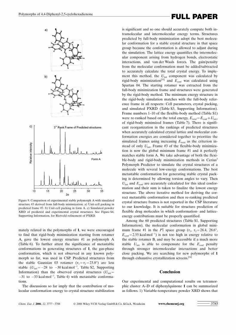
Figure 9. Comparison of experimental stable polymorph A with simulatedstructure #3 derived from full-body minimization. a) Unit-cell packing inpredicted frame #3. b) Unit-cell packing in form A. c) Simulated powderXRD of predicted and experimental crystal structures. See Figure S4,Supporting Information, for Rietveld refinement of PXRD.
Chem. Eur. J. 2006, 12, 3777 – 3788 L 2006 Wiley-VCH Verlag GmbH&Co. KGaA, Weinheim www.chemeurj.org 3785
FULL PAPERPolymorphs of 4,4-Diphenyl-2,5-cyclohexadienone

form A is the thermodynamic polymorph and form B is thekinetic phase. The enantiotropic relationship means thatlattice-energy differences as small as 0.3 kcalmol�1
(~1.3 kJmol�1) computed using the COMPASS force field(Cerius2) are experimentally verified. This value serves as abenchmark for future work on crystal structures that lie in ashallow potential energy well such as concomitant and/orconformational polymorphs. 2) We have shown that weakbut directional C�H···O interactions promote multiple mole-cules in the asymmetric unit and moreover that a shortH···O distance in a particular polymorph relates to a high Z’value of that crystal structure. Z’ is 4 in kinetic polymorph Band 1 in thermodynamic polymorph A. 3) The reason forconformational polymorphism in 1 is the presence of severallow-energy, interconverting conformations in solution. Thecrystal structures of 1 are a compromise between the mini-mization of intramolecular (rotamer) and intermolecular(interaction) energies. Crystal packing stabilizes the meta-stable molecular conformation of 1 in the solid state. 4)Thermodynamic polymorph A is reproduced as frame #3 infull-body minimized structure prediction based on Ulatt. Re-ranking of frames by including the Econf contribution to thecrystal energy gives global minimum structure #1 whichmatches form A. This exercise gives a posteriori validity toCerius2 Polymorph Predictor for a conformationally flexiblemedium-sized organic molecule. Our results on prototypesystem 1 are currently being examined and evaluated inother polymorphic clusters.
Experimental Section
Synthesis : Compound 1 was prepared in four steps as shown inScheme 1.[35]
trans-Stilbene (1.1 g, 6.0 mmol) in CH2Cl2 (60 mL) was added dropwiseto a stirred solution of m-CPBA (1.14 g, 6.6 mmol) in dry CH2Cl2(15 mL) at 0 8C. The reaction was continued for 30 h. The mixture waswashed with a NaHCO3 solution and water. The resulting epoxide wasextracted with CH2Cl2 and evaporated in vacuo to give pure trans-stil-bene oxide (1.2 g, 95%).
trans-Stilbene oxide (1.20 g, 6.0 mmol)was stirred with BF3·Et2O (0.5 mL,225 mg, 3.0 mmol) in dry CH2Cl2(60 mL) for 30 min at 0 8C. The reac-tion mixture was washed with water(2X50 mL) and the solvent evaporatedto give diphenyl acetaldehyde(960 mg, 80%).
Ethanolic KOH (3n, 0.5 mL) wasadded dropwise over a period of 5 minto a mixture of diphenyl acetaldehyde(700 mg, 3.6 mmol) and methyl vinylketone (MVK, 0.3 mL, 3.7 mmol) indry THF (30 mL) at 0 8C. The mixturewas stirred for 2 h at 0 8C and then for2 h at room temperature. Neutraliza-tion with 20% HCl, extraction withEtOAc, and work up gave the crude
product. Purification by column chromatography yielded pure 4,4-di-phenyl-2-cyclohexenone (440 mg, 50%).
DDQ (908 mg, 4.0 mmol) and a catalytic amount of p-TsOH (15 mg,0.1 mmol) was added to a solution of the above cyclohexenone (248 mg,1.0 mmol) in 1,4-dioxane (40 mL) and the solution was refluxed for 72 h.After cooling, the reaction mixture was filtered through Celite and thefiltrate was diluted with CH2Cl2 and washed with 10% NaOH solution(3X30 mL). Workup gave the crude product which was purified on asilica gel column to give pure 4,4-diphenyl-2,5-cyclohexadienone 1(100 mg, 40%). M.p. 120 8C. 1H NMR (400 MHz, CDCl3, 25 8C, TMS):d=6.38 (d, J=10 Hz, 2H, a-enone Hs), 7.25–8.15 (m, 12H, Ph+b-enoneHs) ppm; IR (KBr): n=3028, 1655 (C=O), 1620, 1487, 1446, 1400, 1267,1226 cm�1.
Polymorphs A—D : The pure solid 1 after column chromatography wasanalyzed by powder XRD. The bulk material (100 mg of the solid) showsall four forms A–D in the concomitant crystallization batch (Figure 3):monoclinic form A 37.5%, triclinic forms B+C 52.0%; orthorhombicform D 10.5%.
Polymorphic forms A, B, and C were crystallized by slow evaporation ofa solution of 1 in 5% EtOAc/n-hexane at ambient temperature. Threetypes of morphologies were observed: needle, block, and plate. Needle-type crystals correspond to form A and block/plate crystals correspond toform B as confirmed by random cell-checking of different crystals. Crys-tallization by slow evaporation at �5 8C in a domestic refrigerator yieldedform A whereas crystal growth from a saturated solution by fast evapora-tion at ambient temperature yielded predominantly form B. Crystals ofform D were obtained from a CH2Cl2/EtOAc/n-hexane solvent mixture.In recent batches over the last 2–3 years we have not found a single crys-tal corresponding to form C in several random cell-checking experiments.
Pure form A: The polymorphic mixture 1 (100 mg) was heated in a testtube at ~70 8C in an oil bath for 30 min and then slowly cooled to roomtemperature. The mixture converted to form A in >95% purity as con-firmed by powder XRD (Figure 5).
Table 7. Ulatt of the lowest energy frame determined by the rigid-body method starting from the molecularconformation in full-body minimized frames #1–10. Econf is calculated in Spartan 04. The simulated structuresof 1 are re-ranked based on the sum of the intra- and intermolecular energies, Etotal [kcalmol�1].
Frame # in full-body method[a]
Spacegroup[b]
Ulatt in rigid-bodymethod [kcalmol�1]
Econf[c]
[kcalmol�1]Etotal=Ulatt+Econf
[kcalmol�1]Re-ranked framebased on Etotal
3 P21[d] �30.93 1.38 �29.55 1
1 P1 �30.83 2.03 �28.80 26 P212121 �30.91 2.25 �28.66 310 C2/c �30.42 1.82 �28.60 47 P1 �30.88 2.59 �28.29 58 C2/c �30.06 1.82 �28.24 65 P21/c �30.85 2.79 �28.06 74 P212121 �30.01 2.14 �27.87 82 P21 �31.03 3.27 �27.76 99 P1 �29.88 2.37 �27.51 10
[a] Taken from Table S1. [b] See Table S3 for matching cell parameters. [c] Relative to the stable gas-phase ro-tamer, Econf=�479812.98 kcalmol�1. [d] Unit cell in full-body/rigid-body minimized structure: 7.712/7.713 Q,8.286/8.289 Q, 10.415/10.412 Q, 75.68/76.658, 322.45/322.44 Q3.
Scheme 1. Synthesis of diphenylquinone 1 from trans-stilbene.
www.chemeurj.org L 2006 Wiley-VCH Verlag GmbH&Co. KGaA, Weinheim Chem. Eur. J. 2006, 12, 3777 – 37883786
A. Nangia, G. J. Kruger et al.

Pure form B : The polymorphic mixture 1 (100 mg) was heated in the alu-minium pan of a powder X-ray diffractometer until the compoundmelted (~120 8C). The cooled solid is form B (powder XRD in Figure 6),as confirmed by unit-cell matching of a few randomly picked crystals.
Differential scanning calorimetry : DSC was performed with a MettlerToledo 822e module. Samples (4–6 mg) were placed in crimped butvented aluminium pans and heated at 10 8C min�1 from 30–200 8C. Theinstrument was purged with a stream of dry nitrogen at 150 mLmin�1.
Spartan 04, Gaussian 03, and Cerius2 computations : Cerius2 simulationsand crystal energy :[26] All simulations were carried out using version 4.8of the Cerius2 molecular modeling software package on a Silicon Graph-ics workstation. Geometry optimization was carried out using densityfunctional theory (DFT) at the B3LYP/6-31G (d,p) level of theory withGaussian 03.[32] The global minimized rotamer of 1 from Gaussian 03 wasentered as the input for the Polymorph Predictor. Crystal structure pre-diction was carried out in six common space groups (P21, P21/c, C2/c,Pbca, P212121, and P1). The cell parameters of predicted frames of theC2/c space group were compared with reduced cell parameters to con-firm that they represent different structures. Reduced cell parameters areused throughout the paper. Atom point charges were assigned using theCOMPASS force field. It was not felt necessary to calculate multipolecharges because molecule 1 does not contain strong hydrogen-bondinggroups. Multipole charges are known to give superior results but the ad-vantage is more evident in strongly hydrogen-bonded structures and thattoo at the expense of an approximate 10-fold increase in computertime.[36] Default options were used throughout with the fine searchoption in Monte Carlo simulations and for the clustering of frames toobtain unique structures. Lattice-energy minimization of predicted struc-tures was carried out without any modifications except for the use of theEwald summation of van der Waals interactions at a cut off of 6.0 Q. Allcalculations were carried out either by relaxing the molecular conforma-tion during the minimization, referred to as the full-body method, or bykeeping the conformation fixed during minimization, the so-called rigid-body method. Full-body lattice energy minimizations were carried outeven though these calculations take approximately five times more com-puter time because this method gives more accurate results for flexiblemolecules such as 1. The lattice energies of the experimental polymorphsof 1 were computed using the Cerius2 program by energy minimization ofcrystal structures using DREIDING 2.21 and COMPASS force fields.Force-field charges were assigned with COMPASS and the charge equili-brium method was used with DREIDING 2.21. COMPASS is better par-ametrized for structure prediction and the energy of organic molecules.Crystal lattice energies were calibrated to account for the number of mol-ecules in the unit cell (per molecule).
Spartan 04 computations : The energies of all 19 conformers were calcu-lated using Spartan 04[25] with crystallographic coordinates as the input;the positions of the hydrogen atoms were reoptimized at the HF/6-31G**level of theory while keeping the heavy atoms fixed. The gas-phase con-formation of 1 was obtained by global energy minimization. The gas-phase rotamer of 1 calculated using Spartan 04 (t1=t2=22.38) is verysimilar to the minimized conformation derived using Gaussian 03 (t1=t2=23.88).
Variable-temperature powder X-ray diffraction : Powder X-ray data werecollected with a PANlytical X’Pert PRO powder X-ray diffractometerusing a parallel beam of monochromated Cu-Ka radiation (l=1.54056 Q)and an X’celerator detector at 40 kV and 40 mA. Diffraction patternswere collected over the 2q range of 5–508. Samples were ground to a par-ticle size of >20 mm and loaded in an 18 mm alumina holder for varia-ble-temperature powder X-ray diffraction data collection and in an alu-minium sample holder with a 10 mm diameter sample cavity for the col-lection of data at room temperature. Vigorous grinding was avoided tominimize potential phase transitions. The program X’Pert High Scorewas used for the processing and comparison of powder patterns. PowderCell 2.3[22] was used for calculating the PXRD patterns and for the profilefitting and Rietveld refinement of unit-cell parameters, a displacementparameter, a background polynomial function, peak shape asymmetryterms, and an overall temperature factor using the known single-crystalstructures of polymorphs A, B, and D as the model. Variable-tempera-
ture powder X-ray diffraction data at a heating rate of 1 8Cmin�1 werecollected at T=28, 39, 49, 59, 69, 79, 89, 94, 98, 102, and 105 8C. Thesample was cooled to room temperature (31 8C) and data were recollect-ed. Powder XRD profiles are plotted in the range of 2q=10–358 for allsamples. There are no significant peaks below 108 and only minor peaksbetween 35 and 508. Wide peaks resulting from the sample holder appearat 25.2 and 34.88.
Acknowledgements
We thank the Department of Science and Technology for research fund-ing (SR/S5/OC-02/2002), for support of the CCD X-ray diffractometer(IRPHA), and the CMSD computational facility. The UPE program ofthe UGC provided infrastructure funds for UH. S.R. and R.B. thank theUGC for fellowships. G.J.K. and Prof. Gautam R. Desiraju (UH) thankthe Indo-South Africa Program on Research Cooperation for financialsupport (DST/INT/SAFR/COP/2001).
[1] W. C. McCrone in Physics and Chemistry of the Organic Solid State,Vol. 2 (Eds.: D. Fox, M. M. Labes, A. Weissberger), Wiley Inter-science, New York, 1965, pp. 725–767.
[2] J. Bernstein, Polymorphism in Molecular Crystals, Clarendon Press,Oxford, 2002.
[3] For some recent papers on polymorphism dealing with these issues,see: a) A. KYlmYn, L. FYbiYn, G. Argay, G. BernPth, Z. Gyarmati, J.Am. Chem. Soc. 2003, 125, 34; b) I. Weissbuch, V. Y. Torbeev, L.Leiserowitz, M. Lahav, Angew. Chem. 2005, 117, 3290; Angew.Chem. Int. Ed. 2005, 44, 3226; c) M. Morimoto, S. Kobatake, M. Irie,Chem. Eur. J. 2003, 9, 621; d) P. Raiteri, R. MartoZYk, M. Parrinello,Angew. Chem. 2005, 117, 3835; Angew. Chem. Int. Ed. 2005, 44,3769; e) P. K. Thallapally, R. K. R. Jetti, A. K. Katz, H. L. Carrell,K. Singh, K. Lahiri, S. Kotha, R. Boese, G. R. Desiraju, Angew.Chem. 2004, 116, 1169; Angew. Chem. Int. Ed. 2004, 43, 1149; f) H.Chow, P. A. W. Dean, D. C. Craig, N. T. Lucas, M. L. Scudder, I. G.Dance, New J. Chem. 2003, 27, 704; g) R. G. Gonnade, M. M.Bhadbhade, M. S. Shashidhar, Chem. Commun. 2004, 2530; h) C.Guo, M. B. Hickey, E. R. Guggenheim, V. Enkelmann, B. M.Foxman, Chem. Commun. 2005, 2220; i) S. Aitipamula, A. Nangia,Chem. Commun. 2005, 3159; j) W. I. F. David, K. Shankland, C. R.Pulham, N. Bladgen, R. J. Davey, M. Song, Angew. Chem. 2005, 117,7194; Angew. Chem. Int. Ed. 2005, 44, 7032.
[4] G. R. Desiraju, Angew. Chem. 1995, 107, 2541; Angew. Chem. Int.Ed. Engl. 1995, 34, 2311.
[5] a) R. J. Davey, Chem. Commun. 2003, 1463; b) N. Blagden, R. J.Davey, Cryst. Growth Des. 2003, 3, 873; c) P. Erk, H. Hengelsberg,M. F. Haddow, R. van Gelder, CrystEngComm 2004, 6, 474; d) J.Bernstein, Chem. Commun. 2005, 5007; e) L. Yu, J. Am. Chem. Soc.2003, 125, 6380; f) P. Vishweshwar, J. A. McMahon, M. Oliveira,M. L. Peterson, M. J. Zaworotko, J. Am. Chem. Soc. 2005, 127,16802; g) C. P. Price, A. L. Grzesiak, A. J. Matzger, J. Am. Chem.Soc. 2005, 127, 5512.
[6] S. R. Bryn, R. R. Pfeiffer, J. G. Stowell, Solid-State Chemistry ofDrugs, SSCI, West Lafayette IN, 1999.
[7] a) X. Zheng, B. Wang, U. Engelert, G. E. Herberich, Inorg. Chem.2001, 40, 3117; b) J. van de Streek, S. Motherwell, Acta Crystallogr.Sect. B 2005, 61, 504. In 2000 the values were 4.1% of organic com-pounds and 5.5% of organometallic compounds (ref. [7a]), which in2004 was updated to 14600 accurate crystal structures out of 325000entries (4.5%) (ref. [7a]).
[8] F. H. Allen, R. Taylor, Chem. Soc. Rev. 2004, 33, 463. The CSD is anarchive of over 350000 small-molecule organic and organometalliccrystal structures (August 2005 release); see www.ccdc.cam.ac.uk
[9] a) L. Yu, G. A. Stephenson, C. A. Mitchell, C. A. Bunnell, S. V.Snorek, J. J. Bowyer, T. B. Borchardt, J. G. Stowell, S. R. Byrn, J.Am. Chem. Soc. 2000, 122, 585; b) S. Chen, I. A. Guzei, L. Yu, J.Am. Chem. Soc. 2005, 127, 9881.
Chem. Eur. J. 2006, 12, 3777 – 3788 L 2006 Wiley-VCH Verlag GmbH&Co. KGaA, Weinheim www.chemeurj.org 3787
FULL PAPERPolymorphs of 4,4-Diphenyl-2,5-cyclohexadienone

[10] a) V. S. S. Kumar, PhD Thesis, University of Hyderabad, 2002 ;b) crystal structures of 3 have been deposited with the CambridgeCrystallographic Data Centre (see ref. [8]): CCDC 293133–293135.
[11] J. Bernstein, R. J. Davey, J.-O. Henck, Angew. Chem. 1999, 111,3646; Angew. Chem. Int. Ed. 1999, 38, 3440.
[12] J. Bernstein in Organic Solid State Chemistry (Ed.: G. R. Desiraju),Elsevier, Amsterdam, 1987, pp. 471–518.
[13] a) V. S. S. Kumar, A. Addlagatta, A. Nangia, W. T. Robinson, C. K.Broder, R. Mondal, I. R. Evans, J. A. K. Howard, F. H. Allen,Angew. Chem. 2002, 114, 4004; Angew. Chem. Int. Ed. 2002, 41,3848; b) A. Nangia in Nanoporous Materials : Science and Engineer-ing (Eds.: G. Q. Lu, X. S. Zhao), Imperial College Press, London,2004, pp. 165–187.
[14] G. R. Desiraju, Acc. Chem. Res. 2002, 35, 565.[15] J. Bernstein, R. E. Davis, L. Shimoni, N.-L. Chang, Angew. Chem.
1995, 107, 1687; Angew. Chem. Int. Ed. Engl. 1995, 34, 1555.[16] V. S. S. Kumar, A. Nangia, Chem. Commun. 2001, 2392.[17] J. W. Steed, CrystEngComm 2003, 5, 169.[18] a) C. P. Brock, L. L. Duncan, Chem. Mater. 1994, 6, 1307; b) C. P.
Brock, Acta Crystallogr. Sect. B 2002, 58, 1025.[19] A. Gavezzotti, G. Fillippini, J. Phys. Chem. 1994, 98, 4831.[20] H. Jacobsen, H. W. Schmalle, A. Messmer, H. Berke, Inorg. Chim.
Acta 2000, 306, 153.[21] K. E. Plass, K. Kim, A. J. Matzger, J. Am. Chem. Soc. 2004, 126,
9042.[22] Rietveld refinement was carried out using Powder Cell 2.3, N.
Krauss, G. Nolze, Federal Institute for Materials Research and Test-ing, Berlin, 2000.
[23] a) J. D. Dunitz, J. Bernstein, Acc. Chem. Res. 1995, 28, 193; b) J.-O.Henck, J. Bernstein, A. Ellern, R. Boese, J. Am. Chem. Soc. 2001,123, 1834.
[24] W. Ostwald, Z. Phys. Chem. 1897, 22, 289. Ostwald�s rule of stages:“When leaving a metastable state, a given chemical system does notseek out the most stable state, rather the nearest metastable onethat can be reached with minimum loss of free energy.”
[25] Spartan 04 computational methods include Hartree–Fock, densityfunction theory, and Moøller–Plesset on an interactive PC interface:see www.wavefun.com
[26] Crystal Packer and Polymorph Predictor of the Cerius2 suite of soft-ware were used for crystal lattice energy calculation and crystalstructure prediction: see www.accelrys.com
[27] A. Dey, M. T. Kirchner, V. R. Vangala, G. R. Desiraju, R. Mondal,J. A. K. Howard, J. Am. Chem. Soc. 2005, 127, 10545.
[28] H. Sun, J. Phys. Chem. B 1998, 102, 7338.[29] a) T. C. Lewis, D. A. Tocher, S. L. Price, Cryst. Growth Des. 2004, 4,
979; b) C. Ouvard, S. L. Price, Cryst. Growth Des. 2004, 4, 1119;c) H. Nowell, S. L. Price, Acta Crystallogr. Sect. B 2005, 61, 558.
[30] a) J. D. Dunitz, A. Gavezzotti, Cryst. Growth Des. 2005, 5, 2180. TheDEtotal maximum is larger in ROY (5 kcalmol�1 between the Y andORP forms) because Econf and Ulatt contributions are additive thanin 1 (<1 kcalmol�1) when the components counter one another.
[31] a) W. D. S. Motherwell, H. L. Ammon, J. D. Dunitz, A. Dzyabchen-ko, P. Erk, A. Gavezzotti, D. W. M. Hofmann, F. J. J. Leusen, J. P. M.Lommerse, W. T. M. Mooij, S. L. Price, H. Scheraga, B. Schweizer,M. U. Schmidt, B. P. van Eijck, P. Verwer, D. E. Williams, Acta Crys-tallogr. Sect. B 2002, 58, 647; b) G. M. Day, W. D. S. Motherwell, H.Ammon, S. X. M. Boerrigter, R. G. Della Valle, E. Venuta, A.Dzyabchenko, J. D. Dunitz, B. Schweizer, B. P. van Eijck, P. Erk,J. C. Facelli, V. E. Bazterra, M. B. Ferraro, D. W. M. Hofmann, F. J. J.Leusen, C. Liang, C. C. Pantelides, P. G. Karamertzanis, S. L. Price,T. C. Lewis, H. Nowell, A. Torrisi, H. A. Scheraga, Y. A. Arnautova,M. U. Schmidt, P. Verwer, Acta Crystallogr. Sect. B 2005, 61, 511.
[32] Gaussian 03 (Revision B.05), M. J. Frisch, G. W. Trucks, H. B. Schle-gel, G. E. Scuseria, M. A. Robb, J. R. Cheeseman, J. A. Montgomer-y, Jr., T. Vreven, K. N. Kudin, J. C. Burant, J. M. Millam, S. S. Iyen-gar, J. Tomasi, V. Barone, B. Mennucci, M. Cossi, G. Scalmani, N.Rega, G. A. Petersson, H. Nakatsuji, M. Hada, M. Ehara, K.Toyota, R. Fukuda, J. Hasegawa, M. Ishida, T. Nakajima, Y. Honda,O. Kitao, H. Nakai, M. Klene, X. Li, J. E. Knox, H. P. Hratchian,J. B. Cross, C. Adamo, J. Jaramillo, R. Gomperts, R. E. Stratmann,O. Yazyev, A. J. Austin, R. Cammi, C. Pomelli, J. W. Ochterski, P. Y.Ayala, K. Morokuma, G. A. Voth, P. Salvador, J. J. Dannenberg,V. G. Zakrzewski, S. Dapprich, A. D. Daniels, M. C. Strain, O.Farkas, D. K. Malick, A. D. Rabuck, K. Raghavachari, J. B. Fores-man, J. V. Ortiz, Q. Cui, A. G. Baboul, S. Clifford, J. Cioslowski,B. B. Stefanov, G. Liu, A. Liashenko, P. Piskorz, I. Komaromi, R. L.Martin, D. J. Fox, T. Keith, M. A. Al-Laham, C. Y. Peng, A. Na-nayakkara, M. Challacombe, P. M. W. Gill, B. Johnson, W. Chen,M. W. Wong, C. Gonzalez, J. A. Pople, Gaussian, Inc., Pittsburgh,PA, 2003.
[33] The value of Ulatt for minimized crystal structures after full-bodyminimization is much lower (ca. �92 kcalmol�1) than rigid-bodyminimized structures (ca. �32 kcalmol�1). The contribution from va-lence terms (bonds, angles, torsions, cross-terms) is around�60 kcalmol�1 for full-body minimization of molecule 1 but valenceterms are not considered in rigid-body minimization (fixed to zero).This difference between the two sets of energies is not a problembecause we are comparing relative energies using the same method.
[34] For CSP and isolation of the polymorph, see: a) A. T. Hulme, S. L.Price, D. A. Tocher, J. Am. Chem. Soc. 2005, 127, 1116; b) G. M.Day, A. V. Trask, W. D. S. Motherwell, W. Jones, Chem. Commun.2006, 54.
[35] a) D. J. Reif, H. O. House, Org. Syn. 1963, Coll. Vol. IV, 860; b) D. J.Reif, H. O. House, Org. Syn. 1963, Coll. Vol. IV, 375; c) A. C. Cope,P. A. Trumbell, E. R. Trumbell, J. Am. Chem. Soc. 1958, 80, 2844;d) H. E. Zimmerman, K. G. Hancock, G. C. Licke, J. Am. Chem.Soc. 1968, 90, 4892; e) K.-B. Chai, P. Sampson, J. Org. Chem. 1993,58, 6807.
[36] G. M. Day, W. D. S. Motherwell, W. Jones, Cryst. Growth Des. 2005,5, 1023.
Received: November 15, 2005Published online: March 3, 2006
www.chemeurj.org L 2006 Wiley-VCH Verlag GmbH&Co. KGaA, Weinheim Chem. Eur. J. 2006, 12, 3777 – 37883788
A. Nangia, G. J. Kruger et al.