concomitant malaria among visceral leishmaniasis in-patients from
TRANSCRIPT

van den Bogaart et al. BMC Public Health 2013, 13:332http://www.biomedcentral.com/1471-2458/13/332
RESEARCH ARTICLE Open Access
Concomitant malaria among visceralleishmaniasis in-patients from Gedarif and SennarStates, Sudan: a retrospective case-control studyErika van den Bogaart1*, Marieke MZ Berkhout1, Ayman BYM Nour2, Pètra F Mens1, Al-Badawi A Talha3,Emily R Adams1, Hashim BM Ahmed4, Samira H Abdelrahman4, Koert Ritmeijer5, Bakri YM Nour2,4
and Henk DFH Schallig1
Abstract
Background: In areas where visceral leishmaniasis (VL) and malaria are co-endemic, co-infections are common.Clinical implications range from potential diagnostic delay to increased disease-related morbidity, as compared toVL patients. Nevertheless, public awareness of the disease remains limited. In VL-endemic areas with unstable andseasonal malaria, vulnerability to the disease persists through all age-groups, suggesting that in these populations,malaria may easily co-occur with VL, with potentially severe clinical effects.
Methods: A retrospective case-control study was performed using medical records of VL patients admitted toTabarakallah and Gedarif Teaching Hospitals (Gedarif State) and Al`Azaza kala-azar Clinic (Sennar State), Sudan(2005-2010). Patients positively diagnosed with VL and malaria were identified as cases, and VL patients withoutmicroscopy-detectable malaria as controls. Associations between patient characteristics and the occurrence of theco-infection were investigated using logistic regression analysis. Confirmation of epidemiological outcomes wasobtained with an independently collected dataset, composed by Médecins Sans Frontières (MSF) at Um-el-Kher andKassab Hospitals, Gedarif State (1998).
Results: The prevalence of malaria co-infection among VL surveyed patients ranged from 3.8 to 60.8%, with amedian of 26.2%. Co-infected patients presented at hospital with deteriorated clinical pictures. Emaciation (OddsRatio (OR): 2.46; 95% Confidence Interval (95% CI): 1.72-3.50), jaundice (OR: 2.52; 95% CI: 1.04-6.09) and moderateanemia (OR: 1.58; 95% CI: 1.10-2.28) were found to be positively associated with the co-infection, while severity ofsplenomegaly (OR: 0.53; 95% CI: 0.35-0.81) and, to a less extent, hepatomegaly (OR: 0.52; 95% CI: 0.27-1.01) appearedto be reduced by concomitant VL and malaria. The in-hospital case-fatality rates did not significantly differ betweenco- and mono-infected patients (OR: 1.13; 95% CI: 0.59-2.17). Conversely, a significantly increased mortality rate (OR:4.38; 95% CI: 1.83-10.48) was observed by MSF amongst co-infected patients enrolled at Um-el-Kher and KassabHospitals, who also suffered an enhanced risk of severe anemia (OR: 3.44; 95% CI: 1.68-7.02) compared to VL mono-infections.
Conclusions: In endemic VL areas with unstable seasonal malaria, like eastern Sudan, VL patients are highlyexposed to the risk of developing concomitant malaria. Prompt diagnosis and effective treatment of malaria areessential to ensure that its co-infection does not result into poor prognoses.
Keywords: Visceral leishmaniasis, Malaria, Co-infection, Prevalence, Mortality, Risk factors
* Correspondence: [email protected] of Biomedical Research, Parasitology Unit, Royal TropicalInstitute (KIT), Amsterdam, the NetherlandsFull list of author information is available at the end of the article
© 2013 van den Bogaart et al.; licensee BioMed Central Ltd. This is an Open Access article distributed under the terms of theCreative Commons Attribution License (http://creativecommons.org/licenses/by/2.0), which permits unrestricted use,distribution, and reproduction in any medium, provided the original work is properly cited.
van den Bogaart et al. BMC Public Health 2013, 13:332 Page 2 of 13http://www.biomedcentral.com/1471-2458/13/332
BackgroundIn areas where malaria is co-endemic with visceral leish-maniasis (VL), co-infections with both diseases are com-mon [1]. Previous observations performed throughcohorts of VL patients report the occurrence of this co-infection across major VL hot spots [2-5], with prevalenceranging from 31% in Sudan [6] to 1.2% in Bangladesh [7].At Amudat Hospital, Uganda, where nearly one out of fiveVL-confirmed patients hospitalized between 2000 and2006 was co-diagnosed with malaria, concomitant malariawas shown to exacerbate symptoms of VL patients,though with no implications for their prognosis [1]. Chil-dren under 10 years of age, notoriously more vulnerableto both malaria and VL, exhibited a twofold higher risk ofbeing co-infected compared to adults. The clinical rele-vance of the VL-malaria co-morbidity, as attested by itsfrequency and severity at Amudat Hospital, highlights therisks associated with disease co-endemicity and suggeststhat other malaria- and VL-endemic areas may experiencesimilar co-infection burdens.Of the 30,000 to 50,000 annual VL cases estimated in
East Africa, the second largest VL focus after South-Asia, half as many (15,700 to 30,300) are thought tooccur in Sudan [8,9]. Hyper-endemic foci are located inthe eastern part of the country, stretching from theWhite Nile in the west to the Ethiopian border in theeast, and from Kassala State in the north across theborder of Southern Sudan up to Upper Nile State in thesouth [10]. Here, the disease has been reported since theearly 1900s [11-13], but it is in the past 30 years that ithas reached dramatic proportions, resulting in recurrentepidemics which have claimed hundreds of thousands oflives [14-16]. Currently, Gedarif State in Eastern State,together with the region of Sennar and Singa in CentralState, accounts for the vast majority of VL cases inSudan [17]. The distribution of the disease within theseareas is wide, erratic and variable, with highly endemicclusters situated near the banks of the Blue Nile Riverand its tributaries (Dinder, Rahad and Atbara Rivers)and the Dinder National Park [18-22]. Here, during theoutbreaks that cyclically recur (every 7-10 years), thedisease can reach incidences of >50 cases per 1000 peryear [22]. Diagnostics and treatment services are avail-able at several governmental hospitals and rural dispens-aries. However, due to the high cost of treatment, manycases have been referred to Médecins sans Frontières-Holland (MSF-H), whose activities at Um-el-Kher andKassab VL treatment centers have resulted in more than24,000 VL patients treated over the last hyper-endemicperiod (1996-2003) [17]. In the early 2000s, after an ini-tial decline in the disease incidence, a new upsurge inthe number of VL cases was recorded [9], leading to theestablishment in January 2010 of a new VL ward (MSF-Switzerland) at Tabarakallah Hospital.
Unstable seasonal malaria, due to Plasmodium falcip-arum, prevails in most areas of Gedarif and Sennar States,where prevalence rates of 1.6% and 1.1%, respectively,were confirmed among the communities surveyed in 2009[23]. This epidemiological pattern of malaria transmissionconsiderably affects the age-related ability of local individ-uals to acquire immunity against clinical malaria, resultinginto seasonal outbreaks that strike all age-groups, albeitwith different incidences [24,25]. Subclinical infections areoften harbored throughout the dry season, resulting intosemi-immunity which ultimately develops within 2 to 3decades [26]. Concomitant exposure of these individualsto Leishmania parasites may, therefore, have dramaticconsequences on ability to control both infections, poten-tially resulting in an aggravated clinical picture.To describe the clinical impact of VL-malaria co-
infections in patients with low malaria immunity, we in-vestigated the epidemiology of VL-malaria co-infectionsin a VL endemic area characterized by unstable seasonalmalaria. By surveying three strategically located hospi-tals, we obtained a semi-representative data collection ofGedarif and Sennar States (2005-2010), whereby the riskfor local VL patients of acquiring a malaria co-infectionwas assessed. The high prevalence of this co-morbidityand its negative impact on patients’ clinical condition, asobserved in this study, highlighted the existence of aclinically significant condition, whose life-threateningimplication was indirectly confirmed by an independentlycollected dataset, gathered by MSF-H at Um-el-Kher andKassab Hospitals, Gedarif State, in 1998.
MethodsStudy areaThe study area lies in Sudan, between the Blue NileRiver in the central region of Sennar State and the lowerAtbara River, which borders Gedarif State in the north-east. The Rahad River and the Dinder River, both tribu-taries of the Blue Nile River, flow across the region withseasonal regime, marked by major floods during therainy season (from June to October) and long periods ofdry-off throughout the rest of the year. Al`Azaza kala-azar Clinic (Sennar State), Gedarif Teaching Hospitaland Tabarakallah Hospital (Gedarif State) (Figure 1)were selected as treatment centers for conducting thesurvey. The hospitals encompass a VL-dedicated ward,where diagnosis and treatment for VL are performed.Gedarif Teaching Hospital, situated in the city ofGedarif, receives patients from the town and surround-ing areas. In addition, difficult clinical cases encounteredat regional level are referred to the hospital, which there-fore serves as Regional Reference Hospital. Al`Azazakala-azar clinic, located along the Dinder River, in prox-imity to the Dinder National Park, is a rural hospital.Together with Gedarif Teaching Hospital, Kassab

Figure 1 Map of Gedarif and Sennar States, Sudan, indicating the study sites and neighboring villages. The study hospitals are shownwith a star, while circles indicate neighboring cities and villages.
van den Bogaart et al. BMC Public Health 2013, 13:332 Page 3 of 13http://www.biomedcentral.com/1471-2458/13/332
Hospital and 4 other health centers in Gedarif andSennar States, the clinic received training and supportfrom MSF-H, as part of a “restructuring program”(2001-2004) for improving local management of VLcases. Tabarakallah Hospital, located in the eastern Atbararegion, 20 km south from the Atbara River, is a rural hos-pital which houses an MSF VL treatment center fromJanuary 2010. After departure of MSF-H from the regionin 2004, many of the medical staff who worked with MSFin an independent Zakat (Islamic charity)-funded clinic inTabarakallah, were employed by the hospital.
Study design and populationA retrospective case-control study was performed onpatients positively diagnosed for VL. Patients with alaboratory-confirmed diagnosis of both VL and malaria athospital admission or during hospitalization were identi-fied as cases, while controls were patients similarly diag-nosed with VL, whose blood smears tested negative formalaria. Laboratory-confirmation of VL was obtained ei-ther through microscopic examination of lymph node,bone marrow or spleen aspirates, or using serologicaltests, such as the direct agglutination test (DAT) (titer≥1:6400) and the rk39 antigen-based dipstick (Kalazar De-tect Rapid TestW), in combination with the clinical casedefinition of the World Health Organization (WHO) [27].
Intensity of Leishmania infection in lymph node or bonemarrow aspirates was categorized according to the WHO-recommended grading system [28,29]. Briefly, parasitaemiasranging from +1 to +4 were defined as smears containing1-10 parasites in 1000, 100, 10 or 1 field, respectively, while10-100 and >100 parasites per field indicated a parasitaemiaof +5 and +6, respectively. Malaria was confirmed bymicroscopic examination of blood smears or by RapidDiagnostic Tests (RDTs) (SD Bioline Malaria Ag P.f/P.v).Diagnosis of other concomitant diseases was based on clin-ical suspicion only. Clinical examination of patients in-cluded assessment of hemoglobin (Hb) levels (Lovibondmethod), spleen size (measured in the anterior axillary lineto the furthest point during quiet breathing) and liver size(measured in midclavicular line during quiet breathing).Nutritional status, as determined by Weight-for-High per-centiles or Body Mass Index (BMI), was directly recorded.Standard treatment for all VL patients, including relapsingcases, consisted of generic sodium stibogluconate ad-ministered parenterally. For malaria, artemisinin deriva-tives (artemether or artesunate), as mono-therapy orin combination with sulfadoxine-pyrimethamine (SP) orlumefantrine, or alternatively quinine were adminis-tered as first-line treatment. Test of cure was regularlyperformed for VL, but not for malaria, by microscopyon lymph node or bone marrow aspirates.
van den Bogaart et al. BMC Public Health 2013, 13:332 Page 4 of 13http://www.biomedcentral.com/1471-2458/13/332
Data collectionMedical records of patients hospitalized between January2005 and December 2010 in the three hospitals, wereretrospectively reviewed. Data from patients positively di-agnosed with VL were collected manually, by using paperCase Record Forms (CRF) specially designed for the study.Data included patients’ demographic details, clinical signsand symptoms, test results, medical treatments and rela-tive outcomes. Upon completion of the collection process,data were single-entered using SPSS 15.0 (SPSS Inc.,Chicago, IL, USA) and Microsoft Excel softwares.
Data analysisDescriptive analyses were conducted to examine the fea-tures of the overall study population and to assess preva-lence and case-fatality rates of the co-infected patients.Statistical analysis was performed using SPSS 15.0 soft-ware. Characteristics of cases and controls were indi-vidually compared, using the Pearson Chi-square test orthe Fisher Exact Probability test. Continuous variableswere categorized in predefined groups: age (<5, 5-9, 10-19, 20-29 and ≥30 years) based on malaria risk, spleensize (0-3 cm, 4-5 cm and ≥6 cm) by percentiles andanemia based on hemoglobin level (no-mild ≥7.3 g/dl,moderate 5.3-7.2 g/dl and severe <5,3 g/dl). As only fewpatients (n = 22, 2.0%) were found to be non-anemic(Hb ≥11 g/dl), while the majority (n = 606, 55.1%) hadmild anemia (Hb 7.3-10.9 g/dl), the groups with no andmild anemic patients were combined in one category,more truly representative of the patient set for anemiarisk assessment. Seasonality of the co-infection was ex-amined by categorizing hospital admissions during wetseason (from June to October) or dry season (fromNovember to May). Odds ratio (OR) with 95% confi-dence intervals (CI) were calculated to test whether avariable significantly differed between cases and controls.Multivariable logistic regression models were used toidentify independent patients’ characteristics associatedwith the co-infection. All variables with a P-value <0.10in the univariate analyses were entered stepwise in themultivariate analysis, whose final model only comprisedsignificant variables (P-value <0.05) and variables whichsignificantly increased the model fit, as assessed by the−2 Log Likelihood test.
MSF’s datasetDemographic and clinical data of VL patients enrolled in aclinical trial conducted by MSF-H at Um-el-Kher andKassab Hospitals (1998) were analyzed for comparison withthe survey outcomes. Both hospitals lie in Gedarif State(Figure 1), in proximity to the Rahad River and the Gedariftown, respectively, where prevalence of malaria and VL av-erages the rates characterizing the survey sites. The trialconducted by MSF aimed to compare efficacy and safety of
branded versus generic sodium stibogluconate for the treat-ment of primary VL. Hence, the study population com-prised patients with a laboratory-confirmed diagnosis of VL(DAT titer ≥1:6400 or demonstration of Leishmania on as-pirates of spleen or lymph node) and no history of previousanti-leishmanial treatment. Microscopic search for malariawas also performed on all study subjects at enrollment.Treatment for VL consisted of either Pentostam or genericsodium stibogluconate given intramuscularly (20 mg/kg/day for 30 days), while SP and quinine were administeredas first-line treatment for uncomplicated and severe mal-aria, respectively. Using the same case-control definition asin the survey, descriptive and statistical analyses (PersonChi-square test) were performed to assess prevalence andmortality rates of the VL-malaria co-infection and to meas-ure its association with explanatory variables.
Ethical approvalData were collected as part of routine patient care, withno need for additional investigations. Data extractionfrom medical records was performed by anonymizingthe information recorded in the CRFs. Ethical approvalfor the study was obtained from the Health Directoratesof Gedarif and Sennar States (3rd January 2011) andfrom the Ethics Review Board of MSF (19th July 2012).
ResultsPrevalence of VL-malaria co-infections in Gedarif andSennar States (2005-2010)A total of 1324 medical records reporting on VL-diagnosed patients hospitalized in Gedarif Teaching Hos-pital, Al`Azaza kala-azar Clinic and Tabarakallah Hospitalduring the study period were reviewed for the survey. Ofthese, 1295 reported on patients with a laboratory-confirmed VL infection, who were included in the study,while the remaining 29 were excluded for lack of labora-tory diagnostic evidence. Microscopy, mainly performedon lymph node (n = 848, 65.5%), bone marrow (n = 428,33.1%) and/or spleen (n = 3, 0.2%) aspirates, was used forthe diagnosis of VL. Serology was performed in 17 pa-tients, 13 of whom were confirmed by DAT and 4 by rk39antigen-based dipstick. Overall, 404 (31.2%) of the VL-confirmed patients were co-diagnosed with malaria athospital admission or during hospitalization. In all casesbut one, in which diagnosis was established with an RDT,malaria was confirmed by microscopy. Stratification of thestudy population by hospital resulted into an unevenly dis-tributed co-infection rate, with a prevalence of 3.8% inGedarif Teaching Hospital (n = 18), followed byTabarakallah Hospital (n = 84) and Al`Azaza kala-azarClinic (n = 302) with 26.2% and 60.8%, respectively. Withthe exception of one P. vivax-infection, all malaria casesfor which the Plasmodium species was determined, wereattributed to P. falciparum (n = 396, 98.0%).

van den Bogaart et al. BMC Public Health 2013, 13:332 Page 5 of 13http://www.biomedcentral.com/1471-2458/13/332
Demographic and clinical features of VL-malaria co-infections in Gedarif and Sennar States (2005-2010)With a median age of 12 years (inter-quartile range 5-23years), nearly three quarters of VL-malaria co-infected pa-tients were under 20 years old. Stratified by age, the per-centage of cases was highest among children agedbetween 0 and 4 years (40.7%, n = 120/296) and progres-sively reduced until 29 years (27.0%, n = 50/185)(data notshown), indicating that the chance of detecting malaria inpatients already diagnosed for VL declined as age in-creased. Residents of Sennar State appeared to be mostlystricken by the co-infection, particularly in the villages ofAl`Azaza (28.0%), Om Bagraa (8.7%) and Jaldook (5.2%).Anemia (hemoglobin level <11 g/dl) was a hallmark ofnearly all co- and mono-infected patients (91% and 79%,respectively), with most patients being diagnosed withmild or moderate anemia on hospital admission. Co-occurrence of malaria in VL patients resulted in increasedseverity of the anemia status, as shown by the decreasein median hemoglobin level (7.0 g/dl, inter-quartile range6.5-8.0 g/dl for cases and 8.0 g/dl, 7.0-9.0 g/dl for controls).Despite most co- and mono-infected patients presentedwith enlarged spleen, frequency (92% for cases, 95% forcontrols) and severity of splenomegaly (median spleensize below the costal margin 4.0 cm, inter-quartile range2.0-6.0 cm for cases and 5.0 cm, 2.0-8.0 cm for controls)appeared to be slightly reduced by concomitant malaria.In Al`Azaza kala-azar Clinic and Tabarakallah Hospital,
most co-infections were detected among young boys(median age 9 and 10 years, inter-quartile range 3-23 yearsand 4-18 years, respectively) (Table 1). In Gedarif Teach-ing Hospital, conversely, cases unlike controls mainlyconsisted of girls (55.6%, n = 10/176) (Table 1), althoughthe difference appeared to be statistically not significant.Seasonal distribution of hospital admissions was similar inall the three hospitals, with most patients presenting athospital during the long dry season (November-May). Co-occurrence of malaria in VL patients resulted in a sharperdivision between dry and wet season, particularly inGedarif Teaching Hospital, where 82.4% of all co-infectedpatients were hospitalized between November and May,emphasizing the peculiar time-trend of these co-infections(in these regions malaria usually occurs during or shortlyafter the rains, while VL clinical cases peak during the dryseason). Finally, with a median disease duration of 12 daysfrom the initial onset of symptoms, co-infected patientspresented at Tabarakallah Hospital earlier than atAl`Azaza kala-azar Clinic (21 days) or Gedarif TeachingHospital (30 days).
Infection intensity and case-fatality rate of VL-malaria co-infections in Gedarif and Sennar States (2005-2010)Intensity of VL infection, as determined by parasitaemiain lymph node or bone marrow aspirates, was directly
related with the frequency of VL-malaria co-infections.Stratified by infection intensity, in fact, the ratio of VL-malaria co-infections was lowest in the groups with lesssevere VL (n = 339/757, 44.8% as determined in lymphnode aspirates and n = 15/259, 5.8% as determined inbone marrow aspirates) and gradually increased as VLinfection intensified (n = 3/4, 75.0% and n = 1/5, 20.0%in lymph node and bone marrow aspirates, respectively,with the highest intensity) (Table 2).During hospitalization, 14 co-infected patients died,
resulting in an overall case-mortality rate of 3.5%. A similarcase-fatality rate was found among the controls (3.1%,n = 27) (OR: 1.13; 95% CI: 0.59-2.17), indicating that con-comitant malaria did not represent a risk factor for poorprognosis. When stratified by malaria treatment, in-hospitalfatalities clustered amongst co-infected patients treatedwith artemether and quinine, whose mortality risk was13-fold (OR: 13.34; 95% CI: 0.77-229.85) and 15-fold (OR:15.35; 95% CI: 0.81-289.33) higher, respectively, than inpatients receiving artesunate+SP (Table 3). Patients with ahigh density of Leishmania parasites were overrepresentedamong those who died, confirming previous findings,whereby high intensities of VL infection increased themortality risk [30]. Here, patients harboring high num-bers of Leishmania parasites in their lymph node orbone marrow were found to be 13 (OR: 13.46; 95% CI:1.34-135.77) and 15 (OR: 14.79; 95% CI: 2.24-97.74)times more likely to die, respectively (Table 2).
Risk factors for VL-malaria co-infections in Gedarif andSennar States (2005-2010)Associations between demographic and clinical variablesand the co-infection, as described by univariate analysis,are summarized in Table 4. Gender and age were identi-fied as risk factors for the VL-malaria co-infection.Significant associations were also found to link the co-infected patients with the treatment center in which theyhave been hospitalized. Neither the rainy season nor theintake of anti-malarial drugs prior to hospitalization sig-nificantly altered the risk for VL patients of acquiringmalaria. Malnourishment was relatively more commonamong cases, resulting in a positive association betweensevere malnutrition and the co-infection (OR: 2.21; 95%CI: 1.01-4.85). A similar positive association was foundwith moderately (OR: 1.73; 95% CI: 1.32-2.28) or se-verely anemic patients (OR: 1.50; 95% CI: 0.99-2.28).The likelihood of being diagnosed with concomitant VLand malaria decreased as spleen size increased: patientswith massive splenomegaly (spleen size ≥6 cm below thecostal margin) were found to be more than twice lesslikely to be co-infected (OR: 0.38; 95% CI: 0.26-0.55)compared with patients with no or minor spleen en-largement. In some patients, malaria displayed an ex-acerbating, although not significant, effect on VL

Table 1 Characteristics of the overall VL population and VL-malaria co-infected patients, stratified by hospital, Sudan(2005-2010)
Treatment center Gedarif TeachingHospital
Tabarakallah Hospital Al`Azaza Kala-azar Clinic
Total population Co-infectedpatients
Totalpopulation
Co-infectedpatients
Totalpopulation
Co-infectedpatients
n (%) n (%) n (%) n (%) n (%) n (%)
Number of patients 468 18 (3.8) 321 84 (26.2) 497 302 (60.8)
Gender 456 18 321 84 497 302
Male 280 (61.4) 8 (44.4) 179 (55.8) 49 (58.3) 254 (51.1) 155 (51.3)
Female 176 (38.6) 10 (55.6) 142 (44.2) 35 (41.7) 243 (48.9) 147 (48.7)
Age 440 15 321 84 492 301
Median (years) (inter-quartile range) 14.0 (7.0-24.5) 13.0 (9.0-29.0) 10.0 (5.0-18.0) 10.0 (4.2-17.7) 9.0 (3.0-23.0) 9.0 (3.0-23.0)
0-4 years 68 (15.5) 2 (13.3) 77 (24.0) 21 (29.8) 151 (30.7) 97 (32.2)
5-9 years 87 (19.8) 3 (20.0) 71 (22.1) 19 (22.6) 99 (20.1) 55 (18.3)
10-19 years 127 (28.9) 5 (33.3) 99 (30.8) 28 (33.3) 91 (18.5) 59 (19.6)
20-29 years 89 (20.2) 2 (13.3) 28 (8.7) 6 (7.1) 68 (13.8) 42 (14.0)
≥30 years 69 (15.7) 3 (20.0) 46 (14.3) 10 (11.9) 83 (16.9) 48 (15.9)
Season 452 17 321 84 488 297
Wet season 151 (33.4) 3 (17.6) 124 (38.6) 32 (38.1) 226 (46.3) 131 (44.1)
Dry season 301 (66.6) 14 (82.4) 197 (61.4) 52 (61.9) 262 (53.7) 166 (55.9)
Duration on-going sickness 433 17 302 82 441 259
Median (days) (inter-quartile range) 30.0 (15.0-40.0) 30.0 (13.5-76.0) 14.0 (9.0-21.0) 12.0 (7.0-14.0) 30.0 (14.0-61.0) 21.0 (14.0-61.0)
van den Bogaart et al. BMC Public Health 2013, 13:332 Page 6 of 13http://www.biomedcentral.com/1471-2458/13/332
infection, as revealed by the increased number of Leish-mania parasites observed in their lymph node or bonemarrow aspirates. Finally, while hepatomegaly was lessfrequently observed in co-infected patients compared tomono-infected patients (OR: 0.29; 95% CI: 0.18-0.48),jaundice (OR: 2.85; 95% CI: 1.43-5.66) and, particularly,weight loss (OR: 3.04; 95% CI: 2.37-3.90) were morecommonly reported among co-infected cases.All 11 variables associated with the VL-malaria co-
infection in the univariate analysis were included in themultivariable models. Due to the high number of miss-ing data in reporting clinical signs and symptoms, three
Table 2 VL-malaria co-infections, stratified by VL infection intdeath, Sudan (2005-2010)
Infection intensity of VL inaspirates
n cases/denominators
% In-hospita(%)
lymph node 377/824 21
+1 339/757 44.8 18 (85.7)
+2 24/44 54.5 2 (9.5)
+3 11/19 57.9 0
+4 3/4 75.0 1 (4.8)
bone marrow 16/264 13
+1 15/259 5.8 11 (84.6)
+2 1/5 20.0 2 (15.4)
*P-value <0.10 based on Fisher Exact Probability tests.
different models were designed to explore the associ-ation between the selected variables and the VL-malariaco-infection: model A, in which the independent riskfactors were identified, model B describing the associ-ation between clinical signs and symptoms and the co-infection and model C, which combines all variables(risk factors, clinical signs and symptoms) in one ana-lysis. Model A (Table 5) shows that, after adjusting forthe other variables included in the model, the treatmentcenter in which co-infected patients were hospitalized,and therefore its catchment area, remained the mostimportant factor associated with the risk of being
ensity, and its association with the overall in-hospital
l deaths n Discharged alive n(%)
OddsRatio
95% ConfidenceInterval
790
727 (92.0) 1
41 (5.2) 1.97 0.44-8.78
19 (2.4) 1.00 0.99-1.00
3 (0.4) 13.46 1.34-135.77*
247
244 (98.8) 1
3 (1.2) 14.79 2.24-97.74*
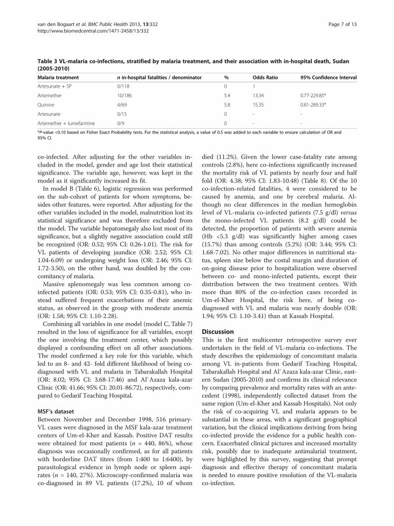
Table 3 VL-malaria co-infections, stratified by malaria treatment, and their association with in-hospital death, Sudan(2005-2010)
Malaria treatment n in-hospital fatalities / denominator % Odds Ratio 95% Confidence Interval
Artesunate + SP 0/118 0 1
Artemether 10/186 5.4 13.34 0.77-229.85*
Quinine 4/69 5.8 15.35 0.81-289.33*
Artesunate 0/15 0 - -
Artemether + lumefantrine 0/9 0 - -
*P-value <0.10 based on Fisher Exact Probability tests. For the statistical analysis, a value of 0.5 was added to each variable to ensure calculation of OR and95% CI.
van den Bogaart et al. BMC Public Health 2013, 13:332 Page 7 of 13http://www.biomedcentral.com/1471-2458/13/332
co-infected. After adjusting for the other variables in-cluded in the model, gender and age lost their statisticalsignificance. The variable age, however, was kept in themodel as it significantly increased its fit.In model B (Table 6), logistic regression was performed
on the sub-cohort of patients for whom symptoms, be-sides other features, were reported. After adjusting for theother variables included in the model, malnutrition lost itsstatistical significance and was therefore excluded fromthe model. The variable hepatomegaly also lost most of itssignificance, but a slightly negative association could stillbe recognized (OR: 0.52; 95% CI: 0.26-1.01). The risk forVL patients of developing jaundice (OR: 2.52; 95% CI:1.04-6.09) or undergoing weight loss (OR: 2.46; 95% CI:1.72-3.50), on the other hand, was doubled by the con-comitancy of malaria.Massive splenomegaly was less common among co-
infected patients (OR: 0.53; 95% CI: 0.35-0.81), who in-stead suffered frequent exacerbations of their anemicstatus, as observed in the group with moderate anemia(OR: 1.58; 95% CI: 1.10-2.28).Combining all variables in one model (model C, Table 7)
resulted in the loss of significance for all variables, exceptthe one involving the treatment center, which possiblydisplayed a confounding effect on all other associations.The model confirmed a key role for this variable, whichled to an 8- and 42- fold different likelihood of being co-diagnosed with VL and malaria in Tabarakallah Hospital(OR: 8.02; 95% CI: 3.68-17.46) and Al`Azaza kala-azarClinic (OR: 41.66; 95% CI: 20.01-86.72), respectively, com-pared to Gedarif Teaching Hospital.
MSF’s datasetBetween November and December 1998, 516 primary-VL cases were diagnosed in the MSF kala-azar treatmentcenters of Um-el-Kher and Kassab. Positive DAT resultswere obtained for most patients (n = 440, 86%), whosediagnosis was occasionally confirmed, as for all patientswith borderline DAT titers (from 1:400 to 1:6400), byparasitological evidence in lymph node or spleen aspi-rates (n = 140, 27%). Microscopy-confirmed malaria wasco-diagnosed in 89 VL patients (17.2%), 10 of whom
died (11.2%). Given the lower case-fatality rate amongcontrols (2.8%), here co-infections significantly increasedthe mortality risk of VL patients by nearly four and halffold (OR: 4.38; 95% CI: 1.83-10.48) (Table 8). Of the 10co-infection-related fatalities, 4 were considered to becaused by anemia, and one by cerebral malaria. Al-though no clear differences in the median hemoglobinlevel of VL-malaria co-infected patients (7.5 g/dl) versusthe mono-infected VL patients (8.2 g/dl) could bedetected, the proportion of patients with severe anemia(Hb <5.3 g/dl) was significantly higher among cases(15.7%) than among controls (5.2%) (OR: 3.44; 95% CI:1.68-7.02). No other major differences in nutritional sta-tus, spleen size below the costal margin and duration ofon-going disease prior to hospitalization were observedbetween co- and mono-infected patients, except theirdistribution between the two treatment centers. Withmore than 80% of the co-infection cases recorded inUm-el-Kher Hospital, the risk here, of being co-diagnosed with VL and malaria was nearly double (OR:1.94; 95% CI: 1.10-3.41) than at Kassab Hospital.
DiscussionThis is the first multicenter retrospective survey everundertaken in the field of VL-malaria co-infections. Thestudy describes the epidemiology of concomitant malariaamong VL in-patients from Gedarif Teaching Hospital,Tabarakallah Hospital and Al`Azaza kala-azar Clinic, east-ern Sudan (2005-2010) and confirms its clinical relevanceby comparing prevalence and mortality rates with an ante-cedent (1998), independently collected dataset from thesame region (Um-el-Kher and Kassab Hospitals). Not onlythe risk of co-acquiring VL and malaria appears to besubstantial in these areas, with a significant geographicalvariation, but the clinical implications deriving from beingco-infected provide the evidence for a public health con-cern. Exacerbated clinical pictures and increased mortalityrisk, possibly due to inadequate antimalarial treatment,were highlighted by this survey, suggesting that promptdiagnosis and effective therapy of concomitant malariais needed to ensure positive resolution of the VL-malariaco-infection.

Table 4 Univariate analysis of characteristics associated with VL-malaria co-infections, Sudan (2005-2010)
Variable Cases n (%) Controls n (%) Crude Odds Ratio 95% Confidence Interval
Gender 404 870
Male 212 (52.5) 501 (57.6) 1
Female 192 (47.5) 369 (42.4) 1.23 0.97-1.56*
Age 400 853
<5 years 120 (30.0) 176 (20.6) 1.53 1.05-2.24*
5-9 years 77 (19.3) 180 (21.1) 0.96 0.64-1.44
10-19 years 92 (23.0) 225 (26.4) 0.92 0.62-1.35
20-29 years 50 (12.5) 135 (15.8) 0.83 0.53-1.30
≥30 years 61 (15.3) 137 (16.1) 1
Treatment center 404 882
Gedarif Teaching Hospital 18 (4.4) 450 (51.0) 1
Tabarakallah Hospital 84 (20.8) 237 (26.9) 8.86 5.20-15.10*
Al`Azaza kala-azar Clinic 302 (74.8) 195 (22.1) 38.72 23.38-64.11*
Season 398 863
Dry season 232 (58.3) 528 (61.2) 1
Wet season 166 (41.7) 335 (38.8) 1.13 0.89-1.44
Previous anti-leishmanial treatment 389 860
No 373 (95.9) 806 (93.7) 1
Yes 16 (4.1) 54 (6.3) 0.64 0.36-1.13
Previous anti-malarial treatment 396 864
No 277 (70.0) 604 (69.9) 1
Yes 119 (30.0) 260 (30.1) 1.00 0.77-1.29
Malnutrition 322 575
None 262 (81.4) 497 (86.4) 1
Mild 17 (5.3) 25 (4.3) 1.29 0.68-2.43
Moderate 29 (9.0) 41 (7.1) 1.34 0.82-2.21
Severe 14 (4.4) 12 (2.1) 2.21 1.01-4.85*
Anemia degree on admission 374 725
Median Hb (inter-quartile range) 7.0 (6.5-8.0) 8.0 (7.0-9.0)
None-mild (Hb ≥7.3 g/dl) 182 (48.7) 445 (61.4) 1
Moderate (Hb 5.3-7.2 g/dl) 149 (39.8) 210 (29.0) 1.73 1.32-2.28*
Severe (Hb <5.3 g/dl) 43 (11.5) 70 (9.7) 1.50 0.99-2.28*
Spleen size 238 563
Median (inter-quartile range) 4.0 (2.0-6.0) 5.0 (2.0-8.0)
0-3 cm 94 (39.5) 150 (26.6) 1
4-5 cm 79 (33.2) 141 (25.0) 0.89 0.61-1.30
≥6 cm 65 (27.3) 272 (48.3) 0.38 0.26-0.55*
Infection intensity in lymph node aspirate 377 447
+1 339 (89.9) 418 (93.5) 1
+2 24 (6.4) 20 (4.5) 1.48 0.80-2.72
+3 11 (2.9) 8 (1.8) 1.70 0.67-4.26
+4 3 (0.8) 1 (0.2) 3.70 0.38-35.72
Infection intensity in bone marrow aspirate 16 248
+1 15 (93.8) 244 (98.4) 1
van den Bogaart et al. BMC Public Health 2013, 13:332 Page 8 of 13http://www.biomedcentral.com/1471-2458/13/332
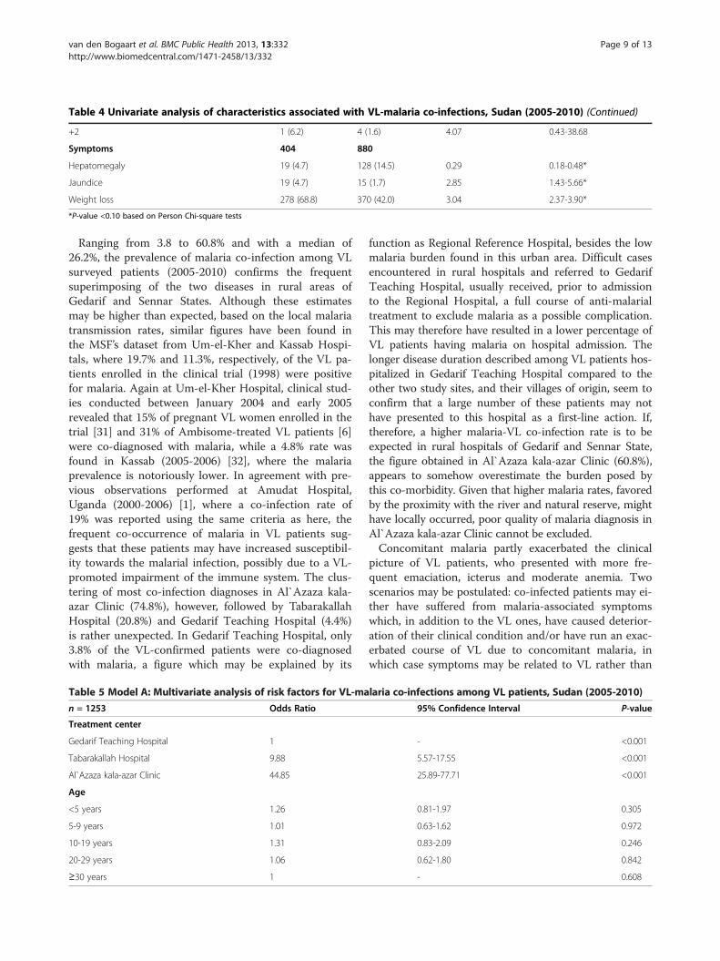
Table 4 Univariate analysis of characteristics associated with VL-malaria co-infections, Sudan (2005-2010) (Continued)
+2 1 (6.2) 4 (1.6) 4.07 0.43-38.68
Symptoms 404 880
Hepatomegaly 19 (4.7) 128 (14.5) 0.29 0.18-0.48*
Jaundice 19 (4.7) 15 (1.7) 2.85 1.43-5.66*
Weight loss 278 (68.8) 370 (42.0) 3.04 2.37-3.90*
*P-value <0.10 based on Person Chi-square tests
van den Bogaart et al. BMC Public Health 2013, 13:332 Page 9 of 13http://www.biomedcentral.com/1471-2458/13/332
Ranging from 3.8 to 60.8% and with a median of26.2%, the prevalence of malaria co-infection among VLsurveyed patients (2005-2010) confirms the frequentsuperimposing of the two diseases in rural areas ofGedarif and Sennar States. Although these estimatesmay be higher than expected, based on the local malariatransmission rates, similar figures have been found inthe MSF’s dataset from Um-el-Kher and Kassab Hospi-tals, where 19.7% and 11.3%, respectively, of the VL pa-tients enrolled in the clinical trial (1998) were positivefor malaria. Again at Um-el-Kher Hospital, clinical stud-ies conducted between January 2004 and early 2005revealed that 15% of pregnant VL women enrolled in thetrial [31] and 31% of Ambisome-treated VL patients [6]were co-diagnosed with malaria, while a 4.8% rate wasfound in Kassab (2005-2006) [32], where the malariaprevalence is notoriously lower. In agreement with pre-vious observations performed at Amudat Hospital,Uganda (2000-2006) [1], where a co-infection rate of19% was reported using the same criteria as here, thefrequent co-occurrence of malaria in VL patients sug-gests that these patients may have increased susceptibil-ity towards the malarial infection, possibly due to a VL-promoted impairment of the immune system. The clus-tering of most co-infection diagnoses in Al`Azaza kala-azar Clinic (74.8%), however, followed by TabarakallahHospital (20.8%) and Gedarif Teaching Hospital (4.4%)is rather unexpected. In Gedarif Teaching Hospital, only3.8% of the VL-confirmed patients were co-diagnosedwith malaria, a figure which may be explained by its
Table 5 Model A: Multivariate analysis of risk factors for VL-ma
n = 1253 Odds Ratio
Treatment center
Gedarif Teaching Hospital 1
Tabarakallah Hospital 9.88
Al`Azaza kala-azar Clinic 44.85
Age
<5 years 1.26
5-9 years 1.01
10-19 years 1.31
20-29 years 1.06
≥30 years 1
function as Regional Reference Hospital, besides the lowmalaria burden found in this urban area. Difficult casesencountered in rural hospitals and referred to GedarifTeaching Hospital, usually received, prior to admissionto the Regional Hospital, a full course of anti-malarialtreatment to exclude malaria as a possible complication.This may therefore have resulted in a lower percentage ofVL patients having malaria on hospital admission. Thelonger disease duration described among VL patients hos-pitalized in Gedarif Teaching Hospital compared to theother two study sites, and their villages of origin, seem toconfirm that a large number of these patients may nothave presented to this hospital as a first-line action. If,therefore, a higher malaria-VL co-infection rate is to beexpected in rural hospitals of Gedarif and Sennar State,the figure obtained in Al`Azaza kala-azar Clinic (60.8%),appears to somehow overestimate the burden posed bythis co-morbidity. Given that higher malaria rates, favoredby the proximity with the river and natural reserve, mighthave locally occurred, poor quality of malaria diagnosis inAl`Azaza kala-azar Clinic cannot be excluded.Concomitant malaria partly exacerbated the clinical
picture of VL patients, who presented with more fre-quent emaciation, icterus and moderate anemia. Twoscenarios may be postulated: co-infected patients may ei-ther have suffered from malaria-associated symptomswhich, in addition to the VL ones, have caused deterior-ation of their clinical condition and/or have run an exac-erbated course of VL due to concomitant malaria, inwhich case symptoms may be related to VL rather than
laria co-infections among VL patients, Sudan (2005-2010)
95% Confidence Interval P-value
- <0.001
5.57-17.55 <0.001
25.89-77.71 <0.001
0.81-1.97 0.305
0.63-1.62 0.972
0.83-2.09 0.246
0.62-1.80 0.842
- 0.608

Table 6 Model B: Multivariate analysis of clinical signs and symptoms associated to VL-malaria co-infections, Sudan(2005-2010)
n = 690 Odds Ratio 95% Confidence Interval P-value
Symptoms
Hepatomegaly 0.52 0.26-1.01 0.052
Jaundice 2.52 1.04-6.09 0.041
Weight loss 2.46 1.72-3.50 <0.001
Spleen size
0-3 cm 1 - 0.007
4-5 cm 0.90 0.60-1.36 0.629
≥6 cm 0.53 0.35-0.81 0.003
Anemia
None - mild (Hb ≥7.3 g/dl) 1 - 0.440
Moderate (Hb 5.3-7.2 g/dl) 1.58 1.10-2.28 0.013
Severe (Hb <5.3 g/dl) 1.10 0.63-1.93 0.737
van den Bogaart et al. BMC Public Health 2013, 13:332 Page 10 of 13http://www.biomedcentral.com/1471-2458/13/332
to malaria. If this latter hypothesis may find its rationalein the increased number of Leishmania parasites ob-served in aspirates of co-infected patients, the firstspeculation may be supported by the peculiar symptompattern. Jaundice, in fact, is rarely described among VLpatients, while it is not uncommon in P. falciparum mal-aria [33]. Weight loss and anemia, on the other hand,are hallmark of both VL and malaria and an increasedseverity of the anemic status, as observed in co-infectedpatients, may therefore be the result of an added effectdisplayed by both diseases on the polyparasitized host.In apparent contradiction is the finding, whereby co-infected patients suffered from less severe hepato-splenomegaly. Suggesting a less advanced state of thediseases in the co-infected patients, the result may beexplained by their earlier hospitalization compared tomono-infected VL patients. Patients with concomitantVL and malaria, in fact, presented at hospital nearly 10
Table 7 Model C: Multivariate analysis of risk factors, clinical siSudan (2005-2010)
n = 690 Odds Ratio
Treatment center
Gedarif Teaching Hospital 1
Tabarakallah Hospital 8.02
Al`Azaza kala-azar Clinic 41.66
Spleen size
0-3 cm B.C.M. 1
4-5 cm B.C.M. 1.20
≥6 cm B.C.M. 1.15
Anemia
None - mild (Hb ≥7.3 g/dl) 1
Moderate (Hb 5.3-7.2 g/dl) 1.28
Severe (Hb <5.3 g/dl) 0.74
days earlier, on average, than those with only VL, pos-sibly due to their more severe symptomatology. Import-antly, this may have also had positive implications fortheir prognosis, which was found to be similar to thecontrols’ one.In antithesis to the positive resolution of VL-malaria co-
infections during the 2005-2010 survey, is the significantlyhigher fatality rate (P-value 0.001) associated with co-infected patients enrolled by MSF at Um-el-Kher andKassab Hospital in 1998. During this trial, co-infected pa-tients were nearly four and half times more likely to diecompared with the VL mono-infected patients, whosemortality (2.8%) on the other hand, compares well to whatfound in the most recent survey (3.1%). Different anti-malarial regimen were used within the two study groups:SP and quinine in 1998 for uncomplicated and severe mal-aria, respectively; artemisinin derivatives (alone or in com-bination) and more rarely quinine between 2005 and
gns and symptoms associated to VL-malaria co-infections,
95% Confidence Interval P-value
- <0.001
3.68-17.46 <0.001
20.01-86.72 <0.001
- 0.706
0.76-1.90 0.428
0.71-1.86 0.563
- 0.219
0.85-1.94 0.239
0.39-1.40 0.356

Table 8 Characteristics of the malarial VL co-infected patients versus the non-malarial VL patients enrolled in theMSF’s clinical trial at Um-el-Kher and Kassab Hospitals, Sudan (1998)
Variable Cases n (%) Controls n (%) Crude Odds Ratio 95% Confidence Interval
Age 89 427
Median (years), (interquartile range) 7 (4-14) 10 (6-17)
Treatment center 89 427
Kassab Hospital 17 (19.1) 134 (31.4) 1
Um-el-Kher Hospital 72 (80.9) 293 (68.6) 1.94 1.10-3.41*
Fatal outcome 89 427
No 79 (88.8) 415 (97.2) 1
Yes 10 (11.2) 12 (2.8) 4.38 1.83-10.48*
Hb level 89 427
Median (g/dl), (interquartile range) 7.5 (6.0-9.0) 8.2 (7.0-9.4)
≥5.3 g/dl 75 (84.3) 405 (94.8) 1
<5.3 g/dl 14 (15.7) 22 (5.2) 3.44 1.68-7.02*
Nutritional status 68 299
Mean Weigh for Height (%) 78.8 80.4
Weight for Height Percent ≥70% 60 (88.2) 279 (93.3) 1
Weight for Height Percent <70% 8 (11.8) 20 (6.7) 1.86 0.78-4.42
Spleen size below the costal margin 80 389
Median (cm), (interquartile range) 6 (3-8) 7 (4-10)
<6 cm 33 (41.2) 144 (37.0) 1
≥6 cm 47 (58.8) 245 (63.0) 0.84 0.51-1.37
Duration on-going sickness 86 422
Median (days), (interquartile range) 30 (15-60) 30 (20-60)
<60 days 62 (72.1) 286 (67.8) 1
≥60 days 24 (27.9) 136 (32.2) 0.81 0.49-1.36
*P-value <0.05 based on Person Chi-square test.
van den Bogaart et al. BMC Public Health 2013, 13:332 Page 11 of 13http://www.biomedcentral.com/1471-2458/13/332
2010. Sudan’s choice to introduce artemisinin-based com-bination therapies (ACTs) for treatment of uncomplicatedand severe malaria was implemented nation-wide in 2004[34], following increasing evidence of resistance againstchloroquine, SP and quinine, for which failure rates up to76.9, 16.1 and 16.7%, respectively, were recorded in east-ern Sudan prior to ACT era [35-38]. The 11.2% mortalityrate of VL-malaria co-infections observed in 1998 atUm-el-Kher Hospital may therefore have partially resultedfrom treatment failures attributable to either SP or quin-ine, besides the more severe malaria course in patients re-ceiving quinine. In fact, no major differences for age,median Hb level, nutritional status, spleen size and dur-ation of on-going disease distinguished the co-infectedpatients’ group at Um-el-Kher and Kassab Hospitals fromthe one surveyed in 2005-2010 and from its relative con-trols. The only exception is to be found in the significantlyhigher number of VL patients who developed severeanemia when co-infected with malaria, similarly to whatwas observed during the 2005-2010 survey, though to aless extent. Hence, concomitant malaria may not only
cause aggravation of VL patients’ clinical condition, but itmay also result in a poorer prognosis, if failed to betreated. Among co-infected patients surveyed in 2005-2010, an increased mortality risk, not ascribable to differ-ences in Leishmania intensities, was observed when quin-ine (P-value 0.07) and artemether (P-value 0.04) wereadministered as antimalarials, suggesting either increasedmalaria severity or inadequate drug treatment.The population surveyed within this study consists of
VL patients residing in over 300 different villages, mainlylocated in Gedarif and Sennar States. Within these dis-tricts, Tabarakallah Hospital and Al`Azaza kala-azarClinic are two rural hospitals receiving patients fromsome of the worst-affected villages. It should be noted,however, that the cohort of VL patients surveyed heremight be sub-representative of the local VL community,as the number of VL-related hospitalizations carried outby the two MSF’s treatment centers in Gedarif Sate(>4000 per year between 1997 and 1999 [22]) exceeds byfar the one recorded by the three study hospitals (1324in total between 2005 and 2010). Other limitations apply
van den Bogaart et al. BMC Public Health 2013, 13:332 Page 12 of 13http://www.biomedcentral.com/1471-2458/13/332
to this study, the most important ones being the lack ofnon-VL malaria infected patients and the quality of diag-nosis. Unlike VL, uncomplicated malaria infections arecommonly treated on an outpatient basis in hospitals,clinics or simple practices, resulting in few data beingsystematically recorded by the different facilities. More-over, malaria patients are rarely found in VL-dedicatedhospitals, such as those surveyed in this study. Thisresulted into the lack of valid malaria controls, essentialto investigate whether VL might predispose or ratherprotect towards a malarial attack and whether it mightinfluence its course and clinical presentation. In absenceof quality control, quality of diagnosis remains question-able. Variable outcomes, as documented in medical re-cords, may have suffered from poor standardization, dueto the different techniques implemented by clinicians inthe different treatment centers and the possible involve-ment of different health workers in filling these files.
ConclusionBased on the results of this study, we conclude that VL pa-tients living in areas with unstable seasonal malaria, such aseastern Sudan, are highly exposed to the risk of developingconcomitant malaria. Large variation in the geographicaldistribution of co-infection cases highlights the presence ofenvironmental and/or social factors, whose identity andrelevance in the risk of co-acquiring VL and malaria still re-main to be elucidated. Clinical concerns should arise whenthe two diseases co-occur in the same patients, as signifi-cant exacerbation of their clinical condition was observed,along with an increased mortality risk, possibly associatedwith inappropriate anti-malarial treatment. Local healthcare policies should take into account the high co-infectionburden borne by VL foci with unstable malaria, byrecommending systematic malaria screening for all VLpatients and ACTs for treatment of malaria.
Competing interestsThe authors declare that they have no competing interests.
Authors’ contributionsEvdB contributed in conceiving of the study, drafted the study protocol andthe manuscript and participated in the data analysis. MB performed the dataentry and the statistical analysis and helped to draft the manuscript. ANperformed the collection and entry of data. PM and EA participated inconceiving of the study and reviewed the study protocol and themanuscript. AT participated in the collection and entry of data and helpedto draft the study protocol. HA participated in the collection and entry ofdata. SA reviewed the study protocol and the manuscript. KR participated inthe data analysis and reviewed the manuscript. BN coordinated thecollection and entry of data, organized local logistics, and helped to reviewthe study protocol and the manuscript. HS conceived the study andparticipated in its design, and reviewed the study protocol and themanuscript. All authors read and approved the final manuscript.
AcknowledgmentsWe are grateful to the staff of Al`Azaza kala-azar Clinic, Gedarif TeachingHospital and Tabarakallah Hospital for their cooperation during the datacollection. In addition, we wish to thank Dr. Masja Straetemans (RoyalTropical Institute of Amsterdam) for providing assistance in the data analysis.
Author details1Department of Biomedical Research, Parasitology Unit, Royal TropicalInstitute (KIT), Amsterdam, the Netherlands. 2Blue Nile National Institute forCommunicable Diseases, University of Gezira, Wad Medani, Sudan. 3Facultyof Medical Laboratory Sciences, University of Gezira, Wad Medani, Sudan.4Department of Medicine, Faculty of Medicine, University of Gedarif, andGedarif Teaching Hospital, Kala Azar Ward, Gedarif, Sudan. 5Public HealthDepartment, Médecins Sans Frontières, Amsterdam, the Netherlands.
Received: 5 August 2012 Accepted: 1 April 2013Published: 11 April 2013
References1. van den Bogaart E, Berkhout MZ, Adams ER, Mens PF, Sentongo E,
Mbulamberi E, Straetemans M, Schallig HDFH, Chappuis F: Prevalence,features and risk factors of malaria co-infections among visceralleishmaniasis patients from Amudat Hospital. Uganda. PLoS Negl Trop Dis2012, 6(4):e1617.
2. Mueller Y, Mbulamberi DB, Odermatt P, Hoffmann A, Loutan L, Chappuis F:Risk factors for in-hospital mortality of visceral leishmaniasis patients ineastern Uganda. Trop Med Int Health 2009, 14(8):910–917.
3. Kolaczinski JH, Reithinger R, Worku DT, Ocheng A, Kasimiro J, Kabatereine N,Brooker S: Risk factors of visceral leishmaniasis in East Africa: a case–control study in Pokot territory of Kenya and Uganda. Int J Epidemiol2008, 37(2):344–352.
4. de Beer P, el Harith A, Deng LL, Semiao–Santos SJ, Chantal B: A killingdisease epidemic among displaced Sudanese population identified asvisceral leishmaniasis. Am J Trop Med Hyg 1991, 44(3):283–9.
5. Nandy A, Addy M, Guha SK, Maji AK, Chaudhuri D, Chatterjee P: Co–existentkala–azar and malaria in India. Trans R Soc Trop Med Hyg 1995, 89(5):516.
6. Mueller M, Ritmeijer K, Balasegaram M, Koummuki Y, Santana MR, DavidsonR: Unresponsiveness to Ambisome in some Sudanese patients with kala–azar. Trans R Soc Trop Med Hyg 2007, 101(1):19–24.
7. Sarker CB, Chowdhury KS, Siddiqui NI, Jamal MF, Rahman S, Momen A, DharDK, Alam KS: Clinical profile of kala–azar in adults: as seen inMymensingh medical college hospital, Mymensingh. Bangladesh.Mymensingh Med J 2003, 12(1):41–4.
8. Alvar J, Vélez ID, Bern C, Herrero M, Desjeux P, Cano J, Jannin J, den Boer M,the WHO Leishmaniasis Control Team: Leishmaniasis worldwide andglobal estimates of its incidence. PLoS One 2012, 7(5):e35671.
9. Burki T: East African countries struggle with visceral leishmaniasis. Lancet2009, 374:371–372.
10. Zijlstra EE, el–Hassan AM: Leishmaniasis in Sudan. Visceral leishmaniasis.Trans R Soc Trop Med Hyg 2001, 95(Suppl 1):27–58.
11. Cummins SL: Kala–azar in the Anglo–Egyptian Sudan. In Third report of theWellcome Tropical Research Laboratories at the Gordon Memorial CollegeKhartoum. London: Baillière, Tindall & Cox; 1908:100–106.
12. Bousfield L: A tour of investigation as to prevalence of kala–azar inKassala and Blue Nile districts, Sudan. J R Army Med Corps 1910,15:161–183. 292–307.
13. Thomson DSB: Kala–azar Commission to investigate the prevalence andcause of the disease in the Eastern Sudan. (1) General report. In Fourthreport of the Wellcome Tropical Research Laboratories at the Gordon MemorialCollege Khartoum. London: Baillière, Tindall & Cox; 1911:142–156.
14. Zijlstra EE, El–Hssan AM, Ismael A, Ghalib HW: Endemic kala–azar in easternSudan: a longitudinal study on the incidence of clinical and subclinicalinfection and post–kala–azar dermal leishmaniasis. Am J Trop Med Hyg1994, 51(6):826–836.
15. El–Hassan AM, Zijlstra EE, Ismael A, Ghalib HW: Recent observations on theepidemiology of kala–azar in the eastern and central states of theSudan. Trop Geogr Med 1995, 47:151–156.
16. Seaman J, Mercer AJ, Sondorp E: The epidemic of visceral leishmaniasis inwestern Upper Nile, Southern Sudan: course and impact from 1984 to1994. Int J Epidemiol 1996, 25:862–971.
17. Malaria consortium: Leishmaniasis control in Eastern Africa: past and presentefforts and future needs. Situation and gap analysis. November 2010. [http://www.malariaconsortium.org/userfiles/file/NTD%20Resources/VL%20EA%20Situation%20Analysis%20Fina_Janl.pdf].
18. Kirk R: Studies in leishmaniasis in the Anglo–Egyptian Sudan. Part I.Epidemiology and general considerations. Trans R Soc Trop Med Hyg 1939,32:533–544.

van den Bogaart et al. BMC Public Health 2013, 13:332 Page 13 of 13http://www.biomedcentral.com/1471-2458/13/332
19. Elnaiem DE, Schorscher J, Bendall A, Obsomer V, Osman ME, Mekkawi AM,Connor SJ, Ashford RW, Thomson MC: Risk mapping of visceralleishmaniasis: the role of local variation in rainfall and altitude on thepresence and incidence of kala–azar in eastern Sudan. Am J Trop MedHyg 2003, 68(1):10–17.
20. EL–Safi SH, Bucheton B, Kheir MM, Musa HA, EL–Obaid M, Hammad A,Dessein A: Epidemiology of visceral leishmaniasis in Atbara River area,eastern Sudan: the outbreak of Barbar El Fugara village (1996–1997).Microbes Infect 2002, 4(14):1439–1447.
21. Ibrahim ME, Lambson B, Yousif AO, Deifalla NS, Alnaiem DA, Ismail A, YousifH, Ghalib HW, Khalil EA, Kadaro A, Barker DC, El Hassan AM: Kala–azar in ahigh transmission focus: an ethnic and geographic dimension. Am J TropMed Hyg 1999, 61(6):941–944.
22. Ritmeijer K, Davidson RN: Royal Society of Tropical Medicine and Hygienejoint meeting with Médecins Sans Frontières at Manson House, London,20 March 2003: field research in humanitarian medical programmes.Médecins Sans Frontières interventions against kala–azar in the Sudan,1989–2003. Trans R Soc Trop Med Hyg 2003, 97(6):609–613.
23. Federal Ministry of Health, the Sudan: Malaria Indicator Survey 2009 in theNorthern states of the Sudan. Cairo, Egypt: Submitted to the World HealthOrganization, Eastern Mediterranean Regional Office; 2010.
24. Theander TG: Unstable malaria in Sudan: the influence of the dry season.Malaria in areas of unstable and seasonal transmission. Lessons fromDaraweesh. Trans R Soc Trop Med Hyg 1998, 92(6):589–592.
25. Giha HA, Rosthoj S, Dodoo D, Hviid L, Satti GM, Scheike T, Arnot DE,Theander TG: The epidemiology of febrile malaria episodes in an area ofunstable and seasonal transmission. Trans R Soc Trop Med Hyg 2000,94(6):645–651.
26. Elhassan IM, Hviid L, Jakobsen PH, Giha H, Satti GM, Arnot DE, Jensen JB,Theander TG: High proportion of subclinical Plasmodium falciparuminfections in an area of seasonal and unstable malaria in Sudan.Am J Trop Med Hyg 1995, 53(1):78–83.
27. World Health Organization: Recommended Surveillance Standards. (WHO/CDS/CSR/ISR/99.2). 2nd edition. Geneva; 1999 [http://data.unaids.org/publications/irc-pub04/surveillancestandards_en.pdf].
28. Chulay JD, Bryceson AD: Quantitation of amastigotes of Leishmaniadonovani in smears of splenic aspirates from patients with visceralleishmaniasis. Am J Trop Med Hyg 1983, 32(3):475–479.
29. World Health Organization Expert Committee: Control of the Leishmaniases.Geneva: WHO Technical Report Series No. 793; 1990.
30. Seaman J, Mercer AJ, Sondorp HE, Herwaldt BL: Epidemic visceralleishmaniasis in Southern Sudan: treatment of severly debilited patientsunder wartime conditions and with limited resources. Ann Intern Med1996, 124(7):664–672.
31. Mueller M, Balasegaram M, Koummuki Y, Ritmeijer K, Santana MR, DavidsonR: A comparison of liposomal amphotericin B with sodiumstibogluconate for the treatment of visceral leishmaniasis in pregnancyin Sudan. J Antimicrob Chemother 2006, 58(4):811–5.
32. Musa AM, Younis B, Fadlalla A, Royce C, Balasegaram M, Wasunna M, HailuA, Edwards T, Omollo R, Mudawi M, Kokwaro G, El–Hassan A, Khalil E:Paromomycin for the treatment of visceral leishmaniasis in Sudan: arandomized, open–label, dose–finding study. PLoS Negl Trop Dis 2010,4(10):e855.
33. Anand AC, Puri P: Jaundice in malaria. J Gastroenterol Hepatol 2005,20(9):1322–1332. Review.
34. Malik EM, Mohamed MM, Elmardi KA, Mowien RM, Elhassan AH, Elamin SB,Mannan AA, Ahmed ES: From chloroquine to artemisinin–basedcombination therapy: the Sudanese experience. Malar J 2006, 5:65.
35. Adam I, Osman ME, Elghzali G, Ahmed GI, Gustafssons LL, Elbashir MI:Efficacies of chloroquine, sulfadoxine–pyrimethamine and quinine in thetreatment of uncomplicated, Plasmodium falciparum malaria in easternSudan. Ann Trop Med Parasitol 2004, 98:661–666.
36. Adam I, Ibrahim MH, A/elbasit IA, Elbashir MI: Efficacy of sulfadoxinpyrimethamine for uncomplicated Plasmodium falciparum malaria in asmall sample of Sudanese children. East Mediterr Health J 2004,10(3):309–314.
37. Salah MT, Mohammed MM, Himeidan YE, Malik EM, Elbashir MI, Adam I:A randomized comparison of sulphadoxine–pyrimethamine andcombination of sulphadoxine pyrimethamine with chloroquine in thetreatment of uncomplicated falciparum malaria in Eastern Sudan. SaudiMed J 2005, 26(1):147–148.
38. Elhassan IM, Satti GH, Ali AE, Fadul I, Elkhalifa AA, Abedelrahim AM, Ming C,Theander TG: The efficacy of artemether in the treatment of Plasmodiumfalciparum malaria in Sudan. Trans R Soc Trop Med Hyg 1993, 87(6):685–686.
doi:10.1186/1471-2458-13-332Cite this article as: van den Bogaart et al.: Concomitant malaria amongvisceral leishmaniasis in-patients from Gedarif and Sennar States, Sudan:a retrospective case-control study. BMC Public Health 2013 13:332.
Submit your next manuscript to BioMed Centraland take full advantage of:
• Convenient online submission
• Thorough peer review
• No space constraints or color figure charges
• Immediate publication on acceptance
• Inclusion in PubMed, CAS, Scopus and Google Scholar
• Research which is freely available for redistribution
Submit your manuscript at www.biomedcentral.com/submit