concentration-dependent activities of the even-skipped...
TRANSCRIPT
Concentration-dependent activities of the even-skipped protein in Drosophila embryos Armen S. Manoukian and Henry M. Krause
Banting and Best Department of Medical Research, University of Toronto, C.H. Best Institute, Toronto, Ontario MSG 1L6 Canada
The Drosophila pair-rule gene even-skipped (eve) encodes a homeo-domain-containing protein (Eve) that is required for the development of both odd- and even-numbered parasegments. We have used a heat shock-inducible eve transgene to study the regulatory functions of Eve in vivo. Transcripts encoded by eight other segmentation genes were monitored for changes in distribution and abundance following short pulses of ectopic Eve expression. Two tiers of response times appeared to distinguish between genes that were direct [fushi tarazu (ftz), odd-skipped (odd), runt (run), paired, and wingless] and indirect [eve, hairy, and engrailed (en)] targets of Eve. Genes that appeared to be directly regulated by Eve were differentially repressed in a concentration-dependent fashion. Interestingly, the run and ftz genes could also be activated by Eve during a brief 20- to 30-rain stage in development. The delayed actions upon the eve and en genes appeared to be mediated by run and odd. As in eve- embryos, these effects on segmentation gene expression patterns caused defects in both odd- and even-numbered parasegments. Four sequential phenotypes could be induced, each of which was attributable to the altered expression of a unique subset of target genes.
[Key Words: even-skipped; segmentation; pair-rule genes; segment polarity genes]
Received May 1, 1992; revised version accepted June 24, 1992.
Segmentation in Drosophila is embryonically controlled by a hierarchy of interactions among several classes of genes (N/isslein-Volhard and Wieschaus 1980; Nfisslein- Volhard et al. 1985). Information is relayed in a temporal progression from the coordinate genes to the gap genes, then from the gap genes to the pair-rule genes and fi- nally, from the pair-rule genes to the segment polarity genes (for review, see Ingham 1988). The even-skipped (eve) gene is considered to be a member of the pair-rule genes (N/isslein-Volhard and Wieschaus 1980). Weak eve mutations (hypomorphs) fit the pair-rule gene criteria, causing deletions of alternate segment-wide regions (Niisslein-Volhard and Wieschaus 1980). Unlike the other pair-rule genes, however, eve null alleles com- pletely abolish segmentation within the trunk of the em- bryo (Nfisslein-Volhard et al. 19851. Thus, eve appears to be a particularly important member of the pair-rule class of genes.
In correspondence to the severity of the eve null phe- notype, eve is expressed in a dynamic fashion through- out the trunk of the embryo (Frasch et al. 1987). Low levels of uniformly distributed eve protein (Eve) resolve into an anterior-to-posterior gradient which, in turn, re- solves into a 7-stripe pattern of expression, followed by a 14-stripe pattern of expression. Eve performs both early and late functions during this 2- to 3-hr period (Goto et al. 1989). During the time that Eve is expressed in seven
stripes, it functions as a primary pair-rule gene, inter- preting spatial cues provided by the gap genes and relay- ing this information to the other pair-rule genes (for re- view, see Pankratz and J/ickle 1990). Eve expression at this time defines the odd-numbered parasegmental pri- mordia (Lawrence et al. 1987}. As the Eve stripes begin to resolve, Eve is required for proper initiation of the seg- ment polarity genes engrailed (en) (DiNardo and O'Far- rell 1987; Lawrence et al. 1987) and wingless (wg)(Ing- ham et al. 1988) in odd-numbered parasegments. These two genes define anterior and posterior parasegmental identities, respectively.
An important issue that remains to be resolved is whether gene interactions such as these are direct or indirect. For example, is Eve a direct activator of the en gene, or does it regulate an intermediary gene whose product regulates en? Eve contains a DNA-binding ho o meo domain (MacDonald et al. 1986; Hoey and Levine 1988) and functions as a sequence-specific transcrip- tional repressor in transfected tissue-culture cells (Han et al. 1989) and in transcriptionally competent extracts (Biggin and Tjian 1989). Although Eve appears to func- tion exclusively as a repressor in vitro, expression pat- tems of several genes in wild-type and mutant embryos (see below) suggest that Eve may function as both a re- pressor and an activator in vivo.
We wished to distinguish between direct and indirect
1740 GENES & DEVELOPMENT 6:1740-1751 © 1992 by Cold Spring Harbor Laboratory ISSN 0890-9369/92 $3.00
Cold Spring Harbor Laboratory Press on August 25, 2019 - Published by genesdev.cshlp.orgDownloaded from
Ectopic e v e causes m u l t i p l e pattern defects
targets of Eve, and to determine whether Eve acts as a transcriptional repressor, an activator, or both. We have addressed these questions by providing short pulses of ectopic Eve expression at different stages of embryogen- esis and monitoring potential Eve target genes for changes in their patterns of expression. Among the genes that we tested, those that might be direct targets of Eve repression include the pair-rule genes fushi - tarazu (ftz) (Carroll and Scott 1986; Hiromi and Gehring 1987), odd- sk ipped (odd) (DiNardo and O'Farrell 1987; Coulter and Wieschaus 1988), runt ( run) ( Ingham and Gergen 1988), and paired (prd)(Baumgartner and Noll 1991), and the segment polarity gene wg (Ingham et al. 1988). Genes postulated to be directly activated by Eve include the eve gene itself (Frasch et al. 1988), the pair-rule gene hairy (h) (Ingham and Gergen 1988), and the segment polarity gene en (DiNardo and O'Farrell 1987).
The results of this study indicate that only a subset of these genes are direct targets of Eve regulatory activities. In general, our data suggest that Eve acts as a transcrip- tional repressor, except during its earliest stages of ex- pression. By regulating different target genes at different developmental stages, ectopic Eve could induce four dif- ferent mutant phenotypes. On the basis of these results, we suggest that Eve functions as a concentration-depen- dent morphogen, with sequential regulatory roles in both the odd- and even-numbered parasegments.
R e s u l t s
Expression of eve in HSEVE embryos
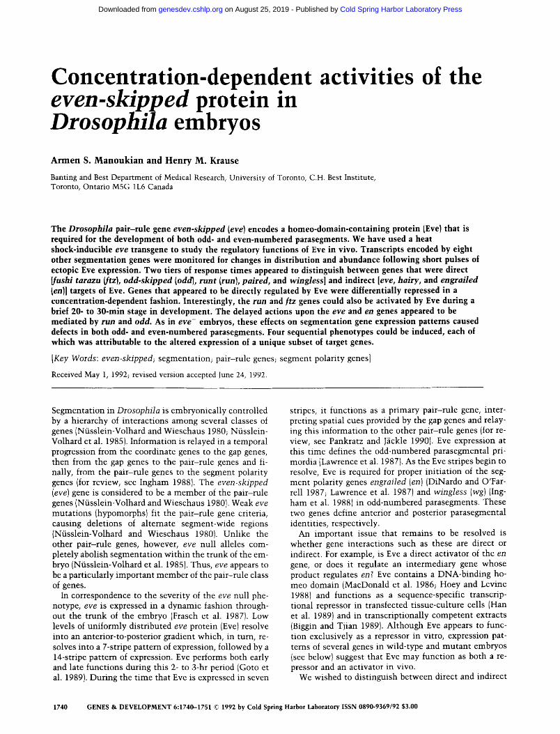
Fly lines containing P-element-mediated insertions of a heat shock promoter (hsp70)-controlled eve gene (PH- SEVE) were obtained from Gary Struhl (Columbia Uni- versity; see Materials and methods). Two PHSEVE-trans- formed lines were used: one with the PHSEVE construct inserted into the second chromosome (HSEVE32), and the other with an insert in the third chromosome (HSEVE19B). Both inserts gave equivalent results in a va-
riety of different genetic backgrounds. Our first goal was to test these lines for heat shock-inducible Eve expres- sion. Figure 1B shows that 30 min after a short 4-min heat shock, Eve was immunologically detected in all nu- clei of HSEVE embryos. Note that the underlying pattern of seven stripes was still visible, indicating that the lev- els of ectopic Eve were probably similar to the levels of endogenous Eve.
It has been suggested that Eve plays a direct role in the activation of its own promoter (Jiang et al. 1991). To test whether ectopic Eve could activate the endogenous eve gene, we looked at eve mRNA expression after adminis- tering 3- to 4-rain heat shock pulses to embryos aged between 2 and 3 hr after egg laying (AEL). Figure 1D shows an embryo fixed 15 min after the initiation of heat shock. At this time, the ectopically induced eve tran- scripts that had been distributed evenly over most of the embryo surface were already on the decline. In embryos fixed 30 rain after heat shock, the heat shock-induced eve transcripts were no longer detectable (Fig. 1E). Tran- scripts of the endogenous eve gene, however, continued to be expressed in a relatively normal seven-stripe pat- tern of expression. To our surprise, in similarly staged embryos (2.5-3 hr AEL) that had been heat-shocked 45 min before fixation, eve transcripts were very weak or undetectable (Fig. IF). This autorepression was not ob- served if embryos were older than 2.5 hr AEL at the time of heat shock. The 45-min delay before the loss of en- dogenous eve expression suggests that this repression was indirect (see below for further discussion). Thus, not only was ectopic Eve incapable of activating the endog- enous eve gene outside of its normal domains of expres- sion, but it caused a premature loss of expression within the domains in which it is normally expressed.
In this experiment and the experiments that follow, the effects described were Eve specific because extended heat shocks (up to 10 rain) had no effects on wild-type embryos (data not shown), and the effects of other heat shock-inducible pair-rule genes varied, depending on the
A i - J - - P
E F
Figure 1. eve expression in HSEVE embryos. Embryos aged 2-3 hr AEL were heat shocked for 4 min and stained for eve protein (A,B) or eve transcripts (C-F). (A, C) Wild-type patterns of protein and mRNA, respec- tively. Twenty minutes after the initiation of heat shock, Eve was detected in all nuclei, with the under- lying pattern of seven stripes still visible (B). (D-F) eve transcripts at 15 (D), 30 (E), and 45 (F) min after the initiation of heat shock. The embryos shown were all fixed at a similar developmental stage (-2.5-3 hr AEL).
GENES & D E V E L O P M E N T 1741
Cold Spring Harbor Laboratory Press on August 25, 2019 - Published by genesdev.cshlp.orgDownloaded from
Manoukian and Krause
particular pair-rule gene that was induced (A.S. Man- oukian and H.M. Krause, unpubl.).
Repression of ftz by ectopic Eve
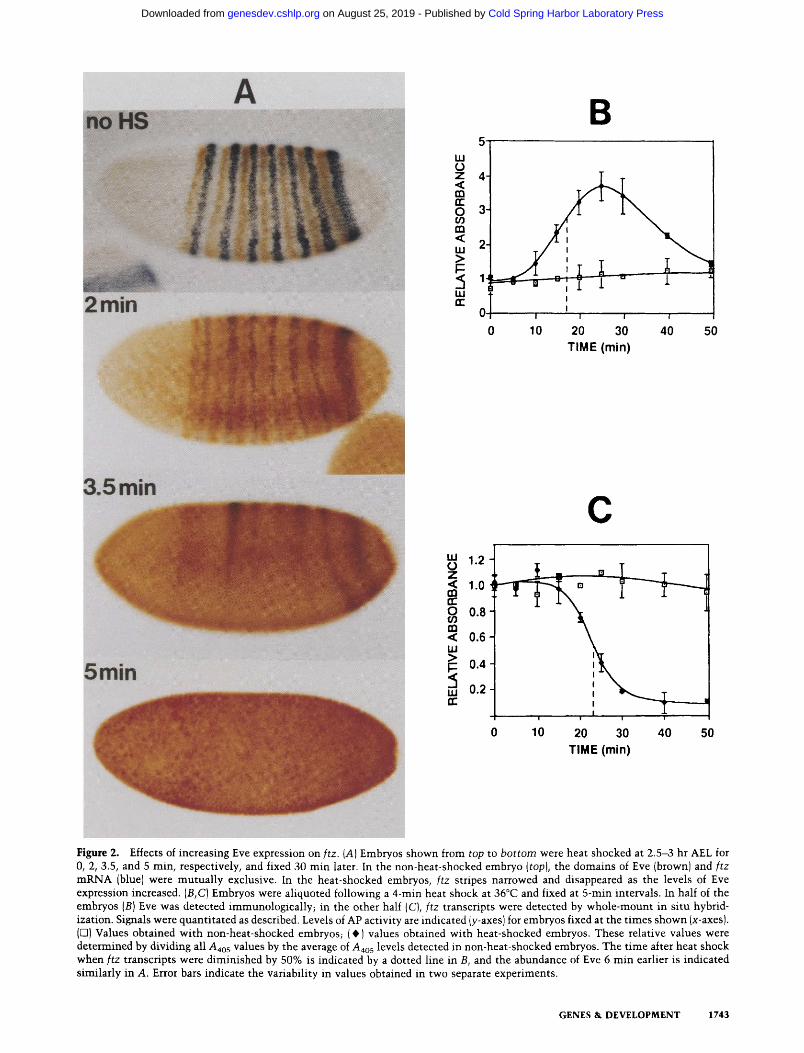
By use of heat shock-inducible transgenes, it is possible to vary the levels of ectopic gene expression by altering the parameters of heat shock. This permits an assess- ment of expression levels that are functionally relevant. One of the suspected targets of Eve is the ftz gene. Figure 2A shows the correlation between the levels of ectopic Eve induced by different durations of heat shock and the subsequent effects on ftz gene expression. Embryos aged 2.5 to 3 hr AEL at the time of heat shock were fixed 30 min later and double-stained for Eve protein (brown} and ftz transcripts (blue). Consistent with the hypothesis that Eve is a repressor of the ftz gene (Carroll and Scott 1986; Frasch and Levine 1987; Hiromi and Gehring 1987; Lawrence and Johnston 1989), stripes of ftz expres- sion diminished in intensity and width as the abundance of ectopic Eve increased. Total repression of ftz tran- scription occurred when heat shocks were - 4 min or longer.
To address the question of whether Eve is a direct or indirect regulator of ftz gene expression, we monitored the levels of Eve and ftz transcripts over the course of an hour following a 4-min heat shock. A very short tempo- ral delay between the rise in levels of Eve and the sub- sequent loss of ftz transcripts would favor a direct inter- action between the two genes. Embryos aliquoted from a common pool of heat-shocked embryos were fixed at 5- to 10-min intervals following heat shock. The levels of Eve and ftz transcripts in equivalent numbers of embryos were quantitated with the aid of secondary antibodies coupled to alkaline phosphatase (AP) and by monitoring AP activity colorimetrically.
Figure 2B shows that ectopic Eve expression could first be detected at 5- to 10-min and peaked at -25 min, after the initiation of heat shock. At their peak, the levels of ectopic Eve were approximately three to four times the levels of Eve detected in non-heat-shocked embryos (or in heat-shocked wild-type embryos; not shown). If eve stripes normally occupy 20-30% of the surface of a blas- toderm embryo (where the majority of nuclei are local- ized), this overall increase in Eve abundance by three- to fourfold should bring the interstripe levels of protein close to the levels that are expressed in the endogenous stripes. This is consistent with the levels of Eve staining in the HSEVE and wild-type embryos shown in Figure 1 (cf. A and BI.
A decrease in the abundance of ftz transcripts in heat- shocked embryos was first detected -15 min after the beginning of heat shock and was essentially complete within 30 min of the initiation of heat shock (Fig. 2C}. The estimated half-life of ftz transcripts (6 min; Edgar et al. 1986) is consistent with the rapid degradation profile of ftz transcripts in HSEVE embryos depicted in Figure 2C. If Eve is a direct repressor of ftz transcription, then ftz transcripts should be reduced in abundance by 50%, - 6 min after Eve reaches sufficient levels to repress the
ftz gene completely. In Figure 2C, it can be seen that ftz expression was reduced by 50% -23 min after the initi- ation of heat shock and that 6 min earlier (17 min after the initiation of heat shock), Eve was approximately two to three times the normal levels of endogenous Eve pro- tein. As discussed above, these levels of ectopic Eve are probably similar to or less than the levels of Eve that are normally confined to the seven stripes. Hence, these re- sults favor the hypothesis that Eve is a direct regulator of the ftz gene. Because the expression patterns of ftz pro- moter-lacZ fusion genes were also repressed within a similar time frame (data not shown), this negative regu- lation by Eve is likely to be mediated by regulatory ele- ments located upstream of the ftz-coding region.
Ectopic Eve acts in a concentration-dependent fashion
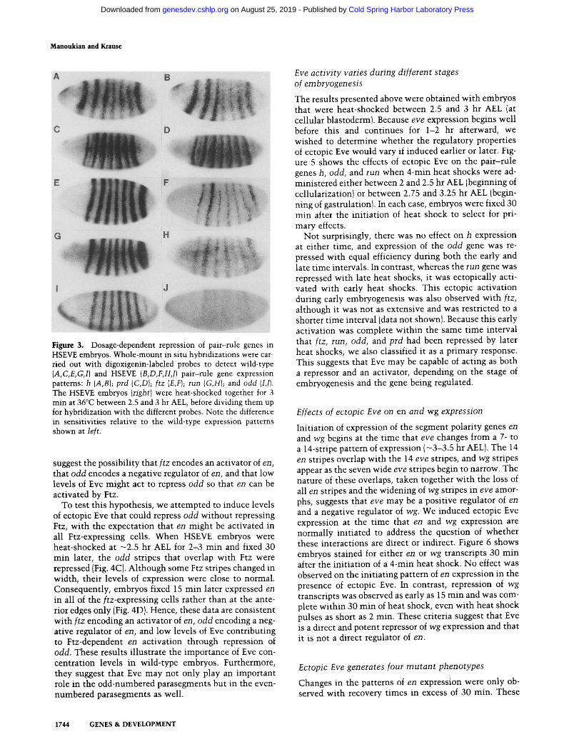
To determine whether other pair-rule genes would also respond to ectopic Eve within the same time frame as ftz, and whether Eve would also act as a repressor of these genes, we performed whole-mount in situ hybrid- izations using sequences from the genes h, run, prd, ftz, and odd as probes {Fig. 3). The embryos shown were heat-shocked in a single batch for 3 min and fixed 30 min later. The only genes that were obviously affected within the 30-min recovery period, and with this duration of heat shock, were the genes ftz, run, and odd. All three genes were repressed but with different levels of effi- ciency. This dosage-dependent variation in sensitivity was also apparent when heat shocks were varied in du- ration (not shown). For example, heat shock durations of only 2 min did not affect ftz and run expression but were still sufficient to repress odd. When the duration of heat shocks was increased to 4 min or longer, all three genes were completely repressed. Four-minute inductions also repressed the prd gene within the same 15- to 30-min recovery period (determined by visual examination only). In contrast, the h and endogenous eve genes were not affected within this 30-min period, even when heat shock pulses were as long as l0 min. These results sug- gest that Eve is a direct regulator of ftz, run, odd, and prd and that each of these genes is differentially repressed by different levels of EVE. The 30- to 45-min delay in the response time of the h and endogenous eve genes sug- gests that they are not direct targets of Eve.
Differential repression of ftz and odd alters en expression
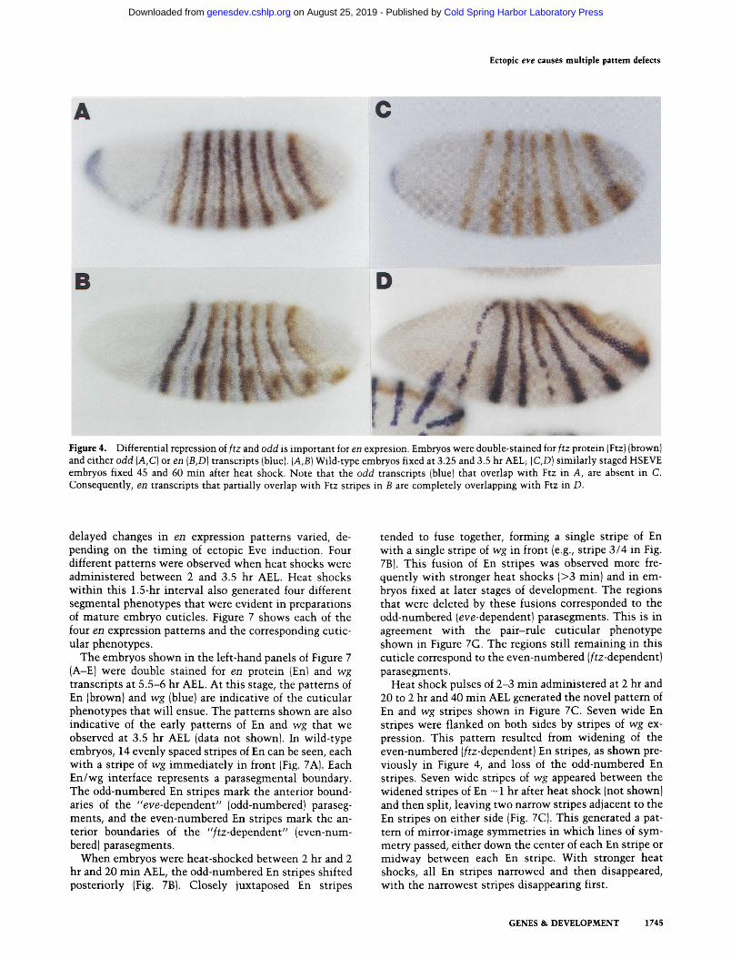
We wished to determine the consequences of the ability of Eve to repress odd at levels that did not repress ftz. This was tested by double-labeling for Ftz (brown) and either odd or en transcripts (blue). Before gastrulation, we found that Ftz and odd stripes were completely over- lapping (not shown). At gastrulation, the Ftz stripes were one to two cells wider than the odd stripes (Fig. 4A). Cells that expressed Ftz and not odd were located where the ftz-dependent en stripes initiated {Fig. 4B). The cells that express ftz and en also express low levels of Eve (Frasch et al. 1987). Taken together, these observations
1742 GENES & DEVELOPMENT
Cold Spring Harbor Laboratory Press on August 25, 2019 - Published by genesdev.cshlp.orgDownloaded from
A
3.5
5rain
w 0 z <
fr- O ffl m < uJ >
w fr
.
01 0
uJ 1.2 0 Z <C 1.0 03 n- O 0.8 03 m ,,:C 0.6 LLI >. i ~ 0.4
w 0.2 fr
B
I I
I 1 I (
10 20 30 40 50 TIME (min)
C
I
, , , , , , , ! ! I i i
0 10 20 30 40 50
TIME (rain)
Figure 2. Effects of increasing Eve expression on ftz. (A) Embryos shown from top to bottom were heat shocked at 2.5-3 hr AEL for 0, 2, 3.5, and 5 rain, respectively, and fixed 30 rain later. In the non-heat-shocked embryo (top), the domains of Eve (brownl and ftz mRNA (blue) were mutually exclusive. In the heat-shocked embryos, ftz stripes narrowed and disappeared as the levels of Eve expression increased. (B,C) Embryos were aliquoted following a 4-min heat shock at 36°C and fixed at 5-min intervals. In half of the embryos (B) Eve was detected immunologically~ in the other half {C), ftz transcripts were detected by whole-mount in situ hybrid- ization. Signals were quantitated as described. Levels of AP activity are indicated (y-axes) for embryos fixed at the times shown (x-axes). (E3) Values obtained with non-heat-shocked embryos; (0) values obtained with heat-shocked embryos. These relative values were determined by dividing all Aao5 values by the average of a4o 5 levels detected in non-heat-shocked embryos. The time after heat shock when ftz transcripts were diminished by 50% is indicated by a dotted line in B, and the abundance of Eve 6 min earlier is indicated similarly in A. Error bars indicate the variability in values obtained in two separate experiments.
GENES & DEVELOPMENT 1743
Cold Spring Harbor Laboratory Press on August 25, 2019 - Published by genesdev.cshlp.orgDownloaded from
Manoukian and Krause
A B
C D
E ~ F
J G FI
Figure 3. Dosage-dependent repression of pair-rule genes in HSEVE embryos. Whole-mount in situ hybridizations were car- ried out with digoxigenin-labeled probes to detect wild-type {A,C,E,G,I) and HSEVE {B,D,F,H,J) pair-rule gene expression pattems: h {A,B); prd {C,D); ftz {E,F); run (G,H); and odd {I,J). The HSEVE embryos {right) were heat-shocked together for 3 min at 36°C between 2.5 and 3 hr AEL, before dividing them up for hybridization with the different probes. Note the difference in sensitivities relative to the wild-type expression patterns shown at left.
suggest the possibil i ty that f t z encodes an activator of en, that odd encodes a negative regulator of en, and that low levels of Eve might act to repress odd so that en can be activated by Ftz.
To test this hypothesis, we attempted to induce levels of ectopic Eve that could repress odd without repressing Ftz, wi th the expectation that en might be activated in all Ftz-expressing ceils. When HSEVE embryos were heat-shocked at -2 .5 hr AEL for 2-3 m i n and fixed 30 min later, the odd stripes that overlap with Ftz were repressed (Fig. 4C). Although some Ftz stripes changed in width, their levels of expression were close to normal. Consequently, embryos fixed 15 min later expressed en in all of the f tz-expressing cells rather than at the ante- rior edges only (Fig. 4D). Hence, these data are consistent wi th f t z encoding an activator of en, odd encoding a neg- ative regulator of en, and low levels of Eve contributing to Ftz-dependent en activation through repression of odd. These results i l lustrate the importance of Eve con- centration levels in wild-type embryos. Furthermore, they suggest that Eve may not only play an important role in the odd-numbered parasegments b u t i n the even- numbered parasegments as well.
Eve ac t iv i ty varies during dif ferent stages of em bryogenesis
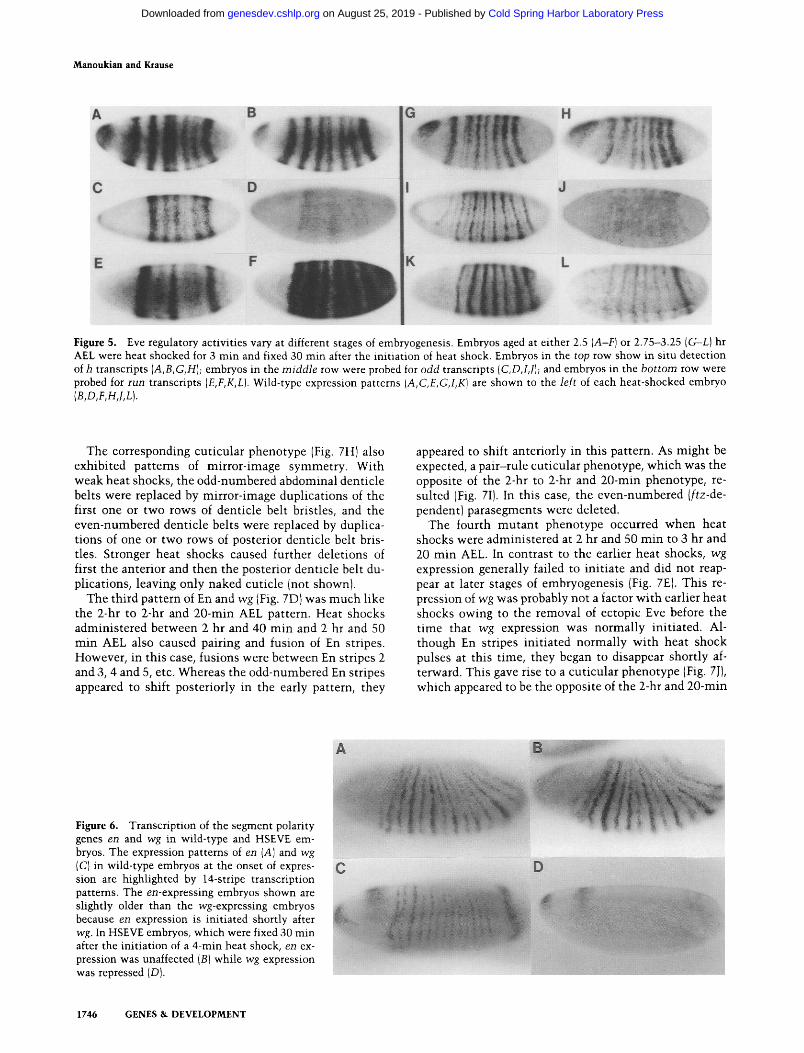
The results presented above were obtained wi th embryos that were heat-shocked between 2.5 and 3 hr AEL (at cellular blastoderm). Because eve expression begins well before this and continues for 1-2 hr afterward, we wished to determine whether the regulatory properties of ectopic Eve would vary if induced earlier or later. Fig- ure 5 shows the effects of ectopic Eve on the pair-rule genes h, odd, and run when 4-min heat shocks were ad- ministered either between 2 and 2.5 hr AEL (beginning of cellularization) or between 2.75 and 3.25 hr AEL (begin- ning of gastrulationl. In each case, embryos were fixed 30 min after the init iat ion of heat shock to select for pri- mary effects.
Not surprisingly, there was no effect on h expression at either time, and expression of the odd gene was re- pressed with equal efficiency during both the early and late t ime intervals. In contrast, whereas the run gene was repressed wi th late heat shocks, it was ectopically acti- vated with early heat shocks. This ectopic activation during early embryogenesis was also observed wi th ftz, although it was not as extensive and was restricted to a shorter t ime interval (data not shown). Because this early activation was complete wi th in the same t ime interval that ftz, run, odd, and prd had been repressed by later heat shocks, we also classified it as a primary response. This suggests that Eve may be capable of acting as both a repressor and an activator, depending on the stage of embryogenesis and the gene being regulated.
Effects of ectopic Eve on en and wg expression
Initiation of expression of the segment polarity genes en and wg begins at the t ime that eve changes from a 7- to a 14-stripe pattern of expression ( -3-3 .5 hr AEL). The 14 en stripes overlap wi th the 14 eve stripes, and wg stripes appear as the seven wide eve stripes begin to narrow. The nature of these overlaps, taken together wi th the loss of all en stripes and the widening of wg stripes in eve amor- phs, suggests that eve may be a positive regulator of en and a negative regulator of wg. We induced ectopic Eve expression at the t ime that en and wg expression are normally initiated to address the question of whether these interactions are direct or indirect. Figure 6 shows embryos stained for either en or wg transcripts 30 rain after the init iat ion of a 4-min heat shock. No effect was observed on the init iat ing pattern of en expression in the presence of ectopic Eve. In contrast, repression of wg transcripts was observed as early as 15 min and was com- plete wi th in 30 m i n of heat shock, even wi th heat shock pulses as short as 2 min. These criteria suggest that Eve is a direct and potent repressor of wg expression and that it is not a direct regulator of en.
Ectopic Eve generates four m u t a n t phenotypes
Changes in the patterns of en expression were only ob- served with recovery t imes in excess of 30 rain. These
1744 GENES & DEVELOPMENT
Cold Spring Harbor Laboratory Press on August 25, 2019 - Published by genesdev.cshlp.orgDownloaded from
Ectopic e v e causes multiple pattern defects
A O
B D
Figure 4. Differential repression of ftz and odd is important for en expresion. Embryos were double-stained for ftz protein (Ftz)(brown} and either odd IA,C) or en (B,D) transcripts (blue). [A,B) Wild-type embryos fixed at 3.25 and 3.5 hr AEL; [C,D) similarly staged HSEVE embryos fixed 45 and 60 min after heat shock. Note that the odd transcripts (blue} that overlap with Ftz in A, are absent in C. Consequently, en transcripts that partially overlap with Ftz stripes in B are completely overlapping with Ftz in D.
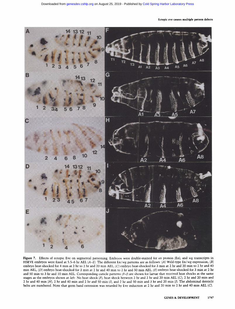
delayed changes in en expression patterns varied, de- pending on the timing of ectopic Eve induction. Four different patterns were observed when heat shocks were administered between 2 and 3.5 hr AEL. Heat shocks within this 1.5-hr interval also generated four different segmental phenotypes that were evident in preparations of mature embryo cuticles. Figure 7 shows each of the four en expression patterns and the corresponding cutic- ular phenotypes.
The embryos shown in the left-hand panels of Figure 7 (A-E) were double stained for en protein fEn) and wg transcripts at 5.5-6 hr AEL. At this stage, the patterns of En (brown) and wg (blue) are indicative of the cuticular phenotypes that will ensue. The patterns shown are also indicative of the early patterns of En and wg that we observed at 3.5 hr AEL (data not shown}. In wild-type embryos, 14 evenly spaced stripes of En can be seen, each with a stripe of wg immediately in front (Fig. 7A). Each En/wg interface represents a parasegmental boundary. The odd-numbered En stripes mark the anterior bound- aries of the "eve-dependent" (odd-numbered) paraseg- ments, and the even-numbered En stripes mark the an- terior boundaries of the " f t z -dependent" (even-num- bered) parasegments.
When embryos were heat-shocked between 2 hr and 2 hr and 20 min AEL, the odd-numbered En stripes shifted posteriorly (Fig. 7B). Closely juxtaposed En stripes
tended to fuse together, forming a single stripe of En with a single stripe of wg in front (e.g., stripe 3/4 in Fig. 7BI. This fusion of En stripes was observed more fre- quently with stronger heat shocks (>3 rain) and in em- bryos fixed at later stages of development. The regions that were deleted by these fusions corresponded to the odd-numbered {eve-dependentl parasegments. This is in agreement with the pair-rule cuticular phenotype shown in Figure 7G. The regions still remaining in this cuticle correspond to the even-numbered (f tz-dependent) parasegments.
Heat shock pulses of 2-3 min administered at 2 hr and 20 to 2 hr and 40 min AEL generated the novel pattern of En and wg stripes shown in Figure 7C. Seven wide En stripes were flanked on both sides by stripes of wg ex- pression. This pattern resulted from widening of the even-numbered (ftz-dependent) En stripes, as shown pre- viously in Figure 4, and loss of the odd-numbered En stripes. Seven wide stripes of wg appeared between the widened stripes of En - 1 hr after heat shock (not shown) and then split, leaving two narrow stripes adjacent to the En stripes on either side (Fig. 7C). This generated a pat- tern of mirror-image symmetries in which lines of sym- metry passed, either down the center of each En stripe or midway between each En stripe. With stronger heat shocks, all En stripes narrowed and then disappeared, with the narrowest stripes disappearing first.
GENES & DEVELOPMENT 1745
Cold Spring Harbor Laboratory Press on August 25, 2019 - Published by genesdev.cshlp.orgDownloaded from
Manoukian and Krause
H
C D r - ~ Ill
E F L
I
Figure 5. Eve regulatory activities vary at different stages of embryogenesis. Embryos aged at either 2.5 (A-F) or 2.75-3.25 (G-L) hr AEL were heat shocked for 3 min and fixed 30 min after the initiation of heat shock. Embryos in the top row show in situ detection of h transcripts (A,B,G,H); embryos in the middle row were probed for odd transcripts (C,D,I,J); and embryos in the bottom row were probed for run transcripts (E,F,K,L). Wild-type expression patterns (A,C,E,G,I,K) are shown to the left of each heat-shocked embryo (B,D,F,H, LL).
The corresponding cuticular phenotype (Fig. 7H) also exhibited patterns of mirror-image symmetry. With weak heat shocks, the odd-numbered abdominal denticle belts were replaced by mirror-image duplications of the first one or two rows of denticle belt bristles, and the even-numbered denticle belts were replaced by duplica- tions of one or two rows of posterior denticle belt bris- tles. Stronger heat shocks caused further deletions of first the anterior and then the posterior denticle belt du- plications, leaving only naked cuticle (not shown).
The third pattern of En and wg (Fig. 7D) was much like the 2-hr to 2-hr and 20-rain AEL pattern. Heat shocks administered between 2 hr and 40 min and 2 hr and 50 min AEL also caused pairing and fusion of En stripes. However, in this case, fusions were between En stripes 2 and 3, 4 and 5, etc. Whereas the odd-numbered En stripes appeared to shift posteriorly in the early pattern, they
appeared to shift anteriorly in this pattern. As might be expected, a pair-rule cuticular phenotype, which was the opposite of the 2-hr to 2-hr and 20-min phenotype, re- sulted (Fig. 7I). In this case, the even-numbered (ftz-de- pendent) parasegments were deleted.
The fourth mutan t phenotype occurred when heat shocks were administered at 2 hr and 50 m i n to 3 hr and 20 min AEL. In contrast to the earlier heat shocks, wg expression generally failed to initiate and did not reap- pear at later stages of embryogenesis (Fig. 7E). This re- pression of wg was probably not a factor wi th earlier heat shocks owing to the removal of ectopic Eve before the t ime that wg expression was normal ly initiated. Al- though En stripes initiated normally wi th heat shock pulses at this time, they began to disappear shortly af- terward. This gave rise to a cuticular phenotype (Fig. 7J), which appeared to be the opposite of the 2-hr and 20-min
Figure 6. Transcription of the segment polarity genes en and wg in wild-type and HSEVE em- bryos. The expression patterns of en (A) and wg (C) in wild-type embryos at the onset of expres- sion are highlighted by 14-stripe transcription patterns. The en-expressing embryos shown are slightly older than the wg-expressing embryos because en expression is initiated shortly after wg. In HSEVE embryos, which were fixed 30 min after the initiation of a 4-min heat shock, en ex- pression was unaffected (B) while wg expression was repressed (D).
C D
1746 GENES & DEVELOPMENT
Cold Spring Harbor Laboratory Press on August 25, 2019 - Published by genesdev.cshlp.orgDownloaded from
Ectopic eve causes multiple pattern defects
===================== ~i i~ii:~:i ~:. ~: -~; :::i~ ~ :
: :i: :: ::i iiil i ! •
Figure 7. Effects of ectopic Eve on segmental patterning. Embryos were double-stained for en protein {En), and w g transcripts in HSEVE embryos were fixed at 5 .5-6 hr AEL (A-E). The different E n / w g patterns are as follows: (A) Wild-type E n / w g expression; (B) embryo heat-shocked for 4 rain at 2 hr to 2 hr and 20 min AEL; (C) embryo heat-shocked for 3 min at 2 hr and 20 min to 2 hr and 40 min AEL; (D) embryo heat-shocked for 3 min at 2 hr and 40 rain to 2 hr and 50 min AEL; (E) embryo heat-shocked for 3 min at 2 hr and 50 rain to 3 hr and 10 min AEL. Corresponding cuticle patterns (F-J) are shown for larvae that received heat shocks at the same stages as the embryos shown at left: No heat shock (F); heat shock between 2 hr and 2 hr and 20 min AEL (G); 2 hr and 20 min and 2 hr and 40 min (H); 2 hr and 40 min and 2 hr and 50 min (I), and 2 hr and 50 min and 3 hr and 20 min (l). The abdominal denticle belts are numbered. Note that germ band extension was retarded by Eve induction at 2 hr and 20 min to 2 hr and 40 min AEL (C).
GENES & DEVELOPMENT 1747
Cold Spring Harbor Laboratory Press on August 25, 2019 - Published by genesdev.cshlp.orgDownloaded from
Manoukian and Krause
to 2-hr and 40-min AEL phenotype; instead of naked cu- ticle, these embryos yielded lawns of denticles.
Discussion
Null mutations of eve disrupt both odd- and even-num- bered parasegments. Our results with a heat shock-in- ducible eve transgene indicate that Eve provides multi- ple regulatory cues in both sets of parasegments and that these functions are sequential. The window of time dur- ing which ectopic Eve generated segmental pheno- types--l.5 hr--is considerably longer than the 15- to 30- min window of sensitivity obtained with the pair-rule gene f t z (Struhl 1985), and can be subdivided into four phases, each characterized by a specific phenotype. The segmental phenotypes caused by earlier Eve inductions were apparent reciprocals of those generated by later in- ductions. These phenotypes and their apparent reciproc- ity can be rationalized in terms of the observed effects on the expression of downstream target genes.
Direct vs. ind irec t targets of Eve regulatory act ivi t ies
By providing short pulses of Eve expression and follow- ing the levels of potential target gene products, we dis- tinguished two different response times. Changes in the expression patterns of five of the eight genes tested (ftz, run, odd, prd, and wg) could be detected within 15 min of a short 2- to 4-min heat shock and were complete within 30 min of heat shock. The remaining three genes (eve, h, and en) showed a delayed response between 30 and 45 min of ectopic Eve induction. A quantitative analysis of Eve and f t z expression levels following heat shock indi- cated that the rapid 15- to 30-min response time was consistent with a direct regulatory interaction between the two genes. A similar response time observed with f t z p r o m o t e r - l a c Z fusion genes indicates that this negative regulation was mediated via sequences located upstream of the f t z -coding region. The other four genes that re- sponded within the same 15- to 30-min interval may also be direct targets of Eve, whereas the three genes that responded later are probably not.
These response times may be of general use in distin- guishing between other direct and indirect gene interac- tions, although several caveats must be considered. For example, heat shocks are believed to alter rates of tran- scription and translation. Interestingly, we did not ob- serve any obvious effects on the expression of genes monitored in this study (even with 15- to 20-min heat shocks). Translation of ectopic eve transcripts also ap- peared to be unaffected by heat shock, as ectopic Eve accumulated at a constant rate, even during heat shocks as long as 1-2 hr (data not shown). We did, however, note short delays in development that were proportional to the duration of heat shock. These were minimal with the 2- to 4-min heat shocks used in this study. Another con- sideration is the size and half-life of transcripts being monitored. In this study all of the transcripts were sim- ilarly sized, and those that have been tested have similar half-lives (Edgar et al. 1989). Another concern is that
some proteins may be active at very low levels of expres- sion. This would shorten the time that is necessary be- tween successive gene interactions.
Regulat ion of the en and eve genes
The loss of en and eve transcripts in eve mutant embryos had suggested previously that Eve could be a direct acti- vator of these two genes (DiNardo and O'Farrell 1987; Frasch et al. 1988}. However, their delayed response to ectopic Eve argues against this possibility. Our results with en indicate that Eve may activate it indirectly by repressing other repressors. One candidate is the product of the odd gene, because Eve rapidly repressed odd, and this repression was followed by activation of en in cells that had expressed odd previously. This indirect cir- cuitry is also consistent with the observation that half of the 14 en stripes that disappear in e v e - embryos reap- pear in e v e - / o d d - double mutant embryos (DiNardo and O'Farrell 1987). The reappearance of only the even- numbered en stripes in e v e - / o d d - embryos suggests that Eve represses other negative regulators of en in the odd-numbered parasegments. One of these may be the product of the run gene, because ectopic run rapidly re- presses the odd-numbered en stripes (first and second phenotypes; A.S. Manoukian and H.M. Krause, in prep.) and Eve was a potent repressor of run (after 2.5 hr AEL).
The lack of a direct response to ectopic Eve by the endogenous eve gene was more of a surprise. In e v e - embryos, stripes of expression driven by the eve pro- moter fail to resolve normally and are then lost prema- turely (Frasch et al. 1988). Jiang et al. (1991) proposed that this regulation was direct because they identified a regulatory element upstream of the eve transcription start site that contained Eve-binding sites and required eve gene activity. To abolish this eve-dependent activity, all Eve-binding sites within a truncated form of the ele- ment had to be destroyed. Jiang et al. (1991) favored re- dundancy of these sites to explain the need to remove them completely. Our data suggest that Eve acts indi- rectly by regulating the expression of an intermediary gene. The product of this gene may no longer be capable of regulating the extensively mutated element con- structed by Jiang and co-workers. As with en, run is a candidate for such a gene, because run appears to be a negative regulator of eve (Ingham and Gergen 19881 A.S. Manoukian and H.M. Krause, in prep.) and ectopic Eve was an effective repressor of run. Alternatively, the fail- ure of ectopic Eve to activate the endogenous eve gene in the interstripe regions might be explained by the absence of a necessary cofactor or by the presence of an overrid- ing repressor.
Eve as a concen tra t ion-dependen t morphogen
In wild-type embryos, the levels of Eve vary dramati- cally. Initially, expression is very low and then resolves into an anterior-to-posterior gradient. Following their formation, the seven stripes polarize with the highest levels of expression at the anterior edges. Finally, at the
1748 GENES & DEVELOPMENT
Cold Spring Harbor Laboratory Press on August 25, 2019 - Published by genesdev.cshlp.orgDownloaded from

Ectopic e v e causes multiple pattern defects
14-stripe phase, the stripes alternate between weak and strong. Our results indicate that these different levels determine the scope of target gene regulation. Each of the five genes that Eve repressed responded to a different level of the ectopic protein: odd and wg by very low levels of Eve, f t z and run by intermediate levels, and prd only by very high levels. The importance of this differ- ential regulation was demonstrated effectively by the relative balance between the levels of Eve, ftz, and odd and the subsequent effects on en expression. With low levels of Eve, the loss of odd allowed expansion of the even-numbered en stripes into all f tz-expressing cells. At slightly higher levels of Eve, f t z was also repressed and en expression was lost altogether.
Possible roles for Eve as a transcriptional act ivator
In addition to acting as a dosage-dependent repressor, Eve had the apparent ability to act as a temporally re- stricted activator. When Eve was induced before cellu- larization (2 hr to 2 hr and 20 rain AEL), ftz and run were activated ectopically rather than being repressed. The rapid response of these two genes suggests that this ac- tivation was direct. Examples of transcription factors that can act as both activators and repressors include the proteins PRTF (pheromone/receptor transcription _fac- tor), which regulates yeast haploid cell mating type (Tan and Richmond 1990), and the glucocorticoid receptor (Diamond et al. 1990). In both cases, the basic activity of these proteins could be altered by specific interactions with other proteins. In a similar fashion, Eve may some- times act as a gene-specific transcriptional activator as well as a repressor, perhaps through specific interactions with different stage- and promoter-specific factors.
Changing regulatory roles of the eve gene
Each of the four phenotypes generated by ectopic Eve is summarized in Figure 8. The similarities of these phe- notypes to known pair-rule and segment polarity mu- tant phenotypes, taken together with changes in the ex- pression patterns of these genes upon Eve induction, sug- gest possible mechanisms for each phenotype. We attribute the reciprocity of the first and third phenotypes to the respective loss of either the eve- or f t z -dependent parasegments. Loss of the eve-dependent parasegments appeared to be caused by indirect repression of the en- dogenous eve gene and the odd-numbered stripes of en owing to the ectopic activation of run. Loss of the ftz- dependent parasegments in the third phenotype is prob- ably the result of the direct repression of ftz. The second and fourth phenotypes also retained either the even- or odd-numbered parasegments, respectively. However, un- like the first and third phenotypes, they were subject to mirror-image duplications owing to the widening or loss of en and wg stripes. In the 2-hr and 20-min to 2-hr and 40-min phenotype, en stripes expanded owing to the re- pression of odd. Expansion of wg was probably the result of the expansion of run and the subsequent repression of eve. In contrast, the fourth phenotype appeared to be
W T
I ) 2 - 2 :20 AEL
2) 2 : 2 0 - 2 :40 A E L
PS 5 PS 6 : PS 7 PS8
WG EN WG EN WG EN WG EN 5 6 7 8
I 1 ! ! EZIB EZIB P'-/~IB PZIB
s e I , 'I ' ' |
E w c
5
3) 2 :40 - 2 : 5 0 A E L
G ! EZlB ~ l B /
7 i 91 : ' ' " 7 " ' ; ' - - - - : - - " , L___,____, ~ [.___,___.,
4) 2 :50 - 3 :10 AEL I Figure 8. Summary of En/wg expression patterns and associ- ated cuticle patterns in HSEVE embryos. The top panel portrays the approximate expression patterns of the segmentation genes addressed in this study between parasegments (PS) 5 and 8. The borders of each parasegment are defined by the juxtaposition of en and wg stripes (denoted by dotted lines). Segmental borders and ventral denticle belt features are shown at the bottom of the panel. The wedge-shaped stripes of eve, ftz, and odd denote the narrowing of these stripes that occurs during gastrulation. The narrow stripes of eve and odd that appear at gastrulation are shown as unlabeled boxes. (Panels 1--4) The four mutant En/wg and cuticle patterns that evolved in HSEVE embryos and the times at which they occurred.
caused by the direct repression of wg and the subsequent loss of en, which has been shown to be wg dependent during early stages of expression (Martinez-Arias et al. 1988; Heemskerk et al. 1991).
These experiments suggest that the endogenous eve gene is required to perform multiple regulatory roles in both the odd- and even-numbered parasegments. The earliest role of Eve may be to assist in the activation of run and ftz, as evidenced by its ability to activate these genes ectopically. Shortly thereafter, Eve appears to act as a repressor of these two genes as well as odd, thereby establishing the odd-numbered parasegments. The nar- rowing of the seven Eve stripes then permits wg expres- sion at the posterior edges of the odd-numbered paraseg- ments. Initiation of en stripes follows at the 14-stripe phase of Eve expression, with the 7 weaker stripes of Eve required to repress odd in the even-numbered paraseg- ments and higher levels to repress both odd and run in the odd-numbered parasegments.
GENES & DEVELOPMENT 1749
Cold Spring Harbor Laboratory Press on August 25, 2019 - Published by genesdev.cshlp.orgDownloaded from
Manoukian and Krause
Opposing roles of Eve and Ftz
A l t h o u g h Eve and Ftz define reciprocal sets of paraseg- ments , Eve appears to func t ion pr imar i ly as a transcrip- t ional repressor, whereas in vi t ro and t issue cul ture stud- ies suggest tha t Ftz acts as a t ranscr ip t ional act ivator (Jaynes and O'Farrel l 1988; Han et al. 1989; Wins low et al. 1989; O k h u m a et al. 1990). In the embryo, both genes are posi t ive regulators of en and negat ive regulators of wg. Perhaps Ftz performs these func t ions in a m a n n e r tha t is c o m p l e m e n t a r y to tha t of Eve, act ing as a direct ac t iva tor of en and an indi rec t repressor of wg.
Clearly, t ranscr ip t iona l repressors can control pat tern- ing as effect ively as t ranscr ip t iona l act ivators. Gene reg- u la t ion by repressors m a y be par t icu lar ly sui ted to genes tha t are ac t iva ted by ub iqu i tous or broadly expressed ac- tivators. This would al low m a n y different genes to be ac- t ivated by a few widely distr ibuted activators and resolved into complex pat terns by specific repressors. This may be the case for mos t of the genes that are regulated by Eve.
Materials and m e t h o d s
Construction and transformation of PHSEVE
Construction of the plasmid PHSEVE and P-element transfor- mation of flies were performed by Gary Struht. Briefly, a HinfI restriction site located 21 bp upstream of the eve ATG and an SspI site located 50 bp downstream of the eve translation stop site were used to isolate the eve-coding region (for eve sequence, see MacDonald et al. 1986). After the addition of EcoRI linkers, the eve sequence was inserted into a vector that contains an hsp70 promoter and tubulin gene 3'-untranslated sequences (Struhl 1989). The hsp70-eve-tubulin hybrid gene was inserted into Carnegie 20 and transformed as described previously (Struhl 1989).
mRNA and protein localization
In general, embryos were collected in cylinders over a 30-rain interval and aged appropriately at 25°C before fixation. When correct staging of the embryos was critical, embryos were col- lected for 20 min and visually staged under halocarbon oil (Wie- schaus and Niisslein-Volhard 1986). The detection of tran- scripts in embryos was achieved by whole-mount in situ hy- bridization with the modifications described by Edgar and O'Farrell (1990). Antibody staining procedures were as de- scribed previously (Krause et al. 1988), except that PBTB {PBS buffer + 0.1% Tween 20 and 1% dry milk powder) was used for blocking and antibody incubations, and biotinylated secondary antibodies were detected with the Vectastain kit {Vector Labo- ratories). In general, double antibody/in situ stainings were per- formed as described, with in situ hybridizations done before the antibody stainings. Alternatively, antibody detection was per- formed first by using PBTH (filter-sterilized 1 x PBS + 0.1% Tween 20, 100 },g/ml of heparin, 100 ~g/ml of tRNA, 0.05 U/ml of RNasin) instead of PBTB, followed by in situ hybrid- ization as described. All embryos were mounted in 80% glyc- erol, 20 mM Tris (pH 7.5}. Embryos in which signals were de- tected by AP staining were subjected to dehydration in 90% and 100% ethanol, to remove background staining and to change the signal from purple to blue, and then rehydrated before mounting.
Protein and RNA measurements
Embryos for kinetic experiments were collected on apple juice plates that were placed in population cages for 30 min. The
embryos were then aged for 2.5 hr at 25°C, transferred to glass coverslips, covered with a thin layer of glycerol to distribute heat evenly, and heat-shocked by floating the coverslips on wa- ter for 4 min in a 36°C water bath. Embryos were then dechori- onated and divided into equal portions. These were maintained at 25°C and fixed at appropriate intervals following heat shock. Protein or mRNA localization was carried out as described with secondary antibodies that were coupled to AP, and with the following modifications. Just before enzymatic detection, em- bryos were washed once in diethanolamine buffer [10 mM di- ethanolamine (pH 9.5), 0.5 mM MgCI2]. The number of embryos per reaction was normalized by adding the equivalent of 50 ~1 of settled embryos to 0.5-ml microcentrifuge tubes. The reactions were developed by rocking for 1 hr at 25°C in 400 ~1 of AP reaction mix [a 5-mg tablet of p-nitrophenyl phosphate (PNP, Sigmal dissolved in 10 ml of diethanolamine buffer]. Reactions were stopped by adding an equal volume of 100 mM EDTA, and activity was determined by measuring the A4os of each super- natant. We found that activities were linear with respect to time during the 1-hr incubation period and that anywhere from 10 to 100 ~tl of settled embryos gave proportional signals. At the end of the !-hr period, embryos were recovered, washed in the usual AP staining buffer and then stained with the insoluble AP sub- strates 5-bromo-4-chloro-3-indolyl phosphate (BCIP) and nitro- blue tetrazolium (NBT). The AP reactions with PNP in dietha- nolamine had no detrimental effects on RNA or protein local- ization within embryos.
It was also possible to extract much of the insoluble purple AP substrate produced by the NBT/BCIP reaction from the em- bryos with 100% ethanol. This was performed by rinsing the embryos once in 100% ethanol and then extracting for 1 hr with occasional shaking at 65°C in 200 o.1 of ethanol. The ethanol was then diluted with 300 ~tl H20, and absorbance was mea- sured at a wavelength of 550 nM. Quantitative results obtained by this method were similar to those obtained by using the soluble AP substrates. We found that for the soluble PNP reac- tions, antibodies that gave a weaker signal gave a more linear result. For Eve detection we used anti-Eve monoclonal antibod- ies obtained from N. Patel {Carnegie Institution of Washington). The ethanol extraction technique worked best with strong sig- nals such as those generated with a polyclonal anti-Eve anti- body obtained from M. Frasch. A higher percentage of the BCIP/ NBT product could be ethanol extracted if the embryos were stained lightly rather than heavily.
Cuticle preparations
Embryos were collected for 20 min on apple juice plates and aged appropriately. Heat shocks were performed by transferring the embryos to glass coverslips, covering them with a thin layer of halocarbon oil, and floating the coverslips on water in a 36°C water bath. Following heat shock, embryos were examined un- der the microscope, and properly staged embryos were trans- ferred to apple juice plates. After 24 hr at 25°C, unhatched em- bryos were dechorionated and then dissected from their vi- telline membranes in Hoyer's medium (Wieschaus and Nfisslein-Volhard 1986). Clearing was carried out for 2-3 days at 65°C in Hoyer's medium.
A c k n o w l e d g m e n t s
We thank Gary Struhl for graciously providing us with his HSEVE lines and for communicating his initial results. Many thanks go to N. Patel and M. Frasch for providing Eve antibod- ies, P. Gergen for providing run and prd cDNAs, D. Coulter for providing odd cDNA, and S. Cot6 for wg cDNA. We also thank D. Coulter, I. Greenblatt, C.J. Ingles and the reviewers for help-
1750 GENES & DEVELOPMENT
Cold Spring Harbor Laboratory Press on August 25, 2019 - Published by genesdev.cshlp.orgDownloaded from

Ectopic e v e causes multiple pattern defects
ful comments on the manuscript and M. Levine for providing encouragement and helpful suggestions. This work was sup- ported by grants from the Medical Research Council (MRC) of Canada and the National Cancer Institute (NCI) of Canada. A.S.M. was supported with funds from the NCI and the Cana- dian Cancer Society, and H.M.K. was supported by a MRC of Canada Scholarship.
The publication costs of this article were defrayed in part by payment of page charges. This article must therefore be hereby marked "advertisement" in accordance with 18 USC section 1734 solely to indicate this fact.
R e f e r e n c e s
Baumgartner, S. and N. Null. 1991. Network of interactions among pair-rule genes regulating paired expression during primordial segmentation of Drosophila. Mech. Dev. 33: 1- 18.
Biggin, M. and R. Tjian. 1989. A purified Drosophila home- odomain protein represses transcription in vitro. Cell 58: 433-440.
Carroll, S. and M. Scott. 1986. Zygotically active genes that affect spatial expression of the fushi tarazu segmentation gene during early Drosophila embryogenesis. Cell 45" 113- 126.
Coulter, D.E. and E. Wieschaus. 1988. Gene activities and seg- mental patterning in Drosophila: Analysis of odd-skipped and pair-rule double mutants. Genes & Dev. 2: 1812-1823.
Diamond, M.I., N.M. Jeffrey, S.K. Yoshinaga, and K.R. Ya- mamoto. 1990. Transcription factor interactions: Selectors of positive or negative regulation from a single regulatory element. Science 249: 1266-1272.
DiNardo, S. and P. O'Farrell. 1987. Establishment and refine- ment of segmental pattern in the Drosophila embryo: Spa- tial control of engrailed expression by pair-rule genes. Genes & Dev. 1: 1212-1225.
Edgar, B. and O'Farrell, P. 1990. The three postblastoderm cell cycles of Drosophila embryogenesis are regulated in G2 by string. Cell 62: 469-480.
Edgar, B., M. Weir, G. Schubiger, and T. Kornberg. 1986. Repres- sion and turnover pattern of fushi tarazu RNA in the early Drosophila embryo. Cell 47: 747-754.
Edgar, B., G. O'Dell, and G. Schubiger. 1989. A negative switch based on negative regulation sharpens stripes in Drosophila embryos. Dev. Genet. 10: 124-142.
Frasch, M. and M. Levine. 1987. Complementary patterns of even-skipped and fushi-tarazu expression involve their dif- ferential regulation by a common set of segmentation genes in Drosophila. Genes & Dev. 2: 981-995.
Frasch, M., C. Rushlow, H. Doyle, and M. Levine. 1987. Char- acterization and localization of the even-skipped protein of Drosophila. EMBO J. 6" 749-759.
Frasch, M., R. Warrior, J. Tugwood, and M. Levine. 1988. Mo- lecular analysis of even-skipped mutants in Drosophila de- velopment. Genes & Dev. 2: 1824-1838.
Goto, T., P.M. Macdonald, and T. Maniatis. 1989. Early and late periodic patterns of even-skipped expression are controlled by distinct regulatory elements that respond to different spa- tial cues. Cell 57: 413-422.
Heemskerk, J., S. DiNardo, R. Kostriken, and P.H. O'Farrell. 1991. Multiple modes of engrailed regulation in the progres- sion towards cell fate determination. Nature 352: 404--410.
Han, K., M. Levine, and J. Manley. 1989. Synergistic activation and repression of transcription by Drosophila homeobox proteins. Cell 56: 573-583.
Hiromi, Y. and W.J. Gehring. 1987. Regulation and function of
the Drosophila segmentation gene fushi tarazu. Cell 50: 963-974.
Hoey, T. and M. Levine. 1988. Divergent homeobox proteins recognize similar sequences in Drosophila. Nature 332: 858-861.
Ingham, P. 1988. The molecular genetics of embryonic pattern formation in Drosophila. Nature 335" 25-34.
Ingham, P. and P. Gergen. 1988. Interactions between the pair- rule genes runt, hairy, even-skipped and fushi tarazu and the establishment of periodic pattern in the Drosophila embryo. Development 104 {Suppl.): 51-60.
Ingham, P., N. Baker, and A. Martinez-Arias. 1988. Regulation of segment polarity genes in the Drosophila blastoderm by fushi tarazu and even-skipped. Nature 331: 73-75.
Jaynes, J.B. and P.H. O'Farrell. 1988. Activation and repression of transcription by homeodomain-containing proteins that bind a common site. Nature 336: 744-749.
Jiang, J., T. Hoey and M. Levine. 1991. Autoregulation of a seg- mentation gene in Drosophila: Combinatorial interaction of the even-skipped borneo box protein with a distal enhancer element. Genes & Dev. 5: 265-277.
Krause, H.M., R. Klemenz, and W.J. Gehring. 1988. Expression, modification and localization of the fushi tarazu protein in Drosophila embryos. Genes & Dev. 2: 1021-1036.
Lawrence, P. and P. Johnston. 1989. Pattern formation in the Drosophila embryo: Allocation to parasegments by even- skipped and fushi tarazu. Development 105: 761-767.
Lawrence, P., P. Johnston, P. MacDonald, and G. Struhl. 1987. Borders of parasegments in Drosophila embryos are delim- ited by the fushi tarazu and even-skipped genes. Nature 328: 440-442.
MacDonald, P.M., P. Ingham, and G. Struhl. 1986. Isolation, structure, and expression of even-skipped: A a second pair- rule gene of Drosophila containing a homeobox. Cell 47: 721-734.
Martinez-Arias, A., N. Baker, and P. Ingham. 1988. Role of seg- ment polarity genes in the definition and maintenance of cell fates in Drosophila. Development 103: 157-170.
N~sslein-Volhard, N. and E. Wieschaus. 1980. Mutations affect- ing segment number and polarity in Drosophila. Nature 287: 795-801.
Nfisslein-Volhard, C., H. Kluding, and G. Jiirgens. 1985. Genes affecting the segmental subdivision of the Drosophila em- bryo. Cold Spring Harbor Syrup. Quant. Biol. 50: 145-154.
Okhuma, Y., M. Horikoshi, R. Roeder, and C. Desplan. 1990. Binding site-dependent direct activation and repression of in vitro transcription by Drosophila homeodomain proteins. Cell 61: 475-484.
Pankratz, M.J. and H. J/ickle. 1990. Making stripes in the Droso- phila embryo. Trends Genet. 6: 287-292.
Struhl, G. 1985. Near-reciprocal phenotypes caused by inacti- vating or indiscriminate expression of the Drosophila seg- mentation gene ftz. Nature 318: 677-680.
1989. Differing strategies for organizing anterior and posterior body patterns in Drosophila embryos. Nature 338: 741-744.
Tan, S. and J.R. Richmond. 1990. DNA binding-induced confor- mational change of the yeast transcriptional activator PRTF. Cell 62: 367-377.
Wieschaus, E. and C. Niisslein-Volhard. 1986. Looking at em- bryos. In Drosophila: A practical approach (ed. D.B. Rob- erts), pp. 199-228. IRL Press, Oxford, England.
Winslow, G.M., H. Shigeo, M. Krasnow, D.S. Hogness, and M.P. Scott. 1989. Transcriptional activation by the Antennapedia and fushi tarazu proteins in cultured Drosophila cells. Cell 57: 1017-1030.
GENES & DEVELOPMENT 1751
Cold Spring Harbor Laboratory Press on August 25, 2019 - Published by genesdev.cshlp.orgDownloaded from
10.1101/gad.6.9.1740Access the most recent version at doi: 6:1992, Genes Dev.
A S Manoukian and H M Krause Drosophila embryos.Concentration-dependent activities of the even-skipped protein in
References
http://genesdev.cshlp.org/content/6/9/1740.full.html#ref-list-1
This article cites 35 articles, 9 of which can be accessed free at:
License
ServiceEmail Alerting
click here.right corner of the article or
Receive free email alerts when new articles cite this article - sign up in the box at the top
Copyright © Cold Spring Harbor Laboratory Press
Cold Spring Harbor Laboratory Press on August 25, 2019 - Published by genesdev.cshlp.orgDownloaded from