computersinbiologyandmedicine - pàgines de la...
TRANSCRIPT
Computers in Biology and Medicine 39 (2009) 130 -- 140
Contents lists available at ScienceDirect
Computers in Biology andMedicine
journal homepage: www.e lsev ier .com/ locate /cbm
Molecularmodeling and dynamics simulation of human cyclin-dependentkinase 3 complexedwith inhibitors
Patrícia Cardoso Pereza, Rafael A. Caceresa,b, Fernanda Canduric, Walter Filgueira de Azevedo Jr.a,∗aFaculdade de Biociências–Laboratório de Bioquímica Estrutural–PUCRS, Av. Ipiranga, Prédio 12-Laboratório 21-Porto Alegre 90619-900, RS, BrazilbPrograma de Pós Graduação em Medicina e Ciências da Saúde, Pontifícia Universidade Católica do Rio Grande do Sul, Porto Alegre–RS, BrazilcInstituto de Química de São Carlos-USP, São Carlos–SP, Brazil
A R T I C L E I N F O A B S T R A C T
Article history:Received 20 April 2007Accepted 20 November 2008
Keywords:Cyclin-dependent kinase 3InhibitorsMolecular dynamics
The complex CDK3–cyclin is involved in the control of the progression of G0. While the mechanisms gov-erning early and late G1 progression are well understood, very little is known about the G0–G1 transition.Human CDK3 is closely related to cyclin-dependent kinase 2 (CDK2). Since there is no crystallographicstructure of human CDK3, this work describes for the first time a molecular model of human CDK3complexed with several inhibitors. Comparison of the binary complexes with different inhibitors stronglyindicates that those inhibitors should inhibit CDK3 as well as CDK2.
© 2008 Elsevier Ltd. All rights reserved.
1. Introduction
Cell cycle is fundamental to the development and function of alleukaryotic life [1]. This process might be extremely regulated. Oneof the most important proteins that control cell cycle progressionis cyclin-dependent kinase (CDK). Each phase of the cell cycle iscontrolled by a specific CDK, progress during G1 is regulated byCDK4, CDK6–cyclins D and CDK2–cyclin D, during G0 the control isperformed by the complex CDK3–cyclin C, progress during S phaseis controlled by the complexes CDK2-cyclin A and CDK1-cyclin A,transition G2/M is controlled by the complex CDK1–cyclin B. Apartfrom cell cycle regulation CDKs also play a role in apoptosis (CDK2), incontrolling cell differentiation in neuronal and muscle cells (CDK5),and in the control of transcription (CDK7–CDK9) [2].
CDKs, a family of serine/threonine protein kinase, formed by twosubunits: the catalytic subunit (CDK) and the regulatory subunit (cy-clins) [3], a protein whose levels raise and fall during cell cycle. CDKsare inactive as monomers, and activation requires binding to the cy-clins, phosphorylation by CDK-activating kinase (CAK) on a specificthreonine residue and dephosphorylation by phosphatases [4,5]. Inaddition to the positive regulatory role of cyclins and CAK, manynegative regulatory proteins (CDK inhibitors, CKIs) have been dis-covered, such as p15, p16, p18, p21 and p27 [6]. Considering thatderegulation of cyclins, and/or alteration, or absence of CKIs havebeen associated with many cancers and the existence of natural CKIs,
∗ Corresponding author. Tel./fax: +555133204529.E-mail address: [email protected] (W.F. de Azevedo).
0010-4825/$ - see front matter © 2008 Elsevier Ltd. All rights reserved.doi:10.1016/j.compbiomed.2008.11.004
this motivates the search for chemical inhibitors [7,8] of CDKs thatcould play an important role in the discovery of new family of anti-tumor agents [9–11].
Recent investigations have identified a new complex CDK3–cyclinC involved in controlling G0/G1 transition, that is, the cell cycle reen-try [12]. Cyclin C associates with CDK3 to drive cells from the G0–G1phase through phosphorylation of a limited number of targets, es-pecially pRb (retinoblastoma protein). This protein when phosphory-lated at residues Thr14 and Tyr15 become inactive, releasing the E2Ftranscriptional factor. The same process is observed in the transitionto G1/S which is regulated by CDK2–cyclin E complex [13].
This article describes the molecular model, of human CDK3 com-plexed with several inhibitors. The understanding of its structuralbasis would lead to discovery or rationalization of drug design pro-cess. Since ATP is the authentic cofactor of CDKs it can be consideredas a “lead compound” for discovery of CDKs inhibitors. ATP partici-pates in 17 intermolecular hydrogen bonds with ATP-binding pocket.This investigation was performed in order to gain further insight intothe structural basis for chemical inhibition of CDK3 [14–16], usingbioinformatics tools.
2. Material and methods
2.1. Molecular modeling
Since there is no crystallographic structure of human CDK3 (NCBIID: NP_001249) we built a structural model based on the structuresavailable for human CDK2. We used 39 crystal structures that con-tain only the complex CDK:inhibitor solved at high resolution, astemplates for modeling. No crystal structure containing cyclin or any
P. Cardoso Perez et al. / Computers in Biology and Medicine 39 (2009) 130–140 131
Table 1All PDB IDs for CDK2 used as templates for modeling CDK3.
PDB ID Inhibitors present in the complexes
1AQ1 Staurosporine1CKP Purvalanol B1DI8 4-[3-Hydroxyanilino]-6,7 dimethoxyquinazoline1E1V NU 20581FVT Oxindole1G5S H7171GII CDK4 inhibitor1GZ8 1-[(2-Amino-6,9-dihydro-1h-purin-6-Yl)oxy]-3-methyl-2-butanol1HCK ATP/Mg1H0W 1-Amino-6-cyclohex-3-enylmethyloxypurine1JSV 4-[(6-amino-4-pyrimidinyl)amino] benzenesulfonamide C1KE5 N-methyl-4-{[(2-oxo-1,2-dihydro-3h-indol-3-ylidene)methyl] amino} benzenesulfonamide1KE6 N-methyl-{4-[2-(7-oxo-6,7-dihydro-8h-[1,3]thiazolo[5,4-e]indol-8-ylidene)hydrazino]phenyl}methanesulfonamide1KE7 3-{[(2,2-dioxido-1,3-dihydro-2-benzothien-5-yl)amino]methylene}-5-(1,3-oxazol-5-yl)-1,3-dihydro-2h-indol-2-one1KE8 4-{[(2-oxo-1,2-dihydro-3h-indol-3-ylidene)methyl]amino}-N-(1,3-thiazol-2-yl)benzenesulfonamide1KE9 3-{[4-({[Amino(imino)methyl] aminosulfonyl) anilino]methylene}-2-oxo-2,3-dihydro-1h-indole1OIT 4-[(4-Imidazo[1,2-a]pyridin-3-ylpyrimidin-2-yl)amino]benzenesulfonamide1P2A trisubstituted naphthostyril inhibitor1PYE [2-Amino-6-(2,6-difluoro-benzoyl)-imidazo[1,2- a]pyridin-3-yl]-phenyl-methanone1URW 2-[4-(N-(3-dimethylaminopropyl)sulphamoyl)anilino]-1V1K 4, 6-bis anilino pyrimidine1VYZ PNU 1812271W0X Olomoucine1Y8Y (5-Chloropyrazolo[1,5-a]pyrimidin-7-yl)-(4- methanesulfonylphenyl)amine1YKR 4-{[6-(2,6-dichlorobenzoyl)imidazo[1,2-a]pyridin-2-yl]amino}benzenesulfonamide2A0C 2-{[(2-{[(1r)-1-(hydroxymethyl)propyl]amino}-9-isopropyl-9h-purin-6-yl)amino]methyl}phenol2A4L Roscovitine2B52 DPH 0425622B53 DIN 2343252B54 DIN 2323052B55 DIN 1013122BHE 5-Bromo-indirubine2BHH 4-Hydroxypiperindinesulfonyl-indirubine2BTR PNU 1988732BTS PNU 2300322C5Y Hydroxy(oxo)(3-{[(2z)-4-[3-(1h-1,2,4-triazol- 1-ylmethyl)phenyl]pyrimidin-2(5h)-ylidene]amino}phenyl)ammonium2C6I Triazolopyrimidine2EXM IsopentenyladenineDFL Dechloroflavopiridol
other proteins were used as templates. The identity between bothsequences is 74.18%, which indicates that CDK2 is a good templateto build a model for CDK3. To build the model for CDK3 we usedParmodel [17]. Parmodel is a web server for automated comparativemodeling and evaluation of protein structure. This web server wasdesigned to integrate the main software used in this process, there-fore it is subdivided in four modules: Parmodel modeling, Parmodelassessment, Parmodel visualization, and Parmodel optimization. Tobuild models, the Parmodel web server relies on MODELLER [18] thatis a program for comparative structure modeling. Parmodel model-ing provides an interface that requires one amino acid sequence andup to eight template structures as input data. To make the server re-spond more rapidly, Parmodel modeling submits jobs to a Beowulfcluster of 16 PCs running Linux. In the modeling procedure all wa-ter molecules were removed from the templates. We used one tem-plate for each model, and this template presents the ligand to beanalyzed. We modeled 39 complexes, presented in Table 1. For eachmodel Parmodel builds 1000 models, and selects the one with thebest stereochemical quality.
2.2. Molecular dynamics simulation protocol
The initial structure for the CDK3 was obtained by homology aspreviously described. A 5ns of molecular dynamics (MD) simula-tion was performed with GROMACS [19] package using the Gromos
96.1 (43A2) force field. The all-atom model of the protein contain2973 atoms and a net molecular charge of +4. Hence, four chloridecounter ions were added using Genion program of the GROMACSsimulation suite to neutralize the positive charge density of the sys-tem, which was then immersed in a cubic box containing a total of21,673 SPC/E water molecules. The initial simulation cell dimensionswere 43.36×63.43×58.08Å and had the protein solvated by a layerof water molecules of at least 10Å in all directions. The simulationcell contained a total of 68,182 atoms.
The simulations were carried out using explicit solvent watermolecules (described by the simple point charge, SPC/E) and pe-riodic boundary conditions (cubic). In the MD protocol, all hydro-gen atoms, ions, and water molecules were first subjected to 200steps of energy minimization by steepest descent to remove closevan der Waals contacts. The system was then submitted to a shortMD with position restrains for a period of 1ps and afterwards itwas performed a full MD without restrains. The temperature of thesystem was then increased from 50–310K in five steps (50–100K,100–150K, 150–200K, 200–250K and 250–310K), and the velocitiesat each step were reassigned according to the Maxwell–Boltzmanndistribution at that temperature and equilibrated for 2ps. Energyminimization and MD were carried out under periodic boundaryconditions. The simulation was computed in the isobaric–isothermal(NPT) ensemble at 310K with the Berendsen temperature couplingand constant pressure of 1 atm with isotropic molecule-based scal-ing [20]. The LINCS algorithm, with a 10−5 Å tolerance, was applied
132 P. Cardoso Perez et al. / Computers in Biology and Medicine 39 (2009) 130–140
to fix all bonds containing a hydrogen atom, allowing the use of atime step of 2 fs in the integration of the equations of motion. Noextra restraints were applied after the equilibration phase. The elec-trostatic interactions between non-ligand atoms were evaluated bythe particle–mesh Ewald method [21] with a charge grid spacing of∼1Å and the charge grid was interpolated on a cubic grid with thedirect sum tolerance set to 1×10⁻⁵. The Lennard-Jones interactionwere evaluated using a 10Å atom-based cutoff [22].
All analysis were performed on the ensemble of system config-urations extracted at 0.5-ps time intervals from the simulation andMD trajectory collection after 2ns of dynamics, to guarantee a com-pletely equilibrated evolution. The MD simulation and results analy-sis were performed on a workstation Intel Xeon Duo–core 1.67GHzand 2Gb RAM.
The convergence of simulation was analyzed in terms of the sec-ondary structure, root mean-square deviation (RMSD) from the ini-tial model structure, and root mean-square fluctuation (RMSF) to es-timate the flexibility of particular regions of CDK3. The analysis werecalculated relative to the last 3ns averaged C� backbone structures,and all coordinate frames from the trajectories were first superim-posed on the initial conformation to remove any effect of overalltranslation and rotation.
2.3. Analysis of the model
The overall stereochemical quality of the final models for CDK3was assessed by the program PROCHECK [23]. Atomic models weresuperposed using the program LSQKAB from CCP4 [24]. The hydro-gen bonds were calculated by LIGPLOT [25], the cutoff was 3.5Å.The contact surfaces for the binary complexes were calculated usingAREAIMOL [24].
2.4. Evaluation of binding affinity
Analysis of the interaction between a ligand and a protein targetis still a scientific endeavor. The affinity and specificity between a lig-and and its protein target depend on directional hydrogen bonds andionic interactions, as well as on shape complementarity of the contactsurfaces of both partners [26]. We used the program SCORE [27] toevaluate the binding affinity of the inhibitors molecules against theCDK2 and CDK3. According to this method, the binding affinity of theligand can be decomposed into the contribution of individual atoms.Each ligand atom obtains a score, called the atomic binding score, in-dicating its role in the binding process. The program reads the struc-ture, assigns atom types and parameters, performs the calculation,and gives the dissociation constant of the given protein–ligand com-plex. The computational results are output into a text file in whichthe detailed information of each ligand atom, including the atomicbinding score, is tabulated.
3. Results and discussion
3.1. Quality of the model
Analysis of the Ramachandran plot of the CDK3 apoenzyme showsthat 92.1% of the residues lie in the most favorable regions, 6% in theadditional allowed regions, and two residues are in the disallowedregion. Most of the 39 CDK3–inhibitor models present similar stere-ochemical quality, which indicates that these models are adequatefor structural studies.
3.2. Overall description
There are over a hundred closely related serine/threonine proteinkinases identified in the human genome, however many of them
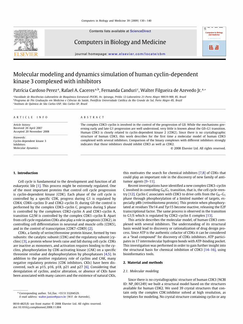
Fig. 1. Alignment of the 10 human CDKs. The identity between CDK2 and CDK3is 74.18%. In black identity residues, in gray similar residues. Asterisk (*) indicatesidentity, colon (:) indicates strong similarity, and period ( · ) indicates weak similarity(due to physico-chemical parameters).
result from alternative splicing or present larger than CDK3sequence, what makes sequence alignment difficult to interpret.Analysis of sequence alignment with 10 human CDKs shows theproximity of CDK3 and CDK2 and also indicates the similarity ofCDK9 with CDK8 and CDK7. Fig. 1 shows the alignment of 10 CDKsequences. The multiple alignments were performed by the program
P. Cardoso Perez et al. / Computers in Biology and Medicine 39 (2009) 130–140 133
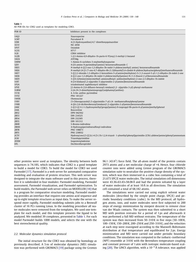
Fig. 2. Secondary structure elements for CDK3 Procheck.
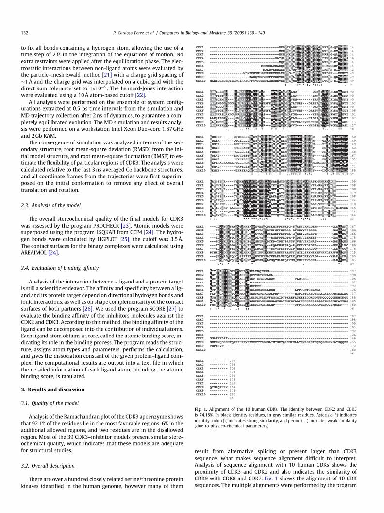
CLUSTAL W. The parameters used were the defaults of the program.This information has been successfully used to model CDK1 [28],CDK9 [29], and CDK5 [30], generating reasonable models. The se-quence of CDK3 (EC 2.7.11.22) consists of 305 amino acids with amolecular weight of 35,045.74da and a theoretical pI of 8.86. Align-ment of other human CDKs indicates that the more closely relatedCDK is CDK2. Analysis of the CDK3 structure shows that it belongsto the class of alpha+beta structures and investigation of the foldindicates two alpha+beta domains with a larger C-terminal mostlyalpha helical, which is characteristic of the protein kinase fold, asexpected. The model of CDK3 is folded into the bilobal structure,with the smaller N-terminal lobe consisting predominantly of �-sheet structure. The N-terminal lobe of CDK3 consists in a sheet offive antiparallel �-strands (�1–�5) and a single large �-helix (�1).The C-terminal domain contains a pseudo-4-helical bundle (�2, �3,�4, �6), a small �-ribbon (�6–�8), and two additional �-helices (�5,�7), as shown in Fig. 2. The ATP biding pocket is found in the cleftbetween the two lobes [31]. Most of the residues present in theATP-binding pocket are hydrophobic. Furthermore, most hydropho-bic residues are conserved in all CDK sequences, except for Ala 31(Glu in CDK8), Phe 82 (His CDK4 and CDK6, Tyr in CDK8 and CDK10)and Leu 83 (Cys in CDK5, CDK9, and CDK10). This hydrophobicpocket shows the ability to accommodate several different geome-tries, such as adenine derivatives and flavonoids. CDK2 inhibitorsidentified so far, present a flat topology with molecular weight be-low 600da, and small number of hydrogen bonds partners. CDK3and CDK2 share several conserved structures, such as: PSTAIRE [32]or cyclin recognition box and the T-loop, containing the activatingphosphorylation site. Fig. 3 shows the CDK3 structure. Furthermore,most of the residues involved in the intermolecular hydrogen bondsobserved in the CDK2 complexes solved by crystallography areconserved in the binary complexes of CDK3.
3.3. Molecular fork
It has been identified in the structures of the complexCDK2:inhibitors a common pattern of intermolecular hydrogenbonds between CDK and inhibitors. This molecular fork is composedof two hydrogen bond acceptors (C&O) and one hydrogen bonddonor (N–H), which allows a wide range of different molecules todock on to the ATP binding pocket, such as: roscovitine, olomoucineand NU2058. Several structures of CDK2 complexed with inhibitorspresent the participation of a molecular fork, composed by a C&O
Fig. 3. CDK3 Structure. A bilobal protein, with a smaller N-terminal composed mainlyof �-strands (dark gray) and a large C-terminal composed of �-helices (light gray).In the cleft the ATP-bonding pocket with ATP.
group of Glu81 and the N–H and C&O group of Leu83, in inter-molecular hydrogen bonds between CDK2 and the inhibitors. Thereare 80 structures of CDK2 complexed with inhibitors only (searchperformed on the PDB [33] on October 2006). All these inhibitorshave pairs of hydrogen bond partners that show complementarity tothe molecular fork on CDK3, most of them involving at least two hy-drogen bonds with the molecular fork. Furthermore, analysis of theCDK inhibitors currently in clinical trials (flavopiridol, roscovitine,

134 P. Cardoso Perez et al. / Computers in Biology and Medicine 39 (2009) 130–140
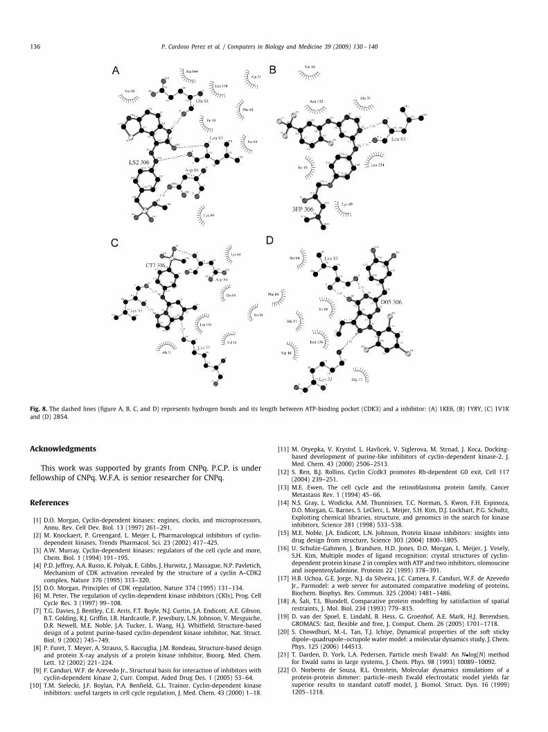
Table 2Hydrogen bonds between the four best inhibitors and CDK3.
Hydrogen bonds between active site and inhibitors
Distance (Å) Contact area (Å2)
LS2 (1KE6) CDK3 332N37 ASP86 OD2 3.06N19 LEU83 O 3.32O11 LEU 83 N 2.79N1 GLU81 O 2.97
2FP (1V1K) CDK3 354N3 LEU83 O 2.76N1 LEU83 N 3.34
CT7 (1Y8Y) CDK3 241O2 ASP86 N 3.10N5 LEU83 O 2.85N3 LEU83 N 3.23N1 LYS33 NZ 3.09
D05 (2B54) CDK3 343N8 LEU83 O 2.91N2 LEU83 N 3.09O9 LYS33 NZ 2.76
indirubin, and UCN-01) strongly indicate that at least two membersof the molecular fork should participate in intermolecular hydrogenbonds, to ensure proper orientation in ATP-binding pocket. Thisfeature may be exploited in the design of future CDK inhibitors.
Analysis of the intermolecular hydrogen bond distances presentin all binary models (CDK3:inhibitors), obtained by homology mod-eling, strongly indicates the conservation of at least one member ofthe molecular fork in hydrogen bonding, an acceptor close to N–Hin Leu83. Inspection of the position of the inhibitors in the ATP-binding pocket of CDK3 indicates one hydrogen bond donor close toC&O in Glu81 and/or Leu83, and an acceptor close to N–H in Leu83.Such simple paradigm is also conserved in all CDK2:inhibitor com-plex structures solved so far. Table 2 presents the intermolecularhydrogen bonds between inhibitors and CDK3.
3.4. Analysis of the MD simulation
MD simulation of the CDK3 structure in the apo form was car-ried out in order to check the structural stability of the structuralmodel proposed for CDK3. Analysis of 5ns dynamics indicates thatthe CDK3 structure is stable, after a rapid increasing during the first250ps, the protein backbone RMSD average and standard deviationover the last 3ns of the CDK3 trajectory was 2.4 ± 0.2Å. A plateau ofRMSD for the system was achieved within 2ns of unrestrained sim-ulation, suggesting that 5ns unrestrained simulation was sufficientfor stabilizing fully relaxed models.
Fig. 4 shows the evolution of the RMSD during the dynamics. Thesuperimposition of the average structure of the CDK3 with the initialmodel (Fig. 5) does not show major conformational changes in com-parison to the initial model, which is consistent with the relativelylow RMSD value. Proteins unbound as previously demonstrated inother MD simulations [34,35] present a higher RMSD value if com-pared with the same protein complexed.
The flexibilities of the proteins were assessed by the RMSF valuesfrom MD of the trajectory which reflects the flexibility of each atomresidue in a molecule (Fig. 6). The major backbone fluctuation occursin the loop region and in the region surrounding the beta–alpha–betafold, whereas regions with the low RMSF correspond exclusivelyto the rigid beta–alpha–beta fold. In a typical RMSF pattern, a low
Fig. 4. The graphic A shows the radius of gyration of all C� of CDK3. The dashedline gives the equilibration phase, the solid line shows the last 3ns of calcula-tion. (B) The graphical representation of root-mean-square deviation (RMSD) ofall C� from starting structure of apoenzime CDK3 as a function of time. Thedashed line gives the equilibration phase, the solid line shows the last 3ns ofcalculation.
Fig. 5. Superposition of the average structure during the MD simulation with theinitial minimized structure of ligand-free CDK3. The structures are presented asribbon diagram. The average structure is colored dark gray; the initial structure iscolored light gray.
RMSF value indicates the well-structured regions while the highvalues indicate the loosely structured loop regions or domainsterminal [36].
In addition, analysis of the structure during the dynam-ics simulation indicates that the regions L1 (turn composed byGlu25–Gly27), L2 (loop Leu37–Pro45), L3 (turn Glu73–Arg74), L4(loop Thr94–Pro100), L5 (turn Tyr159–His161) and L6 (big loop

P. Cardoso Perez et al. / Computers in Biology and Medicine 39 (2009) 130–140 135
Fig. 6. Graphical representation of root-mean-square fluctuations (RMSF) of all C�from starting structure of models as a function of time. The graphic shows theaverage RMSF of last 3ns of simulation of apoenzyme CDK3.
Fig. 7. Correlation between pkd for inhibitors against CDK2 and CDK3.
composed by Gly220–Gly247) present higher RMSF values, whichstrongly indicates that these regions are the most flexible in theCDK3 structure. Most of these regions involve loops as has beenobserved during the MD simulation of CDK2 [37].
Although, the region on the graphic that shows highest RMSF,around L6 (Gly220–Gly247), in CDK2 is related as a region where aregulatory protein known as CskHs binds. This protein is also knownas p9. Two homologues of this protein were identified in humans(CskHs1 and CskHs2)[38].
3.5. Interactions of inhibitors with CDK3
The binding affinity of a protein–ligand is one of the struc-tural properties of a target protein that is essential for specificitywith a ligand. Hydrogen bonds are directly responsible for speci-ficity and affinity between protein and its ligand. Analysis of thecontact areas and intermolecular hydrogen bonds were not con-clusive when we tried to relate to the affinity of the inhibitorsagainst CDK2 and CDK3. In order to identify whether the CDK2inhibitors may be used to inhibit CDK3 as well, we determinedthe affinity constant using the program SCORE for all binary com-plexes studied in this work, for both kinases: CDK2 and CDK3.Fig. 7 shows the pkd for both CDKs. Similar approaches to estimateligand-binding affinity have been successfully employed to studyhuman purine nucleoside phosphorylase in complex with differentligands [34].
The correlation coefficient between CDK3 pkd and CDK2 pkdis around 0.87, which strongly indicates correlation between bothpKds, Previously published comparison between experimental andpredicted pKds considered existence of correlation for values above0.6 [39]. This high correlation coefficient may indicate that CDK2ligands can also inhibit CDK3 activity. Analysis of the pkd for thebinary complexes of CDK3 indicates that the best inhibitors are: n-methyl-{4-[2- (7-oxo-6, 7-dihydro-8h-[1,3]thiazolo[5,4-e]indol-8-ylidene)hydrazino]phenyl}methanesulfonamide (PDB code: 1KE6),(2r)-1-(dimethylamino)-3-{4-[(6-{[2-fluoro-5-(trifluoromethyl)phe-nyl] amino}pyrimidin-4-yl)amino]phenoxy}propan-2-ol (PDB code:1V1K), (5-chloropyrazolo[1,5-a]pyrimidin-7-yl)-(4-methanesulfonylphenyl)amine (PDB code: 1Y8Y) and din-232305 6-(3,4-dihydroxy-benzyl)-3-ethyl-1-(2,4,6-trichlorophenyl)-1h-pyrazolo[3,4-d]pyrim-idin-4(5h)-one (PDB code: 2B54). Fig. 8 shows the intermolecu-lar hydrogen bonds for these complexes. These inhibitors presentlow IC50 values against CDK2, which strongly indicates that theseinhibitors should also potently inhibit CDK3.
4. Conclusions
CDK3 presents high sequential identity with CDK2 (74.18%) whichallows the homology modeling of CDK3, using CDK2 structure astemplate. Analysis of the binary complexes of CDK3 with 39 differentinhibitors indicates the participation of the molecular fork of CDKin intermolecular hydrogen bonds. Comparison of the structures ofthe binary complexes of several inhibitors, previously identified forCDK2, with the complexes with CDK3 strongly indicates that thoseinhibitors should inhibit CDK3 as well. In MD simulation the CDK3model demonstrated a good stability in solution corroborating thereliability of model, showing a higher flexibility in loops and turnsas expected. Therefore development of CDK3 inhibitors may profitby the experience and findings of several years of research in thedevelopment of CDK2 inhibitors.
5. Summary
This article describes structural models for 39 binary complexesof cyclin-dependent kinase 3 (CDK3) and inhibitors. This is the firstdescription of structural models for human CDK3. The protein com-plex CDK3–cyclin is involved in the control of the progression ofphase G0. While the mechanisms governing early and late G1 pro-gression are well understood, much less is known about the G0–G1transition. Most of the structural studies on human CDKs are focusedon CDK2. Human CDK3 is closely related to others CDKs such ascyclin-dependent kinase 2 (CDK2). Since there is no crystallographicstructure available for human CDK3, this work describes the molec-ular model of human CDK3 complexed with several inhibitors. Anal-ysis of the binary complexes of CDK3 with 39 different inhibitorsindicates the participation of the molecular fork of CDK in inter-molecular hydrogen bonds. In order to identify whether the CDK2inhibitors may be used to inhibit CDK3 as well, we determined theaffinity constant using the program SCORE for all binary complexesstudied in this work, for both kinases: CDK2 and CDK3. Further-more, comparison of the binary complexes with different inhibitorsstrongly indicates that those inhibitors should inhibit CDK3 aswell as CDK2.
Conflict of interest statement
We, the authors, have no conflict of interest related to themanuscript entitled “Molecular modeling and dynamics simulationof human CDK3 in complexed with Inhibitors”.
136 P. Cardoso Perez et al. / Computers in Biology and Medicine 39 (2009) 130–140
Fig. 8. The dashed lines (figure A, B, C, and D) represents hydrogen bonds and its length between ATP-binding pocket (CDK3) and a inhibitor: (A) 1KE6, (B) 1Y8Y, (C) 1V1Kand (D) 2B54.
Acknowledgments
This work was supported by grants from CNPq. P.C.P. is underfellowship of CNPq. W.F.A. is senior researcher for CNPq.
References
[1] D.O. Morgan, Cyclin-dependent kinases: engines, clocks, and microprocessors,Annu. Rev. Cell Dev. Biol. 13 (1997) 261–291.
[2] M. Knockaert, P. Greengard, L. Meijer L, Pharmacological inhibitors of cyclin-dependent kinases, Trends Pharmacol. Sci. 23 (2002) 417–425.
[3] A.W. Murray, Cyclin-dependent kinases: regulators of the cell cycle and more,Chem. Biol. 1 (1994) 191–195.
[4] P.D. Jeffrey, A.A. Russo, K. Polyak, E. Gibbs, J. Hurwitz, J. Massague, N.P. Pavletich,Mechanism of CDK activation revealed by the structure of a cyclin A–CDK2complex, Nature 376 (1995) 313–320.
[5] D.O. Morgan, Principles of CDK regulation, Nature 374 (1995) 131–134.[6] M. Peter, The regulation of cyclin-dependent kinase inhibitors (CKIs), Prog. Cell
Cycle Res. 3 (1997) 99–108.[7] T.G. Davies, J. Bentley, C.E. Arris, F.T. Boyle, N.J. Curtin, J.A. Endicott, A.E. Gibson,
B.T. Golding, R.J. Griffin, I.R. Hardcastle, P. Jewsbury, L.N. Johnson, V. Mesguiche,D.R. Newell, M.E. Noble, J.A. Tucker, L. Wang, H.J. Whitfield, Structure-baseddesign of a potent purine-based cyclin-dependent kinase inhibitor, Nat. Struct.Biol. 9 (2002) 745–749.
[8] P. Furet, T. Meyer, A. Strauss, S. Raccuglia, J.M. Rondeau, Structure-based designand protein X-ray analysis of a protein kinase inhibitor, Bioorg. Med. Chem.Lett. 12 (2002) 221–224.
[9] F. Canduri, W.F. de Azevedo Jr., Structural basis for interaction of inhibitors withcyclin-dependent kinase 2, Curr. Comput. Aided Drug Des. 1 (2005) 53–64.
[10] T.M. Sielecki, J.F. Boylan, P.A. Benfield, G.L. Trainor, Cyclin-dependent kinaseinhibitors: useful targets in cell cycle regulation, J. Med. Chem. 43 (2000) 1–18.
[11] M. Otyepka, V. Krystof, L. Havlicek, V. Siglerova, M. Strnad, J. Koca, Docking-based development of purine-like inhibitors of cyclin-dependent kinase-2, J.Med. Chem. 43 (2000) 2506–2513.
[12] S. Ren, B.J. Rollins, Cyclin C/cdk3 promotes Rb-dependent G0 exit, Cell 117(2004) 239–251.
[13] M.E. Ewen, The cell cycle and the retinoblastoma protein family, CancerMetastasis Rev. 1 (1994) 45–66.
[14] N.S. Gray, L. Wodicka, A.M. Thunnissen, T.C. Norman, S. Kwon, F.H. Espinoza,D.O. Morgan, G. Barnes, S. LeClerc, L. Meijer, S.H. Kim, D.J. Lockhart, P.G. Schultz,Exploiting chemical libraries, structure, and genomics in the search for kinaseinhibitors, Science 281 (1998) 533–538.
[15] M.E. Noble, J.A. Endicott, L.N. Johnson, Protein kinase inhibitors: insights intodrug design from structure, Science 303 (2004) 1800–1805.
[16] U. Schulze-Gahmen, J. Brandsen, H.D. Jones, D.O. Morgan, L. Meijer, J. Vesely,S.H. Kim, Multiple modes of ligand recognition: crystal structures of cyclin-dependent protein kinase 2 in complex with ATP and two inhibitors, olomoucineand isopentenyladenine, Proteins 22 (1995) 378–391.
[17] H.B. Uchoa, G.E. Jorge, N.J. da Silveira, J.C. Camera, F. Canduri, W.F. de AzevedoJr., Parmodel: a web server for automated comparative modeling of proteins,Biochem. Biophys. Res. Commun. 325 (2004) 1481–1486.
[18] A. Šali, T.L. Blundell, Comparative protein modelling by satisfaction of spatialrestraints, J. Mol. Biol. 234 (1993) 779–815.
[19] D. van der Spoel, E. Lindahl, B. Hess, G. Groenhof, A.E. Mark, H.J. Berendsen,GROMACS: fast, flexible and free, J. Comput. Chem. 26 (2005) 1701–1718.
[20] S. Chowdhuri, M.-L. Tan, T.J. Ichiye, Dynamical properties of the soft stickydipole–quadrupole–octupole water model: a molecular dynamics study, J. Chem.Phys. 125 (2006) 144513.
[21] T. Darden, D. York, L.A. Pedersen, Particle mesh Ewald: An N•log(N) methodfor Ewald sums in large systems, J. Chem. Phys. 98 (1993) 10089–10092.
[22] O. Norberto de Souza, R.L. Ornstein, Molecular dynamics simulations of aprotein-protein dimmer: particle–mesh Ewald electrostatic model yields farsuperior results to standard cutoff model, J. Biomol. Struct. Dyn. 16 (1999)1205–1218.

P. Cardoso Perez et al. / Computers in Biology and Medicine 39 (2009) 130–140 137
[23] R.A. Laskowski, M.W. MacArthur, D.S. Moss, J.M. Thornton, PROCHECK: aprogram to check the stereochemical quality of protein structures, J. Appl.Cryst. 26 (1993) 283–291.
[24] Collaborative Computational Project, Number 4. The CCP4 Suite: Programs forProtein Crystallography, Acta Crystallogr. 50 (1994) 760–763.
[25] A.C. Wallace, R.A. Laskowski, J.M. Thornton, LIGPLOT: a program generateschematic diagrams of protein–ligand interactions, Protein Eng. Des. Sel. 8(1995) 127–134.
[26] W.F. de Azevedo Jr, H.-.J. Mueller-Dieckmann, U. Schulze-Gahmen, P.J. Worland,E. Sausville, S.H. Kim, Structural basis for specificity and potency of a flavonoidinhibitor of human CDK2, a cell cycle kinase, Proc. Natl. Acad. Sci. USA 93(1996) 2735–2740.
[27] R. Wang, L. Liu, L. Lai, Y. Tang, SCORE: a new empirical method for estimatingthe binding affinity of a protein–ligand complex, J. Mol. Model. 4 (1998) 379–394.
[28] F. Canduri, H.B. Uchoa, W.F. de Azevedo Jr, Molecular models of cyclin-dependent kinase 1 complexed with inhibitors, Biochem. Biophys. Res. Commun.324 (2004) 661–666.
[29] W.F. de Azevedo Jr, F. Canduri, N.J.F. Silveira, Structural basis for inhibitionof cyclin-dependent kinase 9 by flavopiridol, Biochem. Biophys. Res. Commun.293 (2002) 566–571.
[30] W.F. de Azevedo Jr., R.T. Gaspar, F. Canduri, J.C. Camera Jr., N.J.F. Silveira,Molecular model of cyclin-dependent kinase 5 complexed with roscovitine,Biochem. Biophys. Res. Commun. 297 (2002) 1154–1158.
[31] U. Schulze-Gahmen, H.L. De Bondt, S.H. Kim, High-resolution crystal structuresof human cyclin-dependent kinase 2 with and without ATP: bound waters andnatural ligand as guides for inhibitor design, J. Med. Chem. 39 (1996) 4540–4546.
[32] L. Meijer, Cyclin-dependent kinases inhibitors as potential anticancer,antineurodegenerative, antiviral and antiparasitic agents, Drug ResistanceUpdates 3 (2000) 83–88.
[33] H.M. Berman, J. Westbrook, Z. Feng, G. Gilliland, T.N. Bhat, H. Weissig, I.N.Shindyalov, P.E. Bourne, The protein data bank, Nucleic Acids Res. 28 (2000)235–242.
[34] R.A. Caceres, L.F.S. Timmers, R. Dias, L.A. Basso, D.S. Santos, W.F. de AzevedoJr, Molecular modeling and dynamics simulations of PNP from Streptococcusagalactiae, Bioorg. Med. Chem. 16 (2008) 4984–4993.
[35] R.A. Caceres, L.F.S. Timmers, A.L. Vivan, C.Z. Schneider, L.A. Basso, W.F. deAzevedo Jr., Molecular modeling and dynamics studies of cytidylate kinase fromMycobacterium tuberculosis H37Rv, J. Mol. Model. 14 (2008) 427–434.
[36] C.S. Alexander, X. Yan, T. Pei, Homology modeling and molecular dynamicssimulations of transmembrane domain structure of human neuronal nicotinicacetylcholine receptor, Biophys. J. 88 (2005) 1009–1017.
[37] B. Zhang, V.B. Tan, K.M. Lim, T.E. Tay, S. Zhuang, Study of the inhibition of cyclin-dependent kinases with roscovitine and indirubin-3'-oxime from moleculardynamics simulations, J. Mol. Model. 13 (2006) 79–89.
[38] Y. Bourne, M.H. Watson, M.J. Hickey, W. Holmes, W. Rocque, S.I. Reed, J.A.Tainer, Crystal structure and mutational analysis of the human CDK2 kinasecomplex with cell cycle-regulatory protein CksHs1, Cell 84 (1996) 863–874.
[39] R. Wang, L. Lai, S. Wang, Further development and validation of empiricalscoring functions for structure-based binding affinity prediction, J. Comput.Aided Mol. Des. 16 (2002) 11–26.
Patricia Perez was born in Porto Alegre, RS, in 1983. She is undergraduate studentin Biology at Catholic University of Rio Grande do Sul, Brazil. Her research interestsare focused on biochemistry and molecular biology, and structural studies of proteintargets by crystallography and molecular modeling.
Rafael Andrade Caceres was born in Esteio, in 1977. He graduated in Chemistryfrom Universidade Luterana do Brasil, Brazil, in 2005. Since 2006, he has been withPontificia Universidade Catolica do Rio Grande do Sul (PUCRS), where he is postgraduated in Structural Biochemistry Laboratory (LaBioQuest). His research interestsfocus on molecular modeling of Ets, molecular docking protein-protein and protein-DNA, molecular dynamic of biosystems.
Fernanda Canduri was born in Sao Jose do Rio Preto/SP, Brazil, in 1977. She receivedthe MD degree in Molecular biophysics from the Universidade Estadual Paulista Júliode Mesquita Filho, Brazil, in 1998 and a PhD in same University in 2001. She is fullProfessor at Sao Paulo State University, Brazil. Her research interests are focusedon biochemistry and molecular biology, and structural studies of protein targets bycrystallography and molecular modeling.
Walter Filgueira de Azevedo was Born in Rio de Janeiro/RJ, Brazil in 1963 andgraduated in Physics in 1990 from University of Sao Paulo, Brazil, he finished hisPhD in 1997 in Applied Physics at Sao Paulo State University, Brazil. He workedas visiting researcher at University of California-Berkeley from 1993-1996, undersupervision of Prof. Sung-Hou Kim. He is full-professor at Catholic University of RioGrande do Sul, Brazil. He published over a hundred articles, most of them focused onbioinformatics and structural biology. A list of his most important articles is below:
1: Pereira JH, Vasconcelos IB, Oliveira JS, Caceres RA, de Azevedo WF Jr., Basso LA,Santos DS.
Shikimate kinase: a potential target for development of novel antitubercular agents.Curr Drug Targets. 2007 Mar;8(3):459-68. Review.PMID: 17348838 [PubMed - indexed for MEDLINE]
2: Marques MR, Pereira JH, Oliveira JS, Basso LA, de Azevedo WF Jr., Santos DS,Palma MS.The inhibition of 5-enolpyruvylshikimate-3-phosphate synthase as a model for de-velopment of novel antimicrobials.Curr Drug Targets. 2007 Mar;8(3):445-57. Review.PMID: 17348837 [PubMed - indexed for MEDLINE]
3: Dias MV, Ely F, Palma MS, de Azevedo WF Jr., Basso LA, Santos DS.Chorismate synthase: an attractive target for drug development against orphanDiseases.Curr Drug Targets. 2007 Mar;8(3): 437-44. Review.PMID: 17348836 [PubMed - indexed for MEDLINE]
4: Canduri F, Perez PC, Caceres RA, de Azevedo WF Jr.Protein kinases as targets for antiparasitic chemotherapy drugs.Curr Drug Targets. 2007 Mar;8(3):389-98. Review.PMID: 17348832 [PubMed - indexed for MEDLINE]
5: Dias MV, Faim LM, Vasconcelos IB, de Oliveira JS, Basso LA, Santos DS, de AzevedoWF Jr.Effects of the magnesium and chloride ions and shikimate on the structure ofshikimate kinase from Mycobacterium tuberculosis.Acta Crystallograph Sect F Struct Biol Cryst Commun. 2007 Jan 1;63(Pt 1):1-6. Epub2006 Dec 16.PMID: 17183161 [PubMed - indexed for MEDLINE]
6: Moreno FB, Bezerra GA, de Oliveira TM, de Souza EP, da Rocha BA, BenevidesRG, Delatorre P, Cavada BS, de Azevedo WF Jr.New crystal forms of Diocleinae lectins in the presence of different dimannosides.Acta Crystallograph Sect F Struct Biol Cryst Commun. 2006 Nov 1;62(Pt 11):1100-3.Epub 2006 Oct 20.PMID: 17077488 [PubMed - indexed for MEDLINE]
7: Krystof V, Cankar P, Frysova I, Slouka J, Kontopidis G, Dzubak P, Hajduch M,Srovnal J, de Azevedo WF Jr., Orsag M, Paprskarova M, Rolcik J, Latr A, Fischer PM,Strnad M.4-arylazo-3,5-diamino-1H-pyrazole CDK inhibitors: SAR study, crystal structure incomplex with CDK2, selectivity, and cellular effects.J Med Chem. 2006 Nov 2;49(22):6500-9.PMID: 17064068 [PubMed - indexed for MEDLINE]
8: Cavada BS, Moreno FB, da Rocha BA, de Azevedo WF Jr., Castellon RE, GoerschGV, Nagano CS, de Souza EP, Nascimento KS, Radis-Baptista G, Delatorre P, Leroy Y,Toyama MH, Pinto VP, Sampaio AH, Barettino D, Debray H, Calvete JJ, Sanz L.cDNA cloning and 1.75 A crystal structure determination of PPL2, an endochitinaseand N-acetylglucosamine-binding hemagglutinin from Parkia platycephala seeds.FEBS J. 2006 Sep;273(17):3962-74.PMID: 16934035 [PubMed - indexed for MEDLINE]
9: Borges JC, Pereira JH, Vasconcelos IB, dos Santos GC, Olivieri JR, Ramos CH, PalmaMS, Basso LA, Santos DS, de Azevedo WF Jr.Phosphate closes the solution structure of the 5-enolpyruvylshikimate-3-phosphatesynthase (EPSPS) from Mycobacterium tuberculosis.Arch Biochem Biophys. 2006 Aug 15;452(2):156-64. Epub 2006 Jun 13.PMID: 16876105 [PubMed - indexed for MEDLINE]
10: Moreno FB, Martil DE, Cavada BS, de Azevedo WF Jr.Crystallization and preliminary X-ray diffraction analysis of an anti-H(O) lectin fromLotus tetragonolobus seeds.Acta Crystallograph Sect F Struct Biol Cryst Commun. 2006 Jul 1;62(Pt 7):680-3.Epub 2006 Jun 26PMID: 16820693 [PubMed - indexed for MEDLINE]
11: Lombardi FR, Anazetti MC, Santos GC, Olivieri JR, de Azevedo WF Jr., Bonilla-Rodriguez GO.Rattlesnake hemoglobins: Functional properties and tetrameric stability.Protein Pept Lett. 2006;13(5):517-23.PMID: 16800809 [PubMed - indexed for MEDLINE]
12: Radis-Baptista G, Moreno FB, de Lima Nogueira L, Martins AM, de OliveiraToyama D, Toyama MH, Cavada BS, de Azevedo WF Jr., Yamane T.Crotacetin, a novel snake venom C-type lectin homolog of convulxin, exhibits anunpredictable antimicrobial activity.
138 P. Cardoso Perez et al. / Computers in Biology and Medicine 39 (2009) 130–140
Cell Biochem Biophys. 2006;44(3):412-23.PMID: 16679528 [PubMed - indexed for MEDLINE]
13: de Azevedo WF Jr., Canduri F, Basso LA, Palma MS, Santos DS.Determining the structural basis for specificity of ligands using crystallographicscreeningCell Biochem Biophys. 2006;44(3):405-11.PMID: 16679527 [PubMed - indexed for MEDLINE]
14: Dias MV, Canduri F, da Silveira NJ, Czekster CM, Basso LA, Palma MS, Santos DS,de Azevedo WF Jr.Molecular models of tryptophan synthase from mycobacterium tuberculosis com-plexed with inhibitors.Cell Biochem Biophys. 2006;44(3):375-84.PMID: 16679524 [PubMed - indexed for MEDLINE]
15: da Silveira NJ, Bonalumi CE, Uchoa HB, Pereira JH, Canduri F, de Azevedo WF.DBMODELING: a database applied to the study of protein targets from genomeprojects.Cell Biochem Biophys. 2006;44(3):366-74.PMID: 16679523 [PubMed - indexed for MEDLINE]
16: Delatorre P, Rocha BA, Gadelha CA, Santi-Gadelha T, Cajazeiras JB, Souza EP,Nascimento KS, Freire VN, Sampaio AH, Azevedo WF Jr., Cavada BS.Crystal structure of a lectin from Canavalia maritima (ConM) in complex withtrehalose and maltose reveals relevant mutation in ConA-like lectins.J Struct Biol. 2006 Jun;154(3):280-6. Epub 2006 Apr 21.PMID: 16677825 [PubMed - indexed for MEDLINE]
17: Oliveira JS, Pereira JH, Canduri F, Rodrigues NC, de Souza ON, de Azevedo WFJr., Basso LA, Santos DS.Crystallographic and pre-steady-state kinetics studies on binding of NADH to wild-type and isoniazid-resistant enoyl-ACP(CoA) reductase enzymes fromMycobacteriumtuberculosis.J Mol Biol. 2006 Jun 9;359(3):646-66. Epub 2006 Apr 21.PMID: 16647717 [PubMed - indexed for MEDLINE]
18: Vivan AL, Dias MV, Schneider CZ, de Azevedo WF Jr., Basso LA, Santos DS.Crystallization and preliminary X-ray diffraction analysis of prephenate dehydratasefrom Mycobacterium tuberculosis H37Rv.Acta Crystallograph Sect F Struct Biol Cryst Commun. 2006 Apr 1;62(Pt 4):357-60.Epub 2006 Mar 10.PMID: 16582484 [PubMed - indexed for MEDLINE]
19: Leopoldino AM, Canduri F, Cabral H, Junqueira M, de Marqui AB, Apponi LH, daFonseca IO, Domont GB, Santos DS, Valentini S, Bonilla-Rodriguez GO, Fossey MA,de Azevedo WF Jr., Tajara EH.Expression, purification, and circular dichroism analysis of human CDK9.Protein Expr Purif. 2006 Jun;47(2):614-20. Epub 2006 Mar 10.PMID: 16580843 [PubMed - indexed for MEDLINE]
20: Cavada BS, Marinho ES, Souza EP, Benevides RG, Delatorre P, Souza LA, Nasci-mento KS, Sampaio AH, Moreno FB, Rustiguel JK, Canduri F, de Azevedo WF Jr.,Debray H.Purification, partial characterization and preliminary X-ray diffraction analysis of amannose-specific lectin from Cymbosema roseum seeds.Acta Crystallograph Sect F Struct Biol Cryst Commun. 2006 Mar 1;62(Pt 3):235-7.Epub 2006 Feb 10.PMID: 16511310 [PubMed - indexed for MEDLINE]
21: Cavada BS, Castellon RE, Vasconcelos GG, Rocha BA, Bezerra GA, Debray H,Delatorre P, Nagano CS, Toyama M, Pinto VP, Moreno FB, Canduri F, Azevedo WF Jr.Crystallization and preliminary X-ray diffraction analysis of a new chitin-bindingprotein from Parkia platycephala seeds.Acta Crystallograph Sect F Struct Biol Cryst Commun. 2005 Sep 1;61(Pt 9):841-3.Epub 2005 Aug 31.PMID: 16511174 [PubMed - indexed for MEDLINE]
22: Gadelha CA, Moreno FB, Santi-Gadelha T, Cajazeiras JB, Rocha BA, Rustiguel JK,Freitas BT, Canduri F, Delatorre P, Azevedo WF Jr., Cavada BS.Crystallization and preliminary X-ray diffraction analysis of a lectin from Canavaliamaritima seeds.Acta Crystallograph Sect F Struct Biol Cryst Commun. 2005 Jan 1;61(Pt 1):87-9. Epub2004 Dec 2.PMID: 16508099 [PubMed - indexed for MEDLINE]
23: Dias MV, Borges JC, Ely F, Pereira JH, Canduri F, Ramos CH, Frazzon J, PalmaMS, Basso LA, Santos DS, de Azevedo WF Jr.
Structure of chorismate synthase from Mycobacterium tuberculosis.J Struct Biol. 2006 May;154(2):130-43. Epub 2006 Jan 17.PMID: 16459102 [PubMed - indexed for MEDLINE]
24: Gadelha CA, Moreno FB, Santi-Gadelha T, Cajazeiras JB, Rocha BA,Assreuy AM, Lima Mota MR, Pinto NV, Passos Meireles AV, Borges JC,Freitas BT, Canduri F, Souza EP, Delatorre P, Criddle DN, de AzevedoWF Jr., Cavada BS.Native crystal structure of a nitric oxide-releasing lectin from the seeds ofCanavalia maritima.J Struct Biol. 2005 Dec;152(3):185-94. Epub 2005 Nov 14.PMID: 16337811 [PubMed - indexed for MEDLINE]
25: Basso LA, da Silva LH, Fett-Neto AG, de Azevedo WF Jr., Moreira Ide S,Palma MS, Calixto JB, Astolfi Filho S, dos Santos RR, Soares MB, SantosDS.The use of biodiversity as source of new chemical entities against definedmolecular targets for treatment of malaria, tuberculosis, and T-cell mediateddiseases–a review.Mem Inst Oswaldo Cruz. 2005 Oct;100(6):475-506. Epub 2005 Nov 8. Review.PMID: 16302058 [PubMed - indexed for MEDLINE]
26: Fadel V, Bettendorff P, Herrmann T, de Azevedo WF Jr., Oliveira EB,Yamane T, Wuthrich K.Automated NMR structure determination and disulfide bond identification of themyotoxin crotamine from Crotalus durissus terrificus.Toxicon. 2005 Dec 1;46(7):759-67. Epub 2005 Sep 26.PMID: 16185738 [PubMed - indexed for MEDLINE]
27: Silva RG, Pereira JH, Canduri F, de Azevedo WF Jr., Basso LA, SantosDS.Kinetics and crystal structure of human purine nucleoside phosphorylase incomplex with 7-methyl-6-thio-guanosine.Arch Biochem Biophys. 2005 Oct 1;442(1):49-58.PMID: 16154528 [PubMed - indexed for MEDLINE]
28: Manhani KK, Arcuri HA, da Silveira NJ, Uchoa HB, de Azevedo WF Jr.,Canduri FMolecular models of protein kinase 6 from Plasmodium falciparum.J Mol Model. 2005 Dec;12(1):42-8. Epub 2005 Nov 4.PMID: 16096806 [PubMed - indexed for MEDLINE]
29: Canduri F, Silva RG, dos Santos DM, Palma MS, Basso LA, Santos DS,de Azevedo WF Jr.Structure of human PNP complexed with ligands.Acta Crystallogr D Biol Crystallogr. 2005 Jul;61(Pt 7):856-62. Epub 2005 Jun 24.PMID: 15983407 [PubMed - indexed for MEDLINE]
30: Silveira NJ, Uchoa HB, Pereira JH, Canduri F, Basso LA, Palma MS,Santos DS, de Azevedo WF Jr.Molecular models of protein targets from Mycobacterium tuberculosis.J Mol Model. 2005 Mar;11(2):160-6. Epub 2005 Mar 10.PMID: 15759144 [PubMed - in process]
31: da Silveira NJ, Arcuri HA, Bonalumi CE, de Souza FP, Mello IM, Rahal P, PinhoJR, de Azevedo WF Jr.Molecular models of NS3 protease variants of the Hepatitis C virus.BMC Struct Biol. 2005 Jan 21;5:1.PMID: 15663787 [PubMed - indexed for MEDLINE]
32: Canduri F, Fadel V, Basso LA, Palma MS, Santos DS, de Azevedo WF Jr.New catalytic mechanism for human purine nucleoside phosphorylase.Biochem Biophys Res Commun. 2005 Feb 18;327(3):646-9.PMID: 15649395 [PubMed - indexed for MEDLINE]
33: Pereira JH, de Oliveira JS, Canduri F, Dias MV, Palma MS, Basso LA,Santos DS, de Azevedo WF Jr.Structure of shikimate kinase from Mycobacterium tuberculosis reveals thebinding of shikimic acid.Acta Crystallogr D Biol Crystallogr. 2004 Dec;60(Pt 12 Pt 2):2310-9. Epub 2004 Nov26.PMID: 15583379 [PubMed - indexed for MEDLINE]
34: Canduri F, Fadel V, Dias MV, Basso LA, Palma MS, Santos DS, deAzevedo WF Jr.Crystal structure of human PNP complexed with hypoxanthine and sulfate ion.Biochem Biophys Res Commun. 2005 Jan 14;326(2):335-8.PMID: 15582582 [PubMed - indexed for MEDLINE]

P. Cardoso Perez et al. / Computers in Biology and Medicine 39 (2009) 130–140 139
35: Ribeiro SP, Mendes MA, Dos Santos LD, de Souza BM, Marques MR, deAzevedo WF Jr., Palma MS.Structural and functional characterization of N-terminally blocked peptidesisolated from the venom of the social wasp Polybia paulista.Peptides. 2004 Dec;25(12):2069-78.PMID: 15572194 [PubMed - indexed for MEDLINE]
36: Uchoa HB, Jorge GE, Freitas Da Silveira NJ, Camera JC Jr, Canduri F, De AzevedoWF Jr.Parmodel: a web server for automated comparative modeling of proteins.Biochem Biophys Res Commun. 2004 Dec 24;325(4):1481-6.PMID: 15555595 [PubMed - indexed for MEDLINE]37: Peres P, Lombardi FR, Dos Santos GC, Olivieri JR, Canduri F, Bonilla-RodriguezGO, de Azevedo WF Jr.Molecular modeling and small angle X-ray scattering studies of Hoplosternum lit-torale cathodic haemoglobin.Biochem Biophys Res Commun. 2004 Dec 10;325(2):487-93.PMID: 15530418 [PubMed - indexed for MEDLINE]
38: Pereira JH, de Oliveira JS, Canduri F, Dias MV, Palma MS, Basso LA, de AzevedoWF Jr., Santos DS.Interaction of shikimic acid with shikimate kinase.Biochem Biophys Res Commun. 2004 Dec 3;325(1):10-7. Retraction in: BioChemBiophys Res Commun. 2005 Sep 2;334(3):967.PMID: 15522194 [PubMed - indexed for MEDLINE]
39: Peres P, de Azevedo Junior WF, Bonilla-Rodriguez GO.Allosteric water and phosphate effects in Hoplosternum littorale hemoglobins.Eur J Biochem. 2004 Nov;271(21):4270-4.PMID: 15511232 [PubMed - indexed for MEDLINE]
40: Dias MV, Ely F, Canduri F, Pereira JH, Frazzon J, Basso LA, Palma MS, de AzevedoWF Jr., Santos DS.Crystallization and preliminary X-ray crystallographic analysis of chorismate syn-thase from Mycobacterium tuberculosis.Acta Crystallogr D Biol Crystallogr. 2004 Nov;60(Pt 11):2003-5. Epub 2004 Oct 20.PMID: 15502309 [PubMed - indexed for MEDLINE]
41: Nolasco DO, Canduri F, Pereira JH, Cortinoz JR, Palma MS, Oliveira JS, Basso LA,de Azevedo WF Jr., Santos DS.Crystallographic structure of PNP from Mycobacterium tuberculosis at 1.9A resolu-tion.Biochem Biophys Res Commun. 2004 Nov 12;324(2):789-94.PMID: 15474496 [PubMed - indexed for MEDLINE]
42: Canduri F, Uchoa HB, de Azevedo WF Jr.Molecular models of cyclin-dependent kinase 1 complexed with inhibitors.Biochem Biophys Res Commun. 2004 Nov 12;324(2):661-6.PMID: 15474478 [PubMed - indexed for MEDLINE]
43: dos Santos Cabrera MP, de Souza BM, Fontana R, Konno K, Palma MS, de AzevedoWF Jr., Neto JRConformation and lytic activity of eumenine mastoparan: a new antimicrobial pep-tide from wasp venom.J Pept Res. 2004 Sep;64(3):95-103.PMID: 15317499 [PubMed - indexed for MEDLINE]
44: da Silveira NJ, Uchoa HB, Canduri F, Pereira JH, Camera JC Jr, Basso LA, PalmaMS, Santos DS, de Azevedo WF Jr.Structural bioinformatics study of PNP from Schistosoma mansoniBiochem Biophys Res Commun. 2004 Sep 10;322(1):100-4.PMID: 15313179 [PubMed - indexed for MEDLINE]
45: Moreno FB, Delatorre P, Freitas BT, Rocha BA, Souza EP, Faco F, Canduri F, CardosoAL, Freire VN, Lima Filho JL, Sampaio AH, Calvete JJ, De Azevedo WF Jr., Cavada BS.Crystallization and preliminary X-ray diffraction analysis of the lectin from Canavaliagladiata seeds.Acta Crystallogr D Biol Crystallogr. 2004 Aug;60(Pt 8):1493-5. Epub 2004 Jul 21.PMID: 15272187 [PubMed - indexed for MEDLINE]
46: Arcuri HA, Canduri F, Pereira JH, da Silveira NJ, Camera Junior JC, de Oliveira JS,Basso LA, Palma MS, Santos DS, de Azevedo Junior WF.Molecular models for shikimate pathway enzymes of Xylella fastidiosa.Biochem Biophys Res Commun. 2004 Jul 30;320(3):979-91.PMID: 15240145 [PubMed - indexed for MEDLINE]
47: Sforca ML, Oyama S Jr, Canduri F, Lorenzi CC, Pertinhez TA, Konno K, Souza BM,Palma MS, Ruggiero Neto J, Azevedo WF Jr., Spisni A.
How C-terminal carboxyamidation alters the biological activity of peptides from thevenom of the eumenine solitary wasp.Biochemistry. 2004 May 18;43(19):5608-17.PMID: 15134435 [PubMed - indexed for MEDLINE]
48: Canduri F, dos Santos DM, Silva RG, Mendes MA, Basso LA, Palma MS, de AzevedoWF, Santos DS.Structures of human purine nucleoside phosphorylase complexed with inosine andddI.Biochem Biophys Res Commun. 2004 Jan 23;313(4):907-14.PMID: 14706628 [PubMed - indexed for MEDLINE]
49: Fadel V, Canduri F, Olivieri JR, Smarra AL, Colombo MF, Bonilla-Rodriguez GO,de Azevedo WF Jr.Crystal structure of hemoglobin from the maned wolf (Chrysocyon brachyurus) usingsynchrotron radiation.Protein Pept Lett. 2003 Dec;10(6):551-9.PMID: 14683506 [PubMed - indexed for MEDLINE]
50: de Azevedo WF Jr., Canduri F, dos Santos DM, Pereira JH, Bertacine Dias MV,Silva RG, Mendes MA, Basso LA, Palma MS, Santos DS.Crystal structure of human PNP complexed with guanine.Biochem Biophys Res Commun. 2003 Dec 19;312(3):767-72.PMID: 14680831 [PubMed - indexed for MEDLINE]
51: Pereira JH, Canduri F, de Oliveira JS, da Silveira NJ, Basso LA, Palma MS, deAzevedo WF Jr., Santos DSStructural bioinformatics study of EPSP synthase from Mycobacterium tuberculosisBiochem Biophys Res Commun. 2003 Dec 19;312(3):608-14.PMID: 14680808 [PubMed - indexed for MEDLINE]
52: Filgueira de Azevedo W Jr, dos Santos GC, dos Santos DM, Olivieri JR, CanduriF, Silva RG, Basso LA, Renard G, da Fonseca IO, Mendes MA, Palma MS, Santos DS.Docking and small angle X-ray scattering studies of purine nucleoside phosphorylase.Biochem Biophys Res Commun. 2003 Oct 3;309(4):923-8.PMID: 13679062 [PubMed - indexed for MEDLINE]
53: Filgueira de Azevedo W Jr, Canduri F, Marangoni dos Santos D, Pereira JH, DiasMV, Silva RG, Mendes MA, Basso LA, Palma MS, Santos DS.Structural basis for inhibition of human PNP by immucillin-H.Biochem Biophys Res Commun. 2003 Oct 3;309(4):917-22.PMID: 13679061 [PubMed - indexed for MEDLINE]
54: dos Santos DM, Canduri F, Pereira JH, Vinicius Bertacine Dias M, Silva RG, MendesMA, Palma MS, Basso LA, de Azevedo WF Jr., Santos DS.Crystal structure of human purine nucleoside phosphorylase complexed with acy-clovir.Biochem Biophys Res Commun. 2003 Aug 29;308(3):553-9.PMID: 12914786 [PubMed - indexed for MEDLINE]
55: de Azevedo WF Jr., Canduri F, dos Santos DM, Silva RG, de Oliveira JS, de CarvalhoLP, Basso LA, Mendes MA, Palma MS, Santos DS.Crystal structure of human purine nucleoside phosphorylase at 2.3A resolution.Biochem Biophys Res Commun. 2003 Aug 29;308(3):545-52.PMID: 12914785 [PubMed - indexed for MEDLINE]
56: Fonseca JC, Honda RT, Delatorre P, Fadel V, Bonilla-Rodriguez GO, de AzevedoWF Jr.Crystallization, preliminary X-ray analysis and molecular-replacement solution ofhaemoglobin-II from the fish matrinxa (Brycon cephalus).Acta Crystallogr D Biol Crystallogr. 2003 Apr;59(Pt 4):752-4. Epub 2003 Mar 25.PMID: 12657802 [PubMed - indexed for MEDLINE]
57: Filgueira de Azevedo W Jr, Gaspar RT, Canduri F, Camera JC Jr, Freitas da SilveiraNJ.Molecular model of cyclin-dependent kinase 5 complexed with roscovitine.Biochem Biophys Res Commun. 2002 Oct 11;297(5):1154-8.PMID: 12372407 [PubMed - indexed for MEDLINE]
58: Filgueira de Azevedo W Jr, Canduri F, Simoes de Oliveira J, Basso LA, Palma MS,Pereira JH, Santos DS.Molecular model of shikimate kinase from Mycobacterium tuberculosis.Biochem Biophys Res Commun. 2002 Jul 5;295(1):142-8.PMID: 12083781 [PubMed - indexed for MEDLINE]
59: de Azevedo WF Jr., Canduri F, da Silveira NJ.Structural basis for inhibition of cyclin-dependent kinase 9 by flavopiridol.
140 P. Cardoso Perez et al. / Computers in Biology and Medicine 39 (2009) 130–140
Biochem Biophys Res Commun. 2002 Apr 26;293(1):566-71.PMID: 12054639 [PubMed - indexed for MEDLINE]
60: Konno K, Hisada M, Fontana R, Lorenzi CC, Naoki H, Itagaki Y, Miwa A, Kawai N,Nakata Y, Yasuhara T, Ruggiero Neto J, de Azevedo WF Jr., Palma MS, Nakajima T.Anoplin, a novel antimicrobial peptide from the venom of the solitary wasp Anopliussamariensis.Biochim Biophys Acta. 2001 Nov 26;1550(1):70-80.PMID: 11738089 [PubMed - indexed for MEDLINE]
61: Canduri F, Teodoro LG, Fadel V, Lorenzi CC, Hial V, Gomes RA, Neto JR, deAzevedo WF Jr.Structure of human uropepsin at 2.45 A resolution.Acta Crystallogr D Biol Crystallogr. 2001 Nov;57(Pt 11):1560-70. Epub 2001 Oct 25.PMID: 11679720 [PubMed - indexed for MEDLINE]
62: de Azevedo WF Jr., Canduri F, Fadel V, Teodoro LG, Hial V, Gomes RA.Molecular model for the binary complex of uropepsin and pepstatin.Biochem Biophys Res Commun. 2001 Sep 14;287(1):277-81.PMID: 11549287 [PubMed - indexed for MEDLINE]
63: Delatorre P, Smarra AL, Fadel V, Canduri F, Dellamano M, Bonilla-Rodriguez GO,de Azevedo WF Jr.Purification, crystallization and Patterson search of haemoglobin IV from the ActaCrystallogr D Biol Crystallogr. 2001 Sep;57(Pt 9):1329-31. Epub 2001 Aug 23.PMID: 11526335 [PubMed - indexed for MEDLINE]
64: Delatorre P, Olivieri JR, Ruggiero Neto J, Lorenzi CC, Canduri F, Fadel V, KonnoK, Palma MS, Yamane T, de Azevedo WF Jr.Preliminary cryocrystallography analysis of an eumenine mastoparan toxin isolatedfrom the venom of the wasp Anterhynchium flavomarginatum micado.Biochim Biophys Acta. 2001 Feb 9;1545(1-2):372-6.PMID: 11342062 [PubMed - indexed for MEDLINE]
65: Honda RT, Delatorre P, Fadel V, Canduri F, Dellamano M, de Azevedo WF Jr.,Bonilla-Rodriguez GO.Crystallization, preliminary X-ray analysis and molecular-replacement solution ofthe carboxy form of haemoglobin I from the fish Brycon cephalus.Acta Crystallogr D Biol Crystallogr. 2000 Dec;56(Pt 12):1685-7.PMID: 11092946 [PubMed - indexed for MEDLINE]
66: Canduri F, Delatorre P, Fadel V, Lorenzi CC, Pereira JH, Olivieri JR, Ruggiero NetoJ, Konno K, Palma MS, Yamane T, de Azevedo WF Jr.Crystallization and preliminary X-ray diffraction analysis of a eumenine mastoparantoxin: a new class of mast-cell degranulating peptide in the wasp venom.Acta Crystallogr D Biol Crystallogr. 2000 Nov;56(Pt 11):1434-6.PMID: 11053843 [PubMed - indexed for MEDLINE]
67: Smarra AL, de Azevedo WF Jr., Fadel V, Delatorre P, Dellamano M, Colombo MF,Bonilla-Rodriguez GOPurification, crystallization and preliminary X-ray analysis of haemoglobin I fromthe armoured catfish Liposarcus anisitsi.Acta Crystallogr D Biol Crystallogr. 2000 Apr;56(Pt 4):495-7.PMID: 10739931 [PubMed - indexed for MEDLINE]
68: Fadel V, Honda RT, Dellamano M, Smarra AL, Delatorre P, Olivieri JR, Bonilla-Rodriguez GO, de Azevedo WF Jr.Purification, crystallization and preliminary x-ray diffraction analysis ofcarboxyhaemoglobin-II from the fish Piaractus mesopotamicus (pacu)Acta Crystallogr D Biol Crystallogr. 2000 Mar;56(Pt 3):366-7.PMID: 10713529 [PubMed - indexed for MEDLINE]
69: Seixas FA, de Azevedo WF Jr., Colombo MF.Crystallization and x-ray diffraction data analysis of human deoxyhaemoglobin A(0)fully stripped of any anions.
Acta Crystallogr D Biol Crystallogr. 1999 Nov;55(Pt 11):1914-6.PMID: 10531493 [PubMed - indexed for MEDLINE]
70: Smarra AL, Fadel V, Dellamano M, Olivieri JR, de Azevedo WF Jr., Bonilla-Rodriguez GO.Crystallization, preliminary X-ray diffraction analysis and Patterson search of oxy-haemoglobin I from the wolf (Chrysocyon brachiurus).Acta Crystallogr D Biol Crystallogr. 1999 Sep;55(Pt 9):1618-9.PMID: 10489466 [PubMed - indexed for MEDLINE]
71: de Azevedo WF Jr., Ward RJ, Gutierrez JM, Arni RK.Structure of a Lys49-phospholipase A2 homologue isolated from the venom ofBothrops nummifer (jumping viper).Toxicon. 1999 Feb;37(2):371-84.PMID: 10078866 [PubMed - indexed for MEDLINE]
72: Canduri F, Teodoro LG, Lorenzi CC, Gomes RA, Fontes MR, Arni RK, de AzevedoJunior WF.Crystallization, preliminary X-ray analysis and Patterson search of a new asparticprotease isolated from human urine.Biochem Mol Biol Int. 1998 Oct;46(2):355-63.PMID: 9801803 [PubMed - indexed for MEDLINE]
73: Ward RJ, de Azevedo WF Jr., Arni RK.At the interface: crystal structures of phospholipases A2.Toxicon. 1998 Nov;36(11):1623-33.PMID: 9792179 [PubMed - indexed for MEDLINE]
74: de Azevedo WF Jr., Ward RJ, Canduri F, Soares A, Giglio JR, Arni RK.Crystal structure of piratoxin-I: a calcium-independent, myotoxic phospholipaseA2-homologue from Bothrops pirajai venom.Toxicon. 1998 Oct;36(10):1395-406.PMID: 9723838 [PubMed - indexed for MEDLINE]
75: Canduri F, Ward RJ, de Azevedo Junior WF, Gomes RA, Arni RK.Purification and partial characterization of cathepsin D from porcine (Sus scrofa)liver using affinity chromatography.Biochem Mol Biol Int. 1998 Jul;45(4):797-803.PMID: 9713704 [PubMed - indexed for MEDLINE]
76: da Silva Giotto MT, Garratt RC, Oliva G, Mascarenhas YP, Giglio JR, Cintra AC,de Azevedo WF Jr., Arni RK, Ward RJ.Crystallographic and spectroscopic characterization of a molecular hinge: conforma-tional changes in bothropstoxin I, a dimeric Lys49-phospholipase A2 homologue.Proteins. 1998 Mar 1;30(4):442-54.PMID: 9533628 [PubMed - indexed for MEDLINE]
77: De Azevedo WF, Leclerc S, Meijer L, Havlicek L, Strnad M, Kim SH.Inhibition of cyclin-dependent kinases by purine analogues: crystal structure ofhuman cdk2 complexed with roscovitine.Eur J Biochem. 1997 Jan 15;243(1-2):518-26.PMID: 9030780 [PubMed - indexed for MEDLINE]
78: De Azevedo WF Jr., Mueller-Dieckmann HJ, Schulze-Gahmen U, Worland PJ,Sausville E, Kim SH.Structural basis for specificity and potency of a flavonoid inhibitor of human CDK2,a cell cycle kinase.Proc Natl Acad Sci U S A. 1996 Apr 2;93(7):2735-40.PMID: 8610110 [PubMed - indexed for MEDLINE]
79: Kim SH, Schulze-Gahmen U, Brandsen J, de Azevedo Junior WF.Structural basis for chemical inhibition of CDK2.Prog Cell Cycle Res. 1996;2:137-45.PMID: 9552391 [PubMed - indexed for MEDLINE]