computational studies of photobiological keto-enol ...872576/fulltext01.pdf · computational...
TRANSCRIPT

Linkoping Studies in Science and TechnologyDissertation No. 1713
Computational Studies of Photobiological
Keto-Enol Reactions and Chromophores
Olle Falklof
Department of Physics, Chemistry, and Biology (IFM)Linkoping University, SE-581 83 Linkoping, Sweden
Linkoping 2015

ISBN 978-91-7685-922-3ISSN 0345-7524
Printed by LiU-Tryck 2015

Abstract
This thesis presents computational chemistry studies of keto-enol reactions andchromophores of photobiological significance.
The first part of the thesis is concerned with two protein-bound chromophoresthat, depending on the chemical conditions, can exist in a number of differentketonic and enolic forms. The first chromophore is astaxanthin, which occursin the protein complex responsible for the deep-blue color of lobster carapace.By investigating how different forms of astaxanthin absorb UV-vis radiation ofdifferent wavelengths, a model is presented that explains the origin of the dramaticcolor change from deep-blue to red upon cooking of live lobsters.
The second chromophore is the oxyluciferin light emitter of fireflies, which isformed in the catalytic center of the enzyme firefly luciferase. To date, thereis no consensus regarding which of the possible ketonic and enolic forms is thekey contributor to the light emission. In the thesis, the intrinsic tendency ofoxyluciferin to prefer one particular form over other possible forms is establishedthrough calculation of keto-enol and acid-base excited-state equilibrium constantsin aqueous solution.
The second part of the thesis is concerned with two families of biologicalphotoreceptors: the blue-light-absorbing LOV-domain proteins and the red-light-absorbing phytochromes. Based on the ambient light environment, these proteinsregulate physiological and developmental processes by switching between inactiveand active conformations. In both families, the conversion of the inactive intothe active conformation is triggered by a chemical reaction of the respective chro-mophore.
The LOV-domain proteins bind a flavin chromophore and regulate processessuch as chloroplast relocation and phototropism in plants. An important stepin the activation of these photoreceptors is a singlet-triplet transition betweentwo electronically excited states of the flavin chromophore. In the thesis, thistransition is used as a prototype example for illustrating, for the first time, theability of first-principles methods to calculate rate constants of inter-excited statephosphorescence events.
Phytochromes, in turn, bind bilin chromophores and are active in the regu-lation of processes like seed germination and flowering time in plants. Followingtwo systematic studies identifying the best way to model the UV-vis absorption
iii

iv
and fluorescence spectra of these photoreceptors, it is demonstrated that stericinteractions between the chromophore and the apoprotein play a decisive role forhow phytochromes are activated by light.

Popularvetenskaplig sammanfattning
Solljus ar en forutsattning for liv pa jorden. Vaxter anvander solljus som energi-kalla for att kunna bilda naring till sig sjalva och aven syre till andra organismer.I ogonen hos manniskor och djur finns specialdesignade celler som omvandlar ljustill elektriska impulser, som sedan tolkas till en bild av syncentrat i hjarnan. Djurhar utvecklat mekanismer for att med hjalp av ljus kunna kamouflera sig, lockatill sig byten och kommunicera med varandra. Den gren av kemin som studerarkemiska processer som antingen initieras av eller ger upphov till ljus kallas forfotokemi. I denna avhandling har datorbaserad modellering anvants for att skapaen battre forstaelse for de grundlaggande kemiska mekanismer som styr olika ljus-kansliga proteiner. For att dessa proteiner ska fungera utnyttjar de en eller flerahjalpmolekyler som absorberar ljus. Dessa molekyler kallas for kromoforer och gerproteinerna deras karakteristiska farger.
Den forsta delen av avhandlingen behandlar kromoforer som kan anta en radolika kemiska former med vitt skilda fotokemiska egenskaper. Det forsta exempletsom studeras ar astaxantin som ger levande humrar deras karakteristiska morkblafarg, men likaledes ar orsak till att humrar blir roda nar man kokar dem. Vadsom ligger bakom denna dramatiska fargforandring har lange varit ett mysterium,men i avhandlingen presenteras en modell som pa atomar niva kan forklara dettafenomen. Vidare undersoks ocksa vilken av de mojliga formerna av kromoforenoxyluciferin som ansvarar for eldflugornas lysande sken.
Den andra delen av avhandlingen behandlar fragestallningar rorande hur bio-logiska fotoreceptorer aktiveras. Biologiska fotoreceptorer ar en grupp av proteinersom kanner av och omsatter radande ljusforhallanden till ett biologiskt gensvar.Speciellt studeras tva olika familjer av biologiska fotoreceptorer. Den forsta famil-jen ar de sa kallade LOV-domanproteinerna, som exempelvis bidrar till att vaxterforedrar att vaxa i riktning mot solen och som aktiveras genom en mekanismsom sker i flera kemiska steg. Vid ett av stegen sker en overgang fran ett ljus-upptagande elektroniskt tillstand till ett reaktivt tillstand vari en kemisk reaktionsedan intraffar. I avhandlingen anvands en nyutvecklad modelleringsmetod for att
v

vi
avgora exakt hur denna overgang sker. Den andra familjen kallas for fytokromeroch reglerar bland annat vaxters varblomning. I avhandlingen undersoks forstvilken modelleringsmetod som ar bast lampad for att bestamma fargen pa det ljussom dessa proteiner tar upp och avger, och darefter studeras den grundlaggandefotokemiska mekanism varigenom fytokromer aktiveras.
Acknowledgements
First of all I would like to thank my supervisor Bo Durbeej, without whom thisthesis would not have been possible. Thank you Bo, for all your invaluable help,support and guidance during the past five years.
I would also like to thank my co-supervisor Patrick Norman for all his help andassistance, and for introducing me to relativistic quantum chemistry.
Thanks to Michele Cianci, Alfons Hadener, John Helliwell, Madeleine Helliwell,Andrew Regan and Ian Watt for collaboration on the astaxanthin project.
I am grateful to Lejla Kronback for taking care of all administrative issues andMagnus Boman for administrating my teaching.
All present and former colleagues in the Theoretical Chemistry group (formerlyComputational Physics group) deserve great thanks for making my working daysmore pleasant. My special thanks go to Baswanth Oruganti and Chang-Feng Fang.
Ett stort tack gar till mamma, pappa, Anneli och Anders for allt ert stod ochfor all er hjalp under skrivandet av den har avhandlingen.
Avslutningsvis vill jag rikta ett stort tack till de som star mig allra narmast:Joanna, Julie och Sander, ni ar ljuset i mitt liv!
Olle FalklofLinkoping, November 2015
vii

Contents
1 Introduction 3
2 Photochemical Modeling 7
2.1 Basic Quantum Chemistry . . . . . . . . . . . . . . . . . . . . . . . 8
2.1.1 Hartree-Fock Theory . . . . . . . . . . . . . . . . . . . . . . 10
2.1.2 Electron Correlation . . . . . . . . . . . . . . . . . . . . . . 11
2.1.3 Density Functional Theory . . . . . . . . . . . . . . . . . . 12
2.2 Time-Dependent Density Functional Theory . . . . . . . . . . . . . 15
2.3 Other Methods for Modeling Excited States . . . . . . . . . . . . . 18
2.3.1 Configuration Interaction Methods . . . . . . . . . . . . . . 18
2.3.2 Coupled Cluster Methods . . . . . . . . . . . . . . . . . . . 19
2.3.3 Multiconfigurational Methods . . . . . . . . . . . . . . . . . 19
2.4 Relativistic Effects . . . . . . . . . . . . . . . . . . . . . . . . . . . 19
2.4.1 The Dirac Equation . . . . . . . . . . . . . . . . . . . . . . 20
2.4.2 Approximate Relativistic Approaches . . . . . . . . . . . . 21
2.5 Environmental Effects . . . . . . . . . . . . . . . . . . . . . . . . . 22
2.5.1 Polarizable Continuum Models . . . . . . . . . . . . . . . . 22
2.5.2 QM/MM Methods . . . . . . . . . . . . . . . . . . . . . . . 25
2.6 Concluding Remarks . . . . . . . . . . . . . . . . . . . . . . . . . . 28
3 Photobiological Keto-Enol Reactions 29
3.1 Keto-Enol Tautomerism . . . . . . . . . . . . . . . . . . . . . . . . 29
3.2 Astaxanthin . . . . . . . . . . . . . . . . . . . . . . . . . . . . . . . 32
3.2.1 Paper I . . . . . . . . . . . . . . . . . . . . . . . . . . . . . 33
3.3 Oxyluciferin . . . . . . . . . . . . . . . . . . . . . . . . . . . . . . . 38
3.3.1 Paper II . . . . . . . . . . . . . . . . . . . . . . . . . . . . . 39
ix

x Contents
4 Biological Photoreceptors 434.1 Overview . . . . . . . . . . . . . . . . . . . . . . . . . . . . . . . . 434.2 LOV-Domain Proteins . . . . . . . . . . . . . . . . . . . . . . . . . 46
4.2.1 Paper III . . . . . . . . . . . . . . . . . . . . . . . . . . . . 474.3 Phytochromes . . . . . . . . . . . . . . . . . . . . . . . . . . . . . . 50
4.3.1 Paper IV . . . . . . . . . . . . . . . . . . . . . . . . . . . . 534.3.2 Paper V . . . . . . . . . . . . . . . . . . . . . . . . . . . . . 554.3.3 Paper VI . . . . . . . . . . . . . . . . . . . . . . . . . . . . 57
5 Concluding Remarks 61
6 Publications Included in the Thesis 75
Paper I 77
Paper II 119
Paper III 155
Paper IV 213
Paper V 249
Paper VI 277

Contents 1
Acronyms
4C Four-component
AXT Astaxanthin
BH Born-Haber
BLUF Blue-light sensors using flavin-adenine dinucleotide
BO Born-Oppenheimer
BV Biliverdin IXα
CASPT2 Complete active space with second-order perturbation theory
CASSCF Complete active space self-consistent field
CC Coupled cluster
CC2 Second-order approximate coupled cluster singles and doubles
CI Configuration interaction
CIS Configuration interaction singles
CIS(D) CIS with perturbation-theory treatment of doubles corrections
DFT Density functional theory
DrBphP Deinococcus radiodurans bacteriophytochrome
FMN Flavin mononucleotide
GGA Generalized gradient approximation
HF Hartree-Fock
HK Hohenberg-Kohn
KS Kohn-Sham
LDA Local density approximation
LF Lumiflavin
LR Linear response
MCSCF Multiconfigurational self-consistent field
MM Molecular mechanics
ONIOM Our N-layered integrated molecular orbital and molecular mechanics
OxyLH2 Oxyluciferin
2 Contents
PCM Polarizable continuum model
PES Potential energy surface
Pfr Far-red-absorbing form of phytochromes
Pr Red-absorbing form of phytochromes
QM Quantum mechanics
QM/MM Quantum mechanics/molecular mechanics
SAC-CI Symmetry-adapted cluster configuration interaction
SS State specific
TD-DFT Time-dependent density functional theory
TF Thomas-Fermi
UVR8 Ultraviolet resistance locus 8
UV-vis Ultraviolet-visible
λmax Absorption maximum
CHAPTER 1
Introduction
Sunlight is a prerequisite for life on Earth. Sunlight provides energy for plantsto make carbohydrates and oxygen, which are used by themselves and other liv-ing organisms. The human eye contains light-sensitive pigments that generateelectrochemical signals for the brain to interpret. Some animals have developedmechanisms that enable production of light inside their bodies such as the colorfuljellyfish in the oceans and the glowing fireflies lighting up the night sky. Theseexamples illustrate only a few of the numerous chemical and biological processesthat involve light. In the field of photochemistry, scientists not only observe suchprocesses, but also try to understand and explain the mechanisms behind them atmolecular, atomic and even electronic levels.
The common denominator for all photochemical processes is that they involveelectronically excited states. According to the International Union of Pure andApplied Chemistry (IUPAC),1 an electronically excited state is “a state of an atomor molecular entity which has greater electronic energy than the ground state of thesame entity.” The chemical properties of a molecule in an excited state is usuallysubstantially different from those in the ground state. For example, a moleculethat is weakly acidic in the ground state may become strongly acidic in an excitedstate, and a chemical bond that has double-bond character in the ground statemay obtain single-bond character in an excited state. Thus, chemistry that is notpossible in the ground state may well occur in an excited state.
Since molecules always strive to reach their lowest possible energy state, aphotochemical reaction is always facing a competition with a number of differ-ent relaxation processes. The relaxation from one electronic state to another canoccur either through light emission (a so-called radiative process) or dissipationof excess energy as heat into the environment (a so-called non-radiative process).Radiative processes include fluorescence and phosphorescence, and can often beobserved using UV-vis (ultraviolet-visible) spectroscopy. Non-radiative processes,
3

4 Introduction
in turn, include internal conversion and intersystem crossing. While fluorescenceand internal conversion take place between states with equal spin multiplicities(e.g., singlet-singlet and triplet-triplet transitions), phosphorescence and intersys-tem crossing involve states of different spin multiplicities (e.g., singlet-triplet andtriplet-singlet transitions). Since none of these processes changes the chemicalidentity of the system at hand, they are usually referred to as photophysical pro-cesses. Another example of a photophysical process is light absorption, whichraises the energy of the system and results in an excited state being formed. Allthese photophysical processes are illustrated in Fig. 1.1.
1
1
1
1
ISC
P
IC F A Triplet state
1
Singlet state
Singlet state
A: Absorption F: Fluorescence IC: Internal conversion ISC: Intersystem crossing P: Phosphorescence
Figure 1.1. Examples of photophysical processes. Here, absorption (A), fluorescence (F)and internal conversion (IC) occur between singlet states, whereas intersystem crossing(ISC) and phosphorescence (P) involve singlet and triplet states.
During the past decades, much effort has been put into the development ofaccurate and cost-effective computational methods for studying photophysical andphotochemical processes. In the early 50’s, Pariser, Parr and Pople implementedone of the first methods to compute and predict UV-vis spectra of unsaturatedmolecules.2,3 Although their method offered an accurate description of π → π∗
excitations within the valence shell, it was not clear how to treat other types oftransitions like Rydberg and n→ π∗ transitions. For a more general treatment ofexcited states, new computational methods were needed.
In the 80’s, multiconfigurational self-consistent field methods, such as the com-plete active space self-consistent field approach by Roos and co-workers,4 becameavailable for calculating, in a balanced way, both ground and excited states. Withthese methods, it was possible to follow the molecular motions in the excitedstates utilizing analytical gradient techniques. This opened up the door for com-putational studies of photochemical reactions. In 1990, Robb and co-workers,5
for example, used this approach to study the photoinduced cycloaddition of twoethylene molecules, and located a conical intersection (surface crossing) betweenthe lowest excited (singlet) state and the ground state.
While multiconfigurational self-consistent field methods in many cases give

5
accurate results, they are computationally expensive and therefore limited in ap-plicability to rather small systems. For modeling large molecular systems, onthe other hand, cheaper methods like configuration interaction singles typicallyperform quite poorly.6 A breakthrough in the modeling of excited states of largemolecular systems was the advent of time-dependent density functional theoryin the late 90’s.7–11 This method has similar computational requirements as theconfiguration interaction singles approach but performs markedly better. Fur-thermore, just like its parent theory (density functional theory), time-dependentdensity functional theory is a formally exact theory.
While time-dependent density functional theory is the standard choice of meth-odology for modeling large molecular systems containing up to, say, hundreds ofatoms, it is still not practical to perform such calculations on systems containingthousands of atoms, such as proteins. In many proteins, however, the photophysi-cal and photochemical processes are relatively localized to the chromophore. At thebeginning of the 21th century, the so-called hybrid quantum mechanics/molecularmechanics approach, which combines the accuracy of quantum chemical meth-ods (needed to describe the chromophore) with the computational efficiency ofmolecular mechanics methods (needed to describe the full system), started to findwidespread use in photochemical applications.12 Among the studies reported inthe literature during that time, one may highlight the study by Olivucci andco-workers investigating the excited-state dynamics of the retinal chromophorepresent in rhodopsin.13 Although significant methodological improvements havebeen made during the past decades, it is still a very challenging task to describephotochemical, as well as photophysical, phenomena. This, together with all thefascinating applications, makes photochemistry an exciting field of research.
The present thesis deals with computational studies of photophysical and pho-tochemical phenomena in biological systems. In the first part of the thesis, quan-tum chemical calculations are performed to study two-protein bound chromophoresthat, depending on the chemical conditions, can exist in a number of different ke-tonic and enolic forms. The first chromophore is astaxanthin, which is boundto a protein complex responsible for the deep-blue color of live lobsters. By in-vestigating how the absorption maxima depends on the chemical identity of theastaxanthin chromophore, a model is presented that explains why lobsters turnred upon cooking.
The second chromophore is the oxyluciferin chromophore that is responsible forthe fluorescence of fireflies, and is formed in a reaction catalyzed by the enzymefirefly luciferase. Today, there is no consensus regarding which of the differentketonic and enolic forms is the main contributor to the observed fluorescence. Inthe thesis, the intrinsic tendency of oxyluciferin to favor one particular form overother possible forms is predicted by calculating keto-enol and acid-base excited-state equilibrium constants in aqueous solution.
In the second part of the thesis, quantum chemical and hybrid quantum me-chanics/molecular mechanics calculations are performed to study two families ofbiological photoreceptors: the blue-light-sensing LOV-domain proteins and thered-light-sensing phytochromes. These proteins exist in inactive and active forms,where the relative concentrations of the two forms govern physiological and devel-
6 Introduction
opmental processes in the host organism. In both families, the conversion from theinactive to the active form begins with a photochemical reaction of the respectivechromophore.
The LOV-domain proteins contain a flavin chromophore and are active in theregulation of processes like chloroplast relocation and phototropism in plants. Acritical step in the activation of LOV-domain proteins is a transition from anelectronically excited singlet state to an electronically excited triplet state. Inthe thesis, this transition is used as an example to illustrate, for the first time,that first-principles calculations can be performed to obtain rate constants forphosphorescence events occuring between two different excited states.
Phytochromes, in turn, contain bilin chromophores and regulate processes suchas seed germination and flowering time in plants. Following two systematic studiesaimed at identifying the optimal way to use quantum chemical methods to modelphotophysical processes of these photoreceptors, it is demonstrated that stericeffects is the key mechanism by which phytochromes control the photochemicalreactivity of their bilin chromophores.
The materials presented in this thesis, which have been revised and extendedfrom a previous licentiate thesis written by the author,14 is organized into fivechapters. In Chapter 2, the theory underlying the methods used are introduced.Chapters 3 and 4 present the background to the different projects and summarizethe key results of the respective papers. Chapter 5 gives some concluding remarks.Finally, the papers, together with their supporting informations, are included asappendices.
CHAPTER 2
Photochemical Modeling
A chemical reaction can be classified as either a thermal or a photochemical reac-tion. While thermal reactions take place exclusively in the electronic ground state,photochemical reactions also involve one or several electronically excited states. Inorder to study photochemical reactions by means of theoretical methods, the com-putational chemist therefore needs to compute, in a balanced way, relevant parts ofboth ground and excited-state potential energy surfaces (this concept is explainedbelow). Although the calculation of a ground-state potential energy surface andits key features (e.g., minima and transition states) is a demanding task, the treat-ment of an excited-state potential energy surface is often even more challenging. Infact, compared to a ground-state potential energy surface, excited-state potentialenergy surfaces usually also involve crossings with and couplings to other excitedstates. To make things even more complicated, there are also many different typesof excited states, each with its own requirements on the methodology.
A photochemical process usually starts with a photoexcitation from the groundstate to an absorbing excited state. To predict which of the possible excited statesis the absorbing one, one can calculate the transition energies and transition dipolemoments to obtain oscillator strengths, which yield information on the intensitiesof the different transitions. Following photoexcitation, the system will start tomove from the so-called Franck-Condon region toward the nearest local minimumon the excited-state potential energy surface of the absorbing state, which in manycases corresponds to the initial stages of a chemical reaction. In other cases,however, the system will not reach the minimum before it decays to a lower-lying electronic state. By performing calculations to explore the excited-statepotential energy surface, one can obtain rate constants for chemical reactions andcompare these values with rate constants for the competing relaxation processes.If a chemical reaction is faster than the competing relaxation processes, then thereaction will occur before the system relaxes into a lower-lying electronic state.
7

8 Photochemical Modeling
Otherwise, the system will lose energy by either emitting light or dissipating excessenergy as heat into the environment.
Closely related to the treament of photochemical processes is the modelingof photophysical properties such as UV-vis absorption and fluorescence spectra.To calculate such spectra, one first uses some optimization algorithm to locatesome relevant points on the ground and excited-state potential energy surfaces.Thereafter, energies and oscillator strengths for a large number of transitions arecomputed to provide the ranges and the forms of the absorption and fluorescenceenergy bands. For such calculations, it is important that all transitions that con-tribute to the bands are described with the same accuracy. However, since allmethods inevitably describe some transitions better than others, it can be a diffi-cult task to reproduce the shape of experimentally recorded spectra.
This chapter will present the general theory underlying the methods used in thethesis for describing photophysical and photochemical processes. All equations aregiven in atomic units (a.u.), which means that m = ~ = e = 4πε0 = 1 (a.u.), wherem is the electron mass, ~ is Planck’s constant divided by 2π, e is the elementarycharge, and ε0 is the vacuum permittivity. Furthermore, to distinguish betweenvarious types of molecular orbitals, indices i and j refer to occupied orbitals, indicesa and b to virtual orbitals, and k, l, m and n to general orbitals throughout thepresentation.
2.1 Basic Quantum Chemistry
The starting point for a non-relativistic quantum mechanical treatment of a molec-ular system consisting of M nuclei and N electrons is the time-dependent Schro-dinger equation (given in atomic units)
HΨ = i∂Ψ
∂t, (2.1)
where Ψ is the wave function describing the state of the system. The Hamiltonianoperator H is in this framework given by
H = Te + Tn + Vee + Vnn + Ven, (2.2)
where Te is the kinetic energy operator of the electrons, Tn is the kinetic energyoperator of the nuclei, and Vee, Vnn and Ven are the electron-electron, nucleus-nucleus and electron-nucleus interaction operators, respectively. If the Hamilto-nian is time-independent, the solution of the time-dependent Schrodinger equationtakes the form
Ψ(R, r, t) = ψ(R, r)e−iEt, (2.3)
where the wave function ψ(R, r) describes a stationary state with energy E, andr and R represent all spatial coordinates of the electrons and nuclei, respectively.By inserting Eq. 2.3 into the time-dependent Schrodinger equation (Eq. 2.1), thetime-independent Schrodinger equation,
2.1 Basic Quantum Chemistry 9
Hψ(R, r) = Eψ(R, r), (2.4)
is obtained. This equation is central in quantum chemistry.Expanding ψ(R, r) in an orthonormal set of electronic wave functions,
{ψei(r;R)}∞i=1, the exact solution of the time-independent Schrodinger equation(Eq. 2.4) can then be written as
ψ(R, r) =
∞∑i=1
ψni(R)ψei(r;R). (2.5)
Here, the nuclear wave functions {ψni(R)}∞i=1 are considered to be expansion co-efficients, and ψei(r;R) depends explicitly on the electronic coordinates and para-metrically on the nuclear coordinates. Multiplying Eq. 2.5 to the left with theelectronic wave function of state j and integrating over the electronic coordinatesgives
(Tn + Eej + 〈ψej |Tn|ψej〉)ψnj + Λ = Eψnj , (2.6)
where the electronic energy, Eej = Eej(R), is a function of the nuclear geometryand Λ represents the so-called non-adiabatic coupling elements.
In the adiabatic approximation the electronic wave function is assumed tobe restricted to one specific electronic state. This means that all non-adiabaticcoupling elements in Eq. 2.6 can be neglected and a simplified equation
(Tn + Eej + 〈ψej |Tn|ψej〉)ψnj = Eψnj (2.7)
for the total energy E is obtained. To further simplify the description, the〈ψej |Tn|ψej〉 term can be neglected, because of the large difference in mass be-tween electrons and nuclei. This approximation, commonly known as the Born-Oppenheimer (BO) approximation, is usually very appropriate, provided that theelectronic wave function is a slowly varying function of the nuclear coordinates.However, for conical intersections or for other situations where two different statesbecome (nearly) degenerate, which frequently occur in photochemical processes,both the adiabatic and BO approximations break down. The resulting equationafter the adiabatic and BO approximations have been introduced is
(Tn + Eej)ψnj = Eψnj , (2.8)
where Eej(R) is a solution of the electronic Schrodinger equation
Heψej(r;R) = Eej(R)ψej(r;R). (2.9)
If ψej describes the lowest (in energy) state, then this is the wave function for theelectronic ground state. All other solutions to the electronic Schrodinger equationthen correspond to electronically excited states.
Another consequence of the large difference in mass between electrons andnuclei is that the nuclear component of the wave function is spatially more localizedthan the electronic component and, accordingly, the electronic motion is assumedto take place in the field of fixed nuclei. By calculating the electronic energy at

10 Photochemical Modeling
different nuclear geometries, a so-called potential energy surface (PES) is obtained.Since the total nucleus-nucleus repulsion energy does not depend on the electroniccoordinates, this energy term can be added to the other energy terms once theelectronic problem has been solved. Thus, the electronic Hamiltonian can bewritten as He = Te + Ven + Vee.
In applications, the Schrodinger equation is solved by dividing the calculationsinto two steps. First, the electronic Schrodinger equation is solved for a fixednuclear geometry, which is the step that this thesis focuses on. Then, the resultingenergy Eej is used for solving the nuclear problem. To simplify the notation, the
electronic Hamiltonian, wave functions and energies are hereafter denoted as H,ψ and E, respectively. Thereby, Eq. 2.9 reads
Hψ = Eψ. (2.10)
Unfortunately, analytic solutions of this equation can only be obtained for one-electron systems. For other systems, further approximations are required.
2.1.1 Hartree-Fock Theory
The Hartree-Fock (HF) method is one of the simplest methods for describingelectron-electron interactions, and is also the starting point for more advancedelectronic-structure methods. In this method, the electron-electron interactionsare treated in a mean-field fashion, which means that every single electron interactswith the average field generated by all the other electrons. Approximating theantisymmetric N -electron wave function in Eq. 2.10 by a single Slater determinantcomposed of orthonormal spin orbitals φi(xj),
ψHF(x1,x2,x3,x4....) =1√N !
∣∣∣∣∣∣∣∣∣∣φ1(x1) φ1(x2) . . . φ1(xN )
φ2(x1) φ2(x2)...
.... . .
...φN (x1) . . . . . . φN (xN )
∣∣∣∣∣∣∣∣∣∣, (2.11)
the HF equations are obtained as
f(xj)φi(xj) = εiφi(xj) (2.12)
by applying the variational principle with the constraint that the spin orbitals re-main orthonormal. The variational principle states that for any trial wave functionψt
E0 ≤〈ψt|H|ψt〉〈ψt|ψt〉
, (2.13)
where E0 is the exact non-relativistic electronic ground-state energy. In Eq. 2.12,εi is the energy corresponding to the φi(xj) spin orbital, where the 4-dimensionalcoordinate xj represents both spatial (rj) and spin (σj) variables. The Fockoperator,
f(xj) = −1
2∇2j + vHF(xj), (2.14)

2.1 Basic Quantum Chemistry 11
is an effective one-electron operator that includes an effective one-electron poten-tial operator vHF(xj) = ven(xj) + J(xj) + K(xj). vHF(xj) describes the inter-actions between an electron with all nuclei and all other electrons. While theHartree (Coulomb) operator J(xj) is a local operator and corresponds to the clas-
sical electrostatic repulsion, the exchange operator K(xj) is a non-local operatorand depends on the spin orbitals throughout space. The exchange operator arisesfrom the antisymmetry requirement of the wave function and separate electronsof parallel spins (Fermi correlation), and is purely quantum mechanical in nature.
Expanding the spin orbitals in a set of basis functions (a so-called basis set)and solving the resulting equations (the so-called Roothaan-Hall equations), theground-state energy is given by
EHF =
N∑i=1
εi −1
2Vee + Vnn =
N∑i=1
εi −1
2(J −K) + Vnn. (2.15)
In this expression, the second term, which has been divided into Coulomb (J)and exchange (K) parts, corrects for the double counting of the electron-electronrepulsion that arises by summing the orbital energies.
2.1.2 Electron Correlation
While HF often captures a large part of the total energy (with a large basis set),15
the remaining energy corresponding to correlated electronic motions is often ofimportance for the description chemical bonding. In a given basis set, a standardway of defining the correlation energy Ecorr is
Ecorr = E0 − EHF, (2.16)
where EHF is the HF energy.Electron correlation effects may be divided into short-range dynamical and
long-range non-dynamical (static) parts. Dynamical correlation arises from thecorrelated movements of electrons. A prototypical example is the movements ofthe two 1s electrons in the ground-state of the helium atom. Examples of wavefunction (ab initio) methods that have been developed to treat dynamical corre-lation include configuration interaction, coupled cluster and many-body perturba-tion theory methods. Non-dynamical correlation, on the other hand, arises fromnear-degeneracies and typically comes into play when chemical bonds dissociateor when different electronic states lie close in energy. In order to capture non-dynamical correlation effects, multiconfigurational methods are required such asthe complete active space self-consistent field method.4 If both dynamical andnon-dynamical correlation effects are at play, the dynamical correlation energycan be added on top of a complete active space self-consistent field energy usingsecond-order perturbation theory.16
Unfortunately, in standard implementations correlated wave function meth-ods are not viable for routine calculations on large molecular systems (hundredsof atoms), as they often scale very poorly (with respect to the number of basisfunctions) and require a large number of basis functions for accurate results. An
12 Photochemical Modeling
alternative way of treating the electron correlation problem is given by densityfunctional theory, which will be described in the next section.
2.1.3 Density Functional Theory
Density functional theory (DFT) offers a route for carrying out correlated calcu-lations at a relatively small computational cost. The central quantity in DFT isthe electron density ρ(r1), which is related to the many-electron wave functionthrough
ρ(r1) = N
∫|ψ(x1,x2, ...,xn)|2dσ1dx2...dxn, (2.17)
where N is number of electrons that follows straightforwardly from the electrondensity as
N =
∫ρ(r)dr. (2.18)
For a molecular system consisting of N electrons subject to an external poten-tial vext(r), due to M nuclei, the many-electron Hamiltonian is given by
H = −N∑i=1
1
2∇2i +
N∑i=1
vext(ri) +∑i<j
1
rij. (2.19)
In a pioneering work by Hohenberg and Kohn,17 they proved that vext(r) isuniquely defined (by up to an additive constant) by the electron density. This is thefirst Hohenberg-Kohn (HK) theorem.17 The consequence of this theorem is thatthe Hamiltonian can be fully recreated from the electron density (H = H[ρ(r)]),and that the electron density determines the wave function and, thereby, all prop-erties of the system at hand. In particular, the exact electronic energy of a systemcan be written as
E [ρ] = T [ρ] + Vee [ρ] + Vext [ρ] = F [ρ] +
∫ρ(r)vext(r)dr, (2.20)
where T [ρ] is kinetic energy functional and F [ρ] is a (unknown) universal func-tional of ρ
F [ρ] = T [ρ] + Vee [ρ] = 〈ψ|T + Vee|ψ〉. (2.21)
Hohenberg and Kohn also showed that for vext(r), the exact ground-state electrondensity ρ0, derived from an N -electron antisymmetric wave function, can be ob-tained by variationally minimizing the energy, E0 = E [ρ0]. This is the second HKtheorem.17
The Kohn-Sham Equations
Thomas-Fermi (TF) theory is considered to be the original version of density func-tional theory. In this theory, the kinetic energy T [ρ] is given by a local functional of

2.1 Basic Quantum Chemistry 13
the electron density, which is exact for a uniform gas of non-interacting electrons.While the TF functional reasonably reproduces atomic energies, it is unable to de-scribe important chemical phenomena such as atomic shell structure and covalentbonds.18 The main flaw in TF theory is the way electron dynamics is incorporatedinto the functional, which causes a large error in the kinetic energy. One way toimprove the TF model is to include (non-local) kinetic energy terms such as thevon Weizacker correction.19 Although this inclusion improves the treatment of thekinetic energy, these orbital-free functionals are still inferior to HF.
Instead of explicitly deriving an expression of the kinetic energy in terms of ρ,Kohn and Sham20 introduced a practical scheme to calculate the electronic energyusing molecular orbitals. In this scheme, Kohn and Sham proposed that a realsystem of interacting electrons moving in an external potential vext(ri) can bereplaced by a fictitious system of non-interacting electrons moving in an effectivepotential veff(ri) constructed in such a way that the density of the fictitious sys-tem is identical to that of the real system. For such a system of non-interactingelectrons, the electronic Hamiltonian is simply
H =
N∑i=1
[−1
2∇2i + veff(ri)
](2.22)
and the exact ground-state wave function is given by a single Slater determinantformed from the orbitals corresponding to the N lowest (in energy) solutions ofthe Kohn-Sham (KS) equations(
−1
2∇2i + veff(ri)
)φi(xi) = εiφi(xi). (2.23)
The key advantage of the KS approach is that the complicated (real) kineticenergy functional T [ρ] can be divided into a large part TS [ρ], which can be calcu-lated exactly, and a small correction term T [ρ]− TS [ρ]. The universal functional,F [ρ], in the KS scheme is thus defined as
F [ρ] = TS [ρ] + J [ρ] + Exc [ρ] , (2.24)
where TS [ρ] is the exact kinetic energy of the system of non-interacting electronsgiven by
TS [ρ] =
N∑i=1
〈φi| −1
2∇2i |φi〉, (2.25)
and J [ρ] is the classical Hartree (Coulomb) repulsion expressed as
J [ρ] =1
2
∫ρ(r1)ρ(r2)
r12dr1dr2. (2.26)
The third term in Eq. 2.24 (Exc [ρ]) is the exchange-correlation functional of thereal system, which is defined as
Exc [ρ] = T [ρ]− TS [ρ] + Vee [ρ]− J [ρ] . (2.27)
14 Photochemical Modeling
Minimizing the exact energy of the real (interacting) system
E [ρ] =
∫ρ(r)vext(r)dr + TS [ρ] + J [ρ] + Exc [ρ] , (2.28)
subject to fixed number of electrons, gives the effective potential of the non-interacting system that generates the same electron density as that of the realsystem. This potential can be written as
veff = vext +δJ [ρ]
δρ(r)+δExc [ρ]
δρ(r). (2.29)
Solving the KS equations using this potential gives the exact electronic ground-state energy of the system within the BO approximation. Unfortunately, since theexact analytic form of the exchange-correlation functional, Exc [ρ], is unknown foran arbitrary density, this functional has to be approximated. On the other hand,since the exchange-correlation energy is much smaller than the other energy terms,this is a significant improvement over orbital-free DFT such as TF theory. In thenext section, some general ideas of the approximations to Exc [ρ] will be discussed.
Exchange-Correlation Functionals
During the past years, a plethora of different exchange-correlation functionals havebeen developed based on theoretical arguments and/or by fitting a number ofparameters to experimental or high-level ab initio data.
The functionals can be divided into several classes.21 In the local density ap-proximation (LDA), the functionals are based on the uniform electron gas model.Since LDA functionals significantly overestimate binding energies, however, theyare not of widespread use in quantum chemistry. This approximation can beimproved by adding energy terms that depend on the gradient of the electron den-sity. This is referred to as the generalized gradient approximation (GGA). Thenext natural step is to introduce higher order derivatives into the functional suchas the laplacian of the electron density. This yields the meta-GGA functionals.Functionals based on the LDA, GGA and meta-GGA approximations are normallyreferred to as pure functionals.
In order to further improve the energies, a fraction of HF (exact) exchange canbe included in the functionals. These so-called hybrid functionals can be classifiedas global or range-separated hybrid functionals. While a global hybrid functionalcontains a fixed amount of HF exchange, the fraction of HF exchange included ina range-separated hybrid functional depends on the interelectronic distance. Sinceonly the range-separated hybrid functionals exhibit the correct −1/r asymptoticbehavior, they usually give a much better description of Rydberg and charge-transfer states than other functionals.
The complexity of the exchange-correlation functional can be increased by in-cluding additional ingredients into the functional such as variables constructedfrom unoccupied KS orbitals. However, although the inclusion of additional vari-ables into the functional allows for more flexibility, there is no systematic wayto improve the accuracy of the results, which means that the performance of
2.2 Time-Dependent Density Functional Theory 15
exchange-correlation functionals have to be carefully assessed through benchmarkcalculations before they are applied to new chemical problems.
The DFT formalism based on the HK theorems17 can in principle be gener-alized to include the lowest state of a given space-spin symmetry, whereby oneperforms separate calculations on the ground and excited states to obtain exci-tation energies (the so-called ∆SCF approach). Although the ∆SCF approachcan be useful if the excited state of interest has a different symmetry than theground state, this methodology is of limited use for asymmetric molecules. Forsuch systems, more general theoretical methods are necessary. One such method istime-dependent density functional theory,7–11 which will be described in the nextsection.
2.2 Time-Dependent Density Functional Theory
In 1984, Runge and Gross7 laid a firm foundation for time-dependent density func-tional theory (TD-DFT) by extending the basic ideas from static DFT into thetreatment of excitation energies and general time-dependent phenomena. In theirpioneering work, they proved that for a given initial state, the time-dependentcharge density ρ(r, t) determines the external potential by up to an additive func-tion of time. They also derived three practical schemes for calculating this density.
For an interacting system of electrons subject to a time-dependent externalpotential
vext(r, t) = v0(r) + v1(r, t) (2.30)
consisting of a static part v0(r) (typically the nuclear Coulomb potential) anda time-dependent perturbation v1(r, t), the time-dependent KS equations for thenon-interacting system take the form
[−1
2∇2 + veff(r, t)]φ(r, t) = i
∂
∂tφ(r, t), (2.31)
where the time-dependent effective potential veff(r, t) can be written as
veff(r, t) = vext(r, t) +
∫ρ(r′, t)
|r − r′|dr′ +
δAxc[ρ]
δρ(r, t). (2.32)
Here, Axc is the time-dependent exchange-correlation functional, which is the ana-logue of Exc of the parent DFT approach. Normally, the adiabatic approximation
vxc[ρ](r, t) =δAxc[ρ]
δρ(r, t)≈ δExc[ρt]
δρt(r)= vxc[ρt](r) (2.33)
is introduced. In this approximation, Axc is assumed to be local in time, whichmakes it possible to use the exchange-correlation functionals of static DFT. Theadiabatic approximation seems to work best for low-lying excited states.10
If the perturbation v1(r, t) is turned on adiabatically, the first-order densityresponse ρ(1) of the interacting system, initially in its ground state, is described

16 Photochemical Modeling
by the so-called linear response function χKS(t, t′, r, r′) of the non-interacting sys-tem.8 This takes the form
ρ(1)(r, t) =
∫χKS(t, t′, r, r′)
[v1(r′, t′) +
∫ρ(1)(r′′, t′)
|r′ − r′′|dr′′
+
∫δ2Exc
δρ(r′)δρ(r′′)ρ(1)(r′′, t′)dr′′
]dr′dt′. (2.34)
Since ρ(1) depends of ρ(1) itself, Eq. 2.34 has to be solved self-consistently with aniterative scheme.
Taking the Fourier transform with respect to time, one can transform Eq. 2.34into the exact frequency-dependent linear density response within the adiabaticapproximation8
ρ(1)(r, ω) =
∫χKS(ω, r, r′)
[v1(r′, ω) +
∫ρ(1)(r′′, ω)
|r′ − r′′|dr′′
+
∫fxc(r′, r′′)ρ(1)(r′′, ω)dr′′
]dr′, (2.35)
where the exchange-correlation kernel
fxc(r′, r′′) =δ2Exc
δρ(r′)δρ(r′′)(2.36)
has been introduced, and χKS(ω, r, r′) can explicitly be expressed in terms of theKS orbitals as
χKS(ω, r, r′) =∑i,a
[φ∗i (r)φa(r)φi(r
′)φ∗a(r′)
ω − (εa − εi)− φi(r)φ∗a(r)φ∗i (r
′)φa(r′)
ω + (εa − εi)
]. (2.37)
An important property of the exact linear density response is that it diverges(has poles) at the true electronic excitation energies of the unperturbed system.However, note that the true excitation energies are generally not identical to thedifference between KS orbital energies ω = εa − εi for which χKS(ω, r, r′) haspoles.9
To obtain the true excitation energies of the interacting system, the exact linearresponse can be parametrized,
ρ(1)(r, ω) =∑i,a
[Xia(ω)φ∗a(r)φi(r) + Yia(ω)φa(r)φ∗i (r)], (2.38)
with the ground-state KS orbitals. Using this parametrization together with theexpression for χKS(ω, r, r′) above (Eq. 2.37), the generalized eigenvalue problem(
A BB∗ A∗
)(XY
)= ω
(−1 00 1
)(XY
)(2.39)

2.2 Time-Dependent Density Functional Theory 17
is obtained, where the eigenvalue ω = ωI0 = (EI − E0)/~ corresponds to the truevertical excitation energy from the ground state to excited state I.8 The matrixelements of A and B, in turn, are defined as
Aia,jb = δabδij(εa − εi) +Kia,jb (2.40)
and
Bia,jb = Kia,bj , (2.41)
where
Kkl,mn =
∫φ∗k(r)φl(r)
(1
|r − r′|+ fxc(r, r′)
)φ∗n(r′)φm(r′)dr′dr. (2.42)
The exact structures of A and B depend on the specific exchange-correlationfunctional used.8
For real orbitals (typically used in practical calculations), the matrix (A−B)becomes positive definite and the dimensionality of the eigenvalue problem can bereduced by reformulating Eq. 2.39 as
(A−B)1/2
(A+B) (A−B)1/2
(X + Y )′
= ω2 (X + Y )′, (2.43)
where (X + Y )′
= (A−B)1/2
(X + Y ).11 This pseudo-eigenvalue equation, pro-posed by Casida,22 is one of the most popular approaches to calculate excitationenergies within TD-DFT.
Eq. 2.43 (or generally Eq. 2.39) provides a route for obtaining all excitationenergies in one single calculation without explicity considering excited-state wavefunctions. The transition dipole moment between the ground state and excitedstate I, 〈ψ0|µ|ψI〉, which can be used to calculate the oscillator strengths, canalso be obtained from the ground-state wave function.10 Furthermore, it is alsopossible to derive analytical gradients without explicitly considering the excited-state wave functions.23–27 The development of analytical gradients in TD-DFTwas an important step in the field of computational photochemistry because itenabled systematic exploration of excited-state PESs of large molecules. This alsomade it possible to obtain photophysical and photochemical properties of suchsystems beyond vertical excitation energies.
The accuracy of TD-DFT excitation energies depends on both occupied andvirtual KS orbitals and energies. With many exhange-correlation functionals,low-lying virtual orbitals are more accurately described than the correspondinghigh-lying ones. Therefore, TD-DFT normally performs best for low-lying valenceexcited states.
Today, TD-DFT has become the primary tool for calculating excited statesof large molecules. However, there are some well-known deficiences associatedwith TD-DFT such as the description of states with charge-transfer or double-excitation character. Although charge-transfer states can normally be describedwith range-separated hybrid functionals such as LC-BLYP,28 CAM-B3LYP,29 and
18 Photochemical Modeling
ωB97X-D,30 it is also of interest to consider some alternative methods availablefor studying excited states of large molecules.
2.3 Other Methods for Modeling Excited States
2.3.1 Configuration Interaction Methods
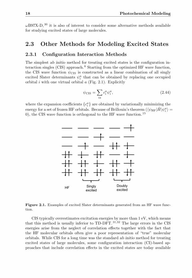
The simplest ab initio method for treating excited states is the configuration in-teraction singles (CIS) approach.6 Starting from the optimized HF wave function,the CIS wave function ψCIS is constructed as a linear combination of all singlyexcited Slater determinants ψai that can be obtained by replacing one occupiedorbital i with one virtual orbital a (Fig. 2.1). Explicitly
ψCIS =∑ia
cai ψai , (2.44)
where the expansion coefficients {cai } are obtained by variationally minimizing the
energy for a set of frozen HF orbitals. Because of Brillouin’s theorem (〈ψHF|H|ψai 〉 =0), the CIS wave function is orthogonal to the HF wave function.15
HF Singly excited
Doubly excited
Figure 2.1. Examples of excited Slater determinants generated from an HF wave func-tion.
CIS typically overestimates excitation energies by more than 1 eV, which meansthat this method is usually inferior to TD-DFT.31,32 The large errors in the CISenergies arise from the neglect of correlation effects together with the fact thatthe HF molecular orbitals often give a poor representation of “true” molecularorbitals. While CIS for a long time was the standard ab initio method for treatingexcited states of large molecules, some configuration interaction (CI)-based ap-proaches that include correlation effects in the excited states are today available
2.4 Relativistic Effects 19
for modeling large systems such as the CIS with perturbation-theory treatment ofdoubles corrections (CIS(D)) approach.33
2.3.2 Coupled Cluster Methods
Another family of methods available for calculating excited states are those basedon coupled cluster (CC) theory. These methods can be classified into differentgroups like the symmetry-adapted cluster configuration interaction (SAC-CI),34
the equation-of-motion CC (EOM-CC)35 and the hierarchy of linear response CCx(CCS, CC2, CC3 ...) methods.36 Although the family of CC methods can sys-tematically be improved to yield very accurate results, only those that includeeffects from single and double excitations such as SAC-CI and CC2 can be usedfor calculations on large molecules. While SAC-CI is often the most sophisticatedCC method available for such systems, CC2 is among the cheapest methods ableto describe electron correlation in excited states.
2.3.3 Multiconfigurational Methods
The final family of ab initio methods that will be presented here are the multicon-figurational self-consistent field (MCSCF)-based methods. In general, a MCSCFwave function is constructed as a linear combination of excited Slater determinants(Fig. 2.1), and is obtained by simultaneously optimizing orbitals and expansioncoefficients. Since it is usually impossible to do so for all possible Slater determi-nants, only a subset of them is selected based on some method-dependent criteria.In the complete active space self-consistent field (CASSCF) method, for example,the linear combination of Slater determinants only includes those that arise froma user-specified selection of so-called active orbitals and electrons. This selectionhas to be governed by some a priori knowledge of the character of the state inquestion, which is a drawback and makes CASSCF a difficult method to use.
While CASSCF captures non-dynamical correlation, it gives a poor descrip-tion of dynamical correlation. To properly account for both dynamical and non-dynamical correlation, the CASSCF wave function can be used as a starting point(reference function) for carrying out complete active space second-order perturba-tion theory (CASPT2)16 calculations. CASPT2 is the most generally applicableand accurate method for the calculation of excited states of small molecular sys-tems. However, although it is regarded as the gold standard for such systems,it is challenging to obtain the same accuracy for large molecules for which largebasis sets and large active spaces can not be used. Therefore, if non-dynamicalcorrelation effects are of minor importance, SAC-CI often provides a much betteralternative.
2.4 Relativistic Effects
One of the most common approximations in quantum chemistry is that relativisticeffects are neglected. Although this is a reasonable approximation in many photo-chemical applications, for one of the systems in the present thesis (i.e., lumiflavin)
20 Photochemical Modeling
relativistic effects actually play a critical role for the observed photochemistry. Inthis section, a brief introduction to the field of relativistic quantum chemistry isgiven. For more complete accounts of relativistic quantum chemistry, the readeris referred to textbooks and reviews in the field such as Refs. 37–41.
2.4.1 The Dirac Equation
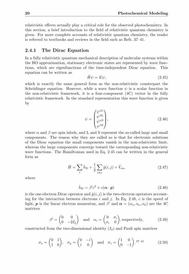
In a fully relativistic quantum mechanical description of molecular systems withinthe BO approximation, stationary electronic states are represented by wave func-tions, which are eigenfunctions of the time-independent Dirac equation. Thisequation can be written as
Hψ = Eψ, (2.45)
which is exactly the same general form as the non-relativistic counterpart theSchrodinger equation. However, while a wave function ψ is a scalar function inthe non-relativistic framework, it is a four-component (4C) vector in the fullyrelativistic framework. In the standard representation this wave function is givenby
ψ =
ψαL
ψβL
ψαS
ψβS
, (2.46)
where α and β are spin labels, and L and S represent the so-called large and smallcomponents. The reason why they are called so is that for electronic solutionsof the Dirac equation the small components vanish in the non-relativistic limit,whereas the large components converge toward the corresponding non-relativisticwave functions. The Hamiltonian used in Eq. 2.45 can be written in the generalform as
H =∑i
hD +1
2
∑i 6=j
g(i, j) + Ven, (2.47)
where
hD = β′c2 + c(α · p) (2.48)
is the one-electron Dirac operator and g(i, j) is the two-electron operators account-ing for the interaction between electrons i and j. In Eq. 2.48, c is the speed oflight, p is the linear electron momentun, and β′ and α = (αx, αy, αz) are the 4Cmatrices
β′ =
(0 00 −2I2
)and αi =
(0 σiσi 0
), respectively, (2.49)
constructed from the two-dimensional identity (I2) and Pauli spin matrices
σx =
(0 11 0
), σy =
(0 −ii 0
)and σz =
(1 00 −1
).37–41 (2.50)

2.4 Relativistic Effects 21
The relativistic effects to the two-electron interaction have two different ori-gins. First, the electromagnetic interactions are mediated by photons, which travelwith a finite velocity (i.e., the speed of light). This means that the interaction isretarded and that an electron will not feel the repulsion from another electroninstantaneously such as in the non-relativistic framework. Second, electrons aremoving charges and, accordingly, interact with the magnetic field generated by thecurrent and by the spins of the other electrons. In contrast to the one-electron partof the Dirac Hamiltonian, no analytic expression is available for g(i, j). However,this operator can be expanded in a perturbative series in c−2 as
g(i, j) =I4 · I4rij
− cαi · cαjc2rij
− (cαi · ∇i)(cαj · ∇j)rij2c2
+O(c−4), (2.51)
where the first term is the instantaneous Coulomb operator, which together withthe one-electron Dirac operator form the so-called Dirac-Coulomb Hamiltonian.37
This is one of most widely used 4C relativistic many-electron Hamiltonians incomputational photochemistry. As an example, a relativistic extension (4C-TD-DFT) of the non-relativistic TD-DFT approach has been derived starting fromthe Dirac-Coulomb Hamiltonian and implemented in relativistic quantum chemicalprograms.42–47 In these implementations, the working equations for the calculationof excitation energies have been obtained in a similar fashion (using responsetheory and the adiabatic approximation) as the corresponding non-relativistic TD-DFT equations outlined in Section 2.2.44–46
Although the Dirac-Coulomb Hamiltonian is sufficiently accurate for manychemical systems where relativistic effects come into play, some cases requiresthe inclusion of the second and third terms in Eq. 2.51, which are the so-calledGaunt and gauge operators. These two operators are here written in terms of therelativistic velocity operators cαi and cαj for particle i and j, respectively. Thesum of the Gaunt and gauge operators gives the so-called Breit operator, whichapproximately account for the (small) effects from the magnetic and retardedelectron-electron interactions.37
2.4.2 Approximate Relativistic Approaches
Despite the fact that much progress have been made in reducing the computationalcost in relativistic calculations based on the 4C Dirac equation described in theprevious section,48,49 such calculations are still much more demanding than thecorresponding non-relativistic calculations. For applications to medium- and large-scale molecular systems, a number of alternative approaches have therefore beendeveloped that account for relativistic effects in a more approximate way.
The first approximation one can make is to transform the fully relativistic4C Dirac Hamiltonian into a two-component Hamiltonian. By keeping only themost important terms, it is possible to effectivly reduce the complexity of thecomputational problem with relatively little loss in accuracy. Examples of two-component Hamiltonians are the zeroth-order regular approximation and Douglas-Kroll-Hess Hamiltonians.39,50,51
22 Photochemical Modeling
Further approximations can often be made for the modeling of organic sys-tems, where the key relativistic correction to the non-relativistic energy is usuallythe spin-orbit coupling. An alternative route for treating relativistic effects havetherefore evolved, which starts from the non-relativistic Schrodinger Hamiltonianand add a spin-orbit coupling operator as a perturbation. Among all spin-orbit op-erators reported in the literature, one may highlight the full one- and two-electronBreit-Pauli and Douglas-Kroll transformed spin-orbit coupling operators, as wellas the effective one-electron mean-field Hamiltonian.52
For the calculation of valence properties like covalent bonds and vibrationalfrequencies, further computational savings can be achieved by replacing the coreorbitals by a relativistic effective core potential. This is the most economic wayto include relativistic effects in the calculations. However, it is important makesure that the quality of the results is sufficiently accurate for the problem to besolved.53
2.5 Environmental Effects
So far, the present chapter has focused on the description of electronic structure,and particularly excited states, using quantum chemical methods, without andwith the inclusion of relativistic effects. Using such methods, it is possible to givea detailed description of the electronic structure of the system at hand. However,such methods are often restricted to molecules consisting of up to a few hundreds ofatoms. This means that unless new approximations are introduced, there are manymolecular systems of interest such as proteins that are impossible to study withcontemporary computers. In order to afford a detailed description of electronicprocesses in large molecular systems, one common approach is to use multiscalemodels, in which the full system is divided into a number of subsystems treatedat different levels of theory.
In this section, two multiscale models will be introduced. The first part is con-cerned with one particular family of continuum solvation models called polarizablecontinuum models, which are typically used to describe solvent effects. The sec-ond part is concerned with the hybrid quantum mechanics/molecular mechanicsapproach, which is widely used for modeling biological systems such as proteins.
2.5.1 Polarizable Continuum Models
In a polarizable continuum model (PCM), a solvated molecule is described at aquantum mechanical (QM) level of theory inside a cavity surrounded by a polariz-able dielectric continuum (characterized by a dielectric constant ε) that representsthe solvent (Fig. 2.2). In such a model, the solute induces a charge distributionon the cavity surface, which in turn interacts with the solute and polarizes itswave function. A PCM description of the solvent enables calculation of solvatedmolecules at a low computational cost but only captures bulk electrostatic ef-fects. If specific solute-solvent interactions are important, the solute model can beexpanded to a supermolecule by adding explicit solvent molecules.40
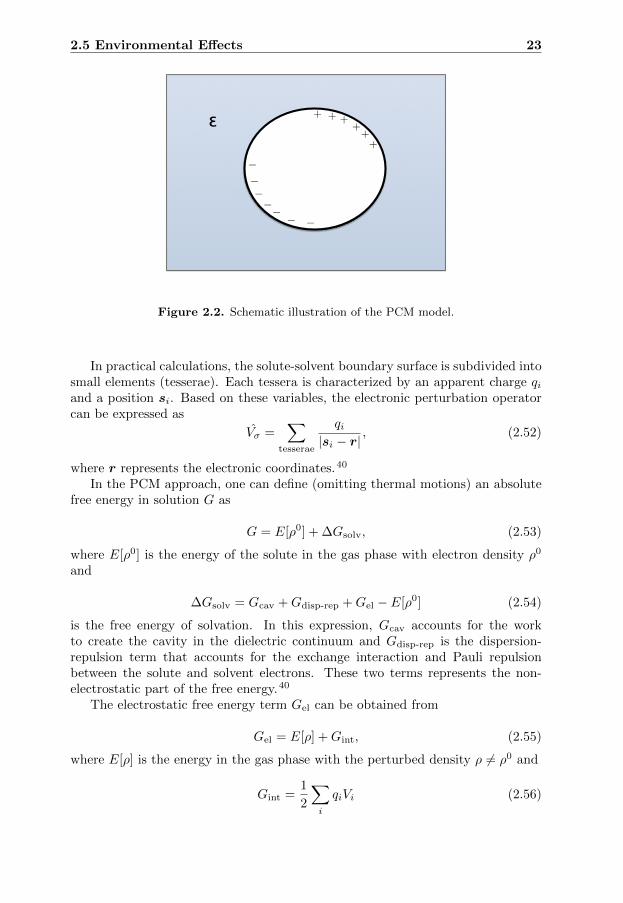
2.5 Environmental Effects 23
+ + + + + +
–
– –
– –
– –
ε
Figure 2.2. Schematic illustration of the PCM model.
In practical calculations, the solute-solvent boundary surface is subdivided intosmall elements (tesserae). Each tessera is characterized by an apparent charge qiand a position si. Based on these variables, the electronic perturbation operatorcan be expressed as
Vσ =∑
tesserae
qi|si − r|
, (2.52)
where r represents the electronic coordinates.40
In the PCM approach, one can define (omitting thermal motions) an absolutefree energy in solution G as
G = E[ρ0] + ∆Gsolv, (2.53)
where E[ρ0] is the energy of the solute in the gas phase with electron density ρ0
and
∆Gsolv = Gcav +Gdisp-rep +Gel − E[ρ0] (2.54)
is the free energy of solvation. In this expression, Gcav accounts for the workto create the cavity in the dielectric continuum and Gdisp-rep is the dispersion-repulsion term that accounts for the exchange interaction and Pauli repulsionbetween the solute and solvent electrons. These two terms represents the non-electrostatic part of the free energy.40
The electrostatic free energy term Gel can be obtained from
Gel = E[ρ] +Gint, (2.55)
where E[ρ] is the energy in the gas phase with the perturbed density ρ 6= ρ0 and
Gint =1
2
∑i
qiVi (2.56)

24 Photochemical Modeling
is the electrostatic solute-solvent interaction free energy. Here, Vi is the totalpotential generated by the solute (both electrons and nuclei) in tessera i. The threeimportant variables ρ, qi and Vi can be determined by variationally minimizing
Gel = 〈ψ|H0 +1
2Vσ|ψ〉 (2.57)
subject to some model specific boundary conditions, where H0 is the gas-phaseHamiltonian. The free energy can thereafter be obtained.40
Non-Equilibrium Solvation in TD-DFT
The dynamic nature of the solvent plays an important role in many photophysicaland photochemical processes such as photon absorption and emission. Duringsuch processes, solvent electronic (fast) degrees of freedom have time to respondto the change in solute electron density, whereas solvent nuclear (slow) degrees offreedom are delayed or even frozen. In order to simulate such a non-equilibriumsituation, each apparent tessera charge qi is divided into a fast
qi,f =εf − 1
ε− 1qi (2.58)
and a slow component
qi,s =ε− εfε− 1
qi, (2.59)
where εf 6= ε is the dielectric constant related to the fast degrees of freedom. In TD-DFT calculations of excited states of solvated molecules, non-equilibrium solvationcan be modeled within either the linear-response (LR) or the state-specific (SS)formalism.40
Within the LR formalism, the time-dependent KS equations are modified byadding a perturbation vPCM(ε). The perturbed KS equations then take the form
[H0KS + vPCM(ε)]φ(r, t) = i
∂
∂tφ(r, t), (2.60)
where H0KS is the time-dependent KS operator in the gas phase. While the vPCM
potential depends on ε if both fast and slow degrees of freedom have time to relax(equilibrium solvation), it depends on εf only if only the fast degrees of freedomhas time to relax (non-equilibrium solvation).54
In the SS implementation of PCM/TD-DFT, the electrostatic interaction partof the free energy of the excited state explicitly depends on the ground-state densityand is given by the expression
G(2)int,SS,neq =
1
2
∑i
q(2)i,f V
(2)i,ρ +
(∑i
q(1)i,s V
(2)i,ρ −
1
2
∑i
q(1)i,s V
(1)i,ρ
)
+1
2
(∑i
q(1)i,s V
(2)i,f −
∑i
q(1)i,s V
(1)i,s
), (2.61)
2.5 Environmental Effects 25
where (2) and (1) represent the excited and ground states, respectively. In Eq. 2.61,some of the potentials and tessera charges depend on the excited state electrondensity and, accordingly, these have to be determined using an iterative proce-dure. Furthermore, Vf and Vs correspond to the potentials relative to the fast and
slow degrees of freedom, respectively, and V(n)ρ is the potential generated by the
density of state (n). Due to the iterative procedure, the SS method is often morecomputationally demanding than the LR approach. However, at least for polarsolvents, the SS approach usually give more accurate results.55
2.5.2 QM/MM Methods
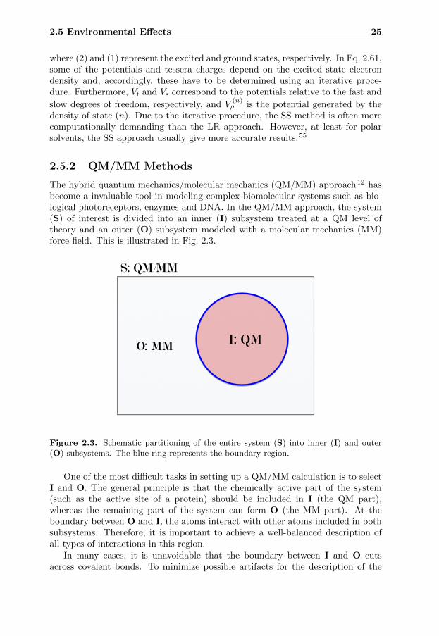
The hybrid quantum mechanics/molecular mechanics (QM/MM) approach12 hasbecome a invaluable tool in modeling complex biomolecular systems such as bio-logical photoreceptors, enzymes and DNA. In the QM/MM approach, the system(S) of interest is divided into an inner (I) subsystem treated at a QM level oftheory and an outer (O) subsystem modeled with a molecular mechanics (MM)force field. This is illustrated in Fig. 2.3.
S: QM/MM
I: QM O: MM
Figure 2.3. Schematic partitioning of the entire system (S) into inner (I) and outer(O) subsystems. The blue ring represents the boundary region.
One of the most difficult tasks in setting up a QM/MM calculation is to selectI and O. The general principle is that the chemically active part of the system(such as the active site of a protein) should be included in I (the QM part),whereas the remaining part of the system can form O (the MM part). At theboundary between O and I, the atoms interact with other atoms included in bothsubsystems. Therefore, it is important to achieve a well-balanced description ofall types of interactions in this region.
In many cases, it is unavoidable that the boundary between I and O cutsacross covalent bonds. To minimize possible artifacts for the description of the
26 Photochemical Modeling
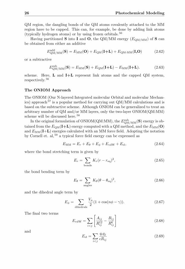
QM region, the dangling bonds of the QM atoms covalently attached to the MMregion have to be capped. This can, for example, be done by adding link atoms(typically hydrogen atoms) or by using frozen orbitals.56
Having partitioned S into I and O, the QM/MM energy (EQM/MM) of S canbe obtained from either an additive
EaddQM/MM(S) = EMM(O) + EQM(I+L) + EQM-MM(I,O) (2.62)
or a subtractive
EsubQM/MM(S) = EMM(S) + EQM(I+L)− EMM(I+L). (2.63)
scheme. Here, L and I+L represent link atoms and the capped QM system,respectively.56
The ONIOM Approach
The ONIOM (Our N-layered Integrated molecular Orbital and molecular Mechan-ics) approach57 is a popular method for carrying out QM/MM calculations and isbased on the subtractive scheme. Although ONIOM can be generalized to treat anarbitrary number of QM and/or MM layers, only the two-layer ONIOM(QM:MM)scheme will be discussed here.58
In the original formulation of ONIOM(QM:MM), the EsubQM/MM(S) energy is ob-
tained from the EQM(I+L) energy computed with a QM method, and the EMM(O)and EMM(I+L) energies calculated with an MM force field. Adopting the notationby Cornell et. al,59 a typical force field energy can be expressed as
EMM = Er + Eθ + Eφ + EvdW + Eel, (2.64)
where the bond stretching term is given by
Er =∑
bonds
Kr(r − req)2, (2.65)
the bond bending term by
Eθ =∑
angles
Kθ(θ − θeq)2, (2.66)
and the dihedral angle term by
Eφ =∑
dihedrals
Vn2
(1 + cos(nφ− γ)). (2.67)
The final two terms
EvdW =∑i<j
[AijR12ij
− BijR6ij
](2.68)
andEel =
∑i<j
qiqjεRij
(2.69)

2.5 Environmental Effects 27
account for the van der Waals and the electrostatic interactions, repectively. Inthe original ONIOM approach, the electrostatic coupling term between the I andO regions is treated at the MM level of theory, which means that the QM electrondensity and nuclear charges has been replaced by fixed atomic charges (so-calledmechanical embedding). Normally, this is a very crude approximation for photo-chemical reactions.
A more accurate way of treating the electrostatic QM-MM coupling term isprovided by the electrostatic embedding (EE) scheme.60 In this embedding scheme,point charges (normally taken from the MM force field) are incorporated into theQM Hamiltonian, which is thus augmented by a term
HEE = −N∑i=1
M ′∑J=1
sJqJ|RJ − ri|
+
M∑I=1
M ′∑J=1
ZIsJqJ|RJ −RI |
. (2.70)
In this expression, the scaling factor sJ prevents severe overpolarization of the QMwave function, qJ is the MM point charge located at RJ , ZI is the nuclear chargeof QM nucleus I, and RI and ri represent the nuclear and electronic coordinates,respectively. The indices i, I and J run over the N QM electrons, the M QMnuclei, and the M ′ MM atoms. Since the additional term in the QM Hamiltoniancaptures the electrostatic interactions between the two subsystems, one has toremove the corresponding classical Coulomb interactions from the total energy.This is done by adding a classical Coulomb term to the EMM(I+L) energy, andthe modified Coulomb term then becomes
EEEMM(I+L) = EMM(I+L) +
M∑I=1
M ′∑J=1
qIsJqJ|RJ −RI |
. (2.71)
Here, the scaling factor sJ is identical to that in Eq. 2.70. Including electrostaticembedding, the QM/MM energy finally reads
Esub,EEQM/MM(S) = EMM(S) + EEE
QM(I+L)− EEEMM(I+L). (2.72)
Although electrostatic embedding is a substantial improvement compared withthe classical mechanical embedding scheme, the general issue concerning the com-patability between the MM charges and the QM electron density still remains.Inherent in the electrostatic embedding scheme is the assumption that the pointcharges give a good representation of the real charge distribution of the MM sys-tem. However, the MM point charges are typically designed together with all otherforce field parameters to give a balanced description of all types of interactions.Another potential source of concern is how the QM method responds to pointcharges. For example, it has been shown for rhodopsins that DFT methods arenot able to fully account for the electrostatic effects.61 Despite these concerns,the QM/MM methodology has today become the state-of-the-art computationaltechnique for modeling complex biomolecular systems.
28 Photochemical Modeling
2.6 Concluding Remarks
In this chapter, a number of methods have been presented that can be used to solvethe non-relativistic electronic Schrodinger equation (or the relativistic electronicDirac equation). Employing one of them to calculate the electronic energy of thesystem for various sets of nuclear coordinates, a stationary description of the mostrepresentative molecular geometries along a PES can be obtained. While such ananalysis is sufficient in many photochemical applications, others requires that alsothe dynamics of the nuclei are taken into account. Although this thesis is concernedwith solving the (static) electronic problem, it is nevertheless worth mentioningthat there are of course methods available for studying the nuclear problem suchas trajectory surface hopping methods,62 where the nuclei are propagated on asurface according to Newton’s equations of motions and the system is able toswitch between different electronic states.
CHAPTER 3
Photobiological Keto-Enol Reactions
Many biological systems rely on a careful control of the relative concentrationsof keto and enol forms (usually referred to as keto and enol tautomers) of keymolecules. DNA, for example, is exclusively built from nucleobases in their ketotautomeric forms, and the conversion of a single base into its enol form is a com-mon source of mutations.63 The underlying control is in many cases executedby enzymes that are able to rapidly adjust the keto and enol concentrations bycatalyzing the interconversion of the two forms.64
Following an introduction to keto-enol tautomerism, this chapter presents myresearch on two photobiologically relevant organic chromophores for which keto-enol reactions play important roles. The two chromophores are astaxanthin andoxyluciferin. Astaxanthin is responsible for the color of lobster carapace and oxy-luciferin is responsible for the light emission of fireflies.
3.1 Keto-Enol Tautomerism
R3
OR1
HR2
R3
OHR1
R2
α
keto form enol form
Figure 3.1. Keto-enol tautomerism.
The conversion of the parent keto form of a carbonyl containing organic molecule
29

30 Photobiological Keto-Enol Reactions
(a ketone or aldehyde) into its enol form involves the shuttling of an hydrogenatom (or a proton) from the α-carbon to the carbonyl oxygen, and is accompaniedby the saturation of the carbonyl bond by an adjacent single bond (Fig. 3.1). Thisconversion can be catalyzed by either an acid or a base. The acid-catalyzed keto-enol tautomerism (Fig. 3.2) is initiated by protonation of the carbonyl oxygenatom, which activates the α-hydrogens. Thereafter, the resulting intermediateloses one of the activated hydrogens to yield the enol form. In most cases, thesecond step is slower than the first.65–67
R3
OR1
HR2
keto form
R3
OHR1
R2
enol form
A
HA
H
R3
OR1
HR2
H
A
R3
OR1
HR2
keto form
R3
OHR1
R2
enol form
A
HA
H
R3
OR1
HR2
H
A
Figure 3.2. Acid-catalyzed keto-enol tautomerism.
The base-catalyzed keto-enol tautomerism (Fig. 3.3), in turn, starts with de-protonation of the acidic α-carbon to give an enolate intermediate, followed byprotonation of the carbonyl oxygen to produce the enol form.65–67 Since the eno-late form is resonance-stabilized, a carbonyl compound is usually a much strongercarbon acid than is an alkane. As an illustrative example, the pKa value for ace-tone is ∼ 40 units smaller than the pKa value for ethane (Fig. 3.4).67 Analogous tothe acid-catalyzed reaction, it is the depronation of the α-carbon that determinesthe rate for the overall base-catalyzed reaction.65–67
R3
OR1
HR2
R3
OR1
R2
keto form enolate
R3
OHR1
R2
enol form
B
B H
B
Figure 3.3. Base-catalyzed keto-enol tautomerism.
There are many factors that affect the equilibrium between keto and enol tau-tomers. Although a carbonyl group is intrinsically more stable than an enol group,the possibility of additional stabilization effects from other functional groups mayshift the equilbrium in favor of the enol tautomer.68–70 Phenol, for example, is

3.1 Keto-Enol Tautomerism 31
OH
HH
HHH
H
HHH
HH
acetone ethane
Figure 3.4. Chemical structures of acetone and ethane.
much more stable than the corresponding keto tautomer (2,4-cyclohexadienone)because of aromaticity (Fig 3.5).65,66 Vinyl alcohol, which is stabilized by bothresonance and intramolecular hydrogen bonding, is another stable enol (Fig 3.5).In fact, while the parent keto acetylacetone form is slightly more stable than vinylalcohol in aqueous solution, the situation is actually reversed in nonpolar solventslike tetrachloromethane that promotes intramolecular interactions.71 Other fac-tors that may shift the equilibrium toward the enol form (or the keto form) areelectron withdrawing/donating groups, steric effects, and temperature.65–67
O OH
OO O OH
2,4-cyclohexadienone phenol
acetylacetone vinyl alcohol
Figure 3.5. Chemical structures of 2,4-cyclohexadienone, phenol, acetylactone and vinylalcohol.
Since the tautomeric equilibria in excited states can be very different fromthose in the ground state, it is in many cases possible to induce an isomerizationbetween two tautomeric forms by light.72–79 Already in 1961, Yang and Rivasshowed that the keto form of ortho-methylbenzophenone (Fig. 3.6) can undergoexcited-state tautomerism into the corresponding enol form in alcohol solutions.Furthermore, Naumov and co-workers have recently used time-resolved emissionspectroscopy to show that photoexcitation can induce enolization of the keto formof 6’-dehydroxyoxyluciferin (Fig. 3.6).79 Several examples of photoketonizationhave also been reported in the literature.75–78 For example, Yankov et al. haveshown that the keto form of dibenzoylmethane (Fig. 3.6), which is unstable in theground state, is formed upon photoexcitation.76
After this brief introduction to keto-enol tautomerism, we will in the followingtwo sections describe two organic chromophores that can exist in a number of
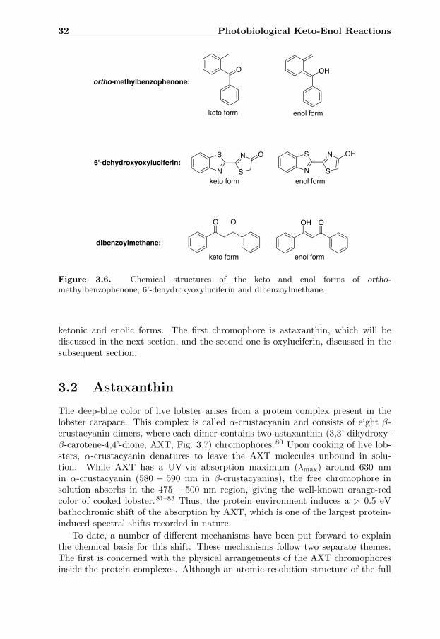
32 Photobiological Keto-Enol Reactions
O OH
N
S
S
N O
N
S
S
N OH
O O OH O
keto form enol form
ortho-methylbenzophenone:
keto form enol form
6'-dehydroxyoxyluciferin:
dibenzoylmethane:
keto form enol form
Figure 3.6. Chemical structures of the keto and enol forms of ortho-methylbenzophenone, 6’-dehydroxyoxyluciferin and dibenzoylmethane.
ketonic and enolic forms. The first chromophore is astaxanthin, which will bediscussed in the next section, and the second one is oxyluciferin, discussed in thesubsequent section.
3.2 Astaxanthin
The deep-blue color of live lobster arises from a protein complex present in thelobster carapace. This complex is called α-crustacyanin and consists of eight β-crustacyanin dimers, where each dimer contains two astaxanthin (3,3’-dihydroxy-β-carotene-4,4’-dione, AXT, Fig. 3.7) chromophores.80 Upon cooking of live lob-sters, α-crustacyanin denatures to leave the AXT molecules unbound in solu-tion. While AXT has a UV-vis absorption maximum (λmax) around 630 nmin α-crustacyanin (580 − 590 nm in β-crustacyanins), the free chromophore insolution absorbs in the 475 − 500 nm region, giving the well-known orange-redcolor of cooked lobster.81–83 Thus, the protein environment induces a > 0.5 eVbathochromic shift of the absorption by AXT, which is one of the largest protein-induced spectral shifts recorded in nature.
To date, a number of different mechanisms have been put forward to explainthe chemical basis for this shift. These mechanisms follow two separate themes.The first is concerned with the physical arrangements of the AXT chromophoresinside the protein complexes. Although an atomic-resolution structure of the full

3.2 Astaxanthin 33
OHOH
HOH
1234 56
7
89
1'2'3'4'5'
6'
7'
8'9'
O
Figure 3.7. Chemical structure of the neutral α-hydroxyketone form of astaxanthin(AXT).
α-crustacyanin complex is not available, Helliwell and co-workers84 have reportedX-ray crystallographic data of a β-crustacyanin that reveal that the binding ofAXT to the apoprotein both changes the conformation of the chromophore from6-s-cis to 6-s-trans, and reduces the twist of the β-ionone end-rings relative tothe polyene chains about the C6-C7 (and C6’-C7’) bonds (see atom numbering inFig. 3.7). Since co-planarization of the end-rings with the polyene chain increasesthe effective conjugation of the chromophore, it follows that the absorption shouldbe bathochromically shifted in the protein. The β-crustacyanin crystal structurealso shows that the two AXT chromophores are paired with their polyene chainslying quite close to each other (∼ 7 A), which would facilitate an exciton-couplingmechanism. However, both computational and experimental studies have shownthat neither conformational changes nor exciton coupling can account for morethan one third of the observed shift.85–87
The second theme, which was suggested already in 1938 by Kuhn and Sorensen,88
is concerned with the keto-enol and acid-base chemistry of AXT. Specifically, al-though the neutral α-hydroxyketone form of AXT (1 in Fig. 3.8) is predominant insolution, these authors suggested that the shift arises as a consequence of the biasof the apoprotein to rather bind AXT in its anionic enolate form (1−). While sucha mechanism has remained largely neglected, Paper I presents experimental andcomputational results showing that this scenario is indeed possible and associatedwith a large bathochromic shift.
3.2.1 Paper I
In order to evaluate the possibility that the bathochromic shift is due to the pres-ence of AXT in its enolate form 1− in α-crustacyanin, the absorption maximum ofthe parent keto form 1 was first calculated with TD-DFT employing three differ-ent functionals (M06-HF, LC-ωPBE and LC-BLYP) and two different basis sets(6-31G(d,p) and aug-cc-pVDZ) in water and methanol solutions. These resultswere then compared with the corresponding absorption maxima obtained for themono-enol (1ed), di-enol (1ded), enolate (1−) forms shown in Fig. 3.8.

34 Photobiological Keto-Enol Reactions
OHOH
HOH
123
4 56
7
89
1'2'3'4'5'
6'
7'
8'9'
C40H52O4
O
HOH
C18H22
1O
OH
1
H+ HA
OH
C18H22
O
OH
1
H
OH
C18H22
O
OH
1
H
+HB
O O+HB
HA HA
B: +
OH
C18H22
O
OHH
+ HA
B: +
HO
OH
C18H22
+ HA
B: +HO
OHOH
1ed
1ded
III
O
Figure 3.8. Chemical structure of the neutral α-hydroxyketone form of AXT (1) andacid-base (HA and B) induced tautomerism of this form producing the enolate (1−),mono-enol (1ed) and di-enol (1ed) forms. Reprinted from Paper I with permission fromthe PCCP Owner Societies.

3.2 Astaxanthin 35
Table 3.1. Calculated absorption maxima (λmax, in nm), excitation energies (∆E, ineV) and oscillator strengths (f) of different forms of AXT.a Reprinted from Paper Iwith permission from the PCCP Owner Societies.
Method/basis setb 1 1ed 1ded 1−
Water solventM06-HF/I λmax 457 463 459 660
∆E 2.71 2.68 2.70 1.88f 4.39 4.48 4.56 4.04
LC-ωPBE/I λmax 473 482 477 658∆E 2.62 2.57 2.60 1.88f 4.53 4.64 4.73 4.43
LC-BLYP/I λmax 463 470 465 643∆E 2.68 2.64 2.66 1.93f 4.59 4.70 4.79 4.45
M06-HF/IIc λmax 470 476 471 666∆E 2.64 2.60 2.63 1.86f 4.31 4.37 4.44 4.17
LC-ωPBE/IIc λmax 482 490 484 665∆E 2.57 2.53 2.56 1.86f 4.47 4.55 4.64 4.45
LC-BLYP/IIc λmax 474 481 475 651∆E 2.62 2.58 2.61 1.90f 4.52 4.61 4.69 4.50
Methanol solventM06-HF/I λmax 456 463 459 668
∆E 2.72 2.68 2.70 1.86f 4.39 4.48 4.56 4.05
LC-ωPBE/I λmax 473 481 477 666∆E 2.62 2.58 2.60 1.86f 4.52 4.64 4.73 4.45
LC-BLYP/I λmax 463 470 465 651∆E 2.68 2.64 2.67 1.90f 4.59 4.70 4.79 4.46
MixtureExperimentd λmax 476 − − −
∆E 2.61 − − −εemax 10000 − − −
aAll calculations based on geometries optimized using B3LYP/6-31G(d,p) in com-bination with a CPCM description of the water or methanol solvent. bBasis set I= 6-31G(d,p); basis set II = aug-cc-pVDZ. cSolvent effects obtained using the cc-pVDZ basis set and added to gas-phase data obtained with the target aug-cc-pVDZbasis set. dExperimental value in a water-methanol mixture. eεmax given in units ofdm3 mol−1 cm−1
Starting with the parent 1 species, we note from Table 3.1 that all three den-sity functionals used for the calculations give very similar results, and that theexperimental value (476 nm) obtained in a water-methanol mixture is reproduced

36 Photobiological Keto-Enol Reactions
with an accuracy of ∼ 20 nm (∼ 0.1 eV). Turning to the calculated absorptionmaxima of the other forms of AXT, it can be seen that neither single (1→ 1ed) nordouble (1 → 1ded) enolization is likely to significantly shift the absorption of 1.Indeed, regardless of the level of theory, it is predicted that such processes induceshifts smaller than 0.05 eV. Notably, however, the calculations predict that theconversion from 1 to 1− is associated with a bathochromic shift in the 0.72− 0.86eV range. Interestingly, this shift is larger than that in β-crustacyanin and sim-ilar to that in α-crustacyanin. This gives support for the hypothesis that thebathochromic shift in α-crustacyanin is due to the presence of AXT in its enolateform 1−. However, how can this result be validated experimentally?
O
HOH
1234 56
7
89
C13H18O3
2
O
O
OH
O
OH
B:
BH+
B:
BH+
B: BH+
O
OH
O
O
Ka(1) Ka(2)
2-dianion
2ed
HO
O
O
O
O
O
O2
Figure 3.9. Chemical structures of 3-hydroxy-4-oxo-β-ionone 2 and its ene-diol tau-tomer 2ed, enolate anion 2− and dianion 2-dianion. Reprinted from Paper I withpermission from the PCCP Owner Societies.
Since similar aqueous solution experiments are not possible, due to aggre-gate formation and the low solubility of AXT in water, the behavior of a water-soluble 3-hydroxy-4-oxo-β-ionone model of AXT (2) was examined using bothexperimental and computational methods. Importantly, the acid-base chemistryof this model compound shown in Fig. 3.9, which retains the trimethylated α-hydroxycyclohexenone of AXT but replaces the rest of the molecule with a simplerconjugated ketone at the 6-position of the ring, is expected to mimic that of AXT.
Comparing the calculated absorption maxima of the various forms of the modelcompound (2, 2ed, 2− and 2-dianion, shown in Fig. 3.9) with the correspondingexperimental maxima, it was found that the calculations accurately reproducethe experimental values for 2 and 2-dianion, and are in qualitative agreementwith the experiments in that the formation of the enolate 2− anion from 2 is

3.2 Astaxanthin 37
associated with a substantial bathochromic shift. These results, together withcomplementary benchmark calculations using CC2 and CASPT2, suggest thatTD-DFT calculations constitute a reliable tool for obtaining a qualitative accurateassessment of how acid-base chemistry influences the absorption maxima of AXTand models thereof.
Having found that the formation of 1− induces a large bathochromic shift, itis of interest to investigate whether this process is at all possible in crustacyanins.While a detailed analysis of the stability of the various forms of AXT in differentenvironments is beyond the scope of Paper I, both calculations and experimentsfound that the pKa of 2 is∼ 10. Since the conjugation is more extensive in 1− thanin 2−, the former anion should be more stable and present at even lower pH. Inthis regard, it is worth noticing that much simpler compounds also having enolateions in which the negative charge is distributed between two oxygen atoms suchas 2,3-dihydroxy-2-propenal (triose reductone) and ascorbic acid have pKa valuesbelow 7 (Fig. 3.10).89,90 Further support for the hypothesis that the 1− form isbound to crustacyanins comes from preliminary B3LYP calculations showing thata key histidine residue shifts the equilibrium between 1− and 1 by at least 8 kJmol− toward the former species. Overall, then, the calculations and experimentspresented in Paper I show that the formation of 1− from 1 is associated with alarge bathochromic shift and is possible in the protein environment.
HO O
H
OH2,3-dihydroxy-2-propenal
O O
H
OHO O
H
OH
O O
OHHO
HO
HOO O
OHO
HO
HOO O
OHO
HO
HO
pKa = 5.2
ascorbic acid
pKa = 4.1
Figure 3.10. Chemical structures of 2,3-dihydroxy-2-propenal and ascorbic acid, andtheir conjugate bases.

38 Photobiological Keto-Enol Reactions
3.3 Oxyluciferin
N
S N
S
O O
N
S N
S
O O
N
S N
S
O O
N
S N
S
O O
N
S N
S
O O
N
S N
S
O O
enol-OxyLH2keto-OxyLH2
phenolate-keto-OxyLH enolate-OxyLHphenolate-enol-OxyLH
OxyL2
pKa(S1)
pKE(S1)
H
HH
H
HpKE(S1)
pKa(S1)pKa(S1)
pKa(S1) pKa(S1)
Figure 3.11. Chemical structures of different forms of oxyluciferin and the excited-stateequilibrium constants for the keto-enol [pKE(S1)] and acid-base [pKa(S1)] reactions thatconnect them. Reprinted from Paper II with permission from John Wiley & Sons.Copyright 2014 Wiley Periodicals, Inc.
The characteristic glow of a firefly derives from a bioluminescent reaction thatoccurs in the catalytic center of the enzyme firefly luciferase. In this reaction, theground-state heterocyclic acid D-luciferin is converted into the chemiexcited (S1,first excited singlet state) oxyluciferin (OxyLH2) light emitter, which subsequentlyemits light.91,92
Depending on the chemical conditions, the OxyLH2 light emitter can exist inany of the six spectrally overlapping chemical forms shown in Fig. 3.11. Thereare two neutral, three mono-anionic and one di-anionic forms. The neutral formsare keto-OxyLH2 and enol-OxyLH2, the mono-anionic forms are phenolate-keto-OxyLH−, phenolate-enol-OxyLH− and enolate-OxyLH−, and the di-anionic formis OxyL2−.93,94
To date, there is no consensus regarding which of the different forms is themain contributor to the light emission in vivo. While both computational95–97 andexperimental98,99 studies have proposed that the phenolate-keto-OxyLH− form isthe preferred light emitter, Naumov and co-workers79 have recently studied anOxyLH2 analog (HOxyLH) and recorded time-resolved emission data indicatingthat it is rather an enolate species (enolate-OxyLH− or OxyL2−) that is responsiblefor the light emission.

3.3 Oxyluciferin 39
In Paper II, the intrinsic tendency of the light emitter to prefer one particularform of OxyLH2 was investigated by calculating the excited-state pKE and pKa
values in aqueous solution for the reactions (shown in Fig. 3.11) connecting thedifferent forms of OxyLH2.
3.3.1 Paper II
Figure 3.12. Structure of the phenolate-keto-OxyLH−-water complex considered.Reprinted from Paper II with permission from John Wiley & Sons. Copyright 2014Wiley Periodicals, Inc.
Using a thermodynamic Born-Haber (BH) cycle, pK values in ground [pKBH(S0)]and excited states [pKBH(S1)] were derived by calculating aqueous solution Gibbsfree energies of reactants (ketones/acids) and products (enols/bases) in the twostates, respectively. In order to minimize inevitable computational errors, thepK(S1) values, henceforth denoted pKBH,corr(S1), were obtained by adding cal-culated [pKBH(S1) − pKBH(S0)] values to experimental ground-state pK values[pKexp(S0)]
pKBH,corr(S1) = pKexp(S0) + [pKBH(S1)− (pKBH(S0)]. (3.1)
Experimental pKexp(S0) values were taken from Ref. 94 or derived from calculatedand experimental data. All calculations were carried out at the ωB97X-D/6-31+G(d,p) level of theory using a hybrid cluster-continuum model including 11explicit water molecules. The explicit water molecules were placed as shown forthe phenolate-keto-OxyLH− anion in Fig. 3.12. The computed pKBH,corr(S1) andexperimental pKexp(S0) are presented in Fig. 3.13.
Starting by comparing keto-OxyLH2 with enol-OxyLH2 (reaction I in Fig. 3.13),we observe that while they are of similar stability in the ground state (pKexp
E (S0) =
−0.39), the pKBH,corrE (S1) value of ∼ 5 is a clear indication that keto-OxyLH2 is
much more stable than enol-OxyLH2 in the excited state. We also observe that theestimated pKBH,corr
a (S1) value for the equilibrium reaction between keto-OxyLH2
and its conjugate base phenolate-keto-OxyLH− (reaction IV) is ∼ 2. Since thereference conditions implicated in the interpretation of pKa values in Paper IIcorrespond to a buffered aqueous solution at pH 7, it seems likely that phenolate-keto-OxyLH− is favored over keto-OxyLH2 in the excited state. Hence, neither

40 Photobiological Keto-Enol Reactions
0 2 4 6 8
10 12 14 16
0 1 2 3 4 5 6 7
-2
0
2
4
6
8
I II III
IV V VI VII VIII IX
pKEexp(S0)
pKEBH,corr(S1)
pKaexp(S0)
pKaBH,corr(S1)
pKE(S
n)
pKa(
Sn)
keto-enol reactions
acid-base reactions
I: keto-OxyLH2 enol-OxyLH2 II: phenolate-keto-OxyLH– enolate-OxyLH–
III: phenolate-keto-OxyLH– phenolate-enol-OxyLH–
IV: keto-OxyLH2 phenolate-keto-OxyLH– V: enol-OxyLH2 phenolate-enol-OxyLH– VI: enol-OxyLH2 enolate-OxyLH–
VII: phenolate-keto-OxyLH– OxyL2– VIII: phenolate-enol-OxyLH– OxyL2–
IX: enolate-OxyLH– OxyL2–
!!!
!!!
!!
!
Figure 3.13. Experimental ground-state and calculated excited-state equilibrium con-stants for the keto-enol and acid-base reactions of OxyLH2. Reprinted from Paper IIwith permission from John Wiley & Sons. Copyright 2014 Wiley Periodicals, Inc.
enol-OxyLH2 nor keto-OxyLH2 seems to be a significant contributor to the lightemission.
Having predicted that the neutral forms are of minor importance for the lightemission, we then proceeded by comparing the three mono-anionic forms. Asfor the phenolate-keto-OxyLH− enolate-OxyLH− equilibrium (reaction II),it can be seen that while the enolate-OxyLH− form dominates in the groundstate, the calculations suggest that it is actually phenolate-keto-OxyLH− thatis more stable in the excited state (pKBH,corr
E (S1) ∼ 6). For the phenolate-keto-OxyLH− phenolate-enol-OxyLH− equilibrium (reaction III), in turn, the
pKBH,corrE (S1) value of ∼ 7 indicates that the phenolate-keto-OxyLH− form is also
favored over this enolic mono-anion. Hence, out of the three mono-anionic forms,only phenolate-keto-OxyLH− appears to come into play.
The results presented above show that phenolate-keto-OxyLH− is intrinsi-cally more stable than keto-OxyLH2, enol-OxyLH2, phenolate-enol-OxyLH− andenolate-OxyLH− in the excited state in aqueous solution, which means that eitherphenolate-keto-OxyLH− or OxyL2− is the preferred excited-state species. Sincethe pKBH,corr
a (S1) value for the phenolate-keto-OxyLH− OxyL2− equilibrium(reaction VII) is ∼ 14, it seems likely that phenolate-keto-OxyLH− is also more

3.3 Oxyluciferin 41
stable than OxyL2− in the excited state. Thus, the key conclusion afforded by theexcited-state equilibrium constants reported in Paper II is that phenolate-keto-OxyLH− is intrinsically the preferred form of OxyLH2 in the excited state, whichsuggests that this species is involved in the bioluminescence of fireflies.

42 Photobiological Keto-Enol Reactions
CHAPTER 4
Biological Photoreceptors
Sunlight is an important energy source in Nature. For many organisms, sunlightis also an important environmental stimulus for activating physiological and de-velopmental processes. Specialized proteins, called biological photoreceptors, havetherefore evolved that detect light and respond to changes in the ambient lightenvironment. In this chapter, a general overview of these proteins will first begiven. Thereafter, my research related to the LOV (light-oxygen-voltage)-domainproteins and the phytochromes, which are two classes of photoreceptors, will bepresented.
4.1 Overview
Biological photoreceptors are involved in signal transduction and utilize light en-ergy to activate physiological and developmental processes in all kingdoms of life.Each photoreceptor uses a chromophore to detect light of a specific wavelengthwithin the UV to near-infrared region of the electromagnetic spectrum. The chro-mophore is either a small organic molecule or is formed from amino-acid residuesof the protein. As shown in Fig. 4.1 and Table 4.1, UVR8 (UV resistance locus 8)photoreceptors employ a tryptophan to respond to UV-B light; xanthopsins usecoumaric acid to sense blue light; cryptochromes as well as BLUF (blue-light sen-sors using flavin-adenine dinucleotide) and LOV-domain proteins employ a flavinchromophore to absorb blue light; rhodopsins use retinals to detect light in theUV to visible red spectral range; phytochromes bind a linear tetrapyrrole (bilin)chromophore to detect light in the red and near-infrared regions; and cyanobacte-riochromes finally contain a bilin chromophore that absorbs light of UV to near-infrared wavelengths.100–104
The biological activity of a photoreceptor depends on the relative concentra-
43

44 Biological Photoreceptors
300 400 500 600 700 800 900 λ/nm
UVR8 rhodopsins
xanthopsins LOV BLUF
cryptochromes
phytochromes
cyanobacteriochromes
Figure 4.1. Spectral sensitivity ranges for different classes of biological photoreceptors.
Table 4.1. Chromophores and primary photochemical events for different classes ofbiological photoreceptors.
class chromophore primary photochemicalevent
UVR8 tryptophana proton-coupledelectron transfera
xanthopsins coumaric acidb isomerizationb
cryptochromes flavin-adenine electron transferc
dinucleotide (FAD)c
BLUF flavin-adenine rearrangement ofdinucleotide (FAD)c hydrogen bondsc
LOV flavin mononucleotide FMN-cysteinyl(FMN)c adduct formationc
rhodopsins retinalb isomerizationb
phytochromes linear tetrapyrrole (bilin)d isomerizationd
cyanobacteriochrome linear tetrapyrrole (bilin)d isomerizationd
aRef. 103. bRef. 100. cRef. 102. dRef. 101.
tions of dark-adapted inactive and biologically active forms. Upon light absorptionby the inactive form, the photoreceptor undergoes a photocycle (Fig. 4.2), whichis started by a photochemical reaction of the chromophore (the so-called primaryphotochemical event, Table 4.1) and involves the formation of the biologically ac-tive form via a number of protein intermediate states. The active form, which istypically produced within microseconds after photoexcitation, passes on a signalto other regulatory systems that activate biological pathways in the host organ-ism leading to physiological responses.105 Since the active form is less stable thanthe inactive form, a thermally driven spontaneous process called dark reversionslowly (milliseconds to many hours) recovers the inactive form, which closes thephotocycle.104 In addition to the dark reversion, photochromic photoreceptors like
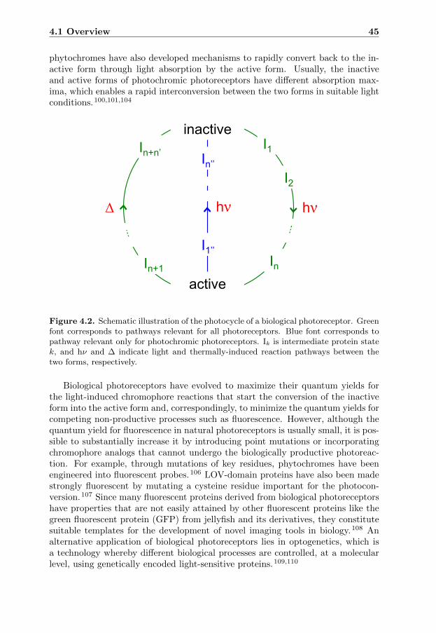
4.1 Overview 45
phytochromes have also developed mechanisms to rapidly convert back to the in-active form through light absorption by the active form. Usually, the inactiveand active forms of photochromic photoreceptors have different absorption max-ima, which enables a rapid interconversion between the two forms in suitable lightconditions.100,101,104
inactive
active
Δ hν hν
I1
In
I2
In+1
In+n’
I1’’
In’’
Figure 4.2. Schematic illustration of the photocycle of a biological photoreceptor. Greenfont corresponds to pathways relevant for all photoreceptors. Blue font corresponds topathway relevant only for photochromic photoreceptors. Ik is intermediate protein statek, and hν and ∆ indicate light and thermally-induced reaction pathways between thetwo forms, respectively.
Biological photoreceptors have evolved to maximize their quantum yields forthe light-induced chromophore reactions that start the conversion of the inactiveform into the active form and, correspondingly, to minimize the quantum yields forcompeting non-productive processes such as fluorescence. However, although thequantum yield for fluorescence in natural photoreceptors is usually small, it is pos-sible to substantially increase it by introducing point mutations or incorporatingchromophore analogs that cannot undergo the biologically productive photoreac-tion. For example, through mutations of key residues, phytochromes have beenengineered into fluorescent probes.106 LOV-domain proteins have also been madestrongly fluorescent by mutating a cysteine residue important for the photocon-version.107 Since many fluorescent proteins derived from biological photoreceptorshave properties that are not easily attained by other fluorescent proteins like thegreen fluorescent protein (GFP) from jellyfish and its derivatives, they constitutesuitable templates for the development of novel imaging tools in biology.108 Analternative application of biological photoreceptors lies in optogenetics, which isa technology whereby different biological processes are controlled, at a molecularlevel, using genetically encoded light-sensitive proteins.109,110
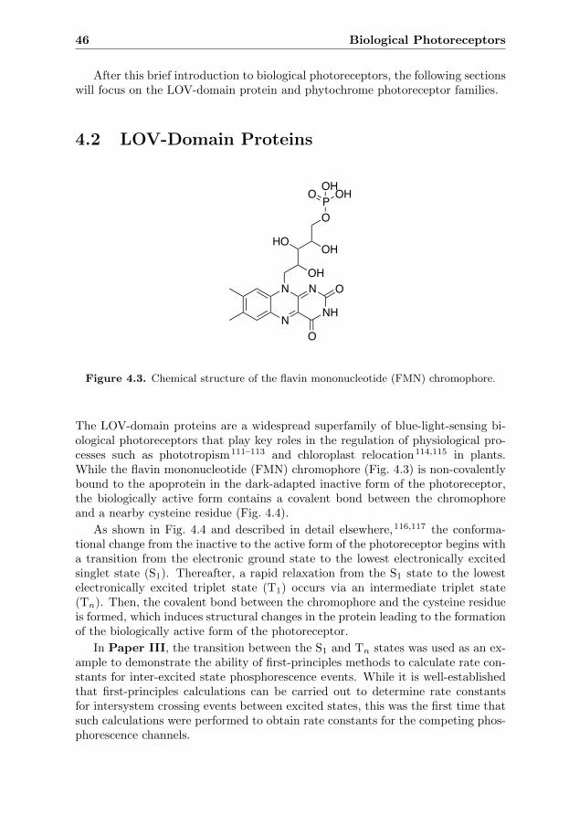
46 Biological Photoreceptors
After this brief introduction to biological photoreceptors, the following sectionswill focus on the LOV-domain protein and phytochrome photoreceptor families.
4.2 LOV-Domain Proteins
N
N
NH
N
O
OOH
HO OH
OP OHO OH
Figure 4.3. Chemical structure of the flavin mononucleotide (FMN) chromophore.
The LOV-domain proteins are a widespread superfamily of blue-light-sensing bi-ological photoreceptors that play key roles in the regulation of physiological pro-cesses such as phototropism111–113 and chloroplast relocation114,115 in plants.While the flavin mononucleotide (FMN) chromophore (Fig. 4.3) is non-covalentlybound to the apoprotein in the dark-adapted inactive form of the photoreceptor,the biologically active form contains a covalent bond between the chromophoreand a nearby cysteine residue (Fig. 4.4).
As shown in Fig. 4.4 and described in detail elsewhere,116,117 the conforma-tional change from the inactive to the active form of the photoreceptor begins witha transition from the electronic ground state to the lowest electronically excitedsinglet state (S1). Thereafter, a rapid relaxation from the S1 state to the lowestelectronically excited triplet state (T1) occurs via an intermediate triplet state(Tn). Then, the covalent bond between the chromophore and the cysteine residueis formed, which induces structural changes in the protein leading to the formationof the biologically active form of the photoreceptor.
In Paper III, the transition between the S1 and Tn states was used as an ex-ample to demonstrate the ability of first-principles methods to calculate rate con-stants for inter-excited state phosphorescence events. While it is well-establishedthat first-principles calculations can be carried out to determine rate constantsfor intersystem crossing events between excited states, this was the first time thatsuch calculations were performed to obtain rate constants for the competing phos-phorescence channels.
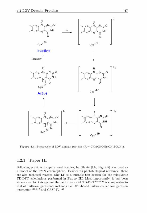
4.2 LOV-Domain Proteins 47
N
N
NH
N
O
OR
CysSH
N
N
NH
N
O
OR
CysSH
N
N
NH
N
O
OR
CysSH
NH
N
NH
N
O
OR
CysS
S1
Tn
hν
Recovery
N
N
NH
N
O
OR
CysSH
T1
NH
N
NH
N
O
OR
CysS
T1
Inactive
Active
Figure 4.4. Photocycle of LOV-domain proteins (R = CH2(CHOH)3CH2PO4H2).
4.2.1 Paper III
Following previous computational studies, lumiflavin (LF, Fig. 4.5) was used asa model of the FMN chromophore. Besides its photobiological relevance, thereare also technical reasons why LF is a suitable test system for the relativisticTD-DFT calculations performed in Paper III. Most importantly, it has beenshown that for this system the performance of TD-DFT118–120 is comparable tothat of multiconfigurational methods like DFT-based multireference configurationinteraction118,119 and CASPT2.121

48 Biological Photoreceptors
N
N
NH
N
O
O1 23
44a5
5a67
8 99a
1010a
Figure 4.5. Chemical structure of lumiflavin (LF). Reprinted from Paper III with per-mission from The Journal of Physical Chemistry A. Copyright 2015 American ChemicalSociety.
To identify the excited states relevant for the singlet and triplet photochemistryof LF, relativistic 4C-TD-DFT/B3LYP/aug-cc-pVDZ calculations were performedto obtain energies and oscillator strengths for transitions from the ground stateto low-lying electronically excited states. From the results presented in Table 4.2,we first note that the transition with the largest oscillator strength (f ∼ 10−1)is the transition to the 1(πHπ
∗L) state (i.e., S1). Since the oscillator strengths for
the transitions to the other excited states are at least two orders of magnitudesmaller (f ≤∼ 10−3), it seems very likely that 1(πHπ
∗L) is the initial absorbing
state. Furthermore, the calculations also predict that the lowest triplet state (i.e.,T1), wherein the chemical reaction with the cysteine residue occurs, is the 3(πHπ
∗L)
state. Finally, since the 3(nN2π∗L) and 3(πH−1π
∗L) states lie between 3(πHπ
∗L) and
1(πHπ∗L), it is possible that the relaxation of the absorbing state into the reactive
state proceeds via both or one of these states.
Table 4.2. Calculated 4C-TD-DFT energies (∆E, in eV) and oscillator strengths (f) fortransitions from the electronic ground state to low-lying electronically excited states ofLF.a Reprinted from Paper III with permission from The Journal of Physical ChemistryA. Copyright 2015 American Chemical Society.
state ∆E f
3(πHπ∗L) 2.04 7.306 · 10−10
3(nN2π∗L) 2.77 1.339 · 10−5
3(πH−1π∗L) 2.80 6.610 · 10−9
1(πHπ∗L) 2.96 1.930 · 10−1
3(nO2π∗L) 2.97 5.358 · 10−3
1(nO2π∗L) 3.12 2.053 · 10−4
1(nN2π∗L) 3.28 3.719 · 10−4
3(πH−2π∗L) 3.36 4.278 · 10−8
3(πHπ∗L+1) 3.58 9.614 · 10−9
aAll calculations carried out in the gas phase using B3LYP, the aug-cc-pVDZ basis set, and a ground-state geometry optimized in a non-relativistic framework.
Having found that the 3(πHπ∗L), 3(nN2π
∗L), 3(πH−1π
∗L) and 1(πHπ
∗L) states are
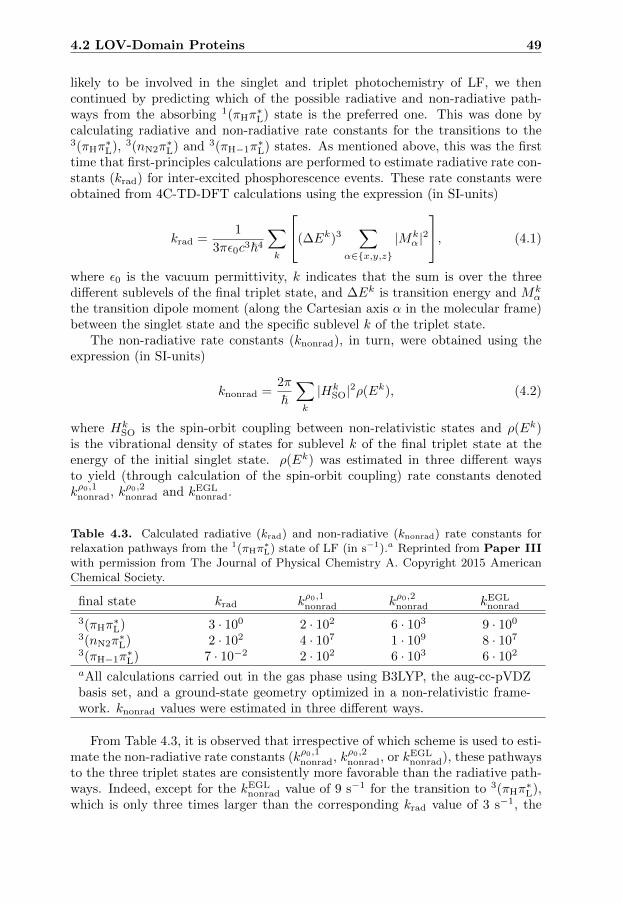
4.2 LOV-Domain Proteins 49
likely to be involved in the singlet and triplet photochemistry of LF, we thencontinued by predicting which of the possible radiative and non-radiative path-ways from the absorbing 1(πHπ
∗L) state is the preferred one. This was done by
calculating radiative and non-radiative rate constants for the transitions to the3(πHπ
∗L), 3(nN2π
∗L) and 3(πH−1π
∗L) states. As mentioned above, this was the first
time that first-principles calculations are performed to estimate radiative rate con-stants (krad) for inter-excited phosphorescence events. These rate constants wereobtained from 4C-TD-DFT calculations using the expression (in SI-units)
krad =1
3πε0c3~4
∑k
(∆Ek)3∑
α∈{x,y,z}
|Mkα|2, (4.1)
where ε0 is the vacuum permittivity, k indicates that the sum is over the threedifferent sublevels of the final triplet state, and ∆Ek is transition energy and Mk
α
the transition dipole moment (along the Cartesian axis α in the molecular frame)between the singlet state and the specific sublevel k of the triplet state.
The non-radiative rate constants (knonrad), in turn, were obtained using theexpression (in SI-units)
knonrad =2π
~∑k
|HkSO|2ρ(Ek), (4.2)
where HkSO is the spin-orbit coupling between non-relativistic states and ρ(Ek)
is the vibrational density of states for sublevel k of the final triplet state at theenergy of the initial singlet state. ρ(Ek) was estimated in three different waysto yield (through calculation of the spin-orbit coupling) rate constants denotedkρ0,1nonrad, kρ0,2nonrad and kEGL
nonrad.
Table 4.3. Calculated radiative (krad) and non-radiative (knonrad) rate constants forrelaxation pathways from the 1(πHπ
∗L) state of LF (in s−1).a Reprinted from Paper III
with permission from The Journal of Physical Chemistry A. Copyright 2015 AmericanChemical Society.
final state krad kρ0,1nonrad kρ0,2nonrad kEGLnonrad
3(πHπ∗L) 3 · 100 2 · 102 6 · 103 9 · 100
3(nN2π∗L) 2 · 102 4 · 107 1 · 109 8 · 107
3(πH−1π∗L) 7 · 10−2 2 · 102 6 · 103 6 · 102
aAll calculations carried out in the gas phase using B3LYP, the aug-cc-pVDZbasis set, and a ground-state geometry optimized in a non-relativistic frame-work. knonrad values were estimated in three different ways.
From Table 4.3, it is observed that irrespective of which scheme is used to esti-mate the non-radiative rate constants (kρ0,1nonrad, kρ0,2nonrad, or kEGL
nonrad), these pathwaysto the three triplet states are consistently more favorable than the radiative path-ways. Indeed, except for the kEGL
nonrad value of 9 s−1 for the transition to 3(πHπ∗L),
which is only three times larger than the corresponding krad value of 3 s−1, the
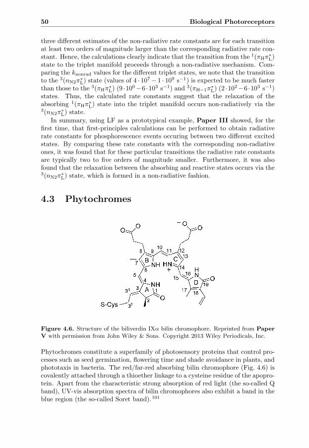
50 Biological Photoreceptors
three different estimates of the non-radiative rate constants are for each transitionat least two orders of magnitude larger than the corresponding radiative rate con-stant. Hence, the calculations clearly indicate that the transition from the 1(πHπ
∗L)
state to the triplet manifold proceeds through a non-radiative mechanism. Com-paring the knonrad values for the different triplet states, we note that the transitionto the 3(nN2π
∗L) state (values of 4 · 107− 1 · 109 s−1) is expected to be much faster
than those to the 3(πHπ∗L) (9 ·100−6 ·103 s−1) and 3(πH−1π
∗L) (2 ·102−6 ·103 s−1)
states. Thus, the calculated rate constants suggest that the relaxation of theabsorbing 1(πHπ
∗L) state into the triplet manifold occurs non-radiatively via the
3(nN2π∗L) state.
In summary, using LF as a prototypical example, Paper III showed, for thefirst time, that first-principles calculations can be performed to obtain radiativerate constants for phosphorescence events occuring between two different excitedstates. By comparing these rate constants with the corresponding non-radiativeones, it was found that for these particular transitions the radiative rate constantsare typically two to five orders of magnitude smaller. Furthermore, it was alsofound that the relaxation between the absorbing and reactive states occurs via the3(nN2π
∗L) state, which is formed in a non-radiative fashion.
4.3 Phytochromes
Figure 4.6. Structure of the biliverdin IXα bilin chromophore. Reprinted from PaperV with permission from John Wiley & Sons. Copyright 2013 Wiley Periodicals, Inc.
Phytochromes constitute a superfamily of photosensory proteins that control pro-cesses such as seed germination, flowering time and shade avoidance in plants, andphototaxis in bacteria. The red/far-red absorbing bilin chromophore (Fig. 4.6) iscovalently attached through a thioether linkage to a cysteine residue of the apopro-tein. Apart from the characteristic strong absorption of red light (the so-called Qband), UV-vis absorption spectra of bilin chromophores also exhibit a band in theblue region (the so-called Soret band).101

4.3 Phytochromes 51
Lumi-R
Pfr
Pr
hν
Lumi-F
Δ
dark reversion
S-Cys
NH
NH
OO
HN
OO
HNO
A
B C
D
+
O
S-Cys
NH
NH
OO
HN
OO
HN
OA
B C
D
+O
hν
Δ
Δ
Figure 4.7. Photocycle of phytochromes. hν and ∆ indicate light and thermally-inducedreaction pathways between the two forms, respectively.
The biological activity of phytochromes depends on the relative concentrationsof a red-absorbing (Pr) and a far-red-absorbing (Pfr) form. In most phytochromes,the Pr form is favored in the dark-adapted inactive state, whereas the Pfr form isfavored in the biologically active state.101 Absorption of red light by Pr triggersthe conversion of Pr into Pfr. This conversion begins with a Z → E photoiso-merization of the bilin chromophore and is followed by a number of thermal steps(Fig. 4.7). Although direct evidence is scarce, it is generelly accepted that thephotoisomerization involves a rotation of pyrrole ring D around the axis of theC15-C16 bond of the methine bridge between rings C and D. Support for such amechanism comes from, for example, X-ray crystallographic data and studies usinga variety of spectroscopic techniques such resonance Raman and magic angle spin-
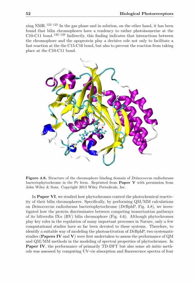
52 Biological Photoreceptors
ning NMR.122–125 In the gas phase and in solution, on the other hand, it has beenfound that bilin chromophores have a tendency to rather photoisomerize at theC10-C11 bond.126–129 Indirectly, this finding indicates that interactions betweenthe chromophore and the apoprotein play a decisive role not only to facilitate afast reaction at the the C15-C16 bond, but also to prevent the reaction from takingplace at the C10-C11 bond.
Figure 4.8. Structure of the chromophore binding domain of Deinococcus radioduransbacteriophytochrome in the Pr form. Reprinted from Paper V with permission fromJohn Wiley & Sons. Copyright 2013 Wiley Periodicals, Inc.
In Paper VI, we studied how phytochromes control the photochemical reactiv-ity of their bilin chromophores. Specifically, by performing QM/MM calculationson Deinococcus radiodurans bacteriophytochrome (DrBphP, Fig. 4.8), we inves-tigated how the protein discriminates between competing isomerization pathwaysof its biliverdin IXα (BV) bilin chromophore (Fig. 4.6). Although phytochromesplay key roles in the regulation of many important processes in Nature, only a fewcomputational studies have so far been devoted to these systems. Therefore, toidentify a suitable way of modeling the photoactivation of DrBphP, two systematicstudies (Papers IV and V) were first undertaken to assess the performance of QMand QM/MM methods in the modeling of spectral properties of phytochromes. InPaper IV, the performance of primarily TD-DFT but also some ab initio meth-ods was assessed by computing UV-vis absorption and fluorescence spectra of four

4.3 Phytochromes 53
synthetic bilin derivatives. In Paper V, in turn, we investigated how the choice ofQM/MM methodology affects the resulting UV-vis absorption spectra of DrBphP.
4.3.1 Paper IV
By performing TD-DFT calculations on four sterically locked bilin chromophores(denoted 5Za15Ea, 5Ea15Ea, 5Zs15Za, 15Za) in aqueous solution, we investi-gated how the choice of density functional affects the resulting absorption andfluorescence spectra. The calculations were performed with a set of representa-tive density functionals including BP86, BLYP and PBE (GGAs), τ -HCTH (ameta-GGA), B3LYP, BHHLYP, PBE0 (hybrid GGAs), M06-2X, M06-HF (hybridmeta-GGAs), LC-BLYP, LC-ωPBE, CAM-B3LYP and ωB97X-D (range-separatedhybrid GGAs). While this set comprises some widely used standard functionalslike B3LYP and PBE0, it also includes functionals that, at least in part, havebeen designed to provide a more accurate description of excited states such asCAM-B3LYP and LC-BLYP.
The structures of the chromophores studied are shown in Fig. 4.9. Absorp-tion maxima were estimated as vertical S0 → S1 excitation energies and fluo-rescence maxima as vertical S1 → S0 emission energies. These approximationswere, in a number of cases, validated by comparing S0 → S1 excitation ener-gies with the corresponding full absorption spectra obtained from a multitudeof Gaussian-convoluted transition energies. Based on optimized geometries usingB3LYP-PCM/SVP, absorption and fluorescence maxima were obtained at the TD-DFT/cc-pVDZ level of theory in combination with a state-specific PCM descrip-tion of the water solvent. While non-equilibrium solvation was used for calculatingabsorption maxima, equilibrium solvation was used for fluorescence maxima.
For the absorption maxima, it was found that for all four chromophores, alldensity functionals give results that are blue-shifted relative to corresponding ex-perimental values. Furthermore, it was also found that the pure GGAs (BP86,BLYP, PBE and τ -HCTH) exhibit better agreement with the experimental valuesthan the hybrid GGAs (BHHLYP, B3LYP and PBE0), as well as than the hy-brid meta-GGAs (M06-2X and M06-HF) and the range-separated hybrid GGAs(LC-BLYP, CAM-B3LYP and ωB97X-D). Specifically, while the pure GGA valuesdeviate from the experimental values by ∼0.15 eV, the values computed with theother functionals differ from the reference values by more than ∼0.3 eV. While thisobservation is not in line with how pure GGAs typically perform,32 the same trendwas actually observed in a QM study on the chemically related porphin molecule.8
For the fluorescence maxima, in turn, it was observed that also for these do thepure GGAs afford the best performance.
Although TD-DFT often yields accurate excitation energies for valence excitedstates, there are some well-known failures with this approach such as the treat-ment of states with double-excitation character. To investigate if such effects areimportant for bilin chromophores, complementary calculations were carried outwith the CASPT2 method. By analyzing the CASSCF wave function underlyingthe CASPT2 calculations, we observed that such effects are of minor importancefor the S1 states.

54 Biological Photoreceptors
NO
NH
NH
N
OH
A
B C
D
R R
+
5Za15Ea
1
23
45
6
789
1011
1213
14
15 16
17
18
19
2122
31
7172
131132
133
181
182
N
NH
NH
N
OH
A
B C
D
R R
+
5Ea15Ea
45
6
89
1011
1213
14
15 16
17
18
19
131132
133
181
182
OH
1
23
21
22
7
31
32 33
N NH N
A
B C
D
R R
5Zs15Za
89
1011
1213
14
15 16
7
O
N
O
+
12
331
3221
4
5
6
71131
132
1718
181182
171
1920
21
N NH N
B C
D
R R
15Za
89
1011
1213
14
15 16
7
O5
6
71131
132
1718
181182
171
19
H+
N
O
HA
12
21
331
32
4
Figure 4.9. Structures of the locked bilin chromophores considered (R =CH2CH2COOH). Reprinted from Paper IV with permission from Elsevier. Copyright2013 Elsevier B.V.

4.3 Phytochromes 55
To further assess the performance of the density functionals, we also comparedthe calculated Stokes shifts (difference between absorption and fluorescence max-ima) with the corresponding experimental values. From this analysis, we foundthat also in this case do the pure GGAs (Stokes shifts in the 0.07−0.10 eV range)show the best agreement with the experimental values (0.04 − 0.07 eV), followedby B3LYP and PBE0 (0.12 − 0.16 eV), which perform slightly better than theother hybrid functionals (0.18 − 0.25 eV). Taken together, the results of PaperIV support the conclusion that the well-established trend that hybrid functionalsperform better than pure functionals does not apply to bilin chromophores.
4.3.2 Paper V
As a natural continuation of Paper IV, Paper V focused on how the choiceof density functional for the QM system affects the computed UV-vis absorp-tion spectra of bilin chromophores in a protein environment. Specifically, usinga two-layer ONIOM(QM:MM) approach, QM/MM calculations were performedto obtain the Q and Soret-band absorption maxima (based on a multitude ofGaussian-convoluted excitation energies) of DrBphP in the Pr form (Fig. 4.8).In the calculations, the full chromophore binding pocket (the BV chromophoreand its neighboring amino-acid residues and water molecules) was relaxed. TheQM system, consisting of the BV chromophore and the side chain of Cys24, wastreated with TD-DFT, whereas the MM part was modeled with the AMBER forcefield. The computed Q and Soret-band absorption maxima are compared with thecorresponding experimental values in Table 4.4.
Table 4.4. Calculated absorption maxima (λmax, in eV) and relative oscillator strengths(fQ/fSoret) of the Q and Soret bands of DrBphP using different density functionals forthe QM system.a Reprinted from Paper V with permission from John Wiley & Sons.Copyright 2013 Wiley Periodicals, Inc.
Functional λmax(Q) λmax(Soret) fQ/fSoret
BP86 1.91 3.15 1.48BLYP 1.90 3.13 1.48B97D 1.91 3.16 1.49τ -HCTH 1.91 3.17 1.49B3LYP 2.05 3.20 1.65PBE0 2.10 3.27 1.54M06-HF 2.26 3.77 1.04LC-BLYP 2.31 3.83 1.08CAM-B3LYP 2.20 3.54 1.19LC-ωPBE 2.29 3.78 1.08Exp.b 1.77 3.26 −aAll QM/MM calculations based on the ONIOM(B3LYP/6-31G(d,p):AMBER)-optimized structure and carried out with the defaultQM system and the 6-31+G(d,p) basis set. bExperimental data from Ref. 130.

56 Biological Photoreceptors
Focusing on the photochemically relevant Q band, we note that the resultsobtained with the pure GGAs (BP86, BLYP, B97D and τ -HCTH) are again inbetter agreement with the experimentally observed absorption maximum than thevalues obtained with the hybrid GGAs (B3LYP and PBE0). Specifically, while thepure GGAs deviate from the experimental value by less than 0.15 eV, the hybridGGAs (B3LYP and PBE0) and also those functionals that have been designed totreat excited states (M06-HF, LC-BLYP, CAM-B3LYP and LC-ωPBE) blue-shiftthis peak by up to 0.5 eV.
Having carried out QM/MM calculations on the DrBphP protein with a num-
ber of density functionals (λ(3)max in Table 4.5), we also investigated how well these
functionals are able to capture the response of the chromophore to the electricfield generated by the protein environment. To this end, complementary TD-DFTcalculations were performed in the gas phase determining the Q-band absorptionmaximum of the bare BV chromophore extracted from the QM/MM optimized
structure (λ(2)max in Table 4.5). In the absence of an experimental Q-band ab-
sorption maximum in the gas phase, the reference value for these calculationswas instead obtained from high-level ab initio calculations. Based on the esti-mated value in the gas phase (1.65 eV) and the experimental value of DrBphP(1.77 eV), it was predicted that the protein environment blue shifts the intrinsic
gas-phase absorption by 0.12 eV (λ(2)max → λ
(3)max in Table 4.5). To solidify this
conclusion, supplementary QM and QM/MM calculations were performed at theCIS(D)/6-31G(d,p) level of theory, yielding a shift of 0.14 eV. This investigationis summarized in Table 4.5.
Interestingly, despite that the pure GGAs show better agreement with the
experimental DrBphP Q-band maximum, it is actually the λ(2)max → λ
(3)max values
obtained with the hybrid functionals that best reproduce the reference value of0.12 eV. Specifically, while the hybrid functionals predict blue shifts of 0.08− 0.11eV and also match the CIS(D) value of 0.14 eV, the pure GGAs give red shifts of0.03− 0.04 eV.
Besides a spectral shift induced by the electrostatic environment, the pro-tein also perturbs the geometry of the chromophore, which may further alter theabsorption. To quantify this geometric effect, complementary high-level ab ini-tio calculations were first carried out to obtain a reference value pertaining to
a relaxed chromophore geometry in isolated conditions (λ(1)max in Table 4.5). By
comparing this value with the corresponding value obtained for the QM/MM ge-ometry, it was predicted that the protein-induced change in geometry blue shifts
the absorption by 0.10 eV (λ(1)max → λ
(2)max in Table 4.5), whereby the total net
effect of the protein environment on the Q-band absorption is approximately 0.12
+ 0.10 ≈ 0.2 eV (λ(1)max → λ
(3)max in Table 4.5). From Table 4.5, it is observed that
all functionals associate the geometric perturbation (λ(1)max → λ
(2)max) with a blue
shift of 0.09−0.17 eV, which is in good agreement with the reference value of 0.10
eV. For the total protein-induced shift (λ(1)max → λ
(3)max), finally, it is found that
the hybrid functionals (0.19 − 0.28 eV) give the best performance relative to thereference value of 0.2 eV.
To summarize the results of Paper V, we conclude that while the pure GGAs

4.3 Phytochromes 57
Table 4.5. Calculated absorption maxima (λmax, in eV) of the Q band of the BVchromophore in gas phase and protein environmentsa Reprinted from Paper V withpermission from John Wiley & Sons. Copyright 2013 Wiley Periodicals, Inc.
λ(1)max λ
(2)max λ
(3)max λ
(1)max λ
(1)max λ
(2)max
Envb → Gas Gas Protein → → →Geoc → Gas Protein Protein λ
(2)max λ
(3)max λ
(3)max
Functional
BP86 1.84 1.94 1.91 0.10 0.07 −0.03BLYP 1.83 1.93 1.90 0.10 0.07 −0.03B97D 1.84 1.95 1.91 0.11 0.07 −0.04τ -HCTH 1.85 1.95 1.91 0.10 0.06 −0.04B3LYP 1.86 1.96 2.05 0.10 0.19 0.09PBE0 1.91 2.00 2.10 0.09 0.19 0.10M06-HF 2.00 2.17 2.26 0.17 0.26 0.09LC-BLYP 2.03 2.20 2.31 0.17 0.28 0.11CAM-B3LYP 1.99 2.12 2.20 0.13 0.21 0.08LC-ωPBE 2.03 2.18 2.29 0.15 0.26 0.11CIS(D)d 1.94 2.05 2.19 0.11 0.25 0.14Reference value 1.55e 1.65e 1.77f 0.10 0.22 0.12aUnless otherwise noted, all calculations carried out with the 6-31+G(d,p) ba-sis set. bEnvironment = Gas: QM calculations carried out in the gas phase.Environment = Protein: QM/MM calculations carried out with the defaultQM system. cGeometry = Gas: Calculations based on the B3LYP/6–31G(d,p)optimized BV structure. Geometry = Protein: Calculations based on theQM/MM optimized structure. dCIS(D) absorption maxima calculated withthe 6–31G(d,p) basis set and obtained as S0 → S1 excitation energies. eSAC-CI-extrapolated value. fExperimental data from Ref. 130.
better reproduce the experimental Q-band absorption maximum of DrBphP, thehybrid functionals account more accurately for spectral shifts induced by the pro-tein environment. These results are very useful for the selection of a suitabledensity functional for studying the phytochrome photoisomerization, which wasthe topic of Paper VI.
4.3.3 Paper VI
Having assessed the accuracy of different density functionals for modeling theabsorption and fluorescence spectra of bilin chromophores in solvent and proteinenvironments in Papers IV and V, Paper VI is concerned with the light-inducedchromophore reaction that initiates the conversion of Pr into Pfr.
QM/MM calculations were first carried out to assess the competition be-tween possible isomerization pathways of the BV chromophore bound to DrBphP(Fig. 4.8). The QM/MM calculations were performed using the scheme employedin Paper V. The QM region was described using the ωB97X-D functional, and

58 Biological Photoreceptors
the MM region was treated with the AMBER force field.
Although the photoisomerization can in principle occur at any of the C4-C5,C5-C6, C9-C10, C10-C11, C14-C15 and C15-C16 bonds of the BV chromophore(atom numbering is shown Fig. 4.6), it was found from an initial Franck-Condon-relaxation-based analysis that only the C4-C5, C9-C10 and C15-C16 bonds areconceivable sites for the photoisomerization.
40
50
60
70
80
0 10 20 30 40 50 60
C4-C5 C9-C10 C10-C11 C15-C16
∆E/k
cal m
ol-1
|∆ω|/°
FC
Figure 4.10. ωB97X-D/AMBER excited-state potential energy curves for the initialstages of photoisomerization at the C4-C5, C9-C10, C10-C11 and C15-C16 bonds of BVin Pr DrBphP. Energies (∆E) and dihedral angles (|∆ω|) are given relative to theirground-state Pr values.
To determine which of the C4-C5, C9-C10 and C15-C16 bonds is the preferredone for photoisomerization, we then mapped the regions of the excited-state poten-tial energy surface corresponding to up to ±50◦ torsional motion along the C3-C4-C5-C6, C8-C9-C10-C11 and C14-C15-C16-C17 dihedral coordinates, respectively.The results from these calculations are summarized in Fig. 4.10.
Notably, it is observed that the energy barrier to complete the ±50◦ rotationat the C15-C16 bond is about 5 kcal mol−1, which is much smaller than the 14and 19 kcal mol−1 required by the reactions at the C4-C5 and C9-C10 bonds,respectively. This is a clear indication that the C15-C16 bond is the preferredphotoisomerization site. While this is in line with the widely accepted view thatthe activation of phytochromes is triggered by a rotation of ring D around the axisof the C15-C16 bond, we then proceeded to investigate why this is so.
One possible explanation is that the chromophore reaction pathways offeredby the apoprotein for the different isomerizations are such that the reaction at theC15-C16 bond is intrinsically more favorable for the BV chromophore. To testthis hypothesis, QM excited-state singlepoint calculations were carried out on theQM/MM chromophore geometries along the C4-C5, C9-C10 and C15-C16 reactionpathways. The resulting energies, denoted EBV, are presented in Fig. 4.11 (top).Notably, however, the C15-C16 intrinsic energy profile is very similar to those for

4.3 Phytochromes 59
photoisomerizations at C4-C5 and C9-C10. This means that another mechanismmust be at play.
∆E/k
cal m
ol-1
|∆ω|/°
45
50
55
60
65
0 10 20 30 40 50 60
EBV
C4-C5 C9-C10 C15-C16
-5
0
5
10
15
0 10 20 30 40 50 60
EvdW
C4-C5 C9-C10 C15-C16
|∆ω|/°
∆E/k
cal m
ol-1
FC
FC
Figure 4.11. (Top) ωB97X-D excited-state potential energy curves for the bare chro-mophore during the initial stages of photoisomerization at the C4-C5, C9-C10 and C15-C16 bonds of BV in Pr DrBphP. (Bottom) AMBER EvdW curves for the initial stagesof photoisomerization at the C4-C5, C9-C10 and C15-C16 bonds of BV in Pr DrBphP.Energies (∆E) and dihedral angles (|∆ω|) are given relative to their ground-state Prvalues.
Another possible explanation is that steric repulsion between the chromophoreand the apoprotein is smaller for the photoisomerization at the C15-C16 bondthan at the C4-C5 and C9-C10 bonds. In this regard, it is worth noticing that,prior to photoexcitation, available Pr crystal structures show that rings A-C aremore tightly packed by the protein than ring D.122,123 To test the hypothesisthat steric effects are critical for the photoactivation, MM singlepoint calculations

60 Biological Photoreceptors
were performed (with the AMBER force field) to obtain the van der Waals energy(EvdW) of the full system for each geometry along the C4-C5, C9-C10 and C15-C16 reaction pathways. Interestingly, Fig. 4.11 (bottom) shows that the EvdW
cost to complete the 50◦ rotation is ∼ 6 and ∼ 10 kcal mol−1 larger at the C4-C5and C9-C10 sites, respectively, than at the C15-C16 site. Hence, steric effects arethe major parts of the ∼ 9 (C4-C5) and ∼ 14 (C9-C10) kcal mol−1 by which thecorresponding reaction barriers in Fig. 4.10 are larger than the barrier at C15-C16.Thus, the key finding from Paper VI is that steric effects play a decisive role forhow phytochromes are activated by light.
CHAPTER 5
Concluding Remarks
This thesis has been devoted to computational studies for improving the under-standing of photobiological keto-enol reactions and chromophores. By performingQM and QM/MM calculations, photophysical and photochemical properties ofthe AXT, OxyLH2, LF and bilin chromophores have been investigated. The keyresults can be summarized as follows:
• The dramatic change in color from deep-blue to red upon cooking of livelobsters seems to originate from a conversion of AXT from its enolate form(1−) to the neutral keto form (1).
• The phenolate-keto-OxyLH− form is the intrinsically preferred excited-statetautomer of the OxyLH2 light emitter of fireflies.
• First-principles methods can be used to obtain rate constants for radiativetransitions from one excited state to another excited state of different spinsymmetry, as demonstrated by 4C relativistic calculations on LF.
• The calculation of absorption and fluorescence spectra of bilin chromophoresand phytochromes are well-amenable to DFT-based QM and QM/MM meth-ods, although it is difficult to find a density functional that is capable of bothreproducing experimental spectra in an absolute sense and accurately predictspectral shifts.
• Steric interactions between the bilin chromophore and the surrounding pro-tein are a key discriminator between the different possible reaction channelsfor the photoactivation of phytochromes.
Needless to say, many unresolved questions remain in these fields of research.As for AXT, it would be of interest to investigate how specific chromophore-protein
61

62 Concluding Remarks
interactions and long-range electrostatic bulk effects influence the absorption max-ima of the various forms of the chromophore inside the binding pocket of crusta-cyanins. While a previous computational study has shown that the neutral ketoform (1) is rather insensitive to short-range chromophore-protein interactions,131
preliminary calculations reported in the thesis indicate that this is not the casefor the enolate form (1−).
For OxyLH2, a natural next step would be to examine the excited-state equi-libria of the chromophore inside the active pocket of firefly luciferase. Althoughthe intrinsic stabilities of the various forms of OxyLH2 studied in this work dogive useful information on the in vivo excited-state equilibria, the considerablechallenge to calculate, e.g., excited-state pKa values in a protein environment is aworthwhile endeavor for future studies.
Having demonstrated that the well-established ability of first-principles meth-ods to describe non-radiative channels for transitions from one excited state toanother excited state of different spin multiplicity, can be supplemented by theability to also describe the corresponding radiative channels, it would be of inter-est to quantitatively assess the competition between radiative and non-radiativechannels for organic molecules exhibiting phosphorescence from high-lying excitedstates.
As for future studies of phytochromes, it would be intriguing to assess whethersteric effects are also important for the photochemical deactivation of the Pfr forminto the Pr form, as well as for the thermal steps of the photocycle. Method-ologically, it is a natural goal to also simulate the molecular dynamics of the fullphotocycle.
Finally, it is my hope that the results presented in this thesis have not onlyprovided a better understanding of the actual systems studied, but will also beuseful for future studies of related systems.

Bibliography
[1] McNaught, A. D., Wilkinson, A., Eds. IUPAC. Compendium of ChemicalTerminology, 2nd ed.; Blackwell Scientific Publications: Oxford, 1997.
[2] Pariser, R.; Parr, R. A Semi-Empirical Theory of the Electronic Spectra andElectronic Structure of Complex Unsaturated Molecules. I. J. Chem.Phys.1953, 21, 466.
[3] Pople, J. A. Electron Interaction in Unsaturated Hydrocarbons. Trans. Fara-day Soc. 1953, 49, 1375.
[4] Roos, B. O.; Taylor, P. R.; Siegbahn, P. E. M. A Complete Active Space SCFMethod (CASSCF) Using a Density Matrix Formulated Super-CI Approach.Chem. Phys. 1980, 48, 157.
[5] Bernardi, F.; De, S.; Olivucci, M.; Robb, M. A. Mechanism of Ground-State-Forbidden Photochemical Pericyclic Reactions: Evidence for Real ConicalIntersections. J. Am. Chem. Soc. 1990, 112, 1737.
[6] Foresman, J. B.; Head-Gordon, M.; Pople, J. A.; Frisch, M. J. Toward aSystematic Molecular Orbital Theory for Excited States. J. Chem. Phys.1992, 96, 135.
[7] Runge, E.; Gross, E. K. U. Density-Functional Theory for Time-DependentSystems. Phys. Rev. Lett. 1984, 52, 997.
[8] Bauernschmitt, R.; Ahlrichs, R. Treatment of Electronic Excitations withinthe Adiabatic Approximation of Time Dependent Density Functional The-ory. Chem. Phys. Lett. 1996, 256, 454.
[9] Petersilka, M.; Gossmann, U. J.; Gross, E. K. U. Excitation Energies fromTime-Dependent Density-Functional Theory. Phys. Rev. Lett. 1996, 76,1212.
63
64 BIBLIOGRAPHY
[10] Casida, M. E.; Jamorski, C.; Casida, K. C.; Salahub, D. R. MolecularExcitation Energies to High-Lying Bound States from Time-DependentDensity-Functional Response Theory: Characterization and Correction ofthe Time-Dependent Local Density Approximation Ionization Threshold.J. Chem. Phys. 1998, 108, 4439.
[11] Stratmann, R. E.; Scuseria, G. E.; Frisch, M. J. An Efficient Implementa-tion of Time-Dependent Density-Functional Theory for the Calculation ofExcitation Energies of Large Molecules. J. Chem. Phys. 1996, 109, 8218.
[12] Warshel, A.; Levitt, M. Theoretical Studies of Enzymic Reactions: Dielec-tric, Electrostatic and Steric Stabilization of the Carbonium Ion in the Re-action of Lysozyme. J. Mol. Biol. 1976, 103, 227.
[13] Frutos, L. M.; Andruniow, T.; Santoro, F.; Ferre, N.; Olivucci, M. Trackingthe Excited-State Time Evolution of the Visual Pigment with Multiconfig-urational Quantum Chemistry. Proc. Natl. Acad. Sci. U. S. A. 2007, 104,7764.
[14] Falklof, O. Photochemical Properties of Phytochrome and Firefly LuciferaseChromophores: A Theoretical Study ; Licentiate Thesis. Linkoping Univer-sity, Sweden, 2014.
[15] Jensen, F. Introduction to Computational Chemistry, 2nd ed.; John Wiley& Sons, 2007.
[16] Andersson, K.; Malmqvist, P.-A.; Roos, B. O. Second-Order PerturbationTheory with a Complete Active Space Self-Consistent Field Reference Func-tion. J. Chem. Phys. 1992, 96, 1218.
[17] Hohenberg, P.; Kohn, W. Inhomogeneous Electron Gas. Phys. Rev. 1964,136, B864.
[18] Nordholm, S. Analysis of Covalent Bonding by Nonergodic Thomas-FermiTheory. J. Chem. Phys. 1987, 86, 363.
[19] von Weizsacker, C. F. Zur Theorie der Kernmassen. Z. Phys. 1935, 96, 431.
[20] Kohn, W.; Sham, L. J. Self-Consistent Equations Including Exchange andCorrelation Effects. Phys. Rev. 1965, 140, A1133.
[21] Perdew, J.; Schmidt, K. Jacob’s Ladder of Density Functional Approxima-tions for the Exchange-Correlation Energy. AIP Conf. Proc. 2001, 577, 1.
[22] Casida, M. E. In Recent Advances in Density Functional Methods Part I ;Chong, D. P., Ed.; World Scientific, 1995.
[23] Van Caillie, C.; Amos, R. D. Geometric Derivatives of Excitation EnergiesUsing SCF and DFT. Chem. Phys. Lett. 1999, 308, 249.
BIBLIOGRAPHY 65
[24] Van Caillie, C.; Amos, R. D. Geometric Derivatives of Density Func-tional Theory Excitation Energies Using Gradient-Corrected Functionals.Chem. Phys. Lett. 2000, 317, 159.
[25] Furche, F.; Ahlrichs, R. Adiabatic Time-Dependent Density FunctionalMethods for Excited State Properties. J. Chem. Phys. 2002, 117, 7433.
[26] Scalmani, G.; Frisch, M. J.; Mennucci, B.; Tomasi, J.; Cammi, R.; Barone, V.Geometries and Properties of Excited States in the Gas Phase and in So-lution: Theory and Application of a Time-Dependent Density FunctionalTheory Polarizable Continuum Model. J. Chem. Phys. 2006, 124, 094107.
[27] Caricato, M.; Mennucci, B.; Tomasi, J.; Ingrosso, F.; Cammi, R.; Corni, S.;Scalmani, G. Formation and Relaxation of Excited States in Solution: A NewTime Dependent Polarizable Continuum Model Based on Time DependentDensity Functional Theory. J. Chem. Phys. 2006, 124, 124520.
[28] Iikura, H.; T., T.; Yanai, T.; Hirao, K. Long-Range Correction Scheme forGeneralized-Gradient-Approximation Exchange Functionals. J. Chem. Phys.2001, 115, 3540.
[29] Yanai, T.; Tew, D. P.; Handy, N. C. A New Hybrid Exchange-Correlation Functional Using the Coulomb-Attenuating Method (CAM-B3LYP). Chem. Phys. Lett. 2004, 393, 51.
[30] Chai, J.-D.; Head-Gordon, M. Long-Range Corrected Hybrid Den-sity Functionals with Damped Atom-Atom Dispersion Corrections.Phys. Chem. Chem. Phys. 2008, 10, 6615.
[31] Dreuw, A.; Head-Gordon, M. Single-Reference Ab Initio Methods for theCalculation of Excited States of Large Molecules. Chem. Rev. 2005, 105,4009.
[32] Jacquemin, D.; Wathelet, V.; Perpete, E.; Adamo, C. Extensive TD-DFTBenchmark: Singlet-Excited States of Organic Molecules. J. Chem. The-ory Comput. 2009, 5, 2420.
[33] Head-Gordon, M.; Rico, J. R.; Oumi, M.; Lee, T. J. A Doubles Correctionto Electronic Excited States from Configuration Interaction in the Space ofSingle Substitutions. Chem. Phys. Lett. 1994, 219, 21.
[34] Nakatsuji, H. Cluster Expansion of the Wave Function. Excited States.Chem. Phys. Lett. 1978, 59, 362.
[35] Stanton, J. F.; Bartlett, R. J. The Equation of Motion Coupled-ClusterMethod. A Systematic Biorthogonal Approach to Molecular Excitation Ener-gies, Transition Probabilities, and Excited State Properties. J. Chem. Phys.1993, 98, 7029.
66 BIBLIOGRAPHY
[36] Christiansen, O.; Koch, H.; Jørgensen, P. The Second-Order ApproximateCoupled Cluster Singles and Doubles Model CC2. Chem. Phys. Lett. 1995,243, 409.
[37] Dyall, K. G.; Fægri, K., Jr. Introduction to Relativistic Quantum Chemistry ;Oxford University Press, 2007.
[38] Liu, W. Ideas of Relativistic Quantum Chemistry. Mol. Phys. 2010, 108,1679.
[39] Saue, T. Relativistic Hamiltonians for Chemistry: A Primer.ChemPhysChem 2011, 12, 3077.
[40] Widmark, P.-O., Ed. European Summerschool in Quantum Chemistry ; 2011.
[41] Pyykko, P. The Physics behind Chemistry and the Periodic Table.Chem. Rev. 2012, 112, 371.
[42] Liu, W.; Hong, G.; Dai, D.; Li, L.; Dolg, M. The Beijing Four-ComponentDensity Functional Program Package (BDF) and Its Application to EuO,EuS, YbO and YbS. Theor. Chem. Acc. 1997, 96, 75.
[43] Liu, W.; Wang, F.; Li, L. The Beijing Density Functional (BDF) ProgramPackage: Methodologies and Applications. J. Theor. Comput. Chem. 2003,2, 257.
[44] Gao, J.; Liu, W.; Song, B.; Liu, C. Time-Dependent Four-Component Rela-tivistic Density Functional Theory for Excitation Energies. J. Chem. Phys.2004, 121, 6658.
[45] Gao, J.; Zou, W.; Liu, W.; Xiao, Y.; Peng, D.; Song, B.; Liu, C. Time-Dependent Four-Component Relativistic Density-Functional Theory for Ex-citation Energies. II. The Exchange-Correlation Kernel. J. Chem. Phys.2005, 123, 054102.
[46] Bast, R.; Jensen, H. J. Aa.; Saue, T. Relativistic Adiabatic Time-DependentDensity Functional Theory Using Hybrid Functionals and Noncollinear SpinMagnetization. Int. J. Quant. Chem. 2009, 109, 2091.
[47] Bast, R.; Jensen, H. J. Aa.; Saue, T.; Visscher, L.; et. al., DIRAC, a Rela-tivistic Ab Initio Electronic Structure Program, Release DIRAC11; 2011.
[48] Stanton, R. E.; Havriliak, S. Kinetic Balance: A Partial Solution to theProblem of Variational Safety in Dirac Calculations. J. Chem. Phys. 1984,81, 1910.
[49] Visscher, L. Approximate Molecular Relativistic Dirac-Coulomb Calcula-tions Using a Simple Coulombic Correction. Theor. Chem. Acc. 1997, 98,68.

BIBLIOGRAPHY 67
[50] Hess, B. A. Relativistic Electronic-Structure Calculations Employing a Two-Component No-Pair Formalism with External-Field Projection Operators.Phys. Rev. A 1986, 33, 3742.
[51] van Lenthe, E.; Baerends, E. J.; Snijders, J. G. Relativistic Regular Two-Component Hamiltonians. J. Chem. Phys. 1993, 99, 4597.
[52] Marian, C. M. Spin-Orbit Coupling and Intersystem Crossing in Molecules.WIREs Comput. Mol. Sci. 2012, 2, 187.
[53] Dyall, K. G.; Taylor, P. R.; Fægri, K., Jr.; Partridge, H. All-Electron Molec-ular Dirac-Hartree-Fock Calculations: The Group IV Tetrahydrides CH4,SiH4, GeH4, and PbH4. J. Chem. Phys. 95, 2583.
[54] Cossi, M.; Barone, V. Time-Dependent Density Functional Theory forMolecules in Liquid Solutions. J. Chem. Phys. 2001, 115, 4708.
[55] Improta, R.; Barone, V.; Scalmani, G.; Frisch, M. J. A State-Specific Po-larizable Continuum Model Time Dependent Density Functional TheoryMethod for Excited State Calculations in Solution. J. Chem. Phys. 2006,125, 054103.
[56] Senn, H. M.; Thiel, W. QM/MM Methods for Biolmolecular Systems.Angew. Chem. Int. Ed. 2009, 48, 1198.
[57] Dapprich, S.; Komaromi, I.; Byun, K. S.; Morokuma, K.; Frisch, M. J. ANew ONIOM Implementation in Gaussian98. Part I. The Calculation ofEnergies, Gradients, Vibrational Frequencies and Electric Field Derivatives.J. Mol. Struct. (THEOCHEM) 1999, 462, 1.
[58] Chung, L. W.; Sameera, W. M. C.; Ramozzi, R.; Page, A. J.; Hatanaka, M.;Petrova, G. P.; Harris, T. V.; Li, X.; Li, Z.; Ke, Z.; Liu, F.; Li, H.-B.,; Ding, L.; Morokuma, K. The ONIOM Method and Its Applications.Chem. Rev. 2015, 115, 5678.
[59] Cornell, W. D.; Cieplak, P.; Bayly, C. I.; Gould, I. R.; Merz, K. M., Jr.;Ferguson, D. M.; Spellmeyer, D. C.; Fox, T.; Caldwell, J. W.; Kollman, P. A.A Second Generation Force Field for the Simulation of Proteins, NucleicAcids, and Organic Molecules. J. Am. Chem. Soc. 1995, 117, 5179.
[60] Vreven, T.; Byun, K. S.; Komaromi, I.; Dapprich, S.; Montgomery, J. A., Jr.;Morokuma, K.; Frisch, M. J. Combining Quantum Mechanics Methods withMolecular Mechanics Methods in ONIOM. J. Chem. Theory Comput. 2006,2, 815.
[61] Wanko, M.; Hoffmann, M.; Strodel, P.; Koslowski, A.; Thiel, W.; Neese, F.;Frauenheim, T.; Elstner, M. Calculating Absorption Shifts for Retinal Pro-teins: Computational Challenges. J. Phys. Chem. B 2005, 109, 3606.

68 BIBLIOGRAPHY
[62] Tully, J. C.; Preston, R. K. Trajectory Surface Hopping Approach to Nona-diabatic Molecular Collisions: The Reaction of H+ with D2. J. Chem. Phys.1971, 55, 562.
[63] Wang, W.; Hellinga, H. W.; Beese, L. S. Structural Evidencefor the Rare Tautomer Hypothesis of Spontaneous Mutagenesis.Proc. Natl. Acad. Sci. U. S. A. 2011, 108, 17644.
[64] Poelarends, G. J.; Veetil, V. P.; Whitman, C. P. The Chemical Versatility ofthe β − α − β Fold: Catalytic Promiscuity and Divergent Evolution in theTautomerase Superfamily. Cell. Mol. Life Sci. 2008, 65, 3606.
[65] Clayden, J.; Greeves, N.; Warren, S.; Wothers, P. Organic Chemistry ; Ox-ford University Press, 2001.
[66] Carey, F. A.; Sundberg, R. J. Advanced Organic Chemistry Part A: Struc-ture and Mechanisms, 5th ed.; Springer, 2007.
[67] McMurry, J. Organic Chemistry, 6th ed.; Thomson Learning: London, 2004.
[68] Chiang, Y.; Kresge, A. J.; Tang, Y. S. pKa and Keto-Enol EquilibriumConstant of Acetone in Aqueous Solution. J. Am. Chem. Soc. 1984, 106,460.
[69] Chiang, Y.; Hojatti, M.; Kresge, A. J.; Schepp, N. P.; Wirz, J. Vinyl Alcohol:Generation and Decay Kinetics in Aqueous Solution and Determination ofthe Tautomerization Equilibrium Constant and Acid Dissociation Constantsof the Aldehyde and Enol Forms. J. Am. Chem. Soc. 1987, 109, 4000.
[70] Keeffe, J. R.; Kresge, A. J.; Schepp, N. P. Keto-Enol Equilibrium Con-stants of Simple Monofunctional Aldehydes and Ketones in Aqueous Solu-tion. J. Am. Chem. Soc. 1990, 112, 4862.
[71] Spencer, J. N.; Holmboe, E. S.; Kirshenbaum, M. R.; Firth, D. W.;Pinto, P. B. Solvent Effects on the Tautomeric Equilibrium of 2,4-Pentanedione. Can. J. Chem. 1982, 60, 1178.
[72] Yang, N. C.; Rivas, C. A New Photochemical Primary Process, the Photo-chemical Enolization of o-Substituted Benzophenones. J. Am. Chem. Soc.1961, 83, 2213.
[73] Sammes, P. G. Photoenolisation. Tetrahedron 1976, 32, 405.
[74] Hart, H. Simple Enols. Chem. Rev. 1979, 79, 515.
[75] Markov, P. Light-Induced Tautomerism of β-Dicarbonyl Compounds.Chem. Soc. Rev. 1984, 13, 69.
[76] Yankov, P.; Saltiel, S.; Petkov, I. Photoketonization of Dibenzoylmethane inPolar Solvents. Chem. Phys. Lett. 1986, 128, 517.

BIBLIOGRAPHY 69
[77] Wan, P.; Budac, D.; Krogh, E. Excited State Carbon Acids: Base CatalysedPhotoketonization of Dibenzosuberenol to Dibenzosuberoone via Initial C-HBond Heterolysis from S1. J. Chem. Soc., Chem. Commun. 1990, 255.
[78] Fita, P.; Luzina, E.; Dziembowska, T.; Kopec, D.; Piatkowski, P.;Radzewicz, C.; Grabowska, A. Keto-Enol Tautomerism of Two StructurallyRelated Schiff Bases: Direct and Indirect Way of Creation of the ExcitedKeto Tautomer. Chem. Phys. Lett. 2005, 416, 305.
[79] Solntsev, K.; Laptenok, S.; Naumov, P. Photoinduced Dynamics of Oxy-luciferin Analogues: Unusual Enol ”Super”photoacidity and Evidence forKeto-Enol Isomerization. J. Am. Chem. Soc. 2012, 134, 16452.
[80] Dellisanti, C. D.; Spinelli, S.; Cambillau, C.; Findlay, J. B. C.; Zagal-sky, P. F.; Finet, S.; Receveur-Brechot, V. Quaternary Structure of Alpha-Crustacyanin from Lobster as Seen by Small-Angle X-Ray Scattering. FEBSLett. 2003, 544, 189.
[81] Buchwald, M.; Jencks, W. P. Optical Properties of Astaxanthin Solutionsand Aggregates. Biochemistry 1968, 7, 834.
[82] Buchwald, M.; Jencks, W. P. Properties of the Crustacyanins and the YellowLobster Shell Pigment. Biochemistry 1968, 7, 844.
[83] Ilagan, R. P.; Christensen, R. L.; Chapp, T. W.; Gibson, G. N.; Pascher, T.;Polıvka, T.; Frank, H. A. Femtosecond Time-Resolved Absorption Spec-troscopy of Astaxanthin in Solution and in α-Crustacyanin. J. Phys. Chem. A2005, 109, 3120.
[84] Cianci, M.; Rizkallah, P. J.; Olczak, A.; Raftery, J.; Chayen, N. E.;Zagalsky, P. F.; Helliwell, J. R. The Molecular Basis of the Col-oration Mechanism in Lobster Shell: β-Crustacyanin at 3.2-A Resolution.Proc. Natl. Acad. Sci. U. S. A. 2002, 99, 9795.
[85] van Wijk, A. A. C.; Spaans, A.; Uzunbajakava, N.; Otto, C.; de Groot, H.J. M.; Lugtenburg, J.; Buda, F. Spectroscopy and Quantum Chemical Mod-eling Reveal a Predominant Contributation of Excitonic Interactions to theBathochromic Shift in α-Crustacyanin, the Blue Carotenoprotein in theCarapace of the Lobster Homarus gammarus. J. Am. Chem. Soc. 2005,127, 1438.
[86] Strambi, A.; Durbeej, B. Excited-State Modeling of the Astaxanthin DimerPredicts a Minor Contribution from Exciton Coupling to the BathochromicShift in Crustacyanin. J. Phys. Chem. B 2009, 113, 5311.
[87] Christensson, N.; Zıdek, K.; Magdaong, N. C. M.; LaFountain, A. M.;Frank, H. A.; Zigmantas, D. Origin of the Bathochromic Shift of Astax-anthin in Lobster Protein: 2D Electronic Spectroscopy Investigation of β-Crustacyanin. J. Phys. Chem. B 2013, 117, 11209.

70 BIBLIOGRAPHY
[88] Kuhn, R.; Sorensen, N. A. Uber die Farbstoffe des Hummers (Astacus gam-marus L.). Angew. Chem. 1938, 51, 465.
[89] Khan, M. M. T.; Martell, A. E. Metal Ion and Metal Chelate CatalyzedOxidation of Ascorbic Acid by Molecular Oxygen. II. Cupric and FerricChelate Catalyzed Oxidation. J. Am. Chem. Soc. 1967, 89, 7104.
[90] Abe, Y.; Horii, H.; Taniguchi, S.; Kamai, K.; Takagi, M. The Autoxidationof 2,3-Dihydroxy-2-propenal (Triose Reductone). The Effects of the pH onthe Rate Constants. Bull. Chem. Soc. Jpn. 1983, 56, 467.
[91] Fraga, H. Firefly Luminescence: A Historical Perspective and Recent Devel-opments. Photochem. Photobiol. Sci. 2008, 7, 146.
[92] Marques, S. M.; Esteves da Silva, J. C. G., Firefly Bioluminescence: AMechanistic Approach of Luciferase Catalyzed Reactions. IUBMB Life 2009,61, 6.
[93] Naumov, P.; Ozawa, Y.; Ohkubo, K.; Fukuzumi, S. Structure and Spec-troscopy of Oxyluciferin, the Light Emitter of the Firefly Bioluminescence.J. Am. Chem. Soc. 2009, 131, 11590.
[94] Rebarz, M.; Kukovec, B.-M.; Maltsev, O.; Ruckebusch, C.; Hintermann, L.;Naumov, P.; Sliwa, M. Deciphering the Protonation and Tautomeric Equilib-ria of Firefly Oxyluciferin by Molecular Engineering and Multivariate CurveResolution. Chem. Sci. 2013, 4, 3803.
[95] Nakatani, N.; Hasegawa, J.-y.; Nakatsuji, H. Red Light in Chemilumines-cence and Yellow-Green Light in Bioluminescence: Color-Tuning Mecha-nism of Firefly, Photinus pyralis, Studied by the Symmetry-Adapted Cluster-Configuration Interaction Method. J. Am. Chem. Soc. 2007, 129, 8756.
[96] Chen, S.-F.; Liu, Y.-J.; Navizet, I.; Ferre, N.; Fang, W.-H.; Lindh, R. System-atic Theoretical Investigation on the Light Emitter of Firefly. J. Chem. The-ory Comput. 2011, 7, 798.
[97] Song, C.-I.; Rhee, Y. Dynamics on the Electronically Excited StateSurface of the Bioluminescent Firefly Luciferase-Oxyluciferin System.J. Am. Chem. Soc. 2011, 133, 12040.
[98] Branchini, B.; Murtiashaw, M.; Magyar, R.; Portier, N.; Ruggiero, M.;Stroh, J. Yellow-Green and Red Firefly Bioluminescence from 5,5-Dimethyloxyluciferin. J. Am. Chem. Soc. 2002, 124, 2112.
[99] Hirano, T.; Hasumi, Y.; Ohtsuka, K.; Maki, S.; Niwa, H.; Yamaji, M.;Hashizume, D. Spectroscopic Studies of the Light-Color Modulation Mech-anism of Firefly (Beetle) Bioluminescence. J. Am. Chem. Soc. 2009, 131,2385.

BIBLIOGRAPHY 71
[100] van der Hoorst, M. A.; Hellingwerf, K. J. Photoreceptor Proteins, “StarActors of Modern Times”: A Review of the Functional Dynamics in theStructure of Representative Members of Six Different Photoreceptor Fami-lies. Acc. Chem. Res. 2004, 37, 13.
[101] Rockwell, N. C.; Lagarias, J. C. A Brief History of Phytochromes.ChemPhysChem 2010, 11, 1172.
[102] Conrad, K. S.; Manahan, C. C.; Crane, B. R. Photochemistry of FlavoproteinLight Sensors. Nat. Chem. Biol. 2014, 10, 801.
[103] Mathes, T.; Heilmann, M.; Pandit, A.; Zhu, J.; Ravensbergen, J.; Kloz, M.;Fu, Y.; Smith, B. O.; Christie, J. M.; Jenkins, G. I.; Kennis, J. T. M. Proton-Coupled Electron Transfer Constitutes the Photoactivation Mechanism ofthe Plant Photoreceptor UVR8. J. Am. Chem. Soc. 2015, 137, 8113.
[104] Ziegler, T.; Moglich, A. Photoreceptor Engineering. Front. Mol. Biosci.2015, 2, 30.
[105] Moglich, A.; Moffat, K. Engineered Photoreceptors as Novel OptogeneticTools. Photochem. Photobiol. Sci. 2010, 9, 1286.
[106] Auldridge, M. E.; Satyshur, K. A.; Anstrom, D. M.; Forest, K. T. Structure-Guided Engineering Enhances a Phytochrome-Based Infrared FluorescentProtein. J. Biol. Chem. 2012, 287, 7000.
[107] Salomon, M.; Christie, J. M.; Knieb, E.; Lempert, U.; Briggs, W. R. Pho-tochemical and Mutational Analysis of the FMN-Binding Domains of thePlant Blue Light Receptor, Phototropin. Biochemistry 2000, 39, 9401.
[108] Shcherbakova, D. M.; Shemetov, A. A.; Kaberniuk, A. A.; Verkhusha, V. V.Natural Photoreceptors as a Source of Fluorescent Proteins, Biosensors, andOptogenetic Tools. Annu. Rev. Biochem. 2015, 84, 519.
[109] Miesenbock, G. Optogenetic Control of Cells and Circuits.Annu. Rev. Cell Dev. Biol. 2011, 27, 731.
[110] Toettcher, J. E.; Voigt, C. A.; Weiner, O. D.; Lim, W. A. The Promise ofOptogenetics in Cell Biology: Interrogating Molecular Circuits in Space andTime. Nat. Methods 2011, 8, 35.
[111] Huala, E.; Oeller, P. W.; Liscum, E.; Han, I.-S.; Larsen, E.; Briggs, W. R.Arabidopsis NPH1: A Protein Kinase with a Putative Redox-Sensing Do-main. Science 1997, 278, 2120.
[112] Christie, J. M.; Reymond, P.; Powell, G. K.; Bernasconi, P.; Raibekas, A. A.;Liscum, E.; Briggs, W. R. Arabidopsis NPH1: A Flavoprotein with theProperties of a Photoreceptor for Phototropism. Science 1998, 282, 1698.
72 BIBLIOGRAPHY
[113] Christie, J. M.; Nozue, M. S. K.; Wada, M.; Briggs, W. R. LOV (Light,Oxygen, or Voltage) Domains of the Blue-Light Photoreceptor Phototropin(NPH1): Binding Sites for the Chromophore Flavin Mononucleotide.Proc. Natl. Acad. Sci. U. S. A. 1999, 96, 8779.
[114] Jarillo, J. A.; Gabrys, H.; Capel, J.; Alonso, J. M.; Ecker, J. R.; Cash-more, A. R. Phototropin-Related NPL1 Controls Chloroplast Relocation In-duced by Blue Light. Nature 2001, 410, 952.
[115] Kagawa, T.; Sakai, T.; Suetsugu, N.; Oikawa, K.; Ishiguro, S.; Kato, T.;Tabata, S.; Okada, K.; Wada, M. Arabidopsis NPL1: A Phototropin Ho-molog Controlling the Chloroplast High-Light Avoidance Response. Science2001, 291, 2138.
[116] Christie, J. M. Phototropin Blue-Light Receptors. Annu. Rev. Plant Biol.2007, 58, 21.
[117] Losi, A.; Gartner, W. Old Chromophores, New Photoactivation Paradigms,Trendy Applications: Flavins in Blue Light-Sensing Photoreceptors. Pho-tochem. Photobiol. 2011, 87, 491.
[118] Salzmann, S.; Tatchen, J.; Marian, C. M. The Photophysics of Flavins: WhatMakes the Difference Between Gas Phase and Aqueous Solution? J. Pho-tochem. Photobiol. A 2008, 198, 221.
[119] Salzmann, S.; Silva-Junior, M. R.; Thiel, W.; Marian, C. M. Influence ofthe LOV Domain on Low-Lying Excited States of Flavin: A CombinedQuantum-Mechanics/Molecular-Mechanics Investigation. J. Phys. Chem. B2009, 113, 15610.
[120] Sikorska, E.; Khmelinskii, I. V.; Koput, J.; Sikorska, M. Electronic Struc-ture of Lumiflavin and Its Analogues in their Ground and Excited States.J. Mol. Struct. (THEOCHEM) 2004, 676, 155.
[121] Climent, T.; Gonzalez-Luque, R.; Merchan, M.; Serrano-Andres, L. Theo-retical Insight into the Spectroscopy of Isoalloxazine, the Flavin Core Ring.J. Phys. Chem. A 2006, 110, 13584.
[122] Wagner, J. R.; Zhang, J.; Brunzelle, J. S.; Vierstra, R. D.; Forest, K. T.High Resolution Structure of Deinococcus Bacteriophytochrome Yields NewInsight into Phytochrome Architecture and Evolution. J. Biol. Chem. 2007,282, 12298.
[123] Essen, L.-O.; Mailliet, J.; Hughes, J. The Structure of a Com-plete Phytochrome Sensory Module in the Pr Ground State.Proc. Natl. Acad. Sci. U. S. A. 2008, 105, 14709.
[124] Spillane, K. M.; Dasgupta, J.; Mathies, R. A. Conformational Homogene-ity and Excited-State Isomerization Dynamics of the Bilin Chromophore inPhytochrome Cph1 from Resonance Raman Intensities. Biophys. J. 2012,102, 709.

BIBLIOGRAPHY 73
[125] Song, C.; Psakis, G.; Kopycki, J.; Lang, C.; Matysik, J.; Hughes, J. TheD-ring, Not the A-ring, Rotates in Synechococcus OS-B’ Phytochrome.J. Chem. Phys. 2014, 289, 2552.
[126] Altoe, P.; Climent, T.; Fusco, G. C. D.; Stenta, M.; Bottoni, A.; Serrano-Andres, L.; Merchan, M.; Orlandi, G.; Garavelli, M. Deciphering IntrinsicDeactivation/Isomerization Routes in a Phytochrome Chromophore Model.J. Phys. Chem. B 2009, 113, 15067.
[127] Durbeej, B. On the Primary Event of Phytochrome: Quantum ChemicalComparison of Photoreactions at C4, C10 and C15. Phys. Chem. Chem. Phys.2009, 11, 1354.
[128] Strambi, A.; Durbeej, B. Initial Excited-State Relaxation of the Bilin Chro-mophores of Phytochromes: A Computational Study. Photochem. Photo-biol. Sci. 2011, 10, 569.
[129] Zhuang, X.; Wang, J.; Lan, Z. Tracking of the Molecular Motion in thePrimary Event of Photoinduced Reactions of a Phytochromobilin Model.J. Phys. Chem. B 2013, 117, 15976.
[130] Wagner, J. R.; Zhang, J.; von Stetten, D.; Gunther, M.; Murgida, M.; Mro-ginski, A.; Walker, J. M.; Forest, K. T.; Hildebrandt, P.; Vierstra, R. D. Mu-tational Analysis of Deinococcus radiodurans Bacteriophytochrome RevealsKey Amino Acids Necessary for the Photochromicity and Proton ExchangeCycle of Phytochromes. J. Biol. Chem. 2008, 283, 12212.
[131] Neugebauer, J.; Veldstra, J.; Buda, F. Theoretical Spectroscopy of Astaxan-thin in Crustacyanin Proteins: Absorption, Circular Dichroism, and NuclearMagnetic Resonance. J. Phys. Chem. B 2011, 115, 3216.

74 BIBLIOGRAPHY

CHAPTER 6
Publications Included in the Thesis
I On the Origin and Variation of Colors in Lobster CarapaceS. Begum, M. Cianci, B. Durbeej, O. Falklof, A. Hadener, J. R. Helliwell,M. Helliwell, A. C. Regan and C. I. F. WattPhys. Chem. Chem. Phys. 2015, 17, 16723
II Distinguishing Between Keto-Enol and Acid-Base Forms of Fire-fly Oxyluciferin Through Calculation of Excited-State EquilibriumConstantsO. Falklof and B. DurbeejJ. Comput. Chem. 2014, 35, 2184
III Inter-Excited State Phosphorescence in the Four-Component Rel-ativistic Kohn-Sham Approximation: A Case Study on LumiflavinO. Falklof, B. Durbeej and P. NormanAccepted for publication in J. Phys. Chem. A, DOI: 10.1021/acs.jpca.5b08908
IV Red-Light Absorption and Fluorescence of Phytochrome Chromophores:A Comparative Theoretical StudyO. Falklof and B. DurbeejChem. Phys. 2013, 425, 19
V Modeling of Phytochrome Absorption SpectraO. Falklof and B. DurbeejJ. Comput. Chem. 2013, 34, 1363
VI QM/MM Modeling Highlights the Importance of Steric Effects inthe Photoactivation of a BacteriophytochromeO. Falklof and B. DurbeejManuscript
75

76 Publications Included in the Thesis
My Contributions to the Papers
I was responsible for carrying out all calculations for all papers. For Papers IIand IV-VI, I planned the research, analyzed the results and wrote the manuscripttogether with Bo Durbeej. For Paper III, I planned the research, analyzed theresults and wrote the manuscript together with Patrick Norman and Bo Durbeej.For Paper I, I planned and analyzed the theoretical work together with Bo Durbeej,and wrote the manuscript together with all co-authors.

Papers
The articles associated with this thesis have been removed for copyright reasons. For more details about these see: http://urn.kb.se/resolve?urn=urn:nbn:se:liu:diva-122614