computational studies of chemical systems: i. a theoretical
TRANSCRIPT

COMPUTATIONAL STUDIES OF CHEMICAL
SYSTEMS:
I. A THEORETICAL INVESTIGATION OF
CLATHRATE HYDRATES
II. CONFORMATIONAL POTENTIAL ENERGY
SURFACE OF TRYPTAMINE
by
Valerie N. McCarthy
BA, Albion College, 2002
Submitted to the Graduate Faculty of
the Department of Chemistry in partial fulfillment
of the requirements for the degree of
Doctor of Philosophy
University of Pittsburgh
2008

UNIVERSITY OF PITTSBURGH
CHEMISTRY DEPARTMENT
This dissertation was presented
by
Valerie N. McCarthy
It was defended on
January 15, 2008
and approved by
Kenneth D. Jordan, Chemistry Department
David Pratt, Chemistry Department
Peter Siska, Chemistry Department
Jeffry Madura, Chemistry Department
Dissertation Director: Kenneth D. Jordan, Chemistry Department
ii

COMPUTATIONAL STUDIES OF CHEMICAL SYSTEMS:
I. A THEORETICAL INVESTIGATION OF CLATHRATE HYDRATES
II. CONFORMATIONAL POTENTIAL ENERGY SURFACE OF
TRYPTAMINE
Valerie N. McCarthy, PhD
University of Pittsburgh, 2008
Hydrogen clathrates have recently been discovered and considered as storage medium for
H2. Hydrogen forms a Type II clathrate structure, with a small and large cage. Multiple
guest hydrogen molecules can occupy both cages (up to two in the small cage and four
in the large cage), although the number of hydrogen molecules occupying the small cage
has been a source of debate in the literature. The goal of this work has been to develop
a polarizable force field for use in molecular dynamics simulations of hydrogen clathrates.
The resulting force field has been coded in the DLPOLY package and simulations of the
system as a function of the number of guest hydrogen molecules have been performed. The
development of the force field, and the results of the simulations are discussed.
In order for a clathrate structure to form, a ’guest’ molecule must be present under ideal
conditions. That is, water does not form a so-called ’self’ hydrate. In order to elucidate the
factors responsible for clathrate formation, ab initio calculations were performed on (H2O)21
and (H2O)20·H2S clusters. The results of these calculations have provided insight into why
water does not form a self hydrate.
Stimulated emission pumping experiments done by the Zwier group have established
bounds on the low energy isomerization barriers between specific minima of tryptamine. In
iii

order to identify the low energy isomerization pathways, the Becke3LYP and RI-MP2 meth-
ods were used to characterize the low-energy minima and the transition states of tryptamine.
In general there is good agreement between theory and experiment, but for a subset of the
isomerization processes, the calculations give significantly higher barriers than deduced from
experiment. Possible causes of this discrepancy are discussed.
iv

TABLE OF CONTENTS
PREFACE . . . . . . . . . . . . . . . . . . . . . . . . . . . . . . . . . . . . . . . . . xvi
1.0 INTRODUCTION . . . . . . . . . . . . . . . . . . . . . . . . . . . . . . . . . 1
1.0.1 Gas hydrates . . . . . . . . . . . . . . . . . . . . . . . . . . . . . . . . 1
1.0.1.1 Stability of H2 clathrates vs. cavity occupancy . . . . . . . . . 1
1.0.1.2 (H2O)21 vs (H2O)20·H2S . . . . . . . . . . . . . . . . . . . . . 2
1.0.2 Conformational potential energy surface of tryptamine . . . . . . . . . 3
2.0 CLATHRATE INTRODUCTION . . . . . . . . . . . . . . . . . . . . . . . 4
2.1 Clathrate review and history . . . . . . . . . . . . . . . . . . . . . . . . . . 5
2.2 Collaborative Research in Chemistry . . . . . . . . . . . . . . . . . . . . . . 9
3.0 DEVELOPMENT OF A MODEL POTENTIAL FOR HYDROGEN
CLATHRATE SIMULATIONS . . . . . . . . . . . . . . . . . . . . . . . . . 10
3.1 Abstract . . . . . . . . . . . . . . . . . . . . . . . . . . . . . . . . . . . . . . 10
3.2 Introduction . . . . . . . . . . . . . . . . . . . . . . . . . . . . . . . . . . . 11
3.3 Theoretical background . . . . . . . . . . . . . . . . . . . . . . . . . . . . . 13
3.3.1 Molecular dynamics simulations . . . . . . . . . . . . . . . . . . . . . 13
3.3.2 Force fields in molecular dynamics simulations . . . . . . . . . . . . . 15
3.3.2.1 Electrostatics and polarization . . . . . . . . . . . . . . . . . . 16
3.3.2.2 van der Waals interaction . . . . . . . . . . . . . . . . . . . . 17
3.3.2.3 Force fields for clathrate structures . . . . . . . . . . . . . . . 18
3.4 Theoretical methods: Developing the hydrogen clathrate force field . . . . . 19
v

3.4.1 Force field for H2O-H2O interactions . . . . . . . . . . . . . . . . . . . 19
3.4.2 Force field for H2O-H2 and H2-H2 interactions . . . . . . . . . . . . . 20
3.4.3 The resulting force field . . . . . . . . . . . . . . . . . . . . . . . . . . 27
3.5 Theoretical methods: Simulations with the hydrogen clathrate force field . . 28
3.5.1 Cage occupancy vs. stability . . . . . . . . . . . . . . . . . . . . . . . 37
3.5.2 Results and discussion . . . . . . . . . . . . . . . . . . . . . . . . . . . 38
3.6 Summary and future direction . . . . . . . . . . . . . . . . . . . . . . . . . . 46
3.7 Acknowledgement . . . . . . . . . . . . . . . . . . . . . . . . . . . . . . . . 47
4.0 STRUCTURE AND STABILITY OF THE (H2O)21 AND (H2O)20·H2S
CLUSTERS: RELEVANCE OF CLUSTER SYSTEMS TO GAS HY-
DRATE FORMATION . . . . . . . . . . . . . . . . . . . . . . . . . . . . . . 48
4.1 Abstract . . . . . . . . . . . . . . . . . . . . . . . . . . . . . . . . . . . . . . 48
4.2 Introduction . . . . . . . . . . . . . . . . . . . . . . . . . . . . . . . . . . . 49
4.3 Methodology . . . . . . . . . . . . . . . . . . . . . . . . . . . . . . . . . . . 49
4.4 Results and Discussion . . . . . . . . . . . . . . . . . . . . . . . . . . . . . . 50
4.4.1 (H2O)21 . . . . . . . . . . . . . . . . . . . . . . . . . . . . . . . . . . . 52
4.4.2 (H2O)20·H2S . . . . . . . . . . . . . . . . . . . . . . . . . . . . . . . . 53
4.5 Conclusion . . . . . . . . . . . . . . . . . . . . . . . . . . . . . . . . . . . . 54
4.6 Acknowledgements . . . . . . . . . . . . . . . . . . . . . . . . . . . . . . . . 54
5.0 DIRECT MEASUREMENT OF THE ENERGY THRESHOLDS TO
CONFORMATIONAL ISOMERIZATION IN TRYPTAMINE: EXPER-
IMENT AND THEORY . . . . . . . . . . . . . . . . . . . . . . . . . . . . . 55
5.1 Abstract . . . . . . . . . . . . . . . . . . . . . . . . . . . . . . . . . . . . . . 55
5.2 INTRODUCTION . . . . . . . . . . . . . . . . . . . . . . . . . . . . . . . . 57
5.3 METHODS . . . . . . . . . . . . . . . . . . . . . . . . . . . . . . . . . . . . 60
5.3.1 Experiment . . . . . . . . . . . . . . . . . . . . . . . . . . . . . . . . . 60
5.3.2 Calculations . . . . . . . . . . . . . . . . . . . . . . . . . . . . . . . . 64
vi

5.4 CONFORMATIONAL ASSIGNMENTS AND CALCULATED STATION-
ARY POINTS ON THE POTENTIAL-ENERGY SURFACE OF TRYPTAMINE 65
5.4.1 Conformational minima . . . . . . . . . . . . . . . . . . . . . . . . . . 65
5.4.2 Transition states . . . . . . . . . . . . . . . . . . . . . . . . . . . . . . 74
5.5 EXPERIMENTAL RESULTS AND ANALYSIS . . . . . . . . . . . . . . . . 74
5.5.1 ”Upstream” LIF spectra . . . . . . . . . . . . . . . . . . . . . . . . . 74
5.5.2 Single conformation SEP spectra . . . . . . . . . . . . . . . . . . . . . 76
5.5.3 SEP-PT spectra . . . . . . . . . . . . . . . . . . . . . . . . . . . . . . 76
5.5.4 SEP-hole-filling spectra . . . . . . . . . . . . . . . . . . . . . . . . . . 82
5.5.5 Studies of deuterated TRA . . . . . . . . . . . . . . . . . . . . . . . . 85
5.6 DISCUSSION . . . . . . . . . . . . . . . . . . . . . . . . . . . . . . . . . . . 90
5.6.1 SEP as a vibrational excitation scheme . . . . . . . . . . . . . . . . . 90
5.6.2 The comparison between experiment and calculations . . . . . . . . . 92
5.6.2.1 Relative energies of the minima . . . . . . . . . . . . . . . . . 94
5.6.2.2 Barrier heights . . . . . . . . . . . . . . . . . . . . . . . . . . 94
5.6.2.3 Isomerization pathways . . . . . . . . . . . . . . . . . . . . . . 95
5.7 CONCLUSIONS . . . . . . . . . . . . . . . . . . . . . . . . . . . . . . . . . 97
5.8 ACKNOWLEDGEMENTS . . . . . . . . . . . . . . . . . . . . . . . . . . . 98
APPENDIX. TRYPTAMINE TRANSITION STATE STRUCTURES . . . 99
BIBLIOGRAPHY . . . . . . . . . . . . . . . . . . . . . . . . . . . . . . . . . . . . 101
vii

LIST OF TABLES
3.1 The energy, ∆En, (kcal/mol) required to load the clathrate with n guest hy-
drogen molecules at 100 K and 1.013 bars. . . . . . . . . . . . . . . . . . . . 40
4.1 Relative energies (kcal/mol) for isomers of (H2O)21 . . . . . . . . . . . . . . . 52
4.2 Relative energies (kcal/mol) for isomers of (H2S)21 . . . . . . . . . . . . . . . 53
5.1 Dihedral angles (degrees) of local minima of tryptamine. For each conformer,
with the exception of G, there is a second structure differing only by exchange
of the two NH2 H atoms. Results from Becke3LYP/6-31+G(d) calculations . 67
5.2 Relative energies (kcal/mol) of the local minima with respect to A. . . . . . . 72
5.3 Relative energies (kcal/mol) of the transition states of tryptamine. . . . . . . 75
viii

LIST OF FIGURES
2.1 The common types of clathrate structures: Type I, Type II and Type H, and
their cage types are displayed. Nomenclature: 51264 indicates a water cage
composed of 12 pentagonal and 4 hexagonal faces. . . . . . . . . . . . . . . . 6
3.1 Schematic diagram of the COS/G2 model for water. The electrostatic prop-
erties are represented by four interaction sites; the two hydrogen atoms, the
M-site, and a polarization charge, which is connected to the M-site by a spring.
There is one van der Waals interaction site at the oxygen atom. . . . . . . . 21
3.2 Selected geometries of the H2O-H2 complex for which cuts through the one-
dimensional potential energy surface were determined by varying the distance
between the monomers. . . . . . . . . . . . . . . . . . . . . . . . . . . . . . . 23
3.3 Selected geometries of the H2-H2 complex for which cuts through the one-
dimensional potential energy surface were determined by varying the distance
between the monomers. . . . . . . . . . . . . . . . . . . . . . . . . . . . . . . 24
3.4 Potential energy curves for the selected geometries of the H2O-H2 system at
the MP2/aug-cc-pV5Z level. The monomers in this and subsequent figures are
rigid. . . . . . . . . . . . . . . . . . . . . . . . . . . . . . . . . . . . . . . . . 25
3.5 Potential energy curves for the selected geometries of the H2-H2 system at the
MP2/aug-cc-pV5Z level. . . . . . . . . . . . . . . . . . . . . . . . . . . . . . 26
ix

3.6 Schematic diagram of the COShyd model for hydrogen. The electrostatic prop-
erties are represented by three interaction sites at the two hydrogen atoms and
the M-site, and the polarization charge, which is connected to the M-site by a
spring. There is one van der Waals interaction site at the center of mass M-site. 28
3.7 The binding energy of the Structure A dimer as a function of intermolecu-
lar distance. The COShyd interaction energy agrees quite well with the PES
generated by MP2/aug-cc-pv5Z calculations. The electrostatic, polarization
and van der Waals contribution to the interaction are also shown. (The po-
larization curve corresponds to the energy cost of separating the two charges
representing the induced dipole.) . . . . . . . . . . . . . . . . . . . . . . . . . 29
3.8 The binding energy of the Structure B dimer as a function of intermolecu-
lar distance. The COShyd interaction energy agrees quite well with the PES
generated by MP2/aug-cc-pv5Z calculations. The electrostatic, polarization
and van der Waals contribution to the interaction are also shown. (The po-
larization curve corresponds to the energy cost of separating the two charges
representing the induced dipole.) . . . . . . . . . . . . . . . . . . . . . . . . . 30
3.9 The binding energy of the Structure C dimer as a function of intermolecu-
lar distance. The COShyd interaction energy agrees quite well with the PES
generated by MP2/aug-cc-pv5Z calculations. The electrostatic, polarization
and van der Waals contribution to the interaction are also shown. (The po-
larization curve corresponds to the energy cost of separating the two charges
representing the induced dipole.) . . . . . . . . . . . . . . . . . . . . . . . . . 31
3.10 The binding energy of the Structure D dimer as a function of intermolecu-
lar distance. The COShyd interaction energy agrees quite well with the PES
generated by MP2/aug-cc-pv5Z calculations. The electrostatic, polarization
and van der Waals contribution to the interaction are also shown. (The po-
larization curve corresponds to the energy cost of separating the two charges
representing the induced dipole.) . . . . . . . . . . . . . . . . . . . . . . . . . 32
x

3.11 The binding energy of the Parallel H2-H2 dimer as a function of intermolec-
ular distance. The COShyd interaction energy agrees quite well with the PES
generated by MP2/aug-cc-pv5Z calculations. The electrostatic, polarization
and van der Waals contribution to the interaction are also shown. (The po-
larization curve corresponds to the energy cost of separating the two charges
representing the induced dipole.) . . . . . . . . . . . . . . . . . . . . . . . . . 33
3.12 The binding energy of the Perpendicular H2-H2 dimer as a function of in-
termolecular distance. The COShyd interaction energy agrees quite well with
the PES generated by MP2/aug-cc-pv5Z calculations. The electrostatic, po-
larization and van der Waals contribution to the interaction are also shown.
(The polarization curve corresponds to the energy cost of separating the two
charges representing the induced dipole.) . . . . . . . . . . . . . . . . . . . . 34
3.13 The binding energy of the Slipped H2-H2 dimer as a function of intermolec-
ular distance. The COShyd interaction energy agrees quite well with the PES
generated by MP2/aug-cc-pv5Z calculations. The electrostatic, polarization
and van der Waals contribution to the interaction are also shown. (The po-
larization curve corresponds to the energy cost of separating the two charges
representing the induced dipole.) . . . . . . . . . . . . . . . . . . . . . . . . . 35
3.14 ab initio and SPC/E potential energy curves for all H2O-H2 structures . . . . 36
3.15 The energy per unit cell for different hydrogen cell occupancy numbers in the
Type II clathrate at 1.013 kbars and 100 K with the Ripmeester and COShyd
potential. . . . . . . . . . . . . . . . . . . . . . . . . . . . . . . . . . . . . . . 41
3.16 The unit cell volume (A 3) for different hydrogen cell occupancy numbers in the
Type II clathrate at 1.013 kbars and 100 K with the Ripmeester and COShyd
potential. . . . . . . . . . . . . . . . . . . . . . . . . . . . . . . . . . . . . . . 42
3.17 The O(water)-H2 M−site radial distribution function for the 1s+0l and 0s+1l
structures at 100 K and 1.013 bar. The peak in the 1s+0l cage is at approxi-
mately 3.7 A, and is located approximately at 4.1 A for the 0s+1l cage. . . . 43
xi

3.18 The O(water)-H2 M−site radial distribution function for the different hydro-
gen guest occupancy numbers at 100 K and 1.013 bar for simulations done
with ripmeester’s potential (a) and COShyd potential (b). The RDF curves
broaden as more hydrogens are added to the large cage, while the small cage
remains singly occupied. The RDF curves are not greatly affected by multiple
occupancies in the large cage when the small cage is doubly occupied. . . . . 44
3.19 The H2 M−site-H2 M−site radial distribution function for the different hydrogen
guest occupancy numbers at 100 K and 1.013 bar for simulations done with
ripmeester’s potential (a) and COShyd potential (b). The RDF curves remain
fairly unchanged for the singly occupied small cage as more hydrogen molecules
are added to the large cage for simulations with both potentials. For the doubly
occupied cage, a split in the peak for the 2s+3l and 2s+4l structures is seen
with the COShyd potential, but this split is only seen in the 2s+3l structure
with the Ripmeester potential. . . . . . . . . . . . . . . . . . . . . . . . . . . 45
4.1 Structures of the UD (20,1)10 and (20,1)8 isomers of (H2O)20, (H2O)21 and
(H2O)20·H2S . . . . . . . . . . . . . . . . . . . . . . . . . . . . . . . . . . . . 51
5.1 (a) Schematic diagram of the spatial temporal arrangement of the experiment.
Selective stimulated emission pumping (SEP) excitation of a single conforma-
tion is carried out early in the supersonic expansion, followed by collisional
recooling either back into the original minimum or into other minima follow-
ing isomerization. The changes in conformational population are detected
downstream using laser-induced fluorescence (LIF). In SEP hole-filling (SEP-
HF) spectroscopy, the first (pump 20 Hz) laser and second (dump 10 Hz) UV
lasers are held fixed while the third UV laser (probe 20 Hz) is scanned. In the
SEP-population transfer (SEP-PT), the dump laser is tuned while the probe
laser is fixed at a unique vibronic transition. In either case, the difference in
fluoresence signal with and without the dump laser is detected downstream. . 58
xii

5.2 (a) LIF excitation spectrum of tryptamine in the region of the S1 ← S0 ori-
gins. Seven conformations are observed under supersonic conditions. The
ethylamine side chain adopts several positions relative to the indole ring as
shown and labeled in the inset. (b) Tryptamine dihedral angle definitions
used in this work. Note that atom 25 is a ”dummy” atom used to simplify the
motions of the amino rotation. . . . . . . . . . . . . . . . . . . . . . . . . . 66
5.3 (a) Schematic PES of the nine calculated minima of tryptamine and their
abbreviated structural designations plotted along two flexible coordinates Ψ
and β corresponding to an amino internal rotation (C17-N20) and a C16-C17
rotation, respectively. (b) The calculated energies of the minima and all as-
sociated transition states interconnecting all minima about the two internal
coordinates. All energies are in kcal/mol from B3LYP/6-31+G(d) optimiza-
tions employing the QST3 algorithm for the transition state structures. . . . 69
5.4 Two minima not shown on the schematic PES which have the ethylamine side
chain in or near the plane of the aromatic ring. (b) Each of the minima has a
mirror image isomer. Conformer B of tryptamine is highlighted as an exam-
ple. These isomers and their mirror images could play a role in isomerization
pathways. . . . . . . . . . . . . . . . . . . . . . . . . . . . . . . . . . . . . . 70
5.5 Energies of the various conformers calculated at the different levels of the-
ory, with conformer I being chosen as the zero of energy. The results la-
beled CCSD(T)/aug-cc-pVDZ were estimated using: E[CCSD(T )/aug−cc−pV DZ] ≈ E[CCSD(T )/6−31+G(d)]E[MP2/6−31+G(d)]+E[MP2/aug−cc− pV DZ] . . . . . . . . . . . . . . . . . . . . . . . . . . . . . . . . . . . . 73
5.6 (a) LIF excitation spectra taken ”upstream” at x/D = 2. (b) LIF excitation
spectrum taken ”downstream” at x/D = 6. The asterisk (*) marks a hot band
of conformer A at 34911 cm−1 due to incomplete cooling. . . . . . . . . . . . 77
xiii

5.7 SEP spectra of conformers (a) A, (b) B, and (c) C. The striking similarities
among the three spectra are due to the Franck-Condon (FC) factors being
localized on the aromatic ring. . . . . . . . . . . . . . . . . . . . . . . . . . . 78
5.8 (a) SEP spectra of A. (be) SEP-PT spectra recorded by monitoring the S1 ←S0 origin transition of the corresponding conformer. . . . . . . . . . . . . . . 80
5.9 (a) An overview LIF spectrum of tryptamine with the expanded region corre-
sponding to vibronic transitions of C(1) and C(2). C(1) and C(2) have vibronic
bands at 413 and 422 cm−1, respectively, which were used to probe the energy
thresholds free of interference from one another. The (b) SEP spectra of A is
compared to the SEP-PT spectra of (c) A → C(1)/C(2), (d) A → C(1), and
(e) A → C(2). . . . . . . . . . . . . . . . . . . . . . . . . . . . . . . . . . . . 81
5.10 The (a) SEP spectrum of B compared to the SEP-PT spectra of (b) B → A,
(c) B → F , and (d) B → C. . . . . . . . . . . . . . . . . . . . . . . . . . . . 83
5.11 Schematic PES for TRA along two flexible internal coordinates. The arrows
signify all of the conformational isomerization reactant-product pairs probed
experimentally. The numbers associated with the arrows denote the lower and
upper bounds to the energy threshold. . . . . . . . . . . . . . . . . . . . . . . 84
5.12 SEP-HF spectra after selective excitation of conformer A to vibrational levels
with energies of (a) 748, (b) 1219, and (c) 1411 cm−1 . . . . . . . . . . . . . 86
5.13 The mass-selected R2PI excitation spectra of deuterated tryptamine in the
(a) triply deuterated mass channel, (b) doubly deuterated mass channel, (c)
singly deuterated mass channel, and (d) the undeuterated tryptamine. The
S1/leftarrowS0 origin transition of tryptamine A is 34,920 cm−1 and the
excitation wavelength for the deuterated tryptamine A is marked at 34,927 to
ensure no interference from the other species in the LIF excitation scheme. . 88
xiv

5.14 The structures indicate the conformational isomerization and deuterated sites.
The SEP spectrum of (a) tryptamine is compared to (d) tryptamine-(d3). The
SEP-PT spectra of (b) A → B and (c) A → F are shown for comparison to (e)
Ad → Bd and (f) Ad → Fd, respectively. Only the threshold to isomerization
of Ad → Fd has any indication of tunneling effects (˜100 cm−1). . . . . . . . . 89
5.15 (a) SEP spectrum of B compared to (c) the SEP spectrum of Bd. The SEP-PT
spectra of (b) B → F exhibits a measured threshold almost identical to the (d)
SEP-PT of Bd → Fd. No tunneling effects are observed in the isomerization
of conformers B into F. . . . . . . . . . . . . . . . . . . . . . . . . . . . . . . 91
A1 Transition-state structures of tryptamine. . . . . . . . . . . . . . . . . . . . . 100
xv

PREFACE
This is the culmination of a scholastic journey that began a long, long time ago, and there
are some very important people who have helped me stay the course during the many times
when I was ready to abandon ship.
I owe my parents my complete gratitude for their hand in who I am today. They have
stood by my side through everything and have loved me just the same for my successes and
my failures, and probably deserve medals for putting up with me during the more trying
experiences of graduate school. My dad (Ed) was the voice in my ear telling me to never ever
give up, and my mom (Cathy) knows me better than anyone and is always able to magically
say the right things. My brother (Mike) and my sister (Stephanie) are my best friends and
their support means the world to me.
I have been lucky to have met such wonderful, funny, and intelligent people throughout
my time in the chemistry department. My graduate school sister and my faithful running
buddy (Inland Trail Marathon champs!), Demetra Czegan, was always up for a run, a work-
out at the gym, a trip to the Monroeville mall, or just hanging out. I will always have a
special place in my heart for the Saturday morning long runs, up before the sun, and solving
all the world’s problems. We also saw some pretty spectacular sunrises. My many, many
thanks to you, DeeDee! I will miss you dearly, but know that we will always remain close,
no matter where life takes us. I also want to thank my good friend and former roommate,
Katherine Stone, for always being able to make me laugh, and for reminding to stop find-
ing things to worry about. Also, Katherine was always game for a good ”Law and Order”
marathon on TV. Mark Ams and I met during our freshman year at Albion College, and
xvi

became good friends and even neighbors during graduate school. Mark is one of the nicest,
most down-to-earth people I know, and I thank him for helping me stay grounded. I will
likely never meet anyone like Kadir Diri, my Jordan group colleague, who is now a cherished
friend. Kadir was a constant source of support, a sounding board, someone who could talk
about quantum mechanics in one minute, and the latest ”South Park” episode the next.
We had so many good times over the years, but what will stand out the most to me is all
of our long discussions about everything under the sun. Somehow, a two-second question
would turn into a two-hour talk. Kadir, you are truly one of a kind. I also want to thank
Jun Cui (another Jordan group colleague) for her friendship and support, and for always
being my ’hole in the ground’. I’d also like to thank Mandy Matson and Kris Takach for
their friendship, and for all the fun times we had. Lunch will never be the same! I want
to especially thank them for being my career counselors and reviewing countless resumes
and cover letters for me. I will continue to be amazed at their seemingly endless patience
with me as we analyzed every single job interview I ever had at least ten times. And finally,
I’d like to acknowledge Bill Valenta, former Assistant Chair. Bill’s door was always open
(even after he left the chemistry department), and I will be forever grateful for his words
of wisdom, kindness, and career advice as I navigated my final year of graduate school, and
finally learned to follow my heart.
Of course, none of this would have been possible without the guidance of my advisor
Ken Jordan, whose passion for science and learning will always be an inspiration to me. Ken
was always available and willing to discuss just about any topic from science, to hockey, to
running. I truly admire and respect Ken for his devotion to the field and to his students.
I would especially like to thank Daniel Schofield and Hao Jiang for their help with the
clathrate projects, as well as all my fellow Jordan group members (past and present) for all
the scientific discussions and moral support throughout the years.
xvii

1.0 INTRODUCTION
The research presented in this dissertation focuses on utilizing electronic structure and molec-
ular dynamics simulation methods to address important chemical problems. Specifically, I
have used quantum mechanical ab initio techniques to study the conformational properties of
biomolecules and have developed a force field for use in dynamics simulations to characterize
gas hydrates. These projects have been in collaboration with experimental groups.
1.0.1 Gas hydrates
Clathrate hydrates are naturally occurring solid cages of water that form around small gas
molecules such as methane, hydrogen, or carbon dioxide when the appropriate conditions
are met. The field of clathrate hydrate chemistry is one ripe with possibilities for research.
Recent experimental work on hydrates has raised the possibility of using clathrates as a
storage and transportation medium for H2, thereby making the possibility of hydrogen as an
alternative fuel source a more realistic goal.1 However, a more comprehensive understanding
of hydrogen clathrates is crucial to the energy industry. The development of a force field for
use in molecular dynamics simulations on hydrogen clathrates is discussed. Additionally, a
main focus of this work is dedicated to understanding the formation mechanisms of clathrates
with regards to (H2O)21 and (H2O)20·H2S clusters.
1.0.1.1 Stability of H2 clathrates vs. cavity occupancy Hydrogen clathrates are a
fairly recent and surprising discovery as it was thought that hydrogen was too small to form
1

a clathrate structure.2 Additionally, hydrogen is one of the only molecules (N2 is another3)
in which multiply occupied cages have been seen. Hydrogen clathrates form a Type II
structure with 136 water molecules forming 24 cages in the unit cell. Sixteen of the cages
are pentagonal dodecahedra (512) and the other eight cages are 16-hedra (51264). In the case
of hydrogen clathrates, hydrogen molecules occupy both cages. The stability of the system
is related to the number of H2 molecules inside the various cavities.
My research has involved developing a polarizable H2-water potential for use in Molecu-
lar Dynamics (MD) and Monte Carlo simulations of the hydrogen hydrates. The resulting
potential uses point charges for the electrostatics and a mobile point charge to represent
induced polarizability, a Buckingham4 term is used for the repulsions and dispersive inter-
actions between the water and hydrogen molecules. This potential was constructed in the
spirit of the charge on a spring (COS) water model.5 The multipoles and polarizabilities
used in the potential as well as the parameters for the H2O-H2 and H2-H2 interactions in the
Buckingham term were determined from MP2/aug-cc-pV5Z calculations.
This potential has been has been shown to closely reproduce ab initio results and has
been coded into the DLPOLY6 package. Simulations on the hydrogen clathrate system as
function of hydrogen guest occupancy numbers have been performed, and the results are in
agreement with experimental data.
1.0.1.2 (H2O)21 vs (H2O)20·H2S To gain insight into the factors responsible for the
different behavior of H2O and H2S with respect to hydrate formation, we have employed the
density functional7,8 and RIMP9,10 electronic structure methods to examine the stability of
the (H2O)21 and (H2O)20·H2S cluster systems. The starting point for the various calculations
was the undistorted dodecahedral (H2O)20 cage. The additional H2O or H2S molecule was
then added to the interior of the cluster, either retaining the ten free OH groups of the cage,
giving the so called (20,1)10 species or with two of the free OH groups reoriented inward to
give the (20,1)8 species.
2

1.0.2 Conformational potential energy surface of tryptamine
The biomolecule, tryptamine, is a neurotransmitter with a flexible ethylamine side chain
and has served as a testing ground for conformation specific infrared spectroscopy. Recent
ab initio calculations have identified 11 low-energy minima of tryptamine, differing in the
orientation and position of the ethylamine side chain.11,12,13 Stimulated emission pumping
(SEP)-hole filling spectroscopy and SEP-induced population transfer spectroscopy have been
used to measure the energy thresholds to conformational isomerization in tryptamine by the
Zwier group at Purdue University.14 This technique has uncovered low-energy isomerization
pathways, but is incapable of providing specific information about the properties of the
transition state molecules.
To aid in the analysis of the experiments and to identify low-energy pathways, we have
carried out a theoretical investigation of the minima and transition states of tryptamine.
We used the density functional, RIMP2, and CCSD(T) electronic structure methods to
characterize the 11 low lying minima on the potential energy surface, and we located the
transition states using the QST315 algorithm.
The theoretical results for most of the pathways are in good agreement with the exper-
imental barriers. However, the calculated pathways involving certain isomerizations have
barriers considerably larger than those seen experimentally.
3

2.0 CLATHRATE INTRODUCTION
4

2.1 CLATHRATE REVIEW AND HISTORY
Clathrate hydrates are naturally occurring solid cages of water that form around small gas
molecules such as methane, hydrogen, or carbon dioxide when the appropriate conditions
are met.16,17 On a molecular scale, single small guest molecules are encaged in cavities
of hydrogen bonded networks of water molecules in a non-stoichiometric ratio. The guest
molecule dictates the type of cage structure that will form with the three most common
and well defined types known as Type I, Type II and Type H, shown in Figure 2.1.18,19,20,21
Type I is the most common naturally occurring clathrate and contains small (0.4-0.55 nm)
guests; Type II occurs mostly in man-made environments and can hold larger guests (0.6-
0.7nm); and Type H occurs in both natural and man-made environments and can hold large
(0.8-0.9 nm) guests.22 Additionally, there are several unique structures formed by bromine,23
dimethyl ether24 and tert-butylamine.25
In Type I and Type II, water molecules form hydrogen bonds in a basic building block
called 512 (pentagonal dodecahedra) because there are 12 faces of pentagonally bonded water
molecules. Both types incorporate additional cages containing hexagonal (two in the 51262
cavity of Type I and four in the 51264 cavity of Type II) faces into the framework of the
structure.17,2 These hexagonal cages can contain larger guest molecules. Although both
cage types are present in each structure, it is not necessary that each be filled for a clathrate
to be stable. In certain cases, the guest molecule may be too large to fit inside the smaller
cage, so these will go empty while the larger cage will be occupied. However, small guests
can fill both cages.
Clathrate hydrates have a long literature history, going back approximately 200 years
when the first observance was reported to the Royal Society and published in 1811.26 Davy
reported that a compound of chlorine (muriatic oxide) and water had a higher melting point
than that of ice and in 1823 Faraday27 measured its composition to be Cl2*(H2O)10. At the
time, clathrates were a bit of a mystery as they did not obey the general rules for chemical
compounds that were just being discovered. It wasn’t until the early 1950s that the Type
5

Figure 2.1: The common types of clathrate structures: Type I, Type II and Type H, and
their cage types are displayed. Nomenclature: 51264 indicates a water cage composed of 12
pentagonal and 4 hexagonal faces.
6

I and Type II structures were identified and characterized,28,29,30,31,32 helping to provide
answers to many of the questions that kept clathrates a mystery during the previous 150
years.
Interest in hydrates began to expand beyond academics in the 1930s when it was dis-
covered that hydrocarbon hydrates form quite easily in gas pipelines, and there was a huge
push for research programs dedicated to understanding hydrate formation in the hope of
preventing blockages.33 Unfortunately, the low temperatures and high pressures typical of
pipelines place them well within hydrate-forming region, and the issue has given rise to the
new field engineering – flow assurance engineering. This field of engineering involves research
programs dedicated to designing methods of avoiding blockages, and to prevent the annual
loss of property and lives that hydrate blockages cause.
During the last few decades, large amounts of methane hydrate have been discovered
in the permafrost regions of the Earth, and off-shore on continental margins.34 There are
differing opinions as to how much natural gas is trapped in clathrates, but even the most
conservative estimates suggest that the amount of energy stored in hydrates is equal to twice
that of all other fossil fuels combined.35 Efforts to cost-effectively harvest gas from such deep-
lying hydrates are underway, and the rates of depletion of the current energy supply have
placed a particular urgency on research in this area. Although it is easy to be excited about
the huge amounts of methane deposits, an unfortunate side effect of this much methane is
indirectly related to global warming, as it is a far worse greenhouse gas than CO2, and may
an important contributor to the greenhouse phenomena.36 Increased global temperatures
cause methane clathrates to decompose releasing methane gas into the atmosphere and thus
contributing to global warming. Given the amount of methane stored in clathrates, this can
produce catastrophic consequences, rendering efforts to understand hydrate formation more
necessary than ever.
In conclusion, there are a multitude of directions and untapped fields of research for one
to consider regarding hydrates, and a comprehensive overview of each possibility is beyond
the scope of this work. Interested readers should consider the references listed at the end of
7

this dissertation. The work presented in this thesis is part of a new NSF initiative designed to
foster collaborations among individuals who have not previously worked together, and tackle
problems that reach beyond academics and have a wider community impact. The work done
under this grant consists of two separate projects and the overall goal is to advance the state
knowledge of clathrate hydrates. Each project is discussed in turn, in the following pages.
8

2.2 COLLABORATIVE RESEARCH IN CHEMISTRY
The NSF CRC Project is a team of chemists, geologists and engineers working together to
advance the state knowledge of gas hydrates. We are currently working with principal inves-
tigators at UC-Irvine to push toward overall goals of developing new experiments to explore
hydrate structures, elucidating the smallest collection of water molecules that form hydrates,
gaining a comprehensive understanding of the diffusion of guest molecules between the cages,
and a better understanding of hydrate formation. This project also includes a community
outreach component of which the purpose is to broaden scientific impact. To establish this,
we have developed a week-long workshop for Irvine area high school teachers in which they
were able to develop clathrate experiments and simulations for use in the classroom, and
we have also developed and maintained a website (http://sag1.chem.pitt.edu/clathrate) in
order to foster communication among the collaborators and to communicate our results to
a broader audience.
9

3.0 DEVELOPMENT OF A MODEL POTENTIAL FOR HYDROGEN
CLATHRATE SIMULATIONS
3.1 ABSTRACT
The use of hydrogen as an alternative fuel resource has gained much attention recently, al-
though there are practical problems with regards to transportation and storage of hydrogen.
Clathrate hydrates have been proposed as means to help overcome some of these obstacles,
although hydrogen clathrates are a relatively new discovery. It is known that multiple hy-
drogen molecules can occupy the different cage types in the Type II clathrate structure, but
there is disagreement in the literature about the exact number of hydrogen guest molecules
that can occupy the smaller of the two cage types. Previous simulations on such clusters
have raised the issue of the importance of including polarization in the force field for molec-
ular dynamics simulations on these structures, although no such force field exists for the
interaction between water and hydrogen. To this end, the work in this section is dedicated
to the development of such a force field, which is modeled after the charge on a spring water
model. The resulting force field was coded into the DLPOLY package for use in hydrogen
hydrate simulations.
10

3.2 INTRODUCTION
As mankind continues to rapidly deplete the readily accessible reserves of natural gas, it is
becoming more urgent that we seek out alternative fuel sources, and there has been a push in
developing the technology for hydrogen usage as an efficient and environmentally clean fuel.
However, effective storage and delivery of hydrogen have proven to be the main (scientific)
obstacle holding back the establishment of a hydrogen-based economy.
The most commonly used form of hydrogen in prototype automobiles is liquid hydrogen
(density of 70 g/L), but up to 40% of the energy content is spent in liquefying the hydrogen
at its low condensation temperature (20 K).37,38 The second most used form is compressed
hydrogen gas, but it has a very low density (15 g/L) at 35 Mpa. Higher pressures could hold
a higher hydrogen content, but safety and logistical concerns render this ineffective. Alter-
native approaches of hydrogen storage include compounds consisting of simple molecules,
and research in this area is ongoing and active.39,1, 40
In considering alternative hydrogen storage mediums, a number of requirements must
be addressed: (1) the storage medium must have a high energy content per unit mass; (2)
high hydrogen content per unit volume; (3) moderate synthesis pressure (ideally < 400 MPa,
which is the pressure that can be reached by a single compressor); (4) near ambient pressure
and moderate temperature for storage; (5) easy hydrogen release and (6) environmentally
friendly byproducts, if any. Alternative storage methods that meet these requirements have
been suggested and include molecular hydrogen adsorption on solids, bonded atomic hydro-
gen in hydrocarbons or in metal hydrides, and more recently, hydrogen clathrates.? Hydrogen
clathrates are especially interesting since preliminary research on H2-H2O systems has sug-
gested that these compounds can retain up to 15.6 mass % molecular hydrogen, which is
significantly above DOE targets.1,40,41 Additionally, the only byproduct upon release of hy-
drogen would be water, rendering clathrates an attractive and inexpensive hydrogen storage
material.40,42,26,27,18,33,43,44,45,41,46,47,48,49
It was previously believed that hydrogen was too small to form a clathrate,50 but re-
11

cently, under high pressure conditions, a hydrogen clathrate with the Type II structure was
synthesized and characterized by Mao et al.,40 from a liquid at a pressure of 200 MPa and a
temperature of 249 K. This structure contains 50 g/L (5.3 mass %) and recovered to ambient
pressure (0.1 MPa) at 77 K, successfully demonstrating the potential for hydrogen storage.
Furthermore, it was surprising when energy-dispersive x-ray diffraction, Raman and infrared
spectroscopy indicated an unusually high H2/H2O ratio.51 The chemical composition could
only be accounted for if multiple occupancies of the clathrate cages are assumed. This mul-
tiple occupancy is a rare phenomenon for clathrates and the only other system to exhibit
this quality is nitrogen clathrates.3
One of the most the most pressing issues hindering hydrogen clathrates as a storage
medium is that an accurate determination of the number of hydrogen molecules that can oc-
cupy the small cage in different pressure-temperature conditions is currently lacking. There
have been enormous experimental and theoretical efforts to address this question, but most
reports are still conflicting.1,52,53,54,55 Mao et al. suggested that four hydrogen molecules
are stored in the large cage, and two are stored in the small cage from optical microscopy
and Raman spectroscopy studies. However, neutron scattering studies by Loshkin et al.48 of
deuterated water and deuterium at 220 MPa and 200-270 K showed that the small cages hold
just one deuterium molecule. Further compounding the confusion are the theoretical studies
carried with different methods. Patchkovskii and Tse52 used Møeller-Plesset perturbation
(MP2) theory to model the small (20-molecule water cluster) and large cage (28-molecule
cluster) with varying numbers of guest hydrogen molecules. They concluded that the small
cages are preferentially occupied by two guest molecules. This disagrees with molecular
dynamics simulations by Alavi et al.54 and Inerbaev et al.,55 in which the small cage is pref-
erentially occupied with one guest hydrogen molecule, agreeing with the neutron scattering
studies. However, Patchkovskii and Yurchenko showed that if quantum effects are included,
the optimal occupancy of the small cage is reduced.56
Clearly, this is a topic with unlimited avenues in which to pursue research. The molecular
dynamics studies performed thus far on the hydrogen clathrate system have used water
12

potentials that have either neglected polarization or have included it in an effective manner.
It has been shown that in order to accurately model hydrogen-bonded systems (such as
clathrate structures), it is necessary to include polarization in the model potential.57,58,59,60
Therefore, the specific goal of the work here is to develop a force field for in use molecular
dynamics simulations that accounts for polarization, which will ultimately be used to study
the stability of the structure II hydrogen clathrate as a function of hydrogen occupancy.
The development of this force field (which will be referred to as COShyd for the remainder
of this document), its implementation into the DLPOLY package, and initial molecular
dynamics simulations on the system for testing the COShyd are discussed in the following
pages. It is hoped that research carried out here will be beneficial to a comprehensive
understanding of the chemical, structural and dynamical properties of hydrogen hydrates
with the ultimate goal of contributing to the advancement of alternative energy technology.
3.3 THEORETICAL BACKGROUND
3.3.1 Molecular dynamics simulations
The time dependent behavior of molecular systems can be calculated with molecular dynam-
ics (MD), through the integration of Newton’s equations of motion:
mid2ri
dt2= Fi. (3.1)
The force can also be expressed as the gradient of the potential energy,
Fi = −∇iV. (3.2)
Combining these two equations yields
−dV
dri
= mid2ri
dt2(3.3)
13

Newton’s equations relate the derivative of the potential energy to the changes in position as
a function of time. The result is a trajectory of positions and velocities as a function of time,
where mi is the mass of particle i, ri is its position and Fi is the force acting on it. From the
trajectory, it is possible to obtain the average values of properties of interest. In practice, the
equations of motion are many-body in nature and cannot be solved analytically. For these
systems, numerical integration techniques such as the Verlet algorithm61 are employed. This
algorithm assumes that the positions and velocities can be approximated as Taylor series
expansions of the coordinate of a particle around time, t.
r(t + δt) = r(t) + δtv(t) +1
2δt2a(t) + · · ·
v(t + δt) = v(t) + δta(t) +1
2δt2b(t) + · · ·
a(t + δt) = a(t) + δtb(t) +1
2δt2c(t) + · · · , (3.4)
where v is the velocity, a is the acceleration, b is the third derivative and so on.
The Verlet algorithm uses positions and accelerations at time t and the positions from
t-δt to calculate new positions at time t+δt. The relationship between these quantities and
the velocities can be expressed as follows:
r(t + δt) = r(t) + δtv(t) +1
2δt2a(t) + · · ·
r(t− δt) = r(t)− δtv(t) +1
2δt2a(t)− · · · .
Summing these equations gives:
r(t + δt) = 2r(t)− r(t− δt) + δt2a(t) · · · . (3.5)
The acceleration is calculated from the force on the atom at time t. While the velocities do
not explicitly appear in the Verlet algorithm, they can be calculated in a variety of ways.
One of the simplest ways is to divide the difference in positions at t+δt and t-δt by 2δt. The
resulting equation is then:
v(t) =r(t + δt)− r(t− δt)
2δt. (3.6)
14

The advantages of this algorithm are that it is straightforward and its storage require-
ments are small, although the precision is sometimes compromised. Variations on the Verlet
algorithm have subsequently been developed. One such variation is the velocity Verlet al-
gorithm, which gives positions, accelerations and a velocity computed at equal times, and
does not compromise precision:
r(t + δt) = r(t) + δtv(t) +1
2δt2a(t) + · · · (3.7)
The velocities are:
v(t + δt) = vt +1
2δt[a(t) + a(t + δt)]. (3.8)
The velocities are computed after the new positions, and the new forces come from the new
positions.
3.3.2 Force fields in molecular dynamics simulations
At the basis of molecular dynamics methods is an interaction potential, which refers to
a functional form for the potential energy of a system, and a set of parameters that are
chosen so that the resulting energies agree with experimental data or with ab initio quantum
mechanical results. The interaction potential is a sum of terms describing the energy required
for distorting a molecule from its equilibrium position.
Vtotal = Vstr + Vbend + Vtors + Vdispersion + Vrepulsion + Velectrostatics + Ecross (3.9)
Estr is the energy for stretching for stretching a bond between two atoms, Ebend is the energy
required for bending an angle, the torsional energy for rotation around a bond is represented
by Etors, Eelectrostatics, Edispersion, Erepulsion describe the non-bonded atom-atom interactions
and Ecross describes the coupling between the stretching, bending and torsional terms.
A major concern in molecular dynamics simulations is obtaining a quality force field while
maintaining reasonable computational costs. In this light, most force fields limit intramolec-
ular interactions, and are designed for rigid molecules. The resulting intermolecular potential
15

is then a sum of pairwise electrostatic and Van der waals terms between the molecules of
interest, which can be written as
Vtotal = Velectrostatic + Vpolarization + Vrepulsion + Vdispersion (3.10)
The various components to the total interaction energy are discussed in the following sections.
3.3.2.1 Electrostatics and polarization Electrons are more attracted to electronega-
tive elements than to less electronegative elements, giving rise to an uneven charge distribu-
tion. This charge distribution can be represented by point charges at various locations on
the molecule. The electrostatic interaction energy arising from attractions and repulsions
between charges on different sites can then be expressed with Coulomb’s law:
Vele =
NA∑i=1
NB∑j=1
qiqj
4πε0Rij
. (3.11)
Where NA and NB are the numbers of point charges in the two molecules. In general, the
number of point charges and the values of the charges are chosen so as to reproduce the
experimental or ab initio values of the dipole and quadrupole moment of the molecule of
interest as determined either by experiment, or high level ab initio quantum calculations.
The quadrupole moment of a system of charges is defined as
Θαβ =1
2
∑i
qi
(3riαriβ − r2
i δαβ
)(3.12)
Electrostatic interactions also arise from changes in the charge distribution of a molecule
or atom that are caused by an external electric field (usually caused by neighboring molecules),
which is polarization. The primary effect of an external electric field is to induce a dipole
moment in the molecule, which is proportional to the electric field with a constant of pro-
portionality, the polarizability, α:
µind = αE (3.13)
It is often difficult to model polarization effects as they tend to be many-body in na-
ture. For example, the induced dipole on molecule A will affect the charge distribution on
16

another molecule, B. The electric field at A will in turn be affected due to the dipole on
B. The presence of other molecules will also influence the interaction. For example, a third
molecule may reduce the size of the electric field on the second molecule and thereby lower
the induction energy contribution to the force field energy. It is these many-body effects
that renders polarization effects computationally demanding. In many cases, such three-
body effects are included implicitly in the pair potential by using water dipole moments that
are larger than the experimental gas phase value. In other models, the charges are constant
and cannot change in response to a change in environment which arises from movements of
atoms during a simulation.
3.3.2.2 van der Waals interaction The remaining component to the total molecular
interaction energy is the van der Waals interaction energy which is a sum of the repulsive and
dispersive interactions. Dispersion forces are the weak attractive forces between molecules
arising from small instantaneous dipoles that occur as the electrons move about the nucleus.
Although electrons are in constant motion, the motions of electrons on atom (or molecule)
affects the motions of the electrons on another atom (or molecule). An instantaneous dipole
in a molecule can induce a dipole in neighboring atoms, giving rise to an attractive interac-
tion. The forces were first explained by Fritz London in 193059 and are often referred to as
London forces.
While dispersion forces act at long distances, repulsive forces arise at short distances
and can be explained in terms of the Pauli principle, which states that no two electrons in
an atom can have the same set of quantum numbers. As the distance between two atoms
decreases, the repulsion energy increases as it is unfavorable for two electrons to occupy the
same orbital space.
The dispersive and repulsive interactions can be modeled in with a force field that in-
cludes a simple empirical expression. In a simulation, it is desirable that this expression be
calculated rapidly, and the best known function is the Lennard-Jones 12-6 function:
Vvdw = 4ε
[(σ
r
)12
−(σ
r
)6]
(3.14)
17

3.3.2.3 Force fields for clathrate structures Although this research focuses exclu-
sively on hydrogen clathrate systems, it is important to point out that work done on other
clathrate systems, particularly on methane systems, can provide insight and direction for
studies on hydrogen systems. As previously discussed, the significance of methane hydrates
is quite obvious, and this system has been the focus of numerous computational studies.
With exception of recent calculations62,63 using the TIP4P-FQ64 model, which allows for
in-plane polarization, most of the molecular dynamics simulations on these systems have
employed models that account for polarization in an effective two-body manner. However,
in their work on methane hydrates, Jiang et al.65 have shown the deficiencies of such non-
polarizable (or in-plane polarizable) models in producing correct descriptions of clathrate
structures. More specifically, the authors demonstrate that the AMOEBA and COS/G2
polarizable models more accurately predict the experimental values for properties such as
the temperature dependence of the lattice constants, radial distribution functions, and vi-
brational spectra.66,5
The AMOEBA water model provides a realistic description of both bulk water and
water clusters;67,68 however, its computational demands and its inability to run in parallel
made it an inappropriate choice for Type II hydrogen clathrate simulations. Instead, the
COS/G2 polarizable model5 was used for H2O-H2O interactions. Previous simulations by
Ripmeester et al.54 on this system have made use of a rigid, non-polarizable model of
hydrogen, using point charges for the electrostatics and adopting a Lennard-Jones function
for describing the van der Waals interactions (from here, this method will be referred to
as the Ripmeester potential). However, this method poorly reproduces ab initio results
for selected one dimensional cuts through the H2O-H2 and H2-H2 potential energy surface.
Therefore, the purpose of this research was to develop and implement a polarizable model
for hydrogen for use in hydrogen hydrate simulations, using the COS/G2 water model as a
guide. Such a force field does not currently exist in the literature, and the importance of
hydrogen hydrates places an urgency on understanding the properties of this system. An
accurate model potential will ultimately help in clarifying the pressing questions regarding
18

hydrogen clathrates, especially regarding the occupancy numbers in the small cage. The
details of developing the model, of coding it into DLPOLY,69 and the results of preliminary
simulations are discussed in the following sections.
3.4 THEORETICAL METHODS: DEVELOPING THE HYDROGEN
CLATHRATE FORCE FIELD
3.4.1 Force field for H2O-H2O interactions
Due to its biological importance, water has been extensively studied by computational meth-
ods. There has been an exhaustive effort put forth by the scientific community in developing
a model potential to accurately describe water. A historical overview of the development of
such a potential can be found in the literature.70,71,72,73,74,75,76,77,78,79 For the most part,
these models make use of pairwise potentials and include many body effects implicitly. Other
force fields use fixed partial charges and include many-body effects in an average, mean field
manner. Some of these interaction models for water include the widely used simple point
charge (SPC) potential developed by Bernal and Fowler,80 which was improved by Stillinger
and Rachman.81 Berendsen et al.82 and Jorgensen et al.83 expanded on this approach to
develop a transferable interaction potential (TIP) that uses fractional charges located on
atomic or symmetry sites of the H2O molecule to describe the electrostatic interactions, and
a Lennard-Jones potential between the oxygen atoms to describe dispersion and repulsion
contributions. However, it has been widely recognized that these types of non-polarizable
water models do not accurately describe the properties of water molecules in different envi-
ronments. There has therefore been a push in developing polarizable models, and the three
main approaches of doing so have been discussed extensively in the literature: polarizable
point dipole,5 fluctuating charge,84,64 and Drude oscillator methods.85
For the hydrogen hydrate simulations discussed here, it was necessary to employ a model
for water than could be extended to water-hydrogen and hydrogen-hydrogen interactions.
19

Additionally, the model needed to be able to run in parallel, and it was necessary that
the model be included in a software package for which the code could be modified and
recompiled. For these reasons, and because it proven successful in reproducing experimental
data of methane hydrates, the COS/G25 water model was used.
In the COS/G2 model, water has one polarizable center, which is a virtual atomic center,
the M-site, that is located 0.22 A from the oxygen atom along the bisector of the HOH angle,
as shown in Figure 3.1. An induced dipole moment on each M-site located at ri is determined
by Equation 3.15
µindi = αi(E
0i + Ep
i ) (3.15)
where E0i is the electric field due to the permanent atomic charges and Ep
i is the field due to
other induced dipoles.
The induced dipole is represented by two charges: one charge is located on the M-site,
and the other charge belongs to a mobile polarization particle that is harmonically attached
to the M-site by a spring. The force constant for the spring is related to the polarizability
and is given byq2pol
αi. This method of representing dipoles requires that an addition term be
included in the intermolecular potential energy expression, which corresponds to the energy
cost of distorting the molecule to its polarized state.
Vpol =1
2
N∑i=1
µindi
αi
(3.16)
The van der Waals terms for the H2O-H2O interaction are accounted for in the COS/G2
model and are expressed with a Lennard-Jones 12-6 function, the parameters for which can
be found in the paper by Yu et al.5
3.4.2 Force field for H2O-H2 and H2-H2 interactions
A polarizable force field describing the H2O-H2 and H2-H2 interactions does not currently
exist in the literature, and the development of such a force field is not a trivial undertaking.
20

Figure 3.1: Schematic diagram of the COS/G2 model for water. The electrostatic properties
are represented by four interaction sites; the two hydrogen atoms, the M-site, and a polar-
ization charge, which is connected to the M-site by a spring. There is one van der Waals
interaction site at the oxygen atom.
21

As a starting point,ab initio quantum mechanics calculations at the MP2/aug-cc-pV5Z86
level were carried out for several one-dimensional cuts through the multidimensional potential
energy surface of both the H2O-H2 and H2-H2 systems. The selected orientations of both
systems are shown in Figure 3.2 and Figure 3.3, and the one dimensional potential energy cuts
are shown in Figure 3.4 and 3.5. The global minimum for the H2O-H2 system is Structure
A, and the global minimum for the H2-H2 system is the perpendicular configuration. The
potential energy surfaces provided a guide for building the molecular dynamics force field. In
particular, the total interaction of each complex as a function of distance should accurately
and precisely match the surfaces generated by the ab initio calculations. In order to do this,
it is necessary to consider each component to the total interaction energy separately.
The electrostatic energy is determined by Coulomb’s law for each system, and the values
and locations of the three point charges for water are those of the COS/G2 model. The point
charges for hydrogen were chosen so as to reproduce the experimental quadrupole value87 of
2.172× 10−40Cm2, and were placed on each hydrogen atom, and on an M-site in the middle
of the H2 bond. The polarization for H2, 0.771 A3 , was determined by ab initio calculations
at the MP2/aug-cc-pV5Z level, which closely matches the experimental value87 of 0.787 A3 ,
and provides a force constant, k of 7208.1 kJ
molA2. These parameters define the electrostatic
and polarization contributions to the total interaction energy. The remaining component to
the total interaction energy is the van der Waals interaction energy.
The interaction between water and hydrogen was modeled with a Buckingham88 function
(Equation 3.17 for H2O-H2 and Equation 3.18 for H2-H2), as it became apparent during this
work that an exponential term was far superior to the Lennard-Jones function for modeling
the repulsive interactions. A repulsive center was placed on the M-site of the H2 molecule,
on each water hydrogen and on the oxygen atom; a dispersion site was placed on the oxygen
atom and on the M-site of H2 for the H2O-H2 and H2-H2 complexes. As discussed by Tang
and Toennies,89 the dispersive functions should be damped at short distances. In this work,
22

(a) Structure A (b) Structure B
(c) Structure C (d) Structure D
Figure 3.2: Selected geometries of the H2O-H2 complex for which cuts through the one-
dimensional potential energy surface were determined by varying the distance between the
monomers.
23

(a) Parallel
(b) Perpendicular
(c) Slipped
Figure 3.3: Selected geometries of the H2-H2 complex for which cuts through the one-
dimensional potential energy surface were determined by varying the distance between the
monomers.
24

Figure 3.4: Potential energy curves for the selected geometries of the H2O-H2 system at the
MP2/aug-cc-pV5Z level. The monomers in this and subsequent figures are rigid.
25

Figure 3.5: Potential energy curves for the selected geometries of the H2-H2 system at the
MP2/aug-cc-pV5Z level.
26

the damping function of Tang and Toennies89 was used (f6(δ, rOM) and f6(δ, rMM)).
Vvdw = AOMe−BOMrOM +∑
AHMe−BHMrHM − f6(δ, rOM)COM
6
r6OM
(3.17)
Vvdw = AMMe−BMMrMM − f6(δ, rMM)CMM
6
r6MM
(3.18)
f6(δ, rOM) = 1− e−δrOM
6∑n=0
(δrOM)n
n!(3.19)
f6(δ, rMM) = 1− e−δrMM
6∑n=0
(δrMM)n
n!(3.20)
The parameters for H2O-H2 and H2-H2 were found by fitting the Etotal − Eelectrostatic −Epolarization residual, where Etotal is the ab initio MP2/aug-cc-pV5Z energy of the dimer of
interest as a function of intermolecular distance. The repulsive parameters A and B, the
dispersion coefficient C6, and the damping parameter, δ, were found by using a least squares
fitting method included in the Mathematica software package.
3.4.3 The resulting force field
The resulting force field for H2 is shown in Figure 3.6, and the energy curves generated from
this model are shown in Figures 3.7, 3.8, 3.9, and 3.10 for each of the H2O-H2 and H2-H2
geometries considered. As can be seen, the potential agrees quite well with the ab initio
curves. The electrostatic and polarization contributions are also shown.
For comparison, the potential energy surfaces generated by the method employed by Rip-
meester et al. are displayed in Figure 3.14. In these potentials, the extended simple point
charge model (SPC/E)90 was used as the intermolecular potential for H2O-H2O interactions.
The isotropic van der Waals potential was used to describe H2-H2 and H2O-H2 interactions,
and a Lennard-Jones potential was used to describe H2-H2 interactions. The intermolecular
potential parameters between unlike atoms were determined from Lorenz-Bertholet combi-
nation rules. It is clear that this potential does not reproduce the ab initio curves very
27

Figure 3.6: Schematic diagram of the COShyd model for hydrogen. The electrostatic prop-
erties are represented by three interaction sites at the two hydrogen atoms and the M-site,
and the polarization charge, which is connected to the M-site by a spring. There is one van
der Waals interaction site at the center of mass M-site.
well. Therefore, including polarization and using a better function for the repulsive interac-
tions leads to a more accurate description of the force field, without significantly increasing
computational expense.
The COShyd model has been coded into the DLPOLY molecular dynamics simulation
package for use in hydrogen hydrate simulations. Molecular dynamics simulations on the
Type II hydrogen clathrates with different guest H2 occupancies were carried out to study
the stability of the system. The results of these simulations are discussed in the following
sections.
3.5 THEORETICAL METHODS: SIMULATIONS WITH THE
HYDROGEN CLATHRATE FORCE FIELD
As discussed in the introduction, there has been debate in the literature about the stability
of the structure II hydrogen clathrate with regards to the number of hydrogen molecules
that can exist inside the small dodecahedral cage. Because of this, and to be able draw com-
28

Figure 3.7: The binding energy of the Structure A dimer as a function of intermolecu-
lar distance. The COShyd interaction energy agrees quite well with the PES generated by
MP2/aug-cc-pv5Z calculations. The electrostatic, polarization and van der Waals contribu-
tion to the interaction are also shown. (The polarization curve corresponds to the energy
cost of separating the two charges representing the induced dipole.)
29

Figure 3.8: The binding energy of the Structure B dimer as a function of intermolecu-
lar distance. The COShyd interaction energy agrees quite well with the PES generated by
MP2/aug-cc-pv5Z calculations. The electrostatic, polarization and van der Waals contribu-
tion to the interaction are also shown. (The polarization curve corresponds to the energy
cost of separating the two charges representing the induced dipole.)
30

Figure 3.9: The binding energy of the Structure C dimer as a function of intermolecu-
lar distance. The COShyd interaction energy agrees quite well with the PES generated by
MP2/aug-cc-pv5Z calculations. The electrostatic, polarization and van der Waals contribu-
tion to the interaction are also shown. (The polarization curve corresponds to the energy
cost of separating the two charges representing the induced dipole.)
31

Figure 3.10: The binding energy of the Structure D dimer as a function of intermolecu-
lar distance. The COShyd interaction energy agrees quite well with the PES generated by
MP2/aug-cc-pv5Z calculations. The electrostatic, polarization and van der Waals contribu-
tion to the interaction are also shown. (The polarization curve corresponds to the energy
cost of separating the two charges representing the induced dipole.)
32

Figure 3.11: The binding energy of the Parallel H2-H2 dimer as a function of intermolec-
ular distance. The COShyd interaction energy agrees quite well with the PES generated by
MP2/aug-cc-pv5Z calculations. The electrostatic, polarization and van der Waals contribu-
tion to the interaction are also shown. (The polarization curve corresponds to the energy
cost of separating the two charges representing the induced dipole.)
33

Figure 3.12: The binding energy of the Perpendicular H2-H2 dimer as a function of inter-
molecular distance. The COShyd interaction energy agrees quite well with the PES generated
by MP2/aug-cc-pv5Z calculations. The electrostatic, polarization and van der Waals contri-
bution to the interaction are also shown. (The polarization curve corresponds to the energy
cost of separating the two charges representing the induced dipole.)
34

Figure 3.13: The binding energy of the Slipped H2-H2 dimer as a function of intermolec-
ular distance. The COShyd interaction energy agrees quite well with the PES generated by
MP2/aug-cc-pv5Z calculations. The electrostatic, polarization and van der Waals contribu-
tion to the interaction are also shown. (The polarization curve corresponds to the energy
cost of separating the two charges representing the induced dipole.)
35

Figure 3.14: ab initio and SPC/E potential energy curves for H2O-H2 and H2-H2 structures
36

parisons between Ripmeester’s model and the newly developed COShyd model, simulations
of the Type II hydrogen clathrate with varying hydrogen occupancies were performed.
3.5.1 Cage occupancy vs. stability
The charge on a spring (COS/G2) model is used as the intermolecular potential for water,
and the newly developed model, COShyd, was used for the H2O-H2 and H2-H2 interactions.
Constant-pressure-constant-temperature molecular dynamics simulations were performed
with the Nose-hoover barostat algorithm91,92 on 2X2X2 Type II hydrogen clathrate super-
cells with the DL-POLY program (version 2.16) after it had been recoded and recompiled
for the new potential. The relaxation times for the thermostat and barostat were 0.5 and
2.0 ps, respectively. The equations of motion were integrated with a time step of 1 fs with
the velocity Verlet scheme. Coulombic long-range interactions were calculated using Ewald
summation93,94 and a potential cutoff was set at R = 8.5A. The simulations were carried
out for 50 ps with the first 10 ps used for equilibration.
The initial input structures for the simulation were provided by Ripmeester and corre-
spond to the experimental Type II clathrate hydrates.95 Diffraction experiments were used
to determine the position of the oxygen atoms, and the positions of the water hydrogen
atoms were chosen so as to minimize the dipole moment of the Type II unit cell, in keeping
consistent with the Bernal-Fowler ice rules, and were obtained from simulations done by van
Klaveren et al.96,97,98
The unit cell volume and energy were determined with N-P-T simulations at 1.013 bars
and 100 K for different guest hydrogen occupancies. In keeping consistent with Ripmeester’s
notation, the occupancy of the small and large cages in the unit cell is given with the
notation is+jl, where i and j are the numbers of guest molecules in the small and large
cages, respectively. Here, i=0,1,2 and j=0,1,2,3,4 are considered where all small (and large)
cages in the super cell carry the same number of hydrogens. The empty cell is not stable
experimentally, but on the order of the simulation time scale, it is metastable.
37

3.5.2 Results and discussion
The unit cell volume and energy content for various H2 occupancies are shown in Figure
3.15 and Figure 3.16. The unit cell volume remains fairly constant up to 1s+2l and then
repulsions among guest molecules and host-guest cause the large cell to expand, which is
most clearly exhibited in the large jump in volume for the 2s+2l structure. The 1s+4l and
2s+2l cages are especially informative since they have the same number of guest hydro-
gens, but they are distributed differently. The unit cell volume greatly expands in going
from the singly occupied small cage to the doubly occupied cage, and continues to increase
as more hydrogen guests are added to the large cage. Although simulations detailing the
variation of the lattice vector lengths for the Type II clathrate with the occupation number
for hydrogen guest molecules have not completed at the time of this writing, Ripmeester’s
simulations demonstrate that as the occupancy increases to 2s+2l, the structure undergoes
a tetragonal distortion. This distortion has not been experimentally observed, and as sug-
gest by Ripmeester et al., this conclusion is based on the specific model potential chosen for
the simulation. The COShyd potential model is more in agreement with high level ab initio
quantum calculations, making simulations with this potential especially appealing.
The energy for loading the Type II hydrogen structure is given by Equation 3.21, where
the isolated rigid-rotor H2 is assigned an energy of 5RT/2 per mole (for an ideal diatomic
gas).99
E(nH2) and E(0H2) are the potential energies of the unit cell with n H2 guests (nH2) and
zero guests (0H2). The results are detailed in Table 3.1. The 1s+4l structure is the most
energetically favorable structure as it has the lowest energy. In considering the 2s+2l and
1s+4l structures, it is clear that double occupancy of the small cage increases the energy by
∼150 kJ/mol. Generally, placing a second hydrogen molecule in the small cage gives rise to a
strong repulsion, which is reflected in the rise in potential energy. Additionally, an increase
in the unit cell potential energy is seen in going from 2s+3l to 2s+4l with Ripmeester’s
force field, although no such increase is seen with the COShyd force field. The results of these
simulations for potential energy and for unit cell volume follow can be seen in Figures 3.15
38

and 3.16 for both force fields.
∆E = E(nH2)− E(0H2)− 5nRT
2(3.21)
The radial distribution functions (O(water)-H2 M−site) in Figure 3.17 demonstrate that
the distance between the guest hydrogen and water in the small cage is approximately 3.7
A and the distance between the guest hydrogen and water in the large cage is about 4.0 A,
in agreement with experimental data.100,48
The O(water)-H2 M−site radial distrubution functions for multiply occupied cages are
shown in Figure 3.18, for simulations done with both the Ripmeester and COShyd potential.
With both potentials, the 1s+nl system (n = 0− 4), the peaks broaden and shifts from 3.7
A for 1s+0l to 3.2 A for 1s+4l as more hydrogen molecules are added. When the small
cage is doubly occupied, the peak remains around 3.0 A as hydrogen molecules are added to
the large cage providing evidence that the RDF is dominated by the guest molecules in the
small cage.
The H2 M−site-H2 M−site RDFs are shown in Figure 3.19 for simulations using both the
Ripmeester and COShyd potential. In both cases, the 1s+nl (n = 2 − 4), the peak is at a
distance of 3.0 A, and for the 2s+nl (n = 2 − 4), the average separation is at about 2.7
A. Additionally, the peak generated from the COShyd potential begins to split as a third
hydrogen molecule is added to the large cage, corresponding to the separation between the
H2 guest molecules in the small and large cages. This split becomes more pronounced when
a fourth hydrogen molecule is added, although it is not seen in the RDFs generated by
simulations with the Ripmeester potential. It is likely that the COShyd force field is able to
predict a geometrical arrangement of the H2 molecules that causes this split, and lowers the
potential energy of the unit cell, as discussed above.
To recap, simulations with COS/G2 model for H2O and COShyd for H2 suggest that single
occupancy in the small cages is preferable since double occupancy raises the energy and likely
causes tetragonal distortion of the unit cell. In terms of storage potential, 2s+4l hydrogen
storage capacity is 5.0 wt %, and decreases to about 3.8 wt % for the singly occupied small
39

Table 3.1: The energy, ∆En, (kcal/mol) required to load the clathrate with n guest hydrogen
molecules at 100 K and 1.013 bars.
na ∆En ∆En/n
8 (0s+1l) -78.6 52.5
16 (1s+0l) -183.5 19.3
24 (1s+1l) -261.5 9.7
32 (1s+2l) -309.6 6.0
40 (1s+3l) -365.6 3.7
48 (1s+4l) -390.3 2.5
48 (2s+2l) -237.6 6.1
56 (2s+3l) -292.6 4.5
64 (2s+4l) -330.1 4.2
aThe notation is+jl corresponds to the numbers of guest molecules (i and j ) in the small (s) and large(l) cage
40

Figure 3.15: The energy per unit cell for different hydrogen cell occupancy numbers in the
Type II clathrate at 1.013 kbars and 100 K with the Ripmeester and COShyd potential.
41

Figure 3.16: The unit cell volume (A 3) for different hydrogen cell occupancy numbers in
the Type II clathrate at 1.013 kbars and 100 K with the Ripmeester and COShyd potential.
42

Figure 3.17: The O(water)-H2 M−site radial distribution function for the 1s+0l and 0s+1l
structures at 100 K and 1.013 bar. The peak in the 1s+0l cage is at approximately 3.7 A,
and is located approximately at 4.1 A for the 0s+1l cage.
43

0
1
2
3
4
5
g(r O
-HM
-site)
1s+0l1s+1l1s+2l1s+3l1s+4l
2 4 6 8
RO-H
M-site (Å)
0
1
2
3
g(r O
-HM
-site)
2s+2l2s+3l2s+4l
(a)
0
1
2
3
4
5
g(r O
-HM
-site)
1s+0l1s+1l1s+2l1s+3l1s+4l
2 4 6 8R
O-HM-site
(Å)
0
1
2
g(r O
-HM
-site)
2s+2l2s+3l2s+4l
(b)
Figure 3.18: The O(water)-H2 M−site radial distribution function for the different hydrogen
guest occupancy numbers at 100 K and 1.013 bar for simulations done with ripmeester’s
potential (a) and COShyd potential (b). The RDF curves broaden as more hydrogens are
added to the large cage, while the small cage remains singly occupied. The RDF curves are
not greatly affected by multiple occupancies in the large cage when the small cage is doubly
occupied.
44

0
1
2
3
4
g(r H
M-s
ite -
HM
-site) 1s+2l
1s+3l1s+4l
2 4 6 8
HM-site
- HM-site
(Å)
0
1
2
3
g(r H
M-s
ite -
HM
-site) 2s+2l
2s+3l2s+4l
(a)
0
1
2
3
g(r H
M-s
ite -
HM
-site)
1s+2l1s+3l1s+4l
2 4 6 8
RH
M-site-H
M-site (Å)
0
1
2
g(r H
M-s
ite-H
M-s
ite)
2s+2l2s+3l2s+4l
(b)
Figure 3.19: The H2 M−site-H2 M−site radial distribution function for the different hydrogen
guest occupancy numbers at 100 K and 1.013 bar for simulations done with ripmeester’s
potential (a) and COShyd potential (b). The RDF curves remain fairly unchanged for the
singly occupied small cage as more hydrogen molecules are added to the large cage for
simulations with both potentials. For the doubly occupied cage, a split in the peak for the
2s+3l and 2s+4l structures is seen with the COShyd potential, but this split is only seen in
the 2s+3l structure with the Ripmeester potential.
45

cage structure. These factors, and the high pressure required for synthesis may limit the
use of clathrates as an effective storage medium for hydrogen, but research dedicated to
overriding these challenges is ongoing, and clathrates remain a promising option.
3.6 SUMMARY AND FUTURE DIRECTION
It has been previously demonstrated that in order to accurately describe properties of hydro-
gen bonded systems, such as clathrates, it is necessary to use a model for H2O that explicitly
includes polarization. In particular, this was shown to be crucial in predicting properties
such as the temperature dependence of lattice constants and vibrational spectra, as well as
reproducing experimental data. These reasons provided motivation for developing a polar-
izable force field for hydrogen clathrate simulations, and the majority of the work presented
here was dedicated to this endeavor. The resulting COShyd model closely predicts the poten-
tial energy surfaces generated by high-level ab intio calculations. The force field was tested
in simulations of the clathrate system with varying occupancies of hydrogen guests in the
small and large cages, and the results are in agreement with experimental data.
It is expected that this working COShyd model can be used in future simulations to ad-
dress issues related to hydrogen storage potential. In particular, simulations of the H2 occu-
pancy under different pressure and temperature regimes may provide experimental guidance
in determining methods of economic synthesis. Further, there has been evidence that pres-
ence of a promoter molecule such as tetrahydrofuran reduces the formation pressure by a
factor of 30.101,102 In these systems, the large cage is filled by the THF, and the small cage
is filled by H2. The THF concentration and pressure range required for formation needs
further investigation. Also, the occupancy number of the small cage in this system is not
entirely clear, illustrating the need for more work.
46

3.7 ACKNOWLEDGEMENT
We acknowledge support from the National Science Foundation (Grant CHE-040474).
47

4.0 STRUCTURE AND STABILITY OF THE (H2O)21 AND (H2O)20·H2S
CLUSTERS: RELEVANCE OF CLUSTER SYSTEMS TO GAS HYDRATE
FORMATION
This work was published as: Valerie N. McCarthy, Kenneth D. Jordan. Chemical Physics
Letters 429 (2006) 166-168
4.1 ABSTRACT
In order to gain insight into the factors responsible for the different behavior of H2O and
H2S with respect to hydrate formation, we have studied theoretically the (H2O)21 and
(H2O)20·H2S cluster systems. For each cluster, two isomers resulting from insertion of an
H2O or H2S molecule into a dodecahedral (H2O)20 cage were examined. The calculations
were performed using the density functional and RIMP2 methods.
48

4.2 INTRODUCTION
H2S is known to form a type I gas hydrate,50 in which the H2S molecules are encapsulated
in dodecahedral and tetrakaidecahedron water cages.103 In contrast, water does not form
a crystalline ”self” hydrate, with water molecules trapped inside polyhedral cages. To gain
insight into the factors responsible for the different behavior of H2O and H2S with respect to
hydrate formation, we have employed the density functional8,104 and RIMP210,9 electronic
structure methods to examine the stability of the (H2O)21 and (H2O)20·H2S cluster systems.
In each case two isomers with the guest H2S (or H2O) molecule inserted inside the most
stable dodecahedral cage were considered. In one isomer, the guest molecule donates two
H-bonds to the O atoms of the cage, with the cage retaining ten free OH groups, and in the
other isomer, two of the free OH groups of the cage are reoriented inward so that the guest
molecule is engaged in a total of four H-bonds (two donor and two acceptor).
4.3 METHODOLOGY
The starting point for the various calculations was the undistorted dodecahedral (H2O)20
cage, hereafter designated as UD. There are multiple such structures differing in the ar-
rangement of the H-bonds.105 In this work we start from the lowest energy isomer of (H2O)20
reported by Xantheas et al.105 The additional H2O or H2S molecule was then added to the
interior of the cluster, either retaining the ten free OH groups of the cage, giving the so
called (20,1)10 species or with two of the free OH groups reoriented inward to give the
(20,1)8 isomer (the structures of the isomers are depicted in Figure 4.1). The specific (20,1)8
isomer considered for (H2O)21 is that located by our group.106 This isomer lies 0.6 kcal/mol
above another (20,1)8 isomer reported by Hartke et al.107 and which was located using the
basin hopping algorithm together with the polarizable TTMF-2 model potential.108 For the
(H2O)20·H2S species the internal water molecule of the two (H2O)21 isomers was replaced by
49

H2S to obtain the initial structures for the geometry optimizations.
All structures were optimized using the Becke3LYP8,104 density functional method to-
gether with the augmented correlation-consistent polarized valence double-zeta (aug-cc-
pVDZ) basis set.109,110,111 Single-point energy calculations were performed using the resolution-
of-the-identity second-order Moller-Plesset perturbation theory (RIMP2)10,9 with both the
aug-cc-pVDZ and aug-cc-pVTZ basis sets and using the Becke3LYP/aug-cc-pVDZ optimized
geometries. The counterpoise correction for basis set superposition error7 was applied to the
RIMP2 results. The harmonic vibrational frequencies were calculated at the B3LYP/6-
31+G(d) level of theory and used to estimate vibrational zero-point energy corrections to
the relative stabilities.
4.4 RESULTS AND DISCUSSION
Figure 4.1 depicts the structures of the undistorted water dodecahedron as well as of the
(20,1)10 and (20,1)8 isomers of (H2O)21 and (H2O)20·H2S. It is clear from this figure that the
cages of the (20,1)8 isomers are highly distorted compared to that of the UD and (20,1)10
species. The (20,1)8 isomer of (H2O)21 is one of a family of closely related structures, which
in turn are similar to the low-lying minima of the H+(H2O)21 cluster which associate the
excess proton with a water monomer on the cluster surface.112
Tables 4.1 and 4.2 report the energies of the (20,1)10 and (20,1)8 isomers of (H2O)21
and (H2O)20·H2S relative to those of the undistorted dodecahedron plus a free H2S or H2O
molecule, with and without corrections for the vibrational zero-point energies. From the data
in these tables, it is evident that the clusters with the internal H2O or H2S molecule are signif-
icantly (∼5 to 10 kcal/mol) more stable with the RIMP2 method than with the Becke3LYP
method. This could reflect, in part, the importance of dispersion interactions in the binding
of the interior monomer. In the subsequent discussion we focus on the RIMP2/aug-cc-pVTZ
results with the counterpoise (CP) correction as these should be the most reliable results for
50

Figure 4.1: Structures of the UD (20,1)10 and (20,1)8 isomers of (H2O)20, (H2O)21 and
(H2O)20·H2S
51

Table 4.1: Relative energies (kcal/mol) for isomers of (H2O)21
Methoda UD+H2O (20,1)8 (20,1)10
B3LYP/aug-cc-pVDZ 0.0 -16.6 -3.5
B3LYP/aug-cc-pVTZ 0.0 -14 -1.7
RIMP2/aug-cc-pVDZ 0.0 (0.0) -26.0 (-19.3) -10.0 (-5.7)
RIMP2/aug-cc-pVTZ 0.0 (0.0) -23.0 (-19.3) -8.0 (-6.1)
HF/aug-cc-pVTZ 0.0 (0.0) -9.4 (-8.9) -2.1 (-1.7)
Relative ZPE 0 4.9 0.8
aThe numbers in paranthesis denote relative energies including counterpoise corrections.
predicting relative stabilities.
4.4.1 (H2O)21
At the RIMP2-CP/aug-cc-pVTZ level, the (20,1)10 and (20,1)8 isomers of (H2O)21 are cal-
culated to be, respectively, 6.1 and 19.3 kcal/mol more stable than the undistorted water
dodecahedron [(UD(20)10] plus a free water molecule. In the (20,1)10 isomer of (H2O)21, the
internal water molecule forms only a single H-bond with one of the water molecules of the
cage, and the cage is only slightly distorted by the presence of the internal water molecule.
In contrast, the cage of the (20,1)8 isomer of (H2O)21 is highly distorted from that of the
undistorted (H2O)20 dodecahedron, primarily due to the reorientation of the two OH-bonds
with the internal water molecule. The large distortion energy (22.1 kcal/mol) is more than
compensated by the 31.0 kcal/mol attractive interaction between the guest water molecule
and the distorted cage. Inclusion of the vibrational zero-point corrections destabilizes the
(20,1)8 and (20,1)10 isomers relative to UD+H2O by 4.9 and 0.8 kcal/mol, respectively. Even
with the vibrational ZPE corrections, the (20,1)8 isomer of (H2O)21 is calculated to be 9.1
52

Table 4.2: Relative energies (kcal/mol) for isomers of (H2S)21
Methoda UD+H2S (20,1)8 (20,1)10
B3LYP/aug-cc-pVDZ 0.0 1.1 0.8
B3LYP/aug-cc-pVTZ 0.0 3.6 2.4
RIMP2/aug-cc-pVDZ 0.0 (0.0) -12.9 (-5.0) -12.5 (-6.3)
RIMP2/aug-cc-pVTZ 0.0 (0.0) -9.8 (-6.2) -10.1 (-7.4)
HF/aug-cc-pVTZ 0.0 (0.0) 4.8 (5.3) 1.2 (1.6)
Relative ZPE 0 2.5 1.2
aThe numbers in paranthesis denote relative energies including counterpoise corrections.
kcal/mol more stable than the (20,1)10 isomer.
4.4.2 (H2O)20·H2S
The (20,1)10 and (20,1)8 isomers of the (H2O)20·H2S cluster are calculated to be, respectively,
7.4 and 6.2 kcal/mol more stable than the perfect dodecahedron plus a free H2S molecule
at the RIMP2-CP/aug-cc-pVTZ level of theory. As in (H2O)21, the dodecahedral cage of
the (20,1)10 isomer of (H2O)20·H2S is only slightly distorted due to the presence of the
guest molecule, whereas the (20,1)8 isomer of (H2O)20·H2S is highly distorted by the guest
molecule. In the (20,1)8 isomer of (H2O)20·H2S, the H2S monomer makes two very weak (as
judged by the bond lengths) H-bonds to the water molecules of the cage.
In contrast to (H2O)21 for which there is a strong preference for the isomer forming four
H-bonds to the internal monomer, for (H2O)20·H2S there is a slight (∼1.2 kcal/mol) prefer-
ence for the (20,1)8 isomer. This is a consequence of the much weaker donor and acceptor
H-bonds between H2S and H2O molecules than between two H2O molecules. Vibrational
ZPE destabilizes the (20,1)8 and (20,1)10 isomers of (H2O)20·H2S by 2.5 and 1.2 kcal/mol,
53

respectively, with respect to the undistorted dodecahedron plus a free H2S molecule. With
inclusion of vibrational ZPE corrections the (20,1)10 isomer is predicted to be 2.5 kcal/mol
more stable than the DD(20,1)8 isomer.
4.5 CONCLUSION
The greater stability of the (20,1)10 isomer of (H2O)20·H2S over the (20,1)8 isomer is consis-
tent with the ability of H2S to form a clathrate with H2O. Moreover, the strong preference
of (H2O)21 for the (20,1)8 isomer over the (20,1)10 isomer is consistent with the failure of
H2O to form a ”self” clathrate. Here it should be noted that the (20,1)8 isomer arrangement
cannot be accommodated into a periodic crystalline structure.
Although the focus here has been on H2O clusters with H2O or H2S molecules inside
(H2O)20 cages, it is instructive to compare these results with those for a methane molecule
inside a dodecahedral cage. To this end, we have also carried out RIMP2 calculations of a
methane molecule inside the (H2O)20 dodecahedral cage. In this case the counterpoise cor-
rected RIMP2/aug-cc-pVDZ calculations indicate that the system is bound by 3.4 kcal/mol,
with respect to the undistorted dodecahedron plus a free methane molecule. This interaction
of the CH4 molecule with the dodecahedral cage is appreciably weaker than that found for
H2O and H2S with the cage.
4.6 ACKNOWLEDGEMENTS
We acknowledge support from the National Science Foundation (Grant CHE-040474).
54

5.0 DIRECT MEASUREMENT OF THE ENERGY THRESHOLDS TO
CONFORMATIONAL ISOMERIZATION IN TRYPTAMINE: EXPERIMENT
AND THEORY
This work was published as: J. R. Clarkson, B. C. Dian, L. Morigg, A. DeFusco, V. Mc-
Carthy, K. D. Jordan, and T. S. Zwier. J. Chem Phys. (122) (2005) 214311.
The author wishes to acknowledge that the work in the experimental sections of the pa-
per was performed by JRC, BCD, and LM. Theoretical calculations were carried out by
VNM with assistance from AD.
5.1 ABSTRACT
The methods of stimulated emission pumping-hole filling spectroscopy (SEP-HFS) and stim-
ulated emission pumping population transfer spectroscopy (SEP-PTS) were applied to the
conformation-specific study of conformational isomerization in tryptamine [TRA, 3 - (2-
aminoethyl)indole]. These experimental methods employ stimulated emission pumping to
selectively excite a fraction of the population of a single conformation of TRA to well-defined
ground-state vibrational levels. This produces single conformations with well-defined inter-
nal energy, tunable over a range of energies from near the zero-point level to well above the
lowest barriers to conformational isomerization. When the SEP step overcomes a barrier
to isomerization, a fraction of the excited population isomerizes to form that product. By
55

carrying out SEP excitation early in a supersonic expansion, these product molecules are
subsequently cooled to their zero-point vibrational levels, where they can be detected down-
stream with a third tunable laser that probes the ground-state population of a particular
product conformer via a unique ultraviolet transition using laser-induced fluorescence. The
population transfer spectra (recorded by tuning the SEP dump laser while holding the pump
and probe lasers fixed) exhibit sharp onsets that directly determine the energy thresholds
for conformational isomerization in a given reactant-product conformer pair. In the ab-
sence of tunneling effects, the first observed transition in a given X-Y PTS constitutes an
upper bound to the energy barrier to conformational isomerization, while the last transi-
tion not observed constitutes a lower bound. The bounds for isomerizing conformer A of
tryptamine to B(688-748 cm−1), C(1)(860-1000 cm−1), C(2)(1219-1316 cm−1), D(1219-1282
cm−1), E(1219-1316 cm−1), and F(688-748 cm−1) are determined. In addition, thresholds for
isomerizing from B to A(< 1562 cm−1), B to F(562-688 cm−1), and out of C(2) to B(< 747
cm−1) are also determined. The A → B and B → A transitions are used to place bounds on
the relative energies of minima B relative to A, with B lying at least 126 cm−1 above A. The
corresponding barriers have been computed using both density functional and second-order
many-body perturbation theory methods in order to establish the level of theory needed
to reproduce experimental results. While most of the computed barriers match experiment
well, the barriers for the A-F and B-F transitions are too high by almost a factor of 2.
Possible reasons for this discrepancy are discussed.
56

5.2 INTRODUCTION
Molecules with several torsional degrees of freedom undergo conformational isomerization
on highly corrugated, multidimensional potential-energy surfaces containing many minima
and an even greater number of transition states separating them. When there are local
minima close in energy to the global minimum, they can acquire significant population,
even when the molecule is cooled in a supersonic expansion. Building off a foundation of
conformation-specific infrared and ultraviolet spectroscopies,113,114,115,116,117,118,119,120,121,122
we have recently been studying the conformational isomerization dynamics of such molecules
using a new experimental protocol developed expressly for this purpose.123,124,125,14 As shown
schematically in Figure 5.1, this protocol involves (i) initial cooling of the molecules of interest
to their zero-point levels, (ii) selective laser excitation of a single ”reactant” conformation
in the mixture while the molecules are still in a high-density region of the expansion where
collisions are prevalent, (iii) isomerization and recooling of the molecules, followed by (iv)
selective detection of a single ”product” conformation downstream in the collision-free region
of the expansion with a probe laser.
The initial studies that used this cool-excite-cool-probe scheme employed infrared exci-
tation of a single conformation via the amide NH stretch fundamentals to initiate conforma-
tional excitation.123,124,125,14 Conformation-selective excitation and detection steps provide
a means of dissecting the complex set of isomerization processes into individual X → Y
reactant-product pairs. When the infrared laser is fixed and the probe laser tuned, one can
determine which products are formed. This scheme is called IR-UV hole-filling spectroscopy
because the ”hole” created in the ground-state population of the excited conformer is used
to fill in population in product conformers. On the other hand, when the IR laser is tuned
while the UV probe laser is fixed to monitor a given product conformer population, the re-
sulting spectrum [called an IR-induced population transfer spectrum (IR-PTS)] records the
fractional population change induced by the infrared laser. By piecing together the results
of a series of IR-population transfer spectra, it is possible to determine the isomerization
57

Figure 5.1: (a) Schematic diagram of the spatial temporal arrangement of the experiment.
Selective stimulated emission pumping (SEP) excitation of a single conformation is carried
out early in the supersonic expansion, followed by collisional recooling either back into the
original minimum or into other minima following isomerization. The changes in conforma-
tional population are detected downstream using laser-induced fluorescence (LIF). In SEP
hole-filling (SEP-HF) spectroscopy, the first (pump 20 Hz) laser and second (dump 10 Hz)
UV lasers are held fixed while the third UV laser (probe 20 Hz) is scanned. In the SEP-
population transfer (SEP-PT), the dump laser is tuned while the probe laser is fixed at
a unique vibronic transition. In either case, the difference in fluoresence signal with and
without the dump laser is detected downstream.
58

product quantum yields following conformation-selective infrared excitation.
One of the nonideal aspects of the infrared excitation scheme is that absorption of one
infrared photon in the hydride stretch fundamental region (˜3500 cm−1) puts 10 kcal/mol
of energy into the excited conformation, well above many of the lowest-energy barriers to
conformational isomerization (3-5 kcal/mol typical).124 Recently, we reported first results
of a variation of the hole-filling method that employs simulated emission pumping (SEP)
rather than infrared excitation.14 SEP is a powerful spectroscopic technique which has been
utilized in a number of ways.126,127,128 With SEP, it is possible to tune widely over ground-
state vibrational energies ranging from the vibrational zero-point level to well above the
lowest-energy barriers to isomerization. In doing so, it is possible to directly measure the
energy thresholds to isomerization for individual X → Y reactant-product pairs. Thus, the
SEP-PT spectra provide a spectroscopic means of placing bounds on the barrier heights
to conformational isomerization. This is an exciting prospect because spectroscopy almost
always characterizes the minima on the potential-energy surface rather than the barriers.
The SEP hole-filling scheme was carried out first on tryptamine [TRA, 3- (2 - aminoethyl)
indole]. In the initial report, we demonstrated the ability of the method to measure the
barriers to conformational isomerization, even in a case where there are several isomers
present.14 In this respect, TRA is quite a challenge because it possesses seven conforma-
tional isomers with significant population, even under conditions of supersonic expansion
cooling.129,11,130,13,131 As a result, there are 42 individual X → Y reactant-product pairs.
Of course, the X → Y and Y → X pairs measure the same barrier from either direction.
The difference in these energy thresholds then constitutes a measure of the energy differences
between the minima; that is,
Ethresh(X → Y )− Ethresh(Y → X) = Emin(Y )− EminX (5.1)
Thus, the combined results from all 42 X → Y pairs could in principle constitute a
complete characterization of the energies of the relevant stationary points on the potential-
energy surface for isomerization.
59

In the present paper, we provide a complete account of the SEP hole-filling studies of
tryptamine. In addition to a more detailed exposition of the full experimental data set,
we have now carried out an extensive set of ab initio and density-functional theoretical
(DFT) calculations of the transition states for conformational isomerization in TRA, for
comparison with the experimental data. As we shall see, the experimental data match well
with calculations for most of the reactant-product pairs measured. However, the thresholds
into conformer well F are computed to be about a factor of 2 higher than the experiment.
The effect of tunneling on the measured isomerization thresholds has been explored by using
tryptamine deuterated at the amino site.
The study of TRA presented here is followed up in an adjoining paper by an analo-
gous study of conformational isomerization in 3-indole propionic acid (IPA) and its water-
containing complexes.132
5.3 METHODS
5.3.1 Experiment
The laser-induced fluorescence (LIF) chamber used in these experiments has been described
in detail elsewhere.123 Those aspects of the experiment that are unique to the SEP hole-
filling method are described here. The three UV laser beams enter the chamber through
CaF2 windows mounted at the Brewster’s angle before passing through a set of baffles con-
taining 2 × 10 mm slits to allow easy spatial movement of the laser beams intersecting the
supersonic expansion. The laser beams intersect the supersonic expansion at right angles
while the fluorescence is collected by a filtered photomultiplier tube (PMT) (ETI 9318QB
with WG320 and WG305 cut-off filters) perpendicular to both the jet and the excitation
beam. TRA (Aldrich) was resistively heated to 400 K in a stainless-steel sample holder.
The sample was entrained in helium (commercial grade, 99.995 %) at a total pressure of 7
bars and expanded through a 20-Hz pulsed valve (Parker General, Series 9, 2-mm orifice)
60

into a vacuum chamber pumped with a roots blower (Leybold, WS-1001) backed by two ro-
tary vane mechanical pumps (Sargent-Welch) to supersonically cool the molecules into their
conformational-specific zero-point levels. Total flow rates of 2×10⁻³ bar L/s in a 1.0-1.5-ms
gas pulse were used, leading to a vacuum chamber pressure of ˜5× 10⁻⁵ bar.
Three independent Nd:YAG-(yttrium aluminum garnet) pumped (Continuum 7000 se-
ries and NY-61) doubled dye lasers (Lumonics HD-500, Lambda Physik Scanmate 2E and
FL3002) were employed. Foundational to all methods was laser-induced fluorescence exci-
tation spectroscopy, in which a single ultraviolet laser (˜0.1 mJ/pulse) was tuned through
the S1 ← S0 origin region of jet-cooled tryptamine. LIF spectroscopy was carried out at
a distance (x) 12 mm downstream from the 2-mm-diameter expansion orifice (D) (corre-
sponding to x/D = 6). SEP spectra were recorded at this same spatial position, with the
pump laser λ1 fixed on the S1 ← S0 origin band of a specific conformer. In the dump step,
the excited-state population was transferred to a particular vibrational level in the ground
state [S0(v)] via stimulated emission induced by a second tunable laser (the ”dump” laser).
Under optimal conditions, as much as 30%-40% of the population in the S1 state of a par-
ticular conformation of TRA can be driven back to S0(v). The magnitude of this population
transfer depends on (i) the laser powers in both pump and dump steps, (ii) the lifetime of
the S1 state, (iii) the delay between pump and dump lasers, (iv) the oscillator strength and
Franck-Condon factors for the dump transitions, and (v) the rate of collisional removal from
the lower level.
The SEP spectra were recorded using the active baseline subtraction mode of a gated
integrator (SRS 250). The dump laser (10 Hz) was pulsed every other time the pump laser
(20 Hz) fires. A negative signal indicates that the dump laser has depleted the fluorescence
signal. Typical pulse energies for the pump and dump steps were 0.1-0.2 and 0.5-1.0 mJ,
respectively. Focusing conditions were chosen to ensure that the dump laser beam was larger
than the pump laser, with the pump and dump lasers focused with 50- and 70-cm focal length
lenses, respectively.
In order to detect the SEP dump transition, the two lasers were delayed somewhere
61

in the range of 2-10 ns, determined by a compromise between maximizing the dip in the
fluorescence signal and minimizing the interference from scattered light from the dump laser.
The gated signal was delayed as far from the scattered light as possible while collecting
sufficient fluorescence to detect the depletion in fluorescence signal created by the dump
laser.
The triple-resonance methods presented here share the pump-dump-probe excitation
scheme with the studies of Kable and Knight133 and Burgi et al.134,135 The unique aspect of
the present work is the place of SEP excitation, sufficiently downstream so that SEP excita-
tion could be cleanly carried out from the vibrational zero-point levels of the conformational
isomers, but in a collisional regime where sufficient collisional cooling occurred subsequent
to the SEP step to recollect the population into the zero-point levels of the various conform-
ers prior to probing downstream. The lasers used for SEP excitation typically intersected
the expansion at x/D = 2 − 2.5, while the probe laser was set to x/D = 6, producing a
delay between pump/dump and probe of 1.8-2.0 µs for optimal signal, determined by the
terminal velocity of a helium expansion (1.8 × 105 cm/s). We estimate a collision rate for
tryptamine with helium at 4 × 10⁹ s−1 at the SEP excitation point in the expansion under
typical expansion conditions, falling off as (x/D).114
Two schemes, differing in terms of which lasers were tuned and which were fixed in
wavelength, were employed for the infrared excitation.123 In SEP-population transfer spec-
troscopy, the pump and probe lasers were fixed on the S1 ← S0 origin transitions of two
conformational isomers (e.g., A and B, respectively), while the dump laser was tuned. As
its name implies, population transfer (PT) spectroscopy monitors the amount of population
transferred from A to B as a function of the internal energy placed in A by the dump laser:
A∗(Eint) → B. (5.2)
In order to selectively highlight the population transfer due to the dump laser, the pump
and probe lasers operated at 20 Hz, while the dump laser operated at 10 Hz. The difference
in signal between successive probe pulses was recorded using active baseline subtraction. By
62

recording the LIF signal size due to conformer B before and after the PT spectrum, the
ordinate can be recorded as a fractional change in the population of the product B induced
by the dump laser.
A second variation, SEP hole-filling spectroscopy (HFS), has both the pump and dump
wavelengths fixed; thereby selectively exciting a particular conformational isomer to a ground-
state vibrational level with well-defined internal energy. In HFS, the probe laser was scanned
to determine where the population went, with gains in the probe signal occurring whenever
transitions due to energetically open product channels were encountered.
Since PT and HF spectroscopies require careful spatial and temporal controls of three
laser beams, two laser checks were developed to assist in setting up the experiment. The
proper timing and spatial arrangement between pump and probe lasers could be set by
pulling back the nozzle so that collisional refilling could not occur, and then maximizing the
dip in the fluorescence signal from conformer A produced by the pump laser fixed on the
same transition. Maximizing the SEP dip was used to maximize the spatial overlap between
pump and dump. These two checks were typically sufficient to observe some signal in the
three-laser experiment. Final optimization of the delay and spatial overlap between pump
and dump was carried out on the three-laser signal under hole-filling conditions.
TRA deuterated at the NH2 and indole NH sites [TRA(d3)] was prepared in order to test
for the effects of tunneling involving the NH2 hydrogens on the measured energy thresholds
for isomerization. Because TRA is relatively insoluble in pure D2O, the sample was dissolved
in excess tetrahydrofuran in the presence of D2O. The exchange was allowed to occur for
several hours before removal of the solvent with a rotary evaporator. The procedure was
repeated three times to bring the sample to near-complete deuteration at the ND2 site. The
resulting purity and further experimental details concerning the deuterated sample will be
presented in the Results section.
63

5.3.2 Calculations
The minimum-energy structures were optimized using the Becke3 LeeYangParr104,8 (LYP)
density-functional method with both the 6-31+G(d)136 and augmented correlation consistent
polarized valence double zeta109,110,111 (aug-cc-pVDZ) basis sets. Transition state structures
were then optimized at the Becke3LYP/6-31+G(d) level and employing the quadratic syn-
chronous transit (QST3)15 algorithm. These calculations were followed up with the resolution
of the identity second-order Møeller-Plesset10,9 (RIMP2)/aug-cc-pVDZ single-point calcula-
tions at the Becke3LYP/6-31+G(d) geometries. In addition, all local minima and a subset
of the transition states were reoptimized at the RIMP2/aug-cc-pVDZ level. For selected
minima and transition state structures, single-point RIMP2/augmented correlation consis-
tent polarized valence triple zeta109,110,111 (aug-cc-pVTZ) and second-order Møeller-Plesset
(MP2)/aug-cc-pVDZ energies were calculated using RIMP2/aug-cc-pVDZ geometries, and
in some cases, MP2/aug-cc-pVDZ geometry optimizations were performed as well. The re-
sults for these calculations are presented in their entirety in supplementary material and will
be referred to throughout the text as necessary.137
The RIMP2 procedure uses a resolution of the identity operator approach to reduce the
computational effort associated with MP2-level calculations. Several studies have shown that
the errors introduced in relative energies due to the use of this approximation is generally
0.1 kcal/mol or less.10 The main advantage of the MP2 and RIMP2 procedures over the
computationally faster Becke3LYP method is the proper treatment of long-range dispersion
interactions.118 The Becke3LYP and MP2 calculations were carried out using the GAUS-
SIAN 03 program138 and the RIMP2 calculations were carried out using the TURBOMOLE
program.139 All Becke3LYP calculations used the ultrafine grid and tight convergence criteria
as implemented in GAUSSIAN 03.
As will be discussed below, there are significant differences in some of the relative energies
obtained from the Becke3LYP and RIMP2 calculations. To gain insight into the origin of
these differences we also carried out coupled cluster singles, doubles (triples) [CCSD(T)]140,141
coupled cluster single point calculations for the various minima using the 6-31+G(d) basis
64

set and the RIMP2 geometries.
5.4 CONFORMATIONAL ASSIGNMENTS AND CALCULATED
STATIONARY POINTS ON THE POTENTIAL-ENERGY SURFACE
OF TRYPTAMINE
The present work on conformational isomerization dynamics of TRA builds off previous stud-
ies that have assigned the observed S1 ← S0 vibronic transitions to particular conformational
isomers.115,129,11,130,13,131 The recent high-resolution study of the origin bands of the isomers
by Nguyen et al.131 have provided convincing evidence for the assignments given in Figure
5.2(a). The labels in the figure make use of a shorthand notation for the conformers which
designates the position of the amino group relative to the indole ring: Anti, gauche on the
phenyl (Gph), and gauche on the pyrrole (Gpy) side of indole.115 The label in parentheses
refers to the direction of the amino nitrogen lone pair relative to the indole ring.
5.4.1 Conformational minima
The flexible ethylamine side chain present in TRA has a total of four coordinates along
which large-amplitude motion can occur [Figure 5.2(b)]. These include internal rotation
about the C17-N20 (dihedral angle ψ), C16-C17 (dihedral angle β), and C15-C16 (dihe-
dral angle α) bonds and inversion of the NH2 group. Table 5.1 summarizes the computed
Becke3LYP values of these torsional angles for all minima located on the potential-energy
surface. The geometrical parameters obtained at the RIMP2 level are reported in Table A
in the supplementary material.
The seven lowest-energy minima (as determined from the Becke3LYP calculations) share
a common configuration about the C15-C16 bond that points the C16-C17 bond approxi-
mately perpendicular to the indole ring (α = ±90◦). The inversion of the NH2 group has
a barrier high enough that it is safe to conclude that the lowest-energy conformers differ
65

Figure 5.2: (a) LIF excitation spectrum of tryptamine in the region of the S1 ← S0 origins.
Seven conformations are observed under supersonic conditions. The ethylamine side chain
adopts several positions relative to the indole ring as shown and labeled in the inset. (b)
Tryptamine dihedral angle definitions used in this work. Note that atom 25 is a ”dummy”
atom used to simplify the motions of the amino rotation.
66

Table 5.1: Dihedral angles (degrees) of local minima of tryptamine. For each conformer,
with the exception of G, there is a second structure differing only by exchange of the two
NH2 H atoms. Results from Becke3LYP/6-31+G(d) calculations
a-type enantiomers b-type enantiomers
Conformer α β γ α β γ
A -76.13 -64.04 122.77 76.13 64.04 -122.77
B -74.43 -62.80 -1.19 74.43 62.80 1.19
H -149.79 -66.73 -121.53 149.79 66.73 121.53
C2 -85.08 65.68 -125.62 85.08 -65.68 125.62
J -68.60 82.57 133.11 68.60 -82.57 -133.11
F -91.15 61.40 2.10 91.15 -61.40 -2.10
C1 -76.06 -179.37 -126.15 76.06 179.37 126.15
E -76.51 178.72 -0.75 76.51 -178.72 0.75
D -75.60 176.27 125.88 75.60 -176.27 -125.88
G -179.99 180.00 0.00 179.99 -180.00 0.00
I -177.92 178.73 125.48 177.92 -178.73 -125.48
67

primarily in the angles of internal rotation about the C16-C17 and C17-N20 bonds (β and
ψ). As a result, it is useful to consider the potential-energy surface as a function of β and
ψ shown in Figure 5.3. The threefold character of the potential along each of these coor-
dinates produces nine conformational minima, seven of which are observed experimentally.
The structures associated with these minima are shown in Figure 5.3(a), and the computed
energies of the minima and transition states (first-order saddle points) are given in Figure
5.3(b). The two unobserved conformers are those in which the nitrogen lone pair points in
toward the indole π cloud [Gpy(in)/H and Gph(in)/J], which destabilizes these minima rel-
ative to the others. In conformer H, this leads to a substantial distortion of the structure in
which the side chain rotates to about −150.0◦ about the C15-C16 bond so that the nitrogen
lone pair is stabilized by interaction with an aromatic CH group. One could argue that the
distortion is so large that structure H does not actually belong on the two-dimensional (2D)
potential-energy surface depicted in Figure 5.3.
Two other minima, labeled G and I in the tables, have the heavy atoms of the ethylamine
side chain lying in or close to the plane of the aromatic ring [Figure 5.4(a)]. These minima
are formed by internal rotation of each of the three ”anti” structures E, C(1), and D about
the C15-C16 bond. These structures highlight the possibility that pathways to isomerization
could swing the ethylamine side chain from one side of the indole plane to the other. As
Figure 5.4(b) shows, each of the conformational minima has a mirror image isomer. These
enantiomers (labeled a and b) are spectroscopically indistinguishable, but could play a role
in isomerization pathways that swing the ethylamine side chain from one side of the indole
ring to the other.
Finally, a combination of inversion and internal rotation of the NH2 group swaps the
positions of the two hydrogens. These spectroscopically indistinguishable isomers [labeled
with and without a prime (‘)] are relevant to isomerization pathways that involve these
coordinates.
The relative energies of the minima computed at several levels of theory are given in
Table 5.2. The relative energies at the Becke3LYP/6-31+G(d) and Becke3LYP/aug-cc-pVDZ
68

Figure 5.3: (a) Schematic PES of the nine calculated minima of tryptamine and their abbre-
viated structural designations plotted along two flexible coordinates Ψ and β corresponding
to an amino internal rotation (C17-N20) and a C16-C17 rotation, respectively. (b) The cal-
culated energies of the minima and all associated transition states interconnecting all minima
about the two internal coordinates. All energies are in kcal/mol from B3LYP/6-31+G(d)
optimizations employing the QST3 algorithm for the transition state structures.
69

Figure 5.4: Two minima not shown on the schematic PES which have the ethylamine side
chain in or near the plane of the aromatic ring. (b) Each of the minima has a mirror image
isomer. Conformer B of tryptamine is highlighted as an example. These isomers and their
mirror images could play a role in isomerization pathways.
70

levels of theory are in fairly good agreement. Stress is placed on the Becke3LYP/6-31+G(d)
level since the transition state optimizations with the Becke3LYP method were carried out
only with the same basis set. Inspection of these results reveals that A, B, C2,and F are
preferentially stabilized by 0.6-1.8 kcal/mol relative to the other minima when going from
the Becke3LYP to the RIMP2 method.
For all minima the relative energies from the RIMP2/aug-cc-pVDZ and MP2/6-31+G(d)
are in fairly good agreement, which indicates that basis set superposition error7 is not the
origin of the discrepancies between the Becke3LYP and MP2 results. In order to determine
the origin of the inconsistencies between the RIMP2 and Becke3LYP results, we also carried
out CCSD(T)/6-31+G(d) calculations at several of the local minima using the Becke3LYP/6-
31+G(d) and RIMP2/aug-cc-pVDZ geometries. In all cases the relative energies obtained
from the CCSD(T) calculations are in good agreement with the RIMP2 results. This leads
us to conclude that the stabilization of A, B, C2, and F relative to the other conformers
is a consequence of a greater importance of dispersion interactions in the former set of
minima. Further support for this conclusion is provided by the results of the Hartree-Fock
calculations, namely, the energy lowering in going from the Hartree-Fock to the MP2 method
is appreciable greater for A, B, C2, and F than for the other conformers.
Figure 5.5 summarizes the energies of the various conformers calculated at the different
levels of theory, with conformer I being chosen as the zero of energy. This figure under-
scores the close agreement between the RIMP2 and CCSD(T) results and the fact that the
Becke3LYP method is giving relative energies roughly intermediate between the Hartree-
Fock and CCSD(T) results. Further it is seen that the dispersion in the relative energies
predicted by the various methods grows as one proceeds from the structures with the chain
”in the plane” (right-hand side) to the structures with one of the amino NH bonds pointed
toward the ring system (left-hand side).
71

Table 5.2: Relative energies (kcal/mol) of the local minima with respect to A.
Theoretical methoda Conformer
A B C1 C2 D E F G H I J
HF/aug-cc-pVDZ//RIMP2/aug-cc-pVDZ 0.00 0.33 -0.14 0.74 -0.16 0.06 1.27 0.25 0.92 0.24 2.51
B3LYP/6-31+G(d)//B3LYP/6-31+G(d) 0.00 0.26 0.74 0.54 0.73 0.59 0.84 1.18 1.17 1.45 2.66
B3LYP/aug-cc-pVDZ//aug-cc-pVDZ 0.00 0.31 0.57 0.56 0.57 0.58 0.96 0.93 1.12 1.00 2.55
RIMP2/aug-cc-pVDZ//B3LYP/6-31+G(d) 0.00 0.62 1.59 0.26 1.59 1.68 1.02 2.41 1.93 2.51 3.28
RIMP2/aug-cc-pVDZ//RIMP2/aug-cc-pVDZ 0.00 0.65 1.83 0.21 1.84 1.91 0.95 2.75 2.24 2.86 3.35
RIMP2/aug-cc-pVTZ//RIMP2/aug-cc-pVDZ 0.00 0.56 1.68 0.39 1.68 1.74 1.01 2.54 2.26 2.64 3.34
MP2/6-31+G(d)//RIMP2/aug-cc-pVDZ 0.00 0.67 1.83 0.39 1.83 1.91 1.20 3.04 2.11 3.19 3.30
CCSD(T)/6-31+G(d)//RIMP2/aug-cc-pVDZ 0.00 0.54 1.48 0.36 1.48 1.54 1.09 2.60 1.67 2.76 3.11
aThe theoretical method specified to the left of the double slash was used to calculate the energies andthat to the right of the double slash was used to optimize the geometries.
72

Figure 5.5: Energies of the various conformers calculated at the different levels of theory,
with conformer I being chosen as the zero of energy. The results labeled CCSD(T)/aug-
cc-pVDZ were estimated using: E[CCSD(T )/aug − cc− pV DZ] ≈ E[CCSD(T )/6 − 31 +
G(d)]E[MP2/6− 31 + G(d)] + E[MP2/aug − cc− pV DZ]
73

5.4.2 Transition states
Searches for transition states connecting the minima were carried out at the Becke3LYP/6-
31+G(d) level of theory. Structural parameters associated with the transition states are given
in Table B of the supplementary material, while their energies relative to the lowest-energy
minimum (conformer A) are collected in Table 5.3.
For the most part, the transition state energies obtained from the Becke3LYP and RIMP2
methods are in fairly good agreement, with the largest changes being in those cases for which
one is proceeding from one of the minima A, B, C2, or F to one of the other minima. In these
cases the energies calculated at the RIMP2 level tend to be about 1 kcal/mol higher than
the corresponding values calculated at the Becke3LYP level. The predictions from theory
serve as a point of comparison for the experimental results described in the next section.
5.5 EXPERIMENTAL RESULTS AND ANALYSIS
5.5.1 ”Upstream” LIF spectra
The hole-filling experimental protocol (Sec. 1.3.1) calls for SEP excitation of a single con-
former to take place out of the vibrational zero-point level of that conformer. In doing so, the
initial internal energy available to the conformer for isomerization has a single, well-defined
value determined by the difference in the wavelengths of the pump and dump lasers. Much
of this selective excitation is achieved by the choice of pump wavelength, which is fixed on
the S0-S1 origin transition of the conformer of interest. In addition, we seek to carry out
excitation at a point in the expansion where the vast majority of the populations of the
conformers have been collapsed into their vibrational zero-point levels. Figure 5.6 proves
that this is the case. There, a LIF excitation spectrum of TRA recorded at the point where
SEP excitation occurs (x/D = 2) is compared with a downstream spectrum at the probe
position (x/D = 6). Apart from the broader rotational band contours at x/D = 2, hot
74

Table 5.3: Relative energies (kcal/mol) of the transition states of tryptamine.
Transition state B3LYP/6-31+G(d)//B3LYP/6-31+G(d) RIMP2/aug-cc-pvDZ//B3LYP/6-31+G(d)
From lowestenergy min-ima
Fromhigherenergyminima
Relative toA
From lowerenergy min-ima
Fromhigherenergyminima
Relative toA
AaBa 3.02 2.76 3.02 3.03 2.40 3.03
AaDa 4.11 3.38 4.11 5.05 3.47 5.05
AaJa 6.34 3.68 6.34 6.16 2.88 6.16
AC2b’ 6.00 5.46 6.00 5.94 5.69 5.94
BaEa 3.94 3.61 4.20 4.72 3.66 5.34
BaFa 4.43 3.85 4.69 4.19 3.80 4.82
C1aC2a 3.73 3.53 4.27 4.85 3.52 5.10
C1aIb 1.01 0.29 1.75 1.28 0.35 2.86
C2aFa 3.33 3.03 3.87 3.49 2.73 3.74
C2aJa 2.64 0.51 3.18 3.44 0.41 3.70
C1aDa 1.53 1.53 2.27 1.68 1.68 3.27
DaEa 2.54 2.40 3.13 2.39 2.29 3.97
DaIa 4.02 3.30 4.75 3.59 2.67 5.18
DaJa 2.78 0.86 3.52 2.64 0.94 4.22
Ea’Eb 4.12 4.12 4.71 3.66 3.66 5.33
EaFa 3.62 3.37 4.21 4.32 3.66 5.34
FaJa 4.38 2.56 5.22 4.87 2.60 5.88
EaG 0.95 0.36 1.54 1.18 0.44 2.86
GIb 2.57 2.30 3.75 2.35 2.25 4.76
HaJb’ 4.90 3.42 6.08 3.96 2.61 5.89
BbFa’ 5.59 5.01 5.85 5.33 4.93 5.95
AaHa 2.42 1.25 2.42 3.01 1.08 3.01
BaHa 3.58 2.67 3.84 3.29 1.99 3.92
C2aHa 6.10 5.46 6.63 6.39 4.72 6.65
C1aHa 3.33 2.89 4.07 3.56 3.22 5.15
C1aEa 2.46 2.31 3.05 2.32 2.23 3.91
75

bands are very weak. The band at 34911 cm−1, marked with an asterisk, is a v” = 1−v′ = 0
hot band of conformer A. By comparing the intensity of this hot band with the correspond-
ing v” = 0 → v′ = 1 vibronic band of TRA(A), we estimate that greater than 95% of the
population at x/D = 2 is in the ground-state zero-point level.
5.5.2 Single conformation SEP spectra
SEP scans of TRA(A), (B), and (C) are shown in Figures 5.7(a) to 5.7(c), respectively,
acquired with the pump laser fixed on the selected S1 ← S0 origin in Figure 5.2(a) while
the dump laser was tuned over the range from 440 to 1580 cm−1 above the zero-point level
in the ground state. SEP scans over this energy region produced vibrationally excited TRA
conformers with energies ranging from below to well above the thresholds to isomerization.
The SEP scan shows a dense vibronic structure throughout this energy range, allowing
narrow bounds to be placed on the isomerization barriers. The striking similarities between
the spectra are due to the Franck-Condon activity isolated primarily in indole ring vibrations,
which are effected only slightly by the conformation of the ethylamine side chain. The
spectrum of band C [Figure 5.7(c)] is not, in fact, a single conformation spectrum, but
rather a composite of the two unresolved transitions C(1) and C(2) in the origin region
[Figure 5.2(a)].
5.5.3 SEP-PT spectra
SEP-induced population transfer spectrum obtained by carrying out SEP excitation on
TRA(A) while monitoring transitions B, D, E, and F with the probe laser are shown in
Figures 5.8(b) (c) (d) and (e), respectively. The abscissa in these scans is the difference in
the wave number between the pump and dump lasers as the dump laser is scanned, which
is equivalent to the initial internal energy of conformer A. Positive-going signals indicate
an increase in the population of the indicated conformer downstream in the expansion. A
comparison of the population transfer spectra with the SEP spectrum in Figure 5.8(a) shows
76

Figure 5.6: (a) LIF excitation spectra taken ”upstream” at x/D = 2. (b) LIF excitation
spectrum taken ”downstream” at x/D = 6. The asterisk (*) marks a hot band of conformer
A at 34911 cm−1 due to incomplete cooling.
77

Figure 5.7: SEP spectra of conformers (a) A, (b) B, and (c) C. The striking similarities
among the three spectra are due to the Franck-Condon (FC) factors being localized on the
aromatic ring.
78
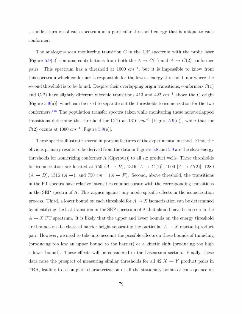
a sudden turn on of each spectrum at a particular threshold energy that is unique to each
conformer.
The analogous scan monitoring transition C in the LIF spectrum with the probe laser
[Figure 5.9(c)] contains contributions from both the A → C(1) and A → C(2) conformer
pairs. This spectrum has a threshold at 1000 cm−1, but it is impossible to know from
this spectrum which conformer is responsible for the lowest-energy threshold, nor where the
second threshold is to be found. Despite their overlapping origin transitions, conformers C(1)
and C(2) have slightly different vibronic transitions 413 and 422 cm−1 above the C origin
[Figure 5.9(a)], which can be used to separate out the thresholds to isomerization for the two
conformers.115 The population transfer spectra taken while monitoring these nonoverlapped
transitions determine the threshold for C(1) at 1316 cm−1 [Figure 5.9(d)], while that for
C(2) occurs at 1000 cm−1 [Figure 5.9(e)].
These spectra illustrate several important features of the experimental method. First, the
obvious primary results to be derived from the data in Figures 5.8 and 5.9 are the clear energy
thresholds for isomerizing conformer A [Gpy(out)] to all six product wells. These thresholds
for isomerization are located at 750 (A → B), 1316 [A → C(1)], 1000 [A → C(2)], 1280
(A → D), 1316 (A →), and 750 cm−1 (A → F ). Second, above threshold, the transitions
in the PT spectra have relative intensities commensurate with the corresponding transitions
in the SEP spectra of A. This argues against any mode-specific effects in the isomerization
process. Third, a lower bound on each threshold for A → X isomerization can be determined
by identifying the last transition in the SEP spectrum of A that should have been seen in the
A → X PT spectrum. It is likely that the upper and lower bounds on the energy threshold
are bounds on the classical barrier height separating the particular A → X reactant-product
pair. However, we need to take into account the possible effects on these bounds of tunneling
(producing too low an upper bound to the barrier) or a kinetic shift (producing too high
a lower bound). These effects will be considered in the Discussion section. Finally, these
data raise the prospect of measuring similar thresholds for all 42 X → Y product pairs in
TRA, leading to a complete characterization of all the stationary points of consequence on
79

Figure 5.8: (a) SEP spectra of A. (be) SEP-PT spectra recorded by monitoring the S1 ← S0
origin transition of the corresponding conformer.
80

Figure 5.9: (a) An overview LIF spectrum of tryptamine with the expanded region corre-
sponding to vibronic transitions of C(1) and C(2). C(1) and C(2) have vibronic bands at
413 and 422 cm−1, respectively, which were used to probe the energy thresholds free of in-
terference from one another. The (b) SEP spectra of A is compared to the SEP-PT spectra
of (c) A → C(1)/C(2), (d) A → C(1), and (e) A → C(2).
81

the potential-energy surface for TRA. Not surprisingly, such a complete characterization is
not possible. The spectra in Figures 5.8 and 5.9 involve observing the population transfer
from a large population conformer (A) to small population conformers (B−F ). The reverse
process, namely, pumping a small population conformer into a large population well, becomes
increasingly hard to observe as the population in the initial conformer well decreases in size.
Despite this difficulty, the thresholds for several other X → Y product pairs were mea-
sured. SEP-induced population transfer spectra pumping TRA(B) monitoring transitions
A,F, and C with the probe laser are shown in Figures 5.10(b) (c), respectively. Both TRA
C(1) and C(2) are being probed when monitoring transition C in the LIF spectrum of Figure
5.2(a). However, based on the lower threshold for A → C(2) than A → C(1), we tentatively
assign this threshold to the B → C(2) process. Similarly, the threshold for pumping con-
formers C(1)/C(2) [whether C(1) or C(2) is not known] back to B was also measured at 750
cm−1, with the likely X → Y pair being C(2) → B. Due to signal-to-noise constraints, only
in the B → F case was it possible to establish a clear lower bound for the threshold.
The combined experimental data on the thresholds for isomerization are summarized on
a 2D potential-energy surface in Figure 5.11. The comparison with theory (Figure 5.3) will
be taken up in the Discussion section.
5.5.4 SEP-hole-filling spectra
An interesting consequence of these three sharp thresholds is that they can be used to
exert some control over which isomerization products are formed by the SEP step. This is
best observed in a hole-filling spectrum, in which the SEP laser wavelengths are fixed to
selectively excite a particular conformer to a well-defined vibrational energy while the probe
laser is scanned. As Figure 5.12(a) shows, when the dump laser is fixed at a wavelength
corresponding to an energy of 750 cm−1 above the zero-point level of A, only conformers B
and F are formed as products. At 1219 cm−1 [Fig. 12(b)], conformer C(2) appears in the
product spectrum, while at 1411 cm−1 [Figure 5.12(c)], C(1), D, and E are added as well.
In Figure 5.12, the population gain in the hot band of A results from incomplete cooling of
82

Figure 5.10: The (a) SEP spectrum of B compared to the SEP-PT spectra of (b) B → A,
(c) B → F , and (d) B → C.
83

Figure 5.11: Schematic PES for TRA along two flexible internal coordinates. The arrows
signify all of the conformational isomerization reactant-product pairs probed experimentally.
The numbers associated with the arrows denote the lower and upper bounds to the energy
threshold.
84

a small fraction of the population of A during the recooling step.
Because the scans in Figure 5.12 are taken with a 20/10/20-Hz configuration, the SEP
hole-filling signal in transition A reflects the population difference between population reach-
ing the ground state via fluorescence versus SEP. The fluorescing molecules produce a ground-
state population dictated by the FranckCondon factors from the fluorescing level, while SEP
produces a population only in a single level. When SEP is to a low-energy state, SEP will
be more efficient than fluorescence at recooling into the reactant well, producing a gain on
transition A, as is observed in Figure 5.12(a). When the dump laser reaches to levels with
higher internal energy [Figures 5.12(b) (c)], SEP will be more efficient at isomerization than
fluorescence, leading to a depletion on transition A.
5.5.5 Studies of deuterated TRA
Two of the four flexible coordinates in TRA involve internal rotation and inversion of the
NH2 group. These motions are implicated in the high-resolution studies of TRA by Nguyen
et al.,131 which have revealed small tunneling splittings (95 MHz) in the origin bands of
C(1) and D. In the present studies of isomerization dynamics, tunneling through the barrier
could produce an energy threshold below the classical barrier height. One way to test for
the effects of tunneling on the isomerization is to compare the isomerization thresholds for
the NH2 and ND2 isotopomers of TRA.
TRA has three exchangeable hydrogens: the two amino hydrogens and the indole NH,
with the former more easily exchanged than the latter. Despite repeated exchange cycles,
complete deuteration at all three sites was never achieved, probably due to exchange back
in the sample compartment. The degree of isotopic substitution was assessed using resonant
two-photon ionization spectroscopy (R2PI). Figure 5.13 shows the R2PI spectra taken while
monitoring the four mass channels of the protonated, singly, doubly, and triply deuterated
TRA. Based on the intensities of the origin transitions, more than 90% of the sample is either
doubly or triply deuterated, with near-complete deuteration at the ND2 site anticipated.
Furthermore, by judicious choice of the SEP pump wavelength on the blue edge of the triply
85

Figure 5.12: SEP-HF spectra after selective excitation of conformer A to vibrational levels
with energies of (a) 748, (b) 1219, and (c) 1411 cm−1
86

deuterated origin (34,927 cm−1, marked in the figure), selective excitation of TRA(d3) was
further enhanced.
Figure 5.14(a) shows the SEP spectrum of conformer A of TRA(d3) with a pump wave-
length set at 34,927 cm−1. Not surprisingly, the spectrum bears a close resemblance to the
spectrum of TRA(A,h3) [Figures 5.14(a) (d)], with small shifts in the frequencies of most
bands. These similarities make it possible to determine the threshold to isomerization with
comparable accuracies on the upper and lower bounds. SEP-PT spectra were recorded for
three reactant-product pairs: A → B, A → F , and B → F . The A → B pair was chosen
because conformers A and B differ primarily in the orientation of the NH2 group, suggest-
ing that tunneling might play a role in the isomerization pathway connecting them. The
other two pairs have F as their product, and therefore represent pathways where the bar-
riers computed on the 2D potential energy surface (PES) (˜0.1-1300 cm−1, Figure 5.3) are
significantly higher than the experiment (<750 cm−1, Figure 5.11).
The SEP-PT spectrum of TRA(A,d3)→ TRA(B,d3) exhibits a threshold to isomerization
[742 cm−1, Figure 5.14(b)] that is identical to the measured threshold in the nondeuterated
system [750 cm−1, Figure 5.8(b)]. Therefore, under the conditions of this experiment, tun-
neling does not shift the threshold for isomerization of TRA A → B.
The corresponding PT scan for A → F is shown in Figure 5.14(c). A comparison of this
scan with the undeuterated analog [Figure 5.14(d)] indicates that the threshold for the d3
isotope is at 832 cm−1, 90 cm−1 above the threshold for the h3 isotope. The triad of bands
near 750 cm−1 is clearly missing despite its large intensity in the SEP spectrum. This result
is surprising because conformers A [Gpy(out)] and F [Gph(up)] differ in both the orientation
and position of the NH2 group relative to the indole ring. Nevertheless, there is a clear shift
in the threshold towards higher values upon deuteration, consistent with tunneling as the
source of the shift.
The corresponding results for B → F [Figure 5.15] show a similar shift, but are less clear
due to a somewhat poorer signal-to-noise ratio in this case. The threshold for TRA(d3) is at
737 cm−1, with no evidence for the transition at 672 cm−1 [Fig. 15(d)]. The corresponding
87

Figure 5.13: The mass-selected R2PI excitation spectra of deuterated tryptamine in the (a)
triply deuterated mass channel, (b) doubly deuterated mass channel, (c) singly deuterated
mass channel, and (d) the undeuterated tryptamine. The S1/leftarrowS0 origin transition
of tryptamine A is 34,920 cm−1 and the excitation wavelength for the deuterated tryptamine
A is marked at 34,927 to ensure no interference from the other species in the LIF excitation
scheme.
88

Figure 5.14: The structures indicate the conformational isomerization and deuterated sites.
The SEP spectrum of (a) tryptamine is compared to (d) tryptamine-(d3). The SEP-PT
spectra of (b) A → B and (c) A → F are shown for comparison to (e) Ad → Bd and (f)
Ad → Fd, respectively. Only the threshold to isomerization of Ad → Fd has any indication
of tunneling effects (˜100 cm−1).
89

band at 685 cm−1 in the spectrum of TRA(h3,B → F ) is observed, but its intensity is
sufficiently small that its absence in the TRA(d3) scan may be simply because it is hidden
in the noise [Figure 5.15(b)].
5.6 DISCUSSION
5.6.1 SEP as a vibrational excitation scheme
The experimental protocol employed in this work uses stimulated emission pumping to ini-
tiate conformational isomerization in a single conformational isomer with a well-defined
internal energy. SEP excitation is an important extension of an earlier work that used in-
frared excitation in the hydride stretch region of the infrared.123,124,125 As noted earlier,
the principle drawback of XH stretch infrared excitation is that many of the lowest-energy
barriers to isomerization are well below the typical XH stretch v = 1 level (˜3500cm−1 = 10
kcal/mol). As such, near-IR excitation never directly probes the barriers to isomerization
in the threshold region. By comparison, the principle advantage of SEP excitation is that it
offers a wide tuning range that stretches from well below the lowest barriers to isomerization
up to energies well in excess of these thresholds.
As a result, by observing the onset of a particular X → Y population transfer, SEP
population transfer spectra provide direct experimental measurements of the energy thresh-
olds for isomerization in individual X → Y reactant-product pairs. Furthermore, in cases
where the thresholds for both X → Y and Y → X pairs can be measured, their difference
constitutes a measure of the relative energies of minima X and Y. Thus, in principle, step-
by-step characterization of all X → Y thresholds constitutes a complete characterization of
the rate-limiting stationary points on the potential-energy surface for isomerization.
SEP excitation also offers exceptional selectivity in the excitation step. Any vibronic
transition that is not overlapped in the ultraviolet spectrum can be used to selectively excite
a particular conformation. With supersonic expansion cooling, vibronic bands typically have
90

Figure 5.15: (a) SEP spectrum of B compared to (c) the SEP spectrum of Bd. The SEP-PT
spectra of (b) B → F exhibits a measured threshold almost identical to the (d) SEP-PT of
Bd → Fd. No tunneling effects are observed in the isomerization of conformers B into F.
91

rotational band contours with widths of 12 cm−1, so different conformers need only shift the
electronic origin by this amount in order to enable selective excitation, a very small fraction
of the excitation energy (˜30,000 cm−1).
Finally, SEP offers the versatility of carrying out SEP using vibronic bands above the
origin in the pump step. In principle, selection of different S1(v) levels offers a powerful
means of changing the FranckCondon intensities to the ground state in the dump step.
These changed FranckCondon factors could be used to narrow the bounds on the threshold
for isomerization. This potential advantage is more one in theory than practice and, in fact,
was not utilized in the present experiment. If intramolecular vibrational redistribution (IVR)
is fast compared to the time delay between pump and dump, then the oscillator strength in
the dump transition is spread over many transitions that reduce the SEP intensity in any
one band considerably. In TRA, we were not able to carry out SEP effectively on bands
more than 200 cm−1 above the S1 origin.
The principle disadvantage of SEP in the context of this experimental protocol is that it
turns a two-laser IR pump/UV probe experiment into a three-laser experiment (SEP pump-
dump, UV probe). Furthermore, in order to use this scheme, the molecule of interest must
have an ultraviolet chromophore capable of efficient SEP, with a sufficiently long lifetime
to stimulate a significant fraction of the excited-state population back down to the ground
state. With the nanosecond lasers employed here, this requires excited-state lifetimes of
several nanoseconds. The tryptamine conformers have excited-state lifetimes of 15 ns.129,131
In addition, the method is not easily adapted to the quantitative measurement of product
quantum yields123,125 because the SEP pump step removes population that is not entirely
brought back to the ground state by the dump step.
5.6.2 The comparison between experiment and calculations
In this section, we compare the experimentally determined energy thresholds for isomer-
ization (Figure 5.10) with computed barrier heights, corrected for zero-point energy effects
(Figure 5.3). This comparison is justified provided that the experimental upper and lower
92

bounds bracket the computed classical barrier height. In order for the upper bound on the
threshold to be an upper bound on the classical barrier height, tunneling must not play a
significant role. The experimental lower bound is a firm lower bound only if the isomeriza-
tion rate at threshold is fast compared to the vibrational cooling rate, producing a negligible
kinetic shift in the threshold.
The measurements on TRA show no evidence for a significant kinetic shift. If vibrational
cooling were competing effectively with isomerization, then the bands near threshold in the
PT spectra should show a reduced intensity (compared to the SEP spectrum) that depends
on the cooling conditions. In TRA, the intensities of transitions above threshold in the PT
spectra faithfully reflect those in the SEP spectra, consistent with isomerization occurring on
a time scale fast compared to cooling. This is consistent with RiceRamspergerKasselMarcus
(RRKM) estimates for the rate constants for isomerization (> 1×10⁹ s−1 at threshold) which
are fast compared to the anticipated cooling rate by helium (23cm1 per collision at a collision
rate of 4 × 10⁹ s−1). A more detailed discussion of the competition between isomerization
and collisional cooling will be taken up in the adjoining paper on 3-indole propionic acid
(IPA). For our purposes here, we will use the lower bounds from the SEP measurements as
the lower bounds on the energy barriers, assuming that the kinetic shifts are negligible.
The data on TRA(d3) (Sec. 1.5.5 E) indicate that tunneling does play a role in isomer-
ization in certain regions of the potential-energy surface; notably, in pathways leading into
well F. Recall that the threshold for A → B isomerization was unchanged by deuteration
at the amino group, while the thresholds for the A → F and B → F channels were raised
by small but measurable amounts (˜100 cm−1). Because this shift is small, it could arise
from some other effect than tunneling; perhaps due to zero-point energy (ZPE) shifts in the
threshold or an increased kinetic shift to the measurement. However, the computed shift in
the barrier height due to ZPE differences between TRA(h3) and TRA(d3) is less than 10
cm−1. Furthermore, the computed RRKM rate constants at threshold for isomerization of
TRA(d3) are only reduced by about 20% by deuteration, and are still large compared to the
rate for cooling. Therefore, tunneling is the most likely explanation for the observed shift
93

in the A → F and B → F isomerization thresholds in TRA(d3). The relationship between
the observed thresholds for A → F and B → F and the classical barrier height depends on
the tunneling pathway, an issue to which we shall return shortly. In other measured X → Y
thresholds, we assume that tunneling plays an insignificant role, and treat the observed
energy threshold as an upper bound to the classical barrier height.
5.6.2.1 Relative energies of the minima A first test of the calculated potential-
energy surface is to compare the relative energies of the minima with the observed relative
populations of the conformers in the expansion in the absence of SEP excitation. One
must be careful not to put too much weight on these populations reflecting energy differ-
ences alone, because it is possible that the cooling in the expansion can remove population
out of the higher-energy minima into the lower ones if the barriers are not too great. In
the case of TRA, we do not observe significant changes in these relative populations as
a function of distance downstream in the expansion (Figure 5.6), and therefore see some
merit in correlating the relative intensities of the observed S0-S1 origins with their relative
energies. The high-resolution spectra of Nguyen et al.131 show that the intensity of the
C(2) origin is about twice that of the C(1) origin. Sorting these intensities from large to
small yields, I(A) > I(B) > I[C(2)] > [I[C(1)], I(D), I(E), andI(F )], where the bands in
brackets have similar intensities. This suggests that the experimental energy ordering is
E(A) < E(B) < E[C(2)] < {E[C(1)], E(D), E(E), andE(F )
This energy ordering matches reasonably well with the DFT energies, while the RIMP2
results would predict greater populations in C(2) and F than observed experimentally. It is
possible that the low experimental barriers separating these minima from B (Figure 5.11)
provide a pathway for removing population from these minima into B early in the expansion
(x/D < 2).
5.6.2.2 Barrier heights For most of the measured A → X thresholds, the calculated
barrier heights, including ZPE corrections, are within a couple of hundred wave numbers
94

of experiment. For instance, the experimental barriers separating A [Gpy(out)] from any
of the three anticonformers [C(1), D and E] are Ethresh ∼ 1300 cm−1 = 3.7 kcal/mol,
consistent with the computed barrier heights for AD and BE (∼ 1300 cm−1 for DFT and
16001700 cm−1 for RIMP2). The A → B threshold is somewhat lower experimentally
(688 < Ethresh < 748 cm−1) than predicted by theory (Ebarrier = 930 cm−1 by both methods),
but still within this 200 cm−1 spread.
The glaring exceptions to this general correspondence between experiment and theory
are the A → F and A → F thresholds. As Figure 5.3 shows, the computed barriers into well
F are all up in the 13001500 cm−1 range, almost a factor of 2 higher than the experiment.
These are the same pathways that show some evidence for tunneling, and it is possible that
tunneling accounts for the entire discrepancy between experiment and theory. However,
the small shift in threshold induced by deuteration seems to argue against such a simple
resolution. Furthermore, since A and F differ along at least two of the flexible coordinates,
one would like to pin down the isomerization pathway, whether tunneling or classical in
nature.
5.6.2.3 Isomerization pathways The experimental thresholds (Figure 5.15) that con-
nect A → B, A → F , and B → F are particularly low, suggesting that there is an efficient
isomerization pathway connecting these minima. As just noted, this pathway may involve
tunneling, but if so, the tunneling penetrates some of the highest computed barriers on the
surface, those that surround minimum F. As noted in the initial communication, the observed
thresholds are consistent with a pathway from A → F (688 < Ethresh < 748 cm−1) that passes
from A → B(688 < Ethresh < 748 cm−1) and then from B → F (566 < Ethresh < 688 cm−1).
The computed potential-energy surface possesses a low-energy trough that connects wells
A, H, and C(2) via the AH and HC(2) barriers. A possible way to resolve these differences
would be to simply swap the assignments of conformers C(2) and F. These two conformers
are both Gph conformers, differing only in the orientation of the NH2 group. As a result, they
have rotational constants which are very close to one another. If this swap in assignments
95

were made, the calculations would predict an A → F barrier of 672 cm−1 based on a
pathway that goes from Gpy(out) to Gpy(in) (Ebarrier = 672cm−1) and then on to Gph(out)
(Ebarrier = 527cm−1). While such a swap is tempting, the arguments that Nguyen used
to arrive at the assignments for C(2) and F are quite persuasive, and are consistent with
previous assignments based on infrared and vibronic level data.131
With or without such a swap in assignments, it is surprising that tunneling would lead
to a shift in the A → F , and B → F thresholds, but produce no shift in A → B, since
the pathways from A or B into F necessarily involve motion of the entire NH2 group from
the Gpy to the Gph position (with an effective tunneling mass of ∼ 16 amu), while A → B
isomerization [Gpy(out) → Gpy(up)] can be accomplished merely by an internal rotation
and/or inversion of the NH2 group (∼2-amu tunneling mass). Of course, this difference in
mass of the tunneling group could be compensated for by a narrower width to the barrier.
Since the likely isomerization pathway for either A → F or B → F involves motion of the
NH2 group over the top of the phenyl ring, one wonders whether the interaction of the NH2
group with the indole π cloud could provide a low-energy tunneling pathway that inverts
the NH2 group as it sweeps across the π cloud. A final resolution of these issues will require
theoretical methods that can directly model the tunneling contribution to the isomerization
dynamics.
Finally, even in a molecule the size of tryptamine, we are quickly losing our ability to
use chemical intuition to identify the important isomerization pathways on the potential-
energy surface. The transition states identified in this work are likely only a subset of those
needed to fully describe the isomerization dynamics. In particular, isomerization pathways
that involve swinging the ethylamine side chain from one side of the indole ring to the other
may play a significant role in the isomerization dynamics. The anti configuration supports
minima G and I in which the ethylamine side chain is nearly in plane. The barriers into
and out of these minima are quite low (∼100 cm−1). Analogous pathways involving motion
of the ethylamine group from the Gpy or Gph positions around the edges of the pyrrole or
phenyl rings deserve further exploration.
96

5.7 CONCLUSIONS
Despite its modest size, tryptamine possesses a potential-energy surface for conformational
isomerization that is a challenge to characterize via experiment and theory. The popula-
tion of TRA molecules is spread over seven conformational minima, even with supersonic
expansion cooling. A complete characterization of the isomerization process would then re-
quire measurement of 42 independent X → Y conformer pairs, illustrating the need for
new experimental tools capable of studying the process in a conformation specific way.
The present paper describes measurements of the energy thresholds for isomerization using
conformation-specific SEP excitation, followed by collisional cooling of the products prior to
conformation-specific detection via laser-induced fluorescence. The method of SEP-induced
population transfer spectroscopy provides a means for measuring the energy thresholds for
isomerization of individual X → Y conformer pairs free from interference from others present
in the expansion.
The application of this method to TRA has demonstrated the power of the method, but
it has also raised many unanswered questions, providing a stimulus for future work. First,
since the method uses collisions as an integral part of the experimental scheme, isomerization
is always viewed in competition with collisional cooling. Ideally, this competition can be used
as a means to quantify the energy-dependent rate of isomerization for individual X(E∗) → Y
pairs. It is hoped that such studies can provide critical new tests of RRKM theory, which is
undergoing close scrutiny by theory, which suggests that in many circumstances, IVR into the
torsional modes may limit the rate of isomerization near threshold. Second, as just discussed
in the preceding section, theoretical methods that can explore the isomerization pathways
more completely, and include the effects of tunneling, are needed. Third, it is important
to compare the results of the present study with analogous studies of isomerization in the
absence of collisions, using methods such as those employed by Pate and co-workers. Finally,
hole-filling methods can be applied to the study of isomerization in a much wider range of
contexts. In the adjoining paper, we will probe the effect of a bound solvent water molecule
97

on the barrier to intramolecular isomerization in 3-indole propionic acid.132
5.8 ACKNOWLEDGEMENTS
We gratefully acknowledge support for the work from the National Science Foundation (Grant
No. CHE-0242818). One of the authors (J.R.C.) acknowledges partial support from the
Purdue Research Foundation for a Graduate Student Assistantship.
98

APPENDIX
TRYPTAMINE TRANSITION STATE STRUCTURES
99

Figure A1: Transition-state structures of tryptamine.
100

BIBLIOGRAPHY
[1] Mao, W.; Mao, H.; Goncharov, A.; Struzhkin, V. V.; Guo, Q.; Hu, J.; Shu, J.; Hem-ley, R.; Somayazulu, M.; Zhaoi, Y. Science 2002, 297, 2247.
[2] Ripmeester, J. A.; Ratcliffe, C.; Klug, D. D.; Tse, J. S. Ann. N. Y. Acad. Sci. 1994,715, 161.
[3] Kuhs, W. F.; Chazallon, B.; Radaelli, P. G.; Pauer, F. J. Incl. Phenom. 1997, 29, 65.
[4] Hill, T. L. J. Chem. Phys. 1948, 16, 399.
[5] Yu, H.; van Gunsteren W. F., J. Chem. Phys. 2004, 121; 19, 9549.
[6] Forester, T. R.; Smith, W. The DLPOLY-2.0 Reference Manual, CCLRC, DaresburyLaboratory, Warrington, England version 2.0 edition 1995.
[7] Boys, S. F.; Bernardi, F. Mol. Phys. 1970, 553.
[8] Lee, C.; Yany, W.; Parr, R. G. Phys. Rev. B: Condens. Matter Mater. Phys. 1988,785.
[9] Bernholdt, D. E.; Harrison, R. J. Chem. Phys. Lett. 1996, 470.
[10] Feyereisen, M.; Fitzgerald, G.; Komornicki, A. Chem. Phys. Lett. 1993, 359.
[11] Park, Y. D.; Rizzo, T. R.; Peteanu, L. A.; Levy, D. H. Journal of Chemical Physics1986, 84, 6539–49.
[12] Philips, L. A.; Levy, D. H. Journal of Chemical Physics 1988, 89, 85–90.
[13] Wu, Y. R.; Levy, D. H. Journal of Chemical Physics 1989, 91, 5278–5284.
[14] Dian, B. C.; Clarkson, J. R.; Zwier, T. S. Science 2004, 303, 1169–1173.
[15] Peng, C.; Schegel, H. B. Israel J. Chem 1993, 33, 449.
101

[16] Sloan, E. D. Clathrate Hydrates of Natural Gases ; Marcel Dekker, New York, 1990.
[17] Atwood, J.; Davies, J. E. D.; Macnicol, D. D. Inclusion Compounds ; Oxford UniversityPress, Oxford, 1991.
[18] Davidson, D. W. Water: A Comprehensive Treastise; Frank, F., Ed.; Plenum PressNew York, 1973 2, year.
[19] Jeffery, G. A.; Macnicol, D. D.; Bishop, R. Comprehensive Supramolecular ChemistryVol. 6 ; 1996.
[20] Ripmeester, J. A.; Tse, J. S.and Ratcliffe, C.; Powell, B. M. Nature 1987, 119, 173.
[21] Udachin, K. A.; Ratcliffe, C.I.and Enright, G. D.; Ripmeester, J. A. Supramol. Chem.1997, 8, 173.
[22] Sloan, E. D. Nature 2003, 426, 353.
[23] Udachin, K. A.; English, N. J.; Ratcliffe, C.I.and Ripmeester, J. A. J. Am. Chem. Soc.197, 119, 11481.
[24] Udachin, K. A. Ratcliffe, C. R. J. A. Angew. Chem. Int. Ed. 2001, 40, 1303.
[25] Mcmullan, R. K.; Jeffery, G. A.; Jordan, T. H. J. Chem. Phys. 1967, 47, 1229.
[26] Davy, H. Philos. Trans. R. Soc. London 1811, 101, 1.
[27] Faraday, M. Philos. Trans. R. Soc. London 1823, 113, 60.
[28] Clausen, W. F. J. Chem. Phys. 1951, 19, 259.
[29] von Stackelberg, M.; Muller, H. R. J. Chem. Phys. 1951, 29, 2425.
[30] Clausen, W. F. J. Chem. Phys. 1951, 19, 1425.
[31] Muller, H. R. von Stackelberg, M. Naturwissenschaften 1952, 39, 20.
[32] Pauling, L.; Marsh, R. E. Pro. Natl. Acad. Sci. U.S.A. 1952, 38, 112.
[33] Hammerschmidt, E. G. Ind. Eng. Chem. 1934, 26, 851.
[34] Suess, E.; Bohrmann, G. an dGreinert, J.; Laush, E. Sci. Am. 1999, 281, 52.
[35] Kvenolden, K. Proct. Int. Japan Natl. Oil Comp, Chiba City, Japan 1998.
[36] Nesbit, E. Nature 1990, 23, 347.
102

[37] Dresselhaus, M. S.; Thomas, I. L. Nature 2001, 414, 332.
[38] Schlapbach, L.; Zuttel, A. Nature 2001, 414, 353.
[39] Dyadin, Y. A.; Larionov, E. G.; Manakov, A. Y.; Zhurko, F. V.; Aladki, E. Y.; V, M. T.;Komarov, V. Y. Mendeleev. Commun. 1991, 171.
[40] Mao, W. L.; Mao, H. K. Proc. Natl. Acad. Sci. 2004, 101, 708.
[41] Vos, W. L.; Finger, L. W.; Hemley, R. J.; Mao, H. K. Phys. Rev. Lett. 1994, 71, 3150.
[42] Powell, H. M. J. Chem. Soc. 1948, 61.
[43] Deaton, W. M.; Frost, E. M., J. U.S. Bur Mines Monogr. 1946, 8.
[44] Waals, J. H. v. d.; Platteeuw, J. C. Adv. Chem. Phys. 1959, 2, 1.
[45] Somayazulu, M. S.; Finger, L. W.; Hemley, R. J.; Mao, H. K. Science 1996, 271, 1400.
[46] Loubeyre, P.; Letoullec, R.; Pinceaux, J. P. Phys. Rev. Lett. 1994, 72, 1360.
[47] www.eere.energy.gov,.
[48] Lokshin, K. A.; Zhao, Y.and He, D.; Mao, W. L.; Mao, H. K.; Hemley, R.J.and Lobanov, M. V.; Greenblatt, M. Phys. Rev. Lett. 2004, 93, 125503.
[49] Mao, W.; Mao, H. K. U.S.A. Patent 6735960 2002.
[50] von Stackelberg, M. Naturwissenschaften 1949, 36, 11–12.
[51] Mao, W. L.; Mao, H. K.; Goncharov, A. F.; Struzhkin, V. V.; Guo, Q.; Hu, J.; Hem-ley, R.; Somayazulu, M.; Y., Z. Science 2002, 297, 2247.
[52] Patchkovskii, S.; Tse, J. Proc. Natl. Acad. Sci. U. S. A. 2003, 100, 14645.
[53] Sluiter, M. F.; Adachi, H.; Belosludov, R. V.; Belosludov, V.; Kawazoe, Y. MaterialsTransactions 2004, 45; 5, 1452.
[54] Alavi, S.; Ripmeester, J. A.; Klug, D. D. J. Chem. Phys. 2005, 123, 024507.
[55] Inerbaev, T. M.; Belosludov, V. R.; Belosludov, R. V.; Sluiter, M.; Kawazoe, Y. Com-putational Materials Science 2006, 36, 229.
[56] Patchkovskii, S.; N., Y. S. Phys. Chem. Chem. Phys. 2004, 6, 4152.
[57] Ponder, J. W.; Case, D. A. Adv. Protein Chem. 2003, 66, 27.
103

[58] Stillinger, F. H. Science 1980, 209, 451.
[59] Halgren, T. A.; Damm, W. Curr. Opin. Struct. Biol. 2001, 11, 236.
[60] Rick, S. W.; Stuart, S. J. Reviews in Computational Chemistry 2002, 89-154, year.
[61] Verlet, L. Physical Review 1967, 159, 98.
[62] English, N. J.; Macelroy, J. M. D. J. Comput. Chem. 2003, 24, 1569.
[63] English, N. J.; Johnson, K. J.; Taylor, C. E. J. Chem. Phys. 2005, 123, 244503.
[64] Rick, S. W.; Stuart, S. J.; Berne, B. J. J. Chem. Phys. 1994, 101, 6141–6156.
[65] Jiang, H.; Jordan, K. D.; Taylor, C. E. J. Phys. Chem. B 2007, 111, 6486.
[66] Ren, P.; Ponder, J. W. J. Phys. Chem. B 2003, 107, 5933.
[67] Ren, P.; Ponder, J. J. J. Phys. Chem. B 2003, 107, 5933.
[68] Ren, P.; Ponder, J. W. J. Chem. Phys. B 2004, 108, 13427.
[69] Smith, W.; Forester, T. R. J. Mol. Graphics 1996, 14, 136.
[70] Eisenberg, D.; Kauzmann, W. The Structure and Properties of Water (ClarendonPress, Oxford, 1969).
[71] Wallqvist, A.; Mountain, R. D. Reviews in Computational Chemistry 1999, 183–247.
[72] Finney, J. L. J. Mol. Liq. 2001, 90, 303.
[73] Guillot, B. J. Mol. Liq. 2002, 101, 219.
[74] Ponder, J. W.; Case, D. A. Adv. Protein Chem. 2003, 66, 27.
[75] Cornell, W. D. Cieplak, P. a. C. I. J. Am. Chem. Soc. 1995, 117, 5179.
[76] MacKerrell, A. D.; Bashford, D.; Bellot, M. J. Phys. Chem. B 1998, 102, 3586.
[77] van Gunsteren W. F.,; Billeter, S. R.; Eising, A. A.; Hunenberger, P. H.; Mark, A. E.;Scott, W. R. P.; Tironi, I. G. Biomolecular Simulation: The GROMOS manual andUser Guide (vdf Hochschulverlag, ETH Zurich, Switzerland) 1996.
[78] Schuler, L. D.; Daura, X.; van Gunsteren W. F., J. Comput. Chem. 2001, 22, 1205.
[79] Jorgensen, W. L.; Maxwell, D. S.; Tirado-Rivers, J. J. Am. Chem. Soc. 1996, 118,11225.
104

[80] Bernal, D.; Fowler, R. H. J. Chem. Phys. 1933, 1, 515.
[81] Stillinger, F. H.; Rahman, A. J. Chem. Phys. 1974, 60, 7943.
[82] Berendsen, H. J. C.; Postma, J. P. M.; Von Gunsteren, W. F.; Hermans, J. 1988, 331.
[83] Jorgensen, W. L.; Chandrasekhar, J.; Madura, J. D.; Impey, R. W.; Klein, M. L. J.Chem. Phys. 1983, 79, 926.
[84] Rick, S. W. J. Chem. Phys. 2001, 114, 2276.
[85] Yu, H. B.; van Gunsteren W. F.and, J. Chem. Phys. 2003, 118, 221.
[86] Peterson, K. A.; Woon, D. B.; Dunning Jr, T. H. J. Chem. Phys. 1994, 100, 7410.
[87] edited by D. R. Lide, CRC Handbook of Chemisry and Physics, 83rd edition 2002.
[88] Buckingham, A. D. Quaterly Reviews of the Chemical Society 1959, 13, 183.
[89] Tang, K. T.; Toennis, J. P. J. Chem. Phys. 1977, 66, 1496.
[90] Berendsen, H. J. C.; Grigera, J. R.; Straatsma, T. P. J. Phys. Chem. 1987, 91, 6269.
[91] Nose, S. J. Chem. Phys. 1984, 81, 511.
[92] Hoover, W. G. J. Chem. Phys. 1985, 81, 511.
[93] Allen, M. P.; Tildesley, D. J. Computer Simulation of Liquids ; Clarendon Press: OxfordUniversity Press, Oxford, 1987.
[94] Ewald, P. Ann. Phys. 1921, 64, 253.
[95] Mak, T. C.; McMullan, R. K. J. Chem. Phys. 1964, 42, 2732.
[96] van Kleveren, E. P.; Michels, J. P. J.; Schouten, J. A.; Klug, D. D.; Tse, J. S. J. Chem.Phys. 2002, 117, 6636.
[97] van Kleveren, E. P.; Michels, J. P. J.; Schouten, J. A.; Klug, D. D.; Tse, J. S. J. Chem.Phys. 2001, 114, 5745.
[98] van Kleveren, E. P.; Michels, J. P. J.; Schouten, J. A.; Klug, D. D.; Tse, J. S. J. Chem.Phys. 2001, 115, 10500.
[99] McQuarrie, D. A. Statistical Mechanics ; University Science Books, 2000.
[100] Florusse, L. J.; Peters, C. J.; Schoonman, J.; Hester, K. C.; Koh, C. A.; Dec, S. F.;Marsh, K. N.; Sloan, E. D. Science 2004, 469.
105

[101] Florusse, L. J.; Peters, C. J.; Schoonman, J. an dHester, K. C.; Koh, C. A.; Dec, S. F.;Marsh, K. N.; Sloan, E. Science 2004, 306, 469.
[102] Lee, H.; Lee, J.-W.; Kin, D. Y.; Park, J.; Seo, Y.-T.; Zeng, H.; Maoudrakovski, I. L.;Ratcliffe, C.I.and Ripmeester, J. A. Nature 2005, 434, 743.
[103] Sloan, E. Soc. Plastic Engineer., Richardson, TX 2000, 21, year.
[104] Becke, A. D. Phys. Rev. A 1988, 3098.
[105] Fanourgakis, G. S.; Apra, E.; Xantheas, S. S. J. Chem. Phys. 2004, 121, 2655.
[106] Cui, J.; Liu, H.; Jordan, K. D. J. Phys. Chem. B 2006.
[107] Hartke, B. Phys. Chem. Chem. Phys. 2003, 5, 275.
[108] Burnham, C. J.; Xantheas, S. S. The Journal of Physical Chemistry 2002, 116, 5115.
[109] Kendall, R. A.; Dunning, T. H.; Harrison, R. J. J. Chem. Phys. 1992, 1481.
[110] Dunning, T. H. J. Chem. Phys. 2002, 6796.
[111] Woon, D. E.; Dunning, T. H.; Harrison, R. J. J. Chem. Phys. 1993, 96, 1481.
[112] Shin, J.-W.; Hammer, N. I.; Diken, E. G.; Johnson, M. A.; Walters, R. S.; Jaeger, T. D.;Duncan, M. A.; Christie, R. A.; Jordan, K. D. Science 2004, 304, 1137–1141.
[113] Robertson, E.; Simons, J. Physical Chemistry Chemical Physics 2001, 3, 1–18.
[114] Zwier, T. Journal of Physical Chemistry A 2001, 105, 8827–8839.
[115] Carney, J. R.; Zwier, T. S. Journal of Physical Chemistry A 2000, 104, 8677–8688.
[116] Carney, J. R.; Zwier, T. S. Chemical Physics Letters 2001, 341, 77.
[117] Dian, B. C.; Longarte, A.; Mercier, S.; Evans, D. A.; Wales, D. J.; Zwier, T. S. J.Chem. Phys. 2002, 117, 10688.
[118] Florio, G. M.; Christie, R. A.; Jordan, K. D.; Zwier, T. S. J. Am. Chem. Soc. 2002,124, 10236.
[119] Chin, W.; Mons, M.; Dognon, J.; Piuzzi, F.; Tardivel, B.; Dimicoli, I. Physical Chem-istry Chemical Physics 2004, 6, 2700–2709.
[120] Fricke, H.; Gerlach, A.; Unterberg, C.; Rzepecki, P.; Schrader, T.; Gerhards, M. Phys-ical Chemistry Chemical Physics 2004, 6, 4636–4641.
106

[121] Gerhards, M.; Unterberg, C.; Gerlach, A.; Jansen, A. Physical Chemistry ChemicalPhysics 2004, 6, 2682–2690.
[122] Nir, E.; Hunig, I.; Kleinermanns, K.; Vries, M. d. ChemPhysChem 2004, 5, 131–137.
[123] Dian, B. C.; Longarte, A.; Winter, P. R.; Zwier, T. S. J. Chem. Phys. 2004, 120,133–147.
[124] Evans, D. A.; Wales, D. J.; Dian, B. C.; Zwier, T. S. J. Chem. Phys. 2004, 120, 148.
[125] Dian, B. C.; Florio, G. M.; Clarkson, J.; Longarte, A.; Zwier, T. S. Journal of ChemicalPhysics 2004, 120, 9033–9046.
[126] Nesbitt, D. J.; Field, R. W. Journal of Physical Chemistry 1996, 100, 12735–12756.
[127] Flynn, G. W.; Parmenter, C. S.; Wodtke, A. M. Journal of Physical Chemistry 1996,100, 12817–12838.
[128] Silva, M.; Jongsma, R.; Field, R.; Wodtke, A. Ann. Rev. Phys. Chem. 2001, 52, 811–852.
[129] Connell, L.; Corcoran, T.; Joireman, P.; Felker, P. Journal of Physical Chemistry 1990,94, 1229–1232.
[130] Philips, L. A.; P., W. S.; Martinez III, S. J.; R., F. G.; Levy, D. H. J. Am. Chem. Soc.1988, 110, 1352.
[131] Nguyen, T. V.; Korter, T. M.; Pratt, D. W. Journal of Molecular Spectroscopy 2005,(in press), year.
[132] Clarkson, J. R.; Baquero, E.; Zwier, T. S. J. Chem. Phys. 2005, 122, 214312.
[133] Kable, S.; Knight, A. Journal of Chemical Physics 1990, 93, 3151–3150.
[134] Burgi, T.; Droz, T.; Leutwyler, S. Chemical Physics Letters 1995, 246, 291–99.
[135] Burgi, T.; Droz, T.; Leutwyler, S. Journal of Chemical Physics 1995, 103, 7228.
[136] Frisch, M. J.; Pople, J. A.; Binkley, J. S. J. Chem. Phys. 1984, 3265.
[137] EPAPS, See EPAPS Document No. E-JCPSA6-122-016523 for the complete re-sults from the calculations including optimized geometric parameters and selectedassociated harmonic frequencies. This document can be reached via a direct linkin the online article’s HTML reference section or via the EPAPS homepage(http://www.aip.org/pubservs/epaps.html).
107

[138] Frisch, M. J. et al. 1995-2004.
[139] Ahlrichs, R.; an d M. Hser, M. B.; Horn, H.; Klmel, C. Chem. Phys. Lett 1989, 162,165–169.
[140] Noga, J.; Bartlett, R. J. J. Chem. Phys. 1987, 86, 7041.
[141] Raghavachari, K.; Trucks, G. W.; Pople, J. A.; Head-Gordon, M. Chem. Phys. Lett.1989, 479.
108