compression neuropathy in the upper extremity · pdf file10/12/2015 1 compression neuropathy...
TRANSCRIPT

10/12/2015
1
COMPRESSION
NEUROPATHY IN
THE UPPER
EXTREMITY Edward L. Birdsong, MD
Allegheny Hand and Upper Extremity Center
DISCLOSURE
The speaker has nothing to disclose
THE BIG THREE
Median nerve
- Carpal Tunnel
- Anterior Interosseous Syndrome
- Pronator Syndrome
Radial Nerve
Radial Tunnel Syndrome
Posterior Interosseous Nerve Syndrome
Wartenberg’s Syndrome
Ulnar Nerve
Cubital Tunnel
Ulnar Tunnel (Guyon’s Canal)

10/12/2015
2
BASIC SCIENCE
• Increased intraneural pressure is a
result of disproportion between volume
of nerve and the space thru which it
passes.
• Degree of axonal injury proportional to
duration and magnitude of compression.
• 30 mmHg = Paresthesias
• 50-60 mmHg = Complete
sensory/motor block
MEDIAN NERVE
• Carpal tunnel syndrome
• Pronator syndrome
• Anterior interosseous nerve syndrome
CARPAL TUNNEL
SYNDROME
• Median nerve compression at the wrist
• Most common compression neuropathy
• U.S. - >$1 billion annual medical costs
• >200,000 surgeries annually

10/12/2015
3
MEDIAN NERVE
ANATOMY
Motor branch - variable,
but most often
extraligamentous
Palmar branch - usually
pierces antebrachial
fascia to lie superficial to
TCL (also variable)
REMEMBER
• Women > Men
• Age > 45-50
• Smoking --- No
• Occupation ---No
ETIOLOGY OF CTS
• Anatomic abnormalities
• Associated medical conditions
• Inflammatory factors
• Fluid imbalances
• Trauma
• Position

10/12/2015
4
ANATOMIC
ABNORMALITIES
• Congenital anomalies
• Persistent median artery
• Proximal lumbrical muscles
• Distal sublimus muscles
ASSOCIATED
MEDICAL
CONDITIONS
• Thyroid disease (hypothyroidism)
• Diabetes
• Pregnancy
INFLAMMATORY
FACTORS
• Rheumatoid arthritis
• Gout
• Infection
10/12/2015
5
FLUID BALANCE
ABNORMALITIES
• Pregnancy
• Hemodialysis - (high correlation
between the side of dialysis access,
and side affected with CTS)
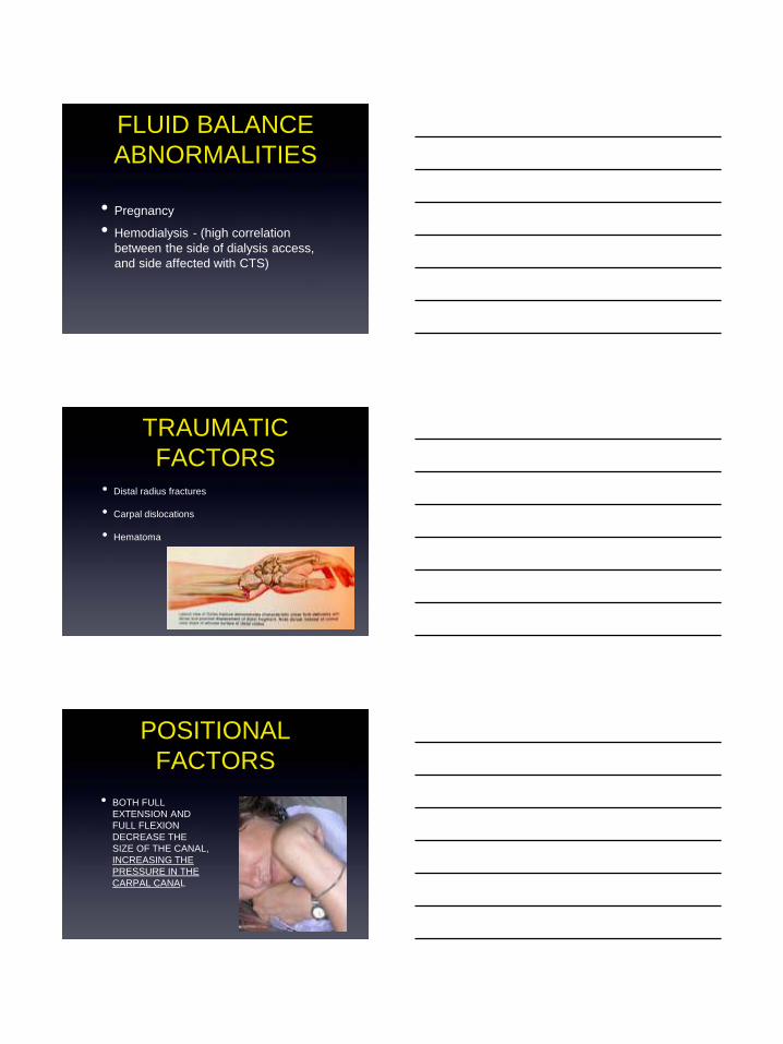
TRAUMATIC
FACTORS • Distal radius fractures
• Carpal dislocations
• Hematoma
POSITIONAL
FACTORS
• BOTH FULL
EXTENSION AND
FULL FLEXION
DECREASE THE
SIZE OF THE CANAL,
INCREASING THE
PRESSURE IN THE
CARPAL CANAL
10/12/2015
6
CLINICAL
PRESENTATION • Numbness, tingling, and pain - radial 3
1/2 digits. Weakness & clumsiness of
grip.
• Pain may radiate proximally into
forearm, arm, and shoulder
• Frequently awakens pt from sleep
(positional)
• Worse with activities - gripping, writing,
driving
CLINICAL STAGING • Early (mild) CTS
intermittent paresthesias, night sxs
wrist flexion may elicit sxs
• Intermediate (moderate) CTS
-more frequent paresthesias, worse with
use
Feeling of numbness, clumsiness
- +/- weakness
• Advanced (severe) CTS
constant impaired sensibility, severe pain
Thenar atrophy, pinch/opposition
weakness
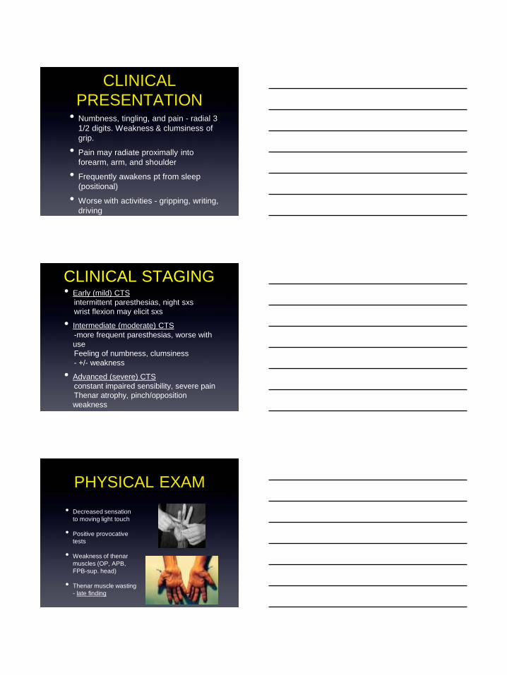
PHYSICAL EXAM
• Decreased sensation
to moving light touch
• Positive provocative
tests
• Weakness of thenar
muscles (OP, APB,
FPB-sup. head)
• Thenar muscle wasting
- late finding

10/12/2015
7
PROVOCATIVE
TESTS
• Tinel’s
• Phalen’s
• Reverse Phalen’s
• Durkin’s ~Sp 90%, Se 87%
NCV / EMG • NCV
-Distal sensory latency >3.5ms
-Motor latency >4.5ms
-Conduction velocity <52m/s
• EMG
-Fibrillations, positive sharp waves, decreased
amplitude of action potentials
• False negative
- 8-25%
- Yes, NCV/EMG negative CTS does exist!
Non- Op Treatment
• Night splints
Effective – wrist in neutral position
Not too tight!
• Steroid injections
80% transient relief – days to months
Only 20% get long-term relief (1year)
Most successful in pts with mild symptoms

10/12/2015
8
Non-OP Treatment
• Oral therapies
Vitamin B6 - no evidence to support use
NSAIDs - no benefit
Gabapentin - no benefit
Corticosteroids - maybe short term
Alpha lipoic acid - maybe short term
SURGICAL
OPTIONS • Considered for patients with persistent
or progressive symptoms, despite non-
operative treatment.
• Transient improvement following
cortisone injection - good prognostic
indicator for surgery.
• Motor denervation on EMG
• Thenar weakness/atrophy
SURGICAL
OPTIONS
• Classic open release
• Mini open release
• Endoscopic release

10/12/2015
9
SURGICAL
OPTIONS
• Risks and long term outcomes
equivalent regardless of technique -
surgeons pref.
• Arguably less initial post op pain, and
possibly earlier return to work with
endoscopic procedure
• most common cause for failure -
incomplete release TCL
SURGICAL
OPTIONS
• Unnecessary to perform:
- internal neurolysis
- tenosynovectomy
- antebrachial fascia release
- concomitant release of Guyon’s canal
POST OP CARE
• Splints – not necessary
• Hand therapy – not necessary
• Antibiotics – not necessary (pre or post-
op
• Return to work – controversial
-depends on kind of work (Duh…)
-2-3 wks for mini-open. Probably
sooner with endo (multiple studies)

10/12/2015
10
COMPLICATIONS
CTR • Incomplete division of TCL
• Damage to PCBMN
• CRPS (RSD)
• Hypertrophic painful scar
• Hematoma
• Bowstringing
REVISION CTR
• 50% experience some relief
• Adjunctive procedures
- hypothenar fat pad flap
- radial forearm fascial flap
- radial artery perforator based flap
ANTERIOR
INTEROSSEOUS NERVE
SYNDROME
• AIN primarily a motor nerve
• Branches from median nerve 4 - 6 cm
distal to elbow
• Passes between 2 heads of PT
• Supplies radial half of FDP (index &
long), FPL, and PQ

10/12/2015
11
AREAS OF
COMPRESSION • Mutiple sites have been implicated
• Deep head of PT
• Origin of FDS – fibrous arch
• Edge of lacertus fibrosis
• Enlarged bicipital tendon bursa
• Accessory head of FPL (Gantzer’s
muscle)
CLINICAL
PRESENTATION
• Complains of ill defined forearm pain
• Inability to flex IP joint of thumb and DIP
of index finger
• EMG/NCS helpful
• Weak forearm pronation
TREATMENT
Complete spontaneous recovery is
common may take 6 - 12 months
• Consider surgery if no motor recovery
after 3-6 months of observation.
• Surgical release of all potential sites of
compression.
• Penetrating trauma – surgery
• Traction injury - observation

10/12/2015
12
• Potential sites of compression:
• Lacertus fibrosus
• Liagament of Struthers
• Origin of FDS – fibrous arch
• Pronator muscle
PRONATOR
SYNDROME
CLINICAL
PRESENTATION
• Numbness and tingling as with CTS
• Numbness may extend to the palm in
the PC branch distribution
• Pain can radiate into the volar forearm
• Night pain not typical complaint
PHYSICAL EXAM
• Palpate for supracondylar process of
distal humerus, proximal to medial
epicondyle
• Check for Tinel’s over proximal volar
forearm (+/- finding)
• May have some motor weakness
• Provocative maneuvers for each
potential site of compression

10/12/2015
13
PROVOCATIVE
TESTS
• Lacertus fibrosus-
resisted elbow flexion
forearm supinated
• Pronator teres-
resisted pronation
elbow extended
• FDS- resisted flexion
long finger PIP
EMG/NCS
• Generally misleading. Usually normal.
• Fibrillation potentials and positive sharp
waves in pronator and FDS may aid in
dx
• XRAY: Look for supra-condylar process
on anterior-medial humerus
TREATMENT • Activity modification - specifically those
involving repetitive flexion/pronation
• Surgical release of all potential sites of
compression yields good results.
-Ligament of Struthers (if present)
-Lacertus fibrosis
-Fascia superficial head of pronator
-Fascial arch of proximal FDS
• Literature reports 90% satisfactory
results

10/12/2015
14
RADIAL NERVE
• BRACHIAL COMPRESSION
SYNDROME
• POSTERIOR INTEROSSEOUS
NERVE SYNDROME
• RADIAL TUNNEL SYNDROME
• SENSORY RADIAL NERVE
COMPRESSION
BRACHIAL
COMPRESSION
• Compression at the arm, due
to: humerus fx, tourniquet
palsey, prolonged postural
compression.
• Usually spontaneous
recovery. If not by 3-4 months,
neurolysis, nerve grafting,
tendon transfers.
• Explore nerve in open
humerus fxs at time of ORIF
PIN SYNDROME • PIN supplies: supinator,
ECRB, EDC, ECU, EDM,
APL, EPB, EIP, and EPL
• 5 sites of potential
compression: fibrous
bands at ant. radial head,
radial recurrent vessels
(Leash of Henry), fibrous
edge of ECRB, proximal
edge of supinator (Arcade
of Frohse- most common
site), distal edge of
supinator.

10/12/2015
15
ETIOLOGY
• Repetitive forearm motion
-Monteggia fx/dislocation
-radial head fx-dislocation
-blunt trauma
-masses -lipomas, ganglion cysts,
-idiopathic.
CLINICAL
PRESENTATION
• Motor nerve, therefore no senssory
complaints
• Difficulty with extension of MP joints of digits
and IP joint of thumb ( IP joints of fingers
intact thru interosseous muscle innervation
by ulnar nerve)
• Wrist extension with radial deviation, due to
loss of ECU function. ECRL functions due to
innervation proximally.
TREATMENT
• Activity modification and splinting first.
• Surgical treatment (after 12 weeks),
involves release of involved structures.
• Patients continue to improve for up to
18 months after surgery

10/12/2015
16
RADIAL TUNNEL
SYNDROME • Primarily a pain syndrome, NOT
associated with motor or sensory
deficits.
• Similar sites of compression to PIN
syndrome. Most common site - Arcade
of Frohse.
• Precurssor to full blown PIN
syndrome??
CLINICAL
PRESENTATION • Deep aching pain in the dorsal-radial
forearm, in the radial neck region.
• Pain radiates from lateral elbow to dorsal
wrist
• Tenderness to palpation of mobile wad over
supinator arch.
• Pain with resisted supination - wrist in ext.
• Pain with passive pronation - wrist in flex.
• Night pain
Diagnostic tests
• EMG/NCT typically normal.
• Injection of local anesthetic radial tunnel
region- pain relief and wrist drop =
diagnostic.

10/12/2015
17
TREATMENT
• Treatment similar to PIN syndrome.
• Conservative first
• Surgery if all else fails
RADIAL SENSORY
NERVE
COMPRESSION • AKA: Wartenberg’s
Syndrome, Cheiralgia
Paresthetica
• Scissor like action of
BR and ECRL tendons
with pronation
compress the nerve.
CLINICAL
PRESENTATION • Paresthesias in dorsal-radial aspect of hand.
• Ill defined pain in radial forearm and wrist.
• Repetitive wrist flexion and ulnar deviation may
exacerbate the sxs.
• Tinel’s over nerve
• Pain with forced pronation
• Diagnostic nerve block relieves pain.
• DDx: DeQuervain’s

10/12/2015
18
ETIOLOGY
• Direct blow
• Handcuffs
• Tight cast
• Tight watch band
• Ex.Fix. pins
TREATMENT
• Splinting and NSAIDs
• Steroid injection – 70% successful
• Avoiding offending activities
• Surgey rare. Involves neurolysis, and
release of fascia between BR and
ECRL. 80-85% reported success
ULNAR NERVE
• Cubital tunnel syndrome - elbow
most common site of ulnar nerve
compression
• Ulnar tunnel syndrome - wrist
Guyon’s canal

10/12/2015
19
CUBITAL TUNNEL
SYNDROME
• Two most common sites of
compression at the elbow:
• Medial epicondylar groove
• Two heads of FCU
CLINICAL
PRESENTATION • Numbness and tingling ulnar 2 digits
• Medial elbow pain, night pain, sxs
worse with elbow flexion
• Wartenberg’s sign- abducted small
finger due to weakness of 3rd palmar
interosseous m.
• Clawing of ulnar two digits - late finding
• Interosseous wasting - late finding
CLINICAL
PRESENTATION • Froment’s sign -
weakness in thumb
adduction with
compensatory FPL
flexion during pinch
• EMG/NCT - Slowing
across the elbow and
low amplitude
sensory and motor
action potentials
• Look for subluxation

10/12/2015
20
TREATMENT • NSAIDs (?) and night
extension splints at 45
degrees and neutral
rotation
• Surgical options:
• In situ decompression
• Medial
epicondylectomy
• Anterior transposition
SURGICAL
OPTIONS • Over the past 15 years, 438 articles
• OUTCOME DATA:
• In situ 86%
• Endo IS 89%
• Med. Epi. 89%
• A.T. subcut 75%
• Intra muscular 85%
• Sub muscular 87%
IMPORTANT STUDY Zlowodski M. et al. JBJS, 89A, 2007
• Meta-analysis of 4 retrospective clinical
trials
• NO difference in clinical outcomes and
motor conduction velocity, when in situ
decompression and anterior
transposition compared

10/12/2015
21
BOTTOM LINE
• NO STATISTICALLY SUPERIOR TECHNIQUE
• Decision based on:
• Surgeon preference
• +/- subluxing nerve
• S/P prior elbow surgery
• Trauma
• Etiology: DJD, RA, tumor, metabolic
neuropathy
REVISION
SURGERY • Submuscular transposition
recommended
• Poor results associated with:
previous submuscular transposition
Age >50
EMG evidence of denervation
ETOH/diabetes
CRPS (RSD)
ULNAR TUNNEL
SYNDROME • Entrapment at the
wrist, at Guyon’s canal
• Zone I - proximal to
bifurcation
• Zone II - from deep
motor branch to just
past fibrous arch of
hypothenar muscles
• Zone III - involves only
superficial sensory
branch

10/12/2015
22
CLINICAL
PRESENTATION
• No sensory deficit on dorsal aspect of hand
(in contrast to cubital tunnel syndrome)
• symptoms vary according to Zone of
compression (Gelberman 1985)
• Zone I - sensory symptoms and motor
weakness
• Zone II - only motor symptoms
• Zone III - only sensory symptoms
Etiology
• Ganglion cysts - most common cause
- Zone I -86%
- Zone II -88%
• Repetitive trauma - bicycles, walkers
• Other - lipomas, ulnar artery
thrombosis, hook of hamate fx, pisiform
dislocation, inflammatory arthritis,
congenital/fibrous bands
DIAGNOSTIC
TESTS
• EMG/NCT - valuable in confirming
diagnosis
• Xrays – carpal tunnel view
• MRI useful if xrays don’t confirm fx or if
ganglion present

10/12/2015
23
TREATMENT
• Padded gloves, splints
• NSAIDs +/-
• Avoiding provocative activities
• Surgical decompression of Guyon’s
canal +/- release of hypothenar muscle
origin
• Removal of space occupying lesions
OTHERS • Lateral antebrachial cutaneous n.
compression
• Thoracic outlet syndrome
• Suprascapular nerve compression
• Musculotaneous nerve compression
• Long thoracic nerve entrapment
• Spinal accessory nerve entrapment
• Axillary nerve entrapment (quadrilateral
space)
THANK YOU!

10/12/2015
24