compression fractures
DESCRIPTION
Compression Fractures PresentationTRANSCRIPT
Eleanor AdamsEleanor AdamsGillian Lieberman, MDGillian Lieberman, MD
Compression FracturesCompression Fractures
Eleanor AdamsEleanor AdamsHarvard Medical School Year IVHarvard Medical School Year IV
Gillian Lieberman, MD Gillian Lieberman, MD
September 2006September 2006

22
Eleanor AdamsEleanor AdamsGillian Lieberman, MDGillian Lieberman, MD
OverviewOverview
•• Spine AnatomySpine Anatomy•• ThoracolumbarThoracolumbar FracturesFractures•• CasesCases•• Compression Fractures, Compression Fractures, DdxDdx•• Radiologic Tests of ChoiceRadiologic Tests of Choice•• Treatment OptionsTreatment Options
CedarsCedars--Sinai Medical Center, www.csmc.edu/7133.htmlSinai Medical Center, www.csmc.edu/7133.html
33
Eleanor AdamsEleanor AdamsGillian Lieberman, MDGillian Lieberman, MD
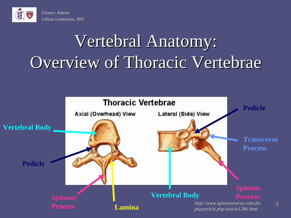
Vertebral Anatomy:Vertebral Anatomy: Overview of Thoracic VertebraeOverview of Thoracic Vertebrae
http://www.spineuniverse.com/dishttp://www.spineuniverse.com/dis playarticle.php/article1286.htmlplayarticle.php/article1286.html
Vertebral Body
Vertebral Body
LaminaSpinous Process
Spinous Process
Pedicle
Pedicle
Transverse Process
44
Eleanor AdamsEleanor AdamsGillian Lieberman, MDGillian Lieberman, MD
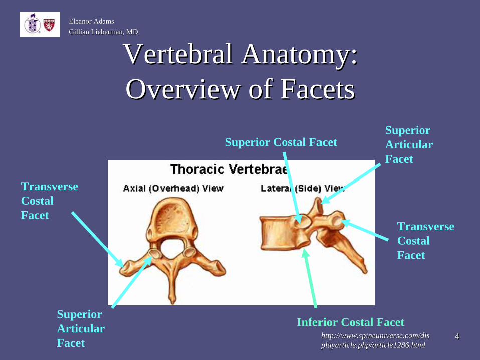
Vertebral Anatomy:Vertebral Anatomy: Overview of FacetsOverview of Facets
Superior Articular Facet
Superior Articular Facet
Transverse Costal Facet
Transverse Costal Facet
Inferior Costal Facet
Superior Costal Facet
http://www.spineuniverse.com/dishttp://www.spineuniverse.com/dis playarticle.php/article1286.htmlplayarticle.php/article1286.html
55
Eleanor AdamsEleanor AdamsGillian Lieberman, MDGillian Lieberman, MD
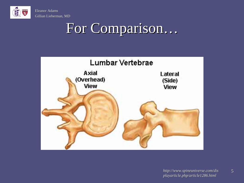
For Comparison…For Comparison…
http://www.spineuniverse.com/dishttp://www.spineuniverse.com/dis playarticle.php/article1286.htmlplayarticle.php/article1286.html
66
Eleanor AdamsEleanor AdamsGillian Lieberman, MDGillian Lieberman, MD
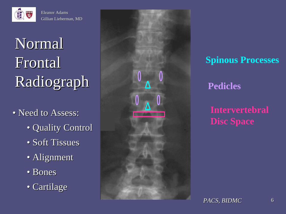
Normal Normal FrontalFrontal RadiographRadiograph
PACS, BIDMCPACS, BIDMC
Spinous Processes
Pedicles
Intervertebral Disc Space
•• Need to Assess: Need to Assess: •• Quality ControlQuality Control•• Soft TissuesSoft Tissues•• AlignmentAlignment•• BonesBones•• CartilageCartilage
77
Eleanor AdamsEleanor AdamsGillian Lieberman, MDGillian Lieberman, MD
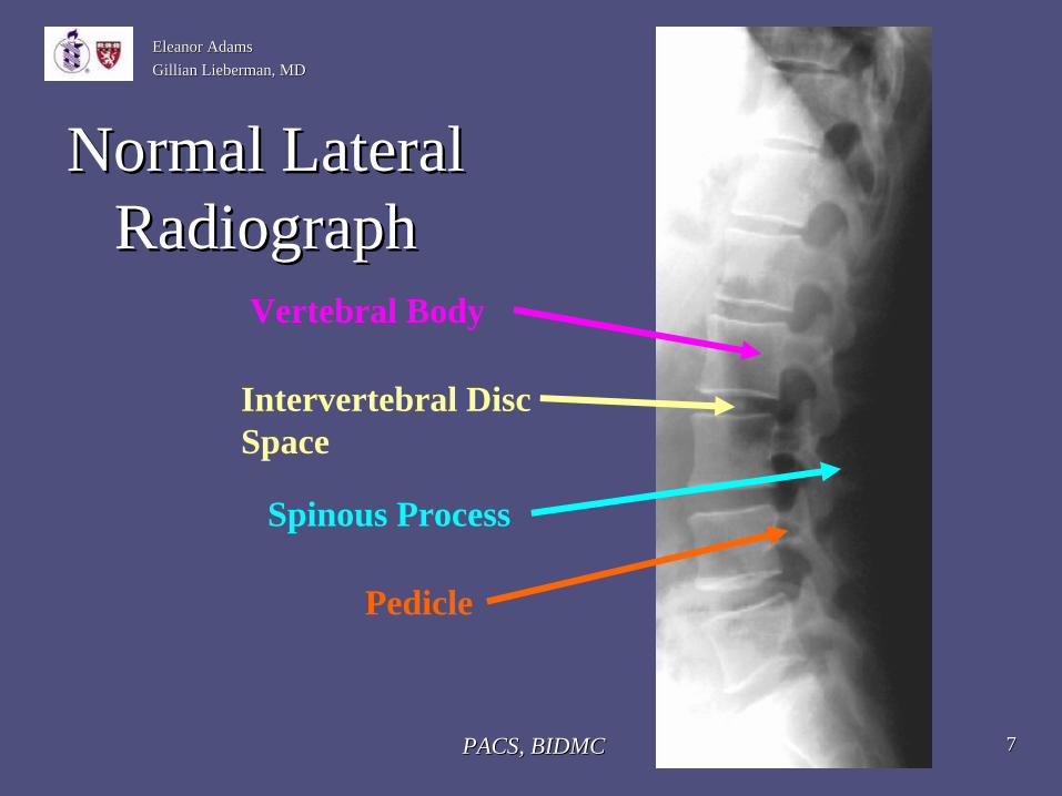
Normal Lateral Normal Lateral RadiographRadiograph
Vertebral Body
Pedicle
Spinous Process
Intervertebral Disc Space
PACS, BIDMCPACS, BIDMC
88
Eleanor AdamsEleanor AdamsGillian Lieberman, MDGillian Lieberman, MD
Spinal ColumnsSpinal Columns
1.) 1.) Anterior columnAnterior column–– Anterior longitudinal ligament, anterior half of the Anterior longitudinal ligament, anterior half of the
vertebral body, disc, and annulusvertebral body, disc, and annulus2.) 2.) Middle columnMiddle column
–– Posterior half of the vertebral body, disc, and Posterior half of the vertebral body, disc, and annulus, and the posterior longitudinal ligament annulus, and the posterior longitudinal ligament
3.) 3.) Posterior columnPosterior column–– Facet joints, Facet joints, ligamentumligamentum flavumflavum, the posterior , the posterior
elements and the interconnecting ligaments.elements and the interconnecting ligaments.
Panjabi et al. 1995Panjabi et al. 1995
99
Eleanor AdamsEleanor AdamsGillian Lieberman, MDGillian Lieberman, MD
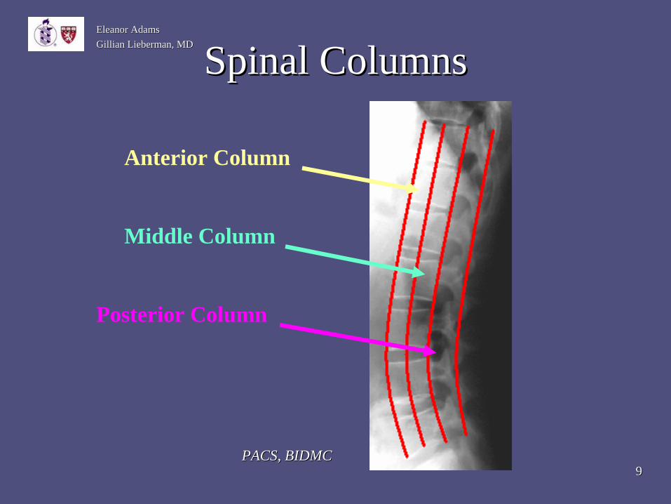
Spinal ColumnsSpinal Columns
Anterior Column
Middle Column
Posterior Column
PACS, BIDMCPACS, BIDMC

1010
Eleanor AdamsEleanor AdamsGillian Lieberman, MDGillian Lieberman, MD
Spinal ColumnsSpinal Columns
•• The spinal canal and cord are located in the The spinal canal and cord are located in the Posterior Column, adjacent to the Middle Posterior Column, adjacent to the Middle ColumnColumn
•• Therefore, fractures in elements in the Middle Therefore, fractures in elements in the Middle or Posterior Columns have the potential to or Posterior Columns have the potential to impinge on the spinal canal and cordimpinge on the spinal canal and cord
•• For this reason, Middle and Posterior Column For this reason, Middle and Posterior Column fractures are considered unstable. fractures are considered unstable.

1111
Eleanor AdamsEleanor AdamsGillian Lieberman, MDGillian Lieberman, MD
Types of FracturesTypes of FracturesType of FractureType of Fracture Column AffectedColumn Affected Stable vs. UnstableStable vs. Unstable
Compression/Wedge Compression/Wedge Anterior OnlyAnterior Only StableStableFractureFracture
Burst fracturesBurst fractures Anterior and MiddleAnterior and Middle UnstableUnstable
Fracture/Dislocation Fracture/Dislocation Anterior, Middle, Anterior, Middle, UnstableUnstableInjuryInjury PosteriorPosterior
Seat belt fracturesSeat belt fractures Anterior, Middle,Anterior, Middle, UnstableUnstablePosteriorPosterior
http://www.spineuniverse.com/displayarticle.php/article1441.html

1212
Eleanor AdamsEleanor AdamsGillian Lieberman, MDGillian Lieberman, MD
Patient LIPatient LI•• Patient LI, an 82 year old female with Patient LI, an 82 year old female with
osteoporosis and mild dementia, presented to osteoporosis and mild dementia, presented to her physician with lower back pain and her physician with lower back pain and posterior leg painposterior leg pain
•• Back pain present for 1Back pain present for 1--2 months2 months•• Difficulty getting out of bed in morning due to Difficulty getting out of bed in morning due to
painpain•• Loss of appetite because of intensity of painLoss of appetite because of intensity of pain

1313
Eleanor AdamsEleanor AdamsGillian Lieberman, MDGillian Lieberman, MD
Patient LIPatient LI
•• Given that Patient LI had Given that Patient LI had osteoporosis, her physician suspected osteoporosis, her physician suspected she had a compression fracture.she had a compression fracture.

1414
Eleanor AdamsEleanor AdamsGillian Lieberman, MDGillian Lieberman, MD
Compression FracturesCompression Fractures•• Osteoporosis is the leading cause of vertebral Osteoporosis is the leading cause of vertebral
compression fractures in the U.S.compression fractures in the U.S.•• 700,000 per year in U.S.700,000 per year in U.S.•• Affect 25% postmenopausal womenAffect 25% postmenopausal women•• Incidence expected to increase fourfold in next 50 Incidence expected to increase fourfold in next 50
yearsyears•• Why Important?Why Important?
–– Pain can lead to immobility and further disabilityPain can lead to immobility and further disability–– 15% increased mortality rate15% increased mortality rate–– Preventable in most casesPreventable in most cases
Old et al., 2004Old et al., 2004
1515
Eleanor AdamsEleanor AdamsGillian Lieberman, MDGillian Lieberman, MD
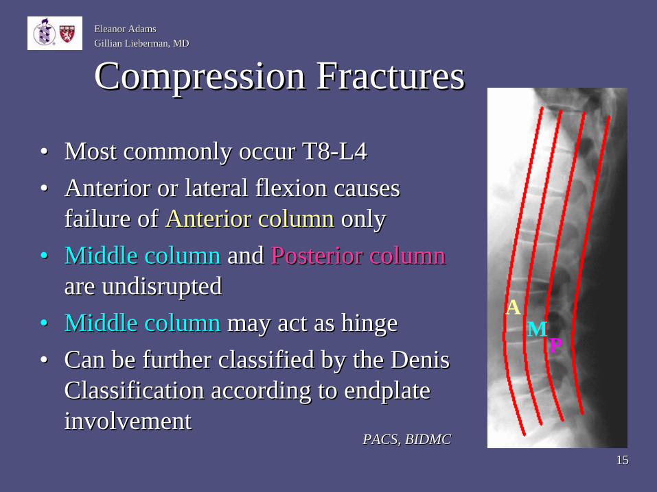
Compression FracturesCompression Fractures
•• Most commonly occur T8Most commonly occur T8--L4L4•• Anterior or lateral flexion causes Anterior or lateral flexion causes
failure of failure of Anterior columnAnterior column onlyonly•• Middle columnMiddle column and and Posterior column Posterior column
are undisruptedare undisrupted•• Middle columnMiddle column may act as hingemay act as hinge•• Can be further classified by the Denis Can be further classified by the Denis
Classification according to endplate Classification according to endplate involvementinvolvement
PM
A
PACS, BIDMCPACS, BIDMC

1616
Eleanor AdamsEleanor AdamsGillian Lieberman, MDGillian Lieberman, MD
Differential DiagnosisDifferential Diagnosis
•• AtraumaticAtraumatic Compression Fracture: Compression Fracture: –– Osteoporosis Osteoporosis
•• Senile/PostSenile/Post--Menopausal Menopausal •• SteroidsSteroids
–– OsteomalaciaOsteomalacia–– PagetsPagets DiseaseDisease–– Multiple MyelomaMultiple Myeloma–– HyperparathyroidismHyperparathyroidism
CedarsCedars--Sinai Medical Center, www.csmc.edu/7133.htmlSinai Medical Center, www.csmc.edu/7133.html

1717
Eleanor AdamsEleanor AdamsGillian Lieberman, MDGillian Lieberman, MD
What is your initial imaging test of What is your initial imaging test of choice?choice?
•• Plain frontal and lateral radiographs are the Plain frontal and lateral radiographs are the initial studies of choiceinitial studies of choice
•• In 20In 20--30% cases multiple fractures are present30% cases multiple fractures are present•• Important to image entire spineImportant to image entire spine
1818
Eleanor AdamsEleanor AdamsGillian Lieberman, MDGillian Lieberman, MD
Radiograph Findings of Radiograph Findings of Compression FracturesCompression Fractures
•• Anterior height of vertebral body is diminishedAnterior height of vertebral body is diminished•• Posterior height of vertebral body is normalPosterior height of vertebral body is normal•• No anterior or posterior translation of vertebral No anterior or posterior translation of vertebral
bodiesbodies•• If anterior compression is >40% when If anterior compression is >40% when
compared to posterior vertebral body height, compared to posterior vertebral body height, suspect burst fracturesuspect burst fracture
1919
Eleanor AdamsEleanor AdamsGillian Lieberman, MDGillian Lieberman, MD
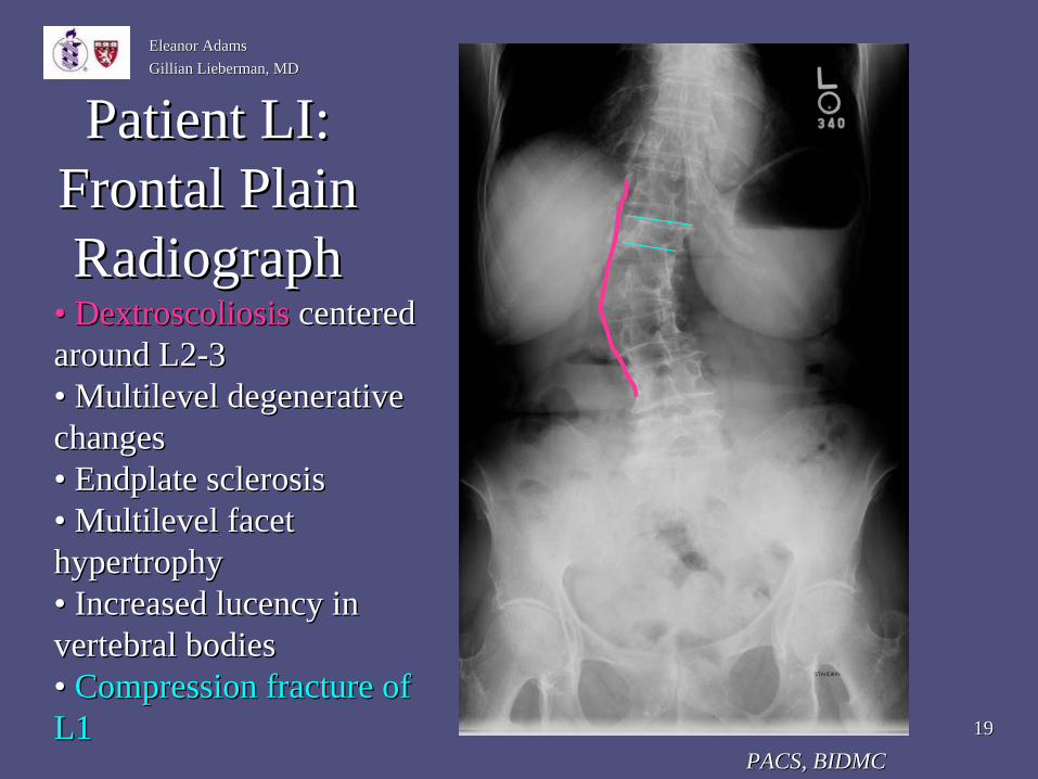
Patient LI: Patient LI: Frontal Plain Frontal Plain Radiograph Radiograph
•• DextroscoliosisDextroscoliosis centered centered around L2around L2--33•• Multilevel degenerative Multilevel degenerative changeschanges•• Endplate sclerosisEndplate sclerosis•• Multilevel facet Multilevel facet hypertrophyhypertrophy•• Increased Increased lucencylucency in in vertebral bodiesvertebral bodies•• Compression fracture of Compression fracture of L1L1
PACS, BIDMCPACS, BIDMC
2020
Eleanor AdamsEleanor AdamsGillian Lieberman, MDGillian Lieberman, MD
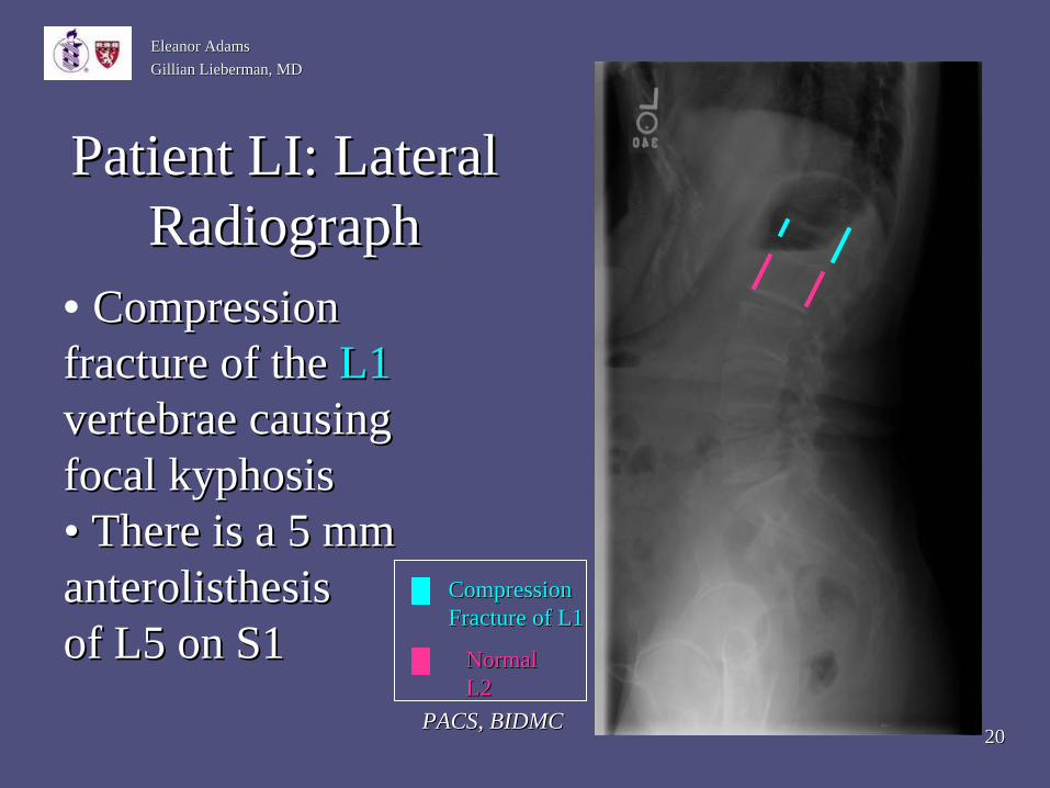
Patient LI: Lateral Patient LI: Lateral RadiographRadiograph
PACS, BIDMCPACS, BIDMC
• Compression Compression fracture of thefracture of the L1 L1 vertebrae causing vertebrae causing focal focal kyphosiskyphosis•• There is a 5 mm There is a 5 mm anterolisthesisanterolisthesisof L5 on S1of L5 on S1
Compression Compression Fracture of L1Fracture of L1
Normal Normal L2L2
2121
Eleanor AdamsEleanor AdamsGillian Lieberman, MDGillian Lieberman, MD
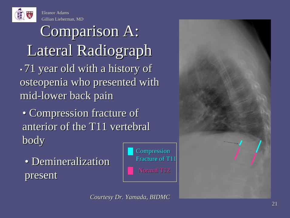
Comparison A: Comparison A: Lateral RadiographLateral Radiograph
•• 71 year old with a history of 71 year old with a history of osteopeniaosteopenia who presented with who presented with midmid--lower back painlower back pain
Courtesy Dr. Yamada, BIDMCCourtesy Dr. Yamada, BIDMC
Normal T12Normal T12
Compression Compression Fracture of T11Fracture of T11
•• Compression fracture of Compression fracture of anterior of the T11 vertebral anterior of the T11 vertebral bodybody
•• Demineralization Demineralization presentpresent

2222
Eleanor AdamsEleanor AdamsGillian Lieberman, MDGillian Lieberman, MD
Comparison B: Comparison B: Lateral RadiographLateral Radiograph
•• 82 year old who 82 year old who suffered a mechanical suffered a mechanical fall at her assisted living fall at her assisted living homehome
•• There is approximately There is approximately a 15% loss of anterior a 15% loss of anterior vertebral body height of vertebral body height of T12T12
Courtesy Dr. Yamada, BIDMCCourtesy Dr. Yamada, BIDMC
Compression Compression Fracture of T12Fracture of T12

2323
Eleanor AdamsEleanor AdamsGillian Lieberman, MDGillian Lieberman, MD
Role of Additional ImagingRole of Additional Imaging•• Role of CT:Role of CT:
•• Use to identify fractures not well visualized on plain filmUse to identify fractures not well visualized on plain film•• Allows for visualization of middle and posterior elementsAllows for visualization of middle and posterior elements
•• Can distinguish between compression fracture and burst fractureCan distinguish between compression fracture and burst fracture•• Can also reveal spinal canal narrowingCan also reveal spinal canal narrowing•• Disadvantage: Disadvantage:
•• Can’t detect horizontal fractures of vertebral bodies or pedicleCan’t detect horizontal fractures of vertebral bodies or pedicles s wellwell
•• Role of MRI:Role of MRI:•• Recommended when patient has suspected spinal cord Recommended when patient has suspected spinal cord
compression or other neurologic symptomscompression or other neurologic symptomsOld et al., 2004Old et al., 2004

2424
Eleanor AdamsEleanor AdamsGillian Lieberman, MDGillian Lieberman, MD
Patient LIPatient LI
•• Given that Patient LI reported posterior leg Given that Patient LI reported posterior leg pain, her physician decided to order an MRI to pain, her physician decided to order an MRI to assess the spinal cord and spinal canalassess the spinal cord and spinal canal

2525
Eleanor AdamsEleanor AdamsGillian Lieberman, MDGillian Lieberman, MD
Patient LI: T2 MRI Patient LI: T2 MRI
PACS, BIDMCPACS, BIDMC
SagittalSagittal
Axial Axial
2626
Eleanor AdamsEleanor AdamsGillian Lieberman, MDGillian Lieberman, MD
Patient LI: MRI FindingsPatient LI: MRI Findings
•• The The conusconus terminates at L1 terminates at L1 •• No evidence for internal No evidence for internal expansileexpansile massmass•• DextroscoliosisDextroscoliosis of the of the lumbosacrallumbosacral spine with spine with
apex at L2/3 apex at L2/3 •• L1 compression fracture L1 compression fracture •• Spinal Spinal stenosisstenosis

2727
Eleanor AdamsEleanor AdamsGillian Lieberman, MDGillian Lieberman, MD
Treatment OptionsTreatment Options
•• NonNon--operative treatment is the standardoperative treatment is the standard–– Pain medication (observe bowel motility)Pain medication (observe bowel motility)–– Brief rest (2Brief rest (2--3 days), encourage early ambulation3 days), encourage early ambulation–– Avoid compression overloads for 2 monthsAvoid compression overloads for 2 months–– Muscle relaxants, external back braces, and Muscle relaxants, external back braces, and
physical therapy may also help physical therapy may also help •• If patients do not respond to conservative If patients do not respond to conservative txtx::
–– PercutaneousPercutaneous VertebroplastyVertebroplasty–– KyphoplastyKyphoplasty
Singh et al., 2006Singh et al., 2006

2828
Eleanor AdamsEleanor AdamsGillian Lieberman, MDGillian Lieberman, MD
SummarySummary
•• Compression fractures common in elderly populationCompression fractures common in elderly population•• Compression fractures are caused by failure of the Compression fractures are caused by failure of the
anterior column onlyanterior column only•• Initial imaging modality of choice is plain filmInitial imaging modality of choice is plain film•• Can use CT or MRI if have concern that the middle Can use CT or MRI if have concern that the middle
or posterior columns are involved, and to evaluate or posterior columns are involved, and to evaluate spinal cordspinal cord
•• Treatment is usually conservativeTreatment is usually conservative

2929
Eleanor AdamsEleanor AdamsGillian Lieberman, MDGillian Lieberman, MD
ReferencesReferences•• De De SmetSmet AA, Robinson RG, Johnson BE, AA, Robinson RG, Johnson BE, LukertLukert BP. Spinal Compression BP. Spinal Compression
Fractures and Osteoporotic Women: Patterns and Relationship to Fractures and Osteoporotic Women: Patterns and Relationship to HyperkyphosisHyperkyphosis. . RadiologyRadiology 1988; 166:4971988; 166:497--500. 500.
•• KrothKroth PJ, Murray MD, McDonald CJ. PJ, Murray MD, McDonald CJ. UndertreatmentUndertreatment of osteoporosis in women, of osteoporosis in women, based on detection of vertebral compression fractures on chest rbased on detection of vertebral compression fractures on chest radiography. adiography. Am J Am J GeriatrGeriatr PharmacotherPharmacother 2004; 2(2):1122004; 2(2):112--118. 118.
•• Old JL, Calvert M. Vertebral Compression Fractures in the ElderlOld JL, Calvert M. Vertebral Compression Fractures in the Elderly. y. Am Am AcadAcad FamFam PhysPhys 2004: 69(1):1112004: 69(1):111--116.116.
•• Panjabi MM, Panjabi MM, OxlandOxland TR, TR, KifuneKifune M, M, ArandArand M, M, WenWen L, Chen A. Validity of the L, Chen A. Validity of the threethree--column theory of column theory of thoracolumbarthoracolumbar fractures. fractures. SpineSpine 1995; 20(10):11221995; 20(10):1122--1127.1127.
•• Singh AK, Singh AK, PilgramPilgram TK, TK, GilulaGilula LA. Osteoporotic Compression Fractures: Outcomes LA. Osteoporotic Compression Fractures: Outcomes after Single versus Multipleafter Single versus Multiple--Level Level PercutaneousPercutaneous VertebroplastyVertebroplasty. . RadiologyRadiology 2006; 2006; 238(1):211238(1):211--220.220.
•• YuhYuh WT, WT, ZacherZacher CK, CK, BarloonBarloon TJ, Sato Y, TJ, Sato Y, SickelsSickels WJ, Hawes DR. Vertebral WJ, Hawes DR. Vertebral Compression Fractures: Distinction between Benign and Malignant Compression Fractures: Distinction between Benign and Malignant Causes with Causes with MR Imaging. MR Imaging. RadiologyRadiology 1989; 172:2151989; 172:215--218.218.
3030
Eleanor AdamsEleanor AdamsGillian Lieberman, MDGillian Lieberman, MD
AcknowledgementsAcknowledgements
•• Special Thanks to:Special Thanks to:
•• Gillian Lieberman, MDGillian Lieberman, MD•• Maryellen Sun, MDMaryellen Sun, MD•• Kei Yamada, MDKei Yamada, MD•• Pamela Pamela LepkowskiLepkowski•• Larry BarbarasLarry Barbaras