comprehensive treatments for congenitally...
TRANSCRIPT

Case ReportComprehensive Treatments for Congenitally Missing Teeth andGeneralized Diastema
Mehran Bahrami,1 Fariba Saleh Saber,2 and Amirreza Hendi3
1Dental Research Center, Department of Prosthodontics, Tehran University of Medical Sciences, School of Dentistry, Tehran, Iran2Department of Prosthodontics, Tabriz University of Medical Sciences, School of Dentistry, Tabriz, Iran3Department of Prosthodontics, Tehran University of Medical Sciences, School of Dentistry, Tehran, Iran
Correspondence should be addressed to Amirreza Hendi; [email protected]
Received 4 April 2017; Accepted 13 June 2017; Published 10 July 2017
Academic Editor: Andrea Scribante
Copyright © 2017 Mehran Bahrami et al. This is an open access article distributed under the Creative Commons AttributionLicense, which permits unrestricted use, distribution, and reproduction in any medium, provided the original work is properlycited.
Congenitally missing teeth and/or hypodontia is a prevalent dental anomaly. There are different treatment options available forthese conditions such as space maintenance, restoring the space by resin-bonded-fixed-partial-dentures (RBFPDs), and dentalimplants. This study addresses the comprehensive treatments for congenitally missing tooth and diastema using interdisciplinaryapproaches. One patient was treated with small-diameter-implants and the other one was treated using an intraoral scanner tomake digital impression and fabricating RBFPDs with CAD/CAM system. Both patients were completely satisfied.
1. Introduction
Hypodontia is used to describe the congenital absence ofone or more primary or secondary teeth. Excluding the thirdmolars, the mandibular second premolars are the most fre-quentlymissing teeth, comprising between 60% to 72% of thetotal number of missing teeth [1].
The congenital absence of teeth and generalized diastemacan seriously affect a young person both physically and emo-tionally, especially when the missing teeth and diastema arelocated in the anterior region of themouth [2]. Complicationsassociated with missing permanent teeth included maloc-clusion, periodontal problems, lack of alveolar-bone-growth,and unfavorable appearance [3].
The treatment options available for these patientsincluded maintenance of the primary teeth, space closureby orthodontic treatment, space maintenance, restoring byRBFPDs, tooth transplantation, or dental implants [2].
Regardless of whether the location is posterior or anterior,RBFPDs have been accepted as an alternative treatmentto conventional fixed-partial-dentures (FPDs) when intactabutments are present and minimal intervention is desired[4]. The main advantage of RBFPDs in comparison to con-ventional FPDs preparations is that they are conservative to
tooth structure. SoRBFPDs can be considered as the interme-diate or definitive restorations following orthodontic treat-ment of congenitally absent teeth. The most common failureof RBFPDs is debonding and the patients should be aware ofthat [2].
Today, the first choice of restoration for a congenitallymissing tooth should be a single-tooth implant [5]. Theaverage implant platform, which is 4.0mm wide, requires aminimummesiodistal space of 1.0mm between the platformand the adjacent tooth to facilitate proper healing and thedevelopment of interdental papilla. So a minimum of 6mmspace is required for the crown.The use of standard-diameterimplants is common in implant treatments, but the lackof interdental space leads to unfavorable aesthetic results.Recently, the use of small-diameter implants has becomemore common [6]. Small-diameter implants (SDIs) or mini-dental implants (MDIs) generally are considered to be lessthan 3mm in diameter. SDIs are introduced to overcomebone-quantity problems with a degree of success comparableto that of standard-diameter implants.The various designs ofSDIs have become more commonly used in recent decadesdue to limitations in the geometry and capacity of the alveolarbone [6, 7].
HindawiCase Reports in DentistryVolume 2017, Article ID 3254873, 5 pageshttps://doi.org/10.1155/2017/3254873
2 Case Reports in Dentistry
Figure 1: Intraoral frontal view before orthodontic treatment shows generalized diastema.
(a) (b)
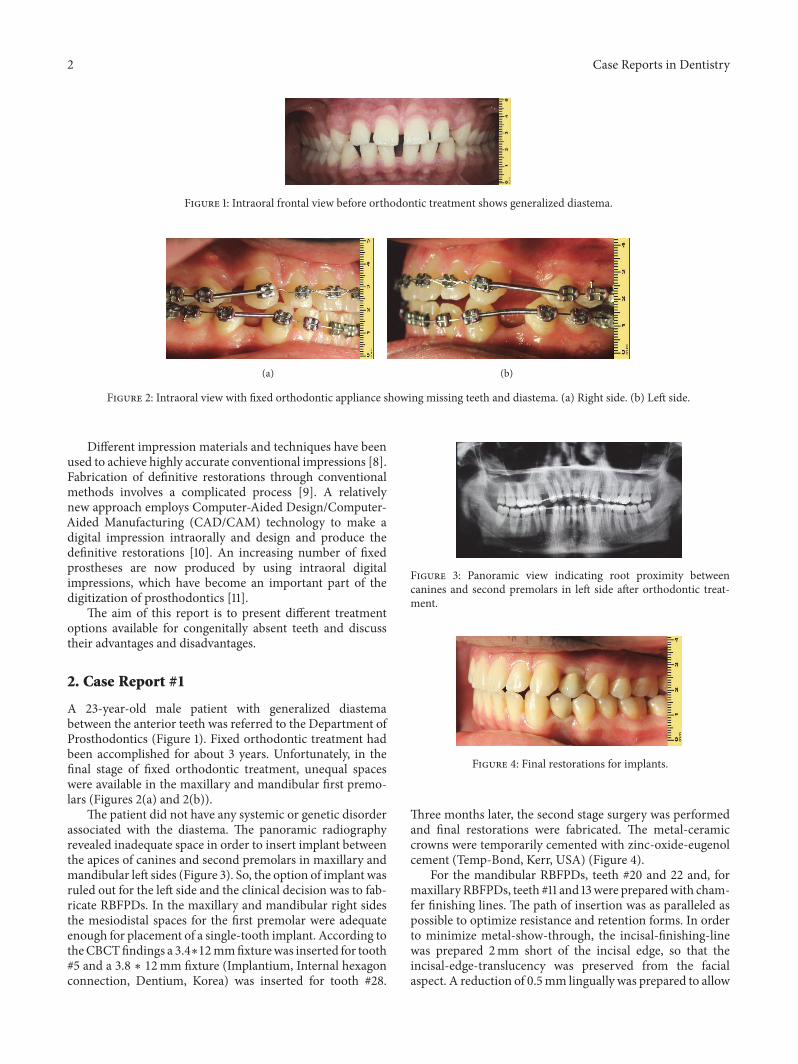
Figure 2: Intraoral view with fixed orthodontic appliance showing missing teeth and diastema. (a) Right side. (b) Left side.
Different impression materials and techniques have beenused to achieve highly accurate conventional impressions [8].Fabrication of definitive restorations through conventionalmethods involves a complicated process [9]. A relativelynew approach employs Computer-Aided Design/Computer-Aided Manufacturing (CAD/CAM) technology to make adigital impression intraorally and design and produce thedefinitive restorations [10]. An increasing number of fixedprostheses are now produced by using intraoral digitalimpressions, which have become an important part of thedigitization of prosthodontics [11].
The aim of this report is to present different treatmentoptions available for congenitally absent teeth and discusstheir advantages and disadvantages.
2. Case Report #1
A 23-year-old male patient with generalized diastemabetween the anterior teeth was referred to the Department ofProsthodontics (Figure 1). Fixed orthodontic treatment hadbeen accomplished for about 3 years. Unfortunately, in thefinal stage of fixed orthodontic treatment, unequal spaceswere available in the maxillary and mandibular first premo-lars (Figures 2(a) and 2(b)).
The patient did not have any systemic or genetic disorderassociated with the diastema. The panoramic radiographyrevealed inadequate space in order to insert implant betweenthe apices of canines and second premolars in maxillary andmandibular left sides (Figure 3). So, the option of implant wasruled out for the left side and the clinical decision was to fab-ricate RBFPDs. In the maxillary and mandibular right sidesthe mesiodistal spaces for the first premolar were adequateenough for placement of a single-tooth implant. According totheCBCTfindings a 3.4∗12mmfixturewas inserted for tooth#5 and a 3.8 ∗ 12mm fixture (Implantium, Internal hexagonconnection, Dentium, Korea) was inserted for tooth #28.
Figure 3: Panoramic view indicating root proximity betweencanines and second premolars in left side after orthodontic treat-ment.
Figure 4: Final restorations for implants.
Three months later, the second stage surgery was performedand final restorations were fabricated. The metal-ceramiccrowns were temporarily cemented with zinc-oxide-eugenolcement (Temp-Bond, Kerr, USA) (Figure 4).
For the mandibular RBFPDs, teeth #20 and 22 and, formaxillary RBFPDs, teeth #11 and 13were preparedwith cham-fer finishing lines. The path of insertion was as paralleled aspossible to optimize resistance and retention forms. In orderto minimize metal-show-through, the incisal-finishing-linewas prepared 2mm short of the incisal edge, so that theincisal-edge-translucency was preserved from the facialaspect. A reduction of 0.5mm lingually was prepared to allow

Case Reports in Dentistry 3
Figure 5: Intraoral digital impressions and interocclusal record.
Figure 6: Resin-bonded-restorations delivery view.
the adequate bulk of metal to obtain enough strength of theretainers. A light chamfer-finishing-line was prepared 1mmsupragingivally. Supragingivally light chamfer maintains thepreparation in the enamel which is essential for the optimalbonding. Subgingival marginsmay also cause some problemsin digital systems. Interproximal-finishing-lines ended at thecenter of the contact areas. The preparation features corre-spond primarily to the optical-scanner-potentiality as wellas to the milling machine capabilities. Undercut areas andsmall spikes or irregular surfaces on the preparation marginwere eliminated. Digital impressions and inter-arch-recordswere made using 3shape intraoral scanner (3shape, Den-mark) (Figure 5). RBFPDs frameworks were designed using3shape dental system and the frameworks were sent to thelaboratory. The frameworks were milled using Cr-Co blocks.The porcelain try-in was done and a mutually protectedocclusion was selected for the patient. The RBFPDs werecemented using resin cement (Panavia, Kuraray America,Inc.) (Figure 6).
The patient was followed up every 6 months during a 2-year period.
3. Case Report #2
A20-year-old female who had congenitallymissingmaxillarycanines with anterior diastema in maxilla and crowding inanterior segment ofmandible was referred to theDepartmentof Prosthodontics. Removable-orthodontic-treatments wereperformed for maxillary and mandibular arches. One of themandibular-central-incisors was extracted to gain sufficientspace and to resolve the crowding. Fixed retainer was usedto maintain the position of mandibular-anterior-teeth. In themaxilla, orthodontic removable retainer was used to elimi-nate the diastema by providing 3mm spaces in the canine
areas (Figures 7(a) and 7(b)). Clinical examination revealedinadequate space to insert regular implants between the lat-erals and first premolars in both sides.Theminimal proximalpreparation of the adjacent teeth was performed to increasethe mesiodistal spaces to 4mm. The RBFPD treatment waspresented to the patient, but she refused to accept teethpreparation. As there was not enough mesial-distal space,one-piece SDIs, 2∗12mm (SlimLine, Dentium, Korea), wereconsidered to replace the maxillary canines.
Immediately following implant placement, provisionalrestorations were fabricated with temporary acrylic resin(UNIFAST� LC, GC co).The provisional restorations had nocentric or eccentric occlusal contact points.
The patient was followed up after one week. In thisappointment, the sutureswere removed. After a healing phaseof three months, the final impression was taken with polyvinyl siloxane impression material (A-silicones, KettenbachGmbH & Co. KG). The porcelain-fused-to-metal crownswere adjusted to have light occlusal contacts in centricocclusion. Partially group-function occlusion was given tothe patient (Figure 8).
The patient was followed up every 6 months for two years(Figure 9).
4. Discussion
Comprehensive treatment of patients with missing teethand/or hypodontia is difficult. It requires a teamwork includ-ing an orthodontist, a prosthodontist, and a surgeon (in caseof implant insertions) to achieve ideal results.
Dental implants may be considered as the best treatmentoption for patients with hypodontia. However, using dentalimplants in patients with hypodontia may be challengingdue to some limitations such as reduced mesiodistal space,poor bone quality and quantity (especially after orthodontictreatment), and compromised implants positions.
The orthodontist plays an important role in determiningand establishing the space requirements for patients withmissing teeth [12]. Then the prosthodontist should reassessthe available space required for the implant fixture using theappropriate radiographs.
In some patients only SDIs should be considered. Thesurvival rate of SDIs appears to be similar to that of regulardiameter implants. Meta-analysis indicated an estimated94.5% for 5-year survival of implant supported single crowns;therefore, the single-tooth implant has become a commontreatment option for the replacement of congenitally absentteeth [6].
In patients with congenitally missing permanent teeth,orthodontic treatment is the gold standard [12]. However,orthodontic treatment can cause some potential risks andcomplications, because teeth undergone orthodontic move-ment may have resorption of cementum and dentine [13].On the other hand, in some patients, it is impossible toinsert even SDIs after orthodontic treatment because of theapices of the adjacent teeth roots. When adjacent teeth havebeen orthodontically moved in order to gain adequate space,radiographic examination often reveals either insufficientinterradicular space available for an implant fixture or an

4 Case Reports in Dentistry
(a) (b)
Figure 7: Intraoral view with removable orthodontic appliance showing missing teeth and diastema. (a) Right side. (b) Left side.
Figure 8: Final restoration delivery view.
Figure 9: Panoramic image after two years of follow-up.
absence of root parallelism [5]. In this condition there maynot be adequate space even to place SDIs between the apices.So the use of RBFPDs will be recommended. Tooth reductionis conservative for RBFPDs preparation because of remainingin the enamel. This is one of the numerous advantages ofthis restoration; however, the three most common compli-cations associated with RBFPDs are debonding (21%), toothdiscoloration (18%), and caries (7%), although it has beenreported that debonding does not appear to affect the patient’ssatisfaction and there is usually limited damage to abutmentteeth [4, 14–16].
One major parameter for clinical success is the fit of arestoration [17]. The CAD/CAM systems are claimed to bemore efficient than the conventional methods [18, 19]. Kugelet al. in an in vitro study showed that there were no significantdifferences in the marginal accuracy and fit of the crownsmade by Lava COS and PVS impressions [20].
Recent systematic reviews have estimated that the five-year survival rates are 87.7% for RBFPDs and over 90% forconventional FPDs. Although these rates are lower than the94.5% success reported for 5-year survival rate of implantsupported single crowns, RBFPDs have the advantages of
being less invasive and requiring a shorter total treatmenttime [14].
5. Conclusions
In the current case reports with congenitally missing teethand diastema, optimal aesthetics and function were providedeither by using small-diameter-implant-retained crowns orRBFPDs.
Additional Points
Key Messages. Optimal aesthetics and function can beprovided by using either small-diameter-implant-retainedcrowns or RBFPDs.
Conflicts of Interest
The authors declare that they have no conflicts of interest.
Acknowledgments
The authors would like to thank Mr. Fazel Hasanzadeh andMr. Sadegh Esfandyari for their laboratory works.
References
[1] B. Svedmyr, “Genealogy and consequences of congenitallymiss-ing second premolars,” Journal of the International Associationof Dentistry for Children, vol. 14, no. 2, pp. 77–82, 1983.
[2] A. L. do Valle, F. C. Lorenzoni, L. M. Martins et al., “A multi-disciplinary approach for the management of hypodontia: Casereport,” Journal of Applied Oral Science, vol. 19, no. 5, pp. 544–548, 2011.
[3] V. Rakhshan, “Congenitally missing teeth (hypodontia): areview of the literature concerning the etiology, prevalence, riskfactors, patterns and treatment,”Dental Research Journal, vol. 12,no. 1, pp. 1–13, 2015.
[4] H. Shimizu, T. Kawaguchi, andY. Takahashi, “The current statusof the design of resin-bonded fixed partial dentures, splints andovercastings,” Japanese Dental Science Review, vol. 50, no. 2, pp.23–28, 2014.
[5] V. G. Kokich andV.O. Kokich, “Congenitallymissingmandibu-lar second premolars: clinical options,” American Journal ofOrthodontics and Dentofacial Orthopedics, vol. 130, no. 4, pp.437–444, 2006.
[6] M. Alharissy and S. Dayoub, “Clinical evaluation of the use ofminiimplants to replace congenitally absent maxillary lateral

Case Reports in Dentistry 5
incisors: a short-term study,” International Dental Research, vol.2, no. 1, pp. 27–32, 2012.
[7] G. J. Christensen, “The increased use of small-diameterimplants,” Journal of the American Dental Association, vol. 140,no. 6, pp. 709–712, 2009.
[8] S. Caputi and G. Varvara, “Dimensional accuracy of resultantcasts made by a monophase, one-step and two-step, and a noveltwo-step putty/light-body impression technique: an in vitrostudy,”The Journal of Prosthetic Dentistry, vol. 99, no. 4, pp. 274–281, 2008.
[9] N. Bittner, T. Hill, and A. Randi, “Evaluation of a one-piecemilled zirconia post and core with different post-and-coresystems: An in vitro study,” Journal of Prosthetic Dentistry, vol.103, no. 6, pp. 369–379, 2010.
[10] A.Mehl, A. Ender,W.Mormann, andT. Attin, “Accuracy testingof a new intraoral 3D camera,” International Journal of Comput-erized Dentistry, vol. 12, no. 1, pp. 11–28, 2009.
[11] S. Ting-Shu and S. Jian, “Intraoral digital impression technique:a review,” Journal of Prosthodontics, vol. 24, no. 4, pp. 313–321,2015.
[12] G. A. Kinzer and V. O. Kokich Jr., “Managing congenitallymissing lateral incisors. Part II: Tooth-supported restorations,”Journal of Esthetic andRestorativeDentistry, vol. 17, no. 2, pp. 76–84, 2005.
[13] M. Wishney, “Potential risks of orthodontic therapy: a criticalreview and conceptual framework,” Australian Dental Journal,vol. 62, pp. 86–96, 2017.
[14] K. A. Durey, P. J. Nixon, S. Robinson, and M. F. W.-Y. Chan,“Resin bonded bridges: techniques for success,” The BritishDental Journal, vol. 211, no. 3, pp. 113–118, 2011.
[15] U. Lally, “Resin-bonded fixed partial dentures past andpresent—an overview,” Journal of the Irish Dental Association,vol. 58, no. 6, pp. 294–300, 2013.
[16] R. Kuijs, A. van Dalen, J. Roeters, and D.Wismeijer, “The resin-bonded fixed partial denture as the first treatment considerationto replace a missing tooth,” International Journal of Prosthodon-tics, vol. 29, no. 4, pp. 337–339, 2016.
[17] A. O. Ali, “Accuracy of digital impressions achieved from fivedifferent digital impression systems,” Dentistry, vol. 5, no. 05,pp. 2–6, 2015.
[18] P. K. Brawek, S. Wolfart, L. Endres, A. Kirsten, and S. Reich,“The clinical accuracy of single crowns exclusively fabricated bydigital workflow—the comparison of two systems,”Clinical OralInvestigations, vol. 17, no. 9, pp. 2119–2125, 2013.
[19] A. Ender, T. Attin, and A. Mehl, “In vivo precision of conven-tional and digital methods of obtaining complete-arch dentalimpressions,” Journal of Prosthetic Dentistry, vol. 115, no. 3, pp.313–320, 2016.
[20] G. Kugel, N. Chaimattayompol, R. Perry, S. Ferreira, S. Sharma,and J. Towers, “Comparison of digital vs. conventional impres-sion systems for marginal accuracy,” Journal of Dental Research,vol. 87, pp. 11–19, 2008.

Submit your manuscripts athttps://www.hindawi.com
Hindawi Publishing Corporationhttp://www.hindawi.com Volume 2014
Oral OncologyJournal of
DentistryInternational Journal of
Hindawi Publishing Corporationhttp://www.hindawi.com Volume 2014
Hindawi Publishing Corporationhttp://www.hindawi.com Volume 2014
International Journal of
Biomaterials
Hindawi Publishing Corporationhttp://www.hindawi.com Volume 2014
BioMed Research International
Hindawi Publishing Corporationhttp://www.hindawi.com Volume 2014
Case Reports in Dentistry
Hindawi Publishing Corporationhttp://www.hindawi.com Volume 2014
Oral ImplantsJournal of
Hindawi Publishing Corporationhttp://www.hindawi.com Volume 2014
Anesthesiology Research and Practice
Hindawi Publishing Corporationhttp://www.hindawi.com Volume 2014
Radiology Research and Practice
Environmental and Public Health
Journal of
Hindawi Publishing Corporationhttp://www.hindawi.com Volume 2014
The Scientific World JournalHindawi Publishing Corporation http://www.hindawi.com Volume 2014
Hindawi Publishing Corporationhttp://www.hindawi.com Volume 2014
Dental SurgeryJournal of
Drug DeliveryJournal of
Hindawi Publishing Corporationhttp://www.hindawi.com Volume 2014
Hindawi Publishing Corporationhttp://www.hindawi.com Volume 2014
Oral DiseasesJournal of
Hindawi Publishing Corporationhttp://www.hindawi.com Volume 2014
Computational and Mathematical Methods in Medicine
ScientificaHindawi Publishing Corporationhttp://www.hindawi.com Volume 2014
PainResearch and TreatmentHindawi Publishing Corporationhttp://www.hindawi.com Volume 2014
Preventive MedicineAdvances in
Hindawi Publishing Corporationhttp://www.hindawi.com Volume 2014
EndocrinologyInternational Journal of
Hindawi Publishing Corporationhttp://www.hindawi.com Volume 2014
Hindawi Publishing Corporationhttp://www.hindawi.com Volume 2014
OrthopedicsAdvances in