composite palatal veneers to restore a case of severe ...024-034.pdf · composite resin (enamel...
TRANSCRIPT
ItalIan Journal of Dental MeDIcIne vol. 2/1-201724
Aim In this case report a conservative approach for the management of dental erosion is described. The restorations are based only on adhesive techniques. Case report A 46 years-old patient presented at the Geneva Erosion Study at Geneva University for diffused dental erosion of intrinsic etiology. The treatment plan was based only on adhesive technique and six palatal veneers were necessary to restore the very compromised front teeth. Moreover, the palatal veneers restorations did not require any tooth preparation, and the teeth kept their vitality. The clinical results at the five year follow-up, confirmed esthetic, biological and mechanical success.Conclusion The adhesive technique described proved to be the most appropriate therapy. and also the overall treatment was more affordable for the patient.
KEYWORDS ABSTRACT
Francesca Vailati, MD, DMD, MSc
Private practice, Senior Lecturer, Department of fixed prosthodontics and occlusion, School of dental medicine, University of Geneva, Switzerland
Conservative approach, Dental erosion, Veneers.
Composite palatal veneers to restore a case of severe dental erosion, from minimally to non invasive dentistry: a 5-year follow-up case report
In the author’s opinion, the dental community should stress more the importance of the tooth preservation (biological success), rather than aim for the “10 guarantee” of their work. A vital tooth restored for the first time with a crown could last 10 years, but if the same tooth looses the vitality and the remaining tooth structure becomes compromised (insufficient ferule), can the “10 year deal” still be guaranteed for the new restoration? The clinician’s capacity to communicate the switch in paradigm (biological success versus the mechanical success) is fundamental, before proposing these extremely conservative treatments, because patients must accept that the restorations are now weaker and eventually will fail over time; however, the restored teeth will keep their integrity and a repair or a replacement with a similar restoration will always be possible. This is not always the case, for example, when a devitalized tooth, restored with a crown, fails because of root fracture.
In this article, the treatment based only on adhesive techniques of a patient affected by several dental erosion is illustrated. His maxillary anterior teeth were supposed to be devitalized in order to be restored with full-crown restorations. The patient refused this aggressive option and sought consultation in the Geneva Erosion Study at Geneva University. A new treatment plan was proposed, based only on adhesive technique; six palatal and six
IntroductionFrequently patients affected by dental erosion are not
immediately treated, since tooth wear still represents an overwhelming challenge. Some clinicians consider erosive wear as a physiological process related to age, rather than a pathology that should be immediately treated. Other dentists, instead, recognize the problem, but they are not comfortable to propose a full-mouth rehabilitation to their patients, especially at the early stage of the disease, waiting for more tooth destruction to happen to justify the treatment. Nowadays, thanks to the adhesive techniques, dental erosion could be immediately addressed, the tooth destruction slowed down, with minimal to no additional tooth loss during the delivering of the restorations (1-22).
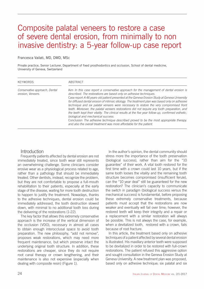
The key factor that allows this extremely conservative approach is the increase of the vertical dimension of the occlusion (VDO), necessary in almost all cases to obtain enough interocclusal space to avoid tooth preparation. The new philosophy, “add not remove”, proposes weak restorations, which may need more frequent maintenance, but which preserve intact the underlying original tooth structure. In addition, these restorations are cheaper, since they do not require root canal therapy or crown lengthening, and their maintenance is also not expensive (especially when dealing with composite resin) (Figure 1).
[email protected] 24 21/04/17 12:35
ItalIan Journal of Dental MeDIcIne vol. 2/1-2017 25
facial veneers (Sandwich approach) were considered to restore his maxillary anterior teeth and to preserve to a maximum the remaining tooth structure. However, the final treatment became even more conservative than expected, since only six palatal veneers were necessary to restore the very compromised teeth. The clinical results (esthetic, biological and mechanical success) at the five year follow-up, confirmed that the adhesive approach chosen was the most appropriate therapy. Not only the palatal veneers restorations did not require any tooth preparation, and the teeth kept their vitality, but also the overall treatment was more affordable for the patient.
Case presentationA 46 year old patient presented to the University
of Geneva, school of Dental Medicine, with the chief complaint that “his teeth were deteriorating at a high speed and he finally wanted to do something about it”. At the anamnesis, the patient remembered that his dentist proposed to restore his dentition by means of crowns and that he was not convinced by this treatment plan. Since then, he had sought dental treatment on an irregular basis. After several years of neglecting his mouth, he was finally addressed to the Geneva Erosion Study, to investigate if other types of treatments than crowns were available. During the
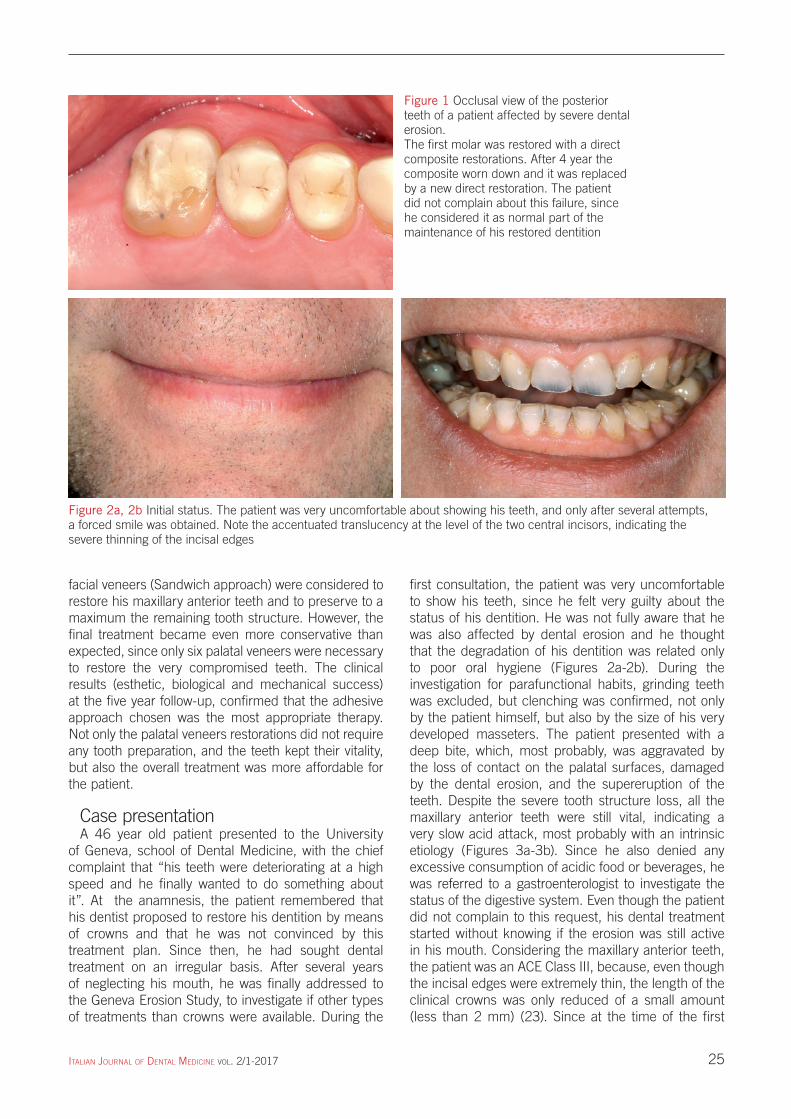
first consultation, the patient was very uncomfortable to show his teeth, since he felt very guilty about the status of his dentition. He was not fully aware that he was also affected by dental erosion and he thought that the degradation of his dentition was related only to poor oral hygiene (Figures 2a-2b). During the investigation for parafunctional habits, grinding teeth was excluded, but clenching was confirmed, not only by the patient himself, but also by the size of his very developed masseters. The patient presented with a deep bite, which, most probably, was aggravated by the loss of contact on the palatal surfaces, damaged by the dental erosion, and the supereruption of the teeth. Despite the severe tooth structure loss, all the maxillary anterior teeth were still vital, indicating a very slow acid attack, most probably with an intrinsic etiology (Figures 3a-3b). Since he also denied any excessive consumption of acidic food or beverages, he was referred to a gastroenterologist to investigate the status of the digestive system. Even though the patient did not complain to this request, his dental treatment started without knowing if the erosion was still active in his mouth. Considering the maxillary anterior teeth, the patient was an ACE Class III, because, even though the incisal edges were extremely thin, the length of the clinical crowns was only reduced of a small amount (less than 2 mm) (23). Since at the time of the first
Figure 1 Occlusal view of the posterior teeth of a patient affected by severe dental erosion.The first molar was restored with a direct composite restorations. After 4 year the composite worn down and it was replaced by a new direct restoration. The patient did not complain about this failure, since he considered it as normal part of the maintenance of his restored dentition
Figure 2a, 2b Initial status. The patient was very uncomfortable about showing his teeth, and only after several attempts, a forced smile was obtained. Note the accentuated translucency at the level of the two central incisors, indicating the severe thinning of the incisal edges
[email protected] 25 21/04/17 12:35

ItalIan Journal of Dental MeDIcIne vol. 2/1-201726
and expensive, not only for the additional six facial veneers, but also for the veneer/onlays considered to restore all the maxillary and mandibular premolars. This plan was, however, simplified, while the treatment was progressing. The case started following a classic three step technique approach (24-26). Two alginate
consultation in the Geneva Erosion Study, however, the ACE classification had not been developed jet, it was planned to restore his maxillary anterior teeth not only with palatal, but also with facial veneers (Sandwich approach). As a result, the initial treatment plan for his full-mouth adhesive rehabilitation was more invasive
Figure 3a, 3b Occlusal and lateral views of the initial status. The palatal destruction was very advanced and the teeth undermined, but due to the patients’ deep bite, the incisal edges fractured very little. The palatal enamel was present only at the cervical level. Interproximal caries weakened the teeth furthermore, nevertheless, they all were still vital. The posterior teeth were also very compromised and the clinical crowns were very short
Figure 4a, 4c Analyzing the lingualized position of the two central incisors, an additive mock-up was considered possible. The laboratory technician slightly bulked the vestibular aspects of the maxillary teeth to reduce the need of tooth preparation, while delivering the facial veneers
Figure 5a, 5b Following the classic three step technique, a maxillary vestibular mock-up was done, which extended up to the first molars (I clinical step). The incisal edges and the occlusal plane were slightly lengthened. The esthetic outcome was of course improved, but the tooth preparation for placing the facial veneers and the veneer/ onlays for the posterior teeth would have been conspicuous, since the vestibular surfaces were almost intact
[email protected] 26 21/04/17 12:35
ItalIan Journal of Dental MeDIcIne vol. 2/1-2017 27
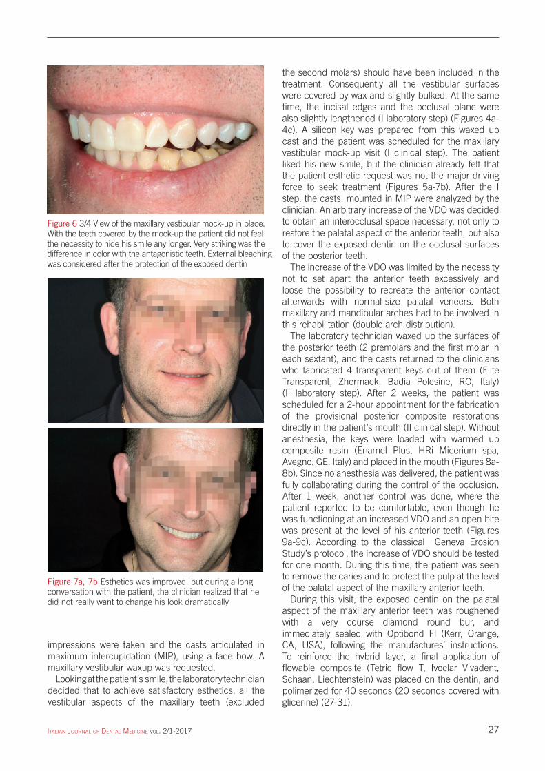
the second molars) should have been included in the treatment. Consequently all the vestibular surfaces were covered by wax and slightly bulked. At the same time, the incisal edges and the occlusal plane were also slightly lengthened (I laboratory step) (Figures 4a-4c). A silicon key was prepared from this waxed up cast and the patient was scheduled for the maxillary vestibular mock-up visit (I clinical step). The patient liked his new smile, but the clinician already felt that the patient esthetic request was not the major driving force to seek treatment (Figures 5a-7b). After the I step, the casts, mounted in MIP were analyzed by the clinician. An arbitrary increase of the VDO was decided to obtain an interocclusal space necessary, not only to restore the palatal aspect of the anterior teeth, but also to cover the exposed dentin on the occlusal surfaces of the posterior teeth.
The increase of the VDO was limited by the necessity not to set apart the anterior teeth excessively and loose the possibility to recreate the anterior contact afterwards with normal-size palatal veneers. Both maxillary and mandibular arches had to be involved in this rehabilitation (double arch distribution).
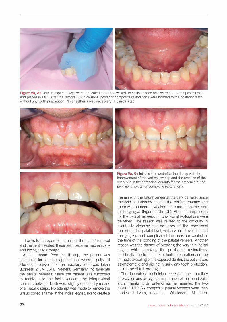
The laboratory technician waxed up the surfaces of the posterior teeth (2 premolars and the first molar in each sextant), and the casts returned to the clinicians who fabricated 4 transparent keys out of them (Elite Transparent, Zhermack, Badia Polesine, RO, Italy) (II laboratory step). After 2 weeks, the patient was scheduled for a 2-hour appointment for the fabrication of the provisional posterior composite restorations directly in the patient’s mouth (II clinical step). Without anesthesia, the keys were loaded with warmed up composite resin (Enamel Plus, HRi Micerium spa, Avegno, GE, Italy) and placed in the mouth (Figures 8a-8b). Since no anesthesia was delivered, the patient was fully collaborating during the control of the occlusion. After 1 week, another control was done, where the patient reported to be comfortable, even though he was functioning at an increased VDO and an open bite was present at the level of his anterior teeth (Figures 9a-9c). According to the classical Geneva Erosion Study’s protocol, the increase of VDO should be tested for one month. During this time, the patient was seen to remove the caries and to protect the pulp at the level of the palatal aspect of the maxillary anterior teeth.
During this visit, the exposed dentin on the palatal aspect of the maxillary anterior teeth was roughened with a very course diamond round bur, and immediately sealed with Optibond Fl (Kerr, Orange, CA, USA), following the manufactures’ instructions. To reinforce the hybrid layer, a final application of flowable composite (Tetric flow T, Ivoclar Vivadent, Schaan, Liechtenstein) was placed on the dentin, and polimerized for 40 seconds (20 seconds covered with glicerine) (27-31).
impressions were taken and the casts articulated in maximum intercupidation (MIP), using a face bow. A maxillary vestibular waxup was requested.
Looking at the patient’s smile, the laboratory technician decided that to achieve satisfactory esthetics, all the vestibular aspects of the maxillary teeth (excluded
Figure 6 3/4 View of the maxillary vestibular mock-up in place. With the teeth covered by the mock-up the patient did not feel the necessity to hide his smile any longer. Very striking was the difference in color with the antagonistic teeth. External bleaching was considered after the protection of the exposed dentin
Figure 7a, 7b Esthetics was improved, but during a long conversation with the patient, the clinician realized that he did not really want to change his look dramatically
[email protected] 27 21/04/17 12:35
ItalIan Journal of Dental MeDIcIne vol. 2/1-201728
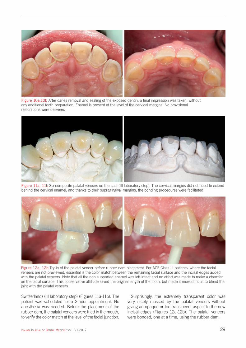
margin with the future veneer at the cervical level, since the acid had already created the perfect chamfer and there was no need to weaken the band of enamel next to the gingiva (Figures 10a-10b). After the impression for the palatal veneers, no provisional restorations were delivered. The reason was related to the difficulty in eventually cleaning the excesses of the provisional material at the palatal level, which would have inflamed the gingiva, and complicated the moisture control at the time of the bonding of the palatal veneers. Another reason was the danger of breaking the very thin incisal edges, while removing the provisional restorations, and finally due to the lack of tooth preparation and the immediate sealing of the exposed dentin, the patient was asymptomatic and did not require any tooth protection, as in case of full coverage.
The laboratory technician received the maxillary impression and an alginate impression of the mandibular arch. Thanks to an anterior jig, he mounted the two casts in MIP. Six composite palatal veneers were then fabricated (Miris, Coltene, Whaledent, Altstatten,
Thanks to the open bite creation, the caries’ removal and the dentin sealed, these teeth became mechanically and biologically stronger.
After 1 month from the II step, the patient was scheduled for a 1-hour appointment where a polyvinyl siloxane impression of the maxillary arch was taken (Express 2 3M ESPE, Seefeld, Germany), to fabricate the palatal veneers. Since the patient was supposed to receive also the facial veneers, the interproximal contacts between teeth were slightly opened by means of a metallic strips. No attempt was made to remove the unsupported enamel at the incisal edges, nor to create a
Figure 8a, 8b Four transparent keys were fabricated out of the waxed up casts, loaded with warmed up composite resin and placed in situ. After the removal, 12 provisional posterior composite restorations were bonded to the posterior teeth, without any tooth preparation. No anesthesia was necessary (II clinical step)
Figure 9a, 9c Initial status and after the II step with the improvement of the vertical overlap and the creation of the open bite in the anterior quadrants for the presence of the provisional posterior composite restorations
[email protected] 28 21/04/17 12:35
ItalIan Journal of Dental MeDIcIne vol. 2/1-2017 29
Switzerland) (III laboratory step) (Figures 11a-11b). The patient was scheduled for a 2-hour appointment. No anesthesia was needed. Before the placement of the rubber dam, the palatal veneers were tried in the mouth, to verify the color match at the level of the facial junction.
Surprisingly, the extremely transparent color was very nicely masked by the palatal veneers without giving an opaque or too translucent aspect to the new incisal edges (Figures 12a-12b). The palatal veneers were bonded, one at a time, using the rubber dam.
Figure 10a,10b After caries removal and sealing of the exposed dentin, a final impression was taken, without any additional tooth preparation. Enamel is present at the level of the cervical margins. No provisional restorations were delivered
Figure 11a, 11b Six composite palatal veneers on the cast (III laboratory step). The cervical margins did not need to extend behind the cervical enamel, and thanks to their supragingival margins, the bonding procedures were facilitated
Figure 12a, 12b Try-in of the palatal veneer before rubber dam placement. For ACE Class III patients, where the facial veneers are not previewed, essential is the color match between the remaining facial surface and the incisal edges added with the palatal veneers. Note that all the non supported enamel was left intact and no effort was made to make a chamfer on the facial surface. This conservative attitude saved the original length of the tooth, but made it more difficult to blend the joint with the palatal veneers
[email protected] 29 21/04/17 12:35
ItalIan Journal of Dental MeDIcIne vol. 2/1-201730
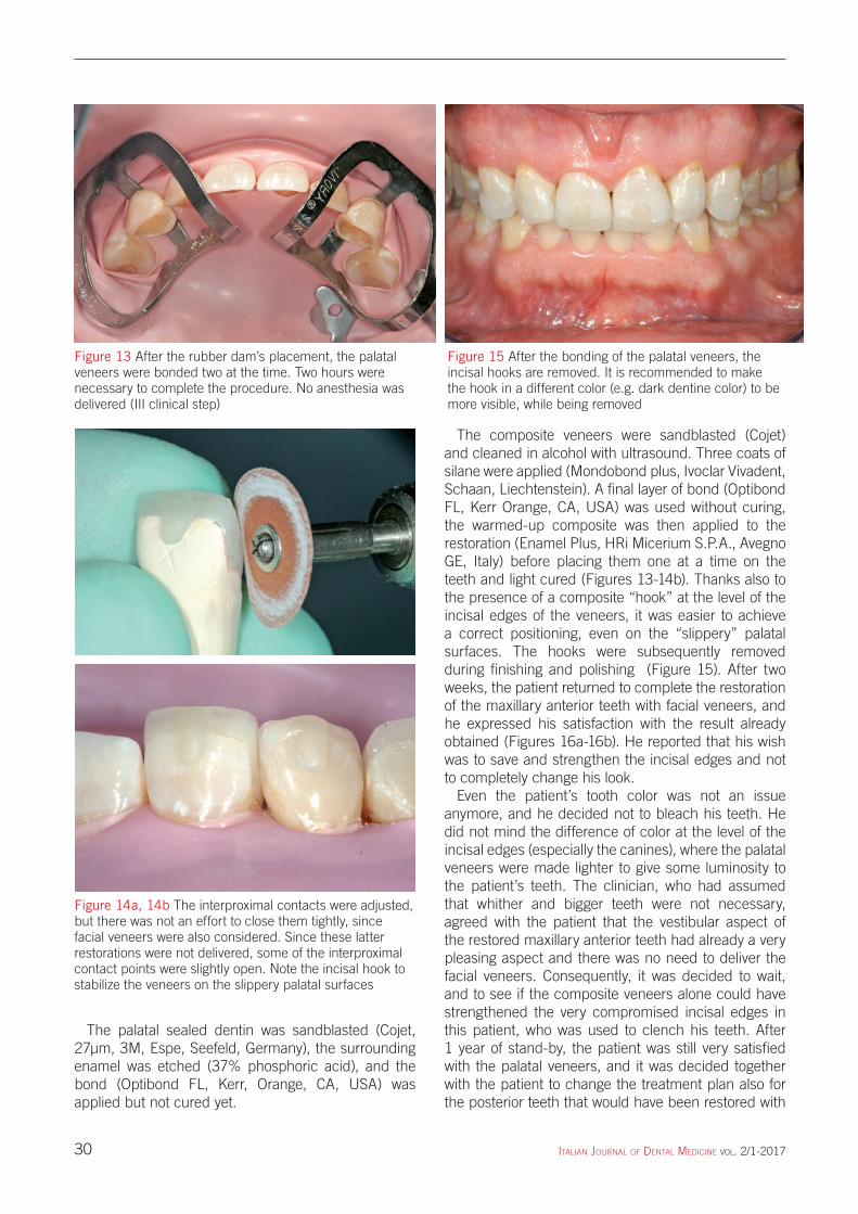
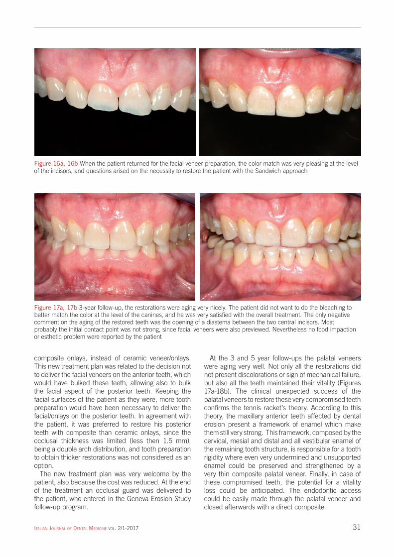
The composite veneers were sandblasted (Cojet) and cleaned in alcohol with ultrasound. Three coats of silane were applied (Mondobond plus, Ivoclar Vivadent, Schaan, Liechtenstein). A final layer of bond (Optibond FL, Kerr Orange, CA, USA) was used without curing, the warmed-up composite was then applied to the restoration (Enamel Plus, HRi Micerium S.P.A., Avegno GE, Italy) before placing them one at a time on the teeth and light cured (Figures 13-14b). Thanks also to the presence of a composite “hook” at the level of the incisal edges of the veneers, it was easier to achieve a correct positioning, even on the “slippery” palatal surfaces. The hooks were subsequently removed during finishing and polishing (Figure 15). After two weeks, the patient returned to complete the restoration of the maxillary anterior teeth with facial veneers, and he expressed his satisfaction with the result already obtained (Figures 16a-16b). He reported that his wish was to save and strengthen the incisal edges and not to completely change his look.
Even the patient’s tooth color was not an issue anymore, and he decided not to bleach his teeth. He did not mind the difference of color at the level of the incisal edges (especially the canines), where the palatal veneers were made lighter to give some luminosity to the patient’s teeth. The clinician, who had assumed that whither and bigger teeth were not necessary, agreed with the patient that the vestibular aspect of the restored maxillary anterior teeth had already a very pleasing aspect and there was no need to deliver the facial veneers. Consequently, it was decided to wait, and to see if the composite veneers alone could have strengthened the very compromised incisal edges in this patient, who was used to clench his teeth. After 1 year of stand-by, the patient was still very satisfied with the palatal veneers, and it was decided together with the patient to change the treatment plan also for the posterior teeth that would have been restored with
The palatal sealed dentin was sandblasted (Cojet, 27μm, 3M, Espe, Seefeld, Germany), the surrounding enamel was etched (37% phosphoric acid), and the bond (Optibond FL, Kerr, Orange, CA, USA) was applied but not cured yet.
Figure 13 After the rubber dam’s placement, the palatal veneers were bonded two at the time. Two hours were necessary to complete the procedure. No anesthesia was delivered (III clinical step)
Figure 14a, 14b The interproximal contacts were adjusted, but there was not an effort to close them tightly, since facial veneers were also considered. Since these latter restorations were not delivered, some of the interproximal contact points were slightly open. Note the incisal hook to stabilize the veneers on the slippery palatal surfaces
Figure 15 After the bonding of the palatal veneers, the incisal hooks are removed. It is recommended to make the hook in a different color (e.g. dark dentine color) to be more visible, while being removed
[email protected] 30 21/04/17 12:35
ItalIan Journal of Dental MeDIcIne vol. 2/1-2017 31
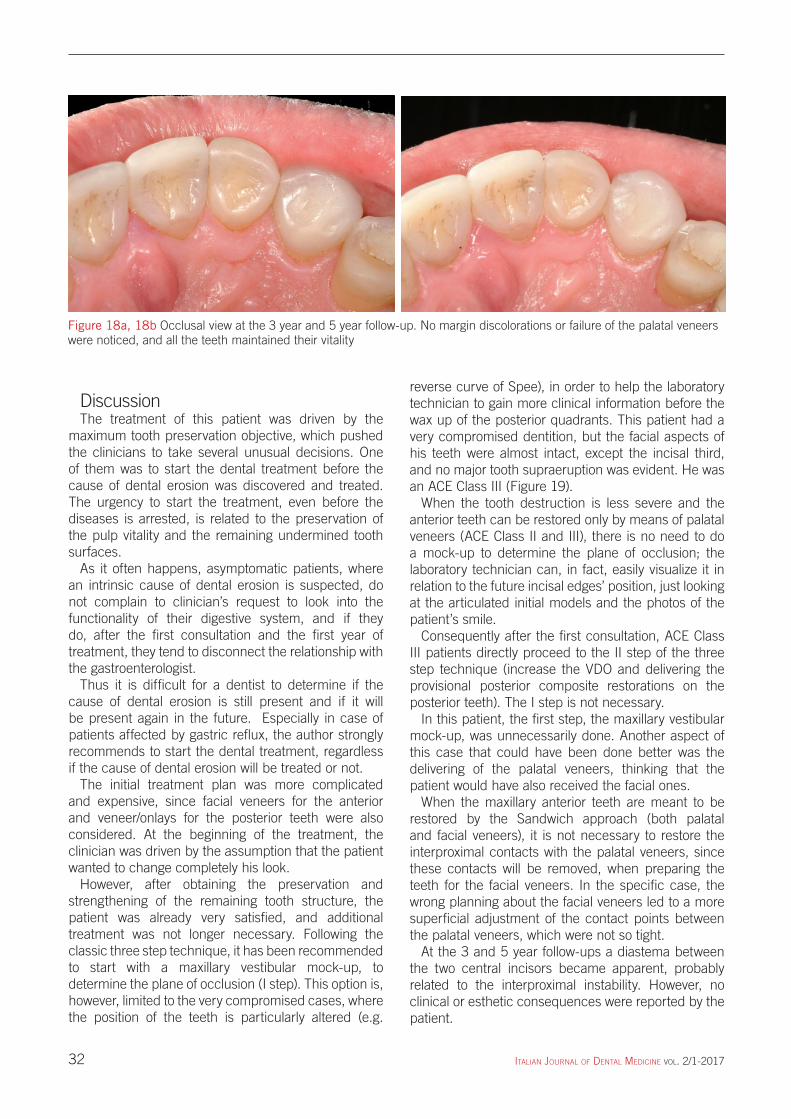
At the 3 and 5 year follow-ups the palatal veneers were aging very well. Not only all the restorations did not present discolorations or sign of mechanical failure, but also all the teeth maintained their vitality (Figures 17a-18b). The clinical unexpected success of the palatal veneers to restore these very compromised teeth confirms the tennis racket’s theory. According to this theory, the maxillary anterior teeth affected by dental erosion present a framework of enamel which make them still very strong. This framework, composed by the cervical, mesial and distal and all vestibular enamel of the remaining tooth structure, is responsible for a tooth rigidity where even very undermined and unsupported enamel could be preserved and strengthened by a very thin composite palatal veneer. Finally, in case of these compromised teeth, the potential for a vitality loss could be anticipated. The endodontic access could be easily made through the palatal veneer and closed afterwards with a direct composite.
composite onlays, instead of ceramic veneer/onlays. This new treatment plan was related to the decision not to deliver the facial veneers on the anterior teeth, which would have bulked these teeth, allowing also to bulk the facial aspect of the posterior teeth. Keeping the facial surfaces of the patient as they were, more tooth preparation would have been necessary to deliver the facial/onlays on the posterior teeth. In agreement with the patient, it was preferred to restore his posterior teeth with composite than ceramic onlays, since the occlusal thickness was limited (less then 1.5 mm), being a double arch distribution, and tooth preparation to obtain thicker restorations was not considered as an option.
The new treatment plan was very welcome by the patient, also because the cost was reduced. At the end of the treatment an occlusal guard was delivered to the patient, who entered in the Geneva Erosion Study follow-up program.
Figure 16a, 16b When the patient returned for the facial veneer preparation, the color match was very pleasing at the level of the incisors, and questions arised on the necessity to restore the patient with the Sandwich approach
Figure 17a, 17b 3-year follow-up, the restorations were aging very nicely. The patient did not want to do the bleaching to better match the color at the level of the canines, and he was very satisfied with the overall treatment. The only negative comment on the aging of the restored teeth was the opening of a diastema between the two central incisors. Most probably the initial contact point was not strong, since facial veneers were also previewed. Nevertheless no food impaction or esthetic problem were reported by the patient
[email protected] 31 21/04/17 12:35
ItalIan Journal of Dental MeDIcIne vol. 2/1-201732
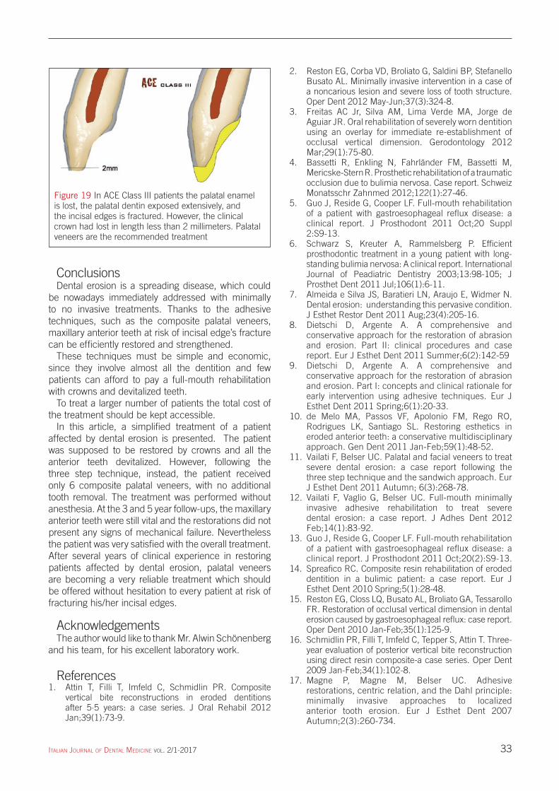
reverse curve of Spee), in order to help the laboratory technician to gain more clinical information before the wax up of the posterior quadrants. This patient had a very compromised dentition, but the facial aspects of his teeth were almost intact, except the incisal third, and no major tooth supraeruption was evident. He was an ACE Class III (Figure 19).
When the tooth destruction is less severe and the anterior teeth can be restored only by means of palatal veneers (ACE Class II and III), there is no need to do a mock-up to determine the plane of occlusion; the laboratory technician can, in fact, easily visualize it in relation to the future incisal edges’ position, just looking at the articulated initial models and the photos of the patient’s smile.
Consequently after the first consultation, ACE Class III patients directly proceed to the II step of the three step technique (increase the VDO and delivering the provisional posterior composite restorations on the posterior teeth). The I step is not necessary.
In this patient, the first step, the maxillary vestibular mock-up, was unnecessarily done. Another aspect of this case that could have been done better was the delivering of the palatal veneers, thinking that the patient would have also received the facial ones.
When the maxillary anterior teeth are meant to be restored by the Sandwich approach (both palatal and facial veneers), it is not necessary to restore the interproximal contacts with the palatal veneers, since these contacts will be removed, when preparing the teeth for the facial veneers. In the specific case, the wrong planning about the facial veneers led to a more superficial adjustment of the contact points between the palatal veneers, which were not so tight.
At the 3 and 5 year follow-ups a diastema between the two central incisors became apparent, probably related to the interproximal instability. However, no clinical or esthetic consequences were reported by the patient.
DiscussionThe treatment of this patient was driven by the
maximum tooth preservation objective, which pushed the clinicians to take several unusual decisions. One of them was to start the dental treatment before the cause of dental erosion was discovered and treated. The urgency to start the treatment, even before the diseases is arrested, is related to the preservation of the pulp vitality and the remaining undermined tooth surfaces.
As it often happens, asymptomatic patients, where an intrinsic cause of dental erosion is suspected, do not complain to clinician’s request to look into the functionality of their digestive system, and if they do, after the first consultation and the first year of treatment, they tend to disconnect the relationship with the gastroenterologist.
Thus it is difficult for a dentist to determine if the cause of dental erosion is still present and if it will be present again in the future. Especially in case of patients affected by gastric reflux, the author strongly recommends to start the dental treatment, regardless if the cause of dental erosion will be treated or not.
The initial treatment plan was more complicated and expensive, since facial veneers for the anterior and veneer/onlays for the posterior teeth were also considered. At the beginning of the treatment, the clinician was driven by the assumption that the patient wanted to change completely his look.
However, after obtaining the preservation and strengthening of the remaining tooth structure, the patient was already very satisfied, and additional treatment was not longer necessary. Following the classic three step technique, it has been recommended to start with a maxillary vestibular mock-up, to determine the plane of occlusion (I step). This option is, however, limited to the very compromised cases, where the position of the teeth is particularly altered (e.g.
Figure 18a, 18b Occlusal view at the 3 year and 5 year follow-up. No margin discolorations or failure of the palatal veneers were noticed, and all the teeth maintained their vitality
[email protected] 32 21/04/17 12:35
ItalIan Journal of Dental MeDIcIne vol. 2/1-2017 33
2. Reston EG, Corba VD, Broliato G, Saldini BP, Stefanello Busato AL. Minimally invasive intervention in a case of a noncarious lesion and severe loss of tooth structure. Oper Dent 2012 May-Jun;37(3):324-8.
3. Freitas AC Jr, Silva AM, Lima Verde MA, Jorge de Aguiar JR. Oral rehabilitation of severely worn dentition using an overlay for immediate re-establishment of occlusal vertical dimension. Gerodontology 2012 Mar;29(1):75-80.
4. Bassetti R, Enkling N, Fahrländer FM, Bassetti M, Mericske-Stern R. Prosthetic rehabilitation of a traumatic occlusion due to bulimia nervosa. Case report. Schweiz Monatsschr Zahnmed 2012;122(1):27-46.
5. Guo J, Reside G, Cooper LF. Full-mouth rehabilitation of a patient with gastroesophageal reflux disease: a clinical report. J Prosthodont 2011 Oct;20 Suppl 2:S9-13.
6. Schwarz S, Kreuter A, Rammelsberg P. Efficient prosthodontic treatment in a young patient with long-standing bulimia nervosa: A clinical report. International Journal of Peadiatric Dentistry 2003;13:98-105; J Prosthet Dent 2011 Jul;106(1):6-11.
7. Almeida e Silva JS, Baratieri LN, Araujo E, Widmer N. Dental erosion: understanding this pervasive condition. J Esthet Restor Dent 2011 Aug;23(4):205-16.
8. Dietschi D, Argente A. A comprehensive and conservative approach for the restoration of abrasion and erosion. Part II: clinical procedures and case report. Eur J Esthet Dent 2011 Summer;6(2):142-59
9. Dietschi D, Argente A. A comprehensive and conservative approach for the restoration of abrasion and erosion. Part I: concepts and clinical rationale for early intervention using adhesive techniques. Eur J Esthet Dent 2011 Spring;6(1):20-33.
10. de Melo MA, Passos VF, Apolonio FM, Rego RO, Rodrigues LK, Santiago SL. Restoring esthetics in eroded anterior teeth: a conservative multidisciplinary approach. Gen Dent 2011 Jan-Feb;59(1):48-52.
11. Vailati F, Belser UC. Palatal and facial veneers to treat severe dental erosion: a case report following the three step technique and the sandwich approach. Eur J Esthet Dent 2011 Autumn; 6(3):268-78.
12. Vailati F, Vaglio G, Belser UC. Full-mouth minimally invasive adhesive rehabilitation to treat severe dental erosion: a case report. J Adhes Dent 2012 Feb;14(1):83-92.
13. Guo J, Reside G, Cooper LF. Full-mouth rehabilitation of a patient with gastroesophageal reflux disease: a clinical report. J Prosthodont 2011 Oct;20(2):S9-13.
14. Spreafico RC. Composite resin rehabilitation of eroded dentition in a bulimic patient: a case report. Eur J Esthet Dent 2010 Spring;5(1):28-48.
15. Reston EG, Closs LQ, Busato AL, Broliato GA, Tessarollo FR. Restoration of occlusal vertical dimension in dental erosion caused by gastroesophageal reflux: case report. Oper Dent 2010 Jan-Feb;35(1):125-9.
16. Schmidlin PR, Filli T, Imfeld C, Tepper S, Attin T. Three-year evaluation of posterior vertical bite reconstruction using direct resin composite-a case series. Oper Dent 2009 Jan-Feb;34(1):102-8.
17. Magne P, Magne M, Belser UC. Adhesive restorations, centric relation, and the Dahl principle: minimally invasive approaches to localized anterior tooth erosion. Eur J Esthet Dent 2007 Autumn;2(3):260-734.
ConclusionsDental erosion is a spreading disease, which could
be nowadays immediately addressed with minimally to no invasive treatments. Thanks to the adhesive techniques, such as the composite palatal veneers, maxillary anterior teeth at risk of incisal edge’s fracture can be efficiently restored and strengthened.
These techniques must be simple and economic, since they involve almost all the dentition and few patients can afford to pay a full-mouth rehabilitation with crowns and devitalized teeth.
To treat a larger number of patients the total cost of the treatment should be kept accessible.
In this article, a simplified treatment of a patient affected by dental erosion is presented. The patient was supposed to be restored by crowns and all the anterior teeth devitalized. However, following the three step technique, instead, the patient received only 6 composite palatal veneers, with no additional tooth removal. The treatment was performed without anesthesia. At the 3 and 5 year follow-ups, the maxillary anterior teeth were still vital and the restorations did not present any signs of mechanical failure. Nevertheless the patient was very satisfied with the overall treatment. After several years of clinical experience in restoring patients affected by dental erosion, palatal veneers are becoming a very reliable treatment which should be offered without hesitation to every patient at risk of fracturing his/her incisal edges.
AcknowledgementsThe author would like to thank Mr. Alwin Schönenberg
and his team, for his excellent laboratory work.
References1. Attin T, Filli T, Imfeld C, Schmidlin PR. Composite
vertical bite reconstructions in eroded dentitions after 5·5 years: a case series. J Oral Rehabil 2012 Jan;39(1):73-9.
Figure 19 In ACE Class III patients the palatal enamel is lost, the palatal dentin exposed extensively, and the incisal edges is fractured. However, the clinical crown had lost in length less than 2 millimeters. Palatal veneers are the recommended treatment
[email protected] 33 21/04/17 12:35
ItalIan Journal of Dental MeDIcIne vol. 2/1-201734
25. Vailati F, Belser UC. Full-mouth adhesive rehabilitation of a severely eroded dentition: the three step technique. Part 2. Eur J Esthet Dent 2008; 3:128-46.
26. Vailati F, Belser UC. Full-mouth adhesive rehabilitation of a severely eroded dentition: the three step technique. Part 1. Eur J Esthet Dent 2008;3:30-44.
27. Magne P, So WS, Cascione D. Immediate dentin sealing supports delayed restoration placement. J Prosthet Dent 2007;98:166-74.
28. Magne P, Kim TH, Cascione D, Donovan TE. Immediate dentin sealing improves bond strength of indirect restorations. J Prosthet Dent 2005;94:511-9.
29. Magne P. Immediate dentin sealing: a fundamental procedure for indirect bonded restorations. J Esthet Restor Dent 2005;17:144-54.
30. Paul SJ, Schärer P. The “Dual Bonding Technique” - A modified method to improve adhesive luting procedures. Int J Periodont Rest Dent 1997;17: 537-45.
31. Bertschinger C, Paul SJ, Lüthy H, Schärer P. Dual application of dentin bonding agents: Effect on bond strength. Am J Dent 1996; 9:115-19
32. Vailati F, Gruetter L, Belser U.C. Facial and palatal veneers to restore maxillary anterior teeth affected by severe wear. Clinical results up to 6 years of service: the geneva erosion study. Eur J Esthet Dent 2013 autumn;8(3):358-75.
18. Hayashi M, Shimizu K, Takeshige F, Ebisu S. Restoration of erosion associated with gastroesophageal reflux caused by anorexia nervosa using ceramic laminate veneers: a case report. Oper Dent 2007 May-Jun;32(3):306-10.
19. Kavoura V, Kourtis SG, Zoidis P, Andritsakis DP, Doukoudakis A. Full-mouth rehabilitation of a patient with bulimia nervosa. A case report. Quintessence Int 2005 Jul-Aug;36(7-8):501-10.
20. Van Roekel NB. Gastroesophageal reflux disease, tooth erosion, and prosthodontic rehabilitation: a clinical report. J Prosthodont 2003 Dec;12(4):255-9.
21. Ambard A, Mueninghoff L. Rehabilitation of a bulimic patient using endosteal implants. J Prosthodont 2002 Sep;11(3):176-80.
22. Bonilla ED, Luna O. Oral rehabilitation of a bulimic patient: a case report. Quintessence Int 2001 Jun;32(6):469-75.
23. Vailati F, Belser UC. Classification and treatment of the anterior maxillary dentition affected by dental erosion: the ACE classification. Int J Periodontics Restorative Dent 2010 Dec;30(6):559-71.
24. Vailati F, Belser UC. Full-mouth adhesive rehabilitation of a severely eroded dentition: the three step technique. Part 3. Eur J Esthet Dent 2008;3:236-57.
[email protected] 34 21/04/17 12:35