complications of fractures non-union dvt damage to nerves and blood vessels compartment syndrome fat...
TRANSCRIPT

Complications of Fractures
•Non-union
•DVT
•Damage to Nerves and Blood Vessels
•Compartment Syndrome
•Fat Emboli
•Infection (Osteomyelitis)
Clinical DecisionsBobbie, age 14, was admitted with fx
left tibia about 10 hrs age. He has a long leg cast.
Report states: “toes warm, pink with good capillary refil, pulses present...pain not controlled with MS.”
You note: p. 88,BP 89/66, r.23, t 98.6. Bobbie reports: ‘toes feel “funny”, left leg hurts at calf.”

Questions
What additional data should you gather?
What is the medical/nursing problem of greatest Priority?
Why did this occur? What actions should you take?
Compartment SyndromeCompartment Syndrome
Compression of structures within a defined boundry; capillary perfusion decreased.
Self-perpetuating edema-ischemia cycle...inc. capillary permeability...arterial obstruction...muscle and tissue death.

Etiology
Normal compartment pressure=10mmHg
Above 30mmHg for 8 hrs = Permanent damage
Factors affecting– dec. BP– dec. oncotic
pressure– inc. capillary
permeability– obs. venous flow– length of time
Causes

Arterial circulation continues; capillary circulation stops!

Events Leading to Compartment Syndrome
Ischemia cycle Edema with inc.
capillary pressure Capillaries
dilate;hydrostatic filtration pressure becomes greater than oncotic pressure of plasma colloid
More fluid leaves capillaries than enters; inc. permeability of capillary walls due to histamine release due to ischemic muscles
Plasma proteins into interstitial fluids
Inc. intramuscular pressure-obstructs venous first, then arterial


Compartment syndrome has many causes.
Describe the etiology in these examples.
compartment

Compartment Syndrome
Types– Acute– Chronic
Microcirculation ceases when compartment pressure = diastolic BP
Lead to Volksmans ischemic contraction
May develop Crush Syndrome:Rhabdomyolosis=Myoglobinuric renal failure

Volksman’s ischemic contracture may result with compartment syndrome!

Compartments Affected Forearm
– deep volar– superficial volar
Lower leg– deep posterior
tibial– anterior with
peroneal nerve– lateral with
superficial peroneal– posterior with sural
Lower leg

•Assessment
•Pain on passive stretch•Progressive pain•Tenseness of muscle compartment•Motor weakness•Dec. sensation•Loss of pulse

Interventions
Ice and elevate Early recognition
– 5 Ps– Pressure monitors
Dec. pressure– Remove what
confines– Eval. response to
meds
Medical/Surgical Interventions
If compartment syndrome present, elevate limb only to heart level, not above!
Prevent complications associated with myogolbinuria
Monitor for compartment syndrome Prevent infection Fasciotomy

Monitor for compartment syndrome
Fasciotomy!
Treatment for compartment syndrome.
Prevent
Treatment

Fat Emboli
Fat globules obstruct blood vessels Causes
– Metabolic: biochemical changes, lipids mobilized and embolize, fatty acids toxic
– Mechanical: fat is liberated due to inc. pressure
Life-threatening: ARDS
Fat emboli
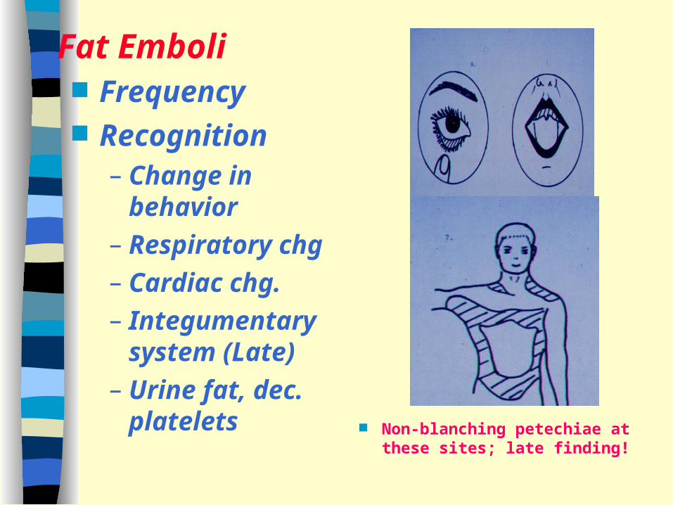
Fat Emboli Frequency Recognition
– Change in behavior
– Respiratory chg– Cardiac chg.– Integumentary
system (Late)– Urine fat, dec.
platelets Non-blanching petechiae at these sites; late finding!

Diagnostic Tests
Blood gasesLung scanChest x-raysLaboratory studies: platelets, urine fat
Nursing diagnosis
Lung changes with fat emboli (ARDS)

Interventions: Fat emboli Prevent:
recognize, immobilize, hydrate
O2 therapy Steroids Fluid volume
replacement Plasma expanders Monitoring