complications of cirrhosis philip c. delich md. the epidemic of cirrhosis nafld/nash hcv hbv ...
TRANSCRIPT
COMPLICATIONS OF CIRRHOSIS
Philip C. Delich MD

THE EPIDEMIC OF CIRRHOSIS
· NAFLD/NASH
· HCV
· HBV

Cirrhosis
· End stage of any chronic liver disease
· Characterized histologically by regenerative nodules surrounded by fibrous tissue
· Clinically there are two types of cirrhosis:· Compensated· Decompensated
DEFINITION OF CIRRHOSIS
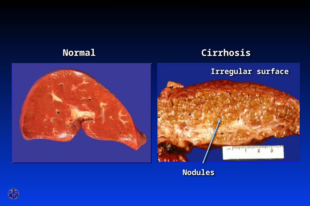
CirrhosisNormal
Nodules
Irregular surface
GROSS IMAGE OF A NORMAL AND A CIRRHOTIC LIVER
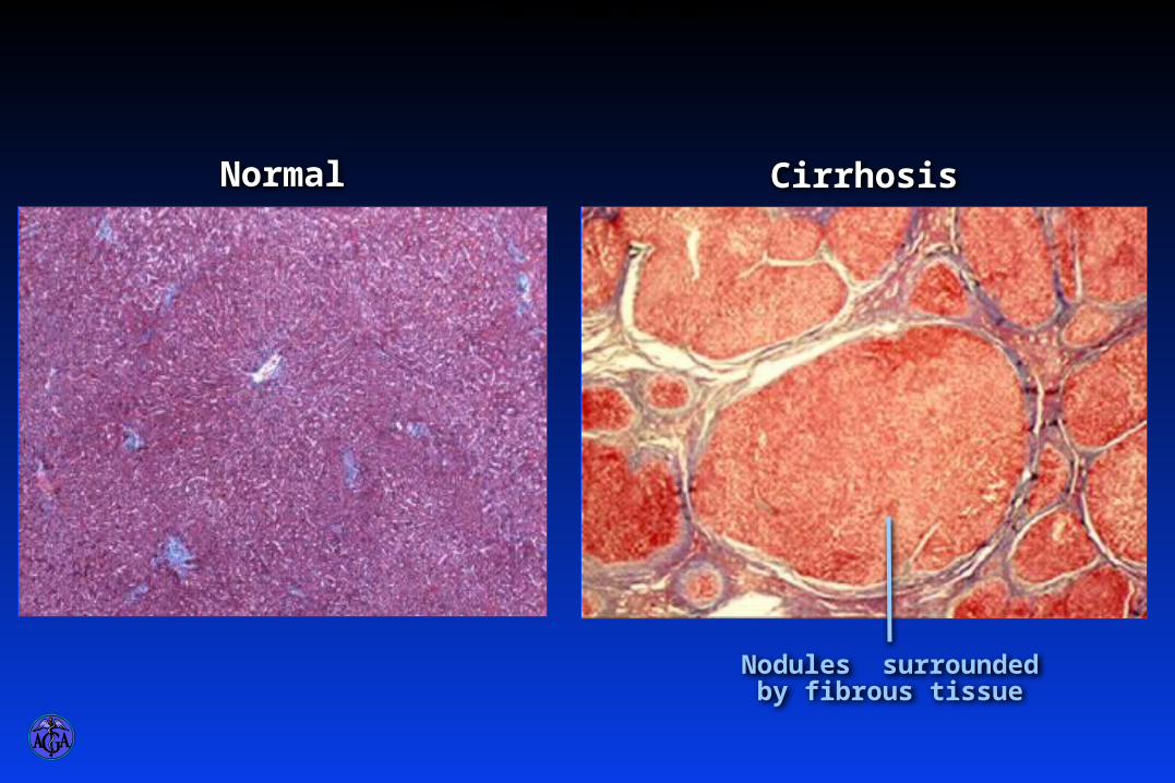
CirrhosisNormal
Nodules surrounded by fibrous tissue
HISTOLOGICAL IMAGE OF A NORMAL AND A CIRRHOTIC LIVER

Diagnosis of Cirrhosis
· Gold standard=liver biopsy· Usually not necessary
Physical exam findings (spiders, palmer erythema, gynecomastia, hepatosplenomegally)
Labs- thrombocytopenia, AST>ALT
Radiologic Evidence

NoYes
Diagnostic Algorithm
Patient with chronic liver disease and any of the following:· Variceal hemorrhage· Ascites· Hepatic encephalopathy
Liver biopsy not necessary for the
diagnosis of cirrhosis
Physical findings:Enlarged left hepatic lobeSplenomegalyStigmata of chronic liver disease
Laboratory findings:ThrombocytopeniaImpaired hepatic synthetic function
Radiological findings:· Small nodular liver· Intra-abdominal collaterals· Ascites· Splenomegaly· Colloid shift to spleen and/or bone marrow
Yes No
Yes No
Liver biopsy
DIAGNOSTIC ALGORITHM
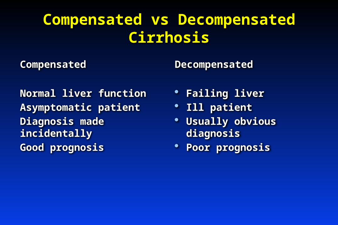
Compensated vs Decompensated Cirrhosis
Compensated
Normal liver function
Asymptomatic patient
Diagnosis made incidentally
Good prognosis
Decompensated
· Failing liver· Ill patient· Usually obvious diagnosis· Poor prognosis

6040 80 100 120 140 1600
40
60
80
20
200
100
Months
Probability of survival
All patients with cirrhosis
Decompensated cirrhosis
180
Decompensation Shortens Survival
Gines et. al., Hepatology 1987;7:122
Median survival~ 9 years
Median survival~ 1.6 years
SURVIVAL TIMES IN CIRRHOSIS

Cirrhosis-proposed new classification
· Stage 1-compensated with no varices-1% mortality
· Stage 2-compensated with varices-3.4%mortality
· Stage 3-ascites- 20% mortality· Stage 4- variceal bleeding-
57%mortality

MANAGEMENT OF WELL COMPENSATED CIRRHOSIS
· Rx. Cause (HCV,HBV, wt. loss in NASH, phlebotomy in hemochromatosis etc)
· Screen for esophageal varices· Screen for hepatocellular carcinoma· Vaccinate against Hepatitis A and B if not
already protected.· Optimize transplant candidacy· Observe for signs of decompensation.

· Decompensated cirrhosis- cirrhosis with signs of liver failure (ascites, edema, hepatic encephalopathy etc)

Complications of cirrhosis
· Ascitesrefractory ascites
SBP
hepatorenal syndrome· Variceal Bleeding· Hepatic Encephalopathy· HCC

Uncomplicated Ascites
· Low Na diet· Combination diuretics
(spironolactone 100mg-400mg/day, furosamide 40mg-160mg/day)
· Therapeutic Paracentesis
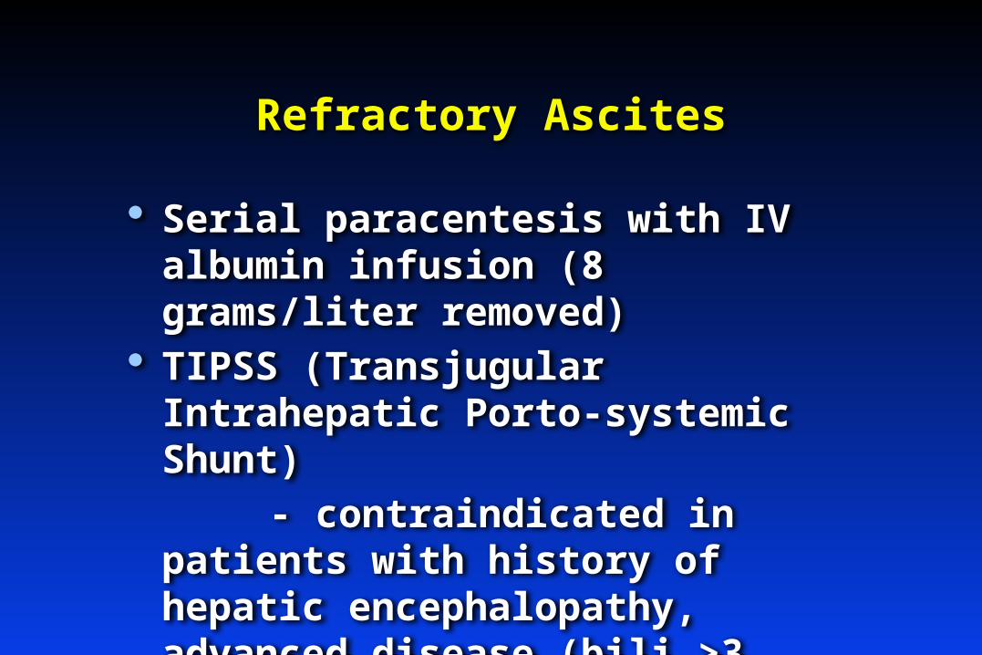
Refractory Ascites
· Serial paracentesis with IV albumin infusion (8 grams/liter removed)
· TIPSS (Transjugular Intrahepatic Porto-systemic Shunt)
- contraindicated in patients with history of hepatic encephalopathy, advanced disease (bili >3 etc), elderly

Transjugular Intrahepatic Portosystemic Shunt
Hepatic vein
Portal veinSplenic vein
Superior mesenteric vein
TIPS
THE TRANSJUGULAR INTRAHEPATIC PORTOSYSTEMIC SHUNT

Spontaneous Bacterial Peritonitis
· Frequent complication· Approx. 30% in hospitalized patients· Diagnosed with paracentesis->250
PMNs per HPF· Rx-3rd gen. cephalosporine x 5 days· 70% recurrence in one
yearprophylactic antibiotics

Hepatorenal Syndrome
· Rapidly progressive renal failure in setting of liver failure
· Absence of other causal factors· No improvement with fluid challenge· oliguria· low urine Na

Rx. Of Hepatorenal syndrome
· Vasoconstrictors (oral midodrine-5 mg TID)
· Octreotide 100ug SQ TID· Volume expansion (IV albumin)

Variceal Bleeding
· Frequent complication (33-50% lifetime risk in cirrhotics)
· High mortality (20%)· Increased risk with progression of
liver disease.· Preventable and treatable

Prevalence of Esophageal Varices in Cirrhosis
%
100
60
40
20
0Overall Child A Child B
80
Child C
Pagliaro et al., In: Portal Hypertension: Pathophysiology and Management, 1994: 72
PREVALENCE OF ESOPHAGEAL VARICES IN CIRRHOSIS

Small varices Large varicesNo varices
7-8%/year 7-8%/year
Varices Increase in Diameter Progressively
Merli et al. J Hepatol 2003;38:266
VARICES INCREASE IN DIAMETER PROGRESSIVELY

Prophylaxis of Variceal Hemorrhage
Diagnosis of Cirrhosis
Endoscopy
No Varices
Follow-up EGD in 2-3 years*
Small Varices
Follow-up EGD in 1-2 years*
Medium/Large Varices
• Stepwise increase until maximally tolerated dose• Continue beta-blocker (life-long)
No Contraindications
ContraindicationsorBeta-blocker intolerance
Beta-blocker therapy
Endoscopic Variceal Band Ligation
*EGD every year in decompensated cirrhosis
MANAGEMENT ALGORITHM FOR THE PROPHYLAXIS OF VARICEAL HEMORRHAGE - SUMMARY
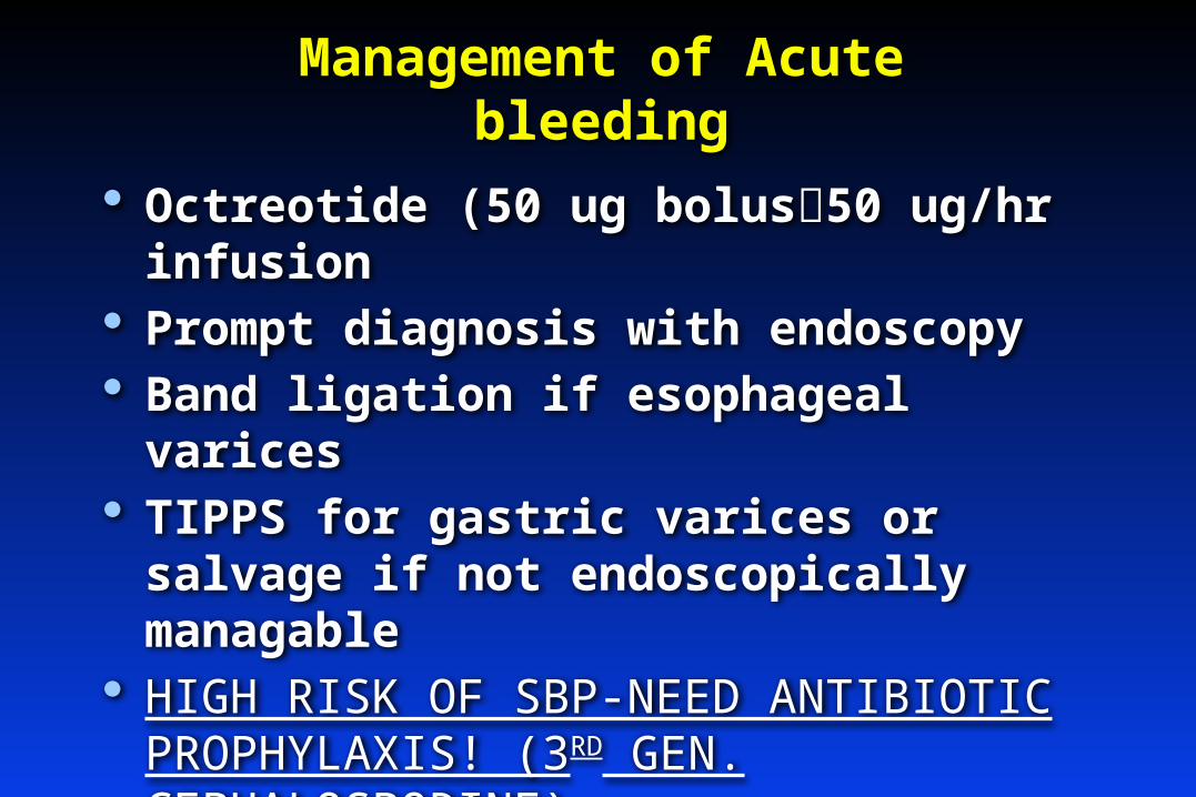
Management of Acute bleeding
· Octreotide (50 ug bolus50 ug/hr infusion
· Prompt diagnosis with endoscopy· Band ligation if esophageal varices· TIPPS for gastric varices or salvage if
not endoscopically managable· HIGH RISK OF SBP-NEED ANTIBIOTIC
PROPHYLAXIS! (3RD GEN. CEPHALOSPORINE)

Endoscopic Variceal Band Ligation
· Bleeding controlled in 90%
· Rebleeding rate 30%
· Compared with sclerotherapy:· Less rebleeding· Lower mortality· Fewer complications· Fewer treatment sessions
ENDOSCOPIC VARICEAL BAND LIGATION

Transjugular Intrahepatic Portosystemic Shunt
Hepatic vein
Portal veinSplenic vein
Superior mesenteric vein
TIPS
THE TRANSJUGULAR INTRAHEPATIC PORTOSYSTEMIC SHUNT

Secondary prophylaxis against variceal bleeding
· Non-selective beta blockers (nadolol or propranolol-tritrate to pulse 60 and tolerance)
· Serial endoscopic banding Q2-4 weeks until varices obliterated then yearly for surveillance.

Hepatic Encephalopathy
· Neuro-psychological disorder resulting from inability of failing liver to remove nitrogenous waste products of protein metabolism from circulation.
· CLINICAL DIAGNOSIS-ammonia levels do not correlate well with diagnosis or stage of encephalopathy.

Hepatic Encephalopathy Is A Clinical Diagnosis
· Clinical findings and history important
· Ammonia levels are unreliable
· Ammonia has poor correlation with diagnosis
· Measurement of ammonia not necessary
· Number connection test
· Slow dominant rhythm on EEG
HEPATIC ENCEPHALOPATHY IS A CLINICAL DIAGNOSIS

Asterixis
ASTERIXIS IS THE HALLMARK IN THE DIAGNOSIS OF HEPATIC ENCEPHALOPATHY

STAGES OF HEPATIC ENCEPHALOPATHY
Confusion
Drowsiness
Somnolence
Coma
1 2 3 4Stage
Stages of Hepatic Encephalopathy

Treatment of Hepatic Encephalopathy
· Identify and treat precipitating factor· Infection· GI hemorrhage· Prerenal azotemia· Sedatives· Constipation
· Lactulose (adjust to 2-3 bowel movements/day)
· Protein restriction, short-term (if at all)
TREATMENT OF HEPATIC ENCEPHALOPATHY

Antibiotics for hepatic encephalopathy
· Generally secondary agents in lactulose failures
· Work through alteration of gut floradecreased absorption of nitrogenous wastes.
· Neomycin (500 mg TID)- cheap-theoretical risk of renal toxicity and deafness
· Xifaxan (400 mg TID)-very expensive perhaps safer and more effective.
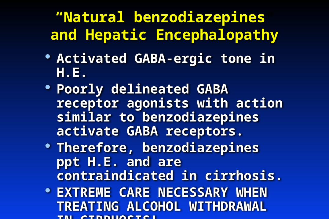
“Natural benzodiazepines” and Hepatic Encephalopathy
· Activated GABA-ergic tone in H.E.· Poorly delineated GABA receptor
agonists with action similar to benzodiazepines activate GABA receptors.
· Therefore, benzodiazepines ppt H.E. and are contraindicated in cirrhosis.
· EXTREME CARE NECESSARY WHEN TREATING ALCOHOL WITHDRAWAL IN CIRRHOSIS!
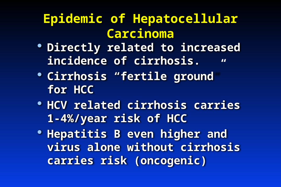
Epidemic of Hepatocellular Carcinoma
· Directly related to increased incidence of cirrhosis.
· Cirrhosis “fertile ground” for HCC· HCV related cirrhosis carries
1-4%/year risk of HCC· Hepatitis B even higher and virus
alone without cirrhosis carries risk (oncogenic)

HCC-Rationale for Screening
· Early tumors potentially resectable (occasionally) and frequently curable with transplantation (Milan criteria).
· Evolving treatments increasingly helpful even if “cure” not feasible (chemoembolization, radiofrequency ablation, cryotherapy)

HCC Screening
· Imaging modalities (US vs. CT vs. MRI)· AFP-probably not useful but still done.· Frequency needs to be Q 6 mo to be
effective.· Screen stage 3 and 4 fibrosis patients.· In hepatitis B, all infected patients at risk
and need to be screened.

Liver Transplantation
· Only definitive treatment· Standard of care in 2014· Virtually all patients with ESLD
should be considered- few absolute contraindications
· Excellent short and long term survival

“Pearls” in management of cirrhosis· Cirrhosis is clinical diagnosis that rarely
requires liver biopsy.· Low platelet count cirrhosis until proven
otherwise.· AST>ALT =cirrhosis (or alcoholic liver disease)· Always consider SBP and when in doubt, rule it
out· Avoid benzodiazepines and use extreme care
in treatment of alcohol withdrawl in cirrhotics.

“Pearls” continued· Screen cirrhotics for varices and HCC
regularly· Hepatic Encephalopathy is a clinical
diagnosis-ammonia levels not useful.· Band esophageal varices to obliteration.· Remember low Na diets in management of
ascites.· Cirrhosis is a catabolic state-low protein
diets rarely useful and usually harmful.· Think about transplantation early in
decompensation.

SURGERY AND CIRRHOSIS
· INCREASED RISK OF ANESTHESIA· INCREASED RISK OF HEPATIC
ENCEPHALOPATHY WITH POST-OP PAIN MANAGEMENT
· VERY HIGH RISK IN MAJOR CAVITIES (CHEST AND ABDOMEN)
· CONTRAINDICATED IN DECOMPENSATED CIRRHOSIS UNLESS TRUE EMERGENCY!

HOSPICE AND END STAGE LIVER DISEASE
· APPROX. 50% MORTALITY OVER 2 YEARS
· BENZODIAZEPINES AND OPIATES RELATIVELY CONTRAINDICATED IN CIRRHOSISHEPATIC COMA!
· PAIN USUALLY NOT AN ISSUE EXCEPT ASCITES RELATED DISTENSION MANAGED BY TAP AND NOT OPIATES!
PREVENTION BEST MEASURE
· MOST CIRRHOSIS IN WESTERN COUNTRIES IS LIFESTYLE RELATED DISEASE (OBESITY/IVDA/ALCOHOL)
· DIAGNOSE AND RX HCV· VACCINATION AGAINST HEPATITIS
A AND B· EDUCATION AND LIFESTYLE
MODIFICATION ESSENTIAL