complications of aom - otitismediasociety.orgotitismediasociety.org/complications of aom.pdf ·...
TRANSCRIPT
This teaching presentation for the ISOM website has been prepared by
Tal Marom MD and Sharon Ovnat Tamir MDDepartment of Otolaryngology-Head and Neck Surgery
Edith Wolfson Medical CenterSackler Faculty of Medicine
Tel Aviv University Holon Israel
Complications of AOM
Acknowledgement
bull This presentation is aimed for teaching purposes of students residents and other allied healthcare workers
bull Please visit the International Society for Otitis Media website for more resources wwwotitismediasocietyorg
What are the Complications of OM
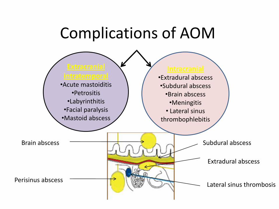
Complications of AOM
Extracranialintratemporal
bullAcute mastoiditisbullPetrositis
bullLabyrinthitis bullFacial paralysis
bullMastoid abscess
IntracranialbullExtradural abscessbullSubdural abscess
bullBrain abscessbullMeningitis
bull Lateral sinus thrombophlebitis
Extradural abscess
Perisinus abscessLateral sinus thrombosis
Subdural abscessBrain abscess
What intratemporal bone complications do you know
How does each complication present
How do you manage each complication
1 Acute coalescent mastoiditis
Definition
It is the inflammation of mucosal lining of antrum and mastoid air cells system
Mastoiditis per se actually occurs with most infections of the middle ear It is not considered a complication until bone destruction occurs
Pathophysiology of acute mastoiditis
bull Production of pus under tension
bull Hyperemic decalcification
bull Osteoclastic resorption of bony walls
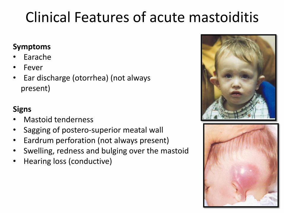
Clinical Features of acute mastoiditis
Symptomsbull Earachebull Feverbull Ear discharge (otorrhea) (not always
present)
Signsbull Mastoid tendernessbull Sagging of postero-superior meatal wallbull Eardrum perforation (not always present)bull Swelling redness and bulging over the mastoidbull Hearing loss (conductive)
Investigations for acute mastoiditis
bull Ear swab for culture amp sensitivity (CampS)
bull HRCT scan of the temporal
bone as well as CT scan of
brain with contrast
bull Myringotomy with pus CampS
bull Blood cultures if indicated
Treatment of acute mastoiditis
Medical treatmentminus Hospitalizationminus Intravenous antibioticsminus Analgesics
Surgical treatmentminusMyringotomy with or without ventilation tube
insertion may be sufficient in mostminus Cortical mastoidectomy if clinical worsening
occurs
2 Acute labyrinthitis
Acute inflammation of the labyrinth due to diffusion of toxins via the round window from the middle ear or due to a labyrinthine fistula caused by hyperemic decalcification
Acute labyrinthitis
Clinical Picture
bull Hearing loss progressive and sensorineural or mixed in nature
bull Attack of vertigo and vomiting mostly during straining sneezing and lifting heavy object
bull Positive fistula test bull Nystagmus may be present to the side of the
affected ear
Acute labyrinthitis
Diagnosis bull High index of suspicionbull Positive fistula test bull HRCT scan of temporal bone will demonstrate fistulaif present
Treatment High dose IV antibioticsCortical mastoidectomy with removal of granulations and
closure of fistula if present
Acute facial nerve paresisparalysis
bull A result of an inflammatory response of the facial nerve within the fallopian canal to the infection
bull The tympanic segment is the most common site to be involved as the facial canal may have dehiscences along this segment
Diagnosis of Facial nerve paralysis
bull Clinical House Brackmann grade must be established and monitoring for progression and recovery made
bull HRCT of the temporal boneperformed to look for bone erosion
Treatment of acute facial nerve paralysis
bull Intravenous antibiotics
bull Cortical mastoidectomy + ventilation tube insertion
bull Excision of granulations over dehiscent facial canal and decompression of the nerve
Extracranial Complications Mastoid abscess -clinical
bull Classically a postaural abscess occurs
bull When the abscess spreads along the mastoid air cell system other sites of collection occur eg
bull Bezoldrsquos abscess abscess over upper part of sternomastoid muscle
bull Lucrsquos abscess abscess
over root of zygoma
bull Citellirsquos abscess abscess
over posterior belly of
digastric
Postaural abscess
Sites of pneumatisation of mastoid air cell system in relation to types of mastoid abscess
Zygomatic cells (Lucrsquos abscess)
Mastoid tip cells (Bezoldrsquos abscess)
Digastric cells (Citellirsquos abscess)
Mastoid cellsPostaural abscess
Extracranial Complications Mastoid abscess -treatment
bull Aspiration or drainage of abscess and pus sent for CampS
bull IV antibiotics
bull Cortical mastoidectomy may be required after 24-48 hours if symptoms persist
Intracranial Complications
bull What are intracranial complications
bull What is the most common intracranial complication
bull Which symptoms do patients presenting with intracranial complications exhibit
bull What are the investigations required to diagnose such complications
Important tenets
bull Intracranial complications should be suspected in a child with recent onset of headache fever convulsionsvomiting not feeding well or focal neurological deficit
bull Contrast enhanced CT scan of the brain is mandatory prior to lumbar puncture when intracranial complication is suspected to avoid ldquoconingrdquo (brain herniation into foramen magnum which causes death)
Intracranial Complications ExtraduralAbscess
Definition
bull Collection of pus against the dura of the middle or posterior cranial fossa
bull Extradural abscess is the commonest intracranial complication of otitis media
Intracranial Complications Extraduralabscess ndash clinical amp treatment
Clinical Picturendash Persistent headache on the side of otitis mediandash Pulsating dischargendash Feverndash May be asymptomatic (discovered during surgery)
Diagnosisndash CT scans reveal the abscess as well as the middleear pathology- MRI reveals associated dural inflammation
Treatmentndash Mastoidectomy and drainage of the abscess
Intra-cranial ComplicationsSubdural Abscess- clinical
Definition
ndash Collection of pus between the dura and the
arachnoid
ndash A rare pathology
Clinical picture
ndash Headache without signs of meningeal irritation
ndash Convulsions
ndash Focal neurological deficit (paralysis loss of sensation visual field defects)
Intracranial Complications Subdural Abscess-Investigation and treatment
Investigations
ndash CT scan
- MRI
Treatment
ndash Drainage or excision (neuro-
surgical consultation required)
ndash High dose IV antibiotics
ndash Mastoidectomy
Intracranial Complications Meningitis-pathology
Definition
ndash Inflammation of the meninges (pia arachnoidand dura)
Pathology
ndash Two forms
bull Circumscribed meningitis no bacteria in CSF
bull Generalized meningitis bacteria are present in CSF
Intracranial Complications Meningitis-clinical
Clinical picture
ndash General symptoms and signs
bull High grade remittent fever restlessness irritability
bull Photophobia and delirium
bull Instability
Intracranial Complications Meningitis -signs of meningeal irritation
Signs of meningeal irritation
bull Nuchal rigidity
bull Positive Kernigrsquos sign difficulty to straighten the
knee while the hip is flexed
bull Positive Brudzinskirsquos sign
ndash passive flexion of one leg results in a similar
movement on the opposite side or
ndash if the neck is passively flexed flexion occurs in the
hips and knees
Intracranial Complications Meningitis- diagnosis and treatment
Diagnosis
bull Lumbar puncture is diagnostic
Treatment
bull High dose IV antibiotics
bull Antipyretics and supportive measures
bull Mastoidectomy to control the ear infection after general condition improves
Intracranial Complications Lateral Sinus Thrombosis- etiology
Definition
bull Thrombophlebitis of the lateral and sigmoid venous sinus most often in the sigmoid sinus
Etiology
bull It usually develops secondary to direct extension from a perisinus abscess due to an advanced otitis media
Lateral sinus thrombosis pathogenesis
Stage 1 Stage 2 Stage 3
Intracranial Complications Lateral Sinus Thrombosis-clinical
Signs of blood invasion
- Fever (spiking) with rigors and chills or persistent fever(septicemia)
ndash Positive Greisingerrsquos sign which is edema and tenderness over the area of the mastoid emissary vein
Signs of increased intracranial pressure
Headache vomiting and papilledema
When the clot extends to the jugular vein the vein might be felt in the neck as a tender cord
Intracranial Complications Lateral Sinus Thrombosis-diagnosis
bull CT scan with contrast ldquodeltardquo
sign
bull MRI MRangiography MR
venography
bull Angiography venography
bull Blood cultures is positive
during the febrile phaseMR venography showing obstructedsigmoid sinus on the right side and good venous filling on the left
Intracranial Complications Lateral Sinus Thrombosis- treatment
Treatment
Medical
bull High dose IV antibiotics and supportive treatment
bull Anticoagulants
Surgical
bull Mastoidectomy with exposure of the affected sinus and the intra-sinus abscess is drained
Intracranial Complications Brain Abscess
Definition
bull Localized suppuration in the brain substance
bull It is most lethal complication of suppurative otitis media
Incidence
bull 50 otogenic brain abscess
bull It is more common in males especially between 10 ndash 30 years of age
Intracranial Complications Brain Abscess- pathology and diagnosis
Pathology
bull Site Temporal lobe or less frequently in the cerebellum
Diagnosis
bull Contrast enhanced
CT scan of the brain
bull Contrast MRI brain
Intracranial Complications Brain Abscess-treatment
Medical
bull Broad-spectrum antibiotics
bull Measures to decrease intracranial pressure
Surgical
bull Neurosurgical drainage or excision of the abscess
bull Mastoidectomy operation after subsidence of the acute stage

Acknowledgement
bull This presentation is aimed for teaching purposes of students residents and other allied healthcare workers
bull Please visit the International Society for Otitis Media website for more resources wwwotitismediasocietyorg
What are the Complications of OM
Complications of AOM
Extracranialintratemporal
bullAcute mastoiditisbullPetrositis
bullLabyrinthitis bullFacial paralysis
bullMastoid abscess
IntracranialbullExtradural abscessbullSubdural abscess
bullBrain abscessbullMeningitis
bull Lateral sinus thrombophlebitis
Extradural abscess
Perisinus abscessLateral sinus thrombosis
Subdural abscessBrain abscess
What intratemporal bone complications do you know
How does each complication present
How do you manage each complication
1 Acute coalescent mastoiditis
Definition
It is the inflammation of mucosal lining of antrum and mastoid air cells system
Mastoiditis per se actually occurs with most infections of the middle ear It is not considered a complication until bone destruction occurs
Pathophysiology of acute mastoiditis
bull Production of pus under tension
bull Hyperemic decalcification
bull Osteoclastic resorption of bony walls
Clinical Features of acute mastoiditis
Symptomsbull Earachebull Feverbull Ear discharge (otorrhea) (not always
present)
Signsbull Mastoid tendernessbull Sagging of postero-superior meatal wallbull Eardrum perforation (not always present)bull Swelling redness and bulging over the mastoidbull Hearing loss (conductive)
Investigations for acute mastoiditis
bull Ear swab for culture amp sensitivity (CampS)
bull HRCT scan of the temporal
bone as well as CT scan of
brain with contrast
bull Myringotomy with pus CampS
bull Blood cultures if indicated
Treatment of acute mastoiditis
Medical treatmentminus Hospitalizationminus Intravenous antibioticsminus Analgesics
Surgical treatmentminusMyringotomy with or without ventilation tube
insertion may be sufficient in mostminus Cortical mastoidectomy if clinical worsening
occurs
2 Acute labyrinthitis
Acute inflammation of the labyrinth due to diffusion of toxins via the round window from the middle ear or due to a labyrinthine fistula caused by hyperemic decalcification
Acute labyrinthitis
Clinical Picture
bull Hearing loss progressive and sensorineural or mixed in nature
bull Attack of vertigo and vomiting mostly during straining sneezing and lifting heavy object
bull Positive fistula test bull Nystagmus may be present to the side of the
affected ear
Acute labyrinthitis
Diagnosis bull High index of suspicionbull Positive fistula test bull HRCT scan of temporal bone will demonstrate fistulaif present
Treatment High dose IV antibioticsCortical mastoidectomy with removal of granulations and
closure of fistula if present
Acute facial nerve paresisparalysis
bull A result of an inflammatory response of the facial nerve within the fallopian canal to the infection
bull The tympanic segment is the most common site to be involved as the facial canal may have dehiscences along this segment
Diagnosis of Facial nerve paralysis
bull Clinical House Brackmann grade must be established and monitoring for progression and recovery made
bull HRCT of the temporal boneperformed to look for bone erosion
Treatment of acute facial nerve paralysis
bull Intravenous antibiotics
bull Cortical mastoidectomy + ventilation tube insertion
bull Excision of granulations over dehiscent facial canal and decompression of the nerve
Extracranial Complications Mastoid abscess -clinical
bull Classically a postaural abscess occurs
bull When the abscess spreads along the mastoid air cell system other sites of collection occur eg
bull Bezoldrsquos abscess abscess over upper part of sternomastoid muscle
bull Lucrsquos abscess abscess
over root of zygoma
bull Citellirsquos abscess abscess
over posterior belly of
digastric
Postaural abscess
Sites of pneumatisation of mastoid air cell system in relation to types of mastoid abscess
Zygomatic cells (Lucrsquos abscess)
Mastoid tip cells (Bezoldrsquos abscess)
Digastric cells (Citellirsquos abscess)
Mastoid cellsPostaural abscess
Extracranial Complications Mastoid abscess -treatment
bull Aspiration or drainage of abscess and pus sent for CampS
bull IV antibiotics
bull Cortical mastoidectomy may be required after 24-48 hours if symptoms persist
Intracranial Complications
bull What are intracranial complications
bull What is the most common intracranial complication
bull Which symptoms do patients presenting with intracranial complications exhibit
bull What are the investigations required to diagnose such complications
Important tenets
bull Intracranial complications should be suspected in a child with recent onset of headache fever convulsionsvomiting not feeding well or focal neurological deficit
bull Contrast enhanced CT scan of the brain is mandatory prior to lumbar puncture when intracranial complication is suspected to avoid ldquoconingrdquo (brain herniation into foramen magnum which causes death)
Intracranial Complications ExtraduralAbscess
Definition
bull Collection of pus against the dura of the middle or posterior cranial fossa
bull Extradural abscess is the commonest intracranial complication of otitis media
Intracranial Complications Extraduralabscess ndash clinical amp treatment
Clinical Picturendash Persistent headache on the side of otitis mediandash Pulsating dischargendash Feverndash May be asymptomatic (discovered during surgery)
Diagnosisndash CT scans reveal the abscess as well as the middleear pathology- MRI reveals associated dural inflammation
Treatmentndash Mastoidectomy and drainage of the abscess
Intra-cranial ComplicationsSubdural Abscess- clinical
Definition
ndash Collection of pus between the dura and the
arachnoid
ndash A rare pathology
Clinical picture
ndash Headache without signs of meningeal irritation
ndash Convulsions
ndash Focal neurological deficit (paralysis loss of sensation visual field defects)
Intracranial Complications Subdural Abscess-Investigation and treatment
Investigations
ndash CT scan
- MRI
Treatment
ndash Drainage or excision (neuro-
surgical consultation required)
ndash High dose IV antibiotics
ndash Mastoidectomy
Intracranial Complications Meningitis-pathology
Definition
ndash Inflammation of the meninges (pia arachnoidand dura)
Pathology
ndash Two forms
bull Circumscribed meningitis no bacteria in CSF
bull Generalized meningitis bacteria are present in CSF
Intracranial Complications Meningitis-clinical
Clinical picture
ndash General symptoms and signs
bull High grade remittent fever restlessness irritability
bull Photophobia and delirium
bull Instability
Intracranial Complications Meningitis -signs of meningeal irritation
Signs of meningeal irritation
bull Nuchal rigidity
bull Positive Kernigrsquos sign difficulty to straighten the
knee while the hip is flexed
bull Positive Brudzinskirsquos sign
ndash passive flexion of one leg results in a similar
movement on the opposite side or
ndash if the neck is passively flexed flexion occurs in the
hips and knees
Intracranial Complications Meningitis- diagnosis and treatment
Diagnosis
bull Lumbar puncture is diagnostic
Treatment
bull High dose IV antibiotics
bull Antipyretics and supportive measures
bull Mastoidectomy to control the ear infection after general condition improves
Intracranial Complications Lateral Sinus Thrombosis- etiology
Definition
bull Thrombophlebitis of the lateral and sigmoid venous sinus most often in the sigmoid sinus
Etiology
bull It usually develops secondary to direct extension from a perisinus abscess due to an advanced otitis media
Lateral sinus thrombosis pathogenesis
Stage 1 Stage 2 Stage 3
Intracranial Complications Lateral Sinus Thrombosis-clinical
Signs of blood invasion
- Fever (spiking) with rigors and chills or persistent fever(septicemia)
ndash Positive Greisingerrsquos sign which is edema and tenderness over the area of the mastoid emissary vein
Signs of increased intracranial pressure
Headache vomiting and papilledema
When the clot extends to the jugular vein the vein might be felt in the neck as a tender cord
Intracranial Complications Lateral Sinus Thrombosis-diagnosis
bull CT scan with contrast ldquodeltardquo
sign
bull MRI MRangiography MR
venography
bull Angiography venography
bull Blood cultures is positive
during the febrile phaseMR venography showing obstructedsigmoid sinus on the right side and good venous filling on the left
Intracranial Complications Lateral Sinus Thrombosis- treatment
Treatment
Medical
bull High dose IV antibiotics and supportive treatment
bull Anticoagulants
Surgical
bull Mastoidectomy with exposure of the affected sinus and the intra-sinus abscess is drained
Intracranial Complications Brain Abscess
Definition
bull Localized suppuration in the brain substance
bull It is most lethal complication of suppurative otitis media
Incidence
bull 50 otogenic brain abscess
bull It is more common in males especially between 10 ndash 30 years of age
Intracranial Complications Brain Abscess- pathology and diagnosis
Pathology
bull Site Temporal lobe or less frequently in the cerebellum
Diagnosis
bull Contrast enhanced
CT scan of the brain
bull Contrast MRI brain
Intracranial Complications Brain Abscess-treatment
Medical
bull Broad-spectrum antibiotics
bull Measures to decrease intracranial pressure
Surgical
bull Neurosurgical drainage or excision of the abscess
bull Mastoidectomy operation after subsidence of the acute stage
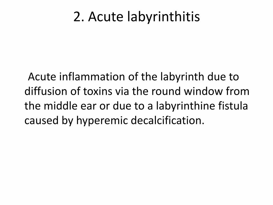
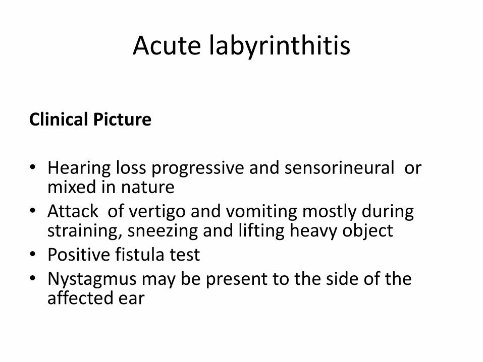
Acute labyrinthitis
Diagnosis bull High index of suspicionbull Positive fistula test bull HRCT scan of temporal bone will demonstrate fistulaif present
Treatment High dose IV antibioticsCortical mastoidectomy with removal of granulations and
closure of fistula if present
Acute facial nerve paresisparalysis
bull A result of an inflammatory response of the facial nerve within the fallopian canal to the infection
bull The tympanic segment is the most common site to be involved as the facial canal may have dehiscences along this segment
Diagnosis of Facial nerve paralysis
bull Clinical House Brackmann grade must be established and monitoring for progression and recovery made
bull HRCT of the temporal boneperformed to look for bone erosion
Treatment of acute facial nerve paralysis
bull Intravenous antibiotics
bull Cortical mastoidectomy + ventilation tube insertion
bull Excision of granulations over dehiscent facial canal and decompression of the nerve
Extracranial Complications Mastoid abscess -clinical
bull Classically a postaural abscess occurs
bull When the abscess spreads along the mastoid air cell system other sites of collection occur eg
bull Bezoldrsquos abscess abscess over upper part of sternomastoid muscle
bull Lucrsquos abscess abscess
over root of zygoma
bull Citellirsquos abscess abscess
over posterior belly of
digastric
Postaural abscess
Sites of pneumatisation of mastoid air cell system in relation to types of mastoid abscess
Zygomatic cells (Lucrsquos abscess)
Mastoid tip cells (Bezoldrsquos abscess)
Digastric cells (Citellirsquos abscess)
Mastoid cellsPostaural abscess
Extracranial Complications Mastoid abscess -treatment
bull Aspiration or drainage of abscess and pus sent for CampS
bull IV antibiotics
bull Cortical mastoidectomy may be required after 24-48 hours if symptoms persist
Intracranial Complications
bull What are intracranial complications
bull What is the most common intracranial complication
bull Which symptoms do patients presenting with intracranial complications exhibit
bull What are the investigations required to diagnose such complications
Important tenets
bull Intracranial complications should be suspected in a child with recent onset of headache fever convulsionsvomiting not feeding well or focal neurological deficit
bull Contrast enhanced CT scan of the brain is mandatory prior to lumbar puncture when intracranial complication is suspected to avoid ldquoconingrdquo (brain herniation into foramen magnum which causes death)
Intracranial Complications ExtraduralAbscess
Definition
bull Collection of pus against the dura of the middle or posterior cranial fossa
bull Extradural abscess is the commonest intracranial complication of otitis media
Intracranial Complications Extraduralabscess ndash clinical amp treatment
Clinical Picturendash Persistent headache on the side of otitis mediandash Pulsating dischargendash Feverndash May be asymptomatic (discovered during surgery)
Diagnosisndash CT scans reveal the abscess as well as the middleear pathology- MRI reveals associated dural inflammation
Treatmentndash Mastoidectomy and drainage of the abscess
Intra-cranial ComplicationsSubdural Abscess- clinical
Definition
ndash Collection of pus between the dura and the
arachnoid
ndash A rare pathology
Clinical picture
ndash Headache without signs of meningeal irritation
ndash Convulsions
ndash Focal neurological deficit (paralysis loss of sensation visual field defects)
Intracranial Complications Subdural Abscess-Investigation and treatment
Investigations
ndash CT scan
- MRI
Treatment
ndash Drainage or excision (neuro-
surgical consultation required)
ndash High dose IV antibiotics
ndash Mastoidectomy
Intracranial Complications Meningitis-pathology
Definition
ndash Inflammation of the meninges (pia arachnoidand dura)
Pathology
ndash Two forms
bull Circumscribed meningitis no bacteria in CSF
bull Generalized meningitis bacteria are present in CSF
Intracranial Complications Meningitis-clinical
Clinical picture
ndash General symptoms and signs
bull High grade remittent fever restlessness irritability
bull Photophobia and delirium
bull Instability
Intracranial Complications Meningitis -signs of meningeal irritation
Signs of meningeal irritation
bull Nuchal rigidity
bull Positive Kernigrsquos sign difficulty to straighten the
knee while the hip is flexed
bull Positive Brudzinskirsquos sign
ndash passive flexion of one leg results in a similar
movement on the opposite side or
ndash if the neck is passively flexed flexion occurs in the
hips and knees
Intracranial Complications Meningitis- diagnosis and treatment
Diagnosis
bull Lumbar puncture is diagnostic
Treatment
bull High dose IV antibiotics
bull Antipyretics and supportive measures
bull Mastoidectomy to control the ear infection after general condition improves
Intracranial Complications Lateral Sinus Thrombosis- etiology
Definition
bull Thrombophlebitis of the lateral and sigmoid venous sinus most often in the sigmoid sinus
Etiology
bull It usually develops secondary to direct extension from a perisinus abscess due to an advanced otitis media
Lateral sinus thrombosis pathogenesis
Stage 1 Stage 2 Stage 3
Intracranial Complications Lateral Sinus Thrombosis-clinical
Signs of blood invasion
- Fever (spiking) with rigors and chills or persistent fever(septicemia)
ndash Positive Greisingerrsquos sign which is edema and tenderness over the area of the mastoid emissary vein
Signs of increased intracranial pressure
Headache vomiting and papilledema
When the clot extends to the jugular vein the vein might be felt in the neck as a tender cord
Intracranial Complications Lateral Sinus Thrombosis-diagnosis
bull CT scan with contrast ldquodeltardquo
sign
bull MRI MRangiography MR
venography
bull Angiography venography
bull Blood cultures is positive
during the febrile phaseMR venography showing obstructedsigmoid sinus on the right side and good venous filling on the left
Intracranial Complications Lateral Sinus Thrombosis- treatment
Treatment
Medical
bull High dose IV antibiotics and supportive treatment
bull Anticoagulants
Surgical
bull Mastoidectomy with exposure of the affected sinus and the intra-sinus abscess is drained
Intracranial Complications Brain Abscess
Definition
bull Localized suppuration in the brain substance
bull It is most lethal complication of suppurative otitis media
Incidence
bull 50 otogenic brain abscess
bull It is more common in males especially between 10 ndash 30 years of age
Intracranial Complications Brain Abscess- pathology and diagnosis
Pathology
bull Site Temporal lobe or less frequently in the cerebellum
Diagnosis
bull Contrast enhanced
CT scan of the brain
bull Contrast MRI brain
Intracranial Complications Brain Abscess-treatment
Medical
bull Broad-spectrum antibiotics
bull Measures to decrease intracranial pressure
Surgical
bull Neurosurgical drainage or excision of the abscess
bull Mastoidectomy operation after subsidence of the acute stage
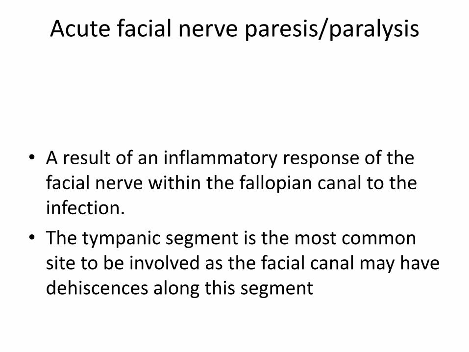
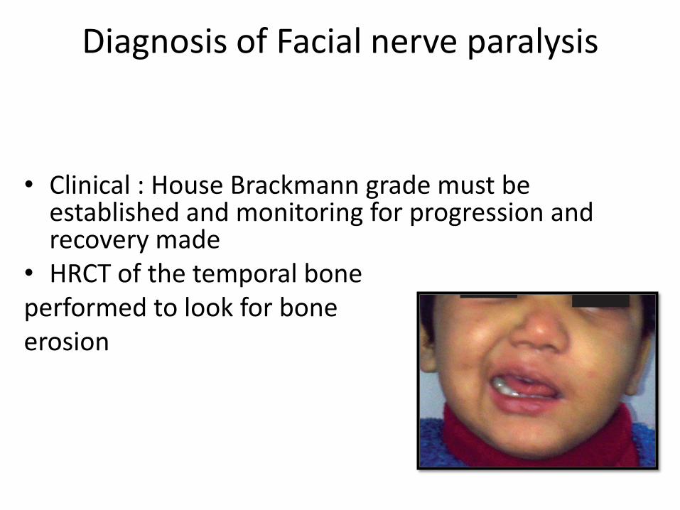
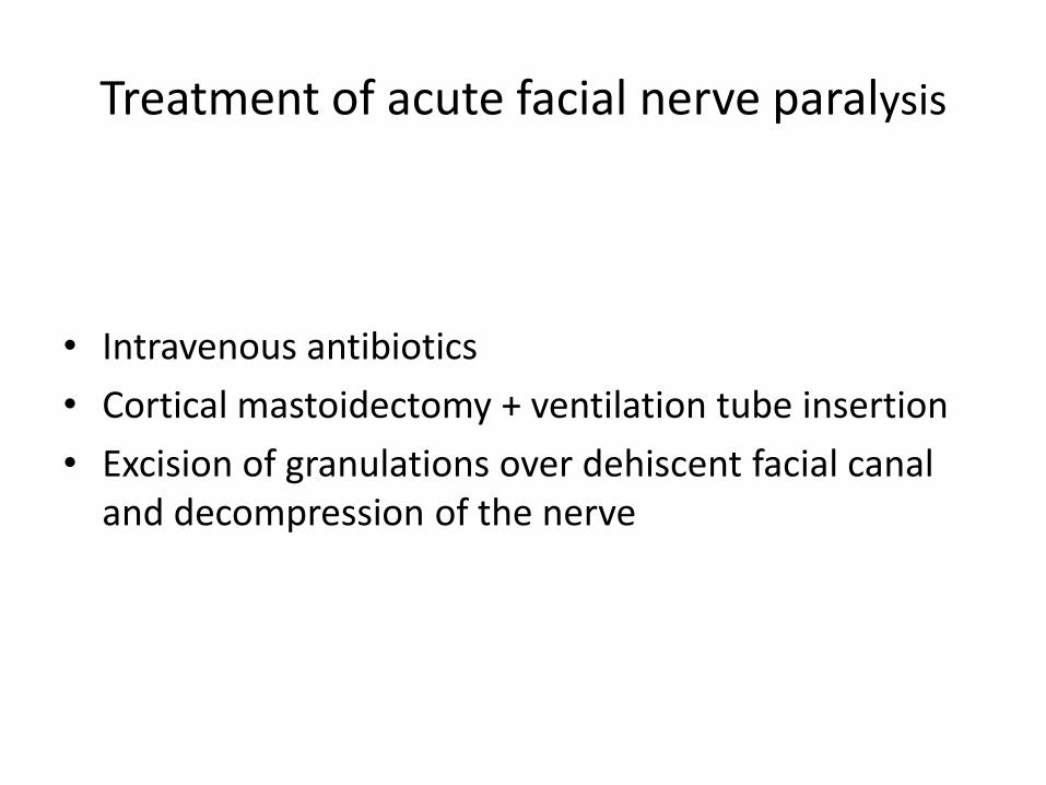
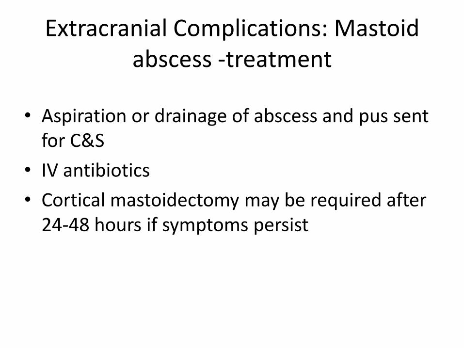
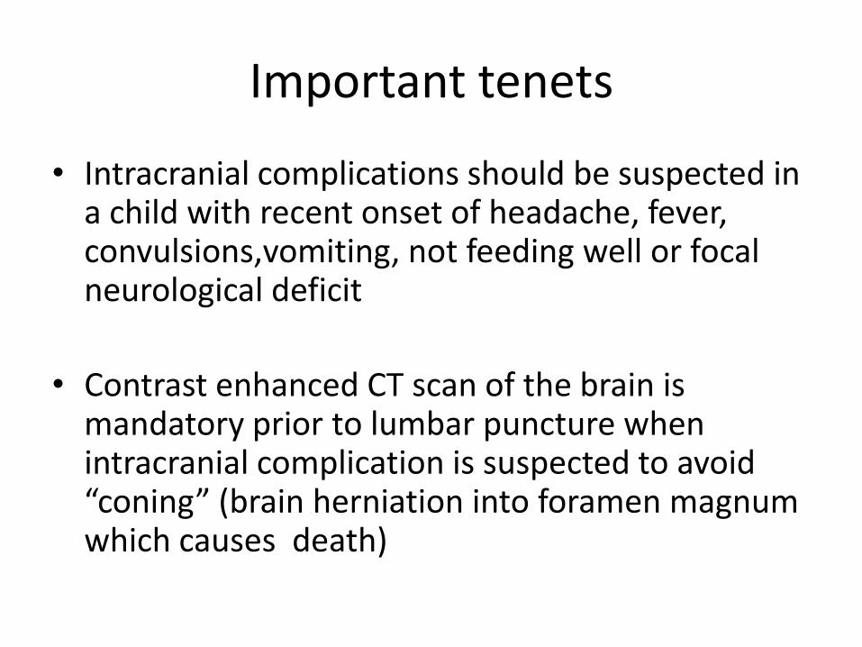
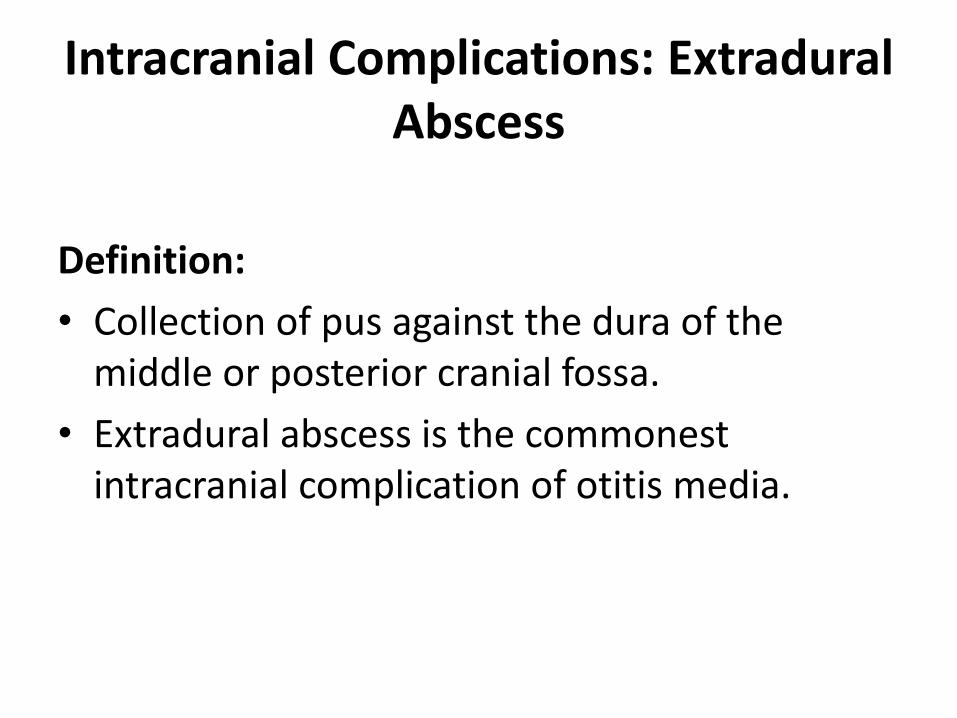
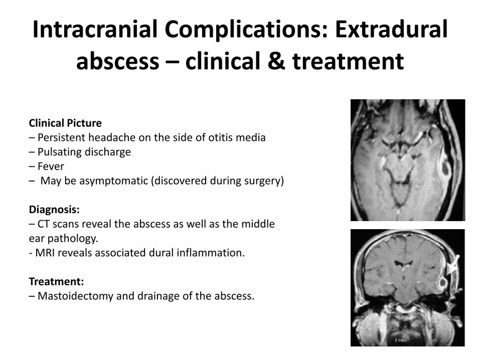
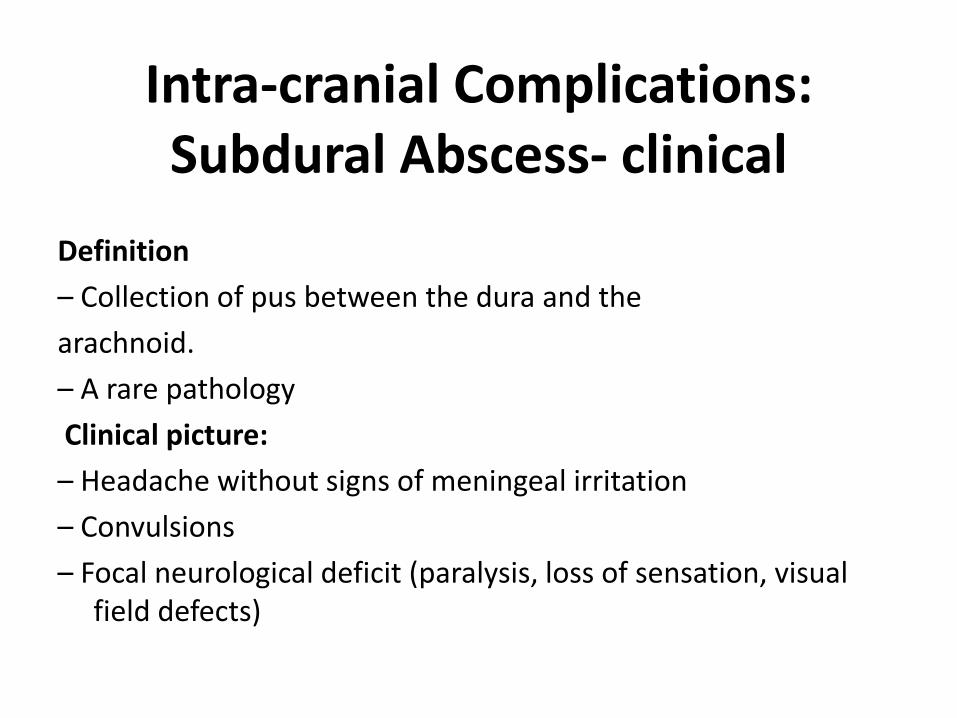
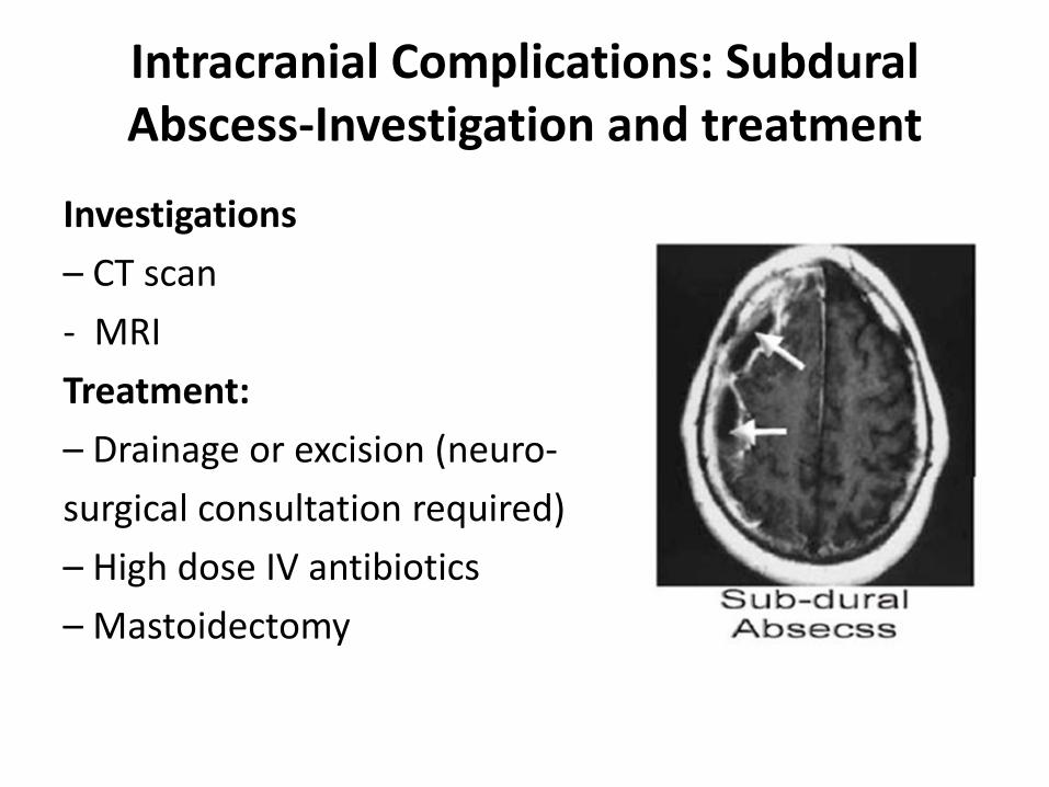
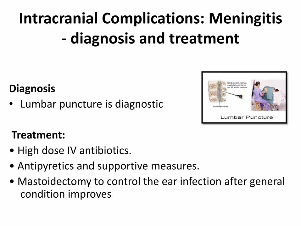
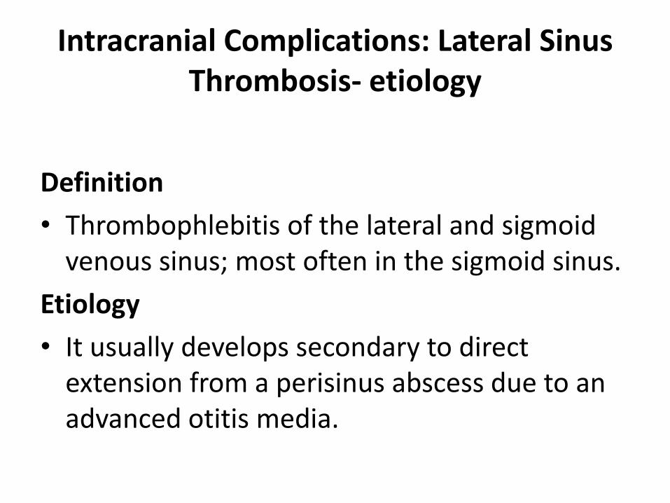
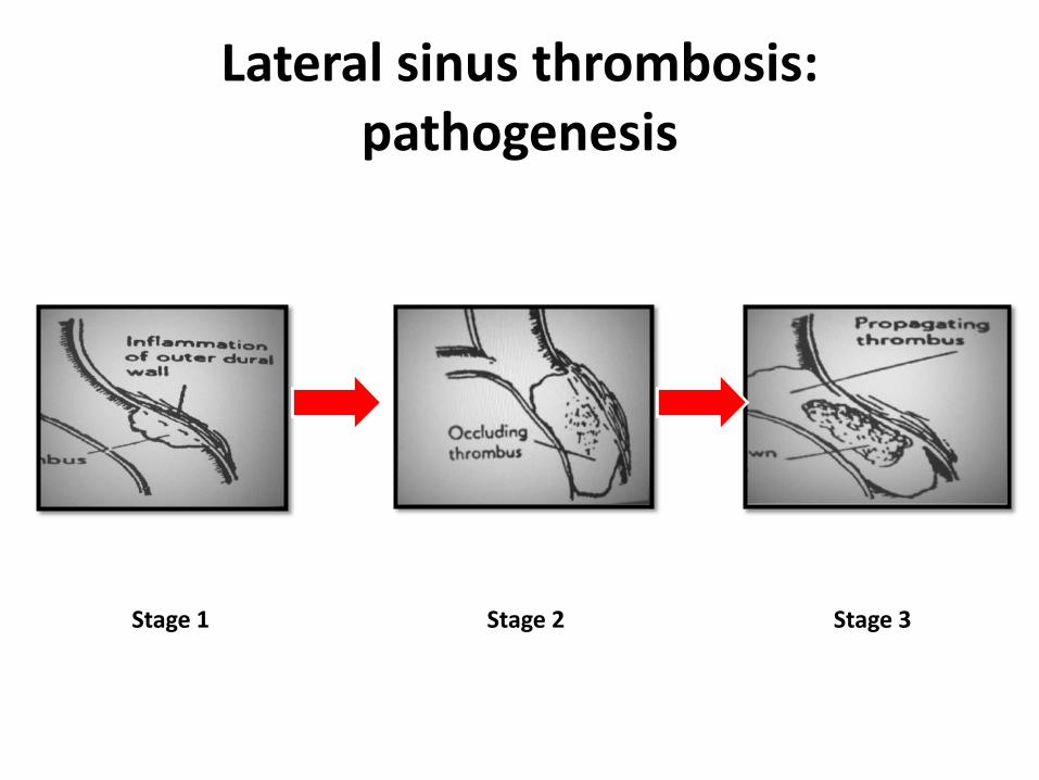
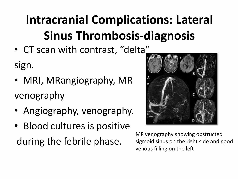
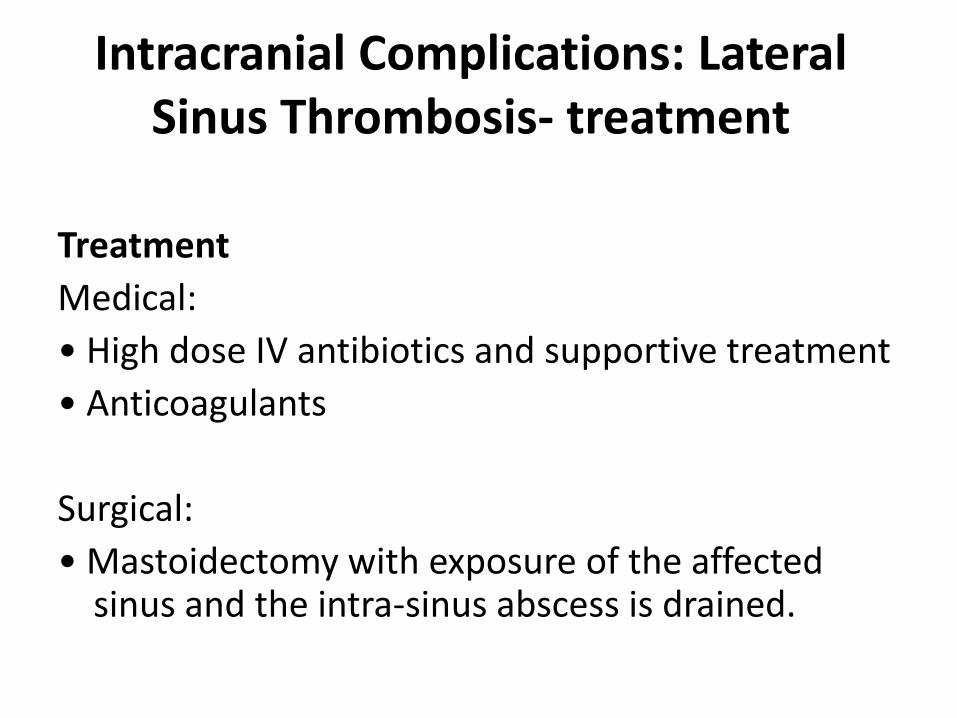
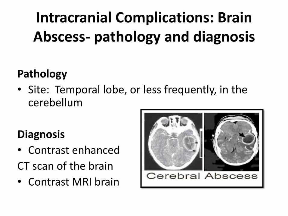
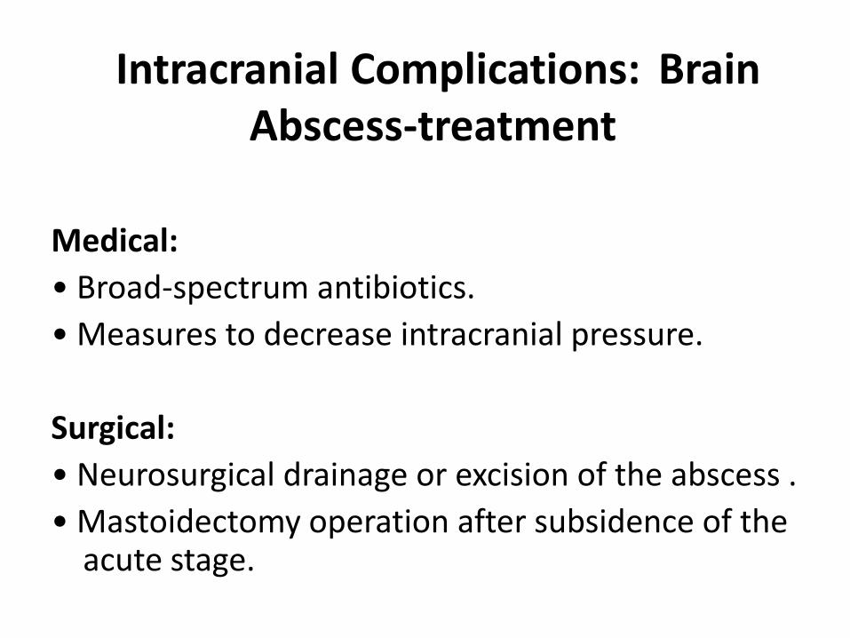
Intracranial Complications Brain Abscess-treatment
Medical
bull Broad-spectrum antibiotics
bull Measures to decrease intracranial pressure
Surgical
bull Neurosurgical drainage or excision of the abscess
bull Mastoidectomy operation after subsidence of the acute stage