completed case study stages with participant responses and
TRANSCRIPT

Patient with Daily Headache Completed Case Study Stages With Participant Responses and
Consultant Interim and Final Summaries
R. Allan Purdy, MD, FRCPC,FACPProfessor of Medicine (Neurology)
Dalhousie University, Halifax, Canada

Patient with Daily Headache Stage 1:
R. Allan Purdy, MD, FRCPC,FACPProfessor of Medicine (Neurology)
Dalhousie University, Halifax, Canada

Learning Issues
• Headaches in the elderly always require a look for a secondary causation
• The history is important as on examination there few clinical signs
• Systemic symptoms have to be sought out to make the diagnosis
• Failure to diagnose this condition has serious consequences

Case History
• JS is a 72-year-old man with a three- month history of diffuse headache, somewhat more left sided and frontal temporal in location. The headache is moderately severe and boring in nature and without relieving or aggravating factors.

Case History
• Over time the headache is gradually worsening and he has begun to feel ill. He develops pain in his face, jaw and tongue.
• He noticed increasing tiredness and some weight loss. He has history of vascular disease
• In the past few weeks he has noticed recurrent episodes of transient loss of vision in his left eye, coming down like a curtain.

Physical Examination
• On examination he looks somewhat ill and in some distress from the pain.
• His blood pressure is 140/90 and PR is 68.• He has no focal neurological signs and his
general examination appears normal. • His fundi showed some atherosclerotic
and hypertensive arteriole changes. • There are no bruits in his head or neck.

Initial Impression
• JS has a steady headache which is not specific for any primary or secondary headache disorder and one which could be a chronic daily headache since it is present more than 15 days a month and longer than 4hrs in duration, however it has yet to last six months which is important in the differential diagnosis.

Other Historical Facts
• JS did not report any triggering or relieving factors. This is very important since most primary headaches, including migraine, tension- type headache and cluster headache, have defi nite modifying factors.
• As this is associated with a progressive worsening over time, it is a definite ‘red flag’ with respect to looking for a secondary cause.
• He has also had his gall bladder removed.

First Stage Questions
• At this stage can you consider three differential diagnoses for this case?
• Is there any other information you would like to know about the case?
• What initial investigations, if any, would you like to consider?

Q1: At this stage can you consider three differential diagnoses for this case?
PARTICIPANT RESPONSES:
Participant (Ireland) "Temporal arteritis Subdural haematoma Neoplasm"
Participant (USA) "1.Temporal arteritis/vascululitis 2.chronic subural or tumor 3.metabolic disorder or endocrine"
Participant (USA) "1. giant cell arteritis 2. sarcoidosis, TB, or syphilis causing a pachymeningitis along the left skull base 3. pituitary adenoma with thyroid dysfunction and extension to the left skull base"
Participant (USA) "1. Temproal arteritis 2. Migraine 3. Carotid stenosis causing a "claudication" type of HA? (Something like the HA somtimes seen in those with moya-moya)"
Participant (USA) "1- Temporal Arteritis/Giant Cell Arteritis 2- Internal carotid artery dissection/occlusion 3- Cytomegalovirus."
Participant (France) "1. temporal arteritis 2. brain tumor 3. cranial infection"
Participant (Turkey) "Temporal arteritis"
Participant (Portugal) "1.Temporal arteritis (probably with polymyalgia rheumatica) 2. Polymyositis 3. Neoplastic meningeal infiltrative disease"
Participant (USA) "Giant cell arteritis (temporal arteritis); vasculitis; malignancy are 3 possibilities"
Participant (Peru) "1. Temporal arteritis 2. Carotid disection 3. Temporomandibular arthritis"

Q2: Is there any other information you would like to know about the case?
PARTICIPANT RESPONSES:
Participant (Portugal) "1. Jaw claudiaction? 2. Pain (and/or ulceration)of the oral mucosa tongue or scalp? 3. Low-grade fever? 4. Passive range of motion in neck far exceeds the active range?"
Participant (USA) "Does he have palpable pulses in his temporal arteries, and are they tender? Does he experience jaw claudication? Does he have ploymyalgia rheumatica? Scalp or tongue atrophy? Is there a positional effect to the HA and is it worse in the mornings? has he seen an ophthalmologist regarding the episodic visual loss?"
Participant (USA) "1- Is there any history of immunosupession? 2- Are there any symptoms or signs (besides weigth loss/tiredness to suggest a malignant process somewhere else aside from the Nervous System? 3-What medications, if any, is the patient taking for his Arterial Hypertension? Compliance? 4- Past history of infectious diseases that may lead to a chronic infectious process in CNS like TB, syphilis or HIV? 5- Is the patient taking any medication/s for headache that may lead to a medication overuse headache?"
Participant (Ireland) "Presence of jaw claudication/fever? Prior history of PMR? "
Participant (USA) "Hx of stroke/MI, evidence of other areas having claudication--i.e. pain in legs relieved with rest. More about vascular risk factors--tobacco, DM, cholesterol, whether he is on meds for his elevated blood pressure. "
Participant (Turkey) "Is there any medication overuse? Any risk factors for stroke?"
Participant (USA) "1. What makes pain worse or better? 2.Is there any problem with sleep? 3.Medications?"
Participant (France) "Does he have any history of cancer or trauma? "
Participant (Peru) "If there are a history of muscular aches, polymyalgia rheumatica"
Participant (USA) "Does he smoke? Any fevers? Coughing? Night sweats? History of head trauma? Rashes?"

PARTICIPANT RESPONSES:
Participant (Portugal) "1.Erythrocyte sedimentation rate? 2. Complete blood count? 3. Plasma electrophoresis "
Participant (Peru) "A eritrocyte sedimentation rate and a temporal biopsy"
Participant (USA) "ESP, CRP, temporal artery biopsy, CBC (check platelet levels), MRI +/- contrast, ANA, RF"
Participant (USA) "Initial: CBC, ESR,C Reactive Protein, Carotid Ultrasonography, Chest X-Ray, CMV tests, Comprehensive Metabolic Panel (including LDH)"
Participant (Ireland) "ESR Temporal artery biopsy CT Brain"
Participant (Turkey) "Sedimentation rate Temporal artery biopsy "
Participant (USA) "1. Metabolic profile,CBC, Sed Rate and C-reactive protein 2. MRI head scan 3. Endocrine tests for thryoid"
Participant (USA) "1. Bloodwork: ESR, CRP, ANA, ACE, TSH, RPR, MHATP, CEA, CBC and CMP 2. MRI with/without of brain 3. LP "
Participant (USA) "Labs: ESR, CRP Imaging: Some type of vascular imaging of head and neck, as well as brain imaging-- carotid US to start, or straight to CT/CTA. "
Participant (France) "CT cranial and brain scan. Biopsy of the temporal artery "
Q3: What initial investigations, if any, would you like to consider?

Stage 1 Summary: Dr A Purdy
I think the group is doing an excellent job with the diagnosis of TA or temporal arteritis being the top one from all that responded. Headache is common in TA and occurs in about 90% of cases with this being the most common systemic vasculitis.
Amaurosis fugax or a symptom of unilateral transient visual loss like a curtain coming down is not common in TA which gives one a reason for pause - some estimate that it occurs in about 12% of cases.Also everyone asked for more information and suggested ordered reasonable tests and asked great questions. It is hard to come up with another working diagnosis other than TA or move in another direction since if TA is the correct diagnosis then of course anyone with that suspicion would have to act on that suspicion. I find and it would be interesting to hear from the participants that in early stages this can be a very difficult diagnosis to make and once made it seems SO obvious.
The headache of TA although a prominent feature is NOT specific in anyway.
I think the real problem with this type of case is to ensure one is not missing another diagnosis, for if truth be told amaourosis fugax is more commonly at TIA symptom of carotid artery disease.
Anyway good work and there is a lot more in the history to date including feeling ill, weight loss and other aches which might suggest TA but he is also a treated hypertensive and has hypertensive changes in his fundi. Thus he has vascular risk factors for non-vasculitic disease.
Anyway, great work and it is nice to see that those that answered so far have the same pattern recognition of the diagnosis from different parts of the world. I will add another question not in the case yet. Would anyone add ASA and how much or do any other urgent tests?
OK, hang in there as they say in North America and move on as there is much more to explore in this case because as you will see the diagnosis may not be the biggest problem to deal with in this case.

Patient with Daily Headache Stage 2:
R. Allan Purdy, MD, FRCPC,FACPProfessor of Medicine (Neurology)
Dalhousie University, Halifax, Canada

More Analysis
• He feels unwell, has fatigue and weight loss, symptoms on their own which could suggest a systemic disease. On their own one might consider something like a malignant process which can present with headaches as well, however one should consider the differential diagnosis more broadly in any patient with constitutional symptoms.

More Analysis
• Also patients with primary headache can have anorexia (tension-type headache) and nausea and vomiting (migraine) but again these are episodic and his age starts to play a role in the diagnostic formulation in his case.

Symptom Analysis
• JS has volunteered symptoms of pain in the face, jaw and tongue, but the latter two are worse with eating and talking, so these need to be evaluated carefully. These symptoms suggest some need for activity to bring them on and are termed, jaw and tongue claudication respectively.

Clinical Pearl
• Claudication suggests the blood supply to the pertinent muscles is compromised to ischemic levels during eating and chewing so that the subsequent pain is most likely ischemic in nature.

Symptom Analysis
• The visual symptoms here are classic for ‘amaurosis fugax’ which implies reduced blood supply to the central retinal artery.
• The classic loss of vision described as a blind coming down is based on the anatomical blood supply to the retina, that is the lower vessels become involved first and then upper arterioles.

Any need to think Otherwise?
• There are no other important features in his history. He has had some mild hypertension over time and prior gall bladder removal, but nothing that suggests any other systemic disorder.

Lab and Investigations
• Preliminary blood work including a complete blood count, CRP and ESR were ordered along with and EKG, chest x-ray and a baseline unenhanced CT scan.
• He also had electrolytes, renal function and lipids done.

Secondary Impression
• A secondary headache in an older patient is likely. Transient ischemic attack or TIA, might be important, because of the visual symptoms, so carotid artery disease needs consideration.
• However the headache is daily which is unusual in a TIA and is progressive so another vascular diagnosis should be kept in mind.

Further impression
• A less likely concern would be cardiac emboli from non-bacterial endocarditis associated with an underlying malignancy, which might fit with some of the general and systemic symptoms.

Second Stage Questions
• At this stage can you narrow your diagnosis a couple of entities?
• Can you analyze the case further based on what you know about this diagnosis?
• Any other investigations you would do and how would they be justified?
• Any immediate treatments options you would consider at this stage?

Q1: At this stage can you narrow your diagnosis to a couple of entities?
PARTICIPANT RESPONSES:
Participant (Canada) "Sounds a lot like temporal or giant cell arteritis to me."
Participant (USA) "Temporal arteritis is by far the most likely diagnosis."
Participant (Portugal) "Temporal arteritis "
Participant (USA) "I would like to know if the studies were normal or not. I believe a vascular cause needs to be looked at carefully."
Participant (Peru) "Basically, a temporal arteritis, and an a carotid dissection"
Participant (Brazil) "a)Temporal arterites or b)other vasculites involving CNS and systemically in immunologic or paraneoplasic base"
Participant (Turkey) "As narrowing the diagnosis malignancy, vascular disease and temporal arteritis are main entities in the differential diagnosis"

Q2: Can you analyze the case further based on what you know about this diagnosis?
PARTICIPANT RESPONSES:
Participant (Portugal) "I would consider treat the patient as the diagnosis is temporal arteritis"
Participant (Brazil) "Symptoms lasting 3 months, no treatment and still no complication is a bit strange for temporal arterites (giant cell)"
Participant (Peru) "Patient have headache and amaurosis fugax, these could be caused by temporal arteritis and carotid disection. Systemic symptoms mean a systemic pathology, like temporal arteritis."
Participant (Canada) "The jaw claudication is quite typical for giant cell arteritis. So are the visual symptoms, although it was my impression that the visual symptoms in giant cell arteritis were usually due to involvement of the ciliary circulation to the optic nerve head, and not due to involvement of the central retinal artery itself. However, in a patient with a new onset headache that is progressively worsening in an elderly patient, giant cell arteritis must be high on the list."
Participant (USA) "I would say he also has polymyalgia rheumatica, frequently touted as the cause of the constitutional symptoms."
Participant (Turkey) "Systemic symptoms and weight loss in (not only elderly) a case with over age 50, physician should suspect malignancy, as a treatable cause of elderly headache or vascular supply problems. Then I prefer to exclude the possibility of malignancy. As I always like to ask medication overuse headache (MOH) in chronic headache. In elderly, physician frequently prescribe analgesics for arthritis. Thus, headache type of MOH may not fit any type of primary headache and systemic complications may associate. It should be discarded. The first thing in the diagnosis is history. Glacuoma should be excluded, too. Vascular problems may also result in jaw claudicating and systemic problems. If above studies are negative, I further on for vascular studies. I perform vascular studies for the exclusion of TIA or stroke but even if the definite diagnosis is temporal arteritis, TA tends to affect the branches of the carotid artery, too. "
Participant (USA) "Ischemic cause for headache in the carotid arties might be a problem as a result of a systemic
disorder."

PARTICIPANT RESPONSES:
Participant (Canada) "I did not see the results of the ESR and CRP. However, high or not, he likely deserves a temporal artery biopsy, especially in he has tender temporal arteries. "
Participant (Portugal) "Temporal artery biopsy"
Participant (Brazil) "As a neurologist I would like to refer the patient to vascular specialist (as far as I know there is a long list of systemic vasculites."
Participant (Peru) "A temporal biopsy and a carotid duplex."
Participant (Turkey) "Systemic symptoms and weight loss in (not only elderly) a case with over age 50, physician should suspect malignancy, as a treatable cause of elderly headache or vascular supply problems. Then I prefer to exclude the possibility of malignancy. If above studies are negative, I further on for vascular studies. I perform vascular studies for the exclusion of TIA or stroke but even if the definite diagnosis is temporal arteritis, TA tends to affect the branches of the carotid artery, too. Sedimentation rate is simple and cheap but not specific. Up to 20-30% of patients with temporal arteritis may have a normal or low ESR. C-reactive protein may be more helpful, if lab is available. I definitely perform temporal artery biopsy, before initiation of the medication. "
Participant (USA) "A temporal artery biopsy should be set up while waiting for ESR results. Also I would perform a carotid ultrasound."
Participant (USA) "I would like to see a MRI/MRA study."
Q3: Any other investigations you would do and how would they be justified?

PARTICIPANT RESPONSES:
Participant (Canada) "His story is clinically very suspicious, and his vision is in peril. He should be started on high dose steroids while the temporal artery biopsy is being arranged. "
Participant (Portugal) "Start immediately prednisone, 60 mg/day, plus aspirin, due the presence of amaurose fugax episodes"
Participant (Peru) "I would prefer realize a biopsy and after that I''d begin corticosteroids."
Participant (Turkey) "If the visual symptoms progress, I will put on steroid and antiaggregant treatment in order to prevent permanent visual loss as early recognition and prompt treatment are critical to prevent permanent ischemic damage to the retina and optic nerve. "
Participant (USA) "Yes, steroid treatment should be started now, even if the ESR is negative. Since he has probable ischemic symptoms already, he should be admitted for IV solumedrol, then switched after 3-5 days to oral high dose Prednisone. I don''t think antiplatelet tx or anticoagulation is indicated, unless a carotid dissection is found."
Participant (Brazil) "If CRP or ESR resulted elucidative I will start predinisone 40 to 60 mg per day. Temporal artery biopsy has low chance to come out positive. In a very smart procedure using US, collecting good sample, 50 to 100 examination at mycroscopy take a too long time."
Q4: Any immediate treatment options you would consider at this stage?

Stage 2 Summary: Dr A Purdy
By now the diagnosis of Temporal Arteritis is not only the working diagnosis, but the actual diagnosis in this case. The indoloent nature of the headache is important.
Gradual worsening headache in an elderly person does ALONE raise the diagnosis especially if the person is ill and has systemic symptoms. It is imperative to start steroid medication to prevent blindness in particular, but there is no guarantee that visual symptoms will not progress or visual loss will be avoided.
The visual symptoms are of a TIA and some have alluded to that in their comments.
There in fact is evidence that ASA AND steroids might help, and I will arrange for that to be send along. We will also be posting some PDFs soon which are excellent resource material on TA.
A malignancy is a good thought in this case but of course is less ''treatable''. I thought dissection an interesting thought but this case has many more features that make this diagnosis untenable. Now the results of the tests will be presented in the next stage with some more relevant questions.

Patient with Daily Headache Stage 3:
R. Allan Purdy, MD, FRCPC,FACPProfessor of Medicine (Neurology)
Dalhousie University, Halifax, Canada

Diagnosis
• The visual symptoms in this case along with jaw and tongue claudication, are very characteristic of temporal arteritis, as well as pain in the shoulders which is termed polymyalgia rheumatica, a feature missing in his early presentation but present later.
• One physical finding helped in the diagnosis, seen on next slide!
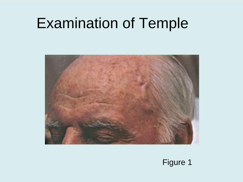
Examination of Temple
Figure 1

Diagnostic Points
• Systemic symptoms of being and “looking” ill are classical but not specific.
• Also remember that “temporal arteritis” is a systemic disorder and a misnomer since other major arteries can be involved including the carotid, basilar and coronary arteries, which could cause a major stroke or myocardial infarction.

The Headache of TA
• The headache in temporal arteritis is not specific but does flag raise the diagnosis early in the case of any elderly patient.
• The pain is usually located over the temporal artery region and is the commonest presenting symptom of the disorder.

Another Clinical Pearl
• Anterior ischemic optic neuropathy or AION can occur in patients with temporal arteritis and can lead to permanent blindness if not recognized and treated and even if treated.
• The diagnosis in his case was supported by the clinical observation of a swollen, tortuous and painful left temporal artery (Figure 1).

Management Issues
• Obtaining results of blood work.• Treatment with corticosteroids.• CT baseline scan to look for other
diagnoses in this case. • Carotid duplex dopplers in this case to rule
out carotid disease with TIA.• Temporal Artery Biopsy

Lab Results
• The results of the blood work can be very informative and strongly support the diagnosis in this case. – The CBC showed a microcytic anemia, the
ESR was 125 (normal 4-16), with a CRP of 42 (normal is less than 10).
– His serum chemistry including was normal.– His EKG was essentially normal.

Comments on Steroids
• Treatment should be started as early as possible to prevent major complications of the arteritis, especially loss of vision, usually due to an AION.
• The treatment is oral steroids with the dosage range of prednisone being 40 mg/day to 80 mg/day, although 60 mg/day or above has been more commonly suggested than lower doses.

Justification for CT
• A CT scan is reasonable in his case as he could have a systemic malignancy with secondary brain metastases or small infarctions from prior carotid TIAs or cardiac microemboli.
• In his case this test was normal for age and that is the usual finding in this disorder.

Comment on MR
• One might wonder why an MR was not done with contrast. Many reasons are:– No indication for MR in this disorder– CT did not show any vascular lesions and
carotid dopplers were normal.– CT is easier to obtain on an emergent basis.– Case NOT a posterior circulation TIA or stroke
and no need to visualize these areas or arteries.

Reason for Carotid Dopplers
• Again this test is reasonable since he presented with amaurosis which usually is due to artery to artery embolic from the internal carotid artery to the ophthalmic artery and retinal vessels.
• In his case the test was normal.

Third Stage Questions
• At this stage how certain of your in this case?
• Any other investigations you would do and how would they be justified?
• Can you suggest long term management recommendations for this case?
• Are there any issues in long term management of cases like this one?

Q1: At this stage how certain are you of your diagnosis in this case?
PARTICIPANT RESPONSES:
Participant (USA) "Quite certain!"
Participant (Portugal) "At this point I don''t think in others diagnosis"
Participant (USA) "I am certain enough to start empiric treatment."
Participant (Brazil) "The visual aspect and pain at the temporal artery, elevated ESR and CRP give reasonable support for diagnosis. Besides that no other relevant investigation finding (CT, Doppler, EKG, blood chemistry)"
Participant (Peru) "Temporal arteritis is the more probable diagnosis considering anamnesis, physical exam and laboratory results. In order to a definite diagnosis is pending the anatomopathological confirmation, but it do not must delay treatment."
Participant (USA) "My #1 Dx was Temporal Arteritis. Very Certain."

Q2: Any other investigations you would do and how would they be justified?
PARTICIPANT RESPONSES:
Participant (USA) "The Temporal artery biopsy is key, with a long sample from the affected side examined under thin cuts. Can consider a doppler of the vessel to target the most affected area for the biopsy sample."
Participant (Portugal) "Could be done an temporal superficial duplex Doppler to find the characteristic hypoechoic halo sign around the lumen of an stenosed or occluded arterial segment. The patient has been submited to an temporal artery biopsy? Results?"
Participant (USA) "I am certain enough to begin empiric treatment. All of the details fit TA well, and the visual picture of the artery is consistent especially if it is non-pulsatile."
Participant (Peru) "I would not do any other investigation"
Participant (USA) "None unless patient develos a change in his symptoms or signs or a new headache."
Participant DD (Neurology/Brazil) "Temporal artery biopsy has been indicated. The visual condition suggests that there is good spot to collect sample. It should be done but even with an excelent procedure the result can be unhelpful. (My experience in 3 cases resulted negative)"

PARTICIPANT RESPONSES:
Participant (USA) "Start steroids 60mg/day, then taper slowly, by 2.5-5mg every 1-3 weeks. Monitor disease progression with ESR or plasma interleukin 6. For flares, can pulse dose with oral or IV steroids. Maintain steroids 1-3 years."
Participant (Brazil) "Steroids during about 6 months and care the side effects (elevation of blood pressure, fotosensibility, weight gain, edema etc...) "
Participant (Portugal) "Yes, Yes and Yes"
Participant (Peru) "Corticosteroid treatment must be tapered over a few weeks slowly, by the end of the first month patient are taking about 40mg prednisone. Slowly tapered to a maintenance dose of 5 mg or a minimum tolerated"
Participant (USA) "Maintain his other health conditions well managed. Be aware and look for any changes in Hx or P.E. that merit any other management."
Participant (USA) "Long term treatment with Prednisone starting at 60 mg/day for 1-2 months (I would err on the side of longer), then a slow taper by about 10 mg/d/month, monitoring for recurrence clinically and with ESR''s."
Q3: Can you suggest long term management recommendations for this case?

PARTICIPANT RESPONSES:
Participant (Portugal) "steroids at least 2 years (depending the clinical and ESR response)and aspirin with no timing limitation"
Participant (Brazil) "As far as I know after 6 months of treatment he will be OK. I have a question... Is ASA reasonable in this case?"
Participant (USA) "Steroids, we all know, are a double edged sword. Diabetes, hypertension, infection, osteoporosis and avascular necrosis, psychiatric changes, myopathy, cataracts, obesity to name a few!! -all need to be monitored carefully."
Participant (Peru) "Some issues respect long term management are: 1. Complications of chronic corticosteroid therapy. 2. Can therapy be suspended in any moment? 3. Other immunosuppresant drugs can be used? Which? How?"
Participant (USA) "Relapses, treated with high-dose steroid pulses; development of steroid intolerance or diabetes."
Participant (USA) "1- Cushingoid features 2- Osteoporosis 3- Avascular hip necrosis"
Q4: Are there any issues in long term management of cases like this one?

Stage 3 Summary: Dr A Purdy
So far this case is going just as I suspected.
You all got the right diagnosis, but you had a wide variety of responses for other possibilities. That really interests me and there is no good explanation for that.
It is possible that the diagnosis was so obvious, which is a good thing, that any second or third diagnosis was jut put in because the question was asked.
I will have to think about those responses some more and I am sorry due to technological reasons that all of you cannot see each others response. I guess I am "learning" the most.
This is an easy diagnosis in this case but in others it would not be and can be missed.
Your investigations are all reasonable but people have various reads on the temporal artery biopsy and other tests. The treatment is generally steroids and by in large is empirical about 1mg per KG, and duration of treatment varies and endpoint for some clinical or lab based.
I prefer clinical.
Some asked about ASA and a Canadian colleague provided me with the following references which appear on the next two pages….

Stage 3 Summary: Dr A Purdy (cont’d)
Rheumatology (Oxford). 2009 Mar;48(3):258-61. Epub 2009 Jan 7.High incidence of severe ischaemic complications in patients with giant cell arteritis irrespective of platelet count and size, and platelet inhibition.
Berger CT, Wolbers M, Meyer P, Daikeler T, Hess C.Department of Internal Medicine, University Hospital Basel, Petersgraben 4,CH-4031 Basel, Switzerland. [email protected]
OBJECTIVE: Vision loss and ischaemic stroke are feared complications in GCA.
We investigated how platelet count and size and platelet inhibition with ASA relate to ischaemic complications in patients with GCA. METHODS: Charts of patients with GCA were retrospectively analysed.
Jaw claudication, amaurosis fugax, blurred vision, ischaemic stroke and permanent visual loss were classified as ''ischaemic events''; ischaemic stroke and permanent visual loss were sub-grouped as ''severe ischaemic events''.
The incidence of ischaemia and the association to the pre-defined covariates age, fever, ESR, platelet count and size and ASA treatment were assessed.
RESULTS: Eighty-five patients (mean age 73 yrs, 60% women, 78% biopsy-proven) were included in the analysis. Of the 85 patients, 62 (73%) presented with ischaemic events, 29/85 patients (34%) with severe ischaemic events.
At the time of diagnosis 22/85 patients (26%) were treated with ASA. Of these 22 patients, 15 (68%) presented with ischaemic events, 7/22 patients (32%) with severe ischaemic events. In multivariate analysis, neither platelet count nor size or ASA treatment were significantly associated with ischaemic or severe ischaemic events.
CONCLUSIONS: The incidence of severe ischaemic events in patients with GCA was high, irrespective of platelet count and size and established ASA treatment.
Arthritis Rheum. 2002 Feb;46(2):457-66.Therapeutic effects of acetylsalicylic acid in giant cell arteritis.Weyand CM, Kaiser M, Yang H, Younge B, Goronzy JJ.Mayo Clinic, 200 First Street SW, Rochester, Minnesota 55905, [email protected]

Stage 3 Summary: Dr A Purdy (cont’d)
Comment in: Arthritis Rheum. 2002 Nov;46(11):3113; author reply 3113-4.
OBJECTIVE: In giant cell arteritis (GCA), inflammatory lesions typically produce interferon-gamma(IFNgamma)-- and nuclear factor kappaB (NF- kappaB)-dependent monokines. Corticosteroids influence disease activity by repressing NF-kappaB-dependent genes but have only marginal effects on IFNgamma.
The current study explored whether acetylsalicylic acid (ASA) had cytokine-repressing activity in GCA and could function as a steroid-sparing agent.
METHODS: Temporal artery-severe combined immunodeficiency (SCID) mouse chimeras were created by engrafting inflamed temporal arteries into SCID mice.
Chimeras were treated with ASA, indomethacin, or dexamethasone for 3 weeks.
Temporal artery grafts were harvested and cytokine message was semiquantified by polymerase chain reaction-enzyme-linked immunosorbent assay. The ability of dexamethasone and ASA to suppress IFNgamma and interleukin-1beta (IL-1beta) messenger RNA and protein production was also tested in vitro using T cell clones and monocytes derived from patients with GCA.
Drug-induced effects on the transcription factors NF-kappaB and activator protein 1 (AP-1) were assessed by electrophoretic mobility shift assays (EMSAs). RESULTS: At clinically relevant doses, 20-100 mg/kg, ASA was a highly effective inhibitor of cytokine transcription in temporal arteries.
While dexamethasone preferentially targeted NF-kappaB-regulated monokines, ASA acted predominantly by suppressing IFNgamma. Indomethacin failed to reduce tissue IFNgamma transcription, which therefore excluded the inhibition of cyclooxygenases as a critical mechanism.
IFNgamma production by T cell clones was highly sensitive to ASA-mediated suppression, whereas IL-1beta production by lipopolysaccharide- stimulated monocytes responded primarily to dexamethasone.
The combination of ASA and dexamethasone had synergistic effects. EMSAs demonstrated that ASA interfered with the formation of AP-1, whereas dexamethasone suppressed the nuclear translocation of NF-kappaB.
CONCLUSION: The results of this study provide evidence of the complementary action of ASA and corticosteroids in suppressing proinflammatory cytokines in the vascular lesions of GCA

Patient with Daily Headache Stage 4:
R. Allan Purdy, MD, FRCPC,FACPProfessor of Medicine (Neurology)
Dalhousie University, Halifax, Canada

The Temporal Artery Biopsy
• The temporal artery biopsy is an important test to do as soon as reasonable. A good section of artery is required to avoid skip lesions.
• It should be done by a surgeon who has a lot of experience obtaining such specimens and read by a pathologist familiar with the disorder.
• Delay of therapy however is not warranted while waiting for the biopsy.

Options on the biopsy result
• If biopsy positive treatment continues and if negative or equivocal, the treatment should be guided by clinical impressions and/or other laboratory tests, especially the ESR.
• See Figure 2 for a biopsy which shows typical arteritis as seen in this classic temporal arteritis.

Temporal artery biopsy

Long-term Followup
• Patients with temporal arteritis can be managed by most practitioners, but referral to a neurologist, internist or rheumatologist maybe helpful for diagnosis, and ongoing management.
• Surgery and pathology are involved early as well as an early ophthalmologic opinion can be helpful for assessment and follow up of the eye symptoms.

Treatment Follow-up
• The duration of therapy depends on his clinical course and follow-up assessments.
• Most patients require high dose steroids for several weeks and this may last up to a year or longer. – Steroid sparing or other immunosuppressive
agents sometimes are employed in some cases. Long term problems with steroids are to be avoided and must be managed.

Back to the Case!
• Shortly after receiving prednisone orally his symptoms started to subside within hours. The next day he was headache free on 60 mgs per day.
• He continued on this dosage for a month and then it was gradually tapered and discontinued several months later as he became asymptomatic in all respects and his blood work normalized.

Final Diagnosis
• This is a case of temporal arteritis presenting with a visual symptom of amaurosis fugax and not AION.
• The case demonstrates the usual features of the disorder as well as concepts about immediate and long term management.
• The headache of this disorder is not specific and on its own non-diagnostic!

Final Case Summary: Dr A Purdy & Dr H Blumenthal
The case is now completed in all stages. The case was well received and started excellent analysis and discussion. Temporal arteritis was the diagnosis and it was considered by everyone who signed onto the site.
Tests were reasonable as I said in the past but treatments were somewhat varied.
Dr. Harvey Blumenthal has a life long experience in dealing with TA and all of its variations. He reviewed the online correspondence and made useful comments.
Before reading them however I thank all who signed on to the COQ as it was fun and I learned a lot, hopefully most of you as well.
Here are Dr. Blumenthal's comments:
"Modern physicians are more aware of TA and the many different clinical syndromes this disease may present with than were our predecessors of even 25 years ago.
The most common and the most feared complication of TA is sudden and irreversible blindness. The prevalence of this catastrophe varied from 60% of cases reported 25 years ago to 6% of cases reported in 2005. This decline in prevalence is attributed to increasing awareness of the condition, and early treatment.
14-30% of TA patients who become blind report amaurosis fugax preceding the blindness. And 64% of TA patients who experience amaurosis fugax will lose vision within one-ten days.
Furthermore, 20-60% of patients blinded in one eye soon lose vision in the other eye.
In conclusion, TA is a medical emergency, and we should initiate corticosteroid treatment immediately if the diagnosis is considered likely. Temporal artery biopsy may be made within a week without fear of obscuring pathological findings.“
*References: Links to a number of online references and downloadable PDFs via Sage are available within the members section of the IHS Website under Learning Centre / CoQ’.

Further References
• Caselli RJ, Hunder GG. Giant Cell Arteritis and Polymyalgia Rheumatica. In Silberstein SD, Lipton RB, Dalessio DJ, editors. Wolff's headache and other head pain, 7th Ed. Oxford: Oxford University Press, 2001:525-535
• Goodwin, J. Temporal Arteritis. Medlink http://www.medlink.com