complementary use of saxs and sans - embl hamburg · complementary use of saxs and sans jill...
TRANSCRIPT

Complementary use of SAXS and SANSComplementary use of SAXS and SANS
Jill Jill TrewhellaTrewhella
University of SydneyUniversity of Sydney

Conceptual diagram of thesmall-angle scattering experiment
The conceptual experiment and theory is the same for X-rays and neutrons, the differences are the physics of the X-ray (electro-magnetic radiation) versus neutron (neutral particle) interactions with matter. Measurement is of the coherent (in phase) scattering from the sample. Incoherent scattering gives and constant background.

Fundamentals
� Neutrons have zero charge and negligible electric dipole and therefore interact with matter via nuclear forces
� Nuclear forces are very short range (a few fermis, where 1 fermi = 10-15 m) and the sizes of nuclei are typically 100,000 smaller than the distances between them.
� Neutrons can therefore travel long distances in material without being scattered or absorbed, i.e. they are and highly penetrating (to depths of 0.1-0.01 m).
� Example: attenuation of low energy neutrons by Al is ~1%/mm compared to >99%/mm for x-rays

Neutrons are particles that have properties of plane waves
They have amplitude and phase

They can be scattered elastically or inelastically
Inelastic scattering changes both direction and magnitude of the neutron wave vector
Elastic scattering changes direction but not the magnitude of the wave vector

It is the elastic, coherent scattering of neutrons that gives rise to small-angle scattering

Coherent scattering is “in phase” and thus can contribute to small-angle scattering. Incoherent scattering is isotropic and in a small-angle scattering experiment and thus contributes to the background signal and degrades signal to noise.
Coherent scattering essentially describes the scattering of a single neutron from all the nuclei in a sample
Incoherent scattering involves correlations between the position of an atom at time 0 and the same atom at time t

The neutron scattering power of an atom is given as b in units of length
Circular wave scattered by nucleus at the origin is:
(-b/r)eikr
b is the scattering length of the nucleus and measures the strength of the neutron-nucleus interaction.
The scattering cross section
σ = 4πb2
..as if b were the radius of the nucleus as seen by the neutron.

� For some nuclei, b depends upon the energy of the incident neutrons because compound nuclei with energies close to those of excited nuclear states are formed during the scattering process.
� This resonance phenomenon gives rise to imaginary components of b. The real part of b gives rise to scattering, the imaginary part to absorption.
� b has to be determined experimentally for each nucleus and cannot be calculated reliably from fundamental constants.

Neutron scattering lengths for isotopes of the same element can have very different neutron scattering properties

As nuclei are point scattering centers, neutron scattering lengths show no angular dependence

At very short wavelengths and low Q, the X-ray coherent scattering cross-section of an atom with Z electrons is 4π(Zr0)2, where r0 = e2/mec2 = 0.28 x 10⁻¹² cm.
Atom Nucleus (10-12 cm) fx-ray for θθθθ = 0 in electrons (and in units of 10-12 cm)a
Hydrogen 1H -0.3742 1.000 (0.28)
Deuterium 2H 0.6671 1.000 (0.28)
Carbon 12C 0.6651 6.000 (1.69)
Nitrogen 14N 0.940 7.000 (1.97)
Oxygen 16O 0.5804 8.000 (2.25)
Phosphorous 31P 0.517 15.000 (4.23)
Sulfur Mostly 32S 0.2847 16.000 (4.5)
b values for nuclei typically found in bio-molecules

Recall:
I(Q) = ⟨⟨⟨⟨ ∫∫∫∫ | ∆∆∆∆ρ ρ ρ ρ e-i(Q•r) dr]|2 ⟩⟩⟩⟩
where ∆∆∆∆ρρρρ=ρρρρparticle - ρρρρsolvent.
As average scattering length density ρ is simply the average of the sum of the scattering lengths (b)/unit volume
Because H (1H) and D (2H) have different signs, by manipulating the H/D ratio in a molecule and/or its solvent one can vary the contrast ∆ρ.
_ _ _
_
_
_

Solvent matching (i.e. matching the scattering density of a molecule with the solvent) facilitates study of on component by rendering another “invisible.”

Planning a neutron scattering experiment
� Choose your data collection strategy (solvent matching or contrast variation?)
� Determine how much sample is needed
� Decide which subunit to label
� What deuteration level is needed in the labeling subunit
� See MULCh*
http://www.mmb/usyd.edu.au/NCVWeb/
*MULCh, Whitten et al, J. Appl. Cryst. 2008 41, 222-226

MULCh
� ModULes for the analysis of neutron Contrast variation data � Contrast, computes neutron contrasts of the components of
a complex� Rg, analyses the contrast dependence of the radius of
gyration to yield information relating to the size and disposition of the labelled and unlabeled components in a complex
� Compost, decomposes the contrast variation data into composite scattering functions containing information on the shape of the lab\led and unlabeled components and their dispositions

Solvent matching
� Best used when you are interested in the shape of one component in a complex, possibly how it changes upon ligand binding or complex formation.
� Requires enough of the component to be solvent matched to complete a contrast variation series to determine required %D2O (~4 x 200-300 µL, ~5 mg/ml).
� Requires 200-300 µL of the labeled complex at 5-10mg/ml.

Solvent Match Point Determination

� Measure data at the %D2O determined to be the solvent match point for the component that you wish to make disappear
C
r (Å)
neutron data
x-ray data
0 20 40 60 80 100 120 140 1600
10
20
30
A
Q (Å-1)0.00 0.05 0.10 0.15 0.20 0.25
10-2
10-1
10 0
10 1
10 2
10 3
10 4
10 5
10 6
x-ray dataneutron data
B x-ray dataneutron data
Ln
I(Q
)
Q2 (1/Å2)0.000 0.001 0.0027
8
9
10
Comoletti et al. (2007)Structure 15, 693-705

~180Ǻ
~130Ǻ ~150Ǻ
Post-synapse
Pre-synapse
Neuroligins
ββββ-Neurexin~70Ǻ

Contrast variation
� To determine the shapes and dispositions of labeled and unlabelled components in a complex
� Requires ≥ 5 x 200-300µL (= 1 – 1.5mL) of your labeled complex at ≥ 5 mg/ml .
� Deuteration level in labeled protein depends upon its size. � Smaller components require higher levels of
deuteration to be distinguished.
� Ideally would like to be able to take data at the solvent match points for the labeled and unlabeled components

KinA2-2DSda complex experiment� Measure sample and solvent blanks at
each contrast point (use a broad range of D2O concentrations)
� Subtract solvent blank data from sample
� Sample to low-q with sufficient frequency to determine large distances accurately (min. 15-20 points in the Guinier region)
� Measure to high enough q to aid in checking background subtraction (q = 0.45 Å-1)
� q = 0.01 - 0.45 is typical range for 10-150 kDa particles, usually requires two detector positions

Use Rg (from MULCh) for Sturhman analysis
222
ρ
β
ρ
α
∆−
∆+= mobs RR
RH = 25.40 ÅRD = 25.3 ÅD = 27.0 Å

Stuhrmann showed that the observed Rg for a scattering object with internal density fluctuations can be expressed as a quadratice function of the contrast ∆ρ:
where Rm is the Rg at infinite contrast, α the second moment of the internal density fluctuations within the scattering object,
and β is a measure of the displacement of the scattering length distribution with contrast
2ρ
β
ρ
α
∆−
∆+= mobs RR
231 ))(( ∫−=r
rrr dV Fρβ
rrrr
321 )( dV F∫−= ρα
_

� zero α implies a homogeneous scattering particle
� positive α implies the higher scattering density is on average more toward the outside of the particle
� negative α places the higher scattering density is on average more toward the inside of the particle

For a two component system in which the difference in scattering density between the two components is large enough, the Stuhhmannrelationship can provide information on the Rg
values for the individual components and their separation using the following relationships:
2222DffRfRfR DHDDHHm ++=
[ ]22222 )()( DffRRff HDDHDHDH −+−−= ρρα
2222)( Dff DHDH ρρβ −=

Use Compost (from MULCh) to solve for I(q)11, I(q)22, I(q)12
II11II1212
II22
)()()()( 1221222211
21 qIqIqIqI ρρρρ ∆∆+∆+∆=

Each experimental scattering profile of a contrast series can be approximated by:
∆ρH(D) (= ρH(D)protein - ρsolvent ) is the mean contrast of the H and D components, IDP, IHP their scattering profiles, and Icrs is the cross term that contains information about their relative positions. The contrast terms can be calculated from the chemical composition, so one can solve for ID, IH, and IHD.
)()()()( 22QIQIQIQI HDDHDDHH ρρρρ ∆∆+∆+∆=
_ _
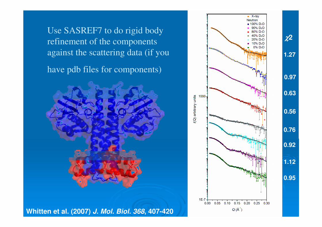
Use SASREF7 to do rigid body refinement of the components against the scattering data (if you
have pdb files for components)
χχχχ2 = 1.27
χχχχ2 = 0.97
χχχχ2 = 0.63
χχχχ2 = 0.56
χχχχ2 = 0.76
χχχχ2 = 0.92
χχχχ2 = 1.12
χχχχ2 = 0.95
χχχχ2
Whitten et al. (2007) J. Mol. Biol. 368, 407-420

Incorporation of deuterium up to 86% of the chemically Non-exchangeable protons can be obtained by using D2O as the deuterium source. Complete deuteration can only be obtained by addition of perdeuterated carbon source (glucose or glycerol).
Use mass spec to determine deuterationlevels.
The described protocols allow the deuteration content in recombinant proteins to be predicted

Neutron scattering sample cells
� Helma quartz cells (high precision path-length, suprasil) – need lots of them!
� Banjo-style (280 µL per 1 mm path length) or rectangular (170 µL per 1 mm path length) cells can be used
� Path lengths are only good to 1%, so good idea to measure sample and solvent background in the same cell if practical, but experiment logistics may prohibit that, so calibrate cells?
� High incoherent scattering for 1H means you always want ≤ 1mm 1H2O in the neutron beam to avoid multiple scattering

Neutrons� Non-ionizing radiation
� Penetrating
� Wavelength and energies available that are suitable for probing structures with dimensions 1-1000s Å
� Coherent scattering lengths that vary randomly with atomic weight and large isotope effect for hydrogen –contrast variation
� Large incoherent scattering cross-section for 1H is a source of noise in small-angle scattering
� Interact weakly with matter and are difficult to produce and detect – therefore should only be used when they provide information that cannot be otherwise obtained.

The sensor histidine kinase KinA - response regulator spo0A in Bacillus subtilis
Sda
KinA
Spo0A
KipAKipI
Failure to initiate DNA replicationDNA damage
Change in N2source
Sporulation
Spo0F
Spo0B
Environmental signal

Our molecular actors
KipIPyrococcus horikoshi
SdaKinABased on H853 Thermotoga maritima
Pro410
His405
Trp
CA
DHp
to sensor domains

HK853 based KinA model predicts the KinAX-ray scattering data

KinA2 Rg = 29.6 Å, dmax = 95 ÅKinA2-Sda2 Rg = 29.1 Å, dmax = 80 Å
KinA2 contracts upon binding 2 Sda molecules

Neutron contrast variation: KinA2:2DSda
222
ρ
β
ρ
α
∆−
∆+= mobs RR
in complex uncomplexedRg KinA2 25.40 Å 29.6 ÅRg 2Sda 25.3 Å 15.4 Å
Separation of centres of mass = 27.0 ÅI(Q) A-1

MONSA: 3D shape restoration for KinA2:2DSda

)()()()( 12212221
21 QIQIQIQI ρρρρ ∆∆+∆+∆=
Component analysis

Rigid-body refinement KinA2-2Sda components
Whitten, Jacques, Whitten, Jacques, LangelyLangely et al., et al., J. J. Mol.BiolMol.Biol. 368. 368, 407, 2007, 407, 2007
9090°°°°°°°°
I(Q) A-1

I(Q) A-1
KinA2-2KipI
Jacques, Jacques, LangelyLangely, Jeffries et al, , Jeffries et al, (2008) (2008) JJ. . Mol.BiolMol.Biol. . 384, 422-435
9090°°°°°°°°
384, 422-435

Pull down assays and Trpfluorescence show mutation of Pro410 abolishes KipIbinding to KinA but Sdacan still bind.
Trp fluorescence confirms that the C-domain of KipIinteracts with KinA

KipI-C domain has a cyclophilin-like structure
Overlay with cyclophilin B
Hydrophobicgroove
3Å crystal structure KipI-C domain

Aromatic side chain density in the hydrophobic groove
Jacques, Jacques, LangelyLangely, Jeffries et al, in review , Jeffries et al, in review J. J. Mol.BiolMol.Biol. . 20082008

The KinA helix containing Pro410 sits in the KipI-
C domain hydrophobic groove

A possible role for cis-trans
isomerization of Pro410 in tightening the helical bundle to transmit the KipI signal to the catalytic domains?
Or is the KipIcyclophilin-like domain simply a
proline binder?

Sda and KipI bind at the base of the KinAdimerization phosphotransfer (DHp) domain

Sda binding does not appear to provide for steric mechanism of inhibition

KipI interacts with that region of the DHpdomain that includes the conserved Pro410

Sda and KipI induce the same contraction of KinA upon binding (4 Å in Rg, 15 Å in Dmax)
DHp helical bundle is a critical conduit for signaling

Contrast variation in biomolecules can take advantage of the fortuitous fact that the major bio-molecular constituents of have mean scattering length densities that are distinct and lie between the values for pure D2O and pure H2OM
ean
scat
teri
ng le
ngth
den
sity
(10
10cm
2 )

DNA and protein have inherent differences in scattering density that can be used in neutron contrast variation experiments
Olah et al., J. Mol. Biol. 249, 576-594, 1995

Doing a Quality Experiment
� After your final gel filtration step, check out your samples with dynamic light scattering
� Carefully calibrate you concentration assay –colorimetric assays are almost useless, a combination of AAA and extinction coefficient of the unfolded protein is best if you are careful about your solvents.
� Compare your data to a well characterized standard(s)� For protein/DNA complexes, standards are more
difficult. Measure the partial specific volume of your particle if you have enough sample – or at least use a good model to calculate it, e.g. see http://geometry.molmovdb.org/NucProt/

Jacques & Trewhella (2010)
“Small-angle Scattering for
Structural Biology;
Expanding the Frontier
While Avoiding the Pitfalls,”
Protein Science 19, 642-657