complement pathway amplifies caspase-11 dependent cell...
TRANSCRIPT
Article
The Rockefeller University Press $30.00J. Exp. Med. 2016 Vol. 213 No. 11 2365–2382www.jem.org/cgi/doi/10.1084/jem.20160027
2365
INT ROD UCT IONSepsis is defined as the presence of a systemic inflammatory response syndrome (SIRS) caused by infection, and is one of the leading causes of death in intensive care units (ICUs); currently, there are more than 19 million cases of sepsis a year worldwide (Rangel-Frausto et al., 1995; Angus et al., 2001; Funk et al., 2009; Angus and van der Poll, 2013). Although the mechanism is not clear, SIRS and an associated infection can develop into severe sepsis, a robust and uncontrollable inflammatory response, which can lead to septic shock and subsequent death (Cerra, 1985; Angus and van der Poll, 2013). Multiple proinflammatory reactions are thought to contribute to the severity of sepsis pathologies (Angus and van der Poll, 2013). These overlapping proinflammatory responses create a complex biological scenario with built-in redundancies, making it difficult to study. Additionally, the proinflammatory pathways that contribute to sepsis have not been fully defined, which compounds the difficulty in developing efficacious di-agnostics and therapeutics. Therefore, a better understanding
of the molecular pathways that contribute to pathogenesis of sepsis is crucial for the development of more effective diag-nostics and therapeutic strategies and in reducing mortality.
Cell death coincides with the production of proinflam-matory cytokines, which in turn are associated with poor outcome in patients with sepsis (Ayala et al., 1996; Hotch-kiss et al., 1997, 1999, 2003; Isogai et al., 1998; Oberholzer et al., 2001; van der Poll and Opal, 2008). Specifically, the caspase-11–dependent cell death pathway has been shown by multiple groups to exacerbate pathologies in an LPS-induced sepsis mouse model (Kayagaki et al., 2011, 2013; Hagar et al., 2013). Caspase-11 is a cytosolic pattern recognition recep-tor (PRR) that plays a critical role in responding to cytoso-lic LPS during Gram-negative bacterial infection and sepsis (Kayagaki et al., 2011, 2013; Aachoui et al., 2013; Hagar et al., 2013). In multiple cell types, caspase-11 expression is induced after initial detection of LPS by TLR4, through Myd88, TRIF, and interferon signaling pathways (Broz and Monack, 2011; Kayagaki et al., 2011; Rathinam et al., 2012; Hagar et al., 2013). Caspase-11 is produced as a monomeric zymogen
Cell death and release of proinflammatory mediators contribute to mortality during sepsis. Specifically, caspase-11–dependent cell death contributes to pathology and decreases in survival time in sepsis models. Priming of the host cell, through TLR4 and interferon receptors, induces caspase-11 expression, and cytosolic LPS directly stimulates caspase-11 activation, promoting the release of proinflammatory cytokines through pyroptosis and caspase-1 activation. Using a CRI SPR-Cas9–mediated ge-nome-wide screen, we identified novel mediators of caspase-11–dependent cell death. We found a complement-related pep-tidase, carboxypeptidase B1 (Cpb1), to be required for caspase-11 gene expression and subsequent caspase-11–dependent cell death. Cpb1 modifies a cleavage product of C3, which binds to and activates C3aR, and then modulates innate immune sig-naling. We find the Cpb1–C3–C3aR pathway induces caspase-11 expression through amplification of MAPK activity down-stream of TLR4 and Ifnar activation, and mediates severity of LPS-induced sepsis (endotoxemia) and disease outcome in mice. We show C3aR is required for up-regulation of caspase-11 orthologues, caspase-4 and -5, in primary human macrophages during inflammation and that c3aR1 and caspase-5 transcripts are highly expressed in patients with severe sepsis; thus, sug-gesting that these pathways are important in human sepsis. Our results highlight a novel role for complement and the Cpb1–C3–C3aR pathway in proinflammatory signaling, caspase-11 cell death, and sepsis severity.
Complement pathway amplifies caspase-11–dependent cell death and endotoxin-induced sepsis severity
Brooke A. Napier,1 Sky W. Brubaker,1 Timothy E. Sweeney,2,3 Patrick Monette,4 Greggory H. Rothmeier,5 Nina A. Gertsvolf,1 Andreas Puschnik,1 Jan E. Carette,1 Purvesh Khatri,2,3 and Denise M. Monack1
1Department of Microbiology and Immunology, 2Division of Biomedical Informatics Research, Stanford University School of Medicine, and 3Institute for Immunity, Transplantation, and Infection, Stanford University, Stanford, CA 94305
4Department of Biology, Middlebury College, Middlebury, VT 057535Digital Team, Blue Bottle Coffee, Oakland, CA, 94607
© 2016 Napier et al. This article is distributed under the terms of an Attribution–Noncommercial–Share Alike–No Mirror Sites license for the first six months after the publication date (see http ://www .rupress .org /terms). After six months it is available under a Creative Commons License (Attribution–Noncommercial–Share Alike 3.0 Unported license, as described at http ://creativecommons .org /licenses /by -nc -sa /3 .0 /).
Correspondence to Denise M. Monack: [email protected]
Abbreviations used: BMDM, murine BM-derived macrophage; Cpb1, carboxypepti-dase B1; CTB, cholera toxin B subunit; gRNA, guide RNA; MDMs, primary human-de-rived macrophage; pt, post-treatment; qRT-PCR, quantitative RT-PCR; SIRS, systemic inflammatory response syndrome.
The
Journ
al o
f Exp
erim
enta
l M
edic
ine
on January 17, 2017D
ownloaded from
Published October 3, 2016
/content/suppl/2016/09/22/jem.20160027.DC1.html Supplemental Material can be found at:

Complement amplifies caspase-11 cell death and endotoxemia | Napier et al.2366
that dimerizes and activates upon detection of cytosolic LPS (Kang et al., 2000). Upon activation of caspase-11, the cell succumbs to pyroptotic cell death, activates caspase-1, and re-leases proinflammatory mediators (Kang et al., 2000; Hagar et al., 2013; Kayagaki et al., 2011, 2013). Caspase-11–dependent release of proinflammatory mediators into the extracellular space during LPS-induced sepsis contributes to host mor-tality (Kayagaki et al., 2011, 2013; Hagar et al., 2013). In hu-mans, caspase-4 and -5 are orthologues to caspase-11 (Shi et al., 2014; Casson et al., 2015), making this an intriguing cell death pathway to understand in the context of sepsis. Thus, an increased understanding of the regulation of caspase-4/5/11–dependent cell death pathway may lead to the identification of novel targets for the diagnosis and treatment of sepsis.
To identify new mediators of caspase-11–dependent cell death, we used a genome-wide CRI SPR-Cas9 knockout screen in macrophages. The results of our screen highlight the complexity of caspase-11 gene expression. Specifically, we identified carboxypeptidase B1 (Cpb1), a complement-re-lated protein, as a novel mediator of caspase-11 gene ex-pression and subsequent caspase-11–dependent cell death in macrophages. Cpb1 modifies a cleavage product of C3, which binds to and activates C3aR, and then modulates innate im-mune signaling. Here, we elucidate the role of the Cpb1–C3–C3aR pathway in enhancing cell autonomous and non–cell autonomous inflammation by amplifying TLR4- and If-nar-dependent expression of proinflammatory genes, includ-ing caspase-11, within macrophages. We find that TLR4- and Ifnar-signaling pathways, but not Ifngr signaling, converge at p38 MAPK signaling downstream of C3aR activation, high-lighting the specificity of this amplification signaling path-way. We identified a role for C3aR, a key mediator of the Cpb1–C3–C3aR signaling pathway, in the early production of proinflammatory mediators, including caspase-11, in vivo and to the severity and outcome of disease in an endotoxemia model of severe sepsis. Furthermore, using a C3aR inhibitor, we show that C3aR may be a candidate for early therapeutic targeting during sepsis.
We demonstrate the clinical relevance of our findings by elucidating the role of C3aR in amplification of caspase-11 human orthologues, caspase-4 and -5, and release of proin-flammatory cytokines in primary human macrophages during inflammation. Furthermore, using multicohort analysis, we find that c3ar1 and caspase-5 are significantly up-regulated in human peripheral blood mononuclear cells (PBMCs) during sepsis, as compared with healthy controls. Thus, C3aR and caspase-5 transcripts may serve as biomarkers for diagnosing sepsis in humans, and this pathway may be targeted for the treatment of sepsis or septic shock.
Complement is known to play an important role in sep-sis through multiple means (Flierl et al., 2008; Markiewski et al., 2008; Rittirsch et al., 2008), and we describe a novel mechanism in which complement amplifies proinflammatory signaling and thus contributes to poor sepsis outcomes. Our results describe the intersection of two known proinflam-
matory innate immune pathways, the caspase-11–dependent cell death pathway and the Cpb1–C3–C3aR complement signaling pathway, that contribute to inflammation and poor outcome during sepsis.
RES ULTSCRI SPR-Cas9 screen in RAW264.7 macrophages identifies mediators of caspase-11–dependent cell deathCaspase-11–dependent cell death is triggered in murine pri-mary BM-derived macrophages (BMDMs) by introducing LPS into the cytosol via cholera toxin B-subunit (CTB; Kay-agaki et al., 2013). LPS and CTB treatment of WT BMDMs results in ∼60% cell death within 16 h post-treatment (pt; Fig. 1 A). Similar to previous studies, this death is caspase-11 dependent and caspase-1 independent (Fig. 1 A; Hagar et al., 2013; Kayagaki et al., 2013). To create a genetically tractable model for caspase-11–dependent cell death, we recapitulated this cell death phenotype in a murine-transformed macro-phage cell line, RAW264.7 (RAW). Treatment of WT RAW cells with LPS and CTB for 16 h results in death of ∼80% of the cells, which is similar to WT BMDMs (Fig. 1 B). Addi-tionally, the requirement of CTB for LPS-induced cell death can be bypassed by transfecting LPS into RAW cells, resulting in ∼40% cell death 16 h pt, similar to BMDMs (Fig. 1 B; Hagar et al., 2013; Kayagaki et al., 2013). Together, these data demonstrate that RAW macrophages respond to intracellular LPS comparably to BMDMs.
To determine whether cell death caused by intracel-lular LPS was dependent on caspase-11, and not caspase-1, we disrupted caspase-11 and -1 using the CRI SPR-Cas9 ge-nome editing system (Cong et al., 2013; Storek et al., 2015). We found that intracellular LPS induced cell death primarily in a caspase-11–dependent, caspase-1–independent manner (Fig. 1 B). Important to note, caspase-1 is directly upstream of caspase-11 on Chromosome 9 in C57BL/6J mice, thus we checked if disruption of either of these genes affected the other (Kang et al., 2000). Disruption of caspase-11 did not affect the protein levels of caspase-1, and vice versa (un-published data). We went on to complement the disrupted caspase-11 gene by expressing a WT copy of caspase-11 tagged at the C terminus with FLAG/HA under the pMSCV promoter. The complemented cell line restored the cell death phenotype to 50% of the WT RAW cell death (Fig. 1 B). Together, these data show that RAW cells are a genetically tractable model and can be used to study caspase-11–depen-dent cell death in macrophages.
Next, we constructed a CRI SPR-Cas9 genome-wide knockout library in WT RAW cells to identify novel medi-ators of caspase-11–dependent cell death (Koike-Yusa et al., 2014). This library is a pool of 87,897 guide RNAs (gRNAs) against 19,150 mouse protein-coding genes expressed from lentiviral vectors (Koike-Yusa et al., 2014). We constructed WT RAW that constitutively express human codon–opti-mized Cas9 nuclease (hCas9) for genome editing (Fig. 1 C). The expression of hCas9 did not affect the cell death kinet-
on January 17, 2017D
ownloaded from
Published October 3, 2016

2367JEM Vol. 213, No. 11
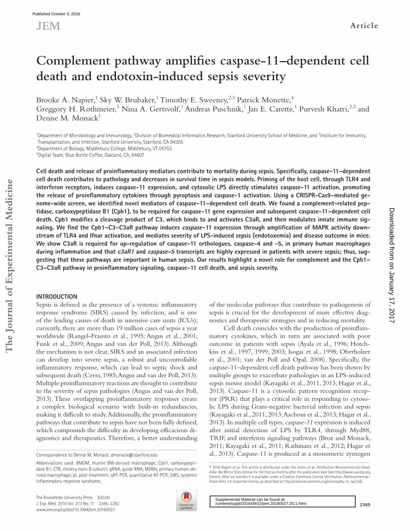
Figure 1. CRI SPR-Cas9 screen identifies Cpb1 to be required for caspase-11 expression, activation, and cell death in macrophages. (A) BMDMs were treated with intracellular LPS via CTB for 16 h, and then cytotoxicity was measured. (B) RAW264.7 transformed murine macrophage cell line (RAW) was treated with CTB/LPS or transfected LPS for 16 h and cytotoxicity was measured. (C) Expression of humanized Cas9 (hCas9) was determined in WT RAW cells that were either untreated or transduced with a lentiviral construct that stably expresses hCas (WT-hCas9), or the same cells were treated with LPS/CTB for 16 h and cytotoxicity was measured. (D) A macrophage genetic screen identified KOs that confer resistance to intracellular LPS stimulation via 16 h treatment with CTB/LPS. Each plot represents a gene (x axis). Genes are ranked on the y axis according to the significance (-LogPvalue) of enrichment, which was calculated by an accumulated hypergeometric distribution function (König et al., 2007), in the CTB/LPS-treated library compared with the nonselected control population. The top 100 hits are highlighted in the extrapolated box. Blue genes, CTB receptor genes; purple genes, complement genes; red, yellow, and green, other hits followed up on. (E–I) WT and KO cell lines were treated with LPS/CTB or transfected with LPS for 16 h and cytotoxicity was measured, (I) with or without the Cpb-inhibitor (MGTA). (J) WT, Cpb1 KO, and Cpb1 KO cells complement with either Cpb1 (Cpb1 KO + Cpb1) or caspase-11 (Cpb1 KO +
on January 17, 2017D
ownloaded from
Published October 3, 2016
Complement amplifies caspase-11 cell death and endotoxemia | Napier et al.2368
ics in WT RAW cells after treatment with LPS and CTB (Fig. 1 C). Next, we transduced the WT-hCas9 cell line with lentiviral vectors created from the gRNA library, as previously described (Koike-Yusa et al., 2014). To identify genes required for caspase-11–dependent cell death, we induced cell death by treating the pooled mutant library with LPS and CTB for 16 h. We expected that surviving cells should have mutations in genes required for caspase-11–dependent cell death.
We calculated enrichment of genes ([% counted in treated cells]/[% counted in untreated cells]) and used these values along with the number of gRNAs per gene that came out of the screen to determine the P-value for each gene (Table S1; Koike-Yusa et al., 2014). As confirmation of our method, our top hit was B4galnt1, which is a gene required for the production of the CTB receptor (GM1 sphingolipid; Fig. 1 D; Wolf et al., 2008; Papatheodorou et al., 2011; Chin-napen et al., 2012; Jobling et al., 2012). Although caspase-11 was not enriched in our screen, it is possible that the caspase-11 gRNAs present in the library were ineffective or lost during amplification of the library. To verify four of our top novel hits, we created knockout RAW cell lines using the gRNAs identified from the screen and treated them with CTB and LPS for 16 h. RAW cells deficient for three of these hits showed a cell death defect after 16 h of CTB and LPS treat-ment, whereas the Acsl6 KO cell line displayed similar levels of cell death compared with WT RAW cells (Fig. 1 E). To ensure that these genes were required for caspase-11–depen-dent cell death, and not CTB-dependent delivery of LPS into the cytosol, we transfected LPS into each knockout cell line. While most of our knockout cell lines had decreased levels of cell death after transfection with LPS compared with WT cells, only the Cpb1KO cell line displayed significant defects after treatment with CTB and LPS, as well as LPS transfec-tion (Fig. 1 E). Together, these data demonstrate that the CRI SPR-Cas9 genome-wide screening method in RAW macro-phages is a useful tool to identify novel genes, such as Cpb1, involved in the caspase-11–dependent cell death pathway.
Cpb1 is required for caspase-11 expression, activation, and cell deathOur CRI SPR-Cas9 genome-wide cell death screen identi-fied Cpb1 as a novel gene required for a caspase-11–depen-dent cell death pathway. To confirm the role of Cpb1 in cell death induced by cytosolic LPS, we created five independent Cpb1 knockout cell lines and found that each of these dis-tinct mutants were ∼50% defective for cell death 16 h pt with CTB and LPS (Fig. 1 F). We confirmed a significant
reduction in cell death after 16-h treatment with intracellular LPS, via CTB or transfection, compared with WT RAW cells in one clone, Cpb1-10 (Fig. 1, G-H). We used a pan-car-boxypeptidase inhibitor (2-mercaptomethyl-3-guanidinoeth-ylthiopropanoic acid [MGTA]) to assess the importance of Cpb1 enzymatic activity during intracellular LPS treatment. We found that during CTB/LPS treatment of macrophages, the presence of MGTA reduces caspase-11–dependent cell death in a dose-dependent manner (Fig. 1 I); thus, underlin-ing the importance of the enzymatic activity of Cpb1 during caspase-11–dependent cell death. Importantly, the cell death defect in Cpb1 KO cells was rescued by expressing a WT copy of Cpb1 under a pMSCV promoter (Fig. 1 J).
We hypothesized that the requirement of Cpb1 was specific to caspase-11–dependent cell death, and not caspase-1-dependent cell death. To test this, we induced caspase-1-dependent cell death via treatment with LPS and nigericin for 9.5 h, and found that Casp11 KO and Cpb1 KO cells died at similar levels as WT cells; however, the Casp1 KO cells had a reduction in cell death after treat-ment (unpublished data). Collectively, these results indicate that Cpb1 contributes specifically to caspase-11–dependent cell death in macrophages.
Caspase-11 activation is a hallmark of caspase-11–de-pendent cell death (Zhang et al., 2013). Thus, we measured caspase-11 activation in WT and KO cell lines by detecting cleavage and release of the C-terminal p30 portion of full-length caspase-11 (pro-casp11) into the supernatant of ac-tivated and dying cells (Kang et al., 2000). 16 h pt of WT cells with CTB and LPS resulted in an increase of pro-casp11 and p30 into the supernatant (SN) as a result of caspase-11 activation and release of cytosolic contents upon cell death (Fig. 1 K). In concordance with our previous data, there were lower levels of pro-casp11 in the SN from Cpb1 KO cells compared with the WT cells after CTB and LPS treatment (Fig. 1 K). In addition, the caspase-11 p30 cleavage product was not detected in the SN from Cpb1 KO cells treated with LPS and CTB (Fig. 1 K). As expected, pro-casp11 and p30 were not detected in the SN from Casp11 KO cell line 16 h pt with LPS and CTB (Fig. 1 K). Interestingly, the level of pro-casp11 in the lysates of Cpb1 KO cells treated with LPS was markedly lower compared with the WT cells treated with LPS suggesting that Cpb1 may contribute to cell death by controlling the level of caspase-11 gene expression (Fig. 1 K). To test this notion, we expressed a WT copy of caspase-11 on an exogenous vector and measured cell death in re-sponse to LPS and CTB. Ectopic expression of caspase-11 in
Casp11) on a transgene were treated with CTB/LPS for 16 h and cytotoxicity was measured. (K) WT and KO cell lines were treated with either LPS or CTB/LPS for 16 h. Supernatants and cell lysates were collected for each sample and detection of pro-caspase-11 (pro-Casp11), cleaved caspase-11 (p30 Casp11), and β-actin was evaluated via Western blot. Indicated cell lines were treated with LPS for 2 h. Relative expression of (L) caspase-11 and caspase-1 or (M) IL-6 and TNF were determined. All relative expression was calculated via qRT-PCR, compared with GAP DH expression. All cytotoxicity was measured by release of LDH. Statistics analyzed via the unpaired Student’s t test. *, P < 0.05; **, P < 0.01; ***, P < 0.001; ****, P < 0.0001. Data are representative of at least two (D) or three (A–C and E–M) independent experiments with three technical replicates each time.
on January 17, 2017D
ownloaded from
Published October 3, 2016

2369JEM Vol. 213, No. 11
Cpb1-deficient cells rescues the cell death defect in response to cytosolic LPS (Fig. 1 J).
Our data indicate that Cpb1 is acting upstream of caspase-11 gene expression and may be involved in amplifi-cation of the TLR4 signaling pathway after stimulation with LPS. We, and others, have shown previously that caspase-11 expression is dependent on MyD88 and TRIF downstream of TLR4 (Kayagaki et al., 2011; Broz et al., 2012; Rathinam et al., 2012). To analyze the contribution of Cpb1 to TLR4-depen-dent caspase-11 gene expression, we treated WT and Cpb1 KO cells with LPS for 2 h to initiate TLR4-dependent ex-pression of proinflammatory genes and measured expression of caspase-11 by quantitative real-time PCR (qRT-PCR) to determine transcript levels. Similar to previous studies, the level of caspase-11 transcript in WT cells is significantly in-creased after 2-h treatment with extracellular LPS (Fig. 1 L). In contrast, the level of caspase-11 gene expression in the Cpb1 KO cell line did not increase after 2 h of extracellular LPS treatment (Fig. 1 L). Of note, caspase-1, which is not influenced by TLR4-dependent signaling, was unaffected in WT and Cpb1 KO cells (Fig. 1 L). Thus, our results indicate that Cpb1 is required for WT levels of caspase-11 expression, activation, and cell death in response to cytosolic LPS.
Our results indicate that Cpb1 somehow impacts TLR4-dependent expression of caspase-11 and perhaps the expression of additional proinflammatory genes. il-6 and tnf are proinflammatory genes induced by TLR4 activation. Thus, we measured il-6 and tnf gene expression in WT and Cpb1 KO cells after LPS stimulation. We found that Cpb1 KO cells have reduced levels of il-6 and tnf compared with WT cells after 2-h LPS treatment (Fig. 1 M). Together, these data imply that Cpb1 is required for amplification of proin-flammatory transcripts, including caspase-11, through the TLR4 signaling pathway.
Cpb1–C3–C3aR pathway is required for TLR4-dependent expression of proinflammatory genes and caspase-11–dependent cell deathCpb1 is a peptidase that cleaves basic resides from the C ter-minus of intra- and extracellular peptides (Moore and Benoi-ton, 1978; Leung et al., 2008). Murine Cpb1 is the orthologue to human CPB, which modifies C3a, a 77-aa anaphylatoxin generated by enzymatic cleavage of C3 during activation of the complement system (Leung et al., 2008). CPB cleaves the C-terminal arginine of C3a to create C3a-desArg (Moore and Benoiton, 1978; Leung et al., 2008). This modified mol-ecule, C3a-desArg, engages the C3aR receptor, a comple-ment receptor that belongs to the rhodopsin family of seven transmembrane, G protein–coupled receptors expressed on both BM-derived myeloid and lymphoid cells (Crass et al., 1996; Martin et al., 1997; Zwirner et al., 1999). Activation of C3aR leads to modulation of innate immune responses during LPS stimulation and Gram-negative bacterial infec-tions, including induction of proinflammatory cytokines IL-6 and TNF (Takabayashi et al., 1996; Mueller-Ortiz et
al., 2006). C3aR activation has been shown to mediate ex-pression of TLR4-dependent transcripts (Zhang et al., 2007). Thus, Cpb1 may modulate TLR4-dependent expression of proinflammatory cytokines through complement receptor C3aR (see also Fig. 7).
To interrogate this hypothesis, we disrupted C3aR and C3 in RAW macrophages using the CRI SPR-Cas9 genome editing system (Fig. S1, B and C). We reasoned that if the Cpb1–C3–C3aR signaling pathway is required for up-regulation of caspase-11 transcripts, then all three of these genes should be necessary for TLR4-dependent expression of caspase-11. As predicted, the level of caspase-11 transcripts was significantly lower in C3aR KO and C3 KO cell lines, compared with WT RAW macrophages after 2-h LPS treatment, similar to Cpb1 KO cells (Fig. 2 A). In addition, the expression of il-6 and tnf transcripts were significantly reduced compared with WT cells, again mimicking the Cpb1 KO cells (Fig. 2 B). These data demonstrate that the Cpb1–C3–C3aR signaling pathway is required for TLR4-dependent up-regulation of proinflammatory transcripts downstream of LPS stimulation.
Because increased caspase-11 gene expression is re-quired for caspase-11–dependent cell death, we measured cell death induced by intracellular LPS stimulation in C3aR KO and C3 KO cell lines. Cell death in C3aR KO and C3 KO cell lines was reduced by 50% compared with WT RAW cells 16 h pt with CTB and LPS (Fig. 2 C). In addition, the ex-pression and release of caspase-11 from C3aR KO and C3 KO cells were notably decreased compared with WT RAW cells 16 h pt with CTB and LPS (Fig. 2 D). Together, these data show that the Cpb1–C3–C3aR complement pathway is required for TLR4-dependent expression of caspase-11 and subsequent caspase-11–dependent cell death. Thus, we reveal a novel contribution of the complement pathway to caspase-11–dependent gene expression and cell death.
We next verified that the Cpb1–C3–C3aR pathway is required for caspase-11–dependent cell death in mu-rine primary BMDMs. BMDMs were generated from WT, caspase-11–deficient (Casp11−/−), C3aR-deficient (C3aR−/−), and C3-deficient (C3−/−) mice to assess caspase-11–depen-dent cell death. Similar to previous publications, >80% of the WT BMDM died by 16 h pt with LPS and CTB (Fig. 2 E). In contrast, treatment of Casp11−/− BMDMs with LPS and CTB resulted in background levels of cell death, ∼20%, 16 h pt (Fig. 2 E). C3- and C3aR-deficient BMDMs had significantly lower levels of cell death compared with WT BMDMs after 16-h treatment with LPS and CTB (Fig. 2 E), suggesting that C3 and C3aR mediate caspase-11–dependent cell death in primary cells. Finally, to further test the role of C3aR in caspase-11–dependent cell death in BMDMs, C3aR-dependent signaling was blocked by adding the C3a receptor antagonist (C3aR-Ant). Treatment of WT BMDMs with C3aR-Ant resulted in a dramatic decrease in LPS-in-duced cell death (Fig. 2 E).
We hypothesize that C3aR and C3 are required for caspase-11–dependent cell death in BMDMs through the
on January 17, 2017D
ownloaded from
Published October 3, 2016

Complement amplifies caspase-11 cell death and endotoxemia | Napier et al.2370
amplification of TLR4-dependent transcripts, as seen in RAW macrophages. Thus, we measured caspase-11, il-6, and tnf gene expression in WT, C3aR−/−, and C3−/− BMDMs after LPS stimulation and found that C3aR−/− and C3−/− BMDMs did not express these transcripts at WT levels (Fig. 2, F and G). The defect in expression of il-6 and tnf transcripts corresponded with a significant reduction in IL-6 and TNF cytokine release from C3aR−/− BMDMs compared with the WT BMDMs (Fig. 2 H). Although the C3−/− and C3aR−/− mice were generated in an Sv129 embryonic background these mice were thoroughly backcrossed and do not retain the known Sv129 5 base-pair deletion at the caspase-11 ge-netic locus (Fig. S2). These data demonstrate that the Cpb1–C3–C3aR pathway is required in transformed macrophages, as well as primary macrophages for expression and release of TLR4-dependent proinflammatory mediators, highlighting
its importance for caspase-11–dependent cell death in re-sponse to intracellular LPS.
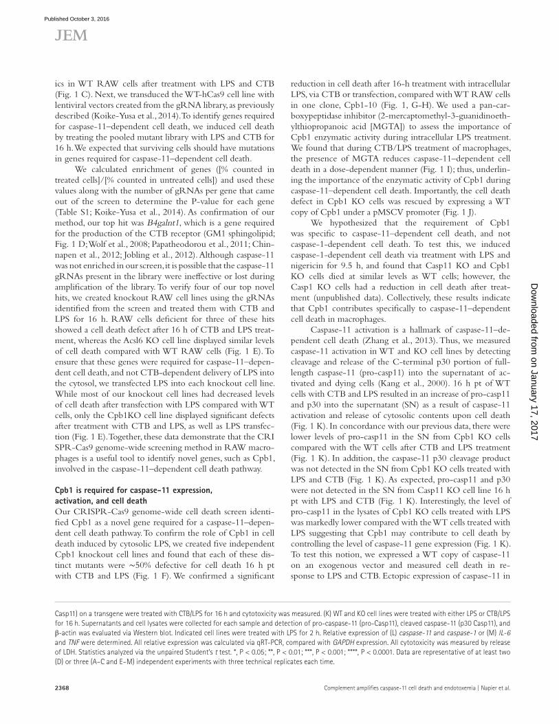
Cpb1–C3–C3aR pathway increases MAPK activity downstream of TLR4 and Ifnar activation in a cell autonomous and non–cell autonomous mannerCaspase-11 gene expression can be induced downstream of TLR4 activation, or interferon signaling pathways through Ifnar and Ifngr activation (Kayagaki et al., 2011; Broz et al., 2012; Rathinam et al., 2012). Although we have shown that the Cpb1–C3–C3aR pathway amplifies proinflamma-tory transcripts, including caspase-11, downstream of TLR4 activation, it is not known whether the Cpb1–C3–C3aR pathway directly modulates Ifnar or Ifngr signaling. To acti-vate Ifnar signaling, primary macrophages were treated with IFN-β, and the levels of caspase-11 and NOS2, Ifnar-de-
Figure 2. Complement amplifies TLR4-dependent transcripts and caspase-11–dependent cell death in macrophages. WT, C3aR KO, and C3 KO RAW cell lines were treated with LPS for 2 h and relative expression of (A) caspase-11 or (B) il-6 and tnf was calculated via qRT-PCR, compared with GAP DH expression. Indicated cell lines were treated with CTB/LPS for 16 h and (C) cytotoxicity was measured or (D) supernatants and cell lysates were collected for Western blot analysis of indicated proteins. (E) WT (control [WT] or C3aR-antagonist treated [C3aR-Ant]), caspase-11–deficient (Casp11−/−), C3aR-de-ficient (C3aR−/−), and C3-deficient (C3−/−) primary BM-derived macrophages (BMDMs) were treated with CTB/LPS for 16 h and cytotoxicity was measured. Indicated BMDMs were treated with LPS for 2 h and the relative expression of (F) caspase-11 or (G) il-6 and tnf was calculated via qRT-PCR, compared with GAP DH expression or (H) analyzed for release of IL-6 and TNF via ELI SA. All cytotoxicity was measured by release of LDH. Statistics analyzed via the unpaired Student’s t test. *, P < 0.05; **, P < 0.01; ***, P < 0.001. Data are representative of at least three independent experiments (A–H), with three tech-nical replicates each time.
on January 17, 2017D
ownloaded from
Published October 3, 2016
2371JEM Vol. 213, No. 11
pendent transcripts, were measured. As expected, there was an amplification of caspase-11 and NOS2 transcripts in WT BMDMs treated with IFN-β for 2 h (Fig. 3 A). In contrast, the levels of caspase-11 and NOS2 transcripts were signifi-cantly lower in the C3aR−/− and C3−/− macrophages treated with IFN-β for 2 h compared with WT BMDMs (Fig. 3 A). In addition, we activated BMDMs with IFN-γ and measured up-regulation of the IFN-γ–dependent genes caspase-11 and NOS2. Interestingly, WT, C3aR−/−, and C3−/− macro-phages expressed caspase-11 and NOS2 transcripts at sim-ilar levels after 2 h of stimulation with IFN-γ (Fig. 3 B). Collectively, these data indicate that the Cpb1–C3–C3aR signaling pathway is important for amplification of Ifnar-de-pendent signaling, but not Ifngr-dependent signaling, during the first 2 h after cytokine stimulation. Surprisingly, this is the first demonstration of C3aR-dependent amplification of Ifnar-dependent signaling. Our results indicate that sig-naling through the Cpb1–C3–C3aR pathway amplifies TLR4-dependent signaling and type I interferon-stimulated
Ifnar signaling in macrophages, but not Ifngr-dependent sig-naling (see also Fig. 7).
It has been described that C3aR activation can amplify MAPK signaling downstream of TLR4 stimulation (Zhang et al., 2007). Additionally, Ifnar signaling leads to downstream activation of MAPKs (Poovassery and Bishop, 2012). Thus, we hypothesize that Cpb1–C3–C3aR pathway amplifica-tion of TLR4- and Ifnar-dependent gene expression con-verges on MAPK signaling within the host cell. We measured phosphorylation of p38 MAP kinase as a read-out of MAPK family activity, to identify the level of MAP kinase activa-tion in WT versus complement-deficient macrophages after various stimuli. After 2 h of treatment with LPS, p38 phos-phorylation in WT BMDMs increases sixfold, whereas the C3aR−/− BMDMs show approximately twofold induction of p38 phosphorylation (Fig. 3 C). Additionally, when BMDMs are treated with IFN-β for 2 h, there is a twofold increase of p38 phosphorylation in WT cells compared with a less than onefold induction in C3aR−/− BMDMs. In congruence
Figure 3. Cpb1–C3–C3aR pathway increases p38 MAPK phosphorylation downstream of TLR4 and Ifnar activation in a cell autonomous and non–cell autonomous manner. WT, C3aR−/−, and C3−/− BMDMs were treated with (A) IFN-β or (B) IFN-γ for 2 h and the relative expression of caspase-11 and NOS2 transcripts were calculated via qRT-PCR, compared with GAP DH expression. (C) WT or C3aR−/− BMDMs were treated with LPS, IFN-β, or IFN-γ for 2 h and lysed for analysis of p38-phosphorylation and β-actin was evaluated via Western blot. (D) WT, Cpb1 KO, or C3aR KO RAW macrophages were treated with LPS for 2 h and supernatants were added to naive Cpb1 KO cells for 2 h. The relative expression of caspase-11, il-6, and tnf-α were calculated via qRT-PCR, compared with GAP DH expression. Statistics were analyzed via the unpaired Student’s t test. *, P < 0.05; ***, P < 0.001; ****, P < 0.0001. Data are representative of at least two (D) or three (A–C) independent experiments, with three technical replicates each time.
on January 17, 2017D
ownloaded from
Published October 3, 2016

Complement amplifies caspase-11 cell death and endotoxemia | Napier et al.2372
with our hypothesis, p38 phosphorylation was not induced in IFN-γ–treated WT or C3aR−/− BMDMs (Fig. 3 C). These data indicate that C3aR activation influences MAPK signal-ing downstream of TLR4 and Ifnar activation, but not Ifngr activation, implicating p38 MAPK as a potential convergence of these signaling pathways (see also Fig. 7).
There are multiple G protein–coupled receptors that enhance MAPK activation through p38 in a cell autonomous and non–cell autonomous manner during inflammation (Ya-mauchi et al., 1997; Robinson and Dickenson, 2001; Kawli et al., 2010; Grimsey et al., 2015). Additionally, human CPB is found in high concentrations in serum (Leung et al., 2008; Chatterjee et al., 2009), C3 is quickly released from mono-cytes and macrophages (Goodrum, 1987), and C3aR is an extracellular receptor, suggesting that this pathway may have non–cell autonomous affects. We have shown thus far that Cpb1–C3–C3aR pathway is enhancing cell autonomous proinflammatory signaling; however, we hypothesize that the Cpb1–C3–C3aR pathway can also work in a non–cell autonomous manner. To test this, we treated WT, Cpb1KO, and C3aR KO RAW macrophages with LPS for 2 h and collected the supernatants. We then treated naive Cpb1 KO cells with the supernatants for 2 h and looked for expression of proinflammatory transcripts. When Cpb1 KO cells were treated with the conditioned WT or C3aR supernatants, both of which have functioning Cpb1 within the superna-tants, caspase-11, IL-6, and TNF transcripts were significantly up-regulated compared with Cpb1 KO cells treated with conditioned supernatants from Cpb1 KO cells (Fig. 3 D). These data confirm that Cpb1–C3–C3aR pathway acts in a cell-autonomous and non–cell autonomous manner during inflammation in macrophages.
Together, our data suggests that the Cpb1–C3–C3aR pathway amplifies proinflammatory mediators by increasing MAPK signaling downstream of TLR4 and Ifnar stimulation and that the effect of this pathway on macrophages can man-ifest in a cell-autonomous and non–cell autonomous man-ner. This data, combined with our previous data showing that this amplification is important for induction of caspase-11 transcripts and caspase-11–dependent cell death, illuminates the potential importance of this proinflammatory amplifi-cation pathway during response to intracellular LPS during Gram-negative bacterial infections and sepsis.
Cbp1–C3–C3aR pathway is required for cell death in response to Gram-negative bacteriaCaspase-11–dependent cell death is triggered by intracellular Gram-negative bacterial pathogens (Rathinam et al., 2012; Aachoui et al., 2013; Hagar et al., 2013; Kayagaki et al., 2013). Treatment with LPS and CTB or transfection of LPS effi-ciently delivers LPS into the cytosol of macrophages, which mimics cytosolic infiltration of LPS during active intracellular Gram-negative bacterial infection (Kayagaki et al., 2011, 2013; Hagar et al., 2013). The LPS from intracellular Gram-nega-tive pathogens Salmonella Typhimurium and Shigella flexneri
have been shown to engage the caspase-11–dependent cell death pathway in macrophages during infection (Aachoui et al., 2013; Hagar et al., 2013; Kayagaki et al., 2013, 2015). We have shown that the Cpb1–C3–C3aR pathway is required for WT levels of caspase-11 protein expression, and subsequent caspase-11–dependent cell death, when exposed to cytosolic LPS. Thus, we hypothesize that the Cpb1–C3–C3aR pathway will be required for caspase-11–dependent cell death during S. Typhimurium and S. flexneri infections. During S. Typh-imurium infection, ∼30% of WT RAW cells are dead 18 h after infection (MOI, 100:1), whereas infected Casp11 KO RAW cells exhibited no detectable cell death above back-ground (∼10%; Fig. 4 A). Interestingly, the levels of cell death in Cpb1 KO, C3aR KO, and C3 KO cell lines infected with S. Typhimurium were similar to those in the infected Casp11 KO cell line (Fig. 4 A). Additionally, RAW macrophages re-quire caspase-11 for WT levels of macrophage cell death after 18 h infection with S. flexneri (MOI 50:1; Fig. 4 B; Kayagaki et al., 2015). Cpb1, C3, and C3aR are also required for WT levels of cell death during S. flexneri infection (Fig. 4 B). Im-portantly, the Cpb1, C3, and C3aR phenotypes cannot be explained by defective bacterial uptake (unpublished data).
To determine whether this pathway is important for caspase-11–dependent cell death in response to Gram-nega-tive bacterial infections in primary macrophages, we infected BMDMs with a S. Typhimurium ΔorgA/fliC strain that triggers caspase-11–dependent cell death (Broz et al., 2012). We find that 18 h after infection 30% of WT BMDMs were dead, whereas the level of cell death in Casp11−/− BMDMs were around background levels (∼10%; Fig. 4 C). Similar to the Casp11−/− BMDMs, BMDMs derived from mice lacking C3aR and C3 show a significant defect in caspase-11–de-pendent cell death after infection (Fig. 4 C). Together, these data reveal a novel importance for the Cpb1–C3–C3aR path-way in caspase-11–dependent cell death during intracellular Gram-negative bacterial infection of macrophages.
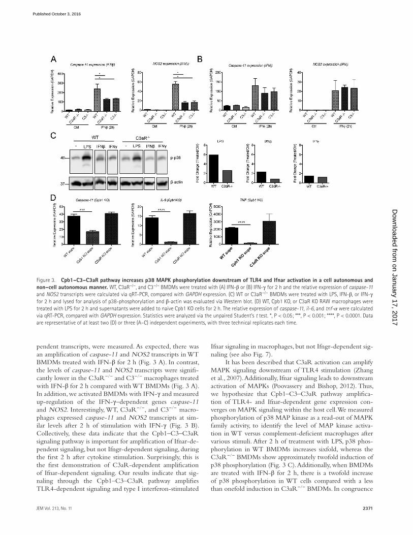
C3aR is required for severity of endotoxemia Caspase-11 promotes disease severity and reduces survival time during severe sepsis induced by LPS, or endotoxemia (Kayagaki et al., 2011, 2013; Hagar et al., 2013). This suggests that caspase-11–dependent cell death, during endotoxemia in mice, increases severity of disease and expedites mortality. We have shown here that the Cpb1–C3–C3aR pathway is important for caspase-11 induced gene expression and sub-sequent caspase-11–dependent cell death in vitro. We hy-pothesize that the Cpb1–C3–C3aR pathway contributes to the severity of endotoxemia by regulating the expression of proinflammatory mediators, such as caspase-11, in vivo. To interrogate this hypothesis, we established a mouse model in which mice receive an i.p. injection with a sublethal dose of LPS, followed by an i.p. injection of a lethal dose of LPS to induce endotoxemia. Proinflammatory gene expression in blood was measured at 2 h after the sublethal LPS injec-tion. Disease severity was assessed after injection of the lethal
on January 17, 2017D
ownloaded from
Published October 3, 2016

2373JEM Vol. 213, No. 11
LPS injection by measuring body temperature and weight loss every 2–4 h pt (Marshall et al., 2005; Gonnert et al., 2011; Gentile et al., 2014).
We found that 2 h after the primary sublethal dose of LPS, C3aR−/− mice had a significant defect in expression of caspase-11 in the blood, compared with WT mice (Fig. 5 A). Additionally, il-6 transcripts in the blood were significantly lower in C3aR−/− compared with WT mice (Fig. 5 B). WT mice injected with the lethal dose of LPS lost a significant amount of weight compared with untreated mice (Fig. 5 C). In addition, within 2–20 h after the lethal dose, the body tem-perature of WT mice decreased dramatically, and was 10°C lower than control mice (Fig. 5 D). In contrast, C3aR−/− mice experienced a moderate weight and temperature loss
compared with WT mice 2–20 h after lethal dose (Fig. 5, C and D). Thus, a key regulator of the Cpb1–C3–C3aR am-plification pathway, C3aR, is required for WT expression of proinflammatory mediators in the blood and contributes to the severity of pathologies during endotoxemia, highlight-ing the importance of this signaling cascade during acute severe sepsis in vivo.
Furthermore, we were interested to know if we could pharmacologically block C3aR activation and decrease the severity of pathologies in mice with endotoxemia. We used a selective nonpeptide antagonist of C3aR (C3aRi; SB290157), shown to be effective in vivo, to block C3aR activation in our mouse model (Ames et al., 2001). Mice injected with a sub-lethal dose of LPS and the C3aR inhibitor displayed a reduced level of caspase-11 and il-6 transcripts in the blood compared with mice that were not given the antagonist (Fig. 5, E and F). After the first injection (6 h), these same mice were in-jected with a lethal dose of LPS and C3aRi and weight and temperature were recorded to track endotoxemia severity. As seen in the C3aR−/− mice, mice treated with the C3aRi had significantly less weight loss (Fig. 5 G). However, the mice treated with C3aRi did not have a difference in tempera-ture loss compared with untreated mice (Fig. 5 H). Notably, the levels of IL-6 detected in the blood of mice treated with C3aRi were significantly lower than control mice 2 h after injection of low dose LPS (Fig. 5 I). These data suggest C3aR as a potential therapeutic target for decreasing endotoxemia severity, and highlight a bifurcation in weight loss and tem-perature loss during sepsis that may be interesting to uncover in future experiments. Finally, we determined whether treat-ment of mice with C3aRi during endotoxemia could influ-ence survival. We induced endotoxemia in WT mice, with and without C3aRi, and found that mice treated with C3aRi are partially protected from LPS-induced mortality (Fig. 5 J). Together, these data suggest that C3aR, a key mediator in the Cpb1–C3–C3aR pathway, plays a role in early expression of inflammatory mediators in the blood during endotoxemia and contributes to endotoxemia severity and disease outcome.
C3aR contributes to enhanced inflammation in primary human macrophagesCaspase-4 and -5 are the potential human orthologues to murine caspase-11, and have been described to play a role in responding to LPS and Gram-negative bacterial pathogens in primary human monocyte–derived macrophages (MDMs; Casson et al., 2015). There is evidence that expression of caspase-4 and -5 is induced upon host cell exposure to LPS; however, the role of this induction in inflammation is not clear (Munday et al., 1995; Lin et al., 2000; Eckhart et al., 2006; Casson et al., 2015; Viganò et al., 2015). Additionally, it is not known what signaling pathways contribute to ex-pression of these caspases downstream of TLR4. Considering our data presented thus far, we hypothesize that C3aR may be required for early induction of caspase-4 and -5 and sub-sequent inflammation. Thus, we first tested the early kinetics
Figure 4. Cpb1–C3–C3aR pathway is required for caspase-11–de-pendent cell death in response to Gram-negative bacterial pathogens. Indicated RAW macrophage cell lines were infected with (A) Salmonella Typhimurium (SL1344; MOI 100:1) or (B) S. flexneri (M90T; MOI 50:1) for 18 h and cytotoxicity was measured. (C) BMDMs were infected with S. Typhimurium (SL1344 ΔorgA/fliC; MOI, 100:1) for 18 h and cytotoxicity was measured. All cytotoxicity was measured by release of LDH. Statistics analyzed via the unpaired Student’s t test. *, P < 0.05; **, P < 0.01; ***, P < 0.001. Data are representative of at least three independent experiments (A–C), with three technical replicates each time.
on January 17, 2017D
ownloaded from
Published October 3, 2016
Complement amplifies caspase-11 cell death and endotoxemia | Napier et al.2374
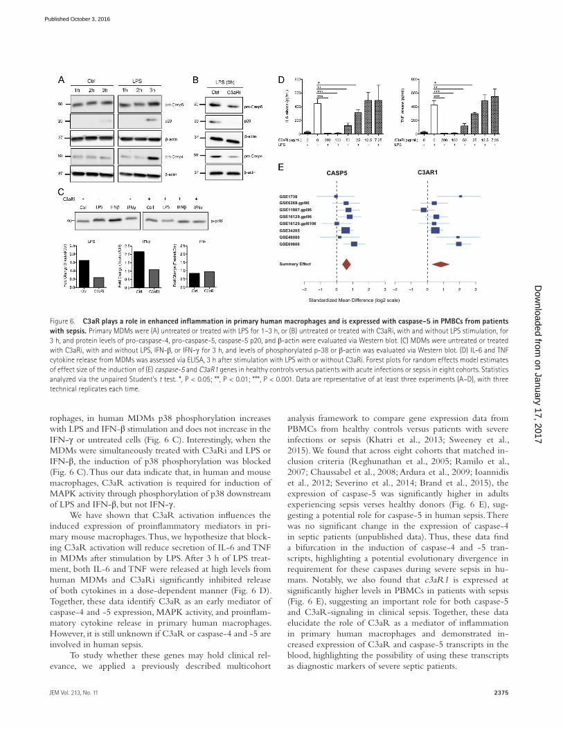
of caspase-4 and -5 expression in MDMs after treatment with LPS. We found that both pro-caspase-4 and -5 were induced after 3 h of stimulation compared with the untreated control (Ctrl; Fig. 6 A). Additionally, at 3 h pt we can detect higher levels of p20 fragment of caspase-5, a potential indicator of caspase-5 activation, in the LPS-treated samples compared with the Ctrl (Fig. 6 A). Thus, caspase-4 and -5 expression, and possibly caspase-5 activation, is induced by LPS treat-ment within 3 h in MDMs.
To elucidate if C3aR is involved in up-regulation of caspase-4 and -5, we treated MDMs with LPS, with and without the C3aR inhibitor (C3aRi). At 3 h pt, C3aRi-
treated MDMs expressed lower levels of pro-caspase-4 and -5 (Fig. 6 B). Additionally, the caspase-5 p20 fragment was not detected after 3 h of LPS and C3aRi treatment in MDMs (Fig. 6 B). Furthermore, our results in mouse macrophages suggest that C3aR is amplifying proinflammatory mediators via increasing p38 MAPK activity downstream of TLR4 and Ifnar activation, but not Ifngr activation (Fig. 3, A–C). Consid-ering these data, we propose that C3aR is modulating inflam-matory signaling pathways in human macrophages similar to what was found in mouse cells. To test this, we treated MDMs with LPS, IFN-β, and IFN-γ for 3 h and looked at fold-change of p38-phosphorylation. As seen in the mouse mac-
Figure 5. C3aR contributes to amplification of proinflammatory mediators in the blood and to the severity of endotoxemia in mice. WT or C3aR-deficient (C3aR−/−) BALBc mice were treated with a sublethal dose of LPS i.p. 2 h pt blood samples were collected and analyzed for expression of (A) caspase-11 or (B) IL-6. Fold-change of gene expression was calculated (Ctrl/LPS treated mice). 6 h pt, mice were treated with a lethal dose of LPS. Weight (C) and temperature (D) were monitored every 2 h for 20 h. Six control mice and eight treated mice were used. Next, WT mice were treated with the same regimen as described with or without C3aRi. As described above, 2 h pt (E) caspase-11 or (F) IL-6 expression was analyzed and fold-change of gene expres-sion was calculated (Ctrl/LPS treated mice), and after 6 h mice were treated with a lethal dose of LPS. Weight (G) and temperature (H) were monitored every 2 h for 20 h. (I) WT mice were treated with LPS and with or without C3aRi i.p. 2 h pt, blood was collected via cardiac bleed and analyzed for IL-6 levels via ELI SA. (J) WT mice were treated with LPS and with or without C3aRi, and survival was monitored. n = 5 mice/group in each independent experiment (E–J). Statistics analyzed via the unpaired Student’s t test. *, P < 0.05; **, P < 0.01; ***, P < 0.001, ****, P < 0.0001. Data are representative of at least two (I–J) or three experiments (A–H), with three technical replicates each time.
on January 17, 2017D
ownloaded from
Published October 3, 2016
2375JEM Vol. 213, No. 11
rophages, in human MDMs p38 phosphorylation increases with LPS and IFN-β stimulation and does not increase in the IFN-γ or untreated cells (Fig. 6 C). Interestingly, when the MDMs were simultaneously treated with C3aRi and LPS or IFN-β, the induction of p38 phosphorylation was blocked (Fig. 6 C). Thus our data indicate that, in human and mouse macrophages, C3aR activation is required for induction of MAPK activity through phosphorylation of p38 downstream of LPS and IFN-β, but not IFN-γ.
We have shown that C3aR activation influences the induced expression of proinflammatory mediators in pri-mary mouse macrophages. Thus, we hypothesize that block-ing C3aR activation will reduce secretion of IL-6 and TNF in MDMs after stimulation by LPS. After 3 h of LPS treat-ment, both IL-6 and TNF were released at high levels from human MDMs and C3aRi significantly inhibited release of both cytokines in a dose-dependent manner (Fig. 6 D). Together, these data identify C3aR as an early mediator of caspase-4 and -5 expression, MAPK activity, and proinflam-matory cytokine release in primary human macrophages. However, it is still unknown if C3aR or caspase-4 and -5 are involved in human sepsis.
To study whether these genes may hold clinical rel-evance, we applied a previously described multicohort
analysis framework to compare gene expression data from PBMCs from healthy controls versus patients with severe infections or sepsis (Khatri et al., 2013; Sweeney et al., 2015). We found that across eight cohorts that matched in-clusion criteria (Reghunathan et al., 2005; Ramilo et al., 2007; Chaussabel et al., 2008; Ardura et al., 2009; Ioannidis et al., 2012; Severino et al., 2014; Brand et al., 2015), the expression of caspase-5 was significantly higher in adults experiencing sepsis verses healthy donors (Fig. 6 E), sug-gesting a potential role for caspase-5 in human sepsis. There was no significant change in the expression of caspase-4 in septic patients (unpublished data). Thus, these data find a bifurcation in the induction of caspase-4 and -5 tran-scripts, highlighting a potential evolutionary divergence in requirement for these caspases during severe sepsis in hu-mans. Notably, we also found that c3aR1 is expressed at significantly higher levels in PBMCs in patients with sepsis (Fig. 6 E), suggesting an important role for both caspase-5 and C3aR-signaling in clinical sepsis. Together, these data elucidate the role of C3aR as a mediator of inflammation in primary human macrophages and demonstrated in-creased expression of C3aR and caspase-5 transcripts in the blood, highlighting the possibility of using these transcripts as diagnostic markers of severe septic patients.
Figure 6. C3aR plays a role in enhanced inflammation in primary human macrophages and is expressed with caspase-5 in PMBCs from patients with sepsis. Primary MDMs were (A) untreated or treated with LPS for 1–3 h, or (B) untreated or treated with C3aRi, with and without LPS stimulation, for 3 h, and protein levels of pro-caspase-4, pro-caspase-5, caspase-5 p20, and β-actin were evaluated via Western blot. (C) MDMs were untreated or treated with C3aRi, with and without LPS, IFN-β, or IFN-γ for 3 h, and levels of phosphorylated p-38 or β-actin was evaluated via Western blot. (D) IL-6 and TNF cytokine release from MDMs was assessed via ELI SA, 3 h after stimulation with LPS with or without C3aRi. Forest plots for random effects model estimates of effect size of the induction of (E) caspase-5 and C3aR1 genes in healthy controls versus patients with acute infections or sepsis in eight cohorts. Statistics analyzed via the unpaired Student’s t test. *, P < 0.05; **, P < 0.01; ***, P < 0.001. Data are representative of at least three experiments (A–D), with three technical replicates each time.
on January 17, 2017D
ownloaded from
Published October 3, 2016

Complement amplifies caspase-11 cell death and endotoxemia | Napier et al.2376
DIS CUS SIONThe multifaceted inflammatory response during sepsis has made the contribution of specific proinflammatory pathways difficult to delineate; however, it is known that caspase-11–dependent cell death contributes to poor sepsis outcomes in model organisms (Kayagaki et al., 2011, 2013; Hagar et al., 2013). Considering the importance of this cell death path-way in disease severity, we used the CRI SPR-Cas9 genome editing system to create a genome-wide unbiased screen in macrophages to look for unique mediators of caspase-11–de-pendent cell death. Using this screen, we identified Cpb1 as a novel mediator of caspase-11 expression and subsequent caspase-11–dependent cell death in macrophages (Fig. 1). Cpb1 is a protein that modifies C3a, an anaphylatoxin product from cleaved complement protein C3, which binds and acti-vates C3aR to modulate immune responses (Takabayashi et al., 1996; Mueller-Ortiz et al., 2006; Leung et al., 2008; Fig. 7). Disruption of Cbp1, C3, and C3aR led to a decrease in ex-pression of caspase-11, and other proinflammatory mediators by reducing p38 MAPK activity, downstream of LPS-depen-dent activation of TLR4 and type-I interferon activation of Ifnar, but not type-II interferon activation of Ifngr signaling (Fig. 1, L–M; Fig. 2; and Fig. 3, A–C). Interestingly, we show the Cpb1–C3–C3aR pathway amplifies inflammatory re-sponses in both a cell autonomous and non–cell autonomous
manner (Fig. 1–3). As seen in Cpb1 KO cells, disruption of C3 and C3aR led to a reduction in caspase-11–dependent cell death (Fig. 2, C and F). Thus, we have elucidated comple-ment as a novel mediator of caspase-11–dependent cell death, through the Cpb1–C3–C3aR signaling pathway.
Expression and activation of caspase-11 is important for cell death induced by Gram-negative bacterial infections, and also acts to exacerbate pathologies during endotoxin-induced sepsis (Broz and Monack, 2011; Kayagaki et al., 2011, 2013, 2015; Aachoui et al., 2013; Hagar et al., 2013). We find that Cpb1–C3–C3aR amplification pathway is important for re-sponding to Gram-negative bacterial pathogens by showing the requirement for Cpb1, C3, and C3aR for caspase-11–de-pendent cell death during S. Typhimurium and S. flexneri infection (Fig. 4, A–C). Furthermore, in a model of severe endotoxin-induced sepsis, C3aR−/− mice had lower levels of caspase-11 and il-6 transcripts in the blood and moderate pa-thologies compared with WT mice (Fig. 5, A–D). We went on to show that inactivation of C3aR with a small nonpeptide C3aR inhibitor (C3aRi) reduces levels of caspase-11 and il-6 transcripts in the blood, IL-6 cytokines levels in the blood, weight loss during severe sepsis, and increased survival, sug-gesting C3aR may be a useful therapeutic target for mediat-ing inflammation during the early stages of sepsis which may influence disease outcome (Fig. 5, E–J).
Figure 7. Model of Cpb1–C3–C3aR pathway amplifying p38 MAPK signaling downstream of TLR4 and Ifnar activation in macrophages. TLR4 and Ifnar activation through their respective ligands induce ex-pression of proinflammatory genes via various mechanisms, including the induction of p38 MAPK (Lee et al., 1994; Sancéau et al., 2000). C3 expression is induced downstream these path-ways (Riches et al., 1988; Maranto et al., 2011), and is quickly released from macrophages (Goodrum, 1987), where it is cleaved into C3a and C3b. Anaphylotoxin C3a can be cleaved ex-tracellularly by Cpb1 into C3a-desArg (Leung et al., 2008; Chatterjee et al., 2009), where it then acts as a ligand for C3aR. We find, the activation of C3aR then amplifies MAPK activ-ity through enhancing p38 MAPK phosphory-lation downstream of TLR4 and Ifnar, but not Ifngr, activation. Due to the extracellular na-ture of this signaling pathway, the Cpb1–C3–C3aR pathway acts as a cell-autonomous and a non–cell autonomous amplification pathway enhancing expression of proinflammatory genes of self- and neighboring macrophages.
on January 17, 2017D
ownloaded from
Published October 3, 2016
2377JEM Vol. 213, No. 11
To understand the clinical relevance of our findings, we looked at the role of C3aR during inflammation in primary human macrophages. We found C3aR plays an integral role in the expression of caspase-11 human orthologues, caspase-4 and -5, and influences p38 MAPK activity downstream of TLR4- and Ifnar-activation, but not Ifngr activation (Fig. 6, A–C). The reduction inflammatory transcripts were reflected in a reduction of proinflammatory cytokine release when cells were treated with C3aRi (Fig. 6 D). Furthermore, we found, by multicohort gene expression analysis, that a caspase-11 orthologue, caspase-5, and c3aR transcripts are up-regulated in PBMCs in clinical sepsis compared with healthy controls, implicating this signaling pathway in humans experiencing sepsis (Fig. 6 E). Together, these data demonstrate that the Cpb1–C3–C3aR signaling pathway as an important mediator of caspase-11–dependent cell death, which is important for the response to Gram-negative bacteria and for severity of pathologies during sepsis. Additionally, this pathway may be playing a role in human sepsis.
Within the Cpb1–C3–C3aR signaling pathway, C3 must be converted into its cleaved product, anaphylatoxin C3a, and then modified by Cpb1 to activate C3aR signal-ing. Thus, supplementing Cpb1 KO cells with C3 should not be able to complement the Cpb1 KO cell death defect. In agreement with this hypothesis, transfection of recombinant C3 into macrophages during stimulation with intracellular LPS does not rescue the Cpb1 KO cell death phenotype (un-published data). These preliminary results further suggest that the enzymatic activity of Cpb1 is required for this signaling pathway to amplify caspase-11–dependent cell death during intracellular LPS stimulation. Future biochemical assays will be essential in following up on this hypothesis.
Additionally, as mentioned previously, Cpb1 works by cleaving the C terminus basic residues (arginine [Arg] and lysine [Lys]) from various peptides, including the anaphyla-toxin C3a (Bokisch and Müller-Eberhard, 1970; Matthews et al., 2004). It is known that an increase in free Arg can in-duce iNOS production in macrophages (Moncada and Higgs, 1991; Bogle et al., 1992; Xia and Zweier, 1997; Lee et al., 2003). Increased iNOS can play a very important role in worsening sepsis outcome and may play a role in induction of caspase-11–dependent cell death (Lupfer et al., 2014). Thus, it is possible that Cpb1 may be contributing to caspase-11–dependent cell death via induction of iNOS. However, when we stimulate WT and iNOS-deficient (iNOS−/−) BMDMs with intracellular LPS, there is no difference in cell death outcome (unpublished data). Accordingly, iNOS is not con-tributing to caspase-11–dependent cell death in macrophages; therefore, we conclude the contribution of an increase in free Arg by Cpb1 is not playing a role in our model.
Our findings support recently published data demon-strating that mice lacking Cpb2, a murine Cpb1 paralogue, had significantly improved survival during polymicrobial sep-sis (Shao et al., 2015). Shao et al. (2015) show that mice that lack Cpb2 had less severe pathologies after polymicrobial sep-
sis was induced by cecal ligation and puncture (CLP) and this was attributed to signaling through another complement re-ceptor, C5aR. Furthermore, loss of Cpb2 is protective during E. coli–induced sepsis, similar to results seen in caspase-11–deficient mice treated with E. coli LPS (Renckens et al., 2005; Kayagaki et al., 2013). We hypothesize that Cpb2 may be playing a redundant role in amplification of proinflammatory transcripts, including caspase-11, and subsequent caspase-11–dependent cell death during sepsis, perhaps through C5aR.
It is notable that we show here that the Cpb1–C3–C3aR complement signaling pathway is acting as an ampli-fication system to enhance cell autonomous and non–cell autonomous TLR4- and Ifnar-dependent signaling required for up-regulation of proinflammatory transcripts, including caspase-11 (Fig. 7). Amplification of TLR signaling by C3aR has been noted previously (Zhang et al., 2007); however, this is the first time that C3aR has been described to affect Ifnar signaling, and these data suggest that signaling downstream of this receptor may be more pleotropic than previously pre-sumed. Notably, we find C3aR is modulating p38 MAPK activity downstream of TLR4 and Ifnar, but not Ifngr, acti-vation. Our data suggests that C3aR directly acts on p38 to induce activity, or another protein downstream of TLR and Ifnar, but upstream of p38. It will be interesting to elucidate the mechanism by which C3aR signaling modifies these sig-naling pathways, and if there other signaling pathways that are affected by C3aR activation during inflammation.
In addition, our data suggest that early signaling path-ways may provide a potential therapeutic target during sep-sis. In congruence with our findings, previous studies have found a close relationship between elevated plasma levels of C3a-desArg in patients with septic shock, and increased risk of developing adult respiratory distress syndrome or multi-ple organ failure, further implicating the Cpb1–C3–C3aR signaling pathway in human disease (Heideman and Hugli, 1984; Bengtson and Heideman, 1988; Heideman et al., 1988; Hack et al., 1989). Because caspase-5 and C3aR gene expres-sion is induced in PBMCs from people with sepsis, it suggests that they may have a role in disease progression.
Our findings identify complement as a key pathway for amplification of proinflammatory cytokine production and caspase-11–dependent cell death and suggest a novel role for complement during bacterial infection and sepsis. Furthermore, we provide evidence that this complement pathway may be important during sepsis in human patients. Considering the compounding difficulty of diagnosing and treating sepsis due to overlapping proinflammatory pathways contributing to disease, our findings suggest that the Cpb1–C3–C3aR pathway could serve as a novel diagnostic and therapeutic target during early stage sepsis.
MAT ERI ALS AND MET HODSCell cultureBMDMs were isolated from the femurs of mice. C3−/− and C3aR−/− BMDMs were generously provided by Beth Stevens
on January 17, 2017D
ownloaded from
Published October 3, 2016
Complement amplifies caspase-11 cell death and endotoxemia | Napier et al.2378
(Harvard Medical School, Boston, MA) and Rick Wetsel (Uni-versity of Texas Medical School, Galveston, TX), respectively. BM cells were plated into sterile Petri dishes and incubated in DMEM supplemented with 10% FBS and 10% macrophage colony-stimulating factor (M-CSF)–conditioned media (col-lected from M-CSF-producing NIH 3T3 cells). BMDMs were incubated at 37°C with 5% CO2 and harvested after 6 d.
RAW264.7 macrophages and constructed KOs were cultured as previously described (Storek et al., 2015). 293T cells were grown in DMEM + 10% heat-inactivated FBS. All cells were incubated at 37°C with 5% CO2.
RAW264.7 KO cell lines and complementationAll RAW264.7 KO cell lines were created using the CRI SPR/Cas9 system (Cong et al., 2013). Target gRNA se-quences were generated using CRI SPR Design and are listed in Table S2. The target gRNAs were then cloned into pX458 following a previously published protocol (Cong et al., 2013). A total of 106 RAW264.7 macrophages were trans-fected with 3 µg pX458 in a 6-well tissue culture treated plate with 6 µl Fugene (Promega). 48 h pt, macrophages were single-cell sorted into 96-well tissue culture–treated plates with 200 µl of DMEM + 10% FBS. After 2 wk of incuba-tion, gDNA from macrophages colonies was purified using QIAamp DNA mini kit (QIA GEN). The targeted DNA se-quence was then amplified and sequenced (Elim Biopharm) with primers (Table S2).
Retroviral constructs were transduced into RAW264.7 macrophages using vesicular stomatitis pseudotyped virus packaged in 293T cells and selected for 48 h with 5 µg/ml puromycin (InvivoGen). Caspase-11 and Cpb1 were cloned in-frame into pMSCV-FLAG-HA-IRES-Puro using Gate-way Cloning, as previously described (Storek et al., 2015).
Cell treatments and LDH assaysFor cytotoxicity assays, RAW264.7 macrophages and BMDMs were plated in 96-well tissue culture treated plates at 50,000 cells per well. When specified, macrophages were treated with 20 µg/ml Cholera toxin B subunit (CTB; List Biological Laboratories) and 20 µg/ml ultra-pure LPS (E. coli O111:B4; InvivoGen), or 0.5 µl per well Fugene and 20 µg/ml ultra-pure LPS and/or recombinant mouse C3 (Ab-nova) in serum-free phenol-red-free OptiMem (11058–021; Gibco). Cytotoxicity was measured via lactate dehydrogenase (LDH) release using CytoTox 96 (nonradioactive cytotoxicity assay; Promega). The percentage of LDH was calculated as follows: (LDH treated/LDH total lysis) × 100.
Real-time PCRMacrophages were treated with 0.1 µg/ml ultra-pure LPS or 100 U/ml recombinant mouse IFN-β or IFN-γ (BioLegend) for 2 h and macrophages were lysed and homogenized in 250 µl TRIzol reagent (MRC). For in vitro samples, RNA was isolated using the RNeasy Mini kit (QIA GEN). cDNA was synthesized from RNA samples using SuperScript III
First-Strand synthesis system (Invitrogen). Gene-specific primers (Table S3) were used to amplify transcripts using FastStart Universal SYBR Green Master (Rox; Roche).
Immunoblot analysisMacrophages were seeded at 106 cells in 6-well tissue culture treated wells and treated as described for 2–16 h. At specified time points, the supernatant was collected and precipitated with 10% trichloracetic acid (TCA) overnight on ice. Pre-cipitated proteins were pelleted at 20,000 g for 30 min at 4°C, washed with ice-cold acetone, air-dried, resuspended in SDS-PAGE sample buffer, and heated to 95°C for 10 min. In parallel, the macrophage monolayer was collected and lysed using RIPA buffer for 30 min on ice with agitation. Protein from 5 × 10⁵ cells was loaded per well of a Crite-rion TGX AnykD Gel (Bio-Rad Laboratories). Mouse West-ern blots were performed with rabbit anti–mouse caspase-1 (Santa Cruz Biotechnology, Inc.) diluted 1:1,000, rat anti–mouse caspase-11 (17D9; Sigma-Aldrich) at 1:500, rabbit anti–mouse phospho-p38 (9211; Cell Signaling Technology) and β-actin (Sigma-Aldrich) diluted 1:2,000. Human West-ern blots were performed with rabbit anti–human caspase-4 (4450; Cell Signaling Technology) and caspase-5 (46680; Cell Signaling Technology) diluted 1:1,000, and rabbit anti–mouse phospho-p38 (9211; Cell Signaling Technology). Quantifica-tion of induction of relative protein expression was measured using Image Lab 5.2.1, as compared with untreated.
RAW264.7 CRI SPR-Cas9 genome wide libraryThe genome-wide mouse lentiviral CRI SPR gRNA pooled library (#50947; Addgene) from K. Yusa (Koike-Yusa et al., 2014) was used to construct this library. The gRNA library was amplified using the available library amplification proto-col, by K. Yusa (Addgene), via the on-plate culture method (Koike-Yusa et al., 2014). The library was then transfected into 293T cells. To maintain 1,000× library, we transfected 5× T-175 tissue culture treated flasks (Falcon) at 11 × 106 cells per flask. We next seeded 50× T-175 culture treated flasks with RAW 264.7 cells constitutively expressing hCas9 at 5 × 10⁶ cells/flask to maintain >1,000× coverage of the gRNA library. We transduced these cells at an MOI of 0.3 (Koike-Yusa et al., 2014) with filtered viral supernatants with 8 µg/ml of pro-tamine sulfate. After 5 d of incubation, the transduced RAW cells were treated with 5 µg/ml of puromycin for selection. 3 d after puromycin treatment, the media was changed and fresh puromycin media was added for an additional 3 d. At 11 d after transduction, the RAW cells were pooled, counted, and frozen back at 10 × 106 cells/vial in 100% FBS.
For our screen, three vials of CRI SPR-Cas9 gRNA li-brary RAW cells were thawed and seeded into three T-175 flasks. One flask was used as an untreated control, and the other two were used as two separate screens. After 24 h of incubation, the media was removed and replaced by serum-free phenol-red-free OptiMem with and without CTB and LPS (at 4 µg per 5 × 10⁴ cells of each). After 16 h of treatment, media was
on January 17, 2017D
ownloaded from
Published October 3, 2016

2379JEM Vol. 213, No. 11
replaced by fresh media and cells were grown for 6 d, media was replaced again and grown for an additional 3 d. At this point, cells were collected for all three conditions and gDNA was isolated using QiaAMP DNA mini kit. The amount of input gDNA for each sample was calculated to achieve 1,000× coverage of the gRNA library (Koike-Yusa et al., 2014). Dual amplification of the gRNA from the lentiviral backbone and then to add index adaptors (P5/P7) for MiSeq was used to create an amplicon library, as described previously (Koike-Yusa et al., 2014). A unique primer was used (5′-TCT TCC GAT CTtcttgtggaaaggacgaaacaccG-3′) for MiSeq sequencing.
Genome-wide screen bioinformatic and statistical analysisA hypergeometric P-value (HGPvalue) calculation was used for genome-wide screen enrichment statistical analysis. All genes listed above P ≤ 0.001 were retained (Table S1). The R version of this code was used and written by Y. Zhou and B. Zhou (Novartis), as previously described (König et al., 2007). All other statistical analysis was done using Prism 6.0 (Graph-Pad Software). The statistical significance was determined by the Student’s t test.
Bacterial cultures and infectionsBacterial strains include WT S. Typhimurium SL1344, SL1344 ΔorgA/fliC mutant, and WT S. flexneri M90T. Infections for both RAW cells and BMDMs were conducted as described previously for BMDMs (Broz and Monack, 2011; Meunier et al., 2014; Kayagaki et al., 2015).
Mice and sepsis mouse modelMice between 5–7 wk were used for in vivo experiments com-paring BALBc mice (000651; The Jackson Laboratory) and C3ar1tm1Cge knock-out mice (C.129S4-C3ar1tm1cge/J; The Jack-son Laboratory). All mouse studies were approved by the insti-tutional animal care and use committees of Stanford University.
In mice tracked for temperature and weight, 1 wk be-fore sepsis, mice were anesthetized locally with 1% lidocaine (4 mg/kg) on the right side hind-back using a 0.5 ml syringe. After 15 min, electronic temperature and ID transponders were implanted subcutaneously in the same right side hind-back region (IPTT-300 transponders; Bio Medic Data System, Inc.) and left to recover for 1 wk. Next, mice were either treated with 200 µl of PBS or LPS (4 mg/kg) via i.p. injection. 2 h pt, 10 µl of blood was collected via tail snip into 150 µl of RNAL-ater (Ambion) and frozen at –80°C. RNA isolation and subse-quent qRT-PCR was performed as previously described in our Materials and methods. At 6 h pt, mice were injected with a le-thal dose of LPS (54 mg/kg) and pathologies were recorded for every 2 h for 20 h (weight and temperature). If the C3aR-in-hibitor (C3aRi; SB290157; Santa Cruz Biotechnology, Inc.) was used, it was injected at 30 mg/kg with LPS in PBS.
Isolating human PBMCsBlood samples from healthy donors were obtained with in-formed consent and collected by research ethics committee
(approved eProtocol; Minimal Risk Research Related Activ-ities at Stanford Blood Center; SQL 79075). Human primary monocyte-derived macrophages (MDM) were prepared by adherence from whole-blood buffy coat fractions from healthy donors. Primary monocytes were cultured in RPMI 1640 with 10% FBS and treated with 30 ng/ml human M-CSF for 6 d. For all assays, 7 d after culture, MDMs were plated at 50,000 cells/well in a 96-well plate and assayed for cytokine release, or plated 1 million cells/well in a 6-well plate and assayed for protein expression level as described above.
Multicohort analysisMulticohort analysis was performed as previously described (Khatri et al., 2013; Sweeney et al., 2015). In brief, a system-atic search was performed in the National Institutes of Health Gene Expression Omnibus for datasets matching the terms: PBMC, sepsis, SIRS, infection, ICU, bacter[wildcard], acute. We retained clinical datasets that studied severe infections, and excluded datasets that included any ex vivo treatment of cells before microarray profiling, arriving at the final seven datasets (Table S1). GSE16129 contained two microarray types, which are treated as two separate cohorts, as previously described (Sweeney et al., 2015).
Online supplemental materialFig. S1 showselectropherograms depicting the biallelic muta-tions for CRI SPR-Cas9 knock-out RAW cell lines Cpb1KO, C3aRKO, and C3KO. Fig. S2 shows alignments of caspase-11 genes. Table S1 is a list of the top 100 enriched hits in the CRI SPR-Cas9 screen. Table S2 lists the gRNAs for each CRI SPR-Cas9 knock-out cell line. Table S3 lists the sequences for each set of primers used in qRT-PCR. Table S4 provides information on the cohorts used in our multi-cohort analysis.
ACK NOW LED GME NTSWe want to thank Sarah Ewald and Kelly Storek for helpful protocols and reagents, and Katie Cumnock for invaluable help with our mouse model. Additionally, we would like to thank Soyon Hung (Beth Steven’s laboratory, Harvard University, Boston, MA) and Stacey Mueller-Ortiz (Rick Wetsel’s laboratory, UT Houston, Houston, TX) for C3−/− and C3aR−/− BMDMs, respectively.
This research was supported by the National Institute of Allergy and Infectious Diseases grants AI095396-05 (to D.M. Monack), DAR PA-15-21-ThoR-FP-006 (to D.M. Monack), and 1F32AI115950-01 (to B.A. Napier).
The authors declare no competing financial interests.
Submitted: 7 January 2016
Accepted: 25 August 2016
REfERENCESAachoui, Y., I.A. Leaf, J.A. Hagar, M.F. Fontana, C.G. Campos, D.E. Zak, M.H.
Tan, P.A. Cotter, R.E. Vance, A. Aderem, and E.A. Miao. 2013. Caspase-11 protects against bacteria that escape the vacuole. Science. 339:975–978. http ://dx .doi .org /10 .1126 /science .1230751
Ames, R.S., D. Lee, J.J. Foley, A.J. Jurewicz, M.A. Tornetta, W. Bautsch, B. Settmacher, A. Klos, K.F. Erhard, R.D. Cousins, et al. 2001. Identification of a selective nonpeptide antagonist of the anaphylatoxin C3a receptor
on January 17, 2017D
ownloaded from
Published October 3, 2016

Complement amplifies caspase-11 cell death and endotoxemia | Napier et al.2380
that demonstrates antiinflammatory activity in animal models. J. Immunol. 166:6341–6348. http ://dx .doi .org /10 .4049 /jimmunol .166 .10 .6341
Angus, D.C., and T. van der Poll. 2013. Severe sepsis and septic shock. N. Engl. J. Med. 369:2063. http ://dx .doi .org /10 .1056 /NEJMra1208623
Angus, D.C., W.T. Linde-Zwirble, J. Lidicker, G. Clermont, J. Carcillo, and M.R. Pinsky. 2001. Epidemiology of severe sepsis in the United States: analysis of incidence, outcome, and associated costs of care. Crit. Care Med. 29:1303–1310. http ://dx .doi .org /10 .1097 /00003246 -200107000 -00002
Ardura, M.I., R. Banchereau, A. Mejias, T. Di Pucchio, C. Glaser, F. Allantaz, V. Pascual, J. Banchereau, D. Chaussabel, and O. Ramilo. 2009. Enhanced monocyte response and decreased central memory T cells in children with invasive Staphylococcus aureus infections. PLoS One. 4:e5446. http ://dx .doi .org /10 .1371 /journal .pone .0005446
Ayala, A., C.D. Herdon, D.L. Lehman, C.A. Ayala, and I.H. Chaudry. 1996. Differential induction of apoptosis in lymphoid tissues during sepsis: variation in onset, frequency, and the nature of the mediators. Blood. 87:4261–4275.
Bengtson, A., and M. Heideman. 1988. Anaphylatoxin formation in sepsis. Arch. Surg. 123:645–649. http ://dx .doi .org /10 .1001 /archsurg .1988 .01400290131023
Bogle, R.G., A.R. Baydoun, J.D. Pearson, S. Moncada, and G.E. Mann. 1992. L-arginine transport is increased in macrophages generating nitric oxide. Biochem. J. 284:15–18. http ://dx .doi .org /10 .1042 /bj2840015
Bokisch, V.A., and H.J. Müller-Eberhard. 1970. Anaphylatoxin inactivator of human plasma: its isolation and characterization as a carboxypeptidase. J. Clin. Invest. 49:2427–2436. http ://dx .doi .org /10 .1172 /JCI106462
Brand, H.K., I.M. Ahout, D. de Ridder, A. van Diepen, Y. Li, M. Zaalberg, A. Andeweg, N. Roeleveld, R. de Groot, A. Warris, et al. 2015. Olfactomedin 4 serves as a marker for disease severity in pediatric respiratory syncytial virus (RSV) infection. PLoS One. 10:e0131927. http ://dx .doi .org /10 .1371 /journal .pone .0131927
Broz, P., and D.M. Monack. 2011. Molecular mechanisms of inflammasome activation during microbial infections. Immunol. Rev. 243:174–190. http ://dx .doi .org /10 .1111 /j .1600 -065X .2011 .01041 .x
Broz, P., T. Ruby, K. Belhocine, D.M. Bouley, N. Kayagaki, V.M. Dixit, and D.M. Monack. 2012. Caspase-11 increases susceptibility to Salmonella infection in the absence of caspase-1. Nature. 490:288–291. http ://dx .doi .org /10 .1038 /nature11419
Casson, C.N., J. Yu, V.M. Reyes, F.O. Taschuk, A. Yadav, A.M. Copenhaver, H.T. Nguyen, R.G. Collman, and S. Shin. 2015. Human caspase-4 mediates noncanonical inflammasome activation against Gram-negative bacterial pathogens. Proc. Natl. Acad. Sci. USA. 112:6688–6693. http ://dx .doi .org /10 .1073 /pnas .1421699112
Cerra, F.B. 1985. The systemic septic response: multiple systems organ failure. Crit. Care Clin. 1:591–607.
Chatterjee, S., O. Lardinois, M.G. Bonini, S. Bhattacharjee, K. Stadler, J. Corbett, L.J. Deterding, K.B. Tomer, M. Kadiiska, and R.P. Mason. 2009. Site-specific carboxypeptidase B1 tyrosine nitration and pathophysiological implications following its physical association with nitric oxide synthase-3 in experimental sepsis. J. Immunol. 183:4055–4066. http ://dx .doi .org /10 .4049 /jimmunol .0900593
Chaussabel, D., C. Quinn, J. Shen, P. Patel, C. Glaser, N. Baldwin, D. Stichweh, D. Blankenship, L. Li, I. Munagala, et al. 2008. A modular analysis framework for blood genomics studies: application to systemic lupus erythematosus. Immunity. 29:150–164. http ://dx .doi .org /10 .1016 /j .immuni .2008 .05 .012
Chinnapen, D.J., W.T. Hsieh, Y.M. te Welscher, D.E. Saslowsky, L. Kaoutzani, E. Brandsma, L. D’Auria, H. Park, J.S. Wagner, K.R. Drake, et al. 2012. Lipid sorting by ceramide structure from plasma membrane to ER for the cholera toxin receptor ganglioside GM1. Dev. Cell. 23:573–586. http ://dx .doi .org /10 .1016 /j .devcel .2012 .08 .002
Cong, L., F.A. Ran, D. Cox, S. Lin, R. Barretto, N. Habib, P.D. Hsu, X. Wu, W. Jiang, L.A. Marraffini, and F. Zhang. 2013. Multiplex genome engineering using CRI SPR/Cas systems. Science. 339:819–823. http ://dx .doi .org /10 .1126 /science .1231143
Crass, T., U. Raffetseder, U. Martin, M. Grove, A. Klos, J. Köhl, and W. Bautsch. 1996. Expression cloning of the human C3a anaphylatoxin receptor (C3aR) from differentiated U-937 cells. Eur. J. Immunol. 26:1944–1950. http ://dx .doi .org /10 .1002 /eji .1830260840
Eckhart, L., C. Kittel, S. Gawlas, F. Gruber, M. Mildner, B. Jilma, and E. Tschachler. 2006. Identification of a novel exon encoding the amino-terminus of the predominant caspase-5 variants. Biochem. Biophys. Res. Commun. 348:682–688. http ://dx .doi .org /10 .1016 /j .bbrc .2006 .07 .104
Flierl, M.A., D. Rittirsch, B.A. Nadeau, D.E. Day, F.S. Zetoune, J.V. Sarma, M.S. Huber-Lang, and P.A. Ward. 2008. Functions of the complement components C3 and C5 during sepsis. FAS EB J. 22:3483–3490. http ://dx .doi .org /10 .1096 /fj .08 -110595
Funk, D., F. Sebat, and A. Kumar. 2009. A systems approach to the early recognition and rapid administration of best practice therapy in sepsis and septic shock. Curr. Opin. Crit. Care. 15:301–307. http ://dx .doi .org /10 .1097 /MCC .0b013e32832e3825
Gentile, L.F., D.C. Nacionales, M.C. Lopez, E. Vanzant, A. Cuenca, B.E. Szpila, A.G. Cuenca, A. Joseph, F.A. Moore, C. Leeuwenburgh, et al. 2014. Host responses to sepsis vary in different low-lethality murine models. PLoS One. 9:e94404. http ://dx .doi .org /10 .1371 /journal .pone .0094404
Gonnert, F.A., P. Recknagel, M. Seidel, N. Jbeily, K. Dahlke, C.L. Bockmeyer, J. Winning, W. Lösche, R.A. Claus, and M. Bauer. 2011. Characteristics of clinical sepsis reflected in a reliable and reproducible rodent sepsis model. J. Surg. Res. 170:e123–e134. http ://dx .doi .org /10 .1016 /j .jss .2011 .05 .019
Goodrum, K.J. 1987. Complement component C3 secretion by mouse mac-rophage-like cell lines. J. Leukoc. Biol. 41:295–301.
Grimsey, N.J., B. Aguilar, T.H. Smith, P. Le, A.L. Soohoo, M.A. Puthenveedu, V. Nizet, and J. Trejo. 2015. Ubiquitin plays an atypical role in GPCR-induced p38 MAP kinase activation on endosomes. J. Cell Biol. 210:1117–1131. http ://dx .doi .org /10 .1083 /jcb .201504007
Hack, C.E., J.H. Nuijens, R.J. Felt-Bersma, W.O. Schreuder, A.J. Eerenberg-Belmer, J. Paardekooper, W. Bronsveld, and L.G. Thijs. 1989. Elevated plasma levels of the anaphylatoxins C3a and C4a are associated with a fatal outcome in sepsis. Am. J. Med. 86:20–26. http ://dx .doi .org /10 .1016 /0002 -9343(89)90224 -6
Hagar, J.A., D.A. Powell, Y. Aachoui, R.K. Ernst, and E.A. Miao. 2013. Cytoplasmic LPS activates caspase-11: implications in TLR4-independent endotoxic shock. Science. 341:1250–1253. http ://dx .doi .org /10 .1126 /science .1240988
Heideman, M., and T.E. Hugli. 1984. Anaphylatoxin generation in multisystem organ failure. J. Trauma. 24:1038–1043. http ://dx .doi .org /10 .1097 /00005373 -198412000 -00006
Heideman, M., B. Norder-Hansson, A. Bengtson, and T.E. Mollnes. 1988. Terminal complement complexes and anaphylatoxins in septic and ischemic patients. Arch. Surg. 123:188–192. http ://dx .doi .org /10 .1001 /archsurg .1988 .01400260068008
Hotchkiss, R.S., P.E. Swanson, J.P. Cobb, A. Jacobson, T.G. Buchman, and I.E. Karl. 1997. Apoptosis in lymphoid and parenchymal cells during sepsis: findings in normal and T- and B-cell-deficient mice. Crit. Care Med. 25:1298–1307. http ://dx .doi .org /10 .1097 /00003246 -199708000 -00015
Hotchkiss, R.S., K.W. Tinsley, P.E. Swanson, K.C. Chang, J.P. Cobb, T.G. Buchman, S.J. Korsmeyer, and I.E. Karl. 1999. Prevention of lymphocyte cell death in sepsis improves survival in mice. Proc. Natl. Acad. Sci. USA. 96:14541–14546. http ://dx .doi .org /10 .1073 /pnas .96 .25 .14541
Hotchkiss, R.S., K.W. Tinsley, and I.E. Karl. 2003. Role of apoptotic cell death in sepsis. Scand. J. Infect. Dis. 35:585–592. http ://dx .doi .org /10 .1080 /00365540310015692
on January 17, 2017D
ownloaded from
Published October 3, 2016

2381JEM Vol. 213, No. 11
Ioannidis, I., B. McNally, M. Willette, M.E. Peeples, D. Chaussabel, J.E. Durbin, O. Ramilo, A. Mejias, and E. Flaño. 2012. Plasticity and virus specificity of the airway epithelial cell immune response during respiratory virus infection. J. Virol. 86:5422–5436. http ://dx .doi .org /10 .1128 /JVI .06757 -11
Isogai, E., H. Isogai, T. Kubota, N. Fujii, S. Hayashi, T. Indoh, S. Takagi, H. Miura, and K. Kimura. 1998. Apoptosis of lymphocytes in mice ad-ministered lipopolysaccharide from Leptospira interrogans. Zentralbl. Veterinarmed. B. 45:529–537.
Jobling, M.G., Z. Yang, W.R. Kam, W.I. Lencer, and R.K. Holmes. 2012. A single native ganglioside GM1-binding site is sufficient for cholera toxin to bind to cells and complete the intoxication pathway. MBio. 3:3. http ://dx .doi .org /10 .1128 /mBio .00401 -12
Kang, S.J., S. Wang, H. Hara, E.P. Peterson, S. Namura, S. Amin-Hanjani, Z. Huang, A. Srinivasan, K.J. Tomaselli, N.A. Thornberry, et al. 2000. Dual role of caspase-11 in mediating activation of caspase-1 and caspase-3 under pathological conditions. J. Cell Biol. 149:613–622. http ://dx .doi .org /10 .1083 /jcb .149 .3 .613
Kawli, T., C. Wu, and M.W. Tan. 2010. Systemic and cell intrinsic roles of Gqalpha signaling in the regulation of innate immunity, oxidative stress, and longevity in Caenorhabditis elegans. Proc. Natl. Acad. Sci. USA. 107:13788–13793. http ://dx .doi .org /10 .1073 /pnas .0914715107
Kayagaki, N., S. Warming, M. Lamkanfi, L. Vande Walle, S. Louie, J. Dong, K. Newton, Y. Qu, J. Liu, S. Heldens, et al. 2011. Non-canonical inflammasome activation targets caspase-11. Nature. 479:117–121. http ://dx .doi .org /10 .1038 /nature10558
Kayagaki, N., M.T. Wong, I.B. Stowe, S.R. Ramani, L.C. Gonzalez, S. Akashi-Takamura, K. Miyake, J. Zhang, W.P. Lee, A. Muszyński, et al. 2013. Noncanonical inflammasome activation by intracellular LPS independent of TLR4. Science. 341:1246–1249. http ://dx .doi .org /10 .1126 /science .1240248
Kayagaki, N., I.B. Stowe, B.L. Lee, K. O’Rourke, K. Anderson, S. Warming, T. Cuellar, B. Haley, M. Roose-Girma, Q.T. Phung, et al. 2015. Caspase-11 cleaves gasdermin D for non-canonical inflammasome signalling. Nature. 526:666–671. http ://dx .doi .org /10 .1038 /nature15541
Khatri, P., S. Roedder, N. Kimura, K. De Vusser, A.A. Morgan, Y. Gong, M.P. Fischbein, R.C. Robbins, M. Naesens, A.J. Butte, and M.M. Sarwal. 2013. A common rejection module (CRM) for acute rejection across multiple organs identifies novel therapeutics for organ transplantation. J. Exp. Med. 210:2205–2221. http ://dx .doi .org /10 .1084 /jem .20122709
Koike-Yusa, H., Y. Li, E.P. Tan, M.C. Velasco-Herrera, and K. Yusa. 2014. Genome-wide recessive genetic screening in mammalian cells with a lentiviral CRI SPR-guide RNA library. Nat. Biotechnol. 32:267–273. http ://dx .doi .org /10 .1038 /nbt .2800
König, R., C.Y. Chiang, B.P. Tu, S.F. Yan, P.D. DeJesus, A. Romero, T. Bergauer, A. Orth, U. Krueger, Y. Zhou, and S.K. Chanda. 2007. A probability-based approach for the analysis of large-scale RNAi screens. Nat. Methods. 4:847–849. http ://dx .doi .org /10 .1038 /nmeth1089
Lee, J.C., J.T. Laydon, P.C. McDonnell, T.F. Gallagher, S. Kumar, D. Green, D. McNulty, M.J. Blumenthal, J.R. Heys, S.W. Landvatter, et al. 1994. A protein kinase involved in the regulation of inflammatory cytokine biosynthesis. Nature. 372:739–746. http ://dx .doi .org /10 .1038 /372739a0
Lee, J., H. Ryu, R.J. Ferrante, S.M. Morris Jr., and R.R. Ratan. 2003. Translational control of inducible nitric oxide synthase expression by arginine can explain the arginine paradox. Proc. Natl. Acad. Sci. USA. 100:4843–4848. http ://dx .doi .org /10 .1073 /pnas .0735876100
Leung, L.L., T. Nishimura, and T. Myles. 2008. Regulation of tissue inflammation by thrombin-activatable carboxypeptidase B (or TAFI). Adv. Exp. Med. Biol. 632:61–69. http ://dx .doi .org /10 .1016 /j .molimm .2008 .07 .010
Lin, X.Y., M.S. Choi, and A.G. Porter. 2000. Expression analysis of the human caspase-1 subfamily reveals specific regulation of the CASP5 gene by
lipopolysaccharide and interferon-gamma. J. Biol. Chem. 275:39920–39926. http ://dx .doi .org /10 .1074 /jbc .M007255200
Lupfer, C.R., P.K. Anand, Z. Liu, K.L. Stokes, P. Vogel, M. Lamkanfi, and T.D. Kanneganti. 2014. Reactive oxygen species regulate caspase-11 expression and activation of the non-canonical NLRP3 inflammasome during enteric pathogen infection. PLoS Pathog. 10:e1004410. http ://dx .doi .org /10 .1371 /journal .ppat .1004410
Maranto, J., J. Rappaport, and P.K. Datta. 2011. Role of C/EBP-β, p38 MAPK, and MKK6 in IL-1β-mediated C3 gene regulation in astrocytes. J. Cell. Biochem. 112:1168–1175. http ://dx .doi .org /10 .1002 /jcb .23032
Markiewski, M.M., R.A. DeAngelis, and J.D. Lambris. 2008. Complexity of complement activation in sepsis. J. Cell. Mol. Med. 12(6a, 6A):2245–2254. http ://dx .doi .org /10 .1111 /j .1582 -4934 .2008 .00504 .x
Marshall, J.C., E. Deitch, L.L. Moldawer, S. Opal, H. Redl, and T. van der Poll. 2005. Preclinical models of shock and sepsis: what can they tell us? Shock. 24(Suppl 1):1–6. http ://dx .doi .org /10 .1097 /01 .shk .0000191383 .34066 .4b
Martin, U., D. Bock, L. Arseniev, M.A. Tornetta, R.S. Ames, W. Bautsch, J. Köhl, A. Ganser, and A. Klos. 1997. The human C3a receptor is expressed on neutrophils and monocytes, but not on B or T lymphocytes. J. Exp. Med. 186:199–207. http ://dx .doi .org /10 .1084 /jem .186 .2 .199
Matthews, K.W., S.L. Mueller-Ortiz, and R.A. Wetsel. 2004. Carboxypeptidase N: a pleiotropic regulator of inflammation. Mol. Immunol. 40:785–793. http ://dx .doi .org /10 .1016 /j .molimm .2003 .10 .002
Meunier, E., M.S. Dick, R.F. Dreier, N. Schürmann, D. Kenzelmann Broz, S. Warming, M. Roose-Girma, D. Bumann, N. Kayagaki, K. Takeda, et al. 2014. Caspase-11 activation requires lysis of pathogen-containing vacuoles by IFN-induced GTPases. Nature. 509:366–370. http ://dx .doi .org /10 .1038 /nature13157
Moncada, S., and E.A. Higgs. 1991. Endogenous nitric oxide: physiology, pathology and clinical relevance. Eur. J. Clin. Invest. 21:361–374. http ://dx .doi .org /10 .1111 /j .1365 -2362 .1991 .tb01383 .x
Moore, G.J., and N.L. Benoiton. 1978. Effect of the basic amino-acid side chain length and the penultimate residue on the hydrolysis of benzoldipeptides by carboxypeptidase B. Can. J. Biochem. 56:315–318. http ://dx .doi .org /10 .1139 /o78 -048
Mueller-Ortiz, S.L., T.J. Hollmann, D.L. Haviland, and R.A. Wetsel. 2006. Ablation of the complement C3a anaphylatoxin receptor causes enhanced killing of Pseudomonas aeruginosa in a mouse model of pneumonia. Am. J. Physiol. Lung Cell. Mol. Physiol. 291:L157–L165. http ://dx .doi .org /10 .1152 /ajplung .00358 .2005
Munday, N.A., J.P. Vaillancourt, A. Ali, F.J. Casano, D.K. Miller, S.M. Molineaux, T.T. Yamin, V.L. Yu, and D.W. Nicholson. 1995. Molecular cloning and pro-apoptotic activity of ICErelII and ICErelIII, members of the ICE/CED-3 family of cysteine proteases. J. Biol. Chem. 270:15870–15876. http ://dx .doi .org /10 .1074 /jbc .270 .26 .15870
Oberholzer, A., C. Oberholzer, and L.L. Moldawer. 2001. Sepsis syndromes: understanding the role of innate and acquired immunity. Shock. 16:83–96. http ://dx .doi .org /10 .1097 /00024382 -200116020 -00001
Papatheodorou, P., J.E. Carette, G.W. Bell, C. Schwan, G. Guttenberg, T.R. Brummelkamp, and K. Aktories. 2011. Lipolysis-stimulated lipoprotein receptor (LSR) is the host receptor for the binary toxin Clostridium difficile transferase (CDT). Proc. Natl. Acad. Sci. USA. 108:16422–16427. http ://dx .doi .org /10 .1073 /pnas .1109772108
Poovassery, J.S., and G.A. Bishop. 2012. Type I IFN receptor and the B cell antigen receptor regulate TLR7 responses via distinct molecular mechanisms. J. Immunol. 189:1757–1764. http ://dx .doi .org /10 .4049 /jimmunol .1200624
Ramilo, O., W. Allman, W. Chung, A. Mejias, M. Ardura, C. Glaser, K.M. Wittkowski, B. Piqueras, J. Banchereau, A.K. Palucka, and D. Chaussabel. 2007. Gene expression patterns in blood leukocytes discriminate patients with acute infections. Blood. 109:2066–2077. http ://dx .doi .org /10 .1182 /blood -2006 -02 -002477
on January 17, 2017D
ownloaded from
Published October 3, 2016

Complement amplifies caspase-11 cell death and endotoxemia | Napier et al.2382
Rangel-Frausto, M.S., D. Pittet, M. Costigan, T. Hwang, C.S. Davis, and R.P. Wenzel. 1995. The natural history of the systemic inflammatory response syndrome (SIRS). A prospective study. JAMA. 273:117–123. http ://dx .doi .org /10 .1001 /jama .1995 .03520260039030
Rathinam, V.A., S.K. Vanaja, L. Waggoner, A. Sokolovska, C. Becker, L.M. Stuart, J.M. Leong, and K.A. Fitzgerald. 2012. TRIF licenses caspase-11-dependent NLRP3 inflammasome activation by gram-negative bacteria. Cell. 150:606–619. http ://dx .doi .org /10 .1016 /j .cell .2012 .07 .007
Reghunathan, R., M. Jayapal, L.Y. Hsu, H.H. Chng, D. Tai, B.P. Leung, and A.J. Melendez. 2005. Expression profile of immune response genes in patients with Severe Acute Respiratory Syndrome. BMC Immunol. 6:2. http ://dx .doi .org /10 .1186 /1471 -2172 -6 -2
Renckens, R., J.J. Roelofs, S.A. ter Horst, C. van ’t Veer, S.R. Havik, S. Florquin, G.T. Wagenaar, J.C. Meijers, and T. van der Poll. 2005. Absence of thrombin-activatable fibrinolysis inhibitor protects against sepsis-induced liver injury in mice. J. Immunol. 175:6764–6771. http ://dx .doi .org /10 .4049 /jimmunol .175 .10 .6764
Riches, D.W., P.M. Henson, L.K. Remigio, J.F. Catterall, and R.C. Strunk. 1988. Differential regulation of gene expression during macrophage ac-tivation with a polyribonucleotide. The role of endogenously derived IFN. J. Immunol. 141:180–188.
Rittirsch, D., M.A. Flierl, and P.A. Ward. 2008. Harmful molecular mechanisms in sepsis. Nat. Rev. Immunol. 8:776–787. http ://dx .doi .org /10 .1038 /nri2402
Robinson, A.J., and J.M. Dickenson. 2001. Activation of the p38 and p42/p44 mitogen-activated protein kinase families by the histamine H(1) receptor in DDT(1)MF-2 cells. Br. J. Pharmacol. 133:1378–1386. http ://dx .doi .org /10 .1038 /sj .bjp .0704200
Sancéau, J., J. Hiscott, O. Delattre, and J. Wietzerbin. 2000. IFN-beta induces serine phosphorylation of Stat-1 in Ewing’s sarcoma cells and mediates apoptosis via induction of IRF-1 and activation of caspase-7. Oncogene. 19:3372–3383. http ://dx .doi .org /10 .1038 /sj .onc .1203670
Severino, P., E. Silva, G.L. Baggio-Zappia, M.K. Brunialti, L.A. Nucci, O. Rigato Jr., I.D. da Silva, F.R. Machado, and R. Salomao. 2014. Patterns of gene expression in peripheral blood mononuclear cells and outcomes from patients with sepsis secondary to community acquired pneumonia. PLoS One. 9:e91886. http ://dx .doi .org /10 .1371 /journal .pone .0091886
Shao, Z., T. Nishimura, L.L. Leung, and J. Morser. 2015. Carboxypeptidase B2 deficiency reveals opposite effects of complement C3a and C5a in a murine polymicrobial sepsis model. J. Thromb. Haemost. 13:1090–1102. http ://dx .doi .org /10 .1111 /jth .12956
Shi, J., Y. Zhao, Y. Wang, W. Gao, J. Ding, P. Li, L. Hu, and F. Shao. 2014. Inflammatory caspases are innate immune receptors for intracellular LPS. Nature. 514:187–192. http ://dx .doi .org /10 .1038 /nature13683
Storek, K.M., N.A. Gertsvolf, M.B. Ohlson, and D.M. Monack. 2015. cGAS and Ifi204 cooperate to produce type I IFNs in response to Francisella infection. J. Immunol. 194:3236–3245. http ://dx .doi .org /10 .4049 /jimmunol .1402764
Sweeney, T.E., A. Shidham, H.R. Wong, and P. Khatri. 2015. A comprehensive time-course-based multicohort analysis of sepsis and sterile inflammation reveals a robust diagnostic gene set. Sci. Transl. Med. 7:287ra71. http ://dx .doi .org /10 .1126 /scitranslmed .aaa5993
Takabayashi, T., E. Vannier, B.D. Clark, N.H. Margolis, C.A. Dinarello, J.F. Burke, and J.A. Gelfand. 1996. A new biologic role for C3a and C3a desArg: regulation of TNF-alpha and IL-1 beta synthesis. J. Immunol. 156:3455–3460.
van der Poll, T., and S.M. Opal. 2008. Host-pathogen interactions in sepsis. Lancet Infect. Dis. 8:32–43. http ://dx .doi .org /10 .1016 /S1473 -3099(07)70265 -7
Viganò, E., C.E. Diamond, R. Spreafico, A. Balachander, R.M. Sobota, and A. Mortellaro. 2015. Human caspase-4 and caspase-5 regulate the one-step non-canonical inflammasome activation in monocytes. Nat. Commun. 6:8761. http ://dx .doi .org /10 .1038 /ncomms9761
Wolf, A.A., M.G. Jobling, D.E. Saslowsky, E. Kern, K.R. Drake, A.K. Kenworthy, R.K. Holmes, and W.I. Lencer. 2008. Attenuated endocytosis and toxicity of a mutant cholera toxin with decreased ability to cluster ganglioside GM1 molecules. Infect. Immun. 76:1476–1484. http ://dx .doi .org /10 .1128 /IAI .01286 -07
Xia, Y., and J.L. Zweier. 1997. Superoxide and peroxynitrite generation from inducible nitric oxide synthase in macrophages. Proc. Natl. Acad. Sci. USA. 94:6954–6958. http ://dx .doi .org /10 .1073 /pnas .94 .13 .6954
Yamauchi, J., M. Nagao, Y. Kaziro, and H. Itoh. 1997. Activation of p38 mitogen-activated protein kinase by signaling through G protein-coupled receptors. Involvement of Gbetagamma and Galphaq/11 subunits. J. Biol. Chem. 272:27771–27777. http ://dx .doi .org /10 .1074 /jbc .272 .44 .27771
Zhang, J.Y., X.D. Chen, Z. Zhang, H.L. Wang, L. Guo, Y. Liu, X.Z. Zhao, W. Cao, Q.H. Xing, and F.M. Shao. 2013. The adenosine deaminase acting on RNA 1 p150 isoform is involved in the pathogenesis of dyschromatosis symmetrica hereditaria. Br. J. Dermatol. 169:637–644. http ://dx .doi .org /10 .1111 /bjd .12401
Zhang, X., Y. Kimura, C. Fang, L. Zhou, G. Sfyroera, J.D. Lambris, R.A. Wetsel, T. Miwa, and W.C. Song. 2007. Regulation of Toll-like receptor-mediated inflammatory response by complement in vivo. Blood. 110:228–236. http ://dx .doi .org /10 .1182 /blood -2006 -12 -063636
Zwirner, J., O. Götze, G. Begemann, A. Kapp, K. Kirchhoff, and T. Werfel. 1999. Evaluation of C3a receptor expression on human leucocytes by the use of novel monoclonal antibodies. Immunology. 97:166–172. http ://dx .doi .org /10 .1046 /j .1365 -2567 .1999 .00764 .x
on January 17, 2017D
ownloaded from
Published October 3, 2016