comparison of wound healing of skin incision on albino rat
TRANSCRIPT

J Med Sci, Volume 51, Number 2, 2019 April: 98-105
98*corresponding author: [email protected]
Journal of the Medical Sciences(Berkala Ilmu Kedokteran)
Volume 51, Number 2, 2019; 98-105 http://dx.doi.org/10.19106/JMedSci005102201901
Submited : 2016-08-10Accepted : 2019-08-20
Keywords:skinwound healingelectrical stimulationTENSHVPCLIDC
Comparison of wound healing of skin incision on albino rat (Rattus norwegicus) by treatment of electrical stimulationsRina Puspasari Herdiawan1, Andri Rezano2*, Vitriana1, Irma Ruslina1, Pritha Pitaloka3, Achadiyani2
1Department of Physical Medicine and Rehabilitation, Faculty of Medicine Universitas Padjadjaran/Hasan Sadikin General Hospital, 2Department of Anatomy, Physiology and Cell Biology, Faculty of Medicine Universitas Padjadjaran, 3Graduate School of Biomedical Sciences Program, Faculty of Medicine Universitas Padjadjaran, Bandung
ABSTRACT
Wound interferes with the equilibrium of skin functions. It disrupts a barrier function of the skin as external barrier of the internal organ from physical, chemical and biological environment. The wound can be easily treated but neglected wound can lead to several complications. Accelerate wound healing will prevent complications and reduce aesthetic problem in anti-aging treatment. Previous studies showed that physical modulation as electrical stimulation could enhance wound healing processes. This study purposed to compare three different modes of electrical stimulation on wound healing such as transcutaneous electrical nerve stimulation (TENS), high voltage pulse current (HVPC) and low-intensity direct current (LIDC). This in vivo study used incisional skin biopsy of albino rat (Rattus norvegicus). Qualitative and quantitative parameters were analyzed to compare three different electrical stimulations on the wound healing response on the epidermis, dermis, inflammation, and angiogenesis phase. The highest histological score on the epidermis and dermis was found on LIDC whereas the highest histological score on the inflammation and angiogenesis phase was found on HVPC. This result of this study may provide useful information for selecting additional treatment for wound healing.
ABSTRAK
Luka mengganggu keseimbangan fungsi kulit sebagai penghalang eksternal organ internal dari lingkungan fisik, kimia dan biologis. Luka dapat dengan mudah diobati tetapi luka yang terabaikan dapat menyebabkan berbagai komplikasi. Mempercepat penyembuhan luka akan mencegah komplikasi dan mengurangi masalah estetika dalam perawatan antipenuaan. Penelitian sebelumnya menunjukkan bahwa modulasi fisik sebagai stimulasi listrik dapat meningkatkan proses penyembuhan luka. Penelitian ini bertujuan membandingkan tiga model stimulasi listrik yang berbeda pada penyembuhan luka yaitu transcutaneous electrical nerve stimulation (TENS), high voltage pulse current (HVPC) and low intensity direct current (LIDC). Penelitian in vivo ini menggunakan biopsy kulit insisional tikus (Rattus norvegicus). Parameter kualitatif dan kuantitatif dianalisis untuk membandingkan tiga stimulasi listrik tersebut pada proses penyembuhan luka pada epidermis, dermis, inflamasi dan angiogenesis. Skor histologi tertinggi pada epidermis dan dermis ditemukan pada LIDC sedangkan skor tertinggi pada inflamasi dan angiogenesis ditemukan pada HVPC. Hasil penelitian ini dapat memberikan informasi yang berguna untuk memilih perawatan tambahan untuk penyembuhan luka.

99
J Med Sci, Volume 51, Number 2, 2019 April: 98-105
INTRODUCTION
Skin is the largest organ of human, weighted more than 16% of total adult weight and 1.2-2.3 m2 wide. Skin has function as external protection system of the internal organs from physical, chemical and biological disturbances. It is composed of 2 layers of epidermis and dermis that are places over the subcutaneous adipose. Epidermis mainly composes as keratinocytes layers and some other cells including melanocytes and Langerhans cells. Epidermis separated with dermis by the basal membrane. Dermis composed with papillary and reticular cells that comprise extra cellular matrix consisting of collagen, elastin, hair follicle and sebaceous gland.1
Wound is a common problem in disturbance of skin function. Wound defines as the impairment of structure and function of the skin by the loss of epithelial integrity caused by physical and chemical trauma. The loss of skin function as barrier from external environment including microorganisms will be impaired. Delayed of wound healing can lead into physiological and aesthetic disturbances.1,2 Wound healing process still become major interest among researcher. Wound healing includes three basic phases: inflammation, proliferation and maturity of the cells. Proliferation phase consists of epithelization, angiogenesis and matrix deposition followed by maturation phase which forms scar tissues and remodeling.3-6
Many studies showed that physical modulation such as ultraviolet radiation, electrical stimulation, electromagnetic field, low energy laser and ultrasound have effects to enhance wound healing.7
Previous studies about electrical stimulation resulted in two important theories. Firstly, human or animal skin have endogenous electrical source. For
the example, external skin layer act electronegatively to internal skin layer. Secondly, part of wounded skin is more positive than intact skin because low intensity electrical current flows to the wounded skin. In injury, skin produced electrical current to stimulate tissue regeneration.7 Based on these findings, previous researcher clarified that electrical stimulation would accelerate wound healing by means of stimulation and enhancement of natural bioelectric current. This theory underlying the mechanism of effectivity of electrical current stimulation in wound healing.4-10
Electrical stimulation mechanism in wound is to copy natural electrical current in wound to elevate galvanotaxis which increases wound healing time process. Negative current will increase vascularization, impact on epidermal cell migration and inhibit bacterial infiltration. Positive current will increase epithelial proliferation and act as a vasoconstrictor.8 Electrical stimulation effects on wound healing are reduces edema around the electrodes, dissolves necrotic tissue, stimulates granulation tissue, increases blood flow, stimulates fibroblast proliferation, induces epidermal cells migration, attracts neutrophils, stimulation of neuritis, vasoconstrictors, stimulates epithelial growth, induces blood clots, and stimulates angiogenesis.8,11,12
Electrical stimulation which commonly use to enhance wound healing are transcutaneous electrical nerve stimulation (TENS), high voltage pulse current (HVPC) and low intensity direct current (LIDC). Preclinical and clinical previous studies only compared wound healing with and without electrical stimulation with one electrical current mode. This study aimed to compare three different modes of electrical stimulation on wound healing i.e. TENS, HVPC and LIDC.

100
Herdiawan RP, et al., Comparison of wound healing...
MATERIALS AND METHODS
Animal
This study was conducted at the laboratory of Faculty of Medicine, Universitas Padjadjaran, Bandung. The protocol of the study has been approved by the Research Ethic Committee, Faculty of Medicine, Universitas Padjadjaran, Bandung. This study used 48 male albino rats (Rattus norvegicus), 10 weeks old, weight 350-400 g, healthy, and active. The subjects were adapted for 7 days. The albino rat weight was measured before and after adaptation, fed regularly with rat pellet and water. Cage lightened for 12 h day and 12 h night. Post adaptation weighting scales were for inclusion screening and divided into 4 groups.
Surgical procedures
All animal’s hair was shaved, washed and given antiseptic solution. Incision area was on the back, about 1 cm lateral from median line. All subject’s skin was stretched and incised horizontally with 2 x 1 cm with 0.5 mm depth then the wound was bandaged with surgical cloth that was damped with NaCl solution.
Electrical stimulation
On second day, the subjects were treated with electrical stimulation. The first group was given TENS procedure. The electrodes were placed on both corner of the wound. Second group
treated with HVPC. The cathodes were placed for 3 days on top of the wound. The day after until 14 days, the anodes were placed on the same location. The counterpart electrodes were placed in proximal or distal of the wound. Intensity of stimulation was based on palpated subject’s contraction. After stimulation, the wound re-dressed with damp surgical cloth. Third group was given LIDC procedure. The anodes were placed upon wound for 3 days. The next day, cathodes were placed on the same area. Counterpart electrodes were placed on proximal or distal of the wound. The fourth group was a control group. After all stimulation, the wound was re-bandaged with damp surgical cloth. All subjects were caged and fed regularly.
Biopsy procedures
On the day 3, 7 and 14, one subjects selected randomly from each group. All the selected subjects were euthanized with ketamine and undergone biopsy. Firstly, the wound was measured with ruler. Biopsy were conducted with rectangular incision 2.5-3.5 cm depth subcutaneously (full thickness wound) and then the tissue was processed for histological preparation and stained with haematoxylin-eosin. The sample was observed under microscope and analyzed qualitatively and quantitatively with histological scoring. Wound healing histological scoring and angiogenesis scoring are shown in TABLE 1 and TABLE 2.
TABLE 1 Wound healing histological examination criteria
Epidermis score0 = Epidermis not intact1 = Epidermis intact, undifferentiated1 = Stratum granulosum forming2 = Stratum corneum forming3 = Normal epidermis morphology and thicknessDermis Score1 = 0 = Dermis not intact1 = Wound filled with cells and loose connective tissue2 = Narrow band of disorganized and highly cellular tissue3 = Indistinct band of disorganized fibrous tissue4 = Normal epidermis morphology and thicknessInflammation Score0 = No inflammation sign1 = Scattered inflammatory cells2 = Moderate amount of inflammatory cells2 = Extensive inflammatory cellsAdapted from: Brownet al.14
101
J Med Sci, Volume 51, Number 2, 2019 April: 98-105
TABLE 2. Angiogenesis examination
1 = Angiogenesis (1 – 2 blood ducts per field)1 = New capillary forming (3 – 4 per field)2 = New capillary forming (5 – 6 per field)3 = New capillary forming and well structured (more than 7 per field)Adapted from Galeanoet al.15
Statistical analysis
Kruskal-Wallis and ANOVA were conducted to analysis any differences among test groups. p-value <0.05 was considered to be statistically significant.13
RESULTS
The wound healing scores of all four groups on day 3, 7 and 14 wound healing process were assessed on the epidermis, dermis, inflammation and angiogenesis phase. The results are shown in TABLE 3.
TABLE 3. Wound healing score (mean ± SD) on day 3, 7, 14 among groups and control.
Biopsy Day Parameter Control HVPC p TENS p LIDC p3 Epidermis 0.5±0.5 0.9±0.4 0.01 1.4±0.9 0.01 1.3±0.9 0.01
Dermis 1.7±0.5 2.1±0.6 <0.01 2.3±0.5 <0.01 2.4±0.6 <0.01Inflammation 2.4±0.7 2.8±0.5 0.03 2.3±0.8 1.0 2.3±1.1 1.0Angiogenesis 1.8±1.4 3.1±1.0 <0.01 2.4±1.1 0.02 2.8±1.2 <0.01
7 Epidermis 0.7±0.5 1.5±0.9 <0.01 1.6±0.7 <0.01 1.5±0.9 <0.01Dermis 1.9±0.4 2.3±0.6 <0.01 2.5±0.5 <0.01 2.3±0.5 <0.01Inflammation 2.8±0.5 2.3±0.9 <0.01 2.1±0.9 <0.01 2.4±0.9 0.04Angiogenesis 3.6±0.9 3.3±1.0 0.13 3.1±1.1 <0.01 3.8±0.5 1.00
14 Epidermis 1.3±1.2 2.7±1.4 <0.01 2.4±1.5 <0.01 3.1±1.1 <0.01Dermis 2.4±0.8 3.2±0.8 <0.01 3.0±0.9 <0.01 3.4±0.7 <0.01Inflammation 1.3±0.9 1.4±1.1 1.0 0.8±0.9 0.02 1.0±0.8 0.19Angiogenesis 2.3±1.4 2.8±1.4 0.12 2.1±1.3 1.0 2.7±1.3 0.5
Microscopic qualitative evaluations from each group in day 3, 7 and 14 were
described in the FIGURE 1-3.
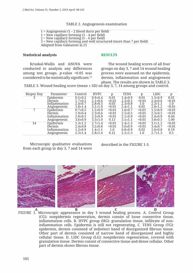
A B C DFIGURE 1. Microscopic appearance in day 3 wound healing process. A. Control Group
(CG): noepidermis regeneration, dermis consist of loose connective tissue, inflammation cells. B. HVPC group (HG): granulation tissue, infiltrate of non-inflammation cells. Epidermis is still not regenerating. C. TENS Group (TG): epidermis, dermis consisted of indistinct band of disorganized fibrous tissue. Other part of dermis consisted of narrow band of disorganized and highly cellular tissue. D. LIDC Group (LG): noepidermis regeneration, covered with granulation tissue. Dermis consist of connective tissue and dense cellular. Other part of dermis shows fibrous tissue.

102
Herdiawan RP, et al., Comparison of wound healing...
A B C DFIGURE 2. Microscopic appearance in day 7 wound healing process. A. CG: wound gap
is not covered with granulation, no epidermis regeneration. Dermis consist of dense connective tissue and cells. B. HG: epidermis, hair follicle and early differentiated dermis. C. TG : normal epidermis with hair follicle growth. Differentiated dermis with sebaceous gland and hair follicle. D. LG : epidermis in early differentiation stage and capillary in differentiated dermis.
A B C DFIGURE 3. Microscopic appearance in day 14. A. CG : differentiation stage of epidermis.
Dermis consist of loose connective tissue. B. HG: epidermis appear like a normal tissue with nicely shaped of corneum layer. Dermis with hair follicle, sebaceousand sweat glands. C. TG: epidermis and dermis with hair follicle. D. LG showed epidermis. Dermis with hair follicle and sebaceousglands.
DISCUSSION
Wound destroyed epidermis layer and induced short circuit on trans epithelial potential (TEP) and produced direct current (DC) to the wound. This current known as current of injury. Electrical stimulation would imitate current of injury to increase galvanotaxis and enhance wound healing processes.11,14
The results of wound healing from every groups in this study evaluated on day 3, 7 and 14 after incision based on the time and onset of healing processes, as described in TABLE 1. In average, wound healing on treatment group have
a better result than control group. This result proved the theory and previous study that stated electrical stimulation enhance and accelerate wound healing.
The results of wound healing on day 3 indicated that all treatment group on epidermis found to be better compared to control (FIGURE 1). This condition occurs because the epithelial process which was depended on current injury.11,15,16 Electrical stimulation to the wound would increase galvanotaxis processes whereas keratinocytes and fibroblasts would migrate to the wound area.8,15 This process could accelerate wound re-epithelialization. TENS group
103
J Med Sci, Volume 51, Number 2, 2019 April: 98-105
had the highest score for this parameter (FIGURE 1). As described earlier, TENS increased blood supply to the skin and supply adequate nutrition to the wound area.17 The advantage of using TENS are inexpensive, simple application and the equipment is portable.
The result of wound healing on the dermis found to be better on LIDC group compare to TENS and HVPC but the differences was not statistically significant (FIGURE 1). The dermis regenerated well, marked with the presence of hair follicle component and sebaceous gland. LIDC imitate injury current which could increase galvanotaxis process to the cells involved on wound healing.15 Placing the anodes on top of the wound assisted better endogenous electric stimulation.18
The result of wound healing on the inflammation found to be better on HPVC group compare to TENS and HVPC (FIGURE 1) HVPC have monophasic wave as LIDC but with shorter pulse duration anodes in HPVC will increase epithelization, since cathodes overcome infection and inflammation and boost granulation.8,19 On the first 3 days, cathodes were placed upon wound. Cathodes stimulation will stimulate galvanotaxis on neutrophil which cleansed debris, bacterial opsonization through complement function and dissolved bacteria through oxidation. Assessment on inflammation process based on density of inflammation cells, and represent strong inflammation. One of the cells is macrophage that produce MIP-1alfa that have role on inflammation.6
Angiogenesis parameter showed that HVPC has highest rate value and significantly difference with TENS and control group. Angiogenesis were trigged by hypoxia, NO, VEGF, FGF-2, chemokines (MCP-1) and MIP-1alfa.6,20
As mentioned earlier, inflammation cells like macrophage will secrete MIP-1 alfa and neutrophil will conduct in hypoxia as the result of oxidation.5 Rate value
of epidermis and dermis on day 7 were on the TENS group although post-hoc test declared the opposite (FIGURE 2). TENS is a back and forth current that the polarity would change minimal one time per second. This were the advantage of TENS that changeable polarity resulted in different stimulation to the wound and the wound could get all stimulation benefit in one treatment.12,17 This assumption was not proved on the result of day 3 and 14 of therapy.
The highest inflammation parameter was on the control group. This condition might be because of this group did not get an electrical stimulation. Post hoc test after ANOVA showed a significant difference between LIDC and TENS for angiogenesis, where the rate value LIDC group were the highest. This result was different with day 3 where the HVPC group has the highest rate. Previous study by Mehmandoust et al. did not asses angiogenesis on wound healing tissue, they only asses the velocity of wound closure and tensile strength of the wound.18 Their study declared that LIDC anodes placement on the first 3 days would increase tensile strength. Electrodes placement of that study were the same with this study. Positive charge will increase migration and proliferation of cells, increasing macrophages to the MIP-1alfa and induced angiogenesis just like mechanism in HVPC.5
Epidermis and dermis regeneration on day 14 had the highest rate value on LIDC group (FIGURE 3). This result proved the theory and previous study by Mehmandoust et al. and Talebi et al. that LIDC elevated epidermis and dermis regeneration.7,18 The advantage of using LIDC was similar with current of injury, and able to elevate galvanotaxis of epidermal cells especially if the anodes placed upon the wound like this study. Although LIDC have a burning potential, it would be prevented if used under 30 min. This theory was a background of the duration of treatment. Result for

104
Herdiawan RP, et al., Comparison of wound healing...
inflammation and angiogenesis in day 14 found to be highest on HVPC group (FIGURE 3). LIDC had the highest value on regeneration of epidermis and dermis. Result parameters of all treatment groups were better than control both statistically and generally. This proved that electrical stimulation modality could be used as additional treatment for wound healing. The treatment choice depends on clinical evaluation and the treatment goals.
CONCLUSION
There are differences on the result of wound healing of skin incision with electrical stimulation treatment of TENS, HVPC and LIDC based on epidermis and dermis regeneration, inflammation reaction and angiogenesis. There is no difference between one electrical current with other for wound healing based on overall parameters. Score and statistical analysis on LIDC are higher than other electrical current in epidermis and dermis regeneration parameter. HVPC give higher score for angiogenesis parameter. There is no specific performance for TENS in any parameters in comparison with other electrical current. This study indicates that every electrical current has advantage or disadvantage and might be useful for wound healing process.
ACKNOWLADGEMENTS
We would like to thank all those who contributed to this research. We also want to thank all those who provided support for this research so that it can be done nicely.
REFERENCES
1. Junqueira L, Carlos. Histologi dasar. Jakarta: Penerbit Buku kedokteran EGC; 1998; 357-8.
2. Barbul A. Wound healing. In:
Brunicardi F, editor. Schwartz’ manual of surgery. 8th. USA: McGraw-Hill Medical Publishing Division. 2006; 165-75.
3. Rosenberg L. Growth Factors. 2006. from: http://www.emedicine.com/ plastic/topic 457.htm.
4. Sonny K. Peran kolagen pada penyembuhan luka. Manado: Universitas Sam Ratulangi; 2004.
5. Steven R, Catherine. D, Chia S, Kang T. The phases of cutaneous wound healing. Experts Rev Mol Med 2003; 5:1-22.
6. Sharon W. Platelet activation and cytokines release: cytokines in wound healing. 2002: from: http://www.scienceboard.org/community/perspectives/cyto.figure1.2002.gif.mht.
7. Talebi G, Torkaman G, Firoozabadi M, Shariat S. Effect of anodal and cathodal microamperage direct current electrical stimulation on injury potential and wound size in guinea pigs. J Rehabil Res Dev 2008; 45(1):153-9.h t t p s : / / d o i . o r g / 1 0 . 1 6 8 2 /JRRD.2007.05.0068
8. Kloth LC. Electrical stimulation for wound healing: a review of evidence from in vitro studies, animal experiments, and clinical trials. Int J Low Extrem Wounds 2005; 4(1):23-44. https://doi.org/10.1177/1534734605275733
9. NN. Normal skin layer2007: cite from: http://www.uspharmacist.com.
10. Moenadjat Y. Buku Pegangan Kursus. Pengetahuan dasar dan keterampilan bedah minoruntuk dokter umum dan mahasiswa kedokteran. Jakarta: FK UI; 2002.
11. Sussman C & Byl N. Electrical stimulation for wound healing. In: Sussman C & Bates-Jansen B, 4th ed. Wound care: a collaborative pracice manual for physical therapist and nurses. Philadelphia: Lippincott Williams & Wilkins, 2012.
12. Hess CL, Howard MA, Attinger CE.

105
J Med Sci, Volume 51, Number 2, 2019 April: 98-105
A review of mechanical adjuncts in wound healing: hydrotherapy, ultrasound, negative pressure therapy, hyperbaric oxygen and electrostimulation. Ann Plast Surg 2003;51(2):210-8.h t t p s : / / d o i . o r g / 1 0 . 1 0 9 7 / 0 1 .SAP.0000058513.10033.6B
13. Dawson B & Trapp RD. Basic and clinical biostatistics, 4th ed. USA: McGraw Hill Companies; 2001.
14. Dalzell M. Tissue healing with electrical stimulation: Wound care and iontophoresis. Philadelphia: FA Davis Company; 1996.
15. Balakatounis KC, Angoulesc AG. Low-intensity electrical stimulation in wound healing: review of the efficacy of externally applied currents resembling the current of injury. Eplasty 2008; 8:e28.
16. Gardner SE, Frantz RA, Schmidt FL. Effect of electrical stimulation on chronic wound healing: a meta-analysis. Wound Repair Regen 1999; 7(6):495-503.https: / /doi .org/10.1046/ j .1524-
475X.1999.00495.x17. Liebano RE, Ferreira LM, Abla
LE. Effect of high frequency transcutaneous electrical nerve stimulation on viability of random skin flap in rats. Acta Cir Bras 2006; 21(3):133-8. h t t p s : / / d o i . o r g / / S 0 1 0 2 -86502006000300003
18. Mehmandoust FG, Torkaman G, Firoozabadi M, Talebi G. Anodal and cathodal pulsed electrical stimulation on skin wound healing in guinea pigs. J Rehabil Res Dev 2007; 44(16):611-8. h t t p s : / / d o i . o r g / 1 0 . 1 6 8 2 /JRRD.2007.01.0007
19. Lampe KE. Electrotherapy in tissue repair. J Hand Ther 1998; 11(2):131-9. ht tps : / /doi .org/10.1016/S0894-1130(98)80011-2
20. NN. The use of clostridial collagenase in clinical practice: mammalian collagenases. 1998. Cite from: www.medscape.com/viewarticle/406066_2.