comparison of two different microbiological test kits for detection of periodontal pathogens
TRANSCRIPT

Acta Odontologica Scandinavica, 2010; 68: 115–121
ORIGINAL ARTICLE
Comparison of two different microbiological test kits for detectionof periodontal pathogens
RALUCA COSGAREA, AMELIE BÄUMER, BERNADETTE PRETZL, SVEN ZEHACZEK &TI-SUN KIM
Department of Operative Dentistry, Section of Periodontology, University Hospital Heidelberg, Heidelberg, Germany
AbstractObjective. The aim of this study was to compare the outcome of two different microbiological tests for detection ofAggregatibacter actinomycetemcomitans (A.a.), Porphyromonas gingivalis (P.g.), Tannerella forsythia (T.f.) and Treponema denticola(T.d.). Material and methods. A total of 69 adult patients with severe chronic (sCP) or aggressive periodontitis (AgP)participated in the study. Microbiological samples were examined for A.a., P.g., T.f. and T.d. using an RNA probe test(PADO) and a real-time polymerase chain reaction test (MERI). Results. For all periodontal pathogens under investigation,the median bacterial counts detected with PADO were smaller compared to those detected with MERI. P.g., T.f. and T.d.could be found in the majority of all patients with both tests. With MERI, A.a. was detected more often (24.6%) than withPADO (18.8%). Only 10.1% of the patients tested positive for A.a. with both tests. Conclusion. Both tests showed a highpercentage of agreement for P.g., T.f. and T.d., but exhibited marked differences in the detection of A.a.
Key Words: Gene probe test, kappa statistics, microbiology, prevalence-and-bias-adjusted kappa, periopathogens
Introduction
Identification of target microorganisms in periodontalpatients with the help of commercially available geneprobe tests has become a valuable diagnostic tool tohelp plan an efficient adjunctive antibiotic therapy.The establishment and progression of periodontal
diseases are based on the presence of high levels ofperiodontal pathogens in the sulcular fluid [1]. Over400 species of microorganism can be detected inperiodontal pockets, but only a few are discussedcurrently in the aetiology and pathogenesis of peri-odontal diseases. The majority of patients diagnosedwith chronic periodontitis show a favourable treat-ment response after mechanical therapy, whichprimarily aims at the decontamination of infectedroot surfaces. The most important constituents ofanti-infective therapy are improvement of oralhygiene, scaling and root planing and periodontalsurgery. Removal of the biofilm and thus reductionof the total bacterial counts in the pockets of peri-odontal patients by means of personal and professio-nal plaque removal is a prerequisite to stop the
progression of the disease. Nevertheless, in certainpatients this treatment regime may not be sufficientto prevent further destruction of tissue and peri-odontal breakdown. Especially if patients are diag-nosed with aggressive periodontitis, mechanicalremoval of plaque and bacterial biofilm does notalways guarantee successful management of theperiodontal infection [2]. For these patients, theuse of an adjunctive antibiotic therapy may beindicated [3].In 1996, the World Workshop in Periodontics des-
cribed Aggregatibacter actinomycetemcomitans (A.a.),Porphyromonas gingivalis (P.g.) and Tannerellaforsythia (T.f.) as microorganisms that play a keyrole in the pathomechanisms of periodontal destruc-tion [4]. Since then, Prevotella intermedia, Treponemadenticola (T.d.), Fusobacterium nucleatum, Parvimonasmicra and Eikenella corrodens have been added asimportant periodontopathogens [2]. Identificationof these microorganisms in periodontal patientswith the help of commercially available gene probetests has become a clinical valuable tool in treatmentplanning and in the selection of adjunctive
Correspondence: Ti-Sun Kim, Sektion Parodontologie, Poliklinik für Zahnerhaltungskunde, Mund-Zahn-Kieferklinik, INF 400, 69120 Heidelberg, Germany.Tel: +49 6221 566022. Fax: +49 6221 567054 . E-mail: [email protected]
(Received 30 June 2009; accepted 28 November 2009)
ISSN 0001-6357 print/ISSN 1502-3850 online � 2010 Informa UK Ltd. (Informa Healthcare, Taylor & Francis AS)DOI: 10.3109/00016350903514848
Act
a O
dont
ol S
cand
Dow
nloa
ded
from
info
rmah
ealth
care
.com
by
McM
aste
r U
nive
rsity
on
11/2
1/14
For
pers
onal
use
onl
y.

antibiotics. In patients who tested positive for A.a., itcould be shown that the mechanical removal of thesubgingival biofilm alone did not ensure a reliableand predictable treatment outcome [5–7]. Further-more, it was shown that A.a. plays a crucial role inthe aetiology of severe chronic periodontitis, aggres-sive periodontitis [8–10] and periodontitis as a man-ifestation of Papillon Lefèvre syndrome [11]. Thesepatients have to be managed with an adequatetherapy protocol, including adjunctive administra-tion of systemic antibiotics, as recommended by theAmerican Academy of Periodontology (2000, 2001).The aim of this study was to compare the out-
come of two different commercially available micro-biological tests which are commonly used in aclinical setting to detect A.a, P.g., T.f. and T.d. inpatients with severe chronic (sCP) and aggressiveperiodontitis (AgP).
Material and methods
Patients
A total of 69 adult subjects participated in thisprospective study. Between 2004 and 2007, patientswere recruited before (n = 45) or after (n = 24)undergoing anti-infective therapy at the Section ofPeriodontology, Department of Operative Dentistry,University Hospital Heidelberg. To be included,patients had to fulfil the criteria for clinical diagnosisof AgP or sCP. Patients who had received anti-biotic therapy within the last 6 months or whoneeded antibiotic prophylaxis before dental treatmentwere excluded. The diagnoses AgP and sCP weredefined according to the International Workshop for aClassification of Periodontal Diseases and Conditions[12]. In the patients awaiting anti-infective therapy,microbiological testing was conducted to identifyA.a.-positive patients, which was a prerequisite foran adjunctive antibiotic treatment. All of the patientswho had received anti-infective therapy had a historyof a subgingival microbiological sample that hadtested positive for A.a. prior to their anti-infectivetreatment [IAI Pado-Test 4.5� RNA probe test kit(PADO); Institut für Angewandte Immunologie,Zuchwil, Switzerland]. In the follow-up of thesepatients, microbiological sampling was repeated toconfirm complete eradication of A.a.Informed written consent to participate in the study
was obtained from the patients.
Clinical examinations
The periodontal parameters were assessed by acalibrated examiner (Section of Periodontology,University Hospital Heidelberg). Pocket probing depth
(PPD) and vertical clinical attachment (CAL-V)were measured to the nearest millimetre using arigid periodontal probe (PCPUNC15; Hu Friedy,Chicago, IL) at six sites per tooth. Bleeding on prob-ing (BOP) was recorded as the percentage of teethwith signs of bleeding 30 s after probing. The cement–enamel junction (CEJ) was defined as a referencepoint for the assessment of CAL-V. If the CEJ hadbeen destroyed by restorative treatment, the referencepoint was represented instead by the most apicallylocated margin of the restoration. Additionally, thegingival bleeding index (GBI) [13] and plaque controlrecord [14] were assessed at four sites per tooth.
Microbiological examination
The microbiological examination was accomplishedwith two commercially available test kits. With thePADO RNA probe test kit, four periodontal patho-gens were detected: A.a., P.g., T.f. and T.d. This testuses oligonucleotide probes complementary to con-served fragments of the 16S rRNA gene that encodesthe rRNA, which forms a subunit of the bacterialribosome. The detection threshold of this test is103 for A.a. and 104 for P.g., T.f. and T.d. Thesecond test used was a commercially available real-time polymerase chain reaction (PCR) test [Meridol�
Paro Diagnostik (MERI); Gaba GmbH, Lörrach,Germany]. The detection threshold of this test is102. The real-time PCR directly records the reactionprocess of amplification. In addition to the specificprimers, the real-time PCR uses a further species-specific DNA fragment (TaqMan probe). This Taq-Man probe binds within the target sequence. Duringduplication of the target sequence, the TaqMan probeis split off from the target sequence and destroyed bythe exonuclease activity of the Taq polymerase. In thisbreakdown of the probe, a fluorescent signal isreleased which is measured online and immediatelyrecorded by means of automatic laser detection in thereactor vessel. The intensity of the fluorescent signal isthus a measure of the amount of the product formed,and is directly proportional to the initial amount of theperiodontal pathogen in the patient samples.Microbiological sampling was performed after
assessment of the periodontal chart according tothe joint statement of the German Society of Peri-odontology and the German Society of Dental, Oraland Maxillofacial Medicine following the manu-facturer’s test protocol. The four deepest pockets infour different quadrants were selected for the sam-pling procedure [6,15]. After removal of supragingivalplaque using cotton pellets, the test site was dried andkept dry with cotton rolls. Two sterile paper pointswere inserted simultaneously to the bottom of theselected pocket. After 10 s the paper points wereremoved and placed in two separate transportation
116 R. Cosgarea et al.
Act
a O
dont
ol S
cand
Dow
nloa
ded
from
info
rmah
ealth
care
.com
by
McM
aste
r U
nive
rsity
on
11/2
1/14
For
pers
onal
use
onl
y.

vials: one for the PADO test and the other for theMERI test. The paper points from the other selectedpockets were placed similarly in the same/respectivevials and the samples were pooled.
Statistical analysis
Sampled data were collected using a software pro-gram for table calculation (Microsoft Excel) and latertransferred into a scientific statistical software pro-gram (SPSS Version 12.0; SPSS Inc., Chicago, IL)for further analysis focusing on special strategies formethod comparisons. For statistical analysis, specialmethods to describe the extent of agreement betweenthe two microbiological test kits were used (kappa-coefficient). Confidence intervals of kappa valueswere calculated with the formulas provided by Fleiss[16]. According to the suggestions of Byrt et al. [17]as well as Sim and Wright [18], prevalence andbias adjustment for kappa was done by addition-ally calculating prevalence-and-bias-adjusted kappa(PABAK) values.Comparison of bacterial counts between both test
kits was performed with a Wilcoxon signed rank testfor paired observations, as distribution analysisrevealed a significant deviation from a normal distri-bution for all bacterial species under investigation.For a two-tailed test, alpha was set to 0.05.To analyse the distribution of differences between
the two test kits, Bland–Altman plots includinglimits of agreement were calculated after logarithmictransformation of the bacterial counts (lg = log 10),referring to 106 counts +1 [19].
Results
Clinical data of patients
A total of 69 patients (45 females; age range 26–71 years; mean age 46.3 ± 9.9 years) were recruitedfor the study between 2004 and 2007. Of thesepatients, 49 (28 females) had been diagnosed withgeneralized sCP and 20 (17 females) with AgP. MeanPPD and CAL-V were 3.76 ± 2.05 and 4.2 ± 2.4 mm,
respectively. Descriptive statistics for PPD andCAL-V of the test sites are listed in Table I.Mean ± SD values were 38.16% ± 20.39% for plaquecontrol record, 11.8% ± 18.7% for GBI and28.95% ± 22.31% for BOP.
Microbiological examination
Dichotomous classification and kappa statistics. Resultsof the microbiological examination were classifieddichotomously into ‘positive’ or ‘negative’ samples.Table II summarizes the prevalences of positiveresults for all the periodontal pathogens under investi-gation, depending on the test kit that had beenapplied. For all periodontal pathogens targeted bythe PADO and MERI tests, the distribution of posi-tive and negative test results was not congruent whencomparing the two kits. Thus, the number of patientswho tested positive with both kits simultaneously wasalways lower than the prevalence of positive resultswith a single kit. For T.f. and T.d., 89.9% and 88.4%of all patients, respectively tested positive with bothkits. For P.g., 69.6% of all patients were positive withthe PADO and MERI tests simultaneously. For A.a.,only 10.1% of the samples tested positive with bothtest kits. Unweighted kappa values (Table III) showedfair agreement for A.a. (k = 0.322), moderate agree-ment for T.f. (k = 0.410) and T.d. (k = 0.506) andgood agreement for P.g. (k = 0.689). Analysis ofsubgroups depending on clinical categories (‘prior toanti-infective therapy’, n = 45; ‘follow-up’, n = 24) alsorevealed a higher percentage of positive test resultsfor all microorganisms with the MERI test compa-red to the PADO test in both subgroups. Further-more, in follow-up patients, prevalences of allmicroorganisms under investigation tended tobe lower compared to those in patients prior toanti-infective therapy (Table II).
PABAK values. After adjustment for differences inprevalence and bias, PABAK values (Table III) variedfrom 0.536 to 0.855, indicating moderate agreementfor A.a. (PABAK = 0.536), good agreement for P.g.
Table I. Descriptive statistics for PPD and CAL-V of the test site teeth (MT4m). All values shown are in millimeters.
Test site 1 Test site 2 Test site 3 Test site 4
PPD CAL-V PPD CAL-V PPD CAL-V PPD CAL-V
Mean 7.4 7.9 7.2 7.9 6.6 6.9 6.7 6.9
SD 1.8 2.6 2.1 2.6 2.1 2.6 1.9 2.4
Min 4 2 4 3 2 3 2 2
Max 14 14 13 15 14 16 12 14
MT4: microbiological analysis of material pooled from 4 different test sites (Multi Site 4).
Periopathogen microbiological testing 117
Act
a O
dont
ol S
cand
Dow
nloa
ded
from
info
rmah
ealth
care
.com
by
McM
aste
r U
nive
rsity
on
11/2
1/14
For
pers
onal
use
onl
y.

(PABAK = 0.768) and very good agreement for T.f.(PABAK = 0.855) and T.d. (PABAK = 0.855).
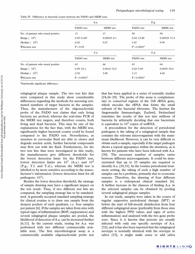
Comparison of bacterial counts. Since distributionanalysis revealed a significant deviation from normal-ity, bacterial counts measured with the PADO andMERI tests were compared with a Wilcoxon signedrank test for paired observations. The results of thisanalysis are listed in Table IV. For all periodontalpathogens under investigation, the MERI testrevealed higher median bacterial counts comparedto the PADO test; the differences proved to bestatistically significant for P.g., T.f. and T.d.
Discussion
The goal of this study was to compare the outcomeof two different commercially available micro-biological tests regarding the detection of A.a., P.g.,T.f. and T.d. in patients with sCP and AgP in order
to obtain further information about the reliability ofthese microbiological tests. Since a gold standardwas not used in this study, the comparison betweenthe two methods was only relative to each other.The inclusion of defined bacterial samples is part ofa study protocol we are currently designing to findout which of the two test systems gives resultsclosest to the correct detection.If microbiological samples from periodontal
patients are analysed, the considerable variabilityin prevalences of these microorganisms may alsodepend on the protocol of the microbiological test,which includes both the sampling and detectionmethods that were applied [20–23]. From studyof the literature it is well known that identificationof periodontal pathogens, especially A.a., can bevery challenging, due to the lack of sensitivity andreproducibility of certain microbiological tests[24,25] and the fact that A.a. often invades theinflamed tissues, which means that this microorgan-ism can be present even if it is not harvested with a
Table III. Kappa statistics and PABAK for all pathogens under investigation.
Unweighted k Standard error 95% CI
A.a. 0.322 0.149 0.031–0.613
PABAK 0.536 0.102 0.337–0.735
P.g. 0.689 0.103 0.487–0.892
PABAK 0.768 0.077 0.617–0.919
T.f. 0.410 0.254 –0.088–0.908
PABAK 0.855 0.062 0.733–0.977
T.d. 0.506 0.213 0.090–0.923
PABAK 0.855 0.062 0.733–0.977
Table II. Agreement between PADO andMERI tests in patients who tested positive for A.a., P.g., T.f. and T.d. Values are given as numbers ofpatients, with percentages in parentheses.
PADO test MERI test MERI and PADO tests
All patients (n = 69)
A.a. 13 (18.8) 17 (24.6) 7 (10.1)
P.g. 50 (72.5) 54 (78.3) 48 (69.6)
T.f. 63 (91.3) 66 (95.7) 62 (89.9)
T.d. 63 (91.3) 64 (92.8) 61 (88.4)
Patients prior to anti-infective therapy (n = 45)
A.a. 12 (26.7) 14 (31.1) 6 (13.3)
P.g. 40 (88.9) 43 (95.6) 40 (88.9)
T.f. 44 (97.8) 45 (100.0) 45 (100.0)
T.d. 43 (95.6) 44 (97.8) 42 (93.3)
Follow-up patients (n = 24)
A.a. 1 (4.2) 3 (12.5) 1 (4.2)
P.g. 10 (41.7) 11(45.8) 8 (33.3)
T.f. 19 (79.2) 21 (87.5) 18 (75.0)
T.d. 20 (83.3) 20 (83.3) 19 (79.2)
118 R. Cosgarea et al.
Act
a O
dont
ol S
cand
Dow
nloa
ded
from
info
rmah
ealth
care
.com
by
McM
aste
r U
nive
rsity
on
11/2
1/14
For
pers
onal
use
onl
y.
subgingival plaque sample. The two test kits thatwere compared in this study show considerabledifferences regarding the methods for assessing esti-mated numbers of target bacteria in the samples.Thus, the manufacturer of the oligonucleotideprobe of the PADO test claims that only livingbacteria are probed, whereas the real-time PCR ofthe MERI test targets, and therefore counts, bothliving and dead bacteria. This may be one of theexplanations for the fact that, with the MERI test,significantly higher bacterial counts could be foundcompared to the PADO test. Nevertheless, asenzymes in crevicular fluid are able to cleave anddegrade nucleic acids, further bacterial compoundsmay flow out with the fluid. Furthermore, for thetwo test kits that were investigated in this study,the manufacturers give different thresholds forthe lowest detection limit: for the PADO test,lowest detection limits are 103 (A.a.) and 104
(P.g., T.f. and T.d.), whereas the MERI test islabelled to be more sensitive according to the manu-facturer’s information (lowest detection limit for allpathogens: 102).Besides the lower detection threshold, the strategy
of sample drawing may have a significant impact onthe test result. Thus, if two different test kits arecompared, the sampling strategy has to be standard-ized. A generally accepted standard sampling strategyfor clinical routine is to draw one sample from thedeepest pockets of each quadrant, i.e. four samplesper patient [6]. If the samples are taken from sites withtypical signs of inflammation (BOP, suppuration) andseveral subgingival plaque samples are pooled, thelikelihood of detection of A.a. can be increased further[6,11]. In the current study, testing for A.a. wasperformed with two different commercially avai-lable tests. The first microbiological assay is acommercially available oligonucleotide probe test
that has been applied in a series of scientific studies[24,26–29]. The probe of this assay is complemen-tary to conserved regions of the 16S rRNA gene,which encodes the rRNA that forms the smallsubunit of the bacterial ribosome. The Institut fürAngewandte Immunologie, Zuchwil, Switzerlandtranslates the results of this test into millions ofbacteria by arbitrarily deciding that one bacteriumis equivalent to 104 copies of ssrRNA.A precondition for the detection of periodontal
pathogens is the taking of a subgingival sample thatcontains the relevant microorganism with the maxi-mum likelihood. However, it can be very difficult toobtain such a sample, especially if the target pathogenshows a topical appearance within the dentition, as isknown for patients who have been infected with A.a.[30]. The necessary number of samples variesbetween different microorganisms. It could be dem-onstrated that up to 25 samples are required toidentify A.a. [30,31]. In the routine periodontal treat-ment setting, the taking of such a high number ofsamples can be a problem, primarily due to economicreasons. Therefore, the drawing of four differentsamples is a widespread clinical standard [6].A further increase in the chances of finding A.a. inthe selected samples can be obtained by poolingseveral subgingival plaque samples.In our study, samples were taken in the course of
regular supportive periodontal therapy (SPT) orbefore the start of full-mouth disinfection from fourdifferent subgingival areas (preferably from those siteswith the highest PPD values and signs of activeinflammation) and analyzed with the two gene probetests. Since it is known that persons are usuallyinfected with only one specific serotype of A.a.[32], and it has also been reported that the subgingivalserotype is normally identical with the serotype insaliva [33], it might make sense to add saliva
Table IV. Difference in bacterial counts between the PADO and MERI tests.
A.a. P.g.
PADO test MERI test PADO test MERI test
No. of patients who tested positive 13 17 50 54
Range (�106) 0.09–0.447 0.00025–4.1 0.02–12.88 0.00025–71.0
Median (�106) 0.02 0.07 3.47 9.90
Wilcoxon test P > 0.05 P < 0.0001a
T.f. T.d.
PADO test MERI test PADO test MERI test
No. of patients who tested positive 63 66 63 64
Range (�106) 0.09–10.1 0.0014–72.0 0.02–5.69 0.0025–35.0
Median (�106) 2.30 3.80 1.11 4.60
Wilcoxon test P < 0.0001a P < 0.0001a
aStatistically significant difference.
Periopathogen microbiological testing 119
Act
a O
dont
ol S
cand
Dow
nloa
ded
from
info
rmah
ealth
care
.com
by
McM
aste
r U
nive
rsity
on
11/2
1/14
For
pers
onal
use
onl
y.

specimens or microbiological samples from the dor-sum of the tongue or the oral mucosa to the materialthat is sent to the laboratory for further analysis.Nevertheless, there will still remain a certain risk ofobtaining a false-negative result.The PADO test is based on the detection of
sequences within the bacterial 16S rRNA genethat are specific for the detected pathogen. The oli-gonucleotide probe that targets this gene ismarked radioactively and shows no cross-reactivitywith related microorganisms. With the use of theseprobes, reactions with homologous sequences ofdifferent species that are closely related to thetarget organism are possible. Probes that haveoriginally been cloned for the DNA of A.a.show cross-reactions with Haemophilus aphrophilus,Haemophilus influenzae, Haemophilus parahaemolyticusand Haemophilus parainfluenzae [34].A final conclusion concerning the clinical relevance
of the different results when comparing the PADOandMERI tests is not possible, as a reference test witha defined bacterial sample was not part of this study.Therefore, it could not be verified whether A.a. wasnot present in the samples of the patients who testednegative or if it was present but could not be detected(false-negative result). On the other hand, especiallynear the lowest detection threshold, the risk of obtain-ing false-positive results with both the PADO andMERI tests has to be discussed. Thus, the results ofthe current study also underline the conclusion of arecent literature review on microbial testing byShaddox and Walker [35], who pointed out thatthe available techniques for the detection ofperiopathogens are still limited.The two commercially available microbiological
tests showed incongruencies regarding the identifica-tion of all four periodontal pathogens, with the MERItest finding more patients with a positive diagnosisand higher bacterial counts. Unweighted kappa andPABAK analysis showed that, for A.a., the agreementbetween both microbiological test kits was muchweaker compared to P.g., T.f. and T.d.
Acknowledgements
The study was supported by the Institut für Ange-wandte Immunologie, Zuchwil, Switzerland andGABA GmbH, Lörrach, Germany, by partially pro-viding the test-kits and performing the analysis for areduced price. There are no conflicts of interestrelevant to the contents of the article for any author.
References
[1] Socransky SS, Haffajee AD. Microbial mechanisms in thepathogenesis of destructive periodontal diseases: a criticalassessment. J Periodontal Res 1991;26:195–209.
[2] Loomer PM. Microbiological diagnostic testing in thetreatment of periodontal diseases. Periodontol 20002004;34:49–56.
[3] Van Winkelhoff AJ, Rodenburg JP, Goené RJ, Abbas F,Winkel EG, de Graaff J. Metronidazole plus amoxicillin inthe treatment of Actinobacillus actinomycetemcomitans asso-ciated periodontitis. J Clin Periodontol 1989;16:128–31.
[4] Zambon JJ. American Academy of Periodontology Con-sensus report periodontal disease. Microbial factors. AnnPeriodontol 1995;67:879–925.
[5] Müller HP, Lange DE, Müller RF. Failure of adjunctiveminocycline-HCl to eliminate oral Actinobacillus actino-mycetemcomitans. J Clin Periodontol 1993;20:498–504.
[6] Mombelli A, Gmür R, Gobbi C, Lang NP. Actinobacillusactinomycetemcomitans in adult periodontitis. I. Topo-graphic distribution before and after treatment.J Periodontol 1994;65:820–6.
[7] Ehmke B, Moter A, Beikler T, Milian E, Flemmig TF.Adjunctive antimicrobial therapy of periodontitis: long-termeffects on disease progression and oral colonization.J Periodontol 2005;76:749–59.
[8] Newmann MG, Socransky SS, Savitt ED, Propas DA,Crawford A. Studies of the microbiology of periodontosis.J Clin Periodontol 1976;47:373–9.
[9] Bragd L, Dahlen G, Wikström M, Slots J. The capability ofActinobacillus actinomycetemcomitans, Bacteroides gin-givalis and Bacteroides intermedius to indicate progressiveperiodontitis: retrospective study. J Clin Periodontol1987;14:95–9.
[10] Tonetti MS, Mombelli A. Early onset periodontitis. AnnPeriodontol 1999;4:39–52.
[11] Schacher B, Baron F, Ludwig B, Valesky E, Noack B,Eickholz P. Periodontal therapy in siblings with Papillon-Lefèvre syndrome and tinea capitis: a report of two cases.J Clin Periodontol 2006;33:829–36.
[12] Armitage GC. Development of a classification system for perio-dontal diseases and conditions. Ann Periodontol 1999;4:1–6.
[13] Ainamo J, Bay I. Problems and proposals for recordinggingivitis and plaque. Int Dent J 1975;25:229–35.
[14] O’Leary TJ, Drake RB, Naylor JE. The plaque control record.J Periodontol 1972;43:38.
[15] Mombelli A, McNabb H, Lang NP. Black-pigmenting gram-negative bacteria in periodontal disease. II. Screeningstrategies for detection of P. gingivalis. J Periodontol Res1991;26:308–13.
[16] Fleiss JL. The measurement of interrater agreement. Statis-tical methods for rates and proportions. Hoboken, NJ: Wiley& Sons; 2003. p. 150–61.
[17] Byrt T, Bishop J, Carlin JB. Bias, prevalence and kappa. J ClinEpidemiol 1993;46:423–9.
[18] Sim J, Wright CC. The kappa statistic in reliability studies:use, interpretation and sample size requirements. Phys Ther2005;85:257–68.
[19] Bland JM, Altman DG. Statistical methods for assessingagreement between two methods of clinical measurement.Lancet 1986;i:307–10.
[20] Kamma JJ, Nakou M, Manti FA. Microbiota of rapidlyprogressive periodontitis lesions in association with clinicalparameters. J Periodontol 1994;65:1073–8.
[21] Kamma JJ, Nakou M, Baehni PC. Clinical and micro-biological characteristics of smokers with early onsetperiodontitis. J Periodontal Res 1999;34:25–33.
[22] Loesche WJ, Syed SA, Schmidt E, Morrison EC. Bacterialprofiles of subgingival plaques in periodontitis. J Periodontol1985;56:447–56.
[23] Moore WEC, Moore LVH. The bacteria of periodontaldiseases. Periodontol 2000 1994;5:66–77.
[24] Dannewitz B, Pohl S, Eickholz P, Kim TS. Clinicaland microbiological effects of a combined mechanic-
120 R. Cosgarea et al.
Act
a O
dont
ol S
cand
Dow
nloa
ded
from
info
rmah
ealth
care
.com
by
McM
aste
r U
nive
rsity
on
11/2
1/14
For
pers
onal
use
onl
y.
antibiotic therapy in patients with Actinobacillusactinomycetemcomitans-associated periodontitis. Am JDent 2007;20:153–6.
[25] Krigar DM, Kaltschmitt J, Krieger JK, Eickholz P. Twosubgingival plaque sampling strategies used with RNA-probes. J Periodontol 2007;78:72–8.
[26] Brochut PF, Marin I, Baehni P, Mombelli A. Predictive valueof clinical and microbiological parameters for the treatmentoutcome of scaling and root planing. J Clin Periodontol2005;32:695–701.
[27] Eguchi T, Koshy G, Umeda M, Iwanami T, Suga J,Momura Y, et al. Microbial changes in patients with acuteperiodontal abscess after treatment detected by PadoTest.Oral Dis 2008;14:180–4.
[28] Kamma JJ, Baehni PC. Five-year maintenance follow-upof early-onset periodontitis patients. J Clin Periodontol2003;30:562–72.
[29] Luterbacher S, Mayfield L, Brägger U, Lang NP. Diagnosticcharacteristics of clinical and microbiological tests for mon-itoring periodontal and peri-implant mucosal tissue condi-tions during supportive periodontal therapy (SPT). Clin OralImplants Res 2000;11:521–9.
[30] Christersson L, Fransson C, Dunford R, Zambon JJ. Sub-gingival distribution of periodontal pathogenic microorgan-isms in adult periodontitis. J Periodontol 1992;63:418–25.
[31] Christersson L, Zambon J. Suppression of subgingival Acti-nobacillus actinomycetemcomitans in localized juvenileperiodontitis by systemic tetracycline. J Clin Periodontol1993;20:395–401.
[32] Saarela M, Asikainen S, Jousimies-Somer H, Asikainen T,von Troil-Lindén B, Alaluusua S. Hybridization patterns ofActinobacillus actinomycetemcomitans serotypes a-e detectedwith an rRNA gene probe. Oral Microbiol Immunol1993;8:111–15.
[33] Lindhe J. Treatment of localized juvenile periodontitis. In:Genco R, Mergenhagen S, editors. Host parasite interactionsin periodontal disease. Washington DC: American Society forMicrobiology; 1982. p. 27.
[34] Savitt E, Keville M, Peros W. DNA probes in the diagnosis ofperiodontal microorganisms. Arch Oral Biol 1990;35(Suppl):153–9.
[35] Shaddox LM, Walker C. Microbial testing in periodontics:value, limitations and future directions. Periodontol 20002009;50:25–38.
Periopathogen microbiological testing 121
Act
a O
dont
ol S
cand
Dow
nloa
ded
from
info
rmah
ealth
care
.com
by
McM
aste
r U
nive
rsity
on
11/2
1/14
For
pers
onal
use
onl
y.