comparison of two commercial recirculated aquacultural
TRANSCRIPT
RESEARCH Open Access
Comparison of two commercial recirculatedaquacultural systems and their microbialpotential in plant disease suppressionSammar Khalil1*, Preeti Panda2, Farideh Ghadamgahi3, AnnaKarin Rosberg1 and Ramesh R Vetukuri3
Abstract
Background: Aquaponics are food production systems advocated for food security and health. Their sustainabilityfrom a nutritional and plant health perspective is, however, a significant challenge. Recirculated aquaculturesystems (RAS) form a major part of aquaponic systems, but knowledge about their microbial potential to benefitplant growth and plant health is limited. The current study tested if the diversity and function of microbialcommunities in two commercial RAS were specific to the fish species used (Tilapia or Clarias) and sampling site(fish tanks and wastewaters), and whether they confer benefits to plants and have in vitro antagonistic potentialtowards plant pathogens.
Results: Microbial diversity and composition was found to be dependent on fish species and sample site. TheTilapia RAS hosted higher bacterial diversity than the Clarias RAS; but the later hosted higher fungal diversity. BothTilapia and Clarias RAS hosted bacterial and fungal communities that promoted plant growth, inhibited plantpathogens and encouraged biodegradation. The production of extracellular enzymes, related to nutrient availabilityand pathogen control, by bacterial strains isolated from the Tilapia and Clarias systems, makes them a promisingtool in aquaponics and in their system design.
Conclusions: This study explored the microbial diversity and potential of the commercial RAS with either Tilapia orClarias as a tool to benefit the aquaponic system with respect to plant growth promotion and control of plantdiseases.
Keywords: Aquaponics, Tilapia, Clarias, Pseudomonas flourescens, Pseudomonas veronii, plant growth promotion,in vitro antagonistic
BackgroundMajor challenges such as climate changes, population in-creases, limited availability of natural resources, andpandemics threaten food security [1], raising urgentneeds to shift to robust and sustainable food productionsystems [2, 3]. As one of the largest food industries glo-bally for animal protein production, aquaculture couldplay a major role in meeting these needs. Its future
expansion will largely rely on land-based recirculatedaquaculture systems (RAS), which enable better controlof rearing conditions with significantly lower water con-sumption and release of nutrients (organic matter, nitro-gen and phosphorous) into lakes, rivers and the sea [4].However, accumulation of nitrates (which is harmful tofish) under RAS conditions is problematic [5]. This canbe ameliorated by plant uptake, so a potential solution isto integrate RAS and hydroponic systems for plant culti-vation in ‘aquaponic systems’. Thus, aquaponic systemsare promising future food production systems with ro-bust environmental profiles and potential to enhance
© The Author(s). 2021 Open Access This article is licensed under a Creative Commons Attribution 4.0 International License,which permits use, sharing, adaptation, distribution and reproduction in any medium or format, as long as you giveappropriate credit to the original author(s) and the source, provide a link to the Creative Commons licence, and indicate ifchanges were made. The images or other third party material in this article are included in the article's Creative Commonslicence, unless indicated otherwise in a credit line to the material. If material is not included in the article's Creative Commonslicence and your intended use is not permitted by statutory regulation or exceeds the permitted use, you will need to obtainpermission directly from the copyright holder. To view a copy of this licence, visit http://creativecommons.org/licenses/by/4.0/.The Creative Commons Public Domain Dedication waiver (http://creativecommons.org/publicdomain/zero/1.0/) applies to thedata made available in this article, unless otherwise stated in a credit line to the data.
* Correspondence: [email protected] of Biosystems and Technology, Swedish University ofAgricultural Sciences, Box 103, 230 53 Alnarp, SwedenFull list of author information is available at the end of the article
Khalil et al. BMC Microbiology (2021) 21:205 https://doi.org/10.1186/s12866-021-02273-4

food security. However, their sustainability needs furtherimprovement, as they are complex and more knowledge isneeded concerning ideal plant nutrient balances in rela-tion to amounts and types of fish feed, system design, andresilience towards pathogen attack and spread of diseases[6]. These aspects are strongly related to RAS conditions.For example, the lack or low availability of elements in fishfeed required for plant growth - such as phosphorous andiron - frequently limits aquaponic systems’ productive effi-ciency [7]. Hence, these nutrients are currently maintainedat required levels by adding extra phosphorus and iron tothe systems. Stabilization of the RAS element of aquapo-nic systems, in terms of water quality parameters such astemperature and pH, is also crucial to meet fish, plant andmicrobial requirements optimally [8, 9] and thereby pro-mote good plant and fish growth.Nevertheless, sustainable approaches for controlling
fish, human and plant pathogens in aquaponic systemsare also needed [6]. Plant root diseases caused by fungalpathogens such as Fusarium, Verticllium, oomycetessuch as Pythium and Phytophthora spp., or bacterialpathogens such as Ralstonia and Xanthomonas spp. arecommonly found in aquatic environments includinghydroponic systems and hence aquaponic systems [10–13]. Biotic and abiotic means to control these pathogensin hydroponic systems, including exploitation of naturalmicrobial communities’ suppressive potential, have beeninvestigated [14–19]. However, further study of the sup-pressive potential of natural microbial communities inaquaponic systems is needed. This is due to the com-plexity of the systems and associated variables related towater quality, fish feed, the fish, plants and microbialtaxa present in compartments from the biofilter to thehydroponic unit (in and through which pathogens mayenter and excessively grow if not controlled). Restric-tions governing pesticides and antibiotics to controlplant and fish diseases, respectively, highlight the needto provide solutions that enhance the sustainability ofaquaponic systems towards pathogen attack.Microbes can suppress pathogens in various ways, in-
cluding competition, production of antibiotics and extra-cellular enzymes, and induction of plant resistance orgrowth-promotion [20]. Microbial communities alsohave confirmed roles in nutrient recycling [21], plantgrowth promotion [22], and protection against pathogenattack [23] in aquaponic systems. Hence, the potentialutility of modulating the microbial habitat and commu-nity in the RAS component of aquaponic systems tocounter fish diseases has been addressed [24].However, more research into microbes’ roles and activ-
ities in RAS is needed to optimize their promotion ofplant growth and suppression of plant diseases. Thus, theobjective of the study presented here was to elucidate mi-crobial diversity in a commercial RAS (with no aquaponic
connection) and its potential to promote plant growth andact against plant pathogens. For these purposes, variationsin the microbial community between systems with twofish species, Tilapia (Oreochromis niloticus) or Clarias(Clarias gariepinus), and between two sampling sites: thewater tank with fish biosolids and wastewater have beenexamined in the current study. The communities’ func-tional roles in terms of production of extracellular en-zymes with known activities against plant pathogens andin nutrient solubilization were also examined. The study isbased on the following hypotheses. First, a RAS (with noaquaponic connection) hosts microbial communities thatare beneficial to plants and antagonistic to pathogens, withcharacteristics that depend on the fish species used andsampling site in the system. Second, the production ofextracellular enzymes and antagonistic potential to controlplant pathogens in vitro are fish species- and site-specific.
ResultsRAS conditionsConditions at the sample collection time in the twotypes of RAS, recirculated aquaculture system, with dif-ferent fish species were similar in terms of water qualityparameters such as pH, temperature, conductivity andcontents of both ammonium and nitrate (Table 1). How-ever, the total weight of Tilapia per tank was far lowerthan the corresponding weight of Clarias (ca. 50 and160 kg, respectively, at the sampling time). Both specieswere fed with the same commercial feed, supplied bySkrettting (https://www.skretting.com/en/), but with aslight difference in composition and larger differences indaily amounts. Tilapia were fed five times per day andClarias 18 times per day.
Microbial abundanceMicrobial enumeration on selective media indicatedmore general bacterial flora and Pseudomonas
Table 1 Growth conditions in each recirculated aquaculturesystem (RAS) populated with either Tilapia or Clarias
Cultivation factors RAS with Tilapia RAS with Clarias
Temperature 21 0 C 20.9 0 C
pH 7.7 8.5
Conductivity** 130 mS cm-1 180 mS cm-1
Ammonium content 2.1 mg / liter 1.8 mg / liter
Nitrate content 280 mg / liter 188 mg / liter
Nitrite content 0,59 mg / liter 0,18 mg / liter
Fish weight 40-600 g per tank 100-4000 g per tank
Feed composition 37 % protein + 10 % fat 44 % protein + 12 % fat
Daily amount 5 times 18 times
** Electrical conductivity
Khalil et al. BMC Microbiology (2021) 21:205 Page 2 of 19

fluorescens in samples collected from the Tilapia RASsystem than the Clarias RAS system (Fig. 1). However,there were differences in amounts of general fungalflora, which were more abundant in samples of Clariaswastewater than in Clarias water tank samples or eithertype of Tilapia RAS samples.
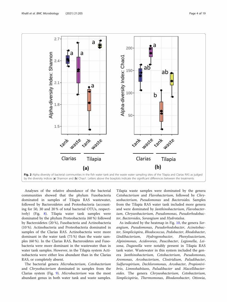
Diversity of the microbial communitiesProcessing Ilumina MiSeq sequencing data revealed thepresence of 4,558 bacteria operational taxonomic units(OTUs) and 405 fungal OTUs in samples of the twoRAS. Total read counts for the bacterial and fungal data-sets were 647, 232 and 530,351, respectively. The bacter-ial communities had significantly higher alpha diversity(according to Shannon indices) in Tilapia RAS watertank samples (p = 0.021) than in Clarias RAS water tanksamples (Fig. 2a). However, bacterial alpha diversity didnot significantly differ (p < 0.05) between Tilapia RASsamples (water or waste) and Clarias RAS wastewatersamples. Calculated Chao1 indices also indicated thatalpha diversity was significantly higher (p = 0.012) insamples of Tilapia RAS wastewater than in Clarias RASwater tank samples, which had the lowest Chao1 indices(Fig. 2b). In further accordance with the Shannon indi-ces, Chao1 indices did not significantly differ betweenTilapia RAS samples (tank or wastewater) and ClariasRAS wastewater samples.Regarding abundance of Pseudomonas, no significant
differences (p < 0.05) in log-transformed Pseudomonascounts between the four types of samples (Tilapia andClarias RAS tank water and wastewater) were found, asshown in Fig. 3. However, although we found no
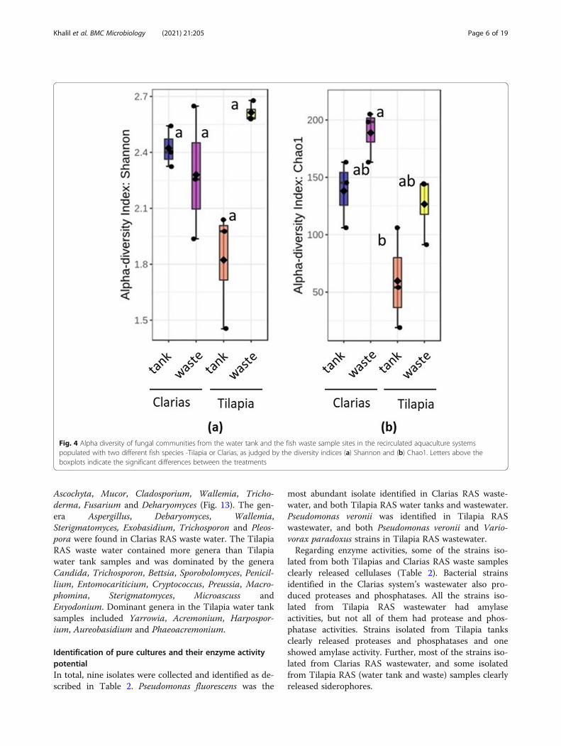
significant differences between treatments in fungalcommunity alpha diversity in terms of Shannon indices(p = 0.211) (Fig. 4a), there were highly significant differ-ences in Chao1 indices (p = 0.008) between treatments.More specifically, they clearly indicated that fungal di-versity was higher in the Clarias RAS wastewater than inthe Tilapia RAS water tank, although Chao 1 indices didnot significantly differ between any other pairs of sampletypes (Fig. 4b).Dendrogram analyses demonstrated that the bacterial
and fungal communities also differed between the Til-apia and Claria cultivation systems (Fig. 5), inter alia inthe bacterial communities in the water tank and waste-water of the Tilapia RAS (Fig. 5a). However, samples ofthese communities also shared similarities in their bac-terial communities. Fungal communities in Tilapia watertank and waste samples also differed (Fig. 5b). By con-trast, there was no clear difference between samples ofthe Clarias RAS in terms of either bacterial or fungalcommunities.The beta diversity metrics clearly distinguished the
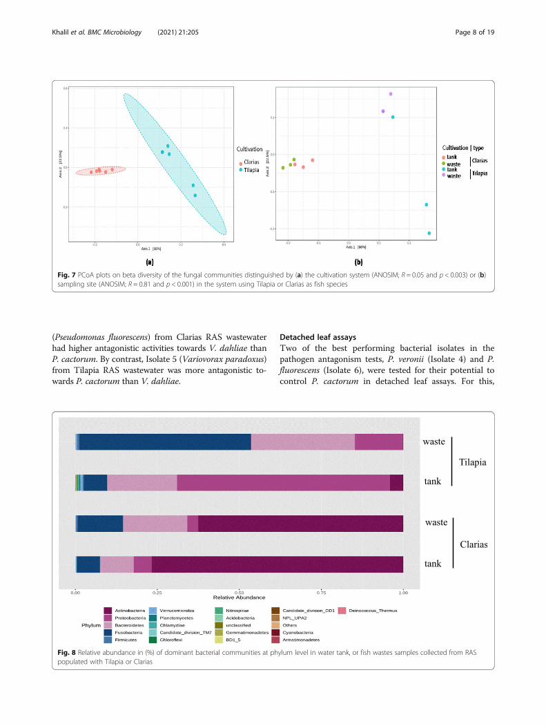
bacterial (Fig. 6) and fungal (Fig. 7) communities associ-ated with the Tilapia and Clarias RAS, and samplingsites in the two systems. The RAS with the two fish spe-cies were clearly separated along the first axis of the gen-erated Principal Coordinate Analysis (PCoA) plot(Fig. 6a) and sampling sites (more prominently for sam-ples from the Tilapia RAS than the Clarias RAS) alongthe second axis (Fig. 6b). No significant differences be-tween sampling site in this respect in the Clarias systemwere detected. Similar patterns in diversity of fungalcommunities were also detected (Fig. 7).
Fig. 1 Microbial colonies isolated from water and waste samples from Tilapia and Clarias RAS and enumerated on 0.1 % Tryptic soya agar (TSA)complemented with cycloheximide (100 µL mL− 1) for enumeration of the general bacterial flora; 0.5 % malt extract agar (MA) for enumeration ofthe general fungal flora; and on King Agar B (KB) with cycloheximide (100 µg mL− 1) for enumeration of fluorescent pseudomonads. Letters abovethe bars indicate the significant differences between the treatments
Khalil et al. BMC Microbiology (2021) 21:205 Page 3 of 19
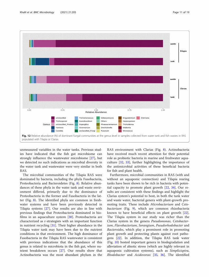
Analyses of the relative abundance of the bacterialcommunities showed that the phylum Fusobacteriadominated in samples of Tilapia RAS wastewater,followed by Bacteroidetes and Proteobacteria (account-ing for 50, 30 and 20 % of total bacterial OTUs, respect-ively) (Fig. 8). Tilapia water tank samples weredominated by the phylum Proteobacteria (60 %) followedby Bacteroidetes (20 %), Fusobacteria and Actinobacteria(10 %). Actinobacteria and Proteobacteria dominated insamples of the Clarias RAS. Actinobacteria were moredominant in the water tank (75 %) than the waste sam-ples (60 %). In the Clarias RAS, Bacteroidetes and Fuso-bacteria were more dominant in the wastewater than inwater tank samples. However, in the Tilapia system Acti-nobacteria were either less abundant than in the ClariasRAS, or completely absent.The bacterial genera Microbacterium, Cetobacterium
and Chrysobacterium dominated in samples from theClarias system (Fig. 9). Microbacterium was the mostabundant genus in both water tank and waste samples.
Tilapia waste samples were dominated by the generaCetobacterium and Flavobacterium, followed by Chry-seobacterium, Pseudomonas and Bacteroides. Samplesfrom the Tilapia RAS water tank included more generaand were dominated by Janthinobacterium, Flavobacter-ium, Chryseobacterium, Pseudomonas, Pseudorhodobac-ter, Bacteroides, Sorangium and Hydrotalea.As indicated by the heatmap in Fig. 10, the genera Sor-
angium, Pseudomonas, Pseudorhodobacter, Acinetobac-ter, Simplicispira, Rhodococcus, Pedobacter, Rhodobacter,Undibacterium, Hydrogenobacter, Phenyloacterium,Alpinimonas, Acidovorax, Paucibacter, Legionella, Lei-sona, Duganella were notably present in Tilapia RAStank water. Wastewater in this system included the gen-era Janthinobacterium, Cetobacterium, Pseudomonas,Aremonas, Arcobacterium, Clostridium, Paludibacter,Sulfurospirium, Dechloromonas, Arcobacter, Propionivi-brio, Limnohabitans, Paludibacter and Macellibacter-oides. The genera Chryseobacterium, Cetobacterium,Simplicispiria, Thermomonas, Rhodanobacter, Ottowia,
Fig. 2 Alpha diversity of bacterial communities in the fish water tank and the waste water sampling sites of the Tilapia and Clarias RAS as judgedby the diversity indices (a) Shannon and (b) Chao1. Letters above the boxplots indicate the significant differences between the treatments
Khalil et al. BMC Microbiology (2021) 21:205 Page 4 of 19

Bergeyella, Prevotella and Clostridium were present inClarias RAS wastewater. The genera Chryseobacterium,Microbacterium, Macellibacteroides and Azospirillumwere predominant in Clarias water tank samples.Analyses of the relative abundance of the fungal com-
munities also revealed differences in the dominant taxadepending on the fish species and sampling site (Fig. 11).Most (> 70 %) of the fungal OTUs were unclassified insamples from the Tilapia cultivation system (either watertank or wastewater). However, the phylum Ascomycotawas more abundant in samples from the Tilapia watertanks than in samples of Tilapia RAS wastewater, inwhich the phylum Basidiomycota was more abundant.There were fewer unclassified fungal OTUs in samples
from Clarias RAS water tanks and wastewater than inthe Tilapia RAS samples. However, their relative abun-dances differed between Clarias water tank and waste
samples. The waste samples were highly dominated bythe phyla Ascomycota, Basidiomycota and unclassifiedphyla of the kingdom Protista. Water samples from theClarias system contained Basidiomycota and Ascomy-cota, but were dominated by an unclassified member ofthe kingdom Protista (Fig. 11).Unclassified fungi also dominated at the genus level
(70 %) in Tilapia and Clarias RAS (Fig. 12). However, Tri-chosporon spp. were abundant in Clarias RAS tank andwastewater (accounting for 25 % of the total fungal OTUs),and Tilapia RAS wastewater. The genus Yarrowia was ex-clusively found in a Tilapia water tank, while the genus Tri-chomonascus was only detected in the Clarias system.The fungal communities’ heatmap showed that mem-
bers of the phylum Basidiomycota in Clarias water tanksamples included the genera Trichosporon, Trichoma-noascus, Phodotonula, Malassezia, Mortierella,
Fig. 3 Log-transformed counts of Pseudomonas communities in the water tank and wastewater site from the Tilapia and Clarias RAS. Lettersabove the boxplots indicate the significant differences between the treatments
Khalil et al. BMC Microbiology (2021) 21:205 Page 5 of 19
Ascochyta, Mucor, Cladosporium, Wallemia, Tricho-derma, Fusarium and Deharyomyces (Fig. 13). The gen-era Aspergillus, Debaryomyces, Wallemia,Sterigmatomyces, Exobasidium, Trichosporon and Pleos-pora were found in Clarias RAS waste water. The TilapiaRAS waste water contained more genera than Tilapiawater tank samples and was dominated by the generaCandida, Trichosporon, Bettsia, Sporobolomyces, Penicil-lium, Entomocariticium, Cryptococcus, Preussia, Macro-phomina, Sterigmatomyces, Microascuss andEnyodonium. Dominant genera in the Tilapia water tanksamples included Yarrowia, Acremonium, Harpospor-ium, Aureobasidium and Phaeoacremonium.
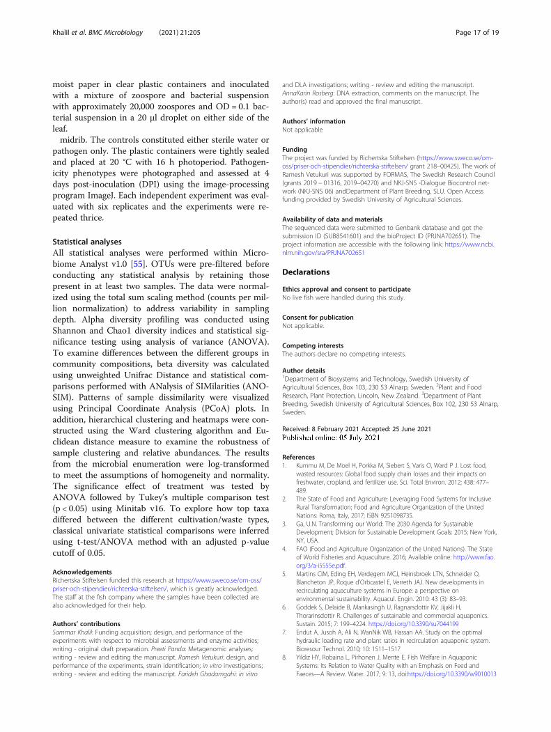
Identification of pure cultures and their enzyme activitypotentialIn total, nine isolates were collected and identified as de-scribed in Table 2. Pseudomonas fluorescens was the
most abundant isolate identified in Clarias RAS waste-water, and both Tilapia RAS water tanks and wastewater.Pseudomonas veronii was identified in Tilapia RASwastewater, and both Pseudomonas veronii and Vario-vorax paradoxus strains in Tilapia RAS wastewater.Regarding enzyme activities, some of the strains iso-
lated from both Tilapias and Clarias RAS waste samplesclearly released cellulases (Table 2). Bacterial strainsidentified in the Clarias system’s wastewater also pro-duced proteases and phosphatases. All the strains iso-lated from Tilapia RAS wastewater had amylaseactivities, but not all of them had protease and phos-phatase activities. Strains isolated from Tilapia tanksclearly released proteases and phosphatases and oneshowed amylase activity. Further, most of the strains iso-lated from Clarias RAS wastewater, and some isolatedfrom Tilapia RAS (water tank and waste) samples clearlyreleased siderophores.
Fig. 4 Alpha diversity of fungal communities from the water tank and the fish waste sample sites in the recirculated aquaculture systemspopulated with two different fish species -Tilapia or Clarias, as judged by the diversity indices (a) Shannon and (b) Chao1. Letters above theboxplots indicate the significant differences between the treatments
Khalil et al. BMC Microbiology (2021) 21:205 Page 6 of 19
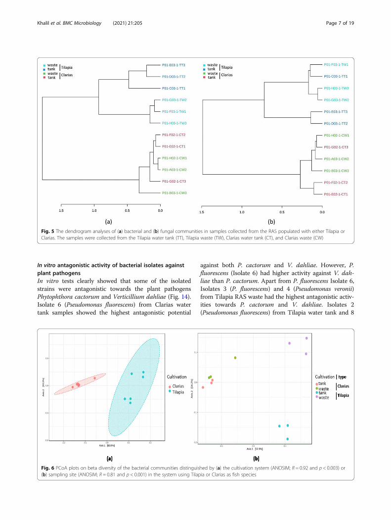
In vitro antagonistic activity of bacterial isolates againstplant pathogensIn vitro tests clearly showed that some of the isolatedstrains were antagonistic towards the plant pathogensPhytophthora cactorum and Verticillium dahliae (Fig. 14).Isolate 6 (Pseudomonas fluorescens) from Clarias watertank samples showed the highest antagonistic potential
against both P. cactorum and V. dahliae. However, P.fluorescens (Isolate 6) had higher activity against V. dah-liae than P. cactorum. Apart from P. fluorescens Isolate 6,Isolates 3 (P. fluorescens) and 4 (Pseudomonas veronii)from Tilapia RAS waste had the highest antagonistic activ-ities towards P. cactorum and V. dahliae. Isolates 2(Pseudomonas fluorescens) from Tilapia water tank and 8
Fig. 5 The dendrogram analyses of (a) bacterial and (b) fungal communities in samples collected from the RAS populated with either Tilapia orClarias. The samples were collected from the Tilapia water tank (TT), Tilapia waste (TW), Clarias water tank (CT), and Clarias waste (CW)
Fig. 6 PCoA plots on beta diversity of the bacterial communities distinguished by (a) the cultivation system (ANOSIM; R = 0.92 and p < 0.003) or(b) sampling site (ANOSIM; R = 0.81 and p < 0.001) in the system using Tilapia or Clarias as fish species
Khalil et al. BMC Microbiology (2021) 21:205 Page 7 of 19
(Pseudomonas fluorescens) from Clarias RAS wastewaterhad higher antagonistic activities towards V. dahliae thanP. cactorum. By contrast, Isolate 5 (Variovorax paradoxus)from Tilapia RAS wastewater was more antagonistic to-wards P. cactorum than V. dahliae.
Detached leaf assaysTwo of the best performing bacterial isolates in thepathogen antagonism tests, P. veronii (Isolate 4) and P.fluorescens (Isolate 6), were tested for their potential tocontrol P. cactorum in detached leaf assays. For this,
Fig. 7 PCoA plots on beta diversity of the fungal communities distinguished by (a) the cultivation system (ANOSIM; R = 0.05 and p < 0.003) or (b)sampling site (ANOSIM; R = 0.81 and p < 0.001) in the system using Tilapia or Clarias as fish species
Fig. 8 Relative abundance in (%) of dominant bacterial communities at phylum level in water tank, or fish wastes samples collected from RASpopulated with Tilapia or Clarias
Khalil et al. BMC Microbiology (2021) 21:205 Page 8 of 19

Fig. 9 Relative abundance (%) of dominant bacterial communities at the genus level in water tank, or fish waste samples collected from RASpopulated with either Tilapia or Clarias as fish species
Fig. 10 The heatmap shows the abundance of dominant bacterial communities at the genus level in water tank and fish waste samplescollected from the Tilapia water tank (TT), Tilapia waste (TW), Clarias water tank (CT), and Clarias waste (CW)
Khalil et al. BMC Microbiology (2021) 21:205 Page 9 of 19

strawberry (Fragaria × ananassa cv. Sonata) leaves wereinoculated with P. cactorum, and started showing symp-toms of infection four days later, while controls inocu-lated with sterile distilled water remained symptomless.These results confirmed the viability of the zoosporesuspensions and absence of the pathogen on uninocu-lated leaflets. Furthermore, treatment with Isolate 6 (P.fluorescens) together with P. cactorum showed potentdisease inhibition. However, Isolate 4 (P. veronii) wasless effective for controlling the progression of diseasecaused by P. cactorum (Fig. 15).
DiscussionOur study provides novel insights into the diversity ofmicrobial communities in commercial RAS, their benefi-cial effects on plants, and antagonistic potential againstplant pathogens. We found that microbial diversity var-ied depending on the fish species populating the systemand sampling point.The effect of fish species on microbial composition is
supported by the beta diversity analyses, which revealedsignificant differences in the bacterial (Fig. 6) and fungal(Fig. 7) communities of the Tilapia and Clarias RAS.The fish species in the system had significant effects in
the water tank environment, in which the bacterial com-munity was richer in the Tilapia RAS (Fig. 2), but the
fungal community was richer in the Clarias RAS (Fig. 4).These findings are consistent with previous indicationsthat the composition of microbial communities isstrongly influenced by associated species of fish [25, 26].The results also agree with previous observations thatfungal diversity was higher in a Clarias system than in aTilapia system [26].In both Tilapia and Clarias systems, sampling site had
no significant effect on the evenness and richness of ei-ther bacterial communities (Fig. 2) or fungal communi-ties (Fig. 4). By contrast, the site effect was evident inthe composition of the bacterial (Fig. 6) and the fungal(Fig. 7) communities in the Tilapia RAS. Moreover, thedendrogram analyses (Fig. 5) showed there were cleardistinctions between the bacterial and fungal communi-ties associated with the two fish species. Differences as-sociated with sampling sites in the Tilapia RAS werealso detected. Microbial diversity in RAS is also a func-tion of the water’s physicochemical properties (such aspH and temperature), the feed, gut microbiome of thefish and nutrient contents [27–29]. In this study the cul-tivation conditions for both fish species were very simi-lar. However, there were substantial differences in theirpopulation density and fish biomass per tank was muchlower in the Tilapia RAS than in the Clarias RAS(Table 1). This may have considerably influenced
Fig. 11 Relative abundance (%) of dominant fungal communities at phylum level in samples collected from water tank and fish waste sites inRAS populated with Tilapia or Clarias
Khalil et al. BMC Microbiology (2021) 21:205 Page 10 of 19
unmeasured variables in the water tanks. Previous stud-ies have indicated that the fish gut microbiome canstrongly influence the wastewater microbiome [27], butwe detected no such indications as microbial diversity inthe water tank and wastewater were very similar in bothRAS.The microbial communities of the Tilapia RAS were
dominated by bacteria, including the phyla Fusobacteria,Proteobacteria and Bacteroidetes (Fig. 8). Relative abun-dances of these phyla in the water tank and waste envir-onment differed, primarily due to the dominance ofProteobacteria in the former and Fusobacteria in the lat-ter (Fig. 8). The identified phyla are common in fresh-water systems and have been previously detected inTilapia systems [27]. Our results are also in line withprevious findings that Proteobacteria dominated in bio-films in an aquaculture system [30]. Proteobacteria arecharacterized as r-strategists with an important functionin nutrient recycling [31]. Their higher abundance in theTilapia water tank may have been due to the nutrientconditions in that environment. The high dominance ofFusobacteria in the Tilapia RAS wastewater is consistentwith previous indications that the abundance of thisgenus is related to microbiota in the fish gut, where nu-trient breakdown occurs [27]. Our results show thatActinobacteria was the most abundant phylum in the
RAS environment with Clarias (Fig. 8). Actinobacteriahave received much recent attention for their potentialrole as probiotic bacteria in marine and freshwater aqua-culture [32, 33], further highlighting the importance ofthe antimicrobial activities of these beneficial bacteriafor fish and plant health.Furthermore, microbial communities in RAS (with and
without an aquaponic connection) and Tilapia rearingtanks have been shown to be rich in bacteria with poten-tial capacity to promote plant growth [22, 34]. Our re-sults are consistent with these findings and highlight theClarias system’s potential to host, in both the tank waterand waste water, bacterial genera with plant-growth pro-moting traits. These include Microbacterium and Ceto-bacterium (Fig. 9), which are common rhizobacteriaknown to have beneficial effects on plant growth [22].The Tilapia system in our study was richer than theClarias system in the genera Pseudomonas, Cetobacter-ium, Flavobacterium, Sorangium, Pseudorhodobacter andBacteroides, which play a prominent role in promotingplant growth and protecting plants against root patho-gens [22]. In addition, the Tilapia RAS tank water(Fig. 10) hosted important genera in biodegradation andalleviation of abiotic stress (which are highly relevant inaquaponic cultivation systems), such as Simplicispira,Rhodobacter and Acidovorax [35, 36]. The identified
Fig. 12 Relative abundance (%) of dominant fungal communities at the genus level in samples collected from water tank and fish wastes in RASpopulated with Tilapia or Clarias
Khalil et al. BMC Microbiology (2021) 21:205 Page 11 of 19
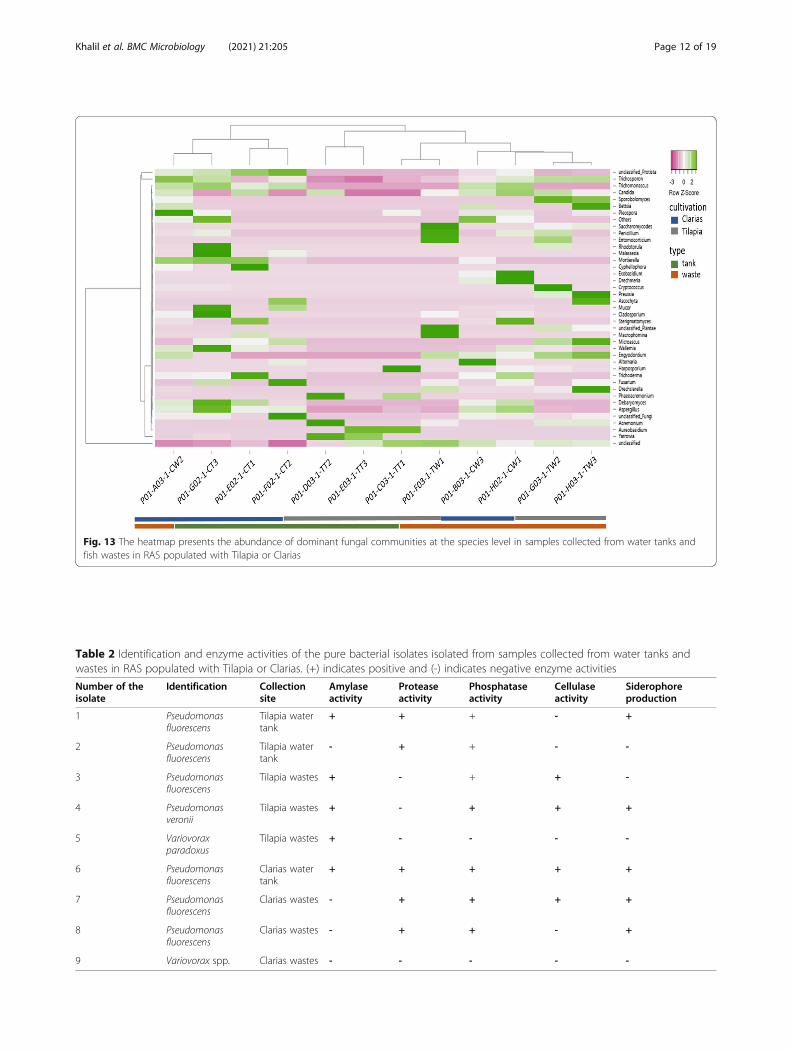
Fig. 13 The heatmap presents the abundance of dominant fungal communities at the species level in samples collected from water tanks andfish wastes in RAS populated with Tilapia or Clarias
Table 2 Identification and enzyme activities of the pure bacterial isolates isolated from samples collected from water tanks andwastes in RAS populated with Tilapia or Clarias. (+) indicates positive and (-) indicates negative enzyme activities
Number of theisolate
Identification Collectionsite
Amylaseactivity
Proteaseactivity
Phosphataseactivity
Cellulaseactivity
Siderophoreproduction
1 Pseudomonasfluorescens
Tilapia watertank
+ + + - +
2 Pseudomonasfluorescens
Tilapia watertank
- + + - -
3 Pseudomonasfluorescens
Tilapia wastes + - + + -
4 Pseudomonasveronii
Tilapia wastes + - + + +
5 Variovoraxparadoxus
Tilapia wastes + - - - -
6 Pseudomonasfluorescens
Clarias watertank
+ + + + +
7 Pseudomonasfluorescens
Clarias wastes - + + + +
8 Pseudomonasfluorescens
Clarias wastes - + + - +
9 Variovorax spp. Clarias wastes - - - - -
Khalil et al. BMC Microbiology (2021) 21:205 Page 12 of 19

genera in Tilapia RAS wastewater (Fig. 10) indicate ashift in bacterial composition between the water tankand waste sampling sites. Genera such as Clostridium,Chryseobacterium, Janthinobacterium, Cetobacteriumand Pseudomonas identified in the waste environmentalso have plant growth-promoting characters [22].
Moreover, the genera Propionivibrio, Sulfurospirium andDechloromonas are involved in nutrient breakdown, andthe removal of certain compounds, e.g., phosphorous byPropionivibrio [37]. Samples from the waste site also in-cluded genera involved in biodegradation and alleviationof abiotic stress such as Limnohabitans and Paludibacter
Fig. 14 Inhibition (%) of radial growth of the plant pathogens Phytophthora cactorum and Verticillium dahliae by the bacterial isolates 1–2 fromTilapia water tanks, isolates 3–5 from Tilapia wastes, isolates 6 from Clarias water tanks and isolates 7–9 from Clarias wastes
Fig. 15 Infection of detached leaflets of strawberry cv. Sonata by Phytophthora cactorum isolate RV4 with and without potential antagonisticbacterial isolates. Treatments from top to bottom rows: A: water only; B: P. cactorum; C: P. cactorum combined with bacterial isolate Pseudomonasfluorescens (number 6 from Clarias wastes); D: P. cactorum combined with bacterial isolate Pseudomonas veronii (number 4 isolated from Tilapiawastes). Wild type RV4 formed large disease lesions on treatment B. Treatment C, with isolate number 6, and treatment D with isolate number 4exhibited no or minimal disease. Leaves were photographed four days after inoculation
Khalil et al. BMC Microbiology (2021) 21:205 Page 13 of 19

[38]. However, the occurrence of Legionella and Aremo-nas in Tilapia RAS water tank and wastewater samplesrequires further investigation as these genera are oppor-tunistic pathogens in aquatic environments and of con-cern in terms of food safety and human health [39]. Ona more positive note, Pseudomonas fluorescens has con-firmed efficiency for controlling the pathogen Aremonas[24], and Pseudomonas spp. were detected in both Til-apia and Clarias RAS (Fig. 3). Pseudomonas fluorescensis a rhizobacterium of the genus Pseudomonas, which weidentified in samples from both our RAS (Table 2). Opti-mizing conditions to favor beneficial microbes in a RASand the root environment in aquaponic systems mightthus be an effective strategy to control fish diseasescaused by pathogens in aquaponic systems.Regarding the fungal communities in our sampled
RAS, we found that they included genera with knownimportance as promoters of plant growth and antago-nists towards plant pathogens. These genera included:Cladosporium, Mortierella [40] and Trichoderma [41] inClarias RAS tank water: Pleospora [42] in Clarias RASwaste water; Acremonium [43] in Tilapia RAS tank waterand Penicillium [40] in Tilapia wastewater samples(Figs. 12 and 13). However, these systems also containedpotentially pathogenic genera for humans and plants,such as Fusarium [43], Candida and Cryptococcus,which pose significant challenges that need further in-vestigation. Further investigation of fungal sequencesand appropriate primers is also needed to enable identi-fication of fungi that could not be classified beyond thelevel of phylum or genus in this study.The bacterial strains isolated from the Tilapia system
showed clear potential to inhibit development of symp-toms of disease caused by two well-known plant patho-gens and hence as biocontrol agents in aquaponicsystems. The only isolated bacterial strain that showedsuch potential and was positively identified and presentin all samples was Pseudomonas fluorescens. However,log transformation of the Pseudomonas data we obtainedindicates that the other strains were also present in allthe investigated samples (Fig. 3), thus strengthening ourfindings concerning the isolated strains. Other less ubi-quitous taxa were Pseudomonas veronii and Vaiovoraxparadoxus in Tilapia RAS wastewater, and Variovorasspp. in Clarias RAS wastewater (Table 2). Pseudomonasspp. generally are important microorganisms that canpromote plant growth as well as producing antimicrobialsubstances that control plant pathogens [18, 20]. Pseudo-monas veronii specifically has high bioremediation po-tential and is found in both soil and water environments[44]. Vaiovorax paradoxus and other Variovoras speciesalso have known ability to promote plant growth [45].Enzyme production is one of the modes of action
through which microorganisms combat pathogen attack
[20]. In aquaculture, probiotic microbial taxa such asActinobacteria, Proteobacteria and Bacteroidetes releasevarious enzymes, such as proteases, chitinases, gluca-nases, amylases, cellulases, and phosphatases that breakdown corresponding nutritional components in theirsubstrates [46]. Results of this study indicate that mostof the strains isolated from the Tilapia system produceproteases, phosphatases, cellulases and siderophores(Table 2). Pseudomonas veronii isolated from TilapiaRAS wastewater (Isolate 4) exhibited ability to produceall of these enzymes. Pseudomonas fluorescens isolatesfrom Clarias RAS tank water (Isolate 6) and Tilapia RASwastewater (Isolate 3) could also produce most of theenzymes. Pseudomonas spp. isolated from both RAS sys-tems are well-known for producing siderophores in plantroots that improve the availability of iron by aiding itsuptake by plant roots [47]. Thus, the isolates in ourstudy may be good candidates for use in aquaponic sys-tems to increase the availability of phosphorous and ironfor plant growth.In vitro investigations of the antagonistic abilities of
the isolated strains highlighted differences in their in-hibitory effects on the growth of pathogens. Pseudo-monas fluorescens (Isolate 6), Pseudomonas fluorescens(Isolate 3) and Pseudomonas veronii (Isolate 4) sup-pressed growth of the root pathogen Phytophthora cac-torum more than the other strains (Fig. 14). Thisdifference may be due to differences in cellulase produc-tion by these strains, which not only breaks down acomponent in the feed, but can also break down cellwalls of pathogens such as P. cactorum. However, in vivoantagonistic effects are needed in aquaponics for theseresults to have practical applicability. Our detached leafassay provides strong evidence of the antagonistic poten-tial of P. veronii (Isolate 4) and P. fluorescens (Isolate 6)against P. cactorum (Fig. 15).
ConclusionsThe current study has contributed new knowledge con-cerning the role of microbial tools in commerciallybased recirculated aquaculture systems (RAS) as pro-moters of plant growth and suppressors of disease. Thisknowledge strengthens the potential application of RASas a part of aquaponic systems, which currently facechallenges regarding plant nutrients and pathogens. Theassemblages of microbial taxa at the level of phyla andgenera both in Tilapia and Clarias RAS suggest the suit-ability of these systems to be used in aquaponic cultiva-tion. However, in terms of promoting plant growth,plant protection and biodegradation, the characteristicsof the richness and composition of the microbial com-munities in the Tilapia system make it the better choicefor application in aquaponic systems. The commercialClarias RAS could also be applied in aquaponic systems,
Khalil et al. BMC Microbiology (2021) 21:205 Page 14 of 19

but principally from a probiotics perspective due to thedominance of Actinobacteria in this system. Pseudo-monas spp. from both Tilapia tank water and waste sam-ples, and Clarias wastes, are good candidates with thepotential to produce extracellular enzymes that enhancenutrient uptake. Although the results suggest consider-able potential for using microbial communities to man-age and control certain aspects of aquaponic systems,our findings need to be strengthened with in vivo studiesto explore further the inhibition of plant pathogens andpositive effects on plant nutrition. Risks arising from thepresence of pathogens also need further investigation.However, our results can still be used as a foundationfor the design of aquaponic systems populated with ei-ther Tilapia or Clarias. Still, these results need furtherinvestigations considering the microbial patterns in theRAS of Tilapia and Clarias in relation to the abiotic andbiotic factors in the system.
Materials & MethodsSample collectionTwo commercial warm and fresh water RAS populatedwith Tilapia (Oreochromis niloticus) or Clarias (Clariasgariepinus) as the fish species and no aquaponic connec-tion were used as the experimental units. The RAS ofTilapia consisted of a water tank of 70 m3 and of 150 m3
of the Clarias system. A filtration unite is connected thesystem including a big biofilter and a Degas column forthe removal of nitrogen and carbon dioxide and additionof oxygen to the system. Three independent replicates(each 5 L water samples) were collected at two differentsites, viz. from the fish water tanks and from the waste-water where the fish faeces were accumulated. Both RASwith each respective fish type were similarly sampled.The collection site was considered as a treatment. Thus,in total, three fish water samples (biological replicates)and three wastewater samples (biological replicates) wererandomly collected from the Tilapia and Clarias systemsrespectively. The samples were then transferred to thelaboratory for subsequent microbial analyses.
Viable count and microbial enumerationThe viable count method was used to quantify themicrobiota in each treatment. Dilution series and enu-meration on selective agar media were applied [48].From this dilution stock, 200 µL aliquots were spread, intriplicate, on the following media: (i) 0.1 % Tryptic soyaagar (TSA, DIFCO 0369-17-6) complemented with cy-cloheximide (100 µL mL-1) to enumerate the generalbacterial flora; (ii) 0.5 % malt extract agar (MA, DIFCO0186-17-7) to enumerate the general fungal flora; and(iii) King Agar B (KB) with cycloheximide (100 µg mL-1)to enumerate the fluorescent pseudomonads. The MAplates were incubated at room temperature for seven
days and the TSA and KB plates were incubated for 24 hat 25 °C.
Microbial community analysesSamples preparationThe total microbiome analyses started by sterile filtra-tion of 1 L of the collected samples through 0.2 μm fil-ters using bottle-top vacuum filtration systems, PES(WVR- Sweden, 514 − 0332). The filtration unit filterwas then transferred to a 50 mL tube, washed with 50mL sterile autoclaved water, followed by vigorous vor-texing for 2 min. The collected material was then centri-fuged at 5000 rpm for 10 min and the pellets werestored at -80 °C.
DNA extractionThe DNA extraction was performed using Enzymo DNApreparation kit (D 4300, Sigma Aldrich) following themanufacturer’s recommendations.
Illumina sequencingThe bacterial and fungal communities were sequencedwith an Illumina MiSeq (2 × 300 bp) at LGC GenomicsGmbH (Berlin, Germany) [49] using Illumina bcl2fastq2.17.1.14 software. The bacterial 16 S ribosomal genewas targeted using the forward primer 341 F (5′-CCTACGGGNGGCWGCAG-3′) and the reverse primer 785R(5′-GACTACHVGGGTATCTAATCC-3′). The fungalforward primer ITS7F (5′-GTG ART CAT CGA ATCTTTG GTT G-3′) and the reverse primer (5′-TCC TCCGCT TAT TGA TAT GC-3′) were used to target theITS2 region for fungal assessment. Data pre-processingand OTU picking from amplicons were performed usingMOTHUR pipelines (version 1.35.1). Reads with a finallength of < 100 bases were discarded and primer, bar-code sequences as well as chimeras were removed. Fortaxonomical classification, alignment against 16 SMothur-Silva SEED r119 reference was performed andsequences from other domains of life were removed. As-signment of operational taxonomic unites, OUTs, wasperformed at the 97 % identity level using the clustersplit method. The fast Tree method was used to gener-ate the phylogenetic trees for 16 S and ITS, respectively.
Microbial activitiesBacterial pure culturesFor each treatment, two single colonies from the TSAplates and two from the KB plates were selected andtransferred to be grown on broth media of tryptic soyabroth (TSB) and King B broth (KBB), respectively. Oneloopful of culture was inoculated into 15 mL of thebroth media and incubated at 25°C with shaking(140 rpm) for 24 h. Bacterial DNA was extracted usingthe Quick-DNA Bacterial Microprep Kit according to
Khalil et al. BMC Microbiology (2021) 21:205 Page 15 of 19

the manufacturer’s recommendations (Zymo Research,USA). The DNA yield and integrity was assessed using aNanoDrop Micro Photometer (NanoDrop Technologies,UK), and agarose gel electrophoresis, respectively. The16s rRNA region of all bacterial isolates was PCR ampli-fied individually with the universal primer pairs, 27F (5’-AGAGTTTGATCMTGGCTCAG-3’) and 907R (5’-CCGTCAATTCMTTTRAGTTT-3’) [50]. PCRs wereperformed using ten ng of DNA with the followingtemperature parameters: initial denaturation step at94 °C for 3 min, followed by 35 cycles at 94 °C for 45 s,50 °C for 30 s, and 72 °C for 30 s, followed by a final ex-tension step of 72 °C for 5 min. The PCR products werepurified using the Qiagen QIAquick PCR PurificationKit (Qiagen, UK). Sanger sequencing for species identifi-cation was carried out at the GATC biotech AG sequen-cing facility (Germany) using 27 F and 907R primers.DNA star software was used (DNASTAR, USA) toanalyze and edit nucleotide sequences obtained from thesequencing platform manually. Resulting sequences with16 s region were searched for matching hits against theNational Center for Biotechnology Information (NCBI)GenBank non-redundant nucleotide database (BLASTn;[51]). Search hits to sequences from records in the data-base were evaluated for coverage and identity and thebest matched NCBI accession was recorded.
Enzyme activitiesFunctional characters of the isolates were assessed byassaying their enzymatic activities. The isolated bacterialcolonies were screened for amylase, protease, phosphat-ase, siderophores and cellulase production on functionalmedia in plate assays [52]. The M9 Minimal Saltsmedium (VWR, Sweden) was used as a base medium.For amylase assays, the isolates were inoculated on M9media amended with 1 % (w/v) starch and incubated for24–72 h at 20 ± 2 °C. After incubation, the plates wereflooded with Lugol iodine, and a zone of clearancearound colonies against the resulting dark backgroundwas taken as an indication of amylase production.For assessing protease activity, the isolates were inocu-
lated on M9 plates amended with skimmed milk (20 mLL− 1), incubated for 24–72 h at 20 ± 2 °C, and formationof a halo around the colonies indicated protease produc-tion. The same incubation conditions and indicative cri-teria were used for assessing cellulase production. Forthis, the M9 plates amended with (1 %) carboxymethylcellulose were used, which were flooded with Congo Redsolution (0.2 % w/v) for 30 min then washed with 1 MNaCl solution.For phosphatase activity assays, tryptose phosphate
agar plates supplemented with Methyl Green (0.05 mgmL− 1) were used. The plates were incubated for 5 daysat 20 ± 2 0 C. Development of green coloration, after
incubation, indicated positive phosphatase activity. Side-rophores production was also assessed, using Chroma-zurol S agar plates inoculated with bacterial isolates andincubated for three days at 20 ± 2 0 C. The formation ofan orange halo around the colonies indicated sidero-phores production.
In vitro antagonistic assayThe bacterial strains isolated were screened for antagon-istic potential towards the oomycete/fungal pathogensPhytophthora cactorum and Verticillium dahlia. Theassay was performed on cornmeal agar (CMA) plate agarplates with the mycelial plug (1 × 1 cm) of the testpathogen placed in the centre followed by streaking testbacterial isolate three cm apart on either side of thepathogen plug and monitored for ten days at 20 °C.Controls constitute only the test pathogens. The experi-ments were repeated thrice with three replicates for eachindependent experiments. The radial growth of thepathogen growth towards test bacteria was measuredand the Growth Inhibition Percentage (GIP) was calcu-lated according to the following formula:
%ð Þ ¼ RC−RTð Þ=RC x 100:
where RC constitutes radial growth of the pathogen inthe control plate (cm) and RT is the radial growth of thepathogen (cm) in the treated plate [53].
Detached leaf assay (DLA)The antagonistic potential of bacterial isolates towardsthe strawberry pathogen P. cactorum was tested in a de-tached leaf assay. The best performing bacterial isolateswith the highest antagonistic activity from the in vitrostudies were selected for the DLA assay. The bacteriawere cultured overnight on Tryptic Soy Broth media at28 °C and pelleted. The pellet was washed and resus-pended in distilled water, followed by density measure-ment and adjustment to OD 0.1 at 600 nm.For zoospore production, P. cactorum isolate RV4 was
cultured as described [54]. For sporangia formation, theagar plugs from the outer edges of freshly growing my-celium colonies were subjected to dark conditions on V8media for three days followed by treatment with auto-claved soil extract solution under light conditions for48 h. The sporangial suspensions obtained were sub-jected to cold treatment at 8 °C to release the zoosporesover an average 2–3 h period depending on the zoo-spore’s release efficiency. The zoospore inoculum wascollected and counted using a haemocytometer to adjustinoculum to 20 000 zoospores per mL.Young strawberry leaflets (cv. Sonata) grown in con-
trolled conditions were used to assess disease develop-ment. The leaves were placed adaxial side facing up on
Khalil et al. BMC Microbiology (2021) 21:205 Page 16 of 19
moist paper in clear plastic containers and inoculatedwith a mixture of zoospore and bacterial suspensionwith approximately 20,000 zoospores and OD = 0.1 bac-terial suspension in a 20 µl droplet on either side of theleaf.midrib. The controls constituted either sterile water or
pathogen only. The plastic containers were tightly sealedand placed at 20 °C with 16 h photoperiod. Pathogen-icity phenotypes were photographed and assessed at 4days post-inoculation (DPI) using the image-processingprogram ImageJ. Each independent experiment was eval-uated with six replicates and the experiments were re-peated thrice.
Statistical analysesAll statistical analyses were performed within Micro-biome Analyst v1.0 [55]. OTUs were pre-filtered beforeconducting any statistical analysis by retaining thosepresent in at least two samples. The data were normal-ized using the total sum scaling method (counts per mil-lion normalization) to address variability in samplingdepth. Alpha diversity profiling was conducted usingShannon and Chao1 diversity indices and statistical sig-nificance testing using analysis of variance (ANOVA).To examine differences between the different groups incommunity compositions, beta diversity was calculatedusing unweighted Unifrac Distance and statistical com-parisons performed with ANalysis of SIMilarities (ANO-SIM). Patterns of sample dissimilarity were visualizedusing Principal Coordinate Analysis (PCoA) plots. Inaddition, hierarchical clustering and heatmaps were con-structed using the Ward clustering algorithm and Eu-clidean distance measure to examine the robustness ofsample clustering and relative abundances. The resultsfrom the microbial enumeration were log-transformedto meet the assumptions of homogeneity and normality.The significance effect of treatment was tested byANOVA followed by Tukey’s multiple comparison test(p < 0.05) using Minitab v16. To explore how top taxadiffered between the different cultivation/waste types,classical univariate statistical comparisons were inferredusing t-test/ANOVA method with an adjusted p-valuecutoff of 0.05.
AcknowledgementsRichertska Stiftelsen funded this research at https://www.sweco.se/om-oss/priser-och-stipendier/richterska-stiftelsen/, which is greatly acknowledged.The staff at the fish company where the samples have been collected arealso acknowledged for their help.
Authors’ contributionsSammar Khalil: Funding acquisition; design, and performance of theexperiments with respect to microbial assessments and enzyme activities;writing - original draft preparation. Preeti Panda: Metagenomic analyses;writing - review and editing the manuscript. Ramesh Vetukuri: design, andperformance of the experiments, strain identification; in vitro investigations;writing - review and editing the manuscript. Farideh Ghadamgahi: in vitro
and DLA investigations; writing - review and editing the manuscript.AnnaKarin Rosberg: DNA extraction, comments on the manuscript. Theauthor(s) read and approved the final manuscript.
Authors’ informationNot applicable
FundingThe project was funded by Richertska Stiftelsen (https://www.sweco.se/om-oss/priser-och-stipendier/richterska-stiftelsen/ grant 218–00425). The work ofRamesh Vetukuri was supported by FORMAS, The Swedish Research Council(grants 2019 − 01316, 2019–04270) and NKJ-SNS -Dialogue Biocontrol net-work (NKJ-SNS 06) andDepartment of Plant Breeding, SLU. Open Accessfunding provided by Swedish University of Agricultural Sciences.
Availability of data and materialsThe sequenced data were submitted to Genbank database and got thesubmission ID (SUB8541601) and the bioProject ID (PRJNA702651). Theproject information are accessible with the following link: https://www.ncbi.nlm.nih.gov/sra/PRJNA702651
Declarations
Ethics approval and consent to participateNo live fish were handled during this study.
Consent for publicationNot applicable.
Competing interestsThe authors declare no competing interests.
Author details1Department of Biosystems and Technology, Swedish University ofAgricultural Sciences, Box 103, 230 53 Alnarp, Sweden. 2Plant and FoodResearch, Plant Protection, Lincoln, New Zealand. 3Department of PlantBreeding, Swedish University of Agricultural Sciences, Box 102, 230 53 Alnarp,Sweden.
Received: 8 February 2021 Accepted: 25 June 2021
References1. Kummu M, De Moel H, Porkka M, Siebert S, Varis O, Ward P J. Lost food,
wasted resources: Global food supply chain losses and their impacts onfreshwater, cropland, and fertilizer use. Sci. Total Environ. 2012; 438: 477–489.
2. The State of Food and Agriculture: Leveraging Food Systems for InclusiveRural Transformation; Food and Agriculture Organization of the UnitedNations: Roma, Italy, 2017; ISBN 9251098735.
3. Ga, U.N. Transforming our World: The 2030 Agenda for SustainableDevelopment; Division for Sustainable Development Goals: 2015; New York,NY, USA.
4. FAO (Food and Agriculture Organization of the United Nations). The Stateof World Fisheries and Aquaculture. 2016; Available online: http://www.fao.org/3/a-i5555e.pdf.
5. Martins CIM, Eding EH, Verdegem MCJ, Heinsbroek LTN, Schneider O,Blancheton JP, Roque d’Orbcastel E, Verreth JAJ. New developments inrecirculating aquaculture systems in Europe: a perspective onenvironmental sustainability. Aquacul. Engin. 2010: 43 (3): 83–93.
6. Goddek S, Delaide B, Mankasingh U, Ragnarsdottir KV, Jijakli H,Thorarinsdottir R. Challenges of sustainable and commercial aquaponics.Sustain. 2015; 7: 199–4224. https://doi.org/10.3390/su7044199
7. Endut A, Jusoh A, Ali N, WanNik WB, Hassan AA. Study on the optimalhydraulic loading rate and plant ratios in recirculation aquaponic system.Bioresour Technol. 2010; 10: 1511–1517
8. Yildiz HY, Robaina L, Pirhonen J, Mente E. Fish Welfare in AquaponicSystems: Its Relation to Water Quality with an Emphasis on Feed andFaeces—A Review. Water. 2017; 9: 13, doi:https://doi.org/10.3390/w9010013
Khalil et al. BMC Microbiology (2021) 21:205 Page 17 of 19
9. Domínguez D, Parisi G, Monsees H, Kloas W, Wuertz S. Decoupled systemson trial: eliminating bottlenecks to improve aquaponic processes. PLoS One.2017; 12(9): https://doi.org/10.1371/journal.pone.0183056
10. Postma J, van Os E, Bonants PJM. Ch 10 – pathogen detection andmanagement strategies in soilless plant growing system. In: Soilless culture:theory and practice. Elsevier B.V., Amsterdam. 2008; 425–457. https://doi.org/10.1016/B978-0-444-52975-6.50012-5
11. Alhussaen K. Pythium and Phytophthora associated with root disease ofhydroponic lettuce. University of Technologie Sydney Faculty of Science.2006, https://opus.lib.uts.edu.au/handle/10453/ 36864
12. Fujiwara K, Aoyama C, Takano M, Shinohara M. Suppression of Ralstoniasolanacearum bacterial wilt disease by an organic hydroponic system. J GenPlant Pathol. 2012; https://doi.org/10. 1007/s10327-012-0371-0.
13. Ito S, Takikawa Y, Tairako K. Black rot of pak-choi in hydroponics caused byXanthomonas campestris pv. Campestris. Japan. J. of phytopath. 2003; DOI:https://doi.org/10.3186/JJPHYTOPATH.69.407.
14. Calvo-bado LA, Pettitt TR, Parsons N, Petch GM, Morgan JAW, Whipps JM.Spatial and Temporal Analysis of the Microbial Community in Slow SandFilters Used for Treating Horticultural Irrigation Water. Appl. Envir. Microbiol.2003; 69: 2116–2125.
15. Chatterton S, Sutton JC, Boland GJ. Timing Pseudomonas chlororaphisapplications to control Pythium aphanidermatum, Pythium dissotocum, androot rot in hydroponic peppers. Biol Cont. 2004; 30: 360–373.
16. Cherif M, Tirilly Y, Belanger RR. Effect of oxygen concentration on plantgrowth, lipid peroxidation, and receptivity of tomato roots to Pythiumunder hydroponic conditions. Eur J Plant Pathol. 1997; 103: 255–264.
17. Ehret DL, Alsanius B, Wohanka W, Menzies JG, Utkhede R. Disinfestation ofrecirculating nutrient solutions in greenhouse horticulture. Agronomie 2001;21: 323–339.
18. Renault D, Déniel F, Benizri E, Sohier D, Barbier G, Rey P.Characterization of Bacillus and Pseudomonas strains with suppressivetraits isolated from tomato hydroponic-slow filtration unit. Can JMicrobiol. 2007; 53: 784–797.
19. Renault D, Vallance J, Déniel F, Wery N, Godon JJ, Barbier G, Rey P. Diversityof bacterial communities that colonize the filter units used for controllingplant pathogens in soilless cultures. Microb Ecol. 2012; 63:170–187.
20. Mazzola M. Assessment and management of soil microbial communitystructure for disease suppression. An. Rev. Phytopath. 2004; 42: 35–59.
21. Zeng Q, Tian X, Wang L. Genetic adaptation of microbial populationspresent in high-intensity catfish production systems with therapeuticoxytetracycline treatment. Sci Rep. 2017;7:1–13.
22. Eck M, Sare AR, Massart S, Schmautz Z, Junge R, Smits THM, Jijakli MH.Exploring Bacterial Communities in Aquaponic Systems. Water. 2019; 11:260, doi:https://doi.org/10.3390/w11020260.
23. Schmautz Z, Graber A, Jaenicke S, Goesmann A, Junge R, Smits THM.Microbial diversity in different compartments of an aquaponics system. Archof Microbiol. 2016b; 613–620.
24. Eissa N, Abou El-Ghiet EN. Efficacy of Pseudomonas fluorescens as biologicalcontrol agents against Aeromonas hydrophila infection in Oreochromisniloticus. World J. Fish Mar. Sci. 2012; 3: 564–569.
25. Jimoh WA, Sulyman T, Taiwo AT. Microbial load and diversity in the gastro-intestinal tract of cultured Nile tilapia (Oreochromis niloticus) and hybridcatfish (Clarias gariepinus ♀ x Heterobranchus bidorsalis ♂) in IlorinMetropolis, Nigeria. Int. J. Aquatic Biol, 2019; 7(5): 271–279, DOI: https://doi.org/10.22034/ijab.v7i5.672
26. Minich JJ, Zhu O, Xu ZZ, Amnon A, Ngochera M, Simwaka M, Allen EE,Zidana H, Knight R. Microbial effects of livestock manure fertilization onfreshwater aquaculture ponds rearing Tilapia (Oreochromis shiranus) andNorth African catfish (Clarias gariepinus). Microbiol Open, 2018; https://doi.org/10.1002/mbo3.716
27. Giatsis C, Sipkema D, Smidt H, Heilig H, Benvenuti G, Verreth J, Verdegem M.The impact of rearing environment on the development of gut microbiotain tilapia larvae. Scientific Reports, 2015; 5: 18206.
28. Liu S, Ren H, Shen L, Lou L, Tian G, Zheng P, Hu B. pH levels drive bacterialcommunity structure in sediments of the Qiantang River as determined by454 pyrosequencing. Front Microbiol 2015; 6: 285.
29. Campbell BJ, Kirchman DL. Bacterial diversity, community structure andpotential growth rates along an estuarine salinity gradient. SME J. 2013; 7:210–220.
30. Schreier HJ, Mirzoyan N, Saito K. Microbial diversity of biological filters inrecirculating aquaculture systems. Cur. Opin. Biotech. 2010; 21: 318–325.
31. Garcia JC, Ketover RDJ, Loh AN, Parsons ML, Urakawa H. Influence offreshwater discharge on the microbial degradation processes of dissolvedorganic nitrogen in a subtropical estuary. Antonie Van Leeuw. 2015; 107:613–632.
32. Das S, Ward LR, Burke S. Prospects of using marine actinobacteria asprobiotics in aquaculture. Appl Microbiol Biotechnol 2008; 81:419–429 DOIhttps://doi.org/10.1007/s00253-008-1731-8.
33. Jami M, Ghanbari M, Kneifel W, Domig KJ. Phylogenetic diversity andbiological activity of culturable Actinobacteria isolated from freshwater fishgut microbiota. Microbiol. Res. 2015: 175: 6–15.
34. Sanchez FA, Vivian-Rogers VR, Urakawa H. Tilapia recirculating aquaculturesystems as a source of plant growth promoting bacteria. Aquacul Res. 2019;50: 2054–2065, DOI: https://doi.org/10.1111/are.14072.
35. Sirakov I, Lutz M, Graber A, Mathis A, Staykov Y, Smits THM, Junge R.Potential for combined biocontrol activity against fungal fish and plantpathogens by bacterial isolates from a model aquaponic system. Water.2016; 8:518.
36. Guo X, Miao Y, Wu B, Ye L, Yu H, Liu S, Zhang X-x. Correlation betweenmicrobial community structure and biofouling as determined by analysis ofmicrobial community dynamics. Bioresource Techn. 2015; 197: 99–105.
37. Albertsen M, McIlroy SJ, Stokholm-Bjerregaard M, Karst SM, Nielsen P.Candidatus Propionivibrio aalborgensis”: A Novel Glycogen AccumulatingOrganism Abundant in Full-Scale Enhanced Biological Phosphorus RemovalPlants. Front. in Microb. 2017;1033. doi: https://doi.org/10.3389/fmicb.2016.01033.
38. Shu D, He Y, Yue H, Wangd O. Microbial structures and communityfunctions of anaerobic sludge in six full-scale wastewater treatment plantsas revealed by 454 high-throughput pyrosequencing. Bioresource Techn.2015; 186: 163–172.
39. Martins P, Cleary DFR, Pires A CC, Rodrigues AM, Quintino V, Calado R,Gomes NCM Molecular Analysis of Bacterial Communities and Detection ofPotential Pathogens in a Recirculating Aquaculture System forScophthalmus maximus and Solea senegalensis. Plos One. 2013; 8 (11):e80847.
40. Tagawa M, Tamaki H, Manome A, Koyama O, Kamagata Y. Isolation andcharacterization of antagonistic fungi against potato scab pathogens frompotato field soils. FEMS Microbiol Lett. 2010; 305: 136–142.
41. Vinalea F, Sivasithamparamb K, Ghisalbertic EL, Marraa R, Wooa SL, Lorito M.Trichoderma–plant–pathogen interactions. Soil Biol. & Bioch. 2008; 40: 1–10.
42. Bailey BA, Apel-Birkhold PC, Akingbe O O, Ryan J L, O’Neill NR, Anderson JD,Nep1 Protein from Fusarium oxysporum Enhances Biological Control of OpiumPoppy by Pleospora papaveracea. Phytopathol. 2000; 90 (8): 812–818.
43. Auer S, Ludwig-Müller J. Biological control of clubroot (Plasmodiophorabrassicae) by the endophytic fungus Acremonium alternatum. J. Endocyt.and Cell Res. 2015; 26: 43–49.
44. Vullo DL, Ceretti HM, Daniel MA, Ramı´rez SAM, Zalts A. Cadmium, zinc andcopper biosorption mediated by Pseudomonas veronii 2E. Bioresour Technol2008; 99: 5574–5581.
45. Han JI, Choi HK, Lee SW, Orwin PM, Kim J, LaRoe SL, Kim TG, O’Neil J,Leadbetter JR, Lee SY, Hur C-G, Spain JC, Ovchinnikova G, Goodwin L, HanC. Complete Genome Sequence of the Metabolically Versatile Plant Growth-Promoting Endophyte Variovorax paradoxus S110. J. of bacterial. 2011; 193(5): 1183–1190, doi:https://doi.org/10.1128/JB.00925-10.
46. Dean RA, Timberlake WE. Production of Cell Wall-Degrading Enzymes byAspergillus nidulans: A Model System for Fungal Pathogenesis of Plants. ThePlant Cell 1989; 1: 265–273.
47. Djibaoui R, Bensoltane A. Effect of iron and growth inhibitors on siderophoresproduction by Pseudomonas fluorescens. Afric J. Biotechnol. 2005; 4 (7): 697–702.
48. Khalil S, Alsanius BW. Dynamics of the indigenous microflora inhabiting theroot zone and the nutrient solution of tomato in a commercial closedgreenhouse system. Gartenbauwissen. 2001;66:188–98.
49. Darlison J, Mogren L, Rosberg A, Grudén M, Minet A, Line C, Mieli M, Bengtsson T.Leaf mineral content govern microbial community structure in the phyllosphere ofspinach (Spinacia oleracea) and rocket (Diplotaxis tenuifolia). Scien. Total. Envir. 2019;675, DOI: https://doi.org/10.1016/j.scitotenv.2019.04.254
50. Morales SE, Holben WE. Empirical testing of 16S rRNA gene PCR primerpairs reveals variance in target specificity and efficacy not suggested by insilico analysis. Appl. Envir. Microbiol. 2009; 75: 2677–2683.
51. Altschul SF, Madden TL, Schäffer A. et al. Gapped BLAST and PS I-BLAST: anew generation of protein database search programs. Nucl. Acid. Res. 1997;25: 3389–3402. doi: https://doi.org/10.1093/nar/25.17.3389
Khalil et al. BMC Microbiology (2021) 21:205 Page 18 of 19

52. Choudhary DK, Agarwal P K, Johri BN. Characterization of functional activityin composted casing amendments used in cultivation of Agaricus bisporus(Lange) Imbach. Ind. J. of Biotech 2009; 8(1): 97–109.
53. Rahman MA, Begum M F, Alam M F. Screening of Trichoderma Isolates as aBiological Control Agent against Ceratocystis paradoxa Causing PineappleDisease of Sugarcane. Mycobiol. 2009; 37(4): 277. doi: https://doi.org/10.4489/MYCO.2009.37.4.277.
54. Vetukuri RR, Tripathy S, Malar CM, Panda A, Kushwaha SK, Chawade A,Andreasson E, Grenville-Briggs LG, Whisson SC. Draft Genome Sequence forthe Tree Pathogen Phytophthora plurivora, Gen. Biol. and Evol. 2018; 10 (9):2432–2442, https://doi.org/10.1093/gbe/evy162
55. Dhariwal A, Chong J, Habib S, King IL, Agellon LB, Xia J. Microbiome Analyst:a web-based tool for comprehensive statistical, visual and metaanalysis ofmicrobiome data. Nucleic Acids Res. 2017;45:W180eW188. https://doi.org/10.1093/nar/gkx295.
Publisher’s NoteSpringer Nature remains neutral with regard to jurisdictional claims inpublished maps and institutional affiliations.
Khalil et al. BMC Microbiology (2021) 21:205 Page 19 of 19