comparison of effects of propofol and dexmedetomidine on
TRANSCRIPT

1
Comparison of Effects of Propofol and Dexmedetomidine on
Motor Evoked Potentials in Neurosurgery: A Prospective
Randomised Single Blinded Interventional Study’
Dissertation submitted for the partial fulfilment for the requirement of
the degree of
DM Neuroanaesthesia
Dr. Soniya Biswas
DM Neuroanaesthesia Resident
SREE CHITRA TIRUNAL INSTITUTE FOR MEDICAL SCIENCES
AND TECHNOLOGY, THIRUVANANTHAPURAM,
KERALA 695011, INDIA
JULY 2019

2
Certificates
DECLARATION
I hereby declare that this thesis titled ‘Comparison of Effects of Propofol
and Dexmedetomidine on Motor Evoked Potentials in Neurosurgery: A
Prospective Randomised Single Blinded Interventional Study’ has been
prepared by me under the capable supervision and guidance of
Dr. Smita V., Associate Professor, Department of Anaesthesiology, Division
of Neuroanaesthesia, Sree Chitra Tirunal Institute for Medical Sciences &
Technology, Thiruvananthapuram.
Date:
Place: Thiruvananthapuram
Dr. Soniya Biswas,
DM Neuroanesthesia Resident,
Division of Neuroanaesthesia,
Department of Anaesthesiology,
SCTIMST, Thiruvananthapuram.

3
Certificates
CERTIFICATE
This is to certify that this thesis titled ‘Comparison of Effects of Propofol
and Dexmedetomidine on Motor Evoked Potentials in Neurosurgery: A
Prospective Randomised Single Blinded Interventional Study’, is a bonafide
work of Dr. Soniya Biswas, DM Neuroanesthesia Resident, and has been
done under my guidance and supervision at Sree Chitra Tirunal Institute for
Medical Sciences & Technology, Thiruvananthapuram. She has shown keen
interest in preparing this project.
Date:
Place: Thiruvananthapuram
Prof (Dr) Manikandan S., MD, PDCC,
Professor,
Head of Division of Neuroanaesthesia
and Neurocritical care,
Department of Anaesthesiology,
SCTIMST, Thiruvananthapuram.

4
Certificates
CERTIFICATE
This is to certify that this thesis titled ‘Comparison of Effects of Propofol
and Dexmedetomidine on Motor Evoked Potentials in Neurosurgery: A
Prospective Randomised Single Blinded Interventional Study’, is a bonafide
work of Dr. Soniya Biswas, DM Neuroanesthesia Resident, and has been
done under my direct guidance and supervision at Sree Chitra Tirunal
Institute for Medical Sciences & Technology, Thiruvananthapuram. She has
shown keen interest in preparing this project.
Date:
Place: Thiruvananthapuram
Dr. Smita V., MD, DM,
Associate Professor,
Division of Neuroanaesthesia and
Neurocritical care,
Department of Anaesthesiology,
SCTIMST, Thiruvananthapuram.

5
Certificates
CERTIFICATE
This is to certify that this thesis titled ‘Comparison of Effects of Propofol
and Dexmedetomidine on Motor Evoked Potentials in Neurosurgery: A
Prospective Randomised Single Blinded Interventional Study’, is a bonafide
work of Dr. Soniya Biswas, DM Neuroanesthesia Resident, and has been
done under my guidance and supervision at Sree Chitra Tirunal Institute for
Medical Sciences & Technology, Thiruvananthapuram. She has shown keen
interest in preparing this project.
Date:
Place: Thiruvananthapuram
Dr Unnikrishnan P., MD, PDCC,
Assistant Professor,
Division of Neuroanaesthesia and
Neurocritical care,
Department of Anaesthesiology,
SCTIMST, Thiruvananthapuram.

6
Certificates
CERTIFICATE
This is to certify that this thesis titled ‘Comparison of Effects of Propofol
and Dexmedetomidine on Motor Evoked Potentials in Neurosurgery: A
Prospective Randomised Single Blinded Interventional Study’, is a bonafide
work of Dr. Soniya Biswas, DM Neuroanesthesia Resident, and has been
done under direct guidance and supervision of Dr. Smita V., MD., DM,
Associate Professor, Division of Neuroanaesthesia Department of
Anaesthesiology at Sree Chitra Tirunal Institute for Medical Sciences &
Technology (SCTIMST), Thiruvananthapuram.
She has shown keen interest in preparing this project.
Date:
Place: Thiruvananthapuram
Dr.Thomas Koshy
Professor (Senior Grade) and Head,
Department of Anaesthesiology,
SCTIMST, Thiruvananthapuram.
.

Acknowledgement
vii
ACKNOWLEDGEMENT
Working on this thesis has been a great learning experience for me. I would not
have been able to complete it if it had not been for the encouragement, support and
advice of so many well-wishers. I must however make a special mention of some of those
who made invaluable contributions:
I express my sincere gratitude to my guide, Dr. Smita V., Associate Professor,
Neuroanaesthesia division, Department of Anaesthesiology, SCTIMST who ploughed
through several preliminary versions of my text, making critical suggestions and posing
challenging questions. Her expertise, guidance, and healthy criticism added considerably
to my experience, which will be a great help in future as well.
I am extremely thankful to Prof. (Dr.) Manikandan S., Professor and Head
Neuroanaesthesia Division, Department of Anaesthesiology, SCTIMST who helped me
take up the cases for the study and understand the technique. His suggestions to improve
upon the thesis work helped me go through this huge task.
I am extremely grateful to Dr Unnikrishnan P., Assistant Professor, Neuroanaesthesia
division, Department of Anaesthesiology, SCTIMST, my co-guide who was always there
to improve upon my ideas and help me frame the writing work right from submitting the
study protocol in the beginning to the thesis project till the end.
I am extremely grateful to Prof. (Dr) Asha Kishore, Director, SCTIMST who
being a true academician of international acclaim and a custodian of novel ideas, for
kindly permitting me to work on my project, in this esteemed institution.
I thank Senior Prof. (Dr.) Thomas Koshy, Head, Department of Anaesthesiology,
SCTIMST for his support and encouragement. A special word of gratitude is due to Dr.
Ajay Prasad Hrishi P and Dr. Ranganatha Praveen for their valuable advice, constructive
criticism and generous help.
I am thankful to Senior Prof. (Dr) Rupa Sreedhar, Department of
Anaesthesiology, SCTIMST who as a part of Technical Advisory Committee gave her
valuable suggestions for improving upon the study protocol.

Acknowledgement
viii
I am grateful and express my sincere thanks to the honourable members of the
Institute Ethics Committee and Technical Advisory Committee for approving my
research protocol.
The Neurosurgery Operation Theatre and Neuro-Surgical Intensive Care Unit
(NSICU) teams were always helpful despite the busy routine. The nurses and doctors
went out of their way to make sure that information required for the study was collected
without a hitch.
I sincerely admire the contribution of all my fellow residents for extending their
unstinted support, timely motivation, sympathetic attitude and unfailing help during the
course of entire study.
I shall fail in my duties if I do not acknowledge my deep gratitude to all those
patients who had volunteered themselves as subjects for this study.
I thank my parents Sunil Biswas and Neena Biswas and my brother Nayan Biswas
who have supported and encouraged me through the many ups and downs.
And an extra special thanks to my husband, Aditya Banerjee, who with his
extraordinary computer abilities and statistical skills made sense of all the names,
numbers and references. I owe my deepest gratitude to him for his patience and
unconditional support for my academic endeavours over the past several years.

ix
Title Page. No
1 INTRODUCTION 1
2 REVIEW OF LITERATURE 4
3 HYPOTHESIS 38
4 AIMS AND OBJECTIVES 39
5 MATERIAL AND METHODS 41
6 RESULTS AND OBSERVATIONS 51
7 DISCUSSION 72
8 LIMITATIONS 80
9 CONCLUSIONS 82
BIBLIOGRAPHY 84
ANNEXURESS 97
PROFORMA
MODIFIED BRICE QUESTIONAIRE
CONSENT FORM
INSTITUTIONAL ETHICS COMMITTEE FORM
TECHNICAL ADVISORY COMMITTEE FORM
PLAGIARISM REPORT
MASTER CHART
Contents

x
Abbrevations
ABBREVIATIONS
IONM -Intraoperative neurophysiological monitoring
MEP (s) -Motor evoked potential(s)
TcMEP -Transcranial motor evoked potential(s)
CMAP - Compound muscle action potential
MAC - Minimum alveolar concentration
TIVA -Total intravenous anaesthesia
SSEP(s) -Somatosensory motor evoked potential(s)
AVM -Arteriovenous malformation
TES - Transcranial electrical stimulation
D waves - Direct waves
I waves -Indirect waves
M waves - Myogenic waves
TMS -Transcranial magnetic stimulation
DOA - depth of Anaesthesia
BIS -Bispectral index
PSI -Patient State Index
EEG -Electroencephalogram
EMG -Electromyogram
SCTIMST -Sree Chitra Tirunal Institute for Medical Sciences and Technology

xi
Abbrevations
Bpm -Beats per minute
CNS - Central Nervous System
HR - Heart Rate
TOF -Train of four
ASA -American society of Anaesthesiologists
MAP -Mean arterial pressure
GABA - Gamma amino butyric acid
µg/kg -microgram per kilogram
µg/kg/hr -microgram per kilogram per hour
µg/kg/min -microgram per kilogram per minute
mg/kg/hr -milligram per kilogram per hour
mg/kg -milligram per kilogram
ng/ml -nanogram per millilitre
µg/ ml -microgram per millilitre
mA -milliampere
µV -microvolt
V -Volt
min - Minutes
msec - milliseconds
Hz - Hertz

xii
Abbrevations
G - Gauge
Kg - Kilogram
Cm - Centimetre
ICU - Intensive Care Unit
RUL - Right Upper Limb
LUL - Left Upper Limb
RLL - Right Lower Limb
LLL - Left Lower Limb
% - percentage
oC - Degree celsius
ANOVA - Analysis of Variances

Introduction
1
INTRODUCTION

Introduction
2
Intraoperative neurophysiological monitoring (IONM) is often used in various
intracranial and spine procedures to prevent damage to eloquent areas, cranial nerves or
motor or sensory tracts. Motor evoked potential (MEP) monitoring is invariably an
essential tool in the armamentarium of the operating surgeons to avoid injury to the motor
tract in various intracranial and spine surgeries. (1)
Transcranial motor evoked potential (TcMEP) monitoring is stimulation of the motor
cortex through the skull and eliciting compound muscle action potentials (CMAP) from
the peripheral muscles to test the intactness of the motor pyramidal pathway. (1)
TcMEP is being used in surgeries for monitoring and mapping of the motor pathways. It
is used in the mapping of the motor cortex in resection of tumours or arteriovenous
malformations located near the motor cortex or in epilepsy surgeries. It is also used in the
subcortical mapping of corticospinal tract. It is also used in brainstem surgeries and in
Chiari malformation. It is also used in vascular surgeries like carotid endarterectomy,
reconstructive surgeries of the neck, aneurysms of the aortic arch and of
thoracoabdominal aorta or intracerebral aneurysms of middle or anterior cerebral arteries.
It is very commonly used in spinal surgeries for extradural or intradural (extramedullary
or intramedullary) tumour resection, embolization of arteriovenous malformations and in
deformity corrective surgeries like scoliosis and spondylolisthesis. (2)
Intraoperatively, there are many factors other than surgical manipulation that can affect
the quality of the CMAP like temperature, blood pressure, partial pressure of expired

Introduction
3
carbon dioxide, oxygen, etc. These factors need to be optimized for correct interpretation
of the MEPs. (2)
The anaesthetic agents can affect the quality of MEP intraoperatively as they inhibit
synaptic transmission. Muscle relaxants antagonize the transmission of signals across the
neuromuscular junction. Inhalational agents suppress the CMAP and should be used at a
lower minimum alveolar concentration (MAC). Opioids seem to have very little effect on
CMAP. Intravenous anaesthetics suppress MEP lesser than inhalational agents, so total
intravenous anaesthesia (TIVA) or combination of intravenous with minimal inhalational
anaesthetic supplementation is used when MEPs are monitored. (3)
TIVA with propofol and opioid is most commonly used for MEP monitoring. (4) As
propofol gets rapidly metabolised, its sedative effects and effects on MEP can be adjusted
quickly. But MEP can get depressed at high doses required to maintain surgical depth,
hence, adjuvant agents that maintain anaesthetic depth without affecting the MEP are
often required. (5)
Dexmedetomidine is a selective alpha-2 agonist. It causes sedation, analgesia,
sympatholysis and minimal respiratory depression. (6) Its addition to the anaesthetic
regimen can reduce hypnotic requirement, especially propofol. Dexmedetomidine has
invariably been used as an adjuvant to various anaesthetic agents and has been found to
have minimal affect on the MEP when combined with other agents. (7) It has found
widespread acceptance in neuroanaesthesia because of its favourable recovery
characteristics and absence of significant impact on cerebral blood flow and intracranial
pressure.

4
Review of Literature
REVIEW OF LITERATURE

5
Review of Literature
MEPs are electrical signals measured from neural tissue or muscle when central motor
pathways are activated. In TcMEP monitoring we stimulate the motor cortex with the
help of scalp electrodes and elicit CMAP from the peripheral muscles. It gives an idea
about the intactness of the motor pyramidal pathway. They complement other
neurophysiologic monitoring techniques like somatosensory evoked potentials (SSEPs),
as motor compromise can occur without sensory evoked potential warning, and vice
versa. (2)
Uses of MEP
The discovery of transcranial electrical stimulation (TES) by Merton and Morton became
a favoured method for corticospinal tract monitoring and has been used in the clinical
setting since many years. It is used in many spinal and intracranial surgeries.
1) Spinal cord surgeries – There are various methods of intraoperative spinal cord
monitoring.
Methods for intraoperative monitoring of spinal cord function
I) Stagnara wake up test – In this the patient is woken up intraoperatively after
lightening the plane of anaesthesia and reversing the neuromuscular blockade and
instructed to move the limbs to ensure the intactness of the motor tracts in the spinal
cord. The test does not check the sensory tracts. It is not a continuous monitoring
and intraoperative awakening with an endotracheal tube in situ can be a traumatic
experience for the patient. (8)
II) Ankle clonus test – It is a test of historical interest. It was usually performed at the
end of the surgery during emergence or at the time of the wake up test. The ankle is

6
Review of Literature
sharply dorsiflexed to look for clonus. Its absence of which indicated spinal injury. It
was a crude test. Factors like anaesthesia could affect its results. Its presence does
not completely rule out spinal cord injury. (8)
III) SSEP – They are elicited by stimulating a peripheral mixed nerve and recording at a
site cephalad to the area at risk.
The anterior two third of the spinal cord is supplied by the anterior spinal artery and
posterior one third is supplied by the two posterior spinal arteries. The SSEPs can
only detect damage to the dorsal aspects of the spinal cord. Patient can present with a
postoperative motor deficit even after an intact intraoperative SSEP. Also SSEP
signals require averaging of around 200 stimuli for eliciting a proper waveform
which can delay the detection of a neural injury.
IV) MEP -A motor deficit is more devastating to the patient than a sensory deficit. But
it is less reliable in patients with a preoperative neurological deficit. Also it cannot
detect injury to the dorsal spinal cord.
V) Combination of SSEP and MEP – There is level 1 evidence that a combination of
SSEP and MEP is a reliable adjunct to spinal cord monitoring to asses spinal cord
integrity. (9)
The application of MEP monitoring in various types of spine surgeries is as follows
a) Spinal cord deformity surgeries
Combination of SSEP and MEP have almost replaced the traditional Stagnara wake up
test especially in scoliosis and other spinal deformity correction surgeries. (10) In a

7
Review of Literature
survey on SSEP monitoring alone in scoliosis surgery, Nuwer et al. had found that
0.063% of patients with intact SSEPs after surgery still had permanent neurological
deficits and concluded that SSEPs are valuable but not adequate for monitoring the
corticospinal tract of spinal cord. (11)
b) Spinal cord tumour resection
MEPs have become a standard of practice in spinal cord tumour resection surgeries
which can be either extramedullary (intradural or extradural) or intramedullary. In many
studies on spinal cord tumour excision surgeries where MEP monitoring was done, there
were no reported cases of paraplegia or quadriplegia, (12, 13) and MEP monitoring
significantly lowered the number of surgically induced injuries in the monitored group of
patients. (14) Transient paraplegia has been reported postoperatively but the motor power
recovered in a couple of hours or days if MEPs had been preserved intraoperatively. (12,
13)
c) Spinal AVM embolization
MEP monitoring can also be used during endovascular embolization of spinal cord
arterio-venous malformation (AVM). A combination of SSEP and MEP monitoring has
been used to identify vessels that supply functional grey and white matter distal to the tip
of microcatheters so as to prevent their accidental embolization. This will prevent
permanent neurological injury to the spinal cord postoperatively. (15)
2) Aneurysms of thoracoabdominal aorta
MEP monitoring has also been used during repair of aneurysms in thoracoabdominal
aorta to detect spinal cord ischaemia. Jacobs et al had found that adjusting the

8
Review of Literature
hemodynamic and surgical strategies according to changes in MEPs could restore spinal
cord ischemia in most patients and prevent early and late paraplegia. (16)
3) Motor mapping
Monitoring muscle MEPs with short train stimuli can also be used for mapping motor
cortex and subcortical motor pathways. It can be used in epilepsy surgeries and tumours
involving motor cortex. In tumour excision surgeries located near motor area TcMEP is a
feasible and safe monitor to predict and prevent postoperative weakness. (17)
4) Cerebral aneurysm surgery
In cerebral aneurysm surgery, MEPs can be used to recognize accidental clamping of
perforating arteries supplying the capsular part of corticospinal tract. In this situation,
SSEPs will remain unaffected but MEPs will disappear. If this condition is not
recognized in time, there can be a postoperative pure motor hemiplegia.(18)
5) Intracranial AVM surgery
MEPs have also been used in intracranial brain AVM excision surgeries located near the
motor cortex. Ichikawa et al had found that in AVM surgeries, MEP monitoring can help
in the detection of blood flow insufficiency or direct injury of the corticospinal tract or
the motor area. (19)
6) Brainstem and cervicomedullary junction surgeries
MEPs also show a high sensitivity in detecting motor deficits in surgeries involving space
occupying lesions in the brainstem. (20) MEPs have also been used in combination with
SSEP during foramen magnum decompression for Arnold Chiari malformation to detect
intraoperative injury to the cervicomedullary junction and to predict postoperative
neurological outcome. (21)

9
Review of Literature
Motor pathways
The brain controls the voluntary and involuntary motor actions through somatic and
autonomic motor system respectively. The somatic motor system consists of the upper
motor neurons which carry majority of the signals from the Brodmann area 4 (motor
cortex) and also signals from the premotor and supplementary motor areas and
somatosensory areas via the corticospinal tract. The neurons of this tract arise from the
pyramidal cells (Betz cells) present in the layer 5 of the cortex. The muscles of the
various body parts are represented on the cortex in a particular fashion which is called as
motor homunculus (Figure 1). Each cerebral hemisphere innervates the muscles of the
opposite side. The homunculus is represented upside down along the precentral gyrus
with facial muscles more inferiorly and laterally and leg muscles more medially and
superiorly. The muscles which are involved in fine movements have more representation
in the homunculus. Hence the hand muscles and muscles of facial expression have larger
areas of representation as compared to trunk muscles. The nerve fibres after originating
from the motor cortex pass through the posterior limb of the internal capsule. They then
form the crus cerebri of the midbrain and pass through the ventral part of pons. 90% of
the fibres decussate to the opposite side at the level of the medulla oblongata and form
the lateral corticospinal tracts. Those fibres that do not cross form the anterior
corticospinal tracts which descend down the spinal cord. These fibres decussate at the
same level (in the anterior white commissure of the spinal cord) where they will synapse
with the cell body of the lower motor neurons in the ventral horn of the spinal cord.
Fibres from the lateral corticospinal tract also synapse at the ventral horns of the spinal
cord. (22, 23) (Figure 2)

10
Review of Literature
Figure 1 – Motor homunculus
Figure 2 Corticospinal tract
The lower motor neurons start from the ventral horn cells (lamina VII and IX) of the grey
matter of the spinal cord. The axons of the lower motor neurons exit the spinal cord and
join their sensory counterparts of the same level and form the spinal nerves which finally
innervate the skeletal muscles. They form a motor unit with the muscle fibres they
innervate. A single motor neuron can innervate various muscle fibres. The connection
between a nerve fibre and a muscle fibre is called neuromuscular junction where
neurotransmitters are released which ultimately leads to muscle contraction. (22)

11
Review of Literature
Both the neurons and muscle fibres are electrically excitable. The resting membrane
potential of the muscle fibres is -60 mV. The potential is maintained by ion channels
present in the cell membrane. Before a muscle contracts, there is depolarisation of the
nerve fibres by generation of action potentials throughout the axon; which causes release
of acetylcholine in the neuromuscular junction. Binding of acetylcholine to its receptors
causes depolarization and generates action potential in the muscle sarcolemma. (22)
Compound muscle action potentials (CMAP) are a summation of action potentials of a
group of muscle fibres.
Technique of MEP monitoring
MEP monitoring requires stimulation of the corticospinal tract either at the level of the
cortex or spinal cord and recording of the evoked potentials either at the level of the
spinal cord or the peripheral muscles.
Stimulation
Motor tract can be either stimulated directly at the level of the cortex or spinal cord or
transcranially .
1) Direct spinal stimulation -Spinal cord stimulation is done intraoperatively and
activates motor tracts but also excites sensory potentials and lower motor neurons via
Ia afferent segmental synapses. (24) Hence, muscle responses cannot be attributed to
only motor tracts. Spinal-elicited responses have been known to miss paraplegia after
scoliosis surgery. (25) Spinally elicited muscle responses might be a valid way to
monitor cauda equina motor axons when central motor tracts are not at risk . (26)

12
Review of Literature
2) Direct cortical stimulation -The amount of current required to elicit MEP is
considerably reduced (5–25 mA) and single pulse stimulus are usually sufficient. (27)
3) Transcranial stimulation – The stimulus is applied through intact skull which
activates corticoefferent motor pathways without sensory stimulation. (28) Unlike the
above mentioned techniques, either electrical or magnetic stimulation can be used.
a) Transcranial electrical stimulation (TES) -
Electrical stimulation of the motor cortex is done with
a) Surface electrodes (cup electrodes)
b) Needle electrodes
c) Cork screw electrodes
Surface electrodes are easy to place and non invasive but they are more prone to
dislodgment and have higher impedance as compared to needle electrodes.
Needle/corkscrew electrodes are most commonly used for TES (2) as they have lower
impedance due to a larger contact area between electrode and tissue which prevents tissue
injury from high stimulus currents by limiting current delivery to the tissue around the
electrodes. They also have a lesser chance of displacement.
Site
The 10 – 20 montage system (American Electroencephalographic Society, 1994) (Figure
3) is used for the placement of the stimulating electrodes. (29)It is an international system
used for applying scalp electrodes for the purpose of recording electroencephalogram
(EEG). 10-20 refers to the distance between the electrodes which is described as the 10%
or 20% of the distance between the nasion and inion and between the two auricles. Each

13
Review of Literature
position is described by a letter which denotes the location of the electrode and a number.
Odd numbers lie on the left side and even numbers on the right side.
Figure 3 – 10-20 montage system
While monitoring upper limb MEPs, stimulating electrodes should be placed at positions
C3 and C4 and can be used to monitor right and left upper limbs by reversing the
polarity.
For lower limb, electrodes can be placed at Cz and Fz. The Cz/Fz stimulating electrodes
have the advantage of stimulating both the lower limbs simultaneously.
The C1/C2 stimulating electrode placement has the advantage of stimulating both upper
and lower limbs with a single stimulus. (2)
Stimulus parameters
TES-MEP can be conducted with constant-voltage stimulators, which adjust the current
to maintain the voltage, or constant-current stimulators, which adjust the voltage to
maintain the current. When constant current stimulation is used, train frequency can be
varied which is not possible with constant voltage stimulation. Thus to enhance MEP

14
Review of Literature
during constant voltage stimulation intraoperatively a greater threshold current needs to
be applied. This situation might arise when impedance increases with drop in body
temperature intraoperatively. For constant current stimulation 40 – 200 mA current can
be used. For constant voltage stimulation a maximum of 500 V can be used.
Single pulse TES is used for monitoring D waves (direct waves).When CMAP are being
monitored under anaesthesia, multi-pulse TES (train of 3 to 9) is preferred as a single
pulse is not enough to bring anterior horn cells to the firing threshold. (30)
If the interval between stimulus pulses in the train is very long, the post-synaptic
potentials do not overlap and CMAP might not get elicited. If it is too short, stimuli are
not effective in firing the corticospinal tract axons because of their refractory periods.
(31) Thus, inter-pulse intervals between 2 and 4 ms (i.e., intra-train pulse repetition rates
of 250 to 500 Hz) are optimal for generating CMAP. (2)
It is better to get baseline recordings with a set of stimulus parameters (stimulus intensity,
number of pulses per train, and inter-pulse interval/pulse rate) under stable level of
anaesthesia, and then adjust the parameters afterwards to obtain MEPs in each patient.
Transcranial electrical stimulation is easier to perform as the stimulating electrodes are
smaller. Focal activation of a selective region of the motor cortex is also easier with
electrical stimulation. The scalp discomfort that could be a major disadvantage of
electrical stimulation in a conscious subject is not a problem during surgery under
anaesthesia. (32)
b) Transcranial magnetic stimulation (TMS) -Magnetic coils are used for stimulation
of the motor cortex. These are quite bulkier as compared to the needle electrodes used

15
Review of Literature
for TES. There is a high degree of variability within the TMS elicited MEPs. (32)
This variability can be due to technical factors like orientation, location, and stability
of the TMS coil. (33) Magnetic stimulation is more diffuse so activation of a selective
region is problematic. Also magnetic stimulation requires specialised equipment
which cannot be used intraoperatively.
Recording of MEP
Type of responses
When the motor cortex is stimulated various types of responses can be elicited.
a) D waves and I waves -D waves or the direct waves are due to direct activation of
the axons of the corticospinal tract and the I waves or the indirect waves which are
due to indirect activation of the corticospinal tract from the interneurons. D-waves
(elicited via single pulse stimulus) are recorded within the spinal cord using
epidural/subdural electrodes. The electrodes should be kept rostral (for control) and
caudal to the region at risk. As there are no synapses between the stimulating and
recording site, multi-pulse stimulation is not required. D waves are resistant to the
effects of anaesthesia and neuromuscular blockade. To improve signal-to-noise
ratio averaging may be required. D-wave monitoring is not recommended below
T10 because of the small number of corticospinal tract fibers below that level. (2) D
wave monitoring is also not useful in cervical cord monitoring due to difficulty in
placement of electrodes rostral to the involved level. Typical recordings use 10–
20 ms time base, 5–20 sweep averaging, and 0.2–2 Hz to 1500–3000 Hz
filtering. (26)

16
Review of Literature
b) M waves/CMAP -When the transcranial stimulation causes the contraction of the
peripheral muscles via the lower motor neurons originating from the anterior horn
cells, CMAP or M waves are generated. (Figure 4) CMAP do not need signal
averaging. CMAP requires synaptic transmission at the anterior horn cell for
which train of stimulus pulses are required. CMAP are affected by anesthesia and
neuromuscular blockade. (34) CMAP usually assess the spinal cord gray matter,
which is more sensitive to ischemia than white matter, and also monitors nerve
roots and peripheral nerves. (35) As CMAP need corticospinal tract conduction,
anterior horn cell transmission and peripheral nerve conduction, they may be
absent even if D-waves are present. (2) CMAP should be recorded from bilateral
limb muscles. CMAP recording do not require averaging as they have good signal
to noise ratio. Filter settings of 10–100 Hz to 1500–3000 Hz with 100 ms time
base are appropriate. (26, 36)
Figure 4 -Response waveforms obtained after motor cortex stimulation

17
Review of Literature
Equipment
MEP monitoring systems are composed of a stimulating output, a recording input, an
amplifier and a computing device. The stimulation unit must be isolated from the main
portion of the stimulator circuitry to avoid a large current flow to the patient in the case
of stimulator malfunction. An electrical grounding is necessary to prevent interfering
signals from the power supply system. Equipment to avoid electromagnetic interference
from cautery etc. should also be placed. (21)
Recording
Needle electrodes are generally preferred as they record larger signals than surface
electrodes. The electrodes and their leadwires should be securely taped to the skin to
prevent displacement. (2) Before the first measurement of MEP is taken up, a review of
electrodes’ impedances should be performed to ensure the correct position in the muscle
and the correct signal transduction from the electrodes to the computing device. A ground
electrode should be placed to improve signal quality and reduce stimulus artefact.
Site
In the upper limb, CMAP can be obtained from hand muscles (abductor pollicis brevis,
abductor digiti minimi, or first dorsal interosseus muscles) because of their higher
representation in the motor homunculus. For CMAP from abductor pollicis brevis,
electrodes should be placed in the midpoint of palmar line drawn between first
metacarpophalangeal joint and carpometacarpal joint. It can be made prominent by
palmar abduction of the thumb. Proximal muscles can also be used. Upper limb MEPs are
used during monitoring of the corticospinal tract in the cervical spinal cord, brainstem, or

18
Review of Literature
the cerebrum. During the thoracic and lumbar spinal cord monitoring, upper limb MEPs
can be used as a control to identify the effects of systemic factors like anaesthesia, that
can affect the lower limb MEPs as well. (2)
Tibialis anterior and abductor hallucis are most commonly used for monitoring in the
lower limbs. Proximal muscles can also be used, but they are less reliable. CMAP can
also be recorded from anal sphincter during lumbar spinal cord surgeries especially cauda
equina. (2)
For abductor hallucis, electrodes should be placed at half the distance between calcaneus
and base of proximal phalanx of great toe. The muscle can be made prominent by
spreading the toes.
MEP monitoring should be done bilaterally. A significant difference in the responses
between the two sides can help detect unilateral pyramidal tract lesion.
Alarm criteria
a) All or none phenomenon – If there is inability to obtain CMAP in the lowest
threshold muscle which was previously elicitable. (2)
b) Threshold criteria – increase in the threshold stimulus intensity required to get
MEP (37)
c) Amplitude criteria – More than 50% drop (preferably upto 80%) in amplitude.
The amplitude is measured between the most positive and negative points of the
CMAP.
d) decrease in the CMAP duration and complexity (38)
e) Latency criteria – A 10% increase in latency.

19
Review of Literature
Alarm criteria based on latency are usually not used during MEP monitoring. (15)
Complications of intraoperative MEP monitoring
During electrical stimulation of the corticospinal tract through the transcranial route,
lighter anaesthesia plane and lack of muscle relaxants can cause complications like
abnormal movements and muscle contraction, respiratory efforts, jaw jerks and tongue
laceration and hemodynamic alterations like hypertension and tachycardia.
Seizures
Electrical brain stimulation can induce a sequence of abnormal neuronal discharges that
can persist as after discharges sometimes progressing to a clinical seizure. Factors that
can contribute to seizure occurrence are stimulus parameters, anaesthetic regimen and
subject predisposition. (39)
Abnormal movement
There is a chance of accidental injury if patient movement happens during handling of a
neural structure. Single pulse TES can avoid such abnormal movements. (40) With pulse
train stimuli, the use of partial neuromuscular blockade can minimize the incidence of
spontaneous movements. Though potentially dangerous in neurosurgeries multiple
studies found the incidence of associated injuries were minimal. (39, 41)

20
Review of Literature
Tongue bite
Tongue bite and rarely mandibular fracture have been reported during MEP monitoring
due to jaw muscle contraction which can be prevented by using soft bite blocks. (39, 41)
Since C3/C4 electrodes are nearer to the area presenting the facial muscles than C1/C2,
stimulation at C3/C4 can produce stronger biting movements and cause tongue
injury. (42)
Arrythmias
Arrhythmias due to hypothalamic stimulation or a seizure discharge are remote
possibilities. (39) Cardiac pacemakers are a relative contraindication to TMS because
there can be magnetic field disruption of the control circuitry.
Scalp injuries
There are no reports of scalp burns with TcMEP using 9-mm cup, adhesive or corkscrew
or needle electrodes. The scalp discomfort of TcMEP is not relevant under anaesthesia.
Headache due to scalp muscle contraction can occur after TMS, but there are no reports
of headache resulting from TES -MEP monitoring. (39)
Contraindications
There are no absolute contraindications to MEP monitoring but there are a few relative
contraindications like epilepsy, cortical lesions, convexity skull defects, raised
intracranial pressure, cardiac disease, intracranial electrodes, vascular clips or shunts and
cardiac pacemakers or other implanted biomedical devices. (39)

21
Review of Literature
Effect of physiological parameters on MEP
There are multiple factors that can affect the MEP waveforms during intraoperative
monitoring.
Temperature
The main effect of hypothermia on MEPs is prolongation of latency. An increase in MEP
stimulation threshold is observed during anaesthesia as the body temperature decreases
which can be due to decrease in motor cortical excitability or due to increase in
impedance of recording electrodes.
Most of the reports suggest that the amplitude of the CMAP disappears below 280C.
Systemic hyperthermia causes significant changes in the latency of CMAP. The latency
changes of CMAP are usually significant at 2-2.5 C above or below baseline, suggesting
a range within which evoked potential studies should be performed. (43, 44) Some
suggest that the amplitude will not alter much if multi-pulse train is used. (45)
Regional spinal hyperthermia above 42°C slightly increase MEP latency and decrease the
amplitude. When spinal cord temperature is more than 45°C, amplitude reduction
becomes irreversible, suggesting permanent neural injury has occured. (46) Irrigation of
spinal cord, brainstem, etc., with cold saline can cause alterations in evoked
responses. (45)
Hypothermia can also change the plasma concentration of anaesthetics and
neuromuscular blockade and influence the MEPs. Leslie et al. demonstrated that a drop in

22
Review of Literature
temperature of 3°C would increase blood propofol concentration by 30% during constant
infusion. (47)
Hypoxia
MEPs are not significantly affected by hypoxia until the partial pressure of oxygen in the
tissue reaches levels that are associated with loss of ATP and cellular function. (48)
Glucose deficiency further aggravates hypoxic inhibition of synaptic transmission. (49)
Blood flow and blood pressure
Mild to moderate reductions in blood pressure do not cause MEP changes. Reliable and
reproducible spinal MEP potentials have been obtained in patients undergoing spinal
surgery during deliberate hypotension to mean arterial pressures of 60 to 70 mmHg. (50)
CMAP generated by transcranial stimuli reflect cellular function at the level of cortex,
spinal cord and muscle. Ischemia at any of these sites can affect CMAP amplitudes. Even
at normal systemic blood pressure, local factors can cause regional ischemia. At the level
of the cortex, cerebral vasospasm and cerebral ischemia from retraction can affect
CMAP. In spinal surgery, the effects of hypotension can get accentuated by spinal
distraction, such that an acceptable limit of systemic hypotension cannot be determined
without monitoring. (51) Peripheral nerve ischemia can occur from positioning and
tourniquet placement.
Intracranial pressure
Raised intracranial pressure affects the cortical structures and causes a reduction in
cortical responses. With increase in intracranial pressure there is a gradual increase in

23
Review of Literature
latency of MEPs until it can no longer be produced. Loss of brainstem responses results
with onset of uncal herniation. (34)
Carbon dioxide
Changes in the partial pressure of carbon dioxide within the normal physiological range
do not affect the CMAP.
Hypercapnia has depressive effects on cortical and anterior horn cell excitability and
peripheral neuromuscular transmission. (52) Animal studies suggest that hypercapnia
(upto 100 mm Hg) increases latency and decreases amplitude of MEP. (53)
Hypocapnia may facilitate cellular transmission at spinal level. (53) Hyperventilation
upto 13 mm Hg has not shown any change in CMAP in both human and animal
models. (54)
Effects of anaesthetic agents on MEP
The anaesthetic agents modulate the activity of various ion channels and
neurotransmitters like gamma amino butyric acid (GABA), N-methyl D aspartate, etc.
and thus reduce the synaptic transmission through the cortex, spinal cord and
neuromuscular junction. As neurophysiological monitoring assesses the integrity of the
central and peripheral nervous system and their synaptic transmission, the recordings can
get depressed by the anaesthetic agents. The cortex, anterior horn of spinal cord and
neuromuscular junction are most susceptible to the anaesthetic effects on MEPs. The
degree and type of affect will depend upon the anaesthetic agent used and the number of

24
Review of Literature
synaptic connections involved. TcMEPs are very sensitive to anaesthetic agents
especially inhalational agents.
Inhalational agents
Volatile anaesthetics such as halogenated agents have an effect on both cortical
pyramidal neurons and interneurons by increasing GABA mediated inhibition. (55)
Burke et al. (56) suggested that halogenated agents significantly decrease the
transmission of impulses at the nodes of Ranvier and thus decrease the excitability of the
corticospinal axons. Also halogenated agents have a suppressive effect at the spinal level
by suppressing the synaptic transmission at the alpha motor neurons.
Volatile anaesthetics, including nitrous oxide and halogenated agents cause a dose-
dependent depression of both CMAP and I waves. They increase latencies, increase
mapping threshold of stimulation and decrease amplitude. Simultaneous use of nitrous
oxide and halogenated agents has a synergistic depressive effect on MEPs. MEP
recording is possible only at low concentrations, for example at 0.2-0.5% of halogenated
agent.
D waves are least affected by anaesthetics but inhalational anaesthetics can increase the
latency and decrease the amplitude of D waves. (56)
Volatile suppression can be partially overcome by using higher-intensity and multi-pulse
stimulation. (57 -60)

25
Review of Literature
Woodforth et al., (60) recorded CMAP in response to single pulse stimulation in patients
anaesthetised with fentanyl and 70% nitrous oxide and found that MEP amplitudes were
very low.
An animal study comparing the effects of halothane, isoflurane and enflurane on MEP by
Zetner et al found that there is a dose-dependent suppression of the CMAP responses,
which was similar with all anaesthetics. Beyond 0.5 MAC of any of the agents, CMAP
were absent. In contrast, D waves were only slightly affected by the anaesthetics. (61)
Pelosi et al found that, isoflurane at concentrations of 0.75 and 1 MAC, produce adequate
CMAP responses in only 61% and 8% of patients, after multi-pulse transcranial electrical
stimulation. When compared to propofol based anaesthetic regimen, the CMAP
amplitudes were lower and reproducibility was lower. (62)
Chong et al had found that sevoflurane used in increasing MAC from 0.3 to 0.5 to 0.7
depressed MEP amplitude from baseline to 66.2%, 41.3%, and 25.3% respectively.
Desflurane at 0.3 MAC produces reliable MEP but at 0.5 and 0.7 MAC depressed MEP
amplitude to 58.4% and 59.9% of baseline. Sevoflurane depressed MEPs more as
compared to desflurane. (63)
Hernandez et al had compared the effects of propofol and sevoflurane on TcMEP and
SSEP in brainstem surgeries and had found that 0.5 MAC sevoflurane or propofol at an
effect-site concentration of 2.5 µg/mL (50-75 µg/kg/min) for maintenance of anaesthesia
with a background infusion of remifentanil (0.25 to 0.35 μg/kg/min) and cisatracurium
(0.03 to 0.04 mg/kg/h) had similar effects on TcMEP ie. decrease in amplitude and
increase in latency. Even though the amplitude was higher and latency was shorter in

26
Review of Literature
propofol group (statistically significant) as compared to sevoflurane group but it was not
clinically significant. (64)
Intravenous anaesthetics
Propofol
Propofol causes depression of cortical activity by selectively suppressing L-type calcium
channels in cortical neurons, as well as opening GABA(A) gated chloride channels. (65)
Propofol also suppresses activation of alpha motor neuron at the level of the spinal gray
matter.
The rapid metabolism and titratability of propofol have made it a popular anaesthetic
agent of choice for use during MEP monitoring in cases which require early
postoperative emergence for neurologic examination. (66) Multi-pulse stimulation
techniques can improve response amplitudes and monitoring success under propofol
anesthesia. (67) When serum propofol levels are maintained at or below 1 µg/mL (20–25
µg/kg/min) and supplemented by opioid/50% N2O, transcranial electric stimulation
applied in 2 to 6 pulses produced CMAP responses in 100% of patients. (68) Serum
propofol concentrations between 1 to 2 µg/mL (25–50 µg/kg/min) caused a 30% to 60%
reduction in CMAP amplitude despite multi-stimulus techniques, even though response
acquisition and reproducibility were well maintained. (68) Above 3 µg/mL (75–100
µg/kg/min), greater variability in response depression has been reported, ranging from
33% to 83% in CMAP amplitude. (69) Response acquisition was adequate in only 60% to
88% of patients above this serum concentration. (69) Scheufler and Zentner used
transcranial magnetic stimulation to demonstrate the effects of propofol as a single agent.
At plasma propofol levels of 3 mcg/mL (75–100 µg/kg/min), increasing stimuli rates
27
Review of Literature
from 2 to 4 pulses more than doubled CMAP amplitudes. At higher serum propofol levels
(≥5 µg/mL [>100 µg/kg/min]), a greater amplitude depression was seen despite
increasing number of impulses, although CMAP responses were recordable. (70)
Benzodiazepines
Benzodiazepines enhance GABA-ergic cortical inhibition by interacting with GABA
receptor. Studies suggest that the excitability of the pyramidal cells is reduced during
sedation with short-acting benzodiazepines (like midazolam) due to an enhancement of
the inhibitory action GABAergic cortical interneurons. (71) The use of benzodiazepines
results in a significant reduction of the amplitudes of MEPs and increase in the cortical
mapping threshold. (72)
Barbiturates
Their mechanism of action is similar to benzodiazepines. Induction bolus can abolish
MEPs for 45-60 min.Thiopentone has a significantly lower incidence of MEP
preservation (20%) as compared to methohexital (50%). (73)
Etomidate
Animal studies have shown that at low dose (0.1 mg/kg) etomidate has an excitatory
cortical effect but at high doses (≥ 0.9 mg/kg) it has a depressive effect on TcMEP.
Etomidate usually affects the amplitude of MEPs. (74)
The influence of four intravenous anesthetic agents on MEPs elicited by TMS was
studied by Taniguchi et al. The patients were anaesthetized by a continuous intravenous
infusion of propofol, etomidate, methohexital, or thiopental. To ensure comparable
effects on level of anaesthesia by each of the agents, an infusion scheme for each of the
28
Review of Literature
drug was computed. A dose-related reduction of the MEP amplitudes was seen in all drug
groups, while the latencies remained constant. MEPs were obtainable at the end of
induction in 14% of the propofol group, 57% of the etomidate group, 53% of the
methohexital group and 20% of the thiopental group. Propofol and thiopental showed
significantly stronger suppression of MEP as compared to etomidate (both P < 0.01) and
to methohexital (P = 0.01 and 0.05, respectively). Etomidate was the least detrimental
agent for intraoperative monitoring of magnetic MEP. (75)
Ketamine
The effect of ketamine on MEP is inconsistent. Glassman et al found that ketamine
enhances MEP (76); while Yang et al observed a non significant decrease in the
amplitude of the MEPs with an increase in latencies. (77) The higher the doses, more
likely is the suppression of MEPs with ketamine.
Opioids
Opioids are used mainly for their analgesic effects and have minimal effects on MEPs. A
bolus dose up to 3 mcg/kg fentanyl does not significantly alter CMAP amplitude. (78) By
decreasing the background spontaneous muscle contractions and associated motor unit
potentials, fentanyl can improve the myogenic responses. The effects of the opioids
fentanyl, alfentanil, and sufentanil on MEP were studied by Thees et al in rabbits. They
concluded that all the three opioids caused a dose dependent suppression of MEP
amplitudes with a greater suppression by fentanyl as compared to sufentanil and
alfentanil. At plasma concentrations maintaining an adequate surgical level of analgesia,
monitoring of MEP with opioid infusion is feasible. (79)
29
Review of Literature
Muscle relaxants
In the presence of a deep blockade, CMAP cannot be elicited. Use of neuromuscular
blockade is completely avoided for monitoring CMAP ideally. But partial neuromuscular
blockade can be used to avoid unwanted movements during the surgery and to allow
easier retraction of the muscles by the surgeon. Neuromuscular blockade can be
evaluated using TOF count or ratio or by comparing the height of twitch response to
single pulse stimulation before and after giving the muscle relaxant. Because of varying
sensitivity of different muscles to muscle relaxants, the blockade should be monitored in
the same muscle groups used for electrophysiological monitoring. (34)
In a study by Kim et al on neurosurgical patients who required intraoperative MEP
monitoring, they found that avoiding neuromuscular blockade led to better MEP
monitoring parameters without an increased incidence of hypotension or spontaneous
movement and respiration as compared to partial blockade. Partial blockade minimally
reduced the MEP amplitudes of upper extremities but the amplitudes of lower extremities
were decreased in all patients with partial blockade, possibly due to the different
sensitivities of muscle groups to neuromuscular drugs. (80)
Techniques to overcome anaesthetic depression
Multi pulse stimulation technique
As mentioned before multi pulse stimulation can improve CMAP response under
anaesthesia.

30
Review of Literature
Titration of anaesthetic drugs
As the concentration of anaesthetic agents like propofol and halogenated volatile
anaesthetic agents is increased, there is a depression of MEP waveforms. To get adequate
and reproducible waveforms the doses of these anaesthetic agents need to be reduced. But
this might lead to a decrease in the anaesthetic depth which will interfere with the surgery
as well as cause discomfort to the patient This can be achieved either by using a
combination of low dose of various anaesthetic agents or by using adjuvants which can
decrease the requirement of the anaesthetic agents while providing an equivalent
anaesthetic effect, but with little effect on MEP.
Combination of drugs
Propofol (75–125 µg/kg/min) along with low dose desflurane (0.3 MAC) has been shown
to produce minimal effects on MEP as compared to baseline. (63) A similar combination
of propofol with low dose sevoflurane (0.3 and 0.5 MAC) depressed CMAP amplitude to
66.2% and 41.3% of the baseline respectively. (63)
Adjuvants
Various agents have been used as adjuvants to decrease the requirement of the
anaesthetic agents especially propofol which is most commonly used for MEP
monitoring.
Ketamine is a valuable adjunct to TIVA anaesthesia for surgical cases that need MEP
monitoring. Extra caution needs to be taken for its side effects such as hallucinatory
potential, increase in the intracranial pressure with intracranial pathology and triggered

31
Review of Literature
epileptic activity in cases of functional cortical mapping, where it can lower the threshold
for stimulation. (77)
Dexmedetomidine
Dexmedetomidine is an alpha-2 adrenergic agonist. It has sedative, anxiolytic,
sympatholytic and minimal respiratory depressant effect. (6) Dexmedetomidine has been
used invariably in intensive care units and perioperatively. It has been used as a
premedication (0.33 -0.67 µg/kg 15 minutes before procedure), as an anaesthetic and
opioid sparing agent intraoperatively (loading dose followed by maintenance), for
postoperative analgesia, for sedation in intensive care units (loading dose not required)
and during procedures like awake fibreoptic intubation, radiological investigations and as
an adjunct to regional anaesthesia. In neuroanaesthesia it has been used during surgeries
like awake craniotomy, deep brain stimulation, etc. (81)
Effect on hemodynamics
Dexmedetomidine is administered with a loading dose of 0.5-1 µg/kg in first ten minutes
and then a continuous infusion of 0.2-0.7 µg/kg/hr. After the initial loading dose there
can be either hypertension or hypotension and bradycardia. The hypertensive response is
mainly due to the vasoconstrictive effect of alpha-2 adrenoceptor stimulation and is most
commonly seen if the loading dose is given over ten minutes. Giving the loading dose
over twenty minutes can reduce the incidence of hypertension. Hypotension and
bradycardia are mostly associated with a large loading dose. Avoiding the loading dose or
reducing it (eg. 0.5 µg/kg) makes hypotension less pronounced. During maintenance,
usually a lower mean arterial pressure and heart rate from the baseline is seen. (81)

32
Review of Literature
Chakrabarti et al found that dexmedetomidine infusion as an adjuvant to propofol
reduced mean arterial pressure and heart rate in cerebellopontine angle surgeries (82) In
patients undergoing MEP monitoring during neurosurgery dexmedetomidine was found
to decrease the heart rate as compared to propofol. The mean arterial pressure was found
to be lower with use of dexmedetomidine. (83, 84) Li et al (85) found that when
dexmedetomidine is used as an adjuvant to propofol, mean arterial pressure was higher as
compared to propofol alone.
Effect on CNS
Dexmedetomidine has sedative, analgesic, anaesthetic sparing and sympatholytic effects.
Its analgesic action occurs at spinal and supraspinal levels. Dexmedetomidine causes
alpha-2 receptor mediated activation of inwardly rectifying potassium gated channels
which leads to membrane hyperpolarisation and inhibits firing of the neurons in the
substantia gelatinosa of spinal cord. This will in turn inhibit the release of nociceptive
neurotransmitters like substance P. Dexmedetomidine also decreases the influx of
calcium ions into the cells and inhibits the release of nociceptive neurotransmitters.
Noradrenergic neurons in the locus coeruleus have a high density of alpha-2 receptors.
Activation of these receptors causes hyperpolarisation of these neurons and thus
inhibiting them from firing. Inhibition of this firing leads to release of GABA from the
preoptic and tuberomammillary nuclei which in turn inhibits the release of histamine in
the cortex and subcortical pathways. This inhibition is the main cause of the drug’s
sedative action. Analgesic effects are also supplemented by the modulation of the
neurotransmitters in the locus coeruleus.(81) Wei et al found that a single dose of

33
Review of Literature
dexmedetomidine (1 µg/kg) facilitated intubation following induction with propofol and
remifentanil without using muscle relaxants pointing towards a possible muscle relaxant
effect of dexmedetomidine. (86)
Dexmedetomidine produces sedative action by hyperpolarizing the noradrenergic neurons
in locus coeruleus. Since it has no action on cortical EEG, the level of sedation produced
by dexmedetomidine cannot be ideally evaluated by standard depth of anaesthesia
monitors which depend on processed EEG.
Depth of anaesthesia monitors
Depth of anaesthesia monitoring is essential during IONM for titration of anaesthetic
drugs. It can be used as a guide to titrate the depth of anaesthesia without increasing the
risk of awareness for optimal MEP monitoring. Processed EEG is most commonly used
for the same. All depth of anaesthesia monitors use processed EEG to obtain a
dimensionless digital number which is easier to interpret. Raw EEG information is
obtained mostly from the frontal region. The EEG signal is then filtered and amplified,
digitised and sent to the device for mathematical processing. The raw EEG is usually
divided into time segments/epochs and processed as segments. Of the different depth of
anaesthesia monitors Bispectral index (BIS) is the most commonly used modality.
BIS
It has a frontal montage sensor and picks up the raw EEG signals from the frontal region
and converts it into a number between 0 (isoelectric EEG) to 100 (fully awake). Its
calculation algorithm involves power spectrum, bispectrum, relative activity in the beta
frequency range, synchronised fast slow activity and burst suppression activity. Apart

34
Review of Literature
from the BIS value it also displays the signal quality index, burst suppression ratio, EEG
and EMG (electromyogram). A value between 40 and 60 indicates adequate depth of
anaesthesia. (87)
Various studies have used BIS monitoring when dexmedetomidine has been used as an
adjuvant to propofol/remifentanil/desflurane/fentanyl and have not reported any
incidence of intraoperative awareness. (83 – 85, 88 - 90)
PSI (Patient State Index)
PSI was approved by FDA in 2000 and uses a four channel frontal EEG montage. The
monitor displays the PSI value (0 -100), density spectral array from both the sides,
unprocessed EEG and EMG, artefact index and burst suppression ratio. Adequate depth
of anaesthesia corresponds to a value between 25 and 50. Many studies have found a
good correlation between PSI and BIS values in propofol/etomidate/
thiopentone/sevoflurane based anaesthesia. (91-94)
A few studies also found a correlation between PSI values and depth of anaesthesia and
sedation in dexmedetomidine based anaesthesia. (95, 96) Sayed et al had found that in
living donors for liver transplantation, PSI guided dexmedetomidine infusion helped to
reduce desflurane and fentanyl consumption with no adverse effects on hemodynamics.
(96)
Effect on MEP
It has been used as an adjuvant to propofol during MEP monitoring to reduce propofol
dose requirement . Most of the studies suggest that dexmedetomidine do not reduce the

35
Review of Literature
amplitude or latency of MEP significantly when used as an adjuvant. (83, 85, 88 - 90).
Most of these studies have used a loading dose of 0.5 to 1 µg/kg followed by a
maintenance infusion of 0.5 µg/kg/hr.
Li et al used dexmedetomidine as an adjuvant to propofol and remifentanil and found that
it did not have any significant effects on amplitude and latency of MEPs in spinal tumour
surgeries as compared to propofol and remifentanil combination. The average dose of
dexmedetomidine used in their study was 0.5 µg/kg loading followed by 0.5 µg /kg/hr.
(85)
Anschel et al studied 18 subjects undergoing scoliosis surgery with propofol and
dexmedetomidine based anaesthesia. They concluded that dexmedetomidine, when used
as part of a TIVA regimen, offered the characteristics of both an anaesthetic and
analgesic and did not significantly obscure the recording of either sensory or motor
evoked potentials and provided the patients with a relatively easy awakening and
recovery postoperatively. (88)
Rozet et al in their study of 40 patients who underwent spine surgeries with propofol,
remifentanil and dexmedetomidine also found that in clinically relevant doses,
dexmedetomidine as an adjunct to TIVA does not seem to alter MEPs and therefore can
be safely used during surgeries requiring monitoring of MEPs. (90)
Bala et al used dexmedetomidine (upto 0.6 ng/ml plasma concentration) as an adjuvant to
desflurane and remifentanil and found that it did not affect the amplitude or the threshold
current required to obtain MEP significantly. (84)

36
A few studies found that dexmedetomidine can cause suppression of MEP especially
after the loading dose. (99, 100) These studies have used a loading dose of 1 µg/kg
calculated on the basis of total body weight. Mahmoud et al studied 40 patients who had
posterior spinal fusion surgery during propofol and remifentanil anaesthesia with
dexmedetomidine as an adjuvant. The authors concluded that dexmedetomidine as an
anaesthetic adjunct to propofol-based TIVA at clinically relevant target plasma
concentrations (0.6–0.8 ng/ml) can significantly decrease the amplitude of TcMEP. (100)
In our study, we used dexmedetomidine as an adjuvant to low dose sevoflurane and
compared its effect on amplitude and latency of motor evoked potentials with that of
propofol.
Bibliography

37
Rationale of the study
Intraoperative MEP monitoring is often compromised by the use of anaesthetic agents that can
depress the MEP waves. Both propofol and sevoflurane are seen to have a dose dependent
depressive effect on TcMEP responses. Though propofol is the preferred agent in the IONM, it
too can produce suppression of MEP in doses required to maintain adequate depth of
anaesthesia. In his study on TcMEP and SSEP in brain stem surgeries, Hernandez et al (64) had
found that both 0.5 MAC sevoflurane and propofol infusion at effect site concentration of 2.5
µg/ml had similar effects on MEPs. The low doses required to maintain adequate MEP
monitoring cannot produce an adequate depth of anaesthesia. Thus an adjuvant to these agents is
required which will facilitate MEP and at the same time maintain an adequate depth of
anaesthesia. And also an adjuvant to either low dose propofol or sevoflurane should have similar
effects on MEP.
Dexmedetomidine is an alpha 2 agonist which has a sedative, anxiolytic, sympatholytic and
anaesthetic sparing effect. Few studies have also suggested a muscle relaxant effect of
dexmedetomidine. (86) There are many studies which have evaluated the effects on MEP of
dexmedetomidine as an adjuvant to low dose propofol but similar studies with sevoflurane are
lacking.
In our study, we have evaluated the effects on MEP of dexmedetomidine as an adjuvant to low
dose sevoflurane and compared it to the effects that a combination of anaesthetic regimens -
propofol and low dose sevoflurane will have on MEP monitoring in neurosurgery. We have also
compared the hemodynamic stability and recovery parameters and complications between the
two anaesthetic regimens. This study will help in formulating an anaesthetic plan which can be
used in neurosurgeries where motor evoked potentials monitoring can be performed without
causing hemodynamic instability or intraoperative awareness and at the same time facilitating
MEP acquisition and surgical procedure so that intraoperative and postoperative complications
can be avoided.
Review of Literature

38
Hypothesis
HYPOTHESIS
Hypothesis
39
Research Hypothesis – Dexmedetomidine in combination with low dose sevoflurane
produces less suppression of amplitude and latency of motor evoked potentials when
compared to a combination of propofol and low dose sevoflurane in patients undergoing
neurosurgery
Null Hypothesis – There is no difference between the effects of dexmedetomidine and
propofol in combination with low dose sevoflurane on amplitude and latency of motor
evoked potentials in patients undergoing neurosurgery
Aims and Objectives
AIMS AND OBJECTIVES

Aims and Objectives
Aim – To compare the effects of propofol and dexmedetomidine on the amplitude and
latency of MEPs in patients undergoing neurosurgery
Objectives – Primary
1) Compare the effects of propofol and dexmedetomidine on amplitude and latency of
motor evoked potentials in patients posted for neurosurgery who require MEP monitoring
intraoperatively
Secondary
2) To compare the hemodynamic parameters of the two anaesthetic regimens containing
propofol and dexmedetomidine during the surgery
3) To compare the recovery profile at the end of the surgery between the two anaesthetic
regimens containing propofol and dexmedetomidine
3) To compare the complications of the two anaesthetic regimens containing propofol and
dexmedetomidine during the surgery

Material and Methods
MATERIAL AND METHODS

Material and Methods
The study was a prospective randomised single blinded interventional study conducted
in the neurosurgery operation theatre of Sree Chitra Tirunal Institute for Medical
Sciences and Technology (SCTIMST), Thiruvananthpuram. Adult patients of age group
18-60 years presenting for any spinal or intracranial surgery requiring MEP monitoring
as part of their operating procedure were considered for recruitment in a consecutive
manner between 1st January 2018 and 31st December 2018.
Inclusion criteria:
Patients presenting for any spinal or intracranial surgery for a lesion involving
the motor cortex or pyramidal tract requiring intraoperative MEP monitoring`
Age 18-60 years
Exclusion criteria
Patient refusal
Age less than 18 years and more than 60 years
Autonomic instability
Long standing Diabetes mellitus (>10 years)
Pregnant & nursing mothers
Motor power grade <3
Neuromuscular disease
Cardiopulmonary disease
Skull defects at the region where electrodes need to be placed
Intracranial apparatus (electrodes, vascular clips and shunts), cardiac pacemakers
or other implanted pumps

Material and Methods
Renal and hepatic disease
History of chronic alcohol use
Allergy to any of the anaesthetic agents used in the study
Preoperative heart rate <50 bpm (beats per minute), presence of heart block
Patients on antihypertensive medication with alpha methyl dopa, clonidine
Patient on beta blockers
After getting approval from the technical advisory committee and institutional ethics
committee of SCTIMST and Central Trial Registry of India (CTRI/2018/07/014709),
the study recruited cases from January 2018.
Informed consent was taken from patients satisfying the inclusion criteria and
consenting patients were included in the study. Patients who met the recruitment criteria
were randomly assigned into two groups of 24 each, labelled as propofol group (Group
P) and dexmedetomidine group (Group D). Randomization was based on a computer
generated random digits table. The patients recruited in the study were blinded to the
group allocation.
On the day of the surgery, patients were shifted to operation theatre. Preinduction
monitoring was started with five lead electrocardiogram, pulse oximetry, non invasive
blood pressure and depth of anaesthesia was monitored with patient state index (PSI)
and baseline values were recorded. An intravenous line was secured and intravenous
fluid was started. After preoxygenation for 3-5 minutes general anaesthesia was induced
with fentanyl (2 μg/kg) and propofol (titrated to loss of consciousness). After adequacy

Material and Methods
of mask ventilation was ensured, vecuronium (0.1 mg/kg) was given. After 3 minutes,
patients were intubated with appropriate size endotracheal tube via direct
laryngoscopy/videolaryngoscopy/fibreoptic bronchoscopy based on patient’s
physiological or pathological considerations. Gas sampling was done through the side
port attached to the ventilator circuit to monitor the end tidal carbon dioxide and
anaesthetic gas levels. Invasive arterial line was inserted after induction, temperature
probe was attached (body temperature was maintained throughout the procedure
between 36-37 0C), and TOF (train of four) monitor was applied to stimulate median
nerve on the normal side (unaffected side). Train of four stimulation was done to assess
the degree of neuromuscular blockade and TOF ratio (ratio of amplitude of fourth twitch
to first twitch) of 0.9 was taken as the starting point (T1) of the MEP monitoring.
MEP monitoring
Needle electrodes were placed over the scalp for electrical stimulation of motor cortex.
MEPs were recorded from bilateral upper and/or lower extremities (according to the
requirement of the case) using needle electrodes. The subdermal EEG needle electrodes
used for the study purpose were 1.5 mm long and were of 27 G (Medtronic, Xomed).
The equipment used for stimulating and recording MEP was Natus Neurology XLTEK
brain monitor. The needle electrodes were placed after positioning and proper cleaning
of the local site with chlorhexidine and 70% ethyl alcohol solution and then were
secured using waterproof adhesive plasters.
A constant current electric stimulus of 70-200 mA (train of 9 pulses at 0.5 Hz) with
interstimulus interval of 2-5 msec was applied transcranially to obtain a baseline
waveform for MEP recording after attaining a TOF ratio of 0.9. The stimulus parameters

Material and Methods
were kept the same as that used for obtaining the baseline for all the subsequent
stimulations.
For placement of the stimulating electrodes 10-20 montage system was used. For
recording MEP in the upper limb; stimulating electrodes were placed at C3 and C4 and
MEP’s were recorded in bilateral abductor pollicis brevis muscle (innervated by median
nerve; C8, T1) using bipolar needle electrodes. For recording MEP in the lower limb;
stimulating electrodes were placed at C1 and C2 and recorded in bilateral abductor
hallucis muscle (innervated by medial plantar nerve; L4, L5) using bipolar needle
electrodes. The distance between the two needle electrodes used for a single muscle
recording was about 2 cm. The connecting wires for the electrodes were tied together to
reduce the impedance. Electrodes inserted at C2 and C4 position were attached to the
anode and those inserted at C1 and C3 were attached to the cathode. After getting MEP
recordings from the left side the polarities of the electrodes were reversed to obtain
recordings from the right side. The location of electrodes among these positions were
dependent on the case requirement. This setting was used in both the groups.
Bite block was placed between the jaws. Ventilation was adjusted to obtain a stable
airway pressure with end tidal carbon dioxide levels between 32 and 40 mm Hg
(adjusted after obtaining an arterial blood gas to correlate with a partial pressure of
carbon dioxide between 35 and 45 mm Hg). In all cases, the PSI was used to monitor the
depth of anaesthesia, with the PSI maintained between 25 and 50 by titrating the level of
the study drug (propofol or dexmedetomidine).
Heart rate, systolic, diastolic and mean arterial pressures, TOF and PSI values were
recorded just after induction, after intubation, every 5 minutes for the first fifteen

Material and Methods
minutes, every 15 minutes till first hour and then every 30 minutes till the end of the
surgery to study the effect of the two modalities of anaesthesia on hemodynamics and
depth of anaesthesia.
Just after induction anaesthesia was maintained with air and oxygen in 1:1 ratio,
sevoflurane (MAC 0.4-0.5) and fentanyl infusion (1 μg/kg/hr) and either propofol or
dexmedetomidine titrated to a PSI value of 25-50.
In Group P, propofol infusion was started just after induction at a rate of 75-100
μg/kg/min and titrated according to the PSI value (25-50).
In Group D, propofol infusion was started just after induction at a rate of 75-100
μg/kg/min and titrated according to the PSI value (25-50) till TOF ratio became
0.9 and continued for an additional 10 minutes. When TOF ratio was 0.9,
dexmedetomidine loading dose was started at a rate of 0.5 μg/kg over 10 minutes. At the
end of the loading dose, the propofol infusion was stopped and dexmedetomidine
infusion was continued at a rate of 0.2-0.7 μg/kg/hr and titrated further according to the
PSI value (25-50).
Amplitude (measured between the most positive and negative points of the CMAP in
microvolts) and latency (measured between stimulus artefact and onset of response in
milliseconds) of CMAP were recorded at the following points in both the groups –
T1–baseline value of MEP obtained when the TOF ratio reached 0.9, and before
dividing the patients to two groups

Material and Methods
T2 –10 min after T1 when the dexmedetomidine loading dose infusion would have just
finished in Group D and in Group P 10 minutes past the point when TOF ratio reached
0.9
T3–10 minutes after T2
T4 – 10 minutes after T3
T5, T6 – recorded during the procedure
Tx – any other time if surgery requires it
Te – at the end of surgery
To study the effect of anaesthetic agents on the motor evoked potential waveforms the
amplitude and latency of the waveforms were measured. More than 50% decrease in the
amplitude and more than 10% prolongation of the latency of CMAP from the baseline
values were defined as significant. Apart from that during the procedure if there was
more than 80% decrease in amplitude of the waveforms, increase in threshold intensity,
decrease in duration and complexity of the waveforms or complete disappearance of the
waveforms, it was considered as an alarming situation and the surgeon was informed so
that corrective measures could be taken up. Heart rate, systolic, mean and diastolic blood
pressures and PSI and also the cumulative amount of the anaesthetic agents and opioids
given till the time of each reading were also noted.
At the time of skin closure, the anaesthetic agents were stopped. The total amount of
propofol, study drug and opioid required were recorded. Time taken for the return of
spontaneous respiration, spontaneous movements, extubation, verbalization and to

Material and Methods
regain orientation from the point of stopping the anaesthetic agents were also noted.
Complications like bradycardia, tachycardia, hypertension, hypotension, unwanted limb
movements or respiratory efforts, injury at the insertion site of electrodes, tongue
laceration and intraoperative awareness were also recorded. Modified Brice
questionnaire was used to detect intraoperative awareness. The questionnaire was used
immediately after the patient was fully oriented to time, place and person, 24 hours and
7 days after the operation. The patients were classified as having definite/possible/no
intraoperative awareness. Any new postoperative neurological deficits within 24 hours
were also noted.
Bradycardia was defined as >20% drop in heart rate from the baseline or a value less
than 60 bpm. Heart rate <50 bpm which lasted for >2 minutes was treated with
intravenous atropine (0.5 mg) bolus administration. Tachycardia was defined as >20%
increase in heart rate from baseline or a value >100 bpm. Hypotension, defined as a
decrease of mean arterial pressure (MAP) >20% from the baseline, was treated using
fluids/vasopressors, to maintain a constant MAP. Hypertension was defined as >20%
increase in MAP from the baseline value. Consistent rise in heart rate and/or MAP
lasting for >1 minute without any confounding factors (neural reflex, iatrogenic changes
in blood pressure for checking hemostasis) was considered as pain stimulus and treated
with 1 µg/kg (microgram per kilogram) bolus of fentanyl.

Material and Methods
Statistical analysis
Motor evoked potentials generate two parameters which have a normal distribution in
the population -
1. Amplitude
2. Latency
Based on a previous study by Yan et al (85), the difference in the means of the two
parameters from the baseline in both the groups and their standard deviations were
considered to calculate the sample size. More than 50% increase in amplitude from the
baseline and more than 10% increase in latency from the baseline were considered as
significant changes.
Table 1 -Calculation of sample size for the study
Trial parameters latency amplitude
Significance level, α 0.05 0.05
Power, 1-β 0.8 0.8
Difference in means 2.18 117
Standard deviation 2.6 140
Allocation ratio 1 1
One/Two sided Two sided Two sided
Total N 48 48
Thus, to get power of the study as 80%, sample size required was 48.

Material and Methods
The baseline parameters and perioperative characteristics were analysed using the
Student’s T test (continuous variables) or the chi square test (qualitative variables).
Kolmogorov Smirnoff test was done to check whether the distribution of data was
normal.
For comparing the amplitude and latency of the CMAP for a given group at T1, T2, T3,
T4 and Te, repeated measures ANOVA with post hoc analysis and Bonferroni correction
was used.
To compare the values of the amplitude and latency between the groups at different time
points T test was used.
For comparing the hemodynamic parameters heart rate and MAP within a group at T1,
T2, T3 and T4 repeated measures ANOVA with post hoc analysis and Bonferroni
correction was used.
To compare the values of the heart rate and mean blood pressure between the groups at
different time points T test was used.
For comparing PSI values at different time intervals and recovery parameters between
the groups T test was used.
To compare the rate of complications between the groups chi square test was used.
The software used for statistical analysis was IBM®SPSS statistics (version 26.0)
software.

Results and Observations
RESULTS AND OBSERVATION

Results and Observations
We screened 58 patients fulfilling the inclusion criteria for the study. 48 patients completed the
study. Five patients were excluded because of inability to obtain transcranial motor evoked
potentials at all the required time points for the study. Four patients were excluded because
neuromuscular relaxation was requested by the neurosurgeon. One patient was excluded because
of SEDLINE PSI sensor failure. The recruitment and follow-up are detailed in the consort
diagram as follows
Figure 5 -Consort Diagram
Enrolment
Allocation
Assessed for eligibility (n=58)
Randomised (n=58)
Allocated to group P (n=29) Allocated to group D (n=29)
Excluded (n=5) Excluded (n=5)
Inability to obtain
MEP (n=3)
Failure of PSI
sensor (n=1)
Muscle relaxant
administration
(n=1)
Inability to
obtain MEP
(n=2)
Muscle
relaxant
administration
(n=3)
Analysis
Analysed (n=24) Analysed (n=24)

Results and Observations
Demographic data
The demographic parameters were comparable in both the groups as shown in Table 2.
Table 2 – Table comparing the demographic data between the two groups
Demographic
characteristics
Group P
(n = 24)
Group D
( n = 24)
P value
Age in years (mean ±
SD)
41.9 (14.1) 40.2 (12.8) 0.67
Weight in kg (mean ±
SD)
62.8 (10.8) 62.8 (10.8) 1.00
Height in cm (mean ±
SD)
160 (5.8) 161 (5.3) 0.85
Gender
[male:female(%)]
11:13
(45.8:54.2)
11:13
(45.8:54.2)
1.00
ASA grade 1 (%)
9 (37.5) 11 (45.8)
0.56 ASA grade 2 (%)
15 (62.5) 13 (54.2)
ASA grade 3/4/5 (%)
0 (0.0) 0 (0.0)
Diagnosis Cervical spine
surgery (%)
14 (58.3) 12 (50.0)
0.40 Thoracic spine
surgery (%)
5 (20.8) 3 (12.5)
Lumbar/sacral
spine surgery
(%)
5 (20.8) 9 (37.5)
Duration of surgery in
min (mean±SD)
207.5 (39.4) 177.9 (64.9) 0.06
Duration of anaesthesia
in min (mean±SD)
266.9 (38.0) 236.4(38.0) 0.06

Results and Observations
Other perioperatve characteristics
Table 3 – Table comparing the motor power of the limbs and baseline current used to
obtain MEP between the two groups
Group P
(n = 24)
Group D
(n = 24)
P
value
Motor power
in RUL (%)
5 13 (54.2) 15 (62.5)
0.56
4 11 (45.8) 9 (37.5)
Motor power
in LUL (%)
5 17 (70.8) 16 (66.7)
0.76
4 7(29.2) 8 (33.3)
Motor power
in RLL (%)
5 19 (79.2) 20 (83.3)
0.71
4 5 (20.8) 4 (16.7)
Motor power
in LLL (%)
5 19 (79.2) 21(87.5)
0.43
4 5 (20.8) 3 (12.5)
Current in mA (mean ±
SD)
127.6 (23.4) 136.1 (23.8) 0.21
The distribution of power grading in all the limbs were comparable in both the sides between the
groups as shown in Table 3. The baseline current for obtaining TcMEP was comparable in both
the groups as shown in Table 3.

Results and Observations
MEP recording
Amplitude
Figure 5 – Line diagram comparing the amplitude of CMAP obtained from right upper
limb (RUL) between Group P and D
Figure 6 -Line diagram comparing the amplitude of CMAP obtained from left upper limb
(LUL) between Group P and D
TE T4 T3 T2 T1
0
50
Propofol
Dexmedetomidine
100
150
200
RUL amplitude
TE T4 T3 T2 T1
0
50
Dexmedetomidine 100
Propofol
150
200
250
LUL amplitude
Am
plit
ude
(µV
) A
mp
litude
(µV
)

Results and Observations
Table 4 – Table comparing the mean amplitude at T1, T2, T3, T4 and Te intergroup (T test)
and intra group (repeated measures ANOVA) in upper limbs
RUL
amp in
µV
T1 (mean ±
SD)
T2 (mean ±
SD)
T3 (mean ±
SD)
T4 (mean ±
SD)
Te (mean ±
SD)
P value
Group
P
(n=24)
184.6
(151.3)
186.3 (153.2) 185.6
(152.6)
185.5
(152.5)
186.2
(152.9)
0.17
Group
D
(n=24)
186.9 (79.4) 164.7 (69.0)* 185.1 (78.6) 185.9
(79.0)
186.8
(79.2)
<0.001*
P value 0.95 0.53 0.99 0.99 0.99
LUL amp in µV
Group
P
(n=24)
192.6(165.8) 192.4(165.8) 192.6(167.9) 192.8
(167.7)
192.9 (166) 0.38
Group
D
(n=24)
186.8(87.0) 164.4(75.7)* 185.1(82.3) 186.7(82.6) 186.8(82.4) <0.001*
P value 0.88 0.45 0.84 0.87 0.87
*significant (p < 0.05)
There was a significant difference between the amplitude values at T1 and T2 in both right and
left upper limbs in Group Dwhereas there was no significant difference between the amplitude
values at T1, T2, T3, T4 and Te of both right and left upper limbs in Group P (Figure 5, Figure 6;
Table 4).
The mean values of amplitude at T1, T2, T3, T4 and Te for both right and left upper limbs were
comparable between the groups (Figure 5, Figure 6; Table 4).

Results and Observations
Lower limb MEP monitoring was done only in thoracic and lumbar cases, ie. 10 patients in Group
P and 12 patients in Group D.
Figure 7 -Line diagram comparing the amplitude of CMAP obtained from right lower limb
(RLL) between Group P and D
Figure 8 -Line diagram comparing the amplitude of CMAP obtained from left lower limb
(LLL) between Group P and D
Te T4 T3 T2 T1
0
50
RLL amp
Dexmedetomidine
100
RLL amp Propofol 150
200
250
RLL Amplitude
Te T4 T3 T2 T1
0
50
LLL amp
Dexmedetomidine
100
LLL amp Propofol 150
200
250
LLL amplitude
Am
plitu
de (
µV
) A
mplitu
de (
µV
)

Results and Observations
Table 5 – Table comparing the mean amplitude at T1, T2, T3, T4 and Te intergroup (T
test) and intra group (repeated measures ANOVA) in lower limbs
RLL amp
in µV
T1 (mean
± SD)
T2 (mean ±
SD)
T3 (mean ±
SD)
T4
(mean
± SD)
Te (mean
± SD)
P value
Group
(n=10)
P 180.2
(83.3)
180.2 (83.2) 180.2 (83.3) 180.3
(83.2)
180.1
(83.3)
0.11
Group
(n=12)
D 179.0
(63.8)
163.1 (65.8)* 172.8 (62.1)* 179.3
(63.6)
179.3
(64.0)
<0.001*
P value 0.97 0.60 0.81 0.97 0.98
LLL amp in µV
Group
(n=10)
P 179.4
(82.9)
180.1 (83.4) 180.2 (83.3) 180.4
(83.0)
180.1
(83.3)
0.45
Group
(n=12)
D 179.2
(63.2)
162.4(65.4)* 173.2(62.8)* 179.3
(63.6)
179.3
(64.0)
<0.001*
P value 0.10 0.58 0.83 0.97 0.98
*significant (p < 0.05)
There was no significant difference between the amplitude values at T1, T2, T3, T4 and Te in
both right and left lower limbs in Group P whereas in Group D the mean values at T2 and T3
were significantly lower than the mean values at T1 (Figure 7, Figure 8, Table 5).

Results and Observations
The mean values of amplitude at T1, T2, T3, T4 and Te for both right and left lower limbs were
comparable between the groups (Figure 7, Figure 8, Table 5).
Latency
Figure 9 -Line diagram comparing the latency of CMAP obtained from right upper limb
(RUL) between Group P and D
Figure 10 -Line diagram comparing the latency of CMAP obtained from left upper limb
(LUL) between Group P and D
RUL latency 40
30
Propofol (n=24)
20
10
Dexmedetomidine
(n=24)
0
T1 T2 T3 T4 TE
TE T4 T3 T2 T1
0
(n=24) 10
Dexmedetomidine
20
Propofol (n=24)
30
40
LUL latency
Late
ncy
(m
s)
late
ncy
(m
s)

Results and Observations
Table 6 – Table comparing the mean latency at T1, T2, T3, T4 and Te intergroup (T test)
and intra group (repeated measures ANOVA) in upper limbs
RUL latency
in ms
T1 (mean
± SD)
T2 (mean
± SD)
T3 (mean
± SD)
T4 (mean
± SD)
Te (mean
± SD)
P value
Group P
(n=24)
25.9 (10.2) 27.1 (10.0) 26.8 (9.5) 27.2 (9.6) 26.3 (9.6) 0.47
Group D
(n=24)
26.6 (4.4) 29(4.5)* 27.8(4.5)* 27.0 (4.4) 26.9(4.5) <0.001*
P value 0.76 0.19 0.34 0.64 0.72
LUL latency in ms
Group P
(n=24)
26.9(4.1) 27.1(4.2) 26.8(4.5) 26.9 (4.3) 26.3(4.8) 0.06
Group D
(n=24)
26.8(4.1) 28.8(4.1)* 27.6(4.1)* 27.0 (4.2) 26.9(4.2) <0.001*
P value 0.83 0.16 0.50 0.91 0.63
*significant (p < 0.05)
There was no significant difference between the latency values at T1, T2, T3, T4 and Te in both
upper limbs in Group P whereas in Group D it was found that the mean value at T2 and T3 were
significantly higher than the mean values at T1 (Figure 9, Figure 10, Table 6).
The mean values at T1, T2, T3, T4 and Te for both right and left upper limbs were comparable
between the groups (Figure 9, Figure 10, Table 6).

Results and Observations
Figure 11 -Line diagram comparing the latency of CMAP obtained from right lower limb
(RLL) between Group P and D
Figure 12 -Line diagram comparing the latency of CMAP obtained from left lower limb
(LLL) between Group P and D
Te T4 T3 T2 T1
0
10
Propofol
Dexmedetomidine
20
30
40
RLL latency
LLL latency 40
30
LLL latency Propofol
20
LLL latency
Dexmedetomidine 10
0
T1 T2 T3 T4 Te
Late
ncy
(m
s)
Late
ncy
(m
s)

Results and Observations
Table 7 – Table comparing the mean latency at T1, T2, T3, T4 and Te intergroup (T test)
and intra group (repeated measures ANOVA) in lower limbs
RLL latency
in ms
T1 (mean
± SD)
T2 (mean
± SD)
T3 (mean
± SD)
T4 (mean
± SD)
Te (mean
± SD)
P
value
Group P
(n=10)
30.3 (5.2) 30.2 (5.3) 30.1 (5.0) 30.2 (4.9) 30.3 (4.7) 0.87
Group D
(n=12)
30.9 (7.3) 32.4 (6.6)* 32.3 (6.5)* 31.9 (6.5)* 30.9(6.9) 0.01*
P value 0.81 0.50 0.39 0.51 0.83
LLL latency in ms
Group P
(n=10)
30.1 (5.0) 30.2 (4.9) 30.2 (5.1) 30.2 (4.9) 30.0 (5.0) 0.35
Group D
(n=12)
30.7 (7.4) 31.9 (6.6)* 32.1 (6.4)* 31.0 (7.3) 30.8 (6.8) 0.002*
P value 0.82 0.50 0.45 0.78 0.76
*significant (p < 0.05)
There was no significant difference between the latency values at T1, T2, T3, T4 and Te in right
lower limb in Group P whereas in Group D it was found that the mean value at T2, T3 and T4
were significantly higher than the mean values at T1 (Figure 11, Figure 12, Table 7).
There was no significant difference between the latency values at T1, T2, T3, T4 and Te in left
lower limb in Group P whereas in Group D it was found that the mean value at T2 and T3 were
significantly higher than the mean values at T1 (Figure 11, Figure 12, Table 7).
The mean values at T1, T2, T3, T4 and Te for both right and left lower limbs were comparable in
between the groups (Figure 11, Figure 12, Table 7).

Results and Observations
In both right and left upper limb in Group D the average drop in amplitude at T2 was around 12%
from baseline at T1. In right upper limb the average increase in latency at T2 and T3 was around
9% and 4.5% respectively from baseline at T1. In left upper limb in Group D, the average
increase in latency at T2 and T3 was 7.4% and 2.9% respectively from the baseline at T1.
In right lower limb the average drop in amplitude at T2 and T3 was 8.8% and 2.6% respectively
from baseline at T1. In left lower limb the average drop in amplitude at T2 and T3 was 9.3% and
3.3% respectively from baseline at T1. In right lower limb the average increase in latency at T2,
T3 and T4 was 4.8%, 4.5% and 3.2% from baseline at T1. In left lower limb the average increase
in latency at T2 and T3 was 3.9% and 4.5% respectively from baseline at T1.
None of these changes were clinically significant.
The dexmedetomidine loading dose given between point T1 and T2 titrated to PSI of 25-50
alongwith background fentanyl and propofol infusion and sevoflurane upto 0.5 MAC did not
cause clinically significant changes in the MEP amplitude and latency in Group D.
Hemodynamic effects
Heart rate
Figure 13 -Line diagram comparing the mean heart rate between Group P and D at
different time interval
Dexmedetomidine
(n=24)
Propofol (n=24)
100
80
60
40
20
0
Heart Rate
Heart
rate
(bpm
)
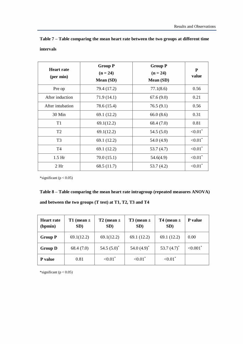
Results and Observations
Table 7 – Table comparing the mean heart rate between the two groups at different time
intervals
Heart rate
(per min)
Group P
(n = 24)
Mean (SD)
Group P
(n = 24)
Mean (SD)
P
value
Pre op 79.4 (17.2) 77.1(8.6) 0.56
After induction 71.9 (14.1) 67.6 (9.0) 0.21
After intubation 78.6 (15.4) 76.5 (9.1) 0.56
30 Min 69.1 (12.2) 66.0 (8.6) 0.31
T1 69.1(12.2) 68.4 (7.0) 0.81
T2 69.1(12.2) 54.5 (5.0) <0.01*
T3 69.1 (12.2) 54.0 (4.9) <0.01*
T4 69.1 (12.2) 53.7 (4.7) <0.01*
1.5 Hr 70.0 (15.1) 54.6(4.9) <0.01*
2 Hr 68.5 (11.7) 53.7 (4.2) <0.01*
*significant (p < 0.05)
Table 8 – Table comparing the mean heart rate intragroup (repeated measures ANOVA)
and between the two groups (T test) at T1, T2, T3 and T4
Heart rate
(bpmin)
T1 (mean ±
SD)
T2 (mean ±
SD)
T3 (mean ±
SD)
T4 (mean ±
SD)
P value
Group P 69.1(12.2) 69.1(12.2) 69.1 (12.2) 69.1 (12.2) 0.00
Group D 68.4 (7.0) 54.5 (5.0)* 54.0 (4.9)* 53.7 (4.7)* <0.001*
P value 0.81 <0.01* <0.01* <0.01*
*significant (p < 0.05)

Results and Observations
After starting dexmedetomidine (after T1), there was a significant drop in the heart rate in Group
D as compared to Group P (p < 0.01) which was sustained throughout the infusion. (Figure 13,
Table 7)
There was no significant difference between the mean heart rate at T1, T2, T3 and T4 in Group P.
(Table 8)
In Group D heart rate was significantly lower at T2, T3 and T4 as compared to T1. (Table 8)
Mean arterial pressure
Figure 14 -Line diagram comparing the mean arterial blood pressure between Group P and
D at different time intervals
Dexmedetomidine
(n=24)
Propofol (n=24)
100 90
80
70
60
50
40
30
20
10
0
Mean arterial pressure
MA
P (m
m H
g)
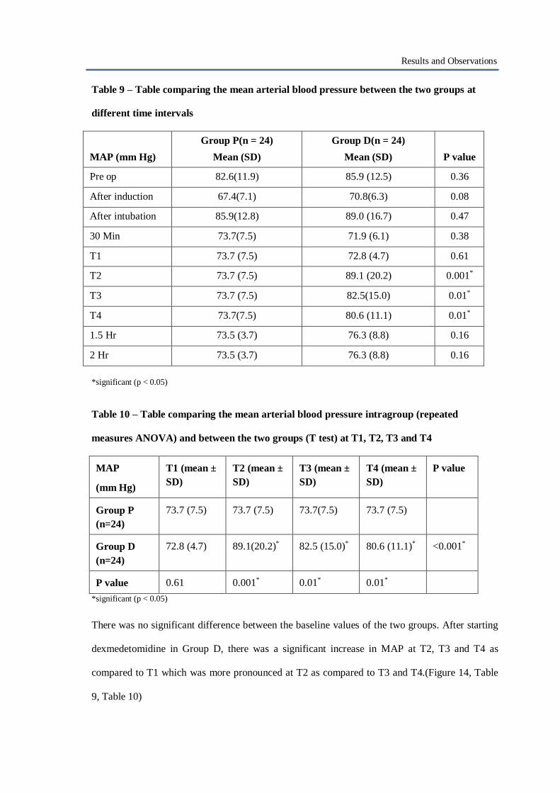
Results and Observations
Table 9 – Table comparing the mean arterial blood pressure between the two groups at
different time intervals
MAP (mm Hg)
Group P(n = 24)
Mean (SD)
Group D(n = 24)
Mean (SD)
P value
Pre op 82.6(11.9) 85.9 (12.5) 0.36
After induction 67.4(7.1) 70.8(6.3) 0.08
After intubation 85.9(12.8) 89.0 (16.7) 0.47
30 Min 73.7(7.5) 71.9 (6.1) 0.38
T1 73.7 (7.5) 72.8 (4.7) 0.61
T2 73.7 (7.5) 89.1 (20.2) 0.001*
T3 73.7 (7.5) 82.5(15.0) 0.01*
T4 73.7(7.5) 80.6 (11.1) 0.01*
1.5 Hr 73.5 (3.7) 76.3 (8.8) 0.16
2 Hr 73.5 (3.7) 76.3 (8.8) 0.16
*significant (p < 0.05)
Table 10 – Table comparing the mean arterial blood pressure intragroup (repeated
measures ANOVA) and between the two groups (T test) at T1, T2, T3 and T4
MAP
(mm Hg)
T1 (mean ±
SD)
T2 (mean ±
SD)
T3 (mean ±
SD)
T4 (mean ±
SD)
P value
Group P
(n=24)
73.7 (7.5) 73.7 (7.5) 73.7(7.5) 73.7 (7.5)
Group D
(n=24)
72.8 (4.7) 89.1(20.2)* 82.5 (15.0)* 80.6 (11.1)* <0.001*
P value 0.61 0.001* 0.01* 0.01*
*significant (p < 0.05)
There was no significant difference between the baseline values of the two groups. After starting
dexmedetomidine in Group D, there was a significant increase in MAP at T2, T3 and T4 as
compared to T1 which was more pronounced at T2 as compared to T3 and T4.(Figure 14, Table
9, Table 10)

Results and Observations
Patient state index (PSI)
Figure 15 – Line diagram comparing PSI values between the groups at different
time intervals
Table 11 – Table comparing the PSI values between the two groups at different time
intervals
PSI
Group P(n = 24)
Mean (SD)
Group D(n = 24)
Mean (SD)
P value
Pre op 94.8 (4.3) 95.0 (2.7) 0.84
After induction 30.3(3.6) 30.5 (2.9) 0.79
After intubation 30.8 (3.1) 31.0 (2.6) 0.84
30 Min 30.3(3) 29.6(1.7) 0.32
T1 29.9(3.2) 30.5(2.2) 0.40
T2 29.9 (3.2) 29.0(2.2) 0.27
T3 29.9 (3.2) 28.6 (2) 0.10
T4 29.9 (3.2) 28.6 (1.9) 0.10
1.5 Hr 29.9(3.4) 28.5 (1.3) 0.10
2 Hr 29.9 (3.4) 28.5 (1.3) 0.10
There was no significant difference between the two groups in terms of PSI values throughout the
operative period. (Figure 15, Table 11)
100
80
60
40
20
0
PSI
Propofol (n=24)
Dexmedetomidine (n=24)
PSI v
alu
e
Pre
op
after
inductio
n
after
intu
ba
tion
30
min
T1
T2
T3
T4
1.5
hr
2 h
r

Results and Observations
Rescue fentanyl
Figure 16 – Graph showing comparison of rescue fentanyl boluses at different time intervals
between the groups
Table 12 – Table showing number of patients receiving fentanyl boluses at different time
intervals between the groups
Group P Group D
Before T1 12 12
Between T1 and T2 0 4
Between T2 and T3 0 1
Between T3 and T4 0 0
After T4 9 5
The graph and the table denote the total number of patients who received fentanyl boluses (1
µg/kg) at different time intervals in each of the group. A continuous infusion of fentanyl was
going on at a rate of 1 µg/kg/hr for all the patients in both the groups. 12 patients each in both the
groups received fentanyl boluses before T1. Between T1 and T2, 4 patients received fentanyl
boluses in group D and 1 patient was given fentanyl bolus between T2 and T3. None of the
12
10
8
6
4
2
0
Propofol
Dexmedetomidine
before betn t1 betn t2 betn t3 after t4
t1 and t2 and t3 and t4

Results and Observations
patients received additional bolus in Group P between T1 and T4. After T4, 9 patients were given
fentanyl boluses in Group P and 5 in Group D. (Figure 16, Table 12)
The average infusion dose of propofol used in group P was 5.6 (±1.23) mg/kg/hr (93.3 ± 20
µg/kg/min). The average loading dose of dexmedetomidine used in Group D was 0.5 µg/kg over
10 minutes and average maintenance dose used was 0.5 µg/kg/hr.
Recovery parameters
Figure 17 – Graph comparing the recovery parameters between the groups
Table 13 – Table showing comparison of the recovery parameters between the groups
Recovery parameters
(min after stopping the
anaesthetic agents)
Group P(n = 24)
Mean (SD)
Group D(n = 24)
Mean (SD)
P value
Spontaneous Respiration 15.5(5.5) 30.7(10.4) p<0.01*
Spontaneous movement 21.2(6.9) 39.0(11.8) p<0.01*
Extubation 26.0(8) 43.3(12.1) p<0.01*
Verbalising 29.0(8.5) 45.6(13.6) p<0.01*
Orientation 38.7(11.3) 48.3(14.5) 0.016*
*significant (p < 0.05)
Propofol (n = 24)
Dexmedetomidine (n=24)
50 45 40 35 30 25 20 15 10
5 0
Recovery Parameters
Tim
e (m
in)

Results and Observations
There was a significant difference in the recovery parameters between both the groups. Time
taken for patient to start breathing spontaneously, move limbs spontaneously, get extubated, start
verbalising and get oriented to time, place and person were significantly prolonged in Group D.
(Figure 17, Table 13)
Complications
Figure 18 – Graph comparing the complications between the two groups
Table 14 – Table showing the complication rates between the two groups
Complications
Group P
(n = 24) (%)
Group D
(n = 24)(%)
P value
Bradycardia 5(20.8) 20(83.3) 0.003*
Hypotension (Hypo) 7(29.2) 7(29.2) 1.00
Hypertension (Hyper) 8(33.3) 10(41.7) 0.55
Abnormal movement 0(0.0) 4 (16.7) 0.038*
*significant (p < 0.05)
0
5
10
15
20
25
Frequency
Complications Group P
Complications Group D

Results and Observations
None of the patients in any group had tachycardia, tongue bite, local injury at pin insertion site,
post operative neurological deficit or intraoperative awareness as determined by modified Brice
questionnaire. The incidence of bradycardia and abnormal movements was significantly higher in
Group D. (Figure 18, Table 14)

Discussion
DISCUSSION

Discussion
Motor evoked potential monitoring is being commonly used in neurosurgeries to
assess the integrity of the motor tracts intraoperatively. Anaesthetic agents have an
inhibitory effect on the synapses and thus most of the agents have a dose dependent
inhibitory effect on MEP. If the doses of the anaesthetic agents are reduced to get an
adequate MEP, there is a risk of intraoperative awareness. Therefore there is a need to
use either a combination of anaesthetic agents or an adjuvant to reduce their inhibitory
effect on MEP and at the same time provide adequate depth of anaesthesia during
intraoperative MEP monitoring. The adjuvants should be able to have a sparing effect
on the dose requirement of the anaesthetic agents as well have have an anaesthetic and
analgesic effects of their own.
Both sevoflurane and propofol have a dose dependent depressive effect on TcMEP
responses.
A study (64) suggests that propofol with effect site concentration of 2.5 µg/ml (50–75
µg/kg/min) and 0.5 MAC sevoflurane had comparable effects on MEP and reproduce
reliable MEPs. But such low doses cannot produce an adequate depth of anaesthesia.
Thus an adjuvant to these agents is required which will facilitate MEP and at the same
time maintain an adequate depth of anaesthesia. And also an adjuvant to either low
dose propofol or sevoflurane should have similar effects on MEP.
There are many studies which have evaluated the effects on MEP of dexmedetomidine
as an adjuvant to low dose propofol (83, 85, 88 - 90) but similar studies with low dose
sevoflurane are lacking.

Discussion
In our study, we have evaluated the effects on MEP of dexmedetomidine as an
adjuvant to low dose sevoflurane and compared it to the effects that a combination of
propofol and low dose sevoflurane will have on MEP monitored during
neurosurgeries. We have also compared the hemodynamic stability and recovery
parameters and complications between the two anaesthetic regimens.
We recorded MEP bilaterally in all patients. Upper limb MEPs were recorded in all the
study patients but lower limb MEPs were recorded in only 10 patients of Group P and
12 patients of Group D who underwent thoracic or lumbar spine surgeries. For the
study purpose, comparison between the amplitude and latency of MEP obtained at four
points T1, T2, T3 and T4, each of which was 10 minutes apart were taken before
surgical stimulation of the spinal cord was done.
There was no significant difference noted between the two groups in terms of
amplitude and latency at any given point of time. In Group P, after point T1 the same
regimen was continued and no significant variations were noted among the subsequent
readings. The addition of loading dose of dexmedetomidine in Group D caused a
statistically significant decrease in amplitude and latency of MEP obtained at point T2
as compared to point T1. Amplitude starts recovering by the point T3 in Group D and
the value at T4 is comparable to baseline T1. Latency also starts recovering by point
T3 in Group D but still a statistically significant difference from the baseline was
found in the latency values at point T3 in Group D. But by T4 the latency mean values
were comparable to that of baseline in Group D.

Discussion
The statistically significant decrease in amplitude and latency seen at T2 could be due
to the combined effect of two drugs ie. propofol and dexmedetomidine loading dose.
Mohamed et al had found a significant decrease in amplitude of MEP or complete
disappearance of MEP after loading dose of dexmedetomidine in spine surgeries
similar to our study but they had used a loading dose of 1 µg/kg with a background of
propofol and remifentanil infusion. (97) In our study we used dexmedetomidine at a
loading dose of 0.5 µg/kg with a background propofol infusion till point T1 and
fentanyl at 1 µg/kg/hr with sevoflurane of 0.4 – 0.5 MAC.
Even though the changes in amplitude and latency were found statistically significant
in Group D they were not clinically significant. Also the amplitude and latency were
comparable between both the groups at any given point.
Similar to our findings, many studies had found that dexmedetomidine when used as
an adjuvant did not depress the MEP response significantly. Li et al, Rozet et al,
Tobias et al and Aschel et al have used dexmedetomidine as an adjuvant to propofol
and had found that as compared to propofol alone there was no significant change in
the amplitude or latency of the motor evoked potentials. (83, 85, 88 - 90)Bala et al
studied the effects of dexmedetomidine as an adjuvant to 4% desflurane on MEP and
had found that upto 0.6 ng/ml plasma concentration of dexmedetomidine did not have
any significant effect on MEP amplitude and threshold current intensity. (84) All of
the above mentioned studies have used a loading dose of 0.5 µg/kg of
dexmedetomidine followed by an infusion of 0.5 µg/kg/hr similar to our study.

Discussion
The PSI values in both the groups were comparable in our study. There are many
studies that have used BIS/PSI to monitor depth of anaesthesia during
dexmedetomidine infusion and have found a good correlation with other scales like
Ramsay sedation score and Observer Assessment alertness/sedation score which
reported good patient satisfaction score postoperatively. (95, 96) The dose of
dexmedetomidine used in most of these studies was 0.5-1 µg/kg loading followed by
0.5 µg/kg/hr maintenance which was similar to our study. Also none of our patients
had any awareness intraoperatively which was evaluated by the modified Brice
questionnaire.
In our study it was seen that there was a significant decrease in the heart rate after
starting the loading dose of dexmedetomidine in Group D. We also found that there
was a significant rise in the mean arterial pressure after the loading dose of
dexmedetomidine. The mean arterial pressure remained on the higher side even during
the maintenance infusion as compared to Group P. After the loading dose of
dexmedetomidine drop in heart rate with hypertension can be seen. (81) Hypertension
is more common when the loading dose is given over ten minutes as in our case. It
mostly occurs due to the intense vasoconstriction due to the stimulating effect of
alpha-2 adrenoceptors. It can be avoided if the loading dose is given over a longer
period. (81) This initial hypertension was also the reason for which according to our
study protocol 5 patients received fentanyl boluses after point T2 without any actual
rise in PSI values.
During the maintenance infusion of dexmedetomidine heart rate in Group D was
significantly lower as compared to Group P. Also the mean arterial pressure in Group
Discussion
D was higher than mean arterial pressure in Group P. Most of the studies have also
found lower heart rate with dexmedetomidine as compared to propofol. In patients
undergoing spinal tumour resection, Li et al had also found the heart rate to be
significantly lower in the groups receiving propofol with dexmedetomidine as
compared to propofol alone. They also found a higher mean arterial pressure in
patients receiving both dexmdetomidine and propofol as compared to propofol alone.
(85) Yan et al in his study on 84 patients who underwent general surgery found that
dexmedetomidine in combination with propofol had higher incidence of hypertension
and lower heart rates. (99) In most of the studies dexmedetomidine infusion was
associated with lower heart rates despite the anaesthetic drug it was used with. (85 –
88, 99)
The amount of analgesic requirement in both the groups seemed equivalent. Between
T1 and T4 points a significant rise in blood pressure was seen in group D after the
loading dose of dexmedetomidine was given. To counter it fentanyl boluses were
given but there was not any actual rise in PSI values and also there was no surgical
stimuli between these two time points. If these boluses are excluded then it can be seen
that after point T4 Group P had a greater analgesic requirement as compared to Group
D. It can be because of analgesic property of dexmedetomidine which propofol lacks.
Chakrabarti et al also found a lesser opioid requirement with dexmedetomidine and
propofol combination as compared to propofol alone. (82)
In our study we found that the patients in Group D took significantly more time for
extubation and getting oriented and verbalising as compared to Group P. This finding
was in contrary to the findings in most of the studies which showed that

Discussion
dexmedetomidine when used either alone or as an adjuvant to propofol did not alter
the recovery parameters significantly. (85, 88, 100, 101) This could be due to the fact
that the elimination half life of dexmedetomidine is around 2-3 hours and its context
sensitive half time increases with increase in the infusion duration from 4 minutes after
a 10 minutes duration to about 250 minutes after an 8 hour infusion. (102) In a study
by Dong et al in orthopaedic procedures they found that even though there was a
higher incidence of smooth emergence with better hemodynamic stability with a
combination of sevoflurane and dexmedetomidine the recovery period (time to
extubation) was significantly prolonged as compared to sevoflurane used alone and
also the recovery period was prolonged when dexmedetomidine and propofol were
used together as compared to propofol alone. (103) Laila et al also reported prolonged
recovery time with dexmedetomidine used as a sole sedative agent during day care
oral and maxillofacial surgeries. (104) It can be that dexmedetomidine used as an
adjuvant can prolong the recovery period because of its sedative and analgesic action
and a long context sensitive half life especially when used for long duration surgeries.
Incidence of bradycardia was significantly higher in Group D. These results were
similar to that of Chakrabarti et al who compared propofol with a combination of
dexmedetomidine and propofol. (82) There were 4 patients in group D that had an
abnormal movement of limbs when the surgeon was working near the nerve roots.
Complications like tongue injury, intraoperative respiratory efforts, needle site injury
or intraoperative awareness were not seen in any of the patients. Abnormal movements
can happen due to absence of muscle relaxation intraoperatively. Animal studies
suggest that propofol has a muscle relaxant property by blocking sarcolemmal sodium

Discussion
channels which can be a probable reason for none of the patients having any abnormal
movements in Group P. (105)

Limitations
LIMITATIONS

Limitations
a) The study was done only in one institute in 48 patients. A larger multicentre trial
is required to better understand its clinical applications.
b) The use of PSI, which evaluates depth of anaesthesia based on processed EEG
cannot be justified completely for depth of anaesthesia monitoring in a
dexmedetomidine based anaesthetic regimen even though previous studies have
justified its use. We supplemented it with postoperative Modified Brice
questionnaire to identify any potential intraoperative awareness in our study
group. There was no incidence of intraoperative awareness in any of our patients.
c) Some of the patients in both the groups had weakness of variable duration (power
of 4) in their limbs but their incidence was similar in both the groups. The
differential effect of the anaesthetic regimens was not separately studied on the
weaker limbs as compared to normal limbs.
d) In our study we used fentanyl as a rescue agent to treat hypertension induced by
dexmedetomidine infusion which must have confounded our results of the
analgesic requirement in both the groups.

Conclusion
CONCLUSION

Conclusion
A combined anaesthetic regimen of intravenous agents with low dose inhalational agents
is commonly used to facilitate motor evoked potential monitoring intraoperatively. In our
study we compared two such combined approaches – low dose sevoflurane with fentanyl
infusion combination with propofol or dexmedetomidine. We concluded that
a) Dexmedetomidine can be used as an adjuvant to low dose sevoflurane and
fentanyl for recording of adequate MEP responses. Even though the responses get
depressed after the loading dose, it is not clinically significant and the responses
(amplitude and latency) recover within 10 to 20 minutes and is comparable to
baseline. It is preferable to finish the loading dose of dexmedetomidine before the
start of surgery.
b) Dexmedetomidine infusion lowers the heart rate significantly. After the loading
dose, it can cause an increase in mean arterial pressure. During dexmedetomidine
infusion, the mean arterial pressures remain higher.
c) Patient’s recovery from anaesthesia is prolonged when dexmedetomidine is used
as an adjuvant to sevoflurane and fentanyl as compared to propofol.
d) Complications like bradycardia and abnormal limb movements are more common
with dexmedetomidine as compared to propofol.

Bibliography
BIBLIOGRAPHY
Bibliography
1) Sutter M, Deletis V, Dvorak J, Eggspuehler A, Grob D, MacDonald D, et al.
Current opinions and recommendations on multimodal intraoperative monitoring
during spine surgeries. Eur Spine J. 2007; 16:232-7.
2) Legatt A D, Emerson R G, Epstein C M, MacDonald D B, Deletis V, Bravo R J, et
al. ACNS Guideline: Transcranial Electrical Stimulation Motor Evoked Potential
Monitoring. J Clin Neurophysiol. 2016; 33:42-50.
3) Deletis V. Basic methodological principles of multimodal intraoperative
monitoring during spine surgeries. Eur Spine J. 2007; 16:147-52.
4) Chen Z. The effects of isoflurane and propofol on intraoperative
neurophysiological monitoring during spinal surgery. J Clin Monit Comput. 2004;
18:303–8.
5) Scheufler KM, Reinacher PC, Blumrich W, Zentner J, Priebe HJ. The modifying
effects of stimulation pattern and propofol plasma concentration on motor-evoked
potentials. Anesth Analg. 2005; 100:440–7.
6) Mariappan R, Ashokkumar H, Kuppuswamy B. Comparing the effects of oral
clonidine premedication with intraoperative dexmedetomidine infusion on
anesthetic requirement and recovery from anesthesia in patients undergoing major
spine surgery. J Neurosurg Anesthesiol. 2014; 26:192–7.
7) Freeman KA, Fullerton DA, Foley LS, Bell MT, Cleveland Jr JC, Weyant MJ, et
al. Spinal cord protection in alpha-2 agonist-mediated increase in glial cell-line
derived neurotropic factor. J Thorac Cardiovasc Surg. 2015; 149:578–86.
8) Raw D A, Beattie J K, Hunter J M. Anaesthesia for spinal surgery in adults. Br J
Anaesth. 2003; 91:886-904.
9) Hadley M N, Shank C D, Rozzelle C J, Walters B C. Guidelines for the Use of
Electrophysiological Monitoring for Surgery of the Human Spinal Column and
Spinal Cord. Neurosurgery. 2017; 81:713–32.
10) Pelosi L, Lamb J, Grevitt M, Mehdian SM, Webb JK, Blumhardt LD. Combined
monitoring of motor and somatosensory evoked potentials in orthopaedic spinal
surgery. Clin neurophysiol. 2002; 113:1082-91.
11) Nuwer MR, Dawson EG, Carlson LG, Kanim LEA, Sherman JE. Somatosensory
evoked potential spinal cord monitoring reduces neurologic deficits after scoliosis

Bibliography
surgery: results of a large multicenter survey. Electroencephalogr Clin
Neurophysiol. 1995; 96:6-11.
12) Kothbauer K, Deletis V, Epstein FJ. Intraoperative spinal cord monitoring for
intramedullary surgery: an essential adjunct. Pediatr Neurosurg. 1997; 26:247–54.
13) Kothbauer K, Deletis V, Epstein FJ, Motor evoked potential monitoring for
intramedullary spinal cord tumor surgery. Neurosurg Focus. 1998; 4:1-9.
14) Sala F, Palandri G, Basso E, Lanteri P, Deletis V, Faccioli F, et al. Motor evoked
potential monitoring improves outcome after surgery for intramedullary spinal
cord tumors: a historical control study. Neurosurgery. 2006; 58:1129-43.
15) Deletis V, Sala F. Intraoperative neurophysiological monitoring of the spinal cord
during spinal cord and spine surgery: A review focus on the corticospinal tracts.
Clin Neurophysiol. 2008; 119: 248–64.
16) Jacobs MJ, Mess W, Mochtar B, Nijenhuis RJ, Statius RG, Schurink GW. The
value of motor evoked potentials in reducing paraplegia during thoracoabdominal
aneurysm repair. J Vasc Surg. 2006; 43:239-46.
17) Zho HH, Kelly PJ. Transcranial Electrical Motor Evoked Potential Monitoring for
Brain Tumor Resection. Neurosurgery. 2001; 48:1075-81.
18) Szelenyi A, Camargo AB, Flamm E, Deletis V. Neurophysiological criteria for
intra-operative prediction of pure motor hemiplegia during aneurysm surgery, J
Neurosurg. 2003; 99:575–8.
19) Ichikawa T, Suzuki K, Sasaki T, Matsumoto M, Sakuma J, Oinuma M, et al.
Utility and the limit of motor evoked potential monitoring for preventing
complications in surgery for cerebral arteriovenous malformation.
Neurosurgery. 2010; 67:222-8.
20) Sarnthein J, Bozinov O, Melone AG, Bertalanffy H. Motor-evoked potentials
(MEP) during brainstem surgery to preserve corticospinal function. Acta
neurochir. 2011; 153:1753-9.
21) Kawasaki Y, Uchida S, Onishi K, Toyokuni M, Okanari K, Fujiki M.
Intraoperative Neurophysiologic Monitoring for Prediction of Postoperative

Bibliography
Neurological Improvement in a Child With Chiari Type I Malformation. J
Craniofac Surg. 2017; 28:1837-41.
22) Stoelting R K, Rathmell J P, Flood P, Shafer S, Stoelting’s handbook of
Pharmacology and Physiology in Anesthetic Practice. 3rd ed. Philadelphia:
Wolten Kluwer Pub. Co.; 2015. 46-55 p.
23) Penfield W, Rasmussen T. The cerebral cortex of man: a clinical study of
localization of function. New York: Hafner Pub. Co.; 1968. 23-27 p.
24) Rose RD. Sensory component of cervically evoked motor potentials. Med
Hypotheses. 1996; 46:577–9.
25) Minahan RE, Sepkuty JP, Lesser RP, Sponseller PD, Kostuik JP. Anterior
spinal cord injury with preserved neurogenic ‘motor’ evoked potentials. Clin
Neurophysiol. 2001; 112:1442–50.
26) MacDonald, Skinner S, Shils J, Yingling C. Intraoperative motor evoked
potential monitoring – A position statement by the American Society of
Neurophysiological Monitoring. Clin Neurophysiol. 2013; 124:2291–316.
27) Szelenyi A, Langer D, Beck J, Raabe A, Flamm ES, Seifert V, et al.
Transcranial and direct cortical stimulation for motor evoked potential
monitoring in intracerebral aneurysm surgery. Clin Neurophysiol. 2007;
37:391-8.
28) Barker AT, Freeston IL, Jalinous R, Jarratt JA. Magnetic stimulation of the
human brain and peripheral nervous system: an introduction and the results of
an initial clinical evaluation. Neurosurgery. 1987; 20:100–9.
29) Wrzosek M. Electroencephalography as a diagnostic technique for canine
neurological diseases. J Vet Res. 2016; 60:181-7.
30) Legatt AD, Ellen R. Grass Lecture: Motor Evoked Potential Monitoring. Am J
Electroneurodiagn Technol. 2004; 44:223-43.
31) Deletis V, Isgum V, Amassian V. Neurophysiological mechanisms underlying
motor evoked potentials in anesthetized humans. Part 1. Recovery time of
corticospinal tract direct waves elicited by pairs of transcranial electrical
stimuli. Clin Neurophysiol. 2001; 112:438-44.
32) Ubags LH, Kalkman CJ, Been HD, Koelman JH, Ongerboer de Visser BW. A
comparison of myogenic motor evoked responses to electrical and magnetic

Bibliography
transcranial stimulation during nitrous oxide/opioid anesthesia. Anesth Analg.
1999; 88:568–72.
33) Chipchase L, Schabrun S, Cohen L, Hodges P, Ridding M, Rothwell J, et al. A
checklist for assessing the methodological quality of studies using transcranial
magnetic stimulation to study the motor system: an international consensus
study. Clin Neurophysiol. 2012; 123:1698–704.
34) Sloan T, Heyer EJ. Anesthesia for intraoperative neurophysiologic monitoring
of the spinal cord. J Clin Neurophysiol. 2002; 19:430-43.
35) MacDonald DB, Dong CC. Spinal cord monitoring during descending aortic
procedures. Handbook of Clinical Neurophysiology. 2008; 8:815-28.
36) Jameson LC, Sloan TB. Monitoring of the brain and spinal cord. Anesthesiol
Clin. 2006; 24:777.
37) Calancie B, Harris W, Broton JG, Alexeeva N, Green BA. "Threshold-level"
multipulse transcranial electrical stimulation of motor cortex for intraoperative
monitoring of spinal motor tracts: description of method and comparison to
somatosensory evoked potential monitoring. J Neurosurg. 1998; 88:457-70.
38) Langeloo DD, Journee HL, Kleuver M, Grotenhuis JA. Criteria for
transcranial electrical motor evoked potential monitoring during spinal
deformity surgery: A review and discussion of the literature. Neurophysiol
Clin. 2007; 37:431-9.
39) MacDonald DB. Safety of Intraoperative Transcranial Electrical Stimulation
Motor Evoked Potential Monitoring. J Clin Neurophysiol. 2002; 19:416–29.
40) Burke D, Hicks RG. Surgical monitoring of motor pathways. J Clin
Neurophysiol. 1998; 15:194–205.
41) Calancie B, Harris W, Brindle GF, Green BA, Landy HJ. Threshold level
repetitive transcranial electrical stimulation for intraoperative monitoring of
central motor conduction. J Neurosurg Spine 1. 2001; 95:161–8.
42) Szelenyi A, Kothbauer KF, Deletis V. Transcranial electric stimulation for
intraoperative motor evoked potential monitoring: Stimulation parameters and
electrode montages. Clin Neurophysiol. 2007; 118:1586-95.

Bibliography
43) Oro J, Haghighi SS. Effects of altering core body temperature on
somatosensory and motor evoked potentials in rats. Spine. 1992; 17:498-503.
44) Maylaerts SA, De Haan P. The effects of regional spinal cord hypothermia on
transcranial myogenic motor evoked potential monitoring and the efficacy of
spinal cord ischaemia detection. J Thorac Cardiovasc Surg. 1999; 118:1038-
45.
45) Sakamoto T, Kawaguchi M. The effect of hypothermia on myogenic motor
evoked potentials to an electrical stimulation with a single pulse and a train of
pulses under propofol/ketamine/fentanyl anaesthesia in rabbits. Anaesth
Analg. 2003; 96:1692-7.
46) Sueda T, Okada K. Evaluation of somatosensory and motor evoked potentials
in thoracoabdominal aortic aneurysm surgery. Jpn J Thorac Cardiovasc Surg.
2000; 48: 60-5.
47) Leslie K, Sessler DI, Bjorksten AR, Moayyeri A. Mild hypothermia alters
propofol pharmacokinetics and increases the duration of action of atracurium.
Anesth Analg. 1995; 80:1007-14.
48) Haghighi SS, Keller BP, Oro JJ, Gibbs SR. Motor-evoked potential changes
during hypoxic hypoxia. Surg neurol. 1993; 39:399-402.
49) Ataka H, Murakami M, Goto S, Moriya H, Hayashi F, Fukuda Y. Effects of
hypoxia on the ventral root motor-evoked potential in the in vitro spinal cord
preparation. Spine. 1996; 21:2095-100.
50) Short LH, Peterson RE, Monagan PD. Physiologic and anesthetic alterations
on spinal-sciatic evoked responses in swine. Anesth Analg. 1993; 76:259–65.
51) Dolan EJ, Transfeld EE, Tator CH, Simmons EH, Hughes KF. The effects of
spinal distraction on regional blood flow in cats. J Neurosurg. 1980; 53:756-
64.
52) King BS, Rampil IJ. Anesthetic depression of spinal motor neurons may
contribute to lack of movement in response to noxious stimuli.
Anesthesiology. 1994; 81:1484–92.

Bibliography
53) Browning JL, Heizer ML, Baskin DS. Variations in corticomotor and
somatosensory evoked potentials: effects of temperature, halothane anesthesia,
and arterial partial pressure of CO2. Anesth Analg. 1992; 74:643–8.
54) Priori A, Berardelli A, Mercuri B, Inghilleri M, Manfredi M. The effect of
hyperventilation on motor cortical inhibition in humans: a study of the
electromyographic silent period evoked by transcranial brain stimulation.
Electroencephalography and Clinical Neurophysiology/Electromyography and
Motor Control. 1995; 97:69-72.
55) Hentschke H, Schwarz C. Antkowiak B. Neocortex is the major target of
sedative concentrations of volatile anesthetics: Strong depression of firing
rates and increase of GABAA receptor mediated inhibition. Eur J Neurosci.
2005; 21:93-102.
56) Burke, Bartley K. The effect of volatile anaesthetics on the excitability of
human corticospinal tract. Brain. 2000; 123:992- 1000.
57) Sakamoto T, Kawaguchi M, Inoue S, Furuya H. Suppressive effect of nitrous
oxide on motor evoked potentials can be reversed by train stimulation n
rabbits under ketamine/ fentanyl anaesthesia, but not with additional propofol.
Br J Anaesth. 2001; 86:395-402.
58) Haghighi SS, Sirintrapun SJ, Keller BP, Oro JJ, Madsen R. Effect of
desflurane anesthesia on transcortical motor evoked potentials. J Neurosurg
Anesthesiol. 1996; 8:47-51.
59) Ubags LH, Kalkman CJ, Been HD. Influence of isoflurane on myogenic motor
evoked potentials to single and multiple transcranial stimuli during nitrous
oxide/opioid anesthesia. Neurosurgery. 1998; 43:90–4.
60) Woodforth IJ, Hicks RG, Crawford MR, Stephen JP, Burke DJ. Variability of
motor evoked potentials recorded during nitrous oxide anaesthesia from the
tibialis anterior muscle after transcranial electrical stimulation. Anesth Analg.
1996; 82:744-9.

Bibliography
61) Zentner J, Albrecht T, Heuser D. Influence of halothane, enflurane, and
isoflurane on motor evoked potentials. Neurosurgery. 1992; 31:298–305.
62) Pelosi L, Stevenson M, Hobbs GJ, Jardine A, Webb JK. Intraoperative motor
evoked potentials to transcranial electrical stimulation during two anaesthetic
regimens. Clin neurophysiol. 2001; 112:1076-87.
63) Chong CT, Manninen P, Sivanaser V, Subramanyam R, Lu N, Venkatraghavan L.
Direct comparison of the effect of desflurane and sevoflurane on intraoperative
motor-evoked potentials monitoring. J Neurosurg Anesthesiol. 2014; 26:306-12.
64) Hernandez-Palazon J, Izura V, Fuentes-Garcia D, Piqueras-Perez
C, Domenech-Asensi P, Falcon-Arana L. Comparison of the Effects of
Propofol and Sevoflurane Combined With Remifentanil on Transcranial
Electric Motor-evoked and Somatosensory-evoked Potential Monitoring
During Brainstem Surgery. J Neurosurg Anesthesiol. 2015; 27:282-88.
65) Martella G, De Persis C, Bonsi P, Natoli S, Cuomo D, Bernardi G, et al.
Inhibition of persistent sodium current fraction and voltage-gated L-type
calcium by propofol in cortical neurons: Implications for its antiepileptic
activity. Epilepsia. 2005; 46:624-35.
66) Kakinohana M, Fuchigami T, Nakamura S, Kawabata T, Sugahara K.
Propofol reduces spinal motor neuron excitability in humans. Anesth Analg.
2002; 94:1586–8.
67) Ubags LH, Kalkman CJ, Been HD, Drummond JC. Differential effects of
nitrous oxide and propofol on myogenic transcranial motor evoked responses
during sufentanil anaesthesia. Br J Anaesth. 1997; 79:590–4.
68) Van Dongen EP, Ter Beek HT, Aarts LP, Schepens MA, Morshuis WJ,
Benning FJ,et al. The effect of two low-dose propofol infusions on the
relationship between sixpulse transcranial electrical stimulation and the
evoked lower extremity muscle response. Acta Anaesthesiol Scand. 2000;
44:799–803.

Bibliography
69) Kawaguchi M, Sakamoto T, Inoue S, Kakimoto M, Furuya H, Morimoto T, et
al. Low-dose propofol as a supplement to ketamine-based anesthesia during
intraoperative monitoring of motor-evoked potentials. Spine. 2000; 25:974–9.
70) Scheufler KM, Zentner J. Motor-evoked potential facilitation during
progressive cortical suppression by propofol. Anesth Analg. 2002; 94:907–12.
71) Schonle PW, Isenberg C, Crozier TA, Dressler D, Machetanz J, Conrad B.
Changes of transcranially evoked motor responses in man by midazolam, a
short-acting benzodiazepine. Neurosci Lett. 1989; 101:321-4.
72) Aggarwal D, Mahajan HK, Chauhan PR. A comparative evaluation of
dexmedetomidine with midazolam as an adjuvant to propofol anesthesia for
spinal surgical procedures under motor evoked potential monitoring. Anaesth
Pain & Intensive Care. 2016; 20:154-8.
73) Nadstawek J, Taniguchi M, Langenbach U, Thees C, Pechstein U, Schnitzler
N. Effect of premedication and barbiturate hypnotics on motor evoked
potentials induced by magnetic cortex stimulation. Anasthesiol Intensivmed
Notfallmed Schmerzther. 1995; 30:361-5.
74) Kochs E, Treede RD, Schulte J. Increase in somatosensory evoked potentials
during anesthesia induction with etomidate. Anaesthesist. 1986; 35:359-64.
75) Taniguchi M, Nadstawek J, Langenbach U, Bremer F, Schramm J. Effects of
Four Intravenous Anesthetic Agents on Motor Evoked Potentials Elicited by
Magnetic Transcranial Stimulation. Neurosurgery. 1993; 33:407–15.
76) Glassman SD, Shields CB, Linden RD, Zhang YP, Nixon AR, Johnson JR.
Anesthetic effects on motor evoked potentials in dogs. Spine. 1993; 18:1083-
9.
77) Yang LH, Lin SM, Lee WY, Liu CC. Intraoperative transcranial electrical
motor evoked potential monitoring during spinal surgery under intravenous
ketamine or etomidate anaesthesia. Acta Neurochir. 1994; 127:191-8.

Bibliography
78) Kalkman CJ, Drummond JC, Ribberink AA, Patel PM, Sano T, Bickford RG.
Effects of propofol, etomidate, midazolam and fentanyl on motor evoked
responses to transcranial electrical or magnetic stimulation in humans.
Anesthesiology. 1992; 76:502-9.
79) Thees C, Scheufler KM, Nadastawek J, Pechstein U, Hanisch M, Juntke R, et
al. Influence of fentanyl, alfentanil, and sufentanil on motor evoked potentials.
J Neurosurg Anesthesiol. 1999; 11:112-8.
80) Kim W H, Lee J J, Lee S M, Park M N, Park S K, Seo D W, et al. Comparison
of motor-evoked potentials monitoring in response to transcranial electrical
stimulation in subjects undergoing neurosurgery with partial vs no
neuromuscular block. Brit J Anaesth. 2013; 110: 567–76.
81) Miller R. Miller’s anesthesia. 8th ed. Vol 1. Philadelphia:Elsevier; 2015. 855 –
860 p.
82) Chakrabarti D, Kamath S, Madhusudan Reddy KR, Srinivas DB, Manohar N,
Masapu D. Effect of adjunctive dexmedetomidine on anesthesia and analgesia
requirement and recovery characteristics during Bispectral Index-guided
anesthesia for cerebello-pontine angle surgeries: A randomized clinical trial. J.
Anaesthesiol Clin Pharmacol. 2018; 34:496-502.
83) Tobias JD. Dexmedetomidine:applications in pediatric critical care and
pediatric anesthesiology. Pediatr Crit Care Med. 2007; 8:115-31.
84) Bala E, Sessler DI, Nair DR, McLain R, Dalton JE, Farag E. Motor and
somatosensory evoked potentials are well maintained in patients given
dexmedetomodine during spine surgery. Anesthesiology. 2008; 109:417-25.
85) Li Yan, Meng L, Peng Y, Qiao H, Guo L, Han R, et al. Effects of
Dexmedetomidine on motor- and somatosensory-evoked potentials in patients
with thoracic spinal cord tumor: a randomized controlled trial. BMC
Anesthesiol. 2016; 16: 51.

Bibliography
86) Wei L, Deng X, Sui J, Wang L, Liu J. Dexmedetomidine Improves Intubating
Conditions Without Muscle Relaxants in Children After Induction With Propofol
and Remifentanil. Anesth Analg. 2015; 121:785-90.
87) Avidan M S, Zhang L, Burnside B, Finkel K J, Searleman A C, Selvidge J A,
et al. Anesthesia Awareness and the Bispectral Index. N Engl J Med. 2008;
358:1097-108.
88) Anschel DJ, Aherne A, Soto RG, Carrion W, Hoegerl C, Nori P, et al.
Successful intraoperative spinal cord monitoring during scoliosis surgery
using a total intravenous anesthetic regimen including dexmedetomidine. J
Clin Neurophysiol. 2008; 25:56–61.
89) Tobias JD, Goble TJ, Bates G, Anderson JT, Hoernschemeyer DG. Effects of
dexmedetomidine on intraoperative motor and somatosensory evoked
potential monitoring during spinal surgery in adolescents. Paediatr Anaesth.
2008; 18:1082–8.
90) Rozet I, Metzner J, Brown M, Treggiari MM, Slimp JC, Kinney G, et al.
Dexmedetomidine does not affect evoked potentials during spine surgery.
Anesth Analg. 2015; 121:492–501.
91) Lee KH, Kim YH, Sung YJ, Oh MK. The Patient State Index is well balanced
for propofol sedation. Hippokratia. 2015; 19:235-8.
92) Drover D R, Lemmens H J, Pierce E T, Plourde G, Loyd G, Ornstein E, et al.
Titration of Delivery and Recovery from Propofol, Alfentanil, and Nitrous
Oxide Anesthesia. Anesthesiology. 2002; 97:82–9.
93) Soehle M, Keuch M, Grube M, Wirz S, Kruer S, Hoeft A, et al. Patient state
index vs bispectral index as measures of the electroencephalographic effects of
propofol. Brit J Anaes. 2010; 105:172-8.

Bibliography
94) Prichep L S, Gugino L D, John E R, Chabot R, Howard B, Merkin H, et al.
The Patient State Index as an indicator of the level of hypnosis under general
anaesthesia. Brit J Anaesth. 2004; 92: 393-9.
95) Park S, Choi JH, Kim HT, Cho Y. Replacement of dexmedetomidine loading
with midazolam for sedation in elderly patients with spinal anesthesia.
Medicine. 2018; 97:e12565.
96) Sayed E, Yassen KA. Intraoperative effect of dexmedetomidine infusion
during living donor liver transplantation: A randomized control trial. Saudi J
Anaesth. 2016; 10:288-94.
97) Mahmoud M, Sadhasivam S, Sestokas AK, Samuels P, McAuliffe J. Loss of
transcranial electric motor evoked potentials during pediatric spine surgery.
Anesthesiology. 2007; 106:393-6.
98) Mahmoud M, Sadhasivam S, Salisbury S, Nick T G, Schnell B, Sestokas A K,
et al. Susceptibility of Transcranial Electric Motor-evoked Potentials to
Varying Targeted Blood Levels of Dexmedetomidine during Spine Surgery.
Anesthesiology. 2010; 112:1364 –73.
99) Han Y, Han L, Dong M, Sun Q, Ding K, Zhang Z, et al. Comparison of a
loading dose of dexmedetomidine combined with propofol or sevoflurane for
hemodynamic changes during anesthesia maintenance: a prospective,
randomized, double-blind, controlled clinical trial. BMC Anesthesiol. 2018;
18:12.
100) Srivastava VK, Agrawal S, Kumar S, Khan S, Sharma S, Kumar R.
Comparative Evaluation of Dexmedetomidine and Propofol Along With Scalp
Block on Haemodynamic and Postoperative Recovery for Chronic Subdural
Haematoma Evacuation Under Monitored Anaesthesia Care. Turk J
Anaesthesiol Reanim. 2018; 46:51–6.

Bibliography
101) Bharathwaj D K, Kamath S S. Comparison of dexmedetomidine versus
propofol-based anaesthesia for controlled hypotension in functional
endoscopic sinus surgery. South Afr J Anaesth Analg. 2018; 0:1–4.
102) Venn RM, Karol MD, Grounds RM. Pharmacokinetics of dexmedetomidine
infusions for sedation of postoperative patients requiring intensive care. Br J
Anaesth. 2002; 88:669-75.
103) Kim DJ, Kim SH, So KY, Jung KT. Effects of dexmedetomidine on smooth
emergence from anaesthesia in elderly patients undergoing orthopaedic
surgery. BMC Anesthesiol. 2015; 15:139.
104) Makary L, Vornik V, Finn R, Lenkovsky F, Mcclelland AL, Thurmon J, et al.
Prolonged Recovery Associated With Dexmedetomidine When Used as a Sole
Sedative Agent in Office-Based Oral and Maxillofacial Surgery Procedures. J
Oral Maxillofac Surg. 2010; 68:386-91.
105) Haeseler G, Stormer M, Bufler J, Dengler R, Hecker H, Piepenbrock S, et al.
Propofol blocks human skeletal muscle sodium channels in a voltage-
dependent manner. Anesth Analg. 2001; 92:1192-8.

Annexures
ANNEXURES

Annexures
ANNEXURE 1
Proforma
Name
Hospital number
Age
Sex if female-pregnant/nursing
Diagnosis
yes no
Informed consent
Autonomic instability
Neurological/neuromuscular
disease
Cardiac/pulmonary/renal/hepatic
disease
Skull defects/injury at local site
of needle electrode insertion
Diabetes(if yes specify duration)
Alcohol use (if yes specify
duration)
Allergy to anesthetic agents
Proposed surgery
Other specifications -

Annexures
Preoperative parameters
Heart rate Blood pressure Saturation
In ward/icu
In ot before
induction
Hemodynamics
Heart rate MAP Saturation PSI TOF
After
induction
After
intubation
10 min
20 min
30 min
45 min
1 hr
1.5 hr
2 hr
2.5 hr
3 hr
3.5 hr
4 hr
4.5 hr
5 hr

Annexures
100
Amount of drugs used intraoperatively –
Duration Study drug Fentanyl Sevoflurane Propofol Rescue
(fentanyl)
1st hr
2nd hr
3rd hr
4th hr
5th hr
When T4/T1 ≥ 0.9
Heart
rate
MAP Saturation EtCO2 PSI TOF Cumulative
dose of
study drug
Cumulative
dose of
fentanyl
Cumulative
dose of
propofol
T1
T2
T3
T4
T5
T6
Tx1
Tx2
Te

Annexures
101
MEP
Left UL Left LL Right UL Right LL
Amplitude Latency Amplitude Latency Amplitude Latency Amplitude Latency
T1
T2
T3
T4
T5
T6
Tx1
Tx2
Te
Recovery
Time (min) – after stopping anaesthetic agents
Respiration
Spontaneous movements
Extubation
Verbal
Cooperation
orientation
ICU discharge

Annexures
102
Any post operative complications –
Complications Frequency
Bradycardia
Tachycardia
Hypertension
Hypotension
Respiratory efforts
Any movement
Tongue bite
Any other injury
Intraoperative awareness

Annexures
103
ANNEXURE 2
Modified Brice Questionnaire
Were you expecting to be completely asleep for this operation (please circle)? YES / NO
1. What is the last thing you remember before going to sleep (please tick one box)?
-Being in the pre-op area ☐ -Seeing the operating room ☐
-Being with family ☐ -Hearing voices ☐
-Feeling mask on face ☐ -Smell of gas ☐
-Burning or stinging in the IV line ☐ -Other [Please write below]:
2. What is the first thing you remember after waking up (please tick one box)?
-Hearing voices ☐ -Feeling breathing tube ☐
-Feeling mask on face ☐ -Feeling pain ☐
-Seeing the operating room ☐ -Being in the recovery room ☐
-Being with family ☐ -Being in ICU ☐
-Nothing ☐ -Other [Please write below]:
3. Do you remember anything between going to sleep and waking up (please tick
box)?
-No ☐
-Yes: -Hearing voices ☐ -Hearing events of the surgery ☐
-Unable to move or breathe ☐ -Anxiety/stress ☐
-Feeling pain ☐ -Sensation of breathing tube ☐
-Feeling surgery without pain ☐ -Other [Please write below]
4. Did you dream during your procedure (please tick box)?
-No ☐ -Yes ☐
-What about [Please write below]:

Annexures
104
5. Were your dreams disturbing to you (please tick box)?
-No ☐ -Yes ☐
6. What was the worst thing about your operation (please tick box)?
-Anxiety ☐ -Pain ☐
-Recovery process ☐ -Unable to carry out usual activities ☐
-Awareness ☐ -Other [Please write below]:

Annexures
105
ANNEXURE 3
PATIENT INFORMATION FORM
Title of the study:
Comparison of effects of propofol and dexmedetomidine on motor evoked potentials in
neurosurgery.
Name of the Investigators:
Dr. Soniya Biswas, Dr. Smita V., Dr. Unnikrishnan P.
You are being requested to participate in this study which compares the effects of propofol and
dexmedetomidine on motor evoked potentials in neurosurgery. We have planned to include about
people from this hospital in this study.
What is motor evoked potential and does it have any side effects?
Motor-evoked potential (MEP) is widely used during neuro surgeries. The MEP is produced by
electrical stimulation of a part of a brain using scalp electrodes. After stimulation, the activity of
the muscles of both arms and legs which gets activated after the stimulation is recorded using
another set of electrodes. As you will be under anesthesia, you will not be aware of this
stimulation and you will also not feel any pain. Sometimes after stimulation there can be some
movement of tongue which will be protected using a bite block so that there is no injury. The
electrodes which are inserted in the arms and legs are minute sized pins. They will be not cause
any pain as you will be under anesthesia and their size is so small that they will also not cause
any injury at the insertion site. The length of the pins is also less so that they will not go very
deep inside. The pin insertion sites will be kept nicely padded.
What is propofol and does it have any side effects?
Propofol is a sedative drug which is given through vein. It is approved for use in neuroanaesthesia
and being used worldwide including our institute. It will be discontinued after surgery and you
will recover from anesthesia. It can cause decrease in blood pressure but we will use it at a rate
which will cause this side effect. It can cause stinging sensation in vein while injection. We will
prevent it by giving low dose of another drug lignocaine.
What is dexmedetomidine and does it have any side effects?
Dexmedetomidine is a sedative drug which is given through vein. Along with the property of
inducing sleep it reduces pain and vomiting. It is approved for use in neuroanaesthesia and being
used worldwide including our institute. It will be discontinued after surgery and you will recover
from anesthesia. Dexmedetomidine in higher doses (> 0.8 mcg/Kg/hour) reduces the heart rate
and blood pressure. However the dose we are using does not have any adverse effects according
to available literature in scientific journals.
If you take part what will you have to do?
You will be included in the study only if you are willing. On the day of surgery you will
be anaesthetized according to the standard protocol of our institute and breathing tube will be
placed for giving artificial breathing. Our study will start after induction. Electrodes will be
placed on your scalp and all the four limbs to monitor motor evoked potentials.To maintain

Annexures
106
anaesthesia during surgery you will be administered anesthetic gases or drugs through vein. You
will be allotted to any one of the two groups:
First group will receive propofol till the end of surgery
Second group will receive propofol for sometime and then dexmedetomidine
Please note that there is no advantage of any of the techniques over another for
maintenance of anesthesia. Both are equally safe and effective. The study purpose is only to
record the effect of these drugs on motor evoked potentials. The quality of surgery will not be
affected by the difference in the anesthesia techniques.
The motor evoked potentials will be recorded at different time intervals during the
surgery. You will not feel any pain or become aware during the recordings. After the end of
surgical procedure the drugs will be stopped.
You will be asked to open your eyes, make hand grip and if they are adequate the
breathing tube will be taken out. Then you will be asked about time, place and date to assess your
recovery from anesthesia.You will be asked about any complaints which will be taken care of.
After that you will be shifted to ICU where postsurgery care will be taken up.
Can you withdraw from this study after it starts?
Your participation in this study is entirely voluntary and you are also free to decide to
withdraw permission to participate in this study. If you do so, this will not affect your usual
treatment at this hospital in any way. In addition, if you experience any side effects, the study will
be stopped and you will be given treatment for the side effects.
What will happen if you develop any study related injury?
We do not expect any injury to happen to you since the anaesthesia technique and
monitoring tools would be same even if you were not part of the study. In addition, adverse
events related to motor evoked potential monitoring is minimal. But if you do develop any side
effects or problems due to the study, the side effects will be treated at no cost to you. We are
unable to provide any monetary compensation, however.
Will you have to pay for the cost of using the devices?
Motor evoked potential monitor is used as a part of anaesthesia procedures for surgery.
So, no extra money will be charged from the patients.
Will your personal details be kept confidential?
The results of this study will be used for thesis submission as a part of academic research
and will be submitted to a medical journal for publication, but you will not be identified by name
in any publication or presentation of results. However, your medical notes may be reviewed by
people associated with the study, without your additional permission, should you decide to
participate in this study.
If you have any further questions, please ask Dr. Soniya Biswas (Principal investigator) mobile
number: 9336693999. Email: [email protected].
For technical advisory committee contact, please ask Dr. Maala Ramanathan, telephone number:
0471-2524234. Email: [email protected]

Annexures
107
CONSENT FORM
Participant’s name: Date of Birth / Age (in years):
I , son/daughter of
Declare that (Please tick boxes)
• I have read the above information provided to me regarding the study: Comparison of effects
of propofol and dexmedetomidine on motor evoked potentials in neurosurgery [ ]
• I have clarified any doubts that I had. [ ]
• I also understand that my participation in this study is entirely voluntary and that I am
free to withdraw permission to continue to participate at any time without affecting my
usual treatment or my legal rights [ ]
• I understand that the study staff and institutional ethics committee members will not need
my permission to look at my health records even if I withdraw from the trial. I agree to
this access [ ]
• I understand that my identity will not be revealed in any information released to third
parties or published [ ]
• I voluntarily agree to take part in this study [ ]
• I have been provided with the contact numbers of the principle investigator, in case I
want to know more about the study and participants rights [ ].
• I received a copy of this signed consent form [ ]
Name:
Signature:
Date:
Name of witness:
Relation to participant:
Signature:
Person Obtaining Consent
I attest that the requirements for informed consent for the medical research project
described in this form have been satisfied. I have discussed the research project with the
participant and explained to him or her in nontechnical terms all of the information contained in
this informed consent form, including any risks and adverse reactions that may reasonably be
expected to occur. I further certify that I encouraged the participant to ask questions and that all
questions asked were answered.
Name:
Signature: Date:

ImcyhnhcW]{Xw ]T\ ioÀjIw. \yqtdm ikv{X{Inbbn tamt«mÀ C³thmIvUv s]m«³jy F¶
\nco£W kwhn[m\¯n s{]ms¸mt^mÄ, UIvkvsaUntämanssU³ F¶nhbpsS {]`mh
¯nsâ XmcXayw.
KthjIcpsS t]cv. tUm. tkmWnb _nizmkv, tUm kvanX hn, tUm D®nIrjvW³ ]n
\yqtdm ikv{X{Inbbn tamt«mÀ C³thmIvUv s]m«³jy F¶ \nco£W
kwhn[m\¯n s{]ms¸mt^mÄ, UIvkvsaUntämanssU³ F¶nhbpsS {]`mh¯nsâ Xmc
Xayw \S¯p¶ ]T\¯n ]¦mfnbmIm³ Xm¦sf £Wn¡p¶p. Cu ]T\¯n Cu Bip
]{Xnbn \n¶pw 70 t]sc ]s¦Sp¸n¡m³ R§Ä Bkq{XWw sN¿p¶p.
tamt«mÀ C³thmIvUv s]m«³jy F¶ \nco£W kwhn[m\w F¶mse´mWv ? AXn\v
]mÀiz^e§fptm?
Xm¦fpsS Xet¨mdnÂ\n¶pw Ahbh§fnte¡v Ne\kw_Ônbmb hnhc§Ä hln
¡p¶ \mUnIÄ¡v ]cp¡ptm F¶dnbm³ \yqtdm ikv{X{InbbnÂ, tamt«mÀ
C³thmIvUv s]m«³jy (Fw. C. ]n.) hym]Iambn D]tbmKn¨phcp¶p. ikv{X{Inb apew
Xm¦Ä¡v ]£mLmXw Dmhm\pÅ km²yX Ipdbv¡m\mWnXv sN¿p¶Xv. Xetbm«nbnÂ
CeIvt{SmUpIÄ LSn¸n¨v, Xet¨mdnsâ Hcp ̀ mKs¯ hfsc sNdnb AfhnepÅ sshZypXn
bp]tbmKn¨v {]tNmZn¸n¨mWv Fw. C. ]n. DXv]mZn¸n¡p¶Xv. {]tNmZ\m\´cw, ssIImepI
fnse t]inIfpsS {]tNmZn¸n¡s¸« Ne\w asämcp kwLw CeIvt{SmUpIfp]tbmKn¨v tcJ
s¸Sp¯pw. Xm¦Ä ab¡¯n\v hnt[bamsW¶Xn\m Cu {]tNmZ\w AdnbnÃ, Xm¦Ä¡v
Hcp thZ\bpw A\p`hs¸SnÃ. Nnet¸mÄ {]tNmZ\m\´cw Ne\apmIm\nSbpÅ
\m¡nsâ kwc£W¯n\mbn ss_äv t»m¡v F¶ kwhn[m\w D]tbmKn¡p¶Xn\m ]cp
s¡m¶pw DmInÃ. ssIImepIfn IS¯p¶ CeIvt{SmUpIÄ hfsc sNdnb ]n¶pIÄ
t]mepÅhbmWv. Xm¦Ä ab¡¯n\v hnt[bamIbm Ah thZ\bpm¡pItbm, hfsc
sNdnb hep¸w am{XapÅXn\m Ah IS¯p¶bnS§fn ]cp¡n\v ImcWamIpItbm
sN¿nÃ. ]n¶pIfpsS \ofw hfsc IpdhmbXn\m DÅnte¡v Bg¯nend§nÃ. ]n¶pIÄ
IS¯nb CS§Ä \¶mbn kwc£n¡s¸Spw. Ah ikv{X{InbbpsS Ahkm\w Xm¦Ä DW
cp¶Xn\p ap³]v amäpw.
s{]ms¸mt^mÄ F´mWv AXn\v ]mÀiz^e§fptm?
ab¡m\mbn kncIfneqsS \ÂIp¶ acp¶mWv s{]ms¸mt^mÄ. AXv ikv{X{Inbmkab¯v
Xm¦sf Dd¡m\mWv \ÂIp¶Xv. AXv AwKoIrXhpw \½psS Øm]\¯nepÄs¸sS temI
sa¼mSpw \yqtdm A\kvXojybv¡v D]tbmKn¡p¶XpamWv. ikv{X{Inbm\´cw AXv
\nÀ¯pIbpw Xm¦Ä ab¡¯n \n¶pWcpIbpw sN¿pw.
DbÀ¶ Afhn \ÂInbm càk½À±w Ipdbv¡m\nSbm¡p¶ Cu acp¶v Cu ]mÀiz^
eapmIm¯ Afhnte \½fp]tbmKn¡pIbpÅp. kncIfn Ip¯nhbv¡pt¼mÄ AXv
sNdnbtXmXn thZ\ Dfhm¡ntb¡mw.
UIvkvsaUntämanssU³ F´mWv? AXn\v ]mÀiz^e§fptm?
ab¡m\mbn kncIfneqsS \ÂIp¶ acp¶mWv UIvkvsaUntämanssU³. Dd¡¯n\v t{]cn
¸n¡pI F¶ KpWt¯msSm¸w thZ\bpw OÀ±nepw Ipdbv¡pIbpw sN¿p¶p. AXv
AwKoIrXhpw \½psS Øm]\¯nepÄs¸sS temIsa¼mSpw \yqtdm A\kvXojybv¡v D]
tbmKn¡p¶XpamWv. ikv{X{Inbm\´cw AXv \nÀ¯pIbpw Xm¦Ä ab¡¯n \n¶pWcp
Ibpw sN¿pw.
]s¦Sp¡pIbmsW¦n Xm¦sf´v sN¿Ww?
Xm¦Ä¡v k½XamsW¦n am{Xta Xm¦sf ]T\¯nepÄs¸Sp¯pIbpÅp. ikv{X{InbmZn
hkw \½psS Øm]\¯nse AwKoIrX \S]Sn{Ia{]Imcw Xm¦sf ab¡pIbpw Ir{Xna
izmkw \ÂIphm\mbn izk\IpgepIÄ LSn¸n¡pIbpw sN¿pw. CXn\ptijamWv \½psS
]T\w Bcw`n¡p¶Xv. tamt«mÀ CthmIvUv s]m«³jy \nco£n¡m³ Xm¦fpsS Xetbm«n
bnepw ssIImepIfnepw CeIvt{SmUpIÄ LSn¸n¡pw. ikv{X{Inbmkab v̄ ab¡w \ne
\nÀ¯m\mbn ab¡m\pÅ hmXI§tfm acpt¶m kncIfneqsS \ÂIns¡mncn¡pw.
Xm¦sf GsX¦nepw Hcp kwL¯nepÄs¸Sp¯pw.
BZys¯ kwL¯nepÅhÀ¡v ikv{X{Inbmhkm\w hsc s{]ms¸mt^mÄ \ÂIpw
cmas¯ kwL¯nepÅhÀ¡v Ipd¨pkabw s{]ms¸mt^mfpw AXn\ptijw UIvkvsa
UntämanssU\pw \ÂIpw.
ab¡w \ne\nÀ¯p¶Xn H¶nÂ\n¶pw hyXyØambn Hcp t\«hpw asäm¶n\nà F¶Xv
Zbhmbn {i²n¡Ww. cpw Hcpt]mse kpc£nXhpw {]tbmP\{]ZhpamWv. Cu acp¶pI
fpsS tamt«mÀ CthmIvUv s]m«³jyenepÅ {]`mhw tcJs¸Sp¯pI F¶Xpam{XamWv Cu
]T\¯nsâ Dt±iw. A\kvXojym\S]SnIfnse hyXymkw ikv{X{InbbpsS KpWs¯
_m[n¡nÃ.
tamt«mÀ CthmIvUv s]m«³jy ikv{X{Inbmthfbn hyXykvX CSthfIfn tcJs¸Sp
¯pw. Ah tcJs¸Sp¯pt¼mÄ Xm¦Ä AdnbpItbm thZ\n¡pItbm CÃ.
ikv{X{InbbpsS Ahkm\w acp¶pIÄ \nÀ¯pw.
Xm¦tfmSv I®v Xpd¡m\pw ssIIÄ apdpsI ]nSn¡phm\pw Bhiys¸t«¡mw Ch
bYmhn[n sN¿p¶ps¦n izk\¯n\mbn LSn¸n¨ IpgepIÄ amäpIbpw sN¿pw. AXn
\ptijw Xm¦tfmSv kabw, Øew, XnbXn F¶nh tNmZn¨v ab¡¯nÂ\n¶pw Xm¦Ä DW
cp¶Xns\ hnebncp¯pw. Xm¦tfmSv ]cmXnIfptmsb¶v tNmZn¡pIbpw Ah {i²n¡p
Ibpw sN¿pw. AXn\ptijw ikv{X{Inbm\´c ]cnNcW¯n\mbn Xm¦sf Xo{h]cnNc
Whn`mK¯nte¡v amäpw.
]T\w Bcw`n¨tijw Xm¦Ä¡v CXnÂ\n¶pw ]n³amdm\mIptam?
Cu ]T\¯nepÅ Xm¦fpsS ]¦mfn¯w ]qÀ®ambpw kzta[bmbpÅXpw Xm¦Ä¡v ]T\
¯n ]s¦Sp¡p¶Xn\pÅ k½Xw ]n³hen¡mhp¶XpamWv. Xm¦Ä A§s\ sN¿p¶Xp

sImv Cu Bip]{XnbnepÅ Xm¦fpsS NnInÕsb Hcphn[¯nepw _m[n¡nÃ. IqSmsX
Xm¦Ä¡v Fs´¦nepw ]mÀiz^e§f\p`hs¸«m ]T\w \nÀ¯pIbpw ]mÀiz^e§Ä¡m
bpÅ NnInÕ \ÂIpIbpw sN¿pw.
]T\hpambn _Ôs¸«v Xm¦Äs¡s´¦nepw ]cp¡pmbm F v́ kw`hn¡pw?
Xm¦Ä ]T\¯nsâ ̀ mKasænepw ab¡m\pÅ kt¦X§fpw \nco£W kwhn[m\hpw
H¶pXs¶bmsW¶Xn\m ]T\kw_Ônbmbn Xm¦Ä¡v ]cp¡pIsfm¶pw R§Ä {]Xo
£n¡p¶nÃ. tamt«mÀ CthmIvUv s]m«³jyepambn _Ôs¸« tZmjIcamb kw`h§fpw
hfsc IpdhmWv. ]t£ ]T\hpambn _Ôs¸«v Xm¦Äs¡s´¦nepw ]mÀiz^e§tfm
{]iv\§tfm Dmbm Ah Xm¦Ä¡v A[nINnehpmImsX NnInÕn¡s¸Spw. F¶ncp
¶mepw km¼¯nIamb \jvS]cnlmcw \ÂIm³ R§Ä¡mhnÃ.
D]IcW§fp]tbmKn¡p¶Xnsâ Nnehv Xm¦Ä \ÂIWtam?
tamt«mÀ CthmIvUv s]m«³jy \nco£Ww ikv{X{Inb¡pthnbpÅ ab¡ensâ `mK
ambn D]tbmKn¡p¶XmWv. BIbm tcmKnbn \n¶pw A[nI Nneshm¶pw CuSm¡nÃ.
Xm¦fpsS hyànKXhnhc§Ä clkyambncn¡ptam
]T\¯nsâ `mKambpÅ KthjW{]_Ô kaÀ¸W¯n\v Cu ]T\¯nse ^e§Ä D]
tbmKn¡pw, Hcp sshZyimkv{X {]kn²oIcW¯n\v \ÂIpw, ]t£ Xm¦sf hyànKXambn
t]cpsImv Xncn¨dnbm\pXIp¶sXm¶pw ]T\^e¯nsâ {]ZÀi\¯ntem {]kn²oIcW
¯ntem DmhnÃ. F¶ncp¶mepw, Xm¦Ä ]T\¯n ]s¦Sp¡m³ Xocpam\n¨m Xm¦
fpsS NnInÕm tcJIÄ ]T\hpambn _Ôs¸«bmfpIÄ Xm¦fpsS {]tXyI A\paXnbn
ÃmsX ]cntim[nt¨¡mw.
Xm¦Ä¡v IpSpXses´¦nepw tNmZy§fps¦n Zbhmbn tUm. tkmWnb _nizmkn
({][m\ KthjI) s\ _Ôs¸SpI. samss_ \¼À. 9336693999, CsabnÂ.
sSIv\n¡Â AssUzkdn I½nän¡mbn Zbhmbn _Ôs¸SpI. tUm amem cma\mY³ t^m¬
\¼À. 0471þ2524234. CsabnÂ. [email protected]

k½X]{Xw
]T\ ioÀjIw. \yqtdm ikv{X{Inbbn tamt«mÀ C³thmIvUv s]m«³jy F¶ \nco£W
kwhn[m\¯n s{]ms¸mt^mÄ, UIvkvsaUntämanssU³ F¶nhbpsS {]`mh¯nsâ Xmc
Xayw.
KthjIcpsS t]cv. tUm. tkmWnb _nizmkv, tUm kvanX hn, tUm D®nIrjvW³ ]n
]s¦Sp¡p¶bmfpsS t]cv ……………..
P\\XobXn/hbÊv (hÀj¯nÂ)…………………
Rm³………………….…………….……….
]p{X³/]p{Xn……………………..……….……………..
{]kvXmhn¡p¶p. (Zbhmbn t_mIvknÀ SnIv ( ) sN¿pI
\yqtdm ikv{X{Inbbn t]inIsf Nen¸n¡p¶ akvXnjv¡`mKs¯ {]tNmZn¸n¡p¶Xnsâ
km²yXIfn s{]ms¸mt^mÄ UIvkvsaUntämanssU³ F¶nhbpsS {]`mh¯nsâ Xmc
Xayw F¶ ]T\kw_Ônbmbn F\n¡v apIfn X¶ hnhc§Ä hmbn¨p.[ ]
F\n¡pmb kwib§Ä ]cnlcn¨p. [ ]
Fsâ Cu ]T\¯nepÅ ]¦mfn¯w kzta[bmbpÅXmsW¶pw ]T\¯n XpScp¶Xn \pÅ A\phmZw GXpkab¯pw Fsâ km[mcWbmbpÅ NnInÕsbtbm \nba]camb
AhImi§sfsbm _m[n¡msX F\n¡v ]n³hen¡m\mIpsa¶pw a\Ênem¡p¶p. [ ]
Rm³ Cu ]T\¯n \n¶pw ]n³amdnbmepw Fsâ NnInÕmtcJIÄ KthjWw \S¯p¶
hÀt¡m Øm]\¯nse ss\XoII½nän AwK§Ät¡m ]cntim[n¡m³ Fsâ k½Xw
Bhiyansöp Rm³ a\Ênem¡p¶p. AXn\\phZn¡p¶p. [ ]
aq¶mw I£nIÄt¡m {]kn²oIcW¯ntem Fsâ hyànhnhc§Ä ssIamdnsöpw Rm³
a\Ênem¡p¶p. [ ]
Rm³ kzta[bm ]T\¯n ]s¦Sp¡m³ k½Xn¡p¶p. [ ]
F\n¡v ]T\s¯¸äntbm ]s¦Sp¡p¶bmfpsS AhImi§sf¸äntbm IpSpXednbWsa
¦n {][m\KthjIsb _Ôs¸Sm³ \¼À X¶n«pv [ ]
Cu k½X]{X¯nsâ H¸n« Hcp {]Xn F\n¡v In«n [ ]
t]cv
H v̧
XnbXn
km£nbpsS t]cv
]¦mfnbpambpÅ _Ôw
H v̧
k½Xw hm§p¶bmÄ
Cu ]{X¯n \ÂInbn«pÅ sshZy KthjW ]²Xn¡pthnbpÅ ImcyhnhcWt¯msS bpÅ k½X]{X¯n\mhiyambsXÃmw Xr]vXnIcamsW¶v Rm³ km£ys¸Sp¯p¶p. ]¦mfn bpambn KthjW]²nsb¸än NÀ¨sN¿pIbpw kmt¦XotIXc ]Z§fp]tbmKn v̈ Imcyhnhc
Wt¯msSbpÅ Cu k½X]{X¯nse FÃm hnhc§fpw bpàn]cambXpw Dmtb¡msa¶v
{]Xo£n¡mhp¶ A]ISkm²yXIfpw {]Xntema {]XnIcW§fpapÄs¸sS hniZoIcn¡pIbpw
sNbvXp. ]¦mfnsb tNmZy§Ä tNmZn¡m³ t{]cn¸n¡pIbpw FÃmtNmZy§Ä¡pw D¯cw
\ÂIpIbpw sNbvXp F¶pw hopw Rm³ km£ys¸Sp¯p¶p.
t]cv
H v̧ XobXn


Plagiarism Checking Result for your Document Page 1 of 28
Plagiarism Checker X Originality Report
Plagiarism Quantity: 5% Duplicate
Sources found:
Click on the highlighted sentence to see sources.
Internet Pages
0% https://www.pubfacts.com/search/Intraope
0% http://www.napavalley.edu/people/briddel
Intraoperative neurophysiological monitoring (IONM) is often used in various intracranial and spine procedures
to prevent damage to eloquent areas, cranial nerves or motor or sensory tracts. Motor evoked potential (MEP)
monitoring is invariably an essential tool in the armamentarium of the operating surgeons to avoid injury to the
motor tract in various intracranial and spine surgeries.
(1) Transcranial motor evoked potential (TcMEP) monitoring is stimulation of the motor cortex through the
skull and eliciting compound muscle action potentials (CMAP) from the peripheral muscles to test the
intactness of the motor pyramidal pathway. (1) TcMEP is being used in surgeries for monitoring and mapping
of the motor pathways. It is used in the mapping of the motor cortex in resection of tumour or arteriovenous
malformation located near the motor cortex or in epilepsy surgeries. It is also used in the subcortical mapping
of corticospinal tract. It is also used in brainstem surgeries and in Chiari malformation.
It is also used in vascular surgeries like carotid endarterectomy, reconstructive surgeries of the neck,
aneurysms of the aortic arch and of thoracoabdominal aorta or intracerebral aneurysms of middle or anterior
cerebral arteries. It is very commonly used in spinal surgeries for extradural or intradural (extramedullary or
intramedullary) tumour resection, embolization of arteriovenous malformations and in deformity corrective
surgeries like scoliosis and spondylolisthesis.
(2) Intraoperatively, there are many factors other than surgical manipulation that can affect the quality of the
CMAP like temperature, blood pressure, partial pressure of expired carbon dioxide, oxygen, etc. These factors
need to be optimized for correct interpretation of the MEPs. (2) The anaesthetic agents can affect the quality
of MEP intraoperatively as they inhibit synaptic transmission. Muscle relaxants antagonize the transmission of
signals across the neuromuscular junction.
Inhalational agents suppress the CMAP and should be used at a lower minimum alveolar concentration
(MAC). Opioids seem to have very little effect on CMAP. Intravenous anaesthetics suppress MEP lesser than
inhalational agents, so total intravenous anaesthesia(TIVA) or combination of intravenous with minimal
0% https://www.sciencedirect.com/science/ar
0% https://www.brainandspinesurgeon.com.au/
0% https://www.ncbi.nlm.nih.gov/pmc/article
0% https://www.ncbi.nlm.nih.gov/pmc/article
0% https://www.researchgate.net/publication
0% https://www.sciencedirect.com/science/ar
0% https://german-medicalgroup.com/page/enc
0% https://thejns.org/view/journals/j-neuro
0% https://www.columbiaspine.org/condition/
0% https://orthopedicny.com/spinal-deformit
0% https://www.ncbi.nlm.nih.gov/pmc/article
0% https://www.sciencedirect.com/science/ar
0% https://www.researchgate.net/publication
0% https://www.researchgate.net/publication
0% https://www.ajol.info/index.php/sajaa/ar
0% https://www.intechopen.com/books/clinica
0% https://www.sciencedirect.com/science/ar
0% https://aneskey.com/anesthesia-for-neuro
0% https://www.ebi.ac.uk/chebi/entityMonthF
0% http://ijsit.com/admin/ijsit_files/USE%2
0% https://www.ncbi.nlm.nih.gov/pmc/article
0% https://www.aievolution.com/ars1901/inde
0% Empty
file:///C:/Users/soniya%20biswas/Documents/PlagiarismCheckerX/Unnamed%20Rep... 27-07-2019
Date Saturday, July 27, 2019
Words 711 Plagiarized Words / Total 14571 Words
Sources More than 545 Sources Identified.
Remarks
Low Plagiarism Detected - Your Document needs Optional
Improvement.
PROPOFOL duration
of
duration of anaesthesi motor current(m
s no age sex asa diagnosis weight height sx a power
rul
lul
rll
lll
A) rul
T1
amplitude latency
T2
amplitude
latency
1 32 f 1 cervical 80 162 240 300 4 4 4 4 100 41 21 45 21
2 18 m 1 thoracic 60 154 180 240 5 5 4 4 100 236 24 236 24
3 58 f 2 lumbar 56 150 180 240 5 5 5 5 99 300 24 312 24
4 51 f 2 cervical 60 156 180 240 4 5 5 5 121 138.8 21.3 138 21.5
5 46 f 2 cervical 70 161 240 300 4 4 5 5 135 687.8 25.3 700 25.8
6 64 m 2 cervical 64 163 180 240 5 5 5 5 112 91 37.7 98.4 37.2
7 20 f 1 cervical 46 150 180 240 4 4 5 5 130 234 20 235 20
8 42 m 2 lumbar 70 165 180 240 5 5 4 4 170 512 26 514 26
9 32 m 1 cervical 64 163 180 240 4 5 5 5 167 57.9 10 60 10
10 30 f 1 thoracic 64 167 180 240 5 5 5 5 150 145 28 145 28
11 60 m 2 cervical 46 160 180 240 4 4 5 5 150 214.7 30.8 209.7 31.7
12 45 f 2 lumbar 72 170 180 240 5 5 5 4 160 250 23 250 23
13 45 f 2 cervical 70 172 300 360 4 4 5 5 100 150 24.5 151 24.5
14 15 m 1 cervical 40 153 240 300 5 5 5 5 100 71 31 74.5 31
15 28 m 1 cervical 56 161 180 240 4 5 5 5 170 46.9 61.2 48 60.3
16 54 m 2 thoracic 64 159 240 300 5 5 5 5 120 200 34 201 34
17 42 f 2 lumbar 70 164 180 240 5 5 5 5 120 123 15 125 15
18 30 m 1 lumbar 65 164 180 240 5 5 5 5 130 234 23 234 23
19 34 f 1 cervical 88 168 240 280 5 5 5 5 133 47.9 22 44.8 23.5
20 45 m 2 cervical 60 162 240 300 4 4 4 4 108 23.7 36.5 22.6 36.7
21 48 f 2 thoracic 50 160 240 300 5 5 4 5 110 112 10 112 10
22 59 f 2 thoracic 60 156 180 240 5 5 5 5 124 219 24 220 24
23 47 m 2 cervical 72 163 180 240 4 5 5 5 113 140 22.3 141 22
24 60 f 2 cervical 60 157 300 350 4 4 5 4 140 154 28 155 28

PROPOFOL
lul
rll
T3 T4 Te t1 t2 t3 t4 te t1
amplitude latency amplitude latency amplitude latency amplitude latency amplitude latency amplitude latency amplitude latency amplitude latency amplitude latency
43 21 41 21 44 21 51 24 53 24 54 23 54 23 56 23
234 24 236 24 234 24 257 25 257 25 254 25 254 25 256 25 33.6 25
300 24 301 24 302 24 311 21 312 21 316 21 316 22 316 21 242.8 31.8
138 21.8 138.9 21.6 138.8 21.7 131.5 25 130 25 130 25 130 25.2 130 26
700 23.3 700 25.8 700 27 814 32 814 34.2 824 33.7 824 33 814.6 32.8
92.6 37.2 91.7 37.8 91.3 37.8 100 27.2 96 27.1 97 26 97 26 97 26
234 20 234 20 235 20 295 24.2 295 24.2 290 23.5 294.5 23.3 294 23.4
511 26 512 26 512 26 453 25 453 25 455 25 453 26 453 25 300 37.5
61 10 60 10 60 11 112 25 115 25 100 26 113 26 115 26
145 28 145 29 147 28 200 28 200 28 201 28 202 28 203 18 218 31.7
205.1 31.5 199.2 31 214 31.8 189 27 189 27 190 27 190 27 191 27
253 23 251 23 253 23 200 25 201 25 201 21 202 21 203 21 137.8 26
154 24.5 154 24.5 154 24.5 120.7 30.2 120.8 30.2 121.2 29.8 121.3 30 120 30
74 31 74 31 74 31 250 24.8 250 24.8 258 22.5 252.2 22.4 252 22.4
48 56.3 48 57.5 48 57.4 122 38.3 122 38.5 121 38.8 120 38.8 121 38.8
201 34 201 34 201 34 178 30 178 30 177.5 30 178 30 178 30 255.7 33
125 15 125 15 125 15 148 20 145 20 145 21 147 22 148 20 240 35.2
235 23 236 23 238 23 235 27 231 27 231 27 231 27 231 27 100 35
55.9 24 50.6 23.3 55 23.3 69 25 68 25 68 25 68 25 69 25
15 36.8 25.4 36.2 14.8 36.5 50 24 46 25 48 24 47 24 47 24
114 9 115 10 115 10 45 25 48 25 48 25 45 25 45 25 138 24.8
221 24 220 24 219 24 169 33 170 33 170 33 170 33 170 33 136 22.5
141 22.5 141 22.6 141 22.6 71 32.1 72 31.8 72 31.9 70 31.8 70 31.8
154 28 154 28 154 28 51 30 51 30 50 30 50 30 50 30

PROPOFOL
t2
amplitude latency
t3
amplitude latency
t4
amplitude latency
te
amplitude latency
lll
t1
amplitude latency
t2
amplitude latency
t3
amp
lat
t4
amp
lat
te
amp
lat
34
25
33.6
25
33.6
26
33
26
33.6
25
34
26
34
25
33.6
25
33
25
242.8 31.8 243 31.8 242.8 32 242 32 242.8 31.8 242.8 31.8 243 32.9 242.8 31.8 242 31.8
300 37.5 300 37.5 300 37.5 300 37.5 300 37.5 300 37.5 300 37.5 300 37.5 300 37.5
218 31.7 218 31.7 218 31.7 218 31.7 210 31.7 218 31.7 218 31.7 218 32 218 31.7
137.8 25.7 137.8 25.7 138 25.7 137.8 25.7 137.8 25.7 137.8 25.7 137.8 25.7 138 25.7 137.8 25
255.7 33 255.7 33 255.7 33 255.7 33 255.7 33 256 33 255.7 33 255 33 255.7 33
240 35.2 240 35.2 240 35.2 240 35.2 240 35.2 240 35.2 240 35.2 240 35.2 240 35
100 35 100 33 100 33 100 33 100 33 100 33 100 33 100 33 100 33
138
25
138
25
139
25
138
25
138
25
138
25
138
25
139
26
138
25
136 22.4 136 23 136 23 136 24 136 23 134 23 135 23 138 23 136 23
PROPOFOL
hemo+psi
+drugs
fentanyl during
bolus stimulatio
frequency n
t1 t2 t3
hr bp map psi propofol fentanyl fentanyl bo hr bp map psi propofol fentanyl fentanyl bo hr bp map
0 72 93/66 70 29 468 201 0 72 93/66 70 29 568 214 0 72 93/66 70
0 105 93/57 60 24 220 190 0 105 93/57 60 24 330 200 0 105 93/57 60
0 72 93/66 69 28 230 199 0 72 93/66 69 28 280 208 0 72 93/66 69
0 74 105/62 70 28 340 170 0 74 105/62 70 28 410 180 0 74 105/62 70
0 57 97/50 69 28 350 210 0 57 97/50 69 28 400 221 0 57 97/50 69
1 62 120/60 87 27 360 324 0 62 120/60 87 27 430 334 0 62 120/60 87
1 62 115/60 78 27 180 454 0 62 115/60 78 27 210 461 0 62 115/60 78
0 88 102/60 71 30 320 210 0 88 102/60 71 30 360 221 0 88 102/60 71
0 70 92/46 61 30 270 180 0 70 92/46 61 30 310 190 0 70 92/46 61
1 70 92/58 69 29 327 418 0 70 92/58 69 29 400 428 0 70 92/58 69
1 65 121/80 89 30 270 228 0 65 121/80 89 30 330 236 0 65 121/80 89
1 84 111/58 76 29 390 303 0 84 111/58 76 29 470 314 0 84 111/58 76
1 54 118/68 75 27 300 309 0 54 118/68 75 27 360 320 0 54 118/68 75
0 64 109/60 76 28 194 210 0 64 109/60 76 28 224 216 0 64 109/60 76
1 60 108/60 73 27 321 340 0 60 108/60 73 27 360 350 0 60 108/60 73
0 64 107/68 72 30 290 182 0 64 107/68 72 30 330 192 0 64 107/68 72
1 60 109/64 71 29 320 281 0 60 109/64 71 29 380 292 0 60 109/64 71
0 74 121/80 84 30 270 230 0 74 121/80 84 30 320 240 0 74 121/80 84
0 80 114/65 76 35 420 198 0 80 114/65 76 35 510 212 0 80 114/65 76
1 60 98/62 68 31 319 296 0 60 98/62 68 31 384 306 0 60 98/62 68
0 79 111/70 73 35 207 232 0 79 111/70 73 35 242 238 0 79 111/70 73
1 47 112/52 72 35 343 502 0 47 112/52 72 35 416 512 0 47 112/52 72
1 66 103/66 70 35 372 366 0 66 103/66 70 35 447 376 0 66 103/66 70
1 69 128/70 89 36 279 416 0 69 128/70 89 36 332 426 0 69 128/70 89

PROPOFOL
psi
propofol fentanyl
t4
fentanyl bo hr
bp
map
psi
propofol fentanyl
hemo+dru
g+psi
preop
fentanyl bo hr bp
map
psi
after induction
hr bp
map
29 668 227 0 72 93/66 70 29 768 240 0 70 117/74 88 90 74 85/53 59
24 380 210 0 105 93/57 60 24 428 220 0 100 123/72 92 100 113 88/45 58
28 330 217 0 72 93/66 69 28 380 226 0 70 117/74 86 100 74 85/53 57
28 480 190 0 74 105/62 70 28 540 200 1 89 135/75 102 98 89 94/50 60
28 450 232 0 57 97/50 69 28 500 243 0 61 100/60 69 95 65 84/50 61
27 513 344 0 62 120/60 87 27 593 354 1 60 140/80 100 98 60 60/30 49
27 240 468 0 62 115/60 78 27 276 475 1 62 105/52 71 90 63 103/50 70
30 400 232 0 88 102/60 71 30 444 242 0 80 140/90 105 84 84 100/70 69
30 350 200 0 70 92/46 61 30 391 210 0 72 110/80 79 90 72 110/72 72
29 470 438 0 70 92/58 69 29 533 448 0 93 127/78 83 91 87 106/61 71
30 390 244 0 65 121/80 89 30 450 252 1 80 120/80 83 95 65 105/70 71
29 550 325 0 84 111/58 76 29 635 336 0 64 126/64 87 96 60 126/82 85
27 425 331 0 54 118/68 75 27 485 342 1 70 110/59 70 91 62 95/59 68
28 254 223 0 64 109/60 76 28 284 230 0 70 110/70 74 90 65 100/60 68
27 303 360 0 60 108/60 73 27 343 370 0 68 121/70 79 94 60 108/60 71
30 386 202 0 64 107/68 72 30 426 212 0 80 121/80 79 99 65 103/60 70
29 440 303 0 60 109/64 71 29 506 314 1 70 112/50 70 98 60 99/50 68
30 370 250 0 74 121/80 84 30 427 260 1 116 118/90 72 90 80 100/60 69
35 600 226 0 80 114/65 76 35 686 240 1 124 120/80 80 95 90 100/60 70
31 449 316 0 60 98/62 68 31 514 326 0 88 106/67 70 98 70 100/70 69
35 277 244 0 79 111/70 73 35 312 250 0 84 115/63 73 98 75 103/55 70
35 489 522 0 47 112/52 72 35 562 532 0 64 154/77 108 99 48 117/54 73
35 522 386 0 66 103/66 70 35 597 396 0 101 113/65 72 98 85 99/70 68
36 385 436 0 69 128/70 89 36 438 446 1 70 130/70 90 98 60 112/65 71

PROPOFOL
after intubation 5 min 10 min 15 min 30 min
psi hr bp map psi hr bp map psi hr bp map psi hr bp map psi hr
35 76 186/85 118 35 76 100/59 60 34 74 76/55 50 30 73 89/30 54 30 72
23 120 116/69 75 24 108 103/73 69 25 101 97/69 60 24 105 83/56 59 23 105
30 76 186/85 120 32 76 100/59 69 30 74 76/55 54 30 73 89/30 60 30 72
26 84 138/87 98 27 95 112/60 72 28 84 120/80 80 29 81 115/80 73 30 74
30 80 110/60 72 31 70 109/60 70 28 65 93/54 69 28 63 95/54 68 28 57
28 62 95/50 69 28 62 95/50 68 27 63 86/42 63 28 62 95/52 68 28 62
26 63 120/70 80 30 64 117/63 75 27 64 116/70 71 27 64 114/61 72 27 62
31 90 140/90 90 31 80 101/67 69 30 82 101/60 69 30 85 100/60 70 30 88
31 90 112/72 76 30 80 110/70 71 30 71 91/47 66 30 67 90/40 67 30 70
30 75 150/94 97 30 75 149/86 98 30 72 121/68 81 30 66 96/57 66 30 70
26 75 130/80 91 30 71 120/82 80 30 65 119/78 79 30 65 120/80 80 30 65
31 64 130/70 92 30 69 112/49 73 30 57 91/41 69 30 59 101/40 68 29 84
28 69 114/64 76 30 63 103/62 70 30 60 100/61 70 30 60 84/49 63 28 54
29 72 111/70 74 28 65 109/70 75 28 65 108/60 71 28 64 108/60 70 28 64
27 70 120/80 80 27 60 105/65 72 27 60 108/60 71 27 60 108/60 70 27 60
32 65 125/80 83 31 65 110/70 76 33 66 109/70 71 32 65 109/68 70 31 64
29 70 120/80 80 30 65 102/60 70 30 64 105/60 70 30 63 107/60 70 30 60
27 97 130/80 89 28 82 120/80 80 29 74 120/80 86 30 75 120/81 84 30 74
33 110 120/85 85 34 86 116/70 75 34 84 115/70 73 35 80 115/70 73 35 80
32 80 122/81 86 31 74 112/72 75 32 59 100/69 69 32 59 100/70 68 32 60
36 90 125/80 87 36 85 120/80 83 35 81 117/70 74 35 78 116/64 75 32 79
35 55 119/60 79 35 51 103/49 69 35 49 99/51 70 36 48 109/54 70 35 47
35 86 112/62 75 36 86 98/57 65 35 86 102/64 69 35 67 108/48 69 35 66
37 68 126/61 89 35 65 130/70 92 35 68 128/70 89 36 68 127/70 90 37 69

PROPOFOL
bp
map
psi
45 min
hr bp
map
psi
1 hr
hr
bp
map
propofol
fentanyl
psi
1.5 hr
hr
bp
map
psi
2 hr
hr
97/66 70 30 73 93/66 70 30 70 111/78 70 768 240 29 70 110/78 70 28 70
82/49 60 23 110 93/57 68 23 113 88/56 60 428 220 24 127 99/52 68 22 106
97/66 69 30 73 93/66 69 28 70 111/78 69 380 226 28 69 109/80 70 28 66
103/50 70 30 73 105/62 70 30 78 99/53 68 540 200 28 90 115/67 74 30 89
93/54 69 28 60 97/50 70 28 66 110/64 70 485 230 28 70 110/64 74 28 72
108/50 87 28 70 120/60 87 27 70 120/60 79 593 354 27 70 108/57 70 27 70
114/60 78 28 60 115/60 78 27 61 118/60 75 276 475 27 60 120/80 71 27 65
90/60 71 30 80 102/60 71 30 80 105/60 69 444 242 30 80 110/70 81 30 80
90/45 61 30 70 92/46 68 30 70 104/46 70 391 210 30 70 110/70 74 30 70
105/59 69 30 68 92/58 69 29 64 99/64 68 533 448 29 63 107/65 80 29 62
120/80 89 30 67 121/80 89 30 68 120/80 73 450 252 30 70 120/80 79 30 65
124/61 76 29 75 111/58 76 30 67 107/51 70 635 336 29 61 102/60 71 30 59
110/61 75 28 52 118/68 75 28 52 118/68 75 485 342 27 56 118/66 74 27 56
109/60 76 28 65 109/60 76 28 65 110/60 70 284 230 28 65 110/60 70 27 64
107/60 73 27 60 108/60 73 27 60 107/62 69 343 370 27 60 105/60 70 27 60
109/68 72 32 65 107/68 72 32 60 108/65 71 426 212 30 60 109/65 70 30 62
108/60 71 30 60 109/64 71 30 63 108/60 70 506 314 29 60 107/63 70 29 60
124/80 84 30 75 121/80 84 30 76 120/80 78 427 260 30 75 121/80 76 30 74
116/70 76 35 78 114/65 76 34 78 115/65 73 686 240 35 80 118/68 72 35 78
99/69 68 32 59 98/62 68 32 59 97/62 68 514 326 31 65 102/65 72 31 62
115/64 73 33 75 111/70 73 34 76 110/70 70 312 250 35 78 110/71 74 35 76
121/70 72 35 47 112/52 72 35 49 112/58 71 562 532 35 48 120/60 81 35 49
107/32 70 35 67 103/66 70 35 78 123/57 83 597 396 35 68 115/69 74 35 68
126/70 89 36 68 128/70 89 36 69 125/60 84 438 446 36 65 122/78 78 37 62

PROPOFOL
bp
map
propofol
fentanyl
psi
2.5 hr
hr bp
map
psi
3 hr
hr
bp
map
propofol
fentanyl
psi
3.5 hr
hr
bp
map
109/78 70 1376 310 27 70 108/78 70 27 70 110/78 70 1984 390 27 70 110/64 70
108/73 68 736 340 23 102 104/63 68 24 101 103/64 68 1044 400 25
108/73 70 648 288 28 66 108/64 70 28 68 109/65 70 916 350 28
115/70 74 960 260 30 90 115/69 74 29 90 112/64 74 1380 320 29
107/63 74 830 300 28 70 104/63 74 28 70 117/72 74 1175 370 28 65 108/62 70
101/60 70 1058 430 27 70 105/60 70 28 70 109/60 70 1523 480 28
116/78 71 460 535 27 69 114/60 71 28 69 112/65 71 644 594 27
115/70 81 748 314 30 80 120/80 81 30 78 122/80 81 1052 384 30
109/68 74 654 270 30 70 108/60 74 30 71 110/70 74 917 340 30
112/63 80 938 515 28 65 120/80 80 28 70 120/80 80 1343 575 28
122/80 79 808 510 31 66 120/78 79 31 65 120/77 79 1166 560 31
111/58 71 1126 378 29 60 112/60 71 29 61 113/60 71 1617 440 29
119/68 74 830 408 28 55 113/64 70 27 54 110/60 70 1175 480 27 56 112/60 72
110/60 70 488 290 28 65 110/60 70 28 65 110/62 70 692 340 28 65 110/64 71
108/60 70 574 430 27 60 108/60 70 27 60 109/62 70 777 486 27
109/68 70 724 272 28 63 109/70 70 30 60 109/60 70 1022 332 30 60 110/67 71
109/60 70 872 390 29 60 110/60 70 29 60 109/58 70 1238 450 29
120/75 76 724 330 30 75 120/70 76 29 76 118/70 76 1021 400 29
119/64 72 1196 350 35 75 116/64 72 35 72 112/64 72 1706 440 35 70 110/70 71
100/70 72 908 480 31 63 107/69 72 31 63 108/60 72 1302 540 31 63 108/60 73
110/70 74 524 300 35 75 110/70 74 35 72 111/70 74 736 360 35 70 112/70 72
130/70 81 1004 890 35 50 128/70 81 35 50 129/71 81 1446 950 35
115/68 74 1050 464 35 68 113/59 74 35 68 114/58 74 1503 534 35
120/75 78 756 520 36 62 120/72 78 36 63 119/79 78 1074 580 36 65 125/70 75

PROPOFOL
propofol fentanyl psi map bp
5 hr
hr psi map bp
4.5 hr
hr propofol fentanyl psi map bp
4 hr
hr psi
propofol
infusion
rate
27 70 110/65 70 2592 470 27 7.6
5.14
4.79
7.1
28 60 104/62 69 1520 440 28 4.94
7.28
4
4.35
4.11
6.33
7.8
6.83
27 57 110/62 71 1520 640 27 57 112/64 71 27 57 110/64 70 1865 700 27 4.94
27 66 110/60 71 896 380 28 5.12
4.13
30 63 110/60 71 1320 392 30 4.67
5.23
4.57
35 70 111/70 71 2216 530 35 5.8
31 63 112/71 71 1696 600 31 6.57
35 72 112/70 70 948 420 35 4.25
7.37
6.3
35 60 118/78 74 1392 650 35 60 120/70 74 35 60 117/74 72 1710 720 35 5.3
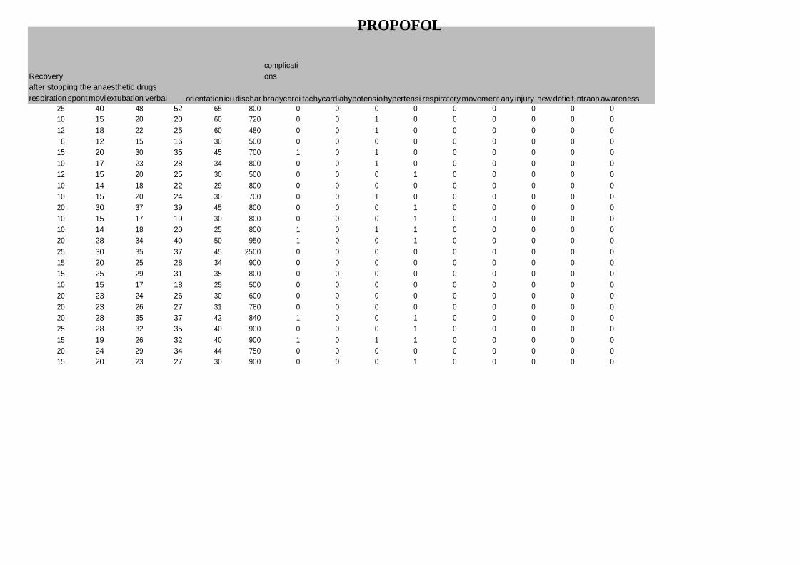
25 40 48 52 65 800 0 0 0 0 0 0 0 0 0
10 15 20 20 60 720 0 0 1 0 0 0 0 0 0
12 18 22 25 60 480 0 0 1 0 0 0 0 0 0
8 12 15 16 30 500 0 0 0 0 0 0 0 0 0
15 20 30 35 45 700 1 0 1 0 0 0 0 0 0
10 17 23 28 34 800 0 0 1 0 0 0 0 0 0
12 15 20 25 30 500 0 0 0 1 0 0 0 0 0
10 14 18 22 29 800 0 0 0 0 0 0 0 0 0
10 15 20 24 30 700 0 0 1 0 0 0 0 0 0
20 30 37 39 45 800 0 0 0 1 0 0 0 0 0
10 15 17 19 30 800 0 0 0 1 0 0 0 0 0
10 14 18 20 25 800 1 0 1 1 0 0 0 0 0
20 28 34 40 50 950 1 0 0 1 0 0 0 0 0
25 30 35 37 45 2500 0 0 0 0 0 0 0 0 0
15 20 25 28 34 900 0 0 0 0 0 0 0 0 0
15 25 29 31 35 800 0 0 0 0 0 0 0 0 0
10 15 17 18 25 500 0 0 0 0 0 0 0 0 0
20 23 24 26 30 600 0 0 0 0 0 0 0 0 0
20 23 26 27 31 780 0 0 0 0 0 0 0 0 0
20 28 35 37 42 840 1 0 0 1 0 0 0 0 0
25 28 32 35 40 900 0 0 0 1 0 0 0 0 0
15 19 26 32 40 900 1 0 1 1 0 0 0 0 0
20 24 29 34 44 750 0 0 0 0 0 0 0 0 0
15 20 23 27 30 900 0 0 0 1 0 0 0 0 0
PROPOFOL
Recovery
after stopping the anaesthetic drugs
respiration spont movi extubation verbal
complicati
ons
orientation icu dischar bradycardi tachycardiahypotensio hypertensi respiratory movement any injury new deficit intraop awareness

Dexmedetomidine- Group D
duration
of
duration anaesthe motor
s no age sex asa diagnosis weight height of sx sia power
rul
lul
rll
lll
current rul
t1
t2
t3
amplitude latency amplitude latency amplitude
1 49 m 2 thoracic 80 168 300 360 5 5 5 5 110 130 25 124 27 130
2 55 f 2 thoracic 60 156 300 360 5 5 4 4 150 312 28 270 31 310
3 50 f 2 cervical 56 158 180 240 4 4 5 5 150 63.8 22 61 24.8 62.8
4 43 f 2 lumbar 60 161 240 300 5 5 5 4 156 200 28 150 31 199
5 28 f 1 thoracic 70 163 240 300 5 5 5 5 100 300 40 270 43 299
6 59 m 2 lumbar 64 160 180 240 5 5 5 5 159 158 22 123 27 158
7 18 f 1 lumbar 46 154 120 180 5 5 5 5 159 198 25 140 29.8 197
8 58 f 2 cervical 70 160 180 240 4 4 5 5 150 140 23.8 125 24 139
9 47 m 2 cervical 64 159 120 180 4 4 5 5 150 172.8 27.1 164 27.5 171
10 49 f 2 lumbar 64 160 300 360 5 5 4 5 160 234 33 212 36 231
11 31 f 1 cervical 46 152 120 180 4 5 5 5 110 118.1 27 110 28 112
12 34 m 1 cervical 72 171 120 180 4 5 5 5 150 72.5 24 72.5 26.2 75.5
13 53 f 2 lumbar 70 168 120 180 5 5 5 5 142 200 25 178 27 198
14 27 m 1 cervical 40 156 120 170 4 4 5 5 142 29.7 26 21.4 27 29
15 37 f 2 cervical 56 156 120 180 4 4 5 5 150 138 30 130 33 137
16 34 m 1 cervical 64 162 120 180 4 5 5 5 100 212 24.3 189 25.4 210
17 56 m 2 cervical 70 169 180 235 4 4 5 5 134 172 35.6 168 35.8 169
18 24 f 1 cervical 65 167 240 300 4 4 5 5 151 150 21 147 22.5 149
19 34 m 1 cervical 88 170 120 180 5 5 5 5 90 304 29 270 31.2 298
20 21 m 1 lumbar 60 162 240 300 5 5 4 5 100 350 25 300 28 345
21 50 f 2 lumbar 50 156 120 178 5 5 4 5 160 248 27 220 31 250
22 22 m 1 lumbar 60 157 180 230 5 5 5 4 143 169 23 140 25 168
23 48 f 2 lumbar 72 162 120 180 5 5 5 5 150 200 24.5 170 27 190
24 38 m 1 cervical 60 160 180 240 4 4 5 5 100 213.4 24 198.7 26.7 213.7
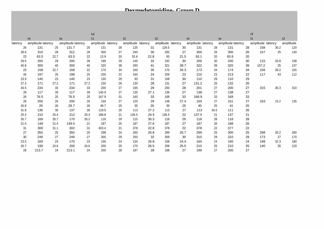
Dexmedetomidine- Group D
t4
te
lul
t1
t2
t3
t4
te
rll
t1
t2
latency amplitude latency amplitude latency amplitude latency amplitude latency amplitude latency amplitude latency amplitude latency amplitude latency amplitude
26 131 25 131.7 25 131 28 125 31 129.5 30 131 28 131 28 158 30.2 120
28.6 310 28 312 28 300 27 240 30 292 27 300 26 300 26 157 25 130
23 63.5 22.7 63.5 22 13.9 20 52.6 22.8 93 21.5 93.1 20 93.9 20
29.5 200 29 200 28 190 29 145 32 192 30 200 30 200 30 115 33.8 108
40.8 300 40 300 40 320 38 290 41 321 39.7 322 39 320 39 157.2 25 137
23 158 22.7 158 22 170 34 165 35 170 34.3 173 34 174 34 156 39.2 155
26 197 25 198 25 200 22 160 24 209 23 210 23 213 22 117 43 112
23.9 140 23 140 23 130 29 93 31 108 30 110 29 110 29
27.3 171 27.2 171 27.2 150 26 130 28 131 27 131.2 26 132 26
34.5 234 33 234 33 200 27 165 29 200 28 201 27 200 27 315 30.3 310
28 117 28 117 28 140.9 27 135 27.1 136 27 138 27 138 27
26 76.5 25 76.5 25 167.9 31 160 33 168 33 168.9 33 169 33
26 200 25 200 25 150 27 120 29 148 27.4 150 27 151 27 163 15.2 135
26.8 29 26 29.7 26 49.7 25 35 26 39 25 40 25 41 25
31.8 136 30 137 30 118.5 26 113 27.2 113 27 113 26.5 111 26
25.3 210 25.4 212 25.3 188.8 21 136.5 24.5 136.5 22 137.5 21 137 21
35.7 169 35.7 170 35.2 118 29 115 30.3 118 29 118 28 118 28
21.5 149 21.4 149.5 21 187 25 187 27.6 187 27 187 26 188 26
31 300 31.1 302 31 403.4 21 378 22.8 378 22 378 22 377 22
27 350 25 350 25 298 24 260 26.8 299 25.7 299 25 300 25 298 35.2 280
30 249 27 249 27 300 29 250 32 309 30 310 29 310 29 173 27 170
23.5 169 23 170 23 156 24 134 26.8 158 24.4 160 24 160 24 199 32.3 180
26.7 190 24.6 200 24.6 200 25 170 26.5 209 25.5 210 25 210 25 140 35 120
26 213.7 24 213.1 24 200 28 187 28 198 27 199 27 200 27
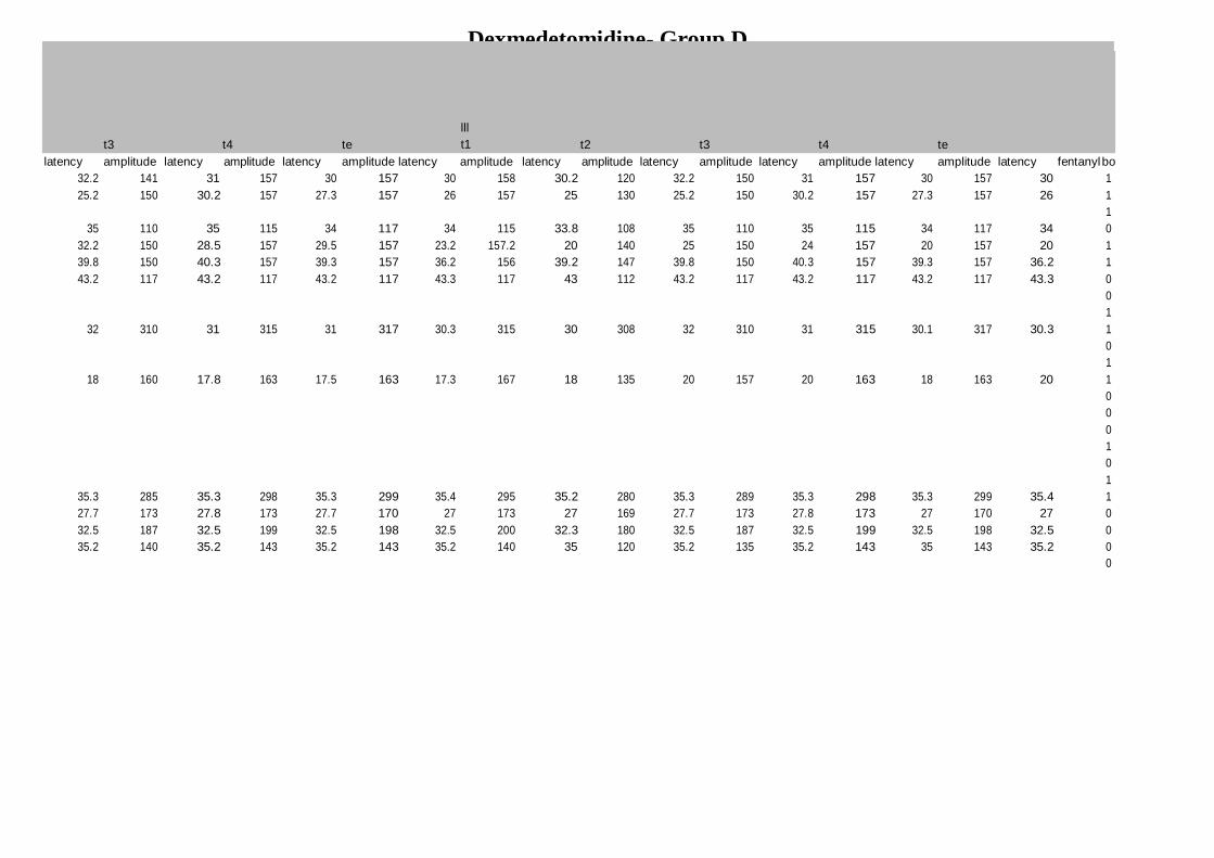
Dexmedetomidine- Group D
t3
t4
te
lll
t1
t2
t3
t4
te
latency amplitude latency amplitude latency amplitude latency amplitude latency amplitude latency amplitude latency amplitude latency amplitude latency fentanyl bo
32.2 141 31 157 30 157 30 158 30.2 120 32.2 150 31 157 30 157 30 1
25.2 150 30.2 157 27.3 157 26 157 25 130 25.2 150 30.2 157 27.3 157 26 1
1
35 110 35 115 34 117 34 115 33.8 108 35 110 35 115 34 117 34 0
32.2 150 28.5 157 29.5 157 23.2 157.2 20 140 25 150 24 157 20 157 20 1
39.8 150 40.3 157 39.3 157 36.2 156 39.2 147 39.8 150 40.3 157 39.3 157 36.2 1
43.2 117 43.2 117 43.2 117 43.3 117 43 112 43.2 117 43.2 117 43.2 117 43.3 0
0
1
32 310 31 315 31 317 30.3 315 30 308 32 310 31 315 30.1 317 30.3 1
0
1
18 160 17.8 163 17.5 163 17.3 167 18 135 20 157 20 163 18 163 20 1
0
0
0
1
0
1
35.3 285 35.3 298 35.3 299 35.4 295 35.2 280 35.3 289 35.3 298 35.3 299 35.4 1
27.7 173 27.8 173 27.7 170 27 173 27 169 27.7 173 27.8 173 27 170 27 0
32.5 187 32.5 199 32.5 198 32.5 200 32.3 180 32.5 187 32.5 199 32.5 198 32.5 0
35.2 140 35.2 143 35.2 143 35.2 140 35 120 35.2 135 35.2 143 35 143 35.2 0
0
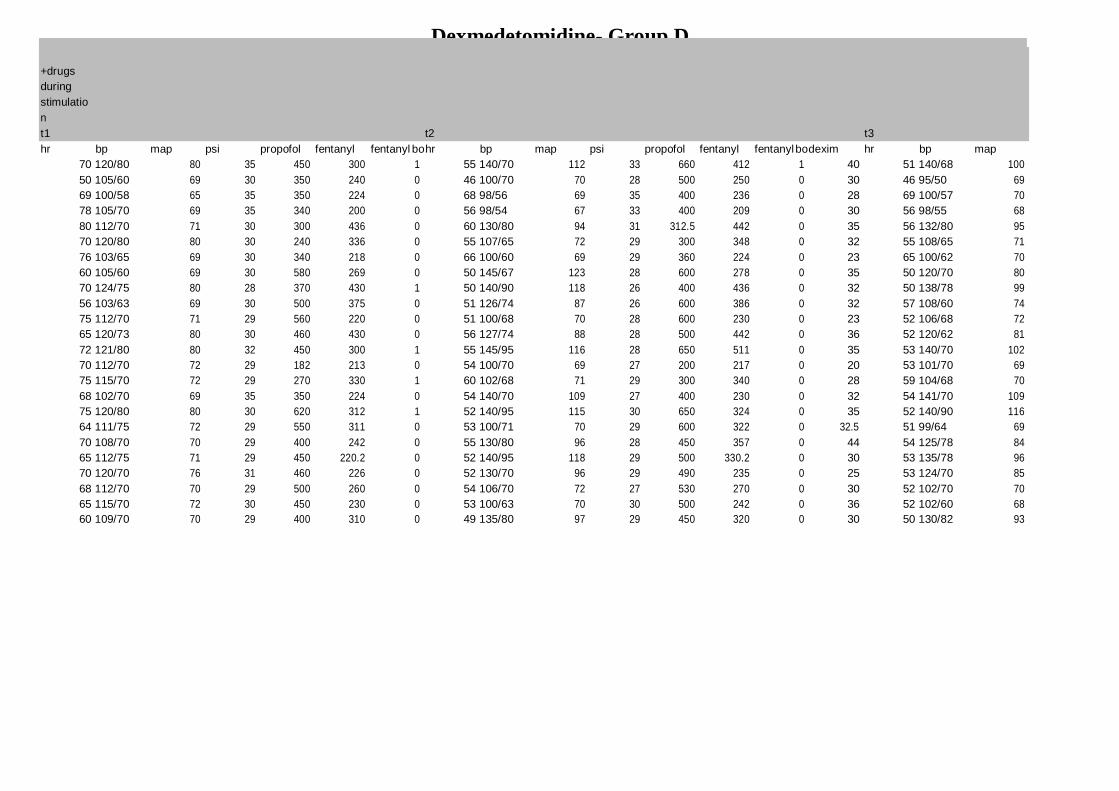
Dexmedetomidine- Group D
+drugs
during
stimulatio
n
t1 t2 t3
hr bp map psi propofol fentanyl fentanyl bo hr bp map psi propofol fentanyl fentanyl bo dexim hr bp map
70 120/80 80 35 450 300 1 55 140/70 112 33 660 412 1 40 51 140/68 100
50 105/60 69 30 350 240 0 46 100/70 70 28 500 250 0 30 46 95/50 69
69 100/58 65 35 350 224 0 68 98/56 69 35 400 236 0 28 69 100/57 70
78 105/70 69 35 340 200 0 56 98/54 67 33 400 209 0 30 56 98/55 68
80 112/70 71 30 300 436 0 60 130/80 94 31 312.5 442 0 35 56 132/80 95
70 120/80 80 30 240 336 0 55 107/65 72 29 300 348 0 32 55 108/65 71
76 103/65 69 30 340 218 0 66 100/60 69 29 360 224 0 23 65 100/62 70
60 105/60 69 30 580 269 0 50 145/67 123 28 600 278 0 35 50 120/70 80
70 124/75 80 28 370 430 1 50 140/90 118 26 400 436 0 32 50 138/78 99
56 103/63 69 30 500 375 0 51 126/74 87 26 600 386 0 32 57 108/60 74
75 112/70 71 29 560 220 0 51 100/68 70 28 600 230 0 23 52 106/68 72
65 120/73 80 30 460 430 0 56 127/74 88 28 500 442 0 36 52 120/62 81
72 121/80 80 32 450 300 1 55 145/95 116 28 650 511 0 35 53 140/70 102
70 112/70 72 29 182 213 0 54 100/70 69 27 200 217 0 20 53 101/70 69
75 115/70 72 29 270 330 1 60 102/68 71 29 300 340 0 28 59 104/68 70
68 102/70 69 35 350 224 0 54 140/70 109 27 400 230 0 32 54 141/70 109
75 120/80 80 30 620 312 1 52 140/95 115 30 650 324 0 35 52 140/90 116
64 111/75 72 29 550 311 0 53 100/71 70 29 600 322 0 32.5 51 99/64 69
70 108/70 70 29 400 242 0 55 130/80 96 28 450 357 0 44 54 125/78 84
65 112/75 71 29 450 220.2 0 52 140/95 118 29 500 330.2 0 30 53 135/78 96
70 120/70 76 31 460 226 0 52 130/70 96 29 490 235 0 25 53 124/70 85
68 112/70 70 29 500 260 0 54 106/70 72 27 530 270 0 30 52 102/70 70
65 115/70 72 30 450 230 0 53 100/63 70 30 500 242 0 36 52 102/60 68
60 109/70 70 29 400 310 0 49 135/80 97 29 450 320 0 30 50 130/82 93
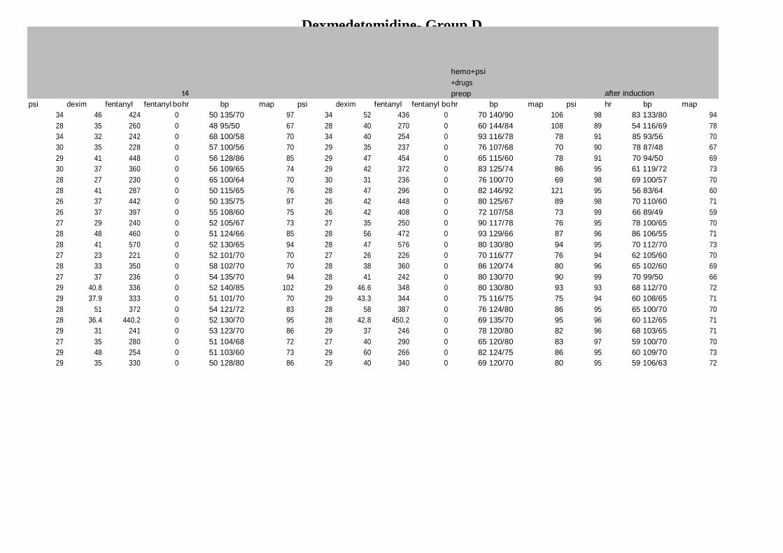
Dexmedetomidine- Group D
t4
hemo+psi
+drugs
preop
after induction
psi dexim fentanyl fentanyl bo hr bp map psi dexim fentanyl fentanyl bo hr bp map psi hr bp map
34 46 424 0 50 135/70 97 34 52 436 0 70 140/90 106 98 83 133/80 94
28 35 260 0 48 95/50 67 28 40 270 0 60 144/84 108 89 54 116/69 78
34 32 242 0 68 100/58 70 34 40 254 0 93 116/78 78 91 85 93/56 70
30 35 228 0 57 100/56 70 29 35 237 0 76 107/68 70 90 78 87/48 67
29 41 448 0 56 128/86 85 29 47 454 0 65 115/60 78 91 70 94/50 69
30 37 360 0 56 109/65 74 29 42 372 0 83 125/74 86 95 61 119/72 73
28 27 230 0 65 100/64 70 30 31 236 0 76 100/70 69 98 69 100/57 70
28 41 287 0 50 115/65 76 28 47 296 0 82 146/92 121 95 56 83/64 60
26 37 442 0 50 135/75 97 26 42 448 0 80 125/67 89 98 70 110/60 71
26 37 397 0 55 108/60 75 26 42 408 0 72 107/58 73 99 66 89/49 59
27 29 240 0 52 105/67 73 27 35 250 0 90 117/78 76 95 78 100/65 70
28 48 460 0 51 124/66 85 28 56 472 0 93 129/66 87 96 86 106/55 71
28 41 570 0 52 130/65 94 28 47 576 0 80 130/80 94 95 70 112/70 73
27 23 221 0 52 101/70 70 27 26 226 0 70 116/77 76 94 62 105/60 70
28 33 350 0 58 102/70 70 28 38 360 0 86 120/74 80 96 65 102/60 69
27 37 236 0 54 135/70 94 28 41 242 0 80 130/70 90 99 70 99/50 66
29 40.8 336 0 52 140/85 102 29 46.6 348 0 80 130/80 93 93 68 112/70 72
29 37.9 333 0 51 101/70 70 29 43.3 344 0 75 116/75 75 94 60 108/65 71
28 51 372 0 54 121/72 83 28 58 387 0 76 124/80 86 95 65 100/70 70
28 36.4 440.2 0 52 130/70 95 28 42.8 450.2 0 69 135/70 95 96 60 112/65 71
29 31 241 0 53 123/70 86 29 37 246 0 78 120/80 82 96 68 103/65 71
27 35 280 0 51 104/68 72 27 40 290 0 65 120/80 83 97 59 100/70 70
29 48 254 0 51 103/60 73 29 60 266 0 82 124/75 86 95 60 109/70 73
29 35 330 0 50 128/80 86 29 40 340 0 69 120/70 80 95 59 106/63 72

Dexmedetomidine- Group D
after intubation 5 min 10 min 15 min 30 min
psi hr bp map psi hr bp map psi hr bp map psi hr bp map psi hr
35 92 144/99 117 34 86 98/57 67 34 85 105/61 71 34 77 125/70 85 34 77
34 63 143/68 116 35 52 97/49 66 32 57 100/54 67 32 54 98/55 67 32 50
35 94 107/62 73 35 88 109/65 75 34 80 105/63 71 35 82 105/65 70 35 89
40 78 109/75 74 40 70 103/68 70 34 67 101/63 69 35 66 105/66 70 35 63
30 78 103/70 71 31 70 104/70 69 30 75 105/70 70 30 72 106/68 70 30 70
30 75 135/85 99 31 70 120/85 85 30 70 120/85 85 30 67 108/70 72 32 67
32 83 117/79 78 31 83 104/64 70 30 80 100/62 69 30 78 87/61 59 30 76
28 58 149/76 123 29 60 136/81 93 30 60 116/64 73 29 59 108/69 70 29 59
28 80 140/80 116 28 70 133/82 95 28 65 128/80 85 28 64 128/76 84 28 60
30 67 98/62 70 29 64 95/52 69 28 61 92/46 62 28 61 99/55 69 28 50
29 86 130/80 98 30 80 120/78 84 29 78 116/76 78 29 70 111/70 74 29 68
30 86 130/58 99 31 80 120/73 83 30 87 113/67 73 29 77 103/56 70 29 77
29 85 131/80 96 30 80 125/70 83 29 78 118/70 78 28 75 119/70 80 29 72
28 68 118/78 76 29 61 112/70 72 29 60 110/70 74 29 61 109/70 71 29 60
30 75 125/78 85 30 72 118/70 75 29 70 115/70 76 29 68 112/68 73 29 65
30 80 110/70 72 30 75 107/60 71 30 70 105/60 70 29 68 107/60 70 29 69
29 80 127/78 86 30 75 121/70 83 30 70 116/67 78 30 68 115/67 73 30 65
29 70 116/71 75 29 64 111/71 76 29 63 110/70 74 29 62 110/70 74 29 62
28 78 114/74 74 30 72 110/70 74 29 70 108/70 71 29 68 107/69 71 29 68
29 78 130/80 98 30 72 125/65 83 30 68 120/80 89 29 65 115/65 76 29 65
31 80 122/84 89 31 74 119/70 76 31 72 116/65 75 30 68 114/62 73 30 68
29 68 121/84 84 30 65 118/74 74 29 65 115/74 74 29 64 111/70 74 29 63
31 70 130/80 96 31 66 121/74 85 30 65 119/70 78 30 64 115/67 75 30 60
29 64 110/70 71 29 60 108/71 76 29 60 109/70 72 29 60 108/69 71 29 60

Dexmedetomidine- Group D
45 min
1 hr
1.5 hr
bp map psi hr bp map psi hr bp map psi propofol dexim fentanyl hr bp map psi
90/46 60 34 68 99/60 70 34 74 131/79 86 32 660 40 548 63 114/69 69 28
90/54 60 31 50 103/56 71 30 50 93/46 68 28 500 31.2 260 60 125/60 71 28
97/56 67 30 70 89/52 67 34 70 102/59 70 34 400 32 242 67 102/60 69 34
101/60 69 35 56 98/64 69 30 55 92/63 67 28 400 35 237 50 93/59 67 28
107/65 70 29 68 106/68 71 30 54 130/80 90 29 312 35 436 54 120/70 72 29
110/70 74 29 68 112/70 75 30 68 113/70 74 30 300 0 336 58 109/65 70 30
104/55 71 30 66 101/60 71 30 69 97/59 69 30 360 23 224 66 109/63 69 29
103/50 71 29 60 102/59 70 29 50 145/67 108 27 600 35 278 50 129/73 89 28
128/75 93 28 60 124/75 83 28 53 140/90 107 26 400 32 436 52 138/80 94 28
103/63 70 27 53 97/62 69 26 50 120/74 83 26 500 32 380 57 111/70 87 28
108/70 73 29 65 108/70 72 29 51 99/54 69 27 600 27 230 52 108/70 69 28
107/56 73 29 75 108/57 72 29 52 124/64 84 28 500 24 454 52 123/62 84 28
115/68 79 29 55 140/92 104 28 54 140/80 95 28 650 35 511 52 125/70 85 28
108/70 71 29 53 99/64 69 27 53 102/64 68 27 200 40 221 52 104/68 68 28
114/67 74 29 59 102/65 70 28 58 107/62 70 28 300 28 330 57 108/70 69 28
110/62 73 29 53 140/70 104 28 52 137/68 87 28 400 32 224 53 135/68 82 28
112/67 72 30 52 145/97 110 30 52 140/89 97 29 650 40.8 336 52 135/70 90 28
109/70 70 29 51 100/62 65 29 51 99/61 68 27 600 38 333 52 105/64 67 28
108/69 72 29 55 130/70 93 28 53 128/71 85 28 450 59 387 52 125/70 82 28
112/75 74 29 53 145/94 112 29 52 140/80 103 28 500 36.4 410 52 135/75 84 28
113/62 73 30 51 136/78 101 28 50 130/70 95 29 490 31 241 53 127/60 81 28
110/70 74 29 62 111/65 72 29 60 112/70 75 29 530 0 260 51 108/70 70 28
110/71 71 30 55 98/62 69 30 54 99/63 69 29 500 48 254 53 102/70 68 29
108/70 72 28 49 135/70 95 28 51 121/80 82 27 450 35 330 50 115/76 74 28

Dexmedetomidine- Group D
2 hr
2.5 hr
3 hr
3.5 hr
hr bp map psi dexim fentanyl hr bp map psi hr bp map psi dexim fentanyl hr bp
59 97/57 69 28 80 618 57 103/63 69 28 57 92/53 65 27 120 668 55 101/63
49 108/52 71 28 62.4 720 43 168/74 120 27 48 129/59 84 27 93.6 880 54 120/65
67 99/64 69 34 56 490 62 92/55 67 27 64 107/65 70 27 84 550
52 95/60 67 28 58 281.6 50 100/62 69 28 50 98/63 65 28 92 345.8 50 97/65
54 112/70 72 29 70 505 53 110/68 71 28 52 110/65 71 28 105 580 52 110/70
58 110/65 70 30 57 408 54 107/64 70 28 54 108/64 70 28 89 480
62 106/62 69 29 46 264
50 120/70 89 28 58 334 50 107/60 70 28 50 105/66 70 27 92 388
52 135/75 94 28 56 605
55 120/80 87 28 56 446 55 130/80 92 28 55 125/70 82 28 82 512 54 120/70
52 104/70 69 28 54 284
52 120/62 84 28 60 526
52 122/68 85 28 58 572
51 106/70 68 28 60 261
57 107/68 69 28 56 390
52 124/70 82 28 56 284
52 130/67 90 28 76 508 52 124/67 82 28 52 120/71 79 28 112 580
52 102/64 67 28 70.5 399 52 105/64 70 27 51 106/64 70 28 103 465 53 105/64
52 120/68 82 28 103 477
52 125/74 84 28 66.4 470 52 120/64 81 28 52 117/73 73 27 96.4 530 52 112/63
53 120/73 81 28 61 395
51 107/62 70 28 60 320 51 101/64 67 28 51 104/65 70 27 90 380
52 105/72 68 29 84 326
50 115/78 74 28 65 390 50 116/70 73 28 51 117/70 73 28 95 450

Dexmedetomidine- Group D
4 hr
4.5 hr
5 hr
map psi hr bp map psi dexim fentanyl hr bp map psi hr bp map psi dexim fentanyl
70 27 55 113/63 73 27 160 728 55 112/64 71 27 63 103/62 69 27 164 778
79 28 53 97/49 69 27 124.8 940 54 99/50 68 27 52 102/57 68 27 156 1000
63
28
51 100/78
70
28 120 399
70 28 52 111/67 71 28 140 650
80 28 54 120/73 79 27 112 578 54 118/76 75 27 54 118/75 74 28 144 644
69 28 53 106/64 69 27 135.5 531
71 27 52 114/73 72 27 120.4 590

respiration spont mov extubation verbal orientation icu dischar bradycardi tachycardiahypotensio hypertensi respiratory movement any injury new deficit intraop awareness
30 45 50 55 64 950 1 0 1 1 0 1 0 0 0
45 55 59 61 70 900 1 0 0 1 0 1 0 0 0
25 35 40 42 50 900 0 0 1 0 0 0 0 0 0
45 48 60 65 65 900 1 0 0 0 0 1 0 0 0
50 51 54 56 58 920 0 0 0 0 0 0 0 0 0
30 34 35 36 40 900 1 0 0 0 0 0 0 0 0
27 35 38 41 45 940 0 0 0 0 0 0 0 0 0
25 35 44 47 50 950 1 0 1 0 0 0 0 0 0
35 45 50 54 57 980 1 0 0 1 0 0 0 0 0
45 60 65 70 75 890 1 0 1 1 0 0 0 0 0
15 20 22 24 24 900 1 0 1 0 0 0 0 0 0
25 30 33 35 36 900 1 0 0 1 0 0 0 0 0
25 30 34 36 37 900 1 0 0 1 0 1 0 0 0
15 19 20 22 24 800 1 0 0 0 0 0 0 0 0
28 32 35 36 36 800 0 0 0 1 0 0 0 0 0
25 35 45 47 50 800 1 0 0 0 0 0 0 0 0
30 45 48 50 53 970 1 0 0 1 0 0 0 0 0
40 45 49 50 52 950 1 0 1 0 0 0 0 0 0
22 24 26 27 27 450 1 0 0 1 0 0 0 0 0
50 65 70 72 72 970 1 0 0 1 0 0 0 0 0
30 40 43 44 44 900 1 0 0 0 0 0 0 0 0
35 45 48 49 49 950 1 0 1 0 0 0 0 0 0
20 28 29 30 34 950 1 0 0 0 0 0 0 0 0
20 35 42 46 48 900 1 0 0 0 0 0 0 0 0
Dexmedetomidine- Group D
Recovery
complicati
ons