comparison of bond strength of …repository-tnmgrmu.ac.in/5062/1/240119717raghunathan.pdfbonded to...
TRANSCRIPT

COMPARISON OF BOND STRENGTH OF PORCELAIN
LAMINATE VENEERS BONDED TO LASER TREATED DENTIN
AND ACID TREATED DENTIN : AN IN VITRO STUDY
A Dissertation submitted
in partial fulfilment of the requirements
for the degree of
MASTER OF DENTAL SURGERY
BRANCH – I
PROSTHODONTICS AND CROWN & BRIDGE
THE TAMILNADU DR.M.G.R. MEDICAL UNIVERSITY
CHENNAI- 600032
2014 – 2017

ADHIPARASAKTHI DENTAL COLLEGE AND HOSPITAL
MELMARUVATHUR – 603319 .
DEPARTMENT OF PROSTHODONTICS AND CROWN &
BRIDGE
CERTIFICATE
This is to cert ify that Dr.J.RAGHUNATHAN, Post Graduate
student (2014-2017) in the Department of Prosthodontics and crown &
bridge, Adhiparasakthi Dental College and Hospital, Melmaruvathur –
603319, has done this dissertation t itled “COMPARISON OF BOND
STRENGTH OF PORCELAIN LAMINATE VENEERS BONDED TO
LASER TREATED DENTIN AND ACID TREATED DENTIN : AN
IN VITRO STUDY.” under our direct guidance and supervision in
partial fulfilment of the regulations laid down by the Tamilnadu
Dr.M.G.R Medical University, Chennai – 600032 for MDS., (Branch-I)
(Prosthodontics and Crown & Bridge) degree examination.
Dr.S.THILLAINAYAGAM MDS.,
Principal
Guide
Dr.A.S.RAMESH MDS.,
Professor & Head
d
Co-Guide
Dr.K.PRABHU MDS.,
Reader

ACKNOWLEDGEMENT
I am extremely grateful to Dr.A.S.Ramesh MDS., Guide,
Professor and Head, Department of Prosthodontics, Adhiparasakthi
Dental College and Hospital, Melmaruvathur. Words cannot express my
gratitude for his quiet confidence in my ability to do the study, his
willingness to help to clear the stumbling bl ocks along the way and his
tremendous patience till the end of the study.
It is my duty to express my thanks to my Co -Guide Dr.K.Prabhu
MDS., Reader for his expert guidance and moral support during the
completion of this study. I consider myself privileg ed, to have studied,
worked and completed my dissertation under them in the department.
My sincere thanks to Dr.S.Thillainayagam MDS., our beloved
Principal, Adhiparasakthi Dental College and Hospital, Melmaruvathur
for providing me with the opportunity to utilize the facilities of the
college.
I thank our Correspondent Dr.T.Ramesh MD., for his vital
encouragement and support.
I am extremely thankful to my teachers Dr.N.Venkatesan MDS.,
Professor , Dr.Arunkumar MDS., Senior lecturer, for their valuable
suggestions, constant encouragement and timely help rendered
throughout this study.

I am thankful and express my gratitude to my previous teachers
Dr.K.Rijesh MDS., Professor , Dr.Lakshmi Devi MDS., Reader , for
their immense help and support for the initiation of this study.
I am extremely grateful to Dr.Premila Suganthan BDS., for
granting me permission to conduct the study in her clinic and helping
me to bring out my study.
I thank Dr.Shyam MDS., Sri Venkateshwara Dental College and
Hospital, Chennai, for helping me with the statistics in the study.
I thank ALMIGHTY GOD for answering my prayers and making
me what I am today.
I owe my gratitude to my mother Mrs.J.Mannammal and my
wife Mrs.D.Anitha who stood beside me as a source of inner strength
and sacrificed so much to make me what I am today. I also thank my
loving daughters R.Jayavarshini & R.Mithra for the unbound love
and joy they brought to my life.
Dr.J.RAGHUNATHAN

DECLARATION
TITLE OF THE DISSERTATION Comparison of bond strength of
porcelain laminate veneers bonded
to laser treated dentin and acid
treated dentin : An in vitro study
PLACE OF THE STUDY Adhiparasakthi Dental College and
Hospital, Melmaruvathur – 603319
DURATION OF THE COURSE 3 years
NAME OF THE GUIDE Dr.A.S.Ramesh MDS.,
NAME OF CO-GUIDE Dr.K.Prabhu MDS.,
I hereby declare that no part of the dissertation will be uti lized
for gaining financial assistance or any promotion without obtaining
prior permission of the Principal, Adhiparasakthi Dental College and
Hospital, Melmaruvathur – 603319. In addition, I declare that no part
of this work will be published either in print or in electronic media
without the guides who has been actively involv ed in dissertation. The
author has the right to reserve for publish work solely with the
permission of the Principal, Adhiparasakthi Dental College and
Hospital, Melmaruvathur – 603319.
Co-Guide Guide & Head of department Signature of cand idate

ABSTRACT
BACKGROUND:
Ideal laminate veneer preparations are supposed to be in enamel
which provides excellent bond strength and ensures the longevity of
the restorations. But certain clinical situations like dental caries
extending to dentin, old restorations require the preparation to be
extended into the dentin; wherein the bond strength gets compromised.
Hard tissue lasers like Er,Cr:YSGG laser has the potential to prepare
dentinal surface for adhesion.
AIM:
The purpose of this study is to co mpare the bond strength of the
conventional acid etch technique and the laser treated dentinal surface
bonded to porcelain laminate veneer restorations.
MATERIALS AND METHODS:
Hundred extracted human maxillary central incisors with
approximately 10 mm anatomic crown length and 8 mm mesio -distal
width are selected. The crown portion of tooth is cut and embedded in
the clear acrylic resin. The labial surface is prepared flat to receive the
porcelain laminates. 50 teeth treated with 37 % phosphoric acid (ac id
etching) and the other 50 teeth are treated with laser etching. Then the
laminate veneers bonded to both these groups. This is kept in disti lled
water for 24 hours and shear bond strengths are tested with Universal
testing machine. Then the results are statistically analysed.
RESULTS:
The mean and standard deviation value of Group A (Acid) is
8.5180 and .22829 with a maximum value of 8.9 and a minimum value
of 8.1. The mean and standard deviation value of Group B (Laser) is
8.4980 and .11156 respectively with a maximum value of 8.7 and a
minimum value of 8.3 .
CONCLUSION:
Within the limitations of this study, 37% ortho -phosphoric acid
(8.5 Mpa) and laser (8.49 Mpa) treated dentin surfaces showed similar
bond strength values. The differences are not s tatist ically significant.
But the results of the Laser etched bond strength were more consistent
than Acid etched bond strength.
(Key words : Porcelain laminate veneers, Acid etching, Laser etching.)

CONTENTS
S.NO: TITLE PAGE NO:
1. INTRODUCTION 1
2. AIM AND OBJECTIVES 6
3. GENERAL REVIEW ON LASERS 7
4. REVIEW OF LITERATURE 18
5. MATERIALS AND METHODS 39
6. RESULTS 50
7. DISCUSSION 56
8. CONCLUSION 70
9. REFERENCES 71
10. ANNEXURE 76

LIST OF PICTURES
FIGURE NO: TITLE PAGE NO:
1. Dental laser wavelengths on the
electromagnetic spectrum
43
2. Collection Of Samples 43
3. Preparing crown portion 44
4. Embedded crown in clear acrylic resin 44
5. Prepared flat labial surface 44
6. Acid etched surface 45
7. Laser settings 45
8. Laser etched surface 45
9. Prepared veneer 46
10. Bonded veneer to tooth surface 46
11. Universal testing machine 46
12. Chisel placed at dentin-veneer interface 47
13. Components of laser unit 47
14. Laser tissue interaction 47
15. Absorption curves of the prime oral
chromophores
48
16. Acid etched Enamel 48
17. Acid etched Dentin 48
18. Laser etched Enamel 49
19. Laser etched Dentin 49

LIST OF TABLES
TABLE NO: TITLE PAGE NO:
1. Shear Bond Strength Values 52
2. Descriptive Statist ics 53
3. Comparison between two groups 53
LIST OF GRAPHS
GRAPH NO: TITLE PAGE NO:
1. Bond Strength Values of Either
Group with Respective Trend Line
54
2. Mean Bond Strength 55

Introduction
1
INTRODUCTION
The populari ty of porcelain laminate veneers has increased due
to its conservative tooth preparation and high end aesthetics .1 The
long-term clinical success of porcelain veneers depends on careful case
selection, treatment planning and tooth preparation .2
Laminates made of porcelain do offer solutions that are both
conservative in nature and esthetically pleasing for the foll owing
clinical situations .3
Discoloration: Teeth discolored by tetracycline staining, devitalization
and fluorosis, and even teeth darkened with age can be given younger,
brighter-looking smiles.3
Enamel Defects: Different types of enamel hypoplasia and
malformations can be masked / corrected .3
Diastemata: Gaps and other multiple unsightly spaces in the anterior
region can be closed.3
Malpositioned Teeth: Indicated in cases where orthodontics is not the
treatment of choice or if the patient has unav ailability of total
treatment time. An i llusion is created by changing the shape, position,
size and surface appearance of a malpositioned / rotated tooth and
making it appear straight. Ex: increasing the length or width of the
anterior teeth.3
Malocclusion: The configuration of lingual surface of anterior teeth
can be modified to develop increased guidance or centric holding cusp
areas in malocclusions or periodontally compromised teeth .3

Introduction
2
Poor Restoration: Teeth with multiple shallow, unaesthetic
restorations on labial surfaces can be restored to naturality .3
Aging: The on going process of aging can result in color changes and
wear in teeth. These teeth may be ideal candidates for improvement by
bleaching or, in certain situations, bleaching with subsequent
veneering.3
Wear Pattern: Porcelain laminates are also useful in those cases that
exhibit slowly progressive wear patterns. If sufficient enamel remains
and the described increase in length is not exces sive, porcelain veneer
can be bonded to the remaining tooth structure to change shape, color,
or function.3
Porcelain laminate veneers are a powerful prosthetic tool to
improve esthetics with minimal loss of tooth structure .4 The technique
requires a shallow reduction of the enamel on the labial surface. Some
authors believe tooth preparation is unnecessary for porcelain veneer
restoration. Tooth preparation is necessary for the following reasons,
they provide for, the strength, restorative area and colo r of the
restoration.2 The prepared area for porcelain veneer should be in the
enamel to maximize the resin bond strength and decrease the tensile
stresses in the porcelain .2
The key to success is by understanding, that the final restoration
obtained is a combined metamerism of the tooth, the resin cement used
for bonding, and the ceramic laminate veneers used for the restorative
procedure.5 The main aim of the tooth preparatio n is that it should be

Introduction
3
simple and minimal reduction of sound tooth structure .6 There are
different approaches in tooth preparation for porcelain laminate
veneers. Tooth preparation for porcelain laminate should be intra -
enamel to maximize the resin bond s trength.7
The resin bonding is a quite reliable and p redictable method in
the enamel. For bonding of ceramic laminate veneer, presence of
enamel thickness is important. Pretreatment of the tooth surface is
essential for establishing a strong bond between the ceramic and both
the enamel and dentin .8
The acid-etching technique with phosphoric acid, introduced by
Buonocore' in 1955, is still used to create an irregular surface of
beveled enamel.9 The retention to the dentinal surface is mainly due t o
the formation of hybrid layer and the micro -mechanical retention
offered by the resin tags embedded in the dentin .1 0
The bonding of resin to enamel surface is gained via micro -
mechanical retention on the irregular surface in cavity preparation.
Therefore, the formation of a hybrid layer and resin tags is essential
for the establishment of a strong bond at the dentin level .1 0
This type
of strong bond can be achieved by complete dissolution of the smear
layer and demineralization of t he intertubular dentin and peritubular
dentin by acid etching method, resulting in an exposed collagen matrix
that can be infiltrated by resin .1 0
Bonding to dentin without dissolution
of smear layer reduces the bond strength because the smear layer
obliterates the tubules and reduces dentinal permeability by 86% .1 1

Introduction
4
In recent years, there has been a growing debate about the use of
lasers for various applications in dentistry, including carious dentin
removal or cavity preparation .1 0
Laser irradiation of dental hard tissues
modifies calcium to phosphorus ratio, reduces carbonate to phosphate
ratio, and leads to the formation of more stable and less acid -soluble
compounds, thus reducing susceptibility to acid attack and caries .1 2
Advancements in laser technology have led to multiple dental
applications such as soft tissue surgery, composite photo -
polymerization, tooth whitening, endodontic procedures, and caries
removal and cavity preparation with minimal pain and discomfort .1 3
Kastler developed the “optical pumping effect” mechanism. LASER is
Light Amplification by Stimulated Emission of Radiation described by
Gordon Gould in 1957. The first laser used was Ruby laser by Maiman.
Then various researches were done regarding this and Maiman found
the therapeutic effect of the laser device as a “bloodless” surgical
machine and used as a “dentist’s drill”. In the year 1990, introduction
of first laser for general dentistry was done by Myers and Myers .1 4
A laser beam is created from a substance known as an active
medium, placed between two optically parallel and extremely high
reflecting mirrors with one of them partially transmitting, and an
energy source to pump active medium. This may either be a solid,
liquid, or gas. They have the property to amplify the ampli tude of the
light wave passing through i t by stimulated emission .1 4

Introduction
5
The pumping source may be electrical or optical. The gain
medium used between pair of mirrors are placed in such a way that
light oscillating between the mirrors passes every time throug h the gain
medium placed in between mirrors, thereafter attaining considerable
amplification which emits through the transmitting mirror .1 4
(figure:1).
Laser etching is a painless procedure making the clinical
procedure highly attractive for routine cli nical use. The surface
produced by laser etching is ideal for adhesion and is also acid
resistant .1 The erbium lasers with very short pulse durations easily
ablade layers of calcified tissue with minimal thermal effects. For
better adhesion to dentin, some authors have suggested the use of
lasers for dentin conditioning, as laser irradiation can provide an
apparently micro retentive surface, free of a smear layer and open
dentinal tubules .1 5
Erbium, Chromium: Yttrium-Scandium-Gallium-Garnet (Er, Cr:
YSGG) and Erbium: Yttrium-Aluminium-Garnet (Er:YAG)lasers can
ablade enamel and dentin effectively because of the high absorption by
water and hydroxyapatite .1 0
Surfaces irradiated by these lasers showed
a characteristic irregular surface area, with clean and without debris
around it, leads to opened dentinal tubules, and micro -irregularit ies
caused by the preferential removal of the inter -tubular dentin,
suggesting that the resultant dentin surface is receptive to adhesive
procedures.1 0

Aim and Objectives
6
AIM AND OBJECTIVES
Ideal laminate veneer preparations are supposed to be in enamel
which provides excellent bond strength and ensures the longevity of
the restorations. But certain clinical situations like dental caries
extending to dentin, old restorations require the preparation to be
extended into the dentin; wherein the bond strength gets compromised.
Hard tissue lasers like Er, Cr:YSGG laser has the potential to prepare
dentinal surface for adhesion.
The purpose of this study is to compare the bond strength of the
conventional acid etch technique and the laser treated dentinal surface
bonded to porcelain laminate veneer restorations.

General Review On Lasers
7
GENERAL REVIEW ON LASERS
HISTORY OF LASERS:
It all started with MAX PLANCK’s work in 1900 that provided
the understanding that light is a form of electromagnetic radiation.
Without this understanding, LASER would not have evolved. The
principle of the laser was first known in 1917, when physicist Albert
Einstein described the theory of stimulated emission. The first laser
was developed by Theodore H. Maiman . Using a theory originally
postulated by Einstein, Maiman created a device where a crystal
medium was stimulated by energy, and radiant, laser light was emitted
from the crystal .1 4
Approximately, the history of lasers begins
similarly to much of modern physics, with Einstein. In 1917, his paper
in Physikialische Zeil, "ZurQuantern Theorie der Strahlung", was the
first discussion of stimulated emmission .1 6
At this stage, the engineers were working towards the creation of
what was termed a MASER (Microwave Amplification by the
Stimulated Emission of Radiation), a device that amplified microwaves
as opposed to l ight and soon found use in microwave communication
systems. Townes and the other engineers believed it to be possible to
create an optical maser, a device for creating powerful beams of l ight
using higher frequency energy to stimulate what was to become termed
the lasing medium.1 7

General Review On Lasers
8
Despite the pioneering work of Townes and Prokhorov, i t was
left to Theodore Maiman in 1960 to invent the first Laser using a
lasing medium of ruby that was stimulated using high energy flashes of
intense light. Townes and Prokhorov were later awarded the Nobel
Science Prize in 1964 for their endeavours .1 6
TYPES OF LASERS1 4
:
According to their sources:
Gas Lasers
Crystal Laser
Semiconductors Lasers
Liquid Lasers
According to the nature of emission:
Continuous Wave
Pulsed Laser
According to their wavelength:
Visible Region
Infrared Region
Far infrared
Near infrared
Ultraviolet Region
According to their site of action:
Soft tissue lasers
Hard tissue lasers

General Review On Lasers
9
GAS LASERS:
The HeNe gas laser was first gas laser introduced, many other
gas discharges have been found to amplify light coherently. Gas lasers
using many different gases have been built and used for many
purposes. The helium-neon laser (HeNe) is able to operate at a number
of different wavelengths, however majority are lased at 633 nm; these
relatively low cost but are highly coherent lasers. Commercial carbon
dioxide (CO2) lasers can emit many hundreds of watts in a single
spatial mode which can be concentrated into a tiny spot. This emission
is in the thermal infrared at 10.6 µm Argon-ion lasers can operate at a
number of lasing transitions between 351 and 528.7 nm. A
nitrogen transverse electrical discharge in gas at atmospheric
pressure (TEA) laser is an inexpensive gas l aser, often home-
built. Metal ion lasers are gas lasers that generate deep like the
ultraviolet wavelengths. Helium-silver (HeAg) 224 nm and neon-
copper (NeCu) 248 nm are two examples .1 4
CONTINUOUS WAVE:
Applications of lasers depend on a beam whose outp ut power is
constant over time and steady when averaged over any longer time
periods, with the very high frequency. The power variations had little
or no impact in the intended application. Such a laser is known as
continuous wave (cw). Many types of lasers can be made to operate in
continuous wave mode to satisfy such an application .1 6

General Review On Lasers
10
PULSED WAVE:
Pulsed operation of lasers refers to any laser not classified as
continuous wave, so that the optical power appears in pulses of some
duration at some repetit ion rate. This encompasses a wide range of
technologies addressing a number of different motivatio ns. Some lasers
are pulsed simply because they cannot be run in continuous mode. In
other cases the application requires the production of pulses having as
large an energy as possible. Since the pulse energy is equal to the
average power divided by the repetition rate, this goal can sometimes
be satisfied by lowering the rate of pulses so that more energy can be
built up in between pulses .1 6
SEMICONDUCTOR DIODE LASERS:
Diode lasers are quite popular due to their compact size and
relatively affordable pricing. A specialized semiconductor that
produces monochromatic light when stimulated electrically is common
to all diode lasers. A simple laser pointer is an example of a diode
laser. Diode lasers can be used in both contact and non -contact mode
and can function with continuous wave or gated pulse modes. They are
not capable of free running pulsed mode. Diode lasers are invisible
near infrared wavelengths and current machines range from 805 –
1064nm. One exception is the Diagnodent caries diagnostic laser wh ich
uses a visible red wavelength of 655 nm. Diode lasers are soft tissue
lasers.1 4

General Review On Lasers
11
The chromophores are pigments such as hemoglobin and melanin,
similar to the Nd:YAG absorption spectrum. Photothermal interactions
predominate whereby diode t issue cutt ing is via thermal energy. They
are quite effective for a host of intraoral soft tissue procedures such as
gingivectomy, biopsy, impression troughing, and frenectomy. Diode
lasers also exhibit bactericida l capabilities and can be used for
adjunctive periodontal procedures. They are also used for laser assisted
tooth whitening. Diode lasers have photobiomodulation properties as
well.1 4
EXCIMER LASERS:
The excimer laser is a special gas laser based on unst able
molecules called excimers. They exist only in the excited state in for
nanoseconds, just long enough for pulsed laser action. Excimer laser
can ablate tissue very effectively via photochemical interaction without
depositing heat therein .1 4
ARGON LASER:
The argon laser operates at a wavelength of 457 to 502
nanometers, using a pulsed or continuous waveform. The argon laser
can be used for a variety of applications, including resin curing and
tooth bleaching. In addition, this laser has a number of soft-tissue
applications, including gingival troughing, esthetic contouring of
gingiva, treatment of oral ulcers, frenectomy and gingivectomy .1 4

General Review On Lasers
12
The primary advantage of the argon laser is that the laser
operates at a wavelength that is absorbed by haemoglobin, which
provides excellent haemostasis. Dentists should be aware that, when
used for resin curing, argon lasers do not necessarily produce a resin
with physical properties superior to those of resins cured with
traditional halogen curing lights. In addition, some resins contain
multiple initiators that activate at different wavelengths. This suggests
that the relatively narrow spectrum of a laser might not be the best
approach to activate the initiators .1 4
HELIUM-NEON LASER:
These types of lasers are one of the forerunners of all lasers
which were at first theoretically proposed and then demonstrated in the
year 1961. Generally only the red 632nm emission was widely applied
as a pointing beam, however becoming replaced by diode lasers of
similar wavelength.1 4
GALLIUM-ARSENIDE /DIODE LASER:
This type of diode laser operates at a wavelength of 904 nm, and
uses a pulsed or continuous waveform and has proven to be successful
with soft-tissue incision and ablation. This laser can be used for the
following: gingival troughing, esthetic contouring of gingival, and
treatment of oral ulcers, frenectomy and gingivectomy. This diode laser
does not affect the inflammatory function of monocytes or endothelial
cells, or the adhesion of endothelial cells. In addition, it can kill some
microbes in the presence of a photosensitizer, as well as some fungi in

General Review On Lasers
13
the presence of some dye photosensitizers. Finally, within certain low -
energy ranges, the diode laser can stimulate the proliferation of
fibroblasts.1 4
NEODYMIUM:YTTRIUM-ALUMINIUM-GARNET(Nd:YAG) LASERS:
Nd:YAG lasers were the first types of true pulsed lasers to be
used for dental procedures in 1990. They are a near infrared
wavelength of 1064 nm. The Nd:YAG laser will penetrate into water to
a depth of 60 mm before it is attenuated to 10% of its original strength.
Therefore, the energy is scattered in soft tissue rather than being
absorbed on the t issue surface as occurs with CO 2 laser energy.
However, since this wavelength is attracted to colo urs, in heavily
pigmented soft tissue such as skin, scattering is about twice as great as
absorption. This heating effect with the Nd:YAG laser is ideal for
ablation of potentially hemorrhagic abnormal t issue, and for hemostasis
of small capillaries and very small venous vessels. Nd:YAG also have
excellent biostimulative properties and unique capacity to stimulate
fibrin formation. This effect is maximized when the pulse duration is
set at 650 microseconds .1 4
However, the scattering effect increases the difficu lty of judging
the depth of penetration, particularly in pale colo ured tissue, since the
surface appearance of the tissue is not a reliable indicator of thermal
damage. The depth of penetration has been estimated to be 2 ± 1 mm in
soft tissue.1 4

General Review On Lasers
14
A Nd:YAG laser device, designed and promoted for oral and
dental applications, can deliver up to 3 watts of power in either a
pulsed (20 pulses per second) or non -pulsed mode, utilizing a specially
designed hand-piece with contact or non-contact probes. This laser also
offers good haemostasis during soft -tissue procedures, which facili tates
a clear operat ing field. In addition, the Nd: YAG laser offers a flexible
fibre delivery system.1 4
The Nd:YAG laser has a number of disadvantages; however, it
has the greatest depth of penetration of all the available dental surgical
laser systems, which means that tissues under the surface are exposed
to laser energy. This is cause for concern because of the risk of
unwanted collateral damage, especially in the underlying bone as well
as the associated postoperative morbidity .1 4
HOLMIUM: YTTRIUM-ALUMINIUM-GARNET( Ho:YAG) LASER:
The Ho:YAG laser operates at a wavelength of 2.1 μm, and uses
a pulsed waveform. This laser is used for soft -tissue incision and
ablation procedures, including the following: gingival troughing,
esthetic contouring of gingival, treatment of oral ulcers, frenectomy
and gingivectomy. The advantages of the Ho :YAG laser center on its
surface effect on tissue. The Ho:YAG laser is less penetrating than the
Nd:YAG laser and, therefore, is faster than the Nd:YAG at cutting soft
tissue. Ho:YAG laser is also bactericidal .1 4

General Review On Lasers
15
CARBON DIOXIDE LASERS:
CO2 Lasers have been available in medicine since the early
1970’s and have been used in dentistry for more than 25 years. They
are a 10,600 nm infrared wavelength, which is highly absorbed by
water. Articulated arms or hollow wave guides are used to transmit
CO2 laser beams and quartz optical fibres cannot be used.1 4
The CO2 gas is in a chamber with nitrogen and hel ium and the
active medium is pumped with an electrical current. CO 2 lasers are
very efficient and exhibit excellent haemostasis . They are currently for
soft tissue uses only. They are continuous wave lasers that can be
operated in gated wave modes, includ ing what are termed as “super -
pulsed” modes. It is important to note even the super -pulsed mode is
not a free running pulsed mode .1 4
These super-pulsed gated modes offer improved surgical control
with less charring of tissue. CO 2 lasers are excellent tools for incising
tissue for multiple purposes. Incisional and excisional biopsies,
frenectomy, gingivectomy, pre-prosthetic procedures are all achieved
with excellent haemostasis , de-epithelialisation of gingival tissue
during periodontal regenerative pro cedures. Sutures are rarely needed
and the controlled thermal effects and sealing of nerve endings often
makes for a very comfortable post -operative experience for the
patient .1 4
The CO2 laser is safe around the implants because the energy is
absorbed into water and not pigments. By affecting the intracellular

General Review On Lasers
16
water of bacteria, the CO 2 wavelength can safely and effectively treat
peri-implanti tis and mucositis , because the energy is not absorbed into
the implant’s surface .1 4
With the CO2 laser, the rapid rise in
intracellular temperature and pressure leads to cellular rupture, as well
as release of vapour and cellular debris, termed the laser plume. The
debris arising from the site of imp act, the char is carbonized tissue by
the laser beam.1 8
ERBIUM AND ERBIUM-CHROMIUM LASERS:
The interest in the Er 3+ laser is based on the wavelength it can
emit i .e. 1.54µm and 2.7 -2.9µm. The former coincides nicely with the
absorption minimum of optical sil ica fibres, al lowing long-range
optical communications. The later wavelength coincides nicely with the
peak of water absorption. As water is contained in every biological
tissue, efficient interaction and dense optical energy deposition is
guaranteed. In medical applications and especially in dentistry, the
Erbium lasers represent highly developed commercial lasers with high
yield and efficiency in tissue removal. For dental hard tissue ablation,
this is currently the type of laser most often used .1 9
ERBIUM:YTTRIUM –ALUMINIUM-GARNET (Er:YAG) LASER:
The laser operates at a wavelength of 2.94 μm and in a pulsed
waveform. The FDA has cleared it for use on cementum and bone, and
it has a variety of hard-tissue applications, including the following
caries removal, cavity preparation in both enamel and dentin,
preparation of root canals. The Er:YAG laser has a number of

General Review On Lasers
17
advantages as it produces clean, sharp margins in enamel and dentin,
and in addition, pulpal safety is not a significant concern because th e
depth of energy penetration is negligible. Pulp may respond even better
to preparations, done with the Er:YAG laser than those done with the
bur. When the Er:YAG laser is used for caries removal, it usually does
not require local anaesthesia. The laser i s antimicrobial when used
within root canals and on root surfaces, and it removes endotoxins
from root surfaces. Finally, vibration from the Er:YAG laser is less
severe than that from the conventional high -speed drill , and it is less
likely to provoke discomfort or pain.1 4
ERBIUM,CHROMIUM:YTTRIUM-SCANDIUM-GALLIUM-
GARNET (Er,Cr:YSGG) LASER:
The Er,Cr:YSGG operates at a wavelength of 2.78 μm, with an
extinction length in water of 1.0 μm. The waveform for the
Er,Cr:YSGG laser is pulsed. The Er,Cr:YSGG laser has several hard-
tissue applications enamel etching, caries removal, cavity preparation,
In-Vitro bone cutt ing with no burning, melting or alteration of the
calcium: phosphorus ratio, root canal preparation .1 4
The Er,Cr:YSGG laser has a number of advantages. Multiple uses
for the Er,Cr:YSGG laser make the economics of providing laser
therapy more feasible. The laser produces a rough surface in enamel
and dentin without significant cracking. In dentin, no smear layer
remains, this suggests good results with bonding. The Er,Cr:YSGG
laser is safe for the pulp. When using the Er,Cr:YSGG laser, no need to
administer local anesthetic for caries removal and cavity preparation .1 4

Review of Literature
18
REVIEW OF LITERATURE
Marco Franchi et al (1995)9 stated that the best removal of dentinal
smear layer resulted from the 37% phosphoric acid treatment; al l the
dentinal tubules orifices appeared to be completely opened. In this
study three standard occlusal cavities with bevelled enamel margins
were prepared on each tooth and etched with the etching solutions of
three dentinal adhesive systems; ( 1 ) 37% phosphoric acid solution. (2)
4.3% oxalic acid and 2.6% aluminium salts solution, and (3) 10%
maleic acid solution. Scanning electron microscopic analysis revealed
that al l the etching solutions affected the enamel surface morphology.
The solution of oxalic acid and aluminium salts removed primarily the
prism core material and partially the periphery of the pr isms, but did
not affect the non bevelled enamel surface. Phosphoric acid and maleic
acid removed both prism core materials and prism periphery; these
specimens also showed areas in which no prism morphology could be
detected. These two acids also removed apatite crystals from the prism
core of the intact enamel surface.
Arturo Martinez-Insua et al (2000)1 2
stated that for both enamel and
dentin, mean tensile strength of bonded brackets obtained after laser
etching were significantly lower than those obtai ned after acid etching.
In this study 80 healthy human premolars were used . Brackets were
cemented to acid-etched enamel, laser -etched enamel, acid-etched
dentin, or laser-etched dentin (20 teeth per group). Dentin was
previously exposed using a high speed hand -piece. Acid etching was

Review of Literature
19
with 37% ortho-phosphoric acid (15 seconds for enamel, 5 seconds for
dentin). Laser etching was with Er:YAG laser (four 200mJ pulses per
second for enamel; four 160 mJ pulses per second for dentin). Brackets
were bonded with auto-curing resin paste, having first applied a primer
(dentin only) and then light cured bonding resin. Tensile strength was
determined with a universal testing machine. Result shows that bond
failure after laser etching was due to micro -cohesive fracture of tooth
tissue.
M Peumans et al (2000)7 stated that there is a general ag reement
among the practitioners that porcelain veneers will play a vital role in
elective dental aesthetics. This places high demands on predictability,
especially with colour matching and masking methods. In addition,
patient acceptance of porcelain veneers in these clinical studies was
high. Porcelain veneers are steadily increasing in populari ty among
today‘s dental practi tioners for conservative restoration of unaesthetic
anterior teeth. This current literature was therefore reviewed in search
for the most important parameters determining the long term success of
porcelain veneers. The adhesive porcelain veneer complex has been
proven to be a strong complex in vitro and in vivo. An optimal bonded
restoration was achieved especially if the preparation wa s located
completely in enamel, if correct adhesive treatment procedures were
carried out and if a suitable luting composite was selected. The
maintenance of aesthetics of porcelain veneers in the medium to long
term was excellent. Major shortcomings of th e porcelain veneer were
described as a relatively large marginal discrepancy, and an

Review of Literature
20
insufficient wear resistance of the luting composite. Although these
shortcomings had no direct impact on the clinical success of porcelain
veneers in the medium term, the ir influence on the overall clinical
performance in the long term is still unknown and therefore needs
further study.
Serdar Usumez et al (2002)2 2
s tated that etching of enamel surface
with an Er,Cr:YSGG hydrokinetic laser system yielded statistically
similar but lower and less predictable bond strengths than did etching
with 37% phosphoric acid for 30 seconds. On the other hand, laser
etching was found to be more practical and faster than conventional
acid etching. Irradiation of enamel with laser energy changes the
physical and chemical characterist ics of the enamel surface, and these
alterations hold promise for the conditioning of enamel for bonding
procedures. This laboratory study examined the influence of laser
irradiation of enamel at 2 different po wer settings with an erbium,
chromium: yttrium, scandium, gall ium garnet (Er,Cr:YSGG)
hydrokinetic laser system on the shear bond strength of orthodontic
appliances and compared these with that of acid etching. The prepared
surfaces of 40 non-carious, intact, extracted premolars were exposed to
laser energy: 20 teeth at 2W setting and 20 teeth at 1W sett ing of the
commercial laser unit. 20 teeth were etched with 37% ortho -phosphoric
acid. Brackets were bonded with an orthodontic no -mix adhesive, and
shear bond strength was determined with a universal testing machine.
Laser etching was found to be more practical and faster than
conventional acid etching.

Review of Literature
21
Mitsuharu Okamoto et al (2003)4 stated that plasma curing for 3
second is sufficient to obtain similar bond strengths and stabili ty to
specimens cured with halogen l ight for 40 second for 1 -mm-thick
porcelain and that plasma curing for 5 second is sufficient even for
porcelain of 2-mm thickness. Plasma and halogen lamp units were used
to photo-cure a composite resin for porcelain bonding. Mean shear
bond strengths to dentin after light curing through porcelain of 1 and
2mm thickness ranged from 20 to 27 MPa before and after thermo -
cycling, indicating no significant effects due to porcelain thickness,
curing method, or thermo-cycling, except for plasma curing for 3
second through porcelain of 2 mm thickness that overcame thermo -
cycling.
GC Lopes et al (2003)1 1
stated that for normal occlusal dentin, no
difference exists in bond strength when 35% phosphoric acid etchant is
applied following the manufacturer‘s suggested time (15 seconds), or
when the time is extended to 30 seconds. Bonding to sclerotic dentin
resulted in lower bond strength to resin composite. Extending the
phosphoric acid etching time can overcome this difficult factor with no
detrimental effect to normal dentin considering the bond strength. In
sclerotic dentin, the Hybrid Layer was very thin, with minimal resin
tags in the dentinal tubules and, when present, they were shorter.
Doubling the etching time resulted in more resin tags with the Hybrid
Layer formation on peritubular dentin. The Hybrid Layer on normal
dentin was thicker when it was acid etched for 30 seconds. Numerous
resin tags were present with both etching times. The results suggest

Review of Literature
22
that the higher mineral amount in sclerotic dentin makes it difficult to
bond to this substrate, resulting in a lower µ-TBS. However, doubling
the etching time resulted in µ -TBS similar to normal dentin.
Aslihan Usumez et al (2003)1 stated that 37% ortho-phosphoric acid
and maleic acid treated enamel surfaces showed statistically similar
bond strength values. Porcelain laminate veneers demonstrated the
highest bond strength to 37% ortho-phosphoric acid etched and
Er,Cr:YSGG hydrokinetic laser system - conditioned tooth surfaces. The
differences were not stat istically different. In this study 40 extracted
caries and restoration free human maxillary central incisors were used.
The teeth were sectioned 2 mm below the cement -enamel junction. The
crowns were embedded in autopolymerising acrylic resin with the
labial surfaces facing up. The labial surfaces were prepared with .05
mm reduction to receive porcelain veneers. The teeth were divided in to
4 groups of 10 specimens. Thirty specimens received one of the
following surface treatments before the bonding of IPS Empress 2
laminate veneers : (1) laser radiation from an Er,Cr:YSGG laser unit ;
(2) 37% ortho-phosphoric acid; and (3) 10% maleic acid. Ten
specimens received no surface treatment and served as the control
group. The veneers were bonded with dual polymerizing resin,
Variolink II. One microtensile specimen from each of the cervical and
incisal thirds measuring 1.2 × 1.2 mm was prepared wit h a slow speed
diamond saw sectioning machine with a diamond rim blade. These
specimens were attached to opposing arms of the microtensile testing
device with cyanoacrylate adhesive and fractured under tension at a

Review of Literature
23
crosshead speed of 1 mm/min, and the maxi mum load at fracture (kg)
was recorded. No statistically significant differences were found
among the bond strengths of veneers bonded to tooth surfaces etched
with Er,Cr:YSGG laser (12.1±4.4 MPa), 37% ortho -phosphoric acid
(13±6.5 MPa), and 10% maleic acid (10.6±5.6 MPa). The control group
demonstrated the lowest bond strength values in all test groups.
Statist ically significant differences were found between the bond
strengths of cervical and incisal sections (p˂.001). Invitro microtensile
bond strengths of porcelain laminate veneers bonded to tooth surfaces
that were laser etched showed results similar to ortho -phosphoric acid
or maleic acid etched tooth surfaces.
John R Calamia et al (2007)5 stated that etched porcelain veneer
technology has demonstrated long-term clinical success. It has proved
to be one of the most successful modali ties of treatment that modern
dentistry has to offer. The relatively few difficulties that have been
encountered may be circumvented or eliminated if the practi tioner pays
close attention to detail. Development of new products and materials is
expected to bring longer term success. Since its introduction more than
two decades ago, etched porcelain veneer restoration has proved to be a
durable and aesthetic modality of treatment. These past 25 years of
success can be attributed to great attention to detail in the following
areas: (1) planning the case, (2) conservative (enamel saving)
preparation of teeth, (3) proper selection of ceramics to use, (4) proper
selection of the materials and methods of cementation of these
restorations, (5) proper finishing and polishing of the restorations,

Review of Literature
24
and (6) proper planning for the continuing maintenance of these
restorations. This art icle discusses failures that could occur if
meticulous at tention is not given to such details. Failures that did
occur structurally and aesthetically warned individuals who were
learning the procedure what to watch for. Some concerns as to newer
products and methods and their effect on the continued success of t his
modality of treatment are also addressed.
Galip Gurel (2007)6 stated that porcelain laminate veneers have been
one of the most used restorations for aesthetics. Although this
approach is one of the most conservative treatment options, some rules
must be followed. Best of all , however, is that it allows for minimal
preparation on the recipient tooth. Tooth preparation is one of the most
important considerations in this technique. Bonding to enamel rather
than dentin provides the best/strongest bond values when we want to
bond porcelain to tooth structure. When a porce lain veneer restoration
is bordered on all margins by enamel, microleakage or debonding of
these restorations is not likely to occur. A main objective of any
restorative case involving these restorations is to keep the preparation
simple and be conservative in reduction of sound tooth structure. Many
other considerations come into play as the preparation becomes more
aggressive and dentin is involved. A rigid veneer behaves differently
when bonded to a rigid surface, such as porcelain, versus a less rigid
surface, such as dentin, and the composite cement can only absorb so
much of the stresses to which the restoration may be exposed. To

Review of Literature
25
minimize effects and possible problems, we should be precise and
careful about case selection and tooth preparation.
Sevil Gurgan et al (2008)3 2
stated that dentin surfaces prepared with
the Er,Cr:YSGG laser and etched with the laser (at either 1.25 or 3
Watt) may provide comparable or increased composite resin bond
strengths, depending on the adhesive used. The present study compared
the in vitro shear bond strength of a nano -hybrid composite resin to
human dentin that was prepared with either the diamond bur or the
Er,Cr:YSGG laser, and treated with two different energy settings of
laser (1.25 and 3 Watt), and two different adhesive systems (a
conventional etch-and-rinse system that requires prior conditioning
with phosphoric acid, and a two-step self etching system). In this
study, they modified the surface morphology by cutting the tooth
surface with a bur or an Er,Cr:YSGG laser, and then conditioned the
surfaces with a bonding agent used with or without phosphoric acid or
with self-etching primer, and they laser -treated them at different
energy settings. In the present study, the highest bond strengths were
achieved by laser preparation, laser etching, and application of Excite.
Excite is a two-step etch and-rinse system, in which the primer and
adhesive resin are combined into one solution. Generally, an etch -and-
rinse procedure involves the use of phosphoric acid that demi neralizes
and exposes the collagen fibres on the dentin surface.

Review of Literature
26
Mathew C A et al (2010)3 stated that the ceramic laminate veneer
remains the prosthetic restoration that best compiles the principles of
present- day aesthetic dentistry. This ―substitute e namel‖ now brings
us closer to achieving the goals of P rosthodontics; to replace human
enamel to its proper structure, shape and colour with this ―bonded
art ificial enamel‖. The history of aesthetic or cosmetic dentistry can be
dated back to the Japanese customs of decorative tooth staining called
―Ohoguro‖ which was documented 4000 years ago. Dr. Charles L.
Pincus introduced the concept of veneering anterior teeth with
laminates when approached by Hollywood directors in 1928. It was
Buonocore's research about the acid etching technique in 1955, which
provided a simple method of increasing adhesion to enamel surface for
acrylic materials. But only after the introduction of light cured
composites in 1970 did the dentist have the necessary working time to
properly shape direct laminate veneers. In the mid 70's and 80's the
composite resin laminate veneers, (with or without facing evolved). At
first the composites were directly bonded to teeth and called
―bonding‖.
Cafer Turkmen et al (2010)1 3
stated that the Er,Cr:YSGG laser-
powered hydrokinetic system etched the enamel surface more
effectively than 37% phosphoric acid for subsequent attachment of
composite material. In this study sixty extracted caries - and restoration
free human maxillary central incisors were used. The teeth were
sectioned 2 mm below the cement -enamel junction. The crowns were
embedded in auto-polymerizing acrylic resin with the labial surfaces

Review of Literature
27
facing up. The labial surfaces were prepared with 0.5 -mm reduction to
receive composite veneers. Thirty specimens were etched with
Er,Cr:YSGG laser. This group was also divided into three subgroups,
and the following three bonding systems were then applied on the laser
groups and the other three unlased groups: (1) 37% phosphoric acid
etch + Bond 1 primer/adhesive (Pentron); (2) Nano bond self-etch
primer (Pentron) + Nano-bond adhesive (Pentron); and (3) all-in-one
adhesive—single dose (Futurabond NR, Voco). All of the groups were
restored with a nanohybrid composite resin (Smile, Pentron). Shear
bond strength was measured. There were no significant differences in
shear bond strength between self -etch primer + adhesive and all -in-one
adhesive systems for non-etched and laser-etched enamel groups (P >
.05).
Vinicius R. Geraldo-Martins et al (2010)3 3
stated that the bonding
effectiveness of adhesives to laser -irradiated dentin may be influenced
not only by the structural substrate alterations induced by the laser, but
also by the characteristics of the adhesive employed. Taking into
consideration the experimental conditions of the present study, it can
be concluded that the use of the Er,Cr:YSGG laser prior to the
application of an etch and rinse adhesive did not negatively influence
the action of the adhesive system in the dentin surface, since the
results obtained for irradiated samples were not statistically different
from those of control groups.

Review of Literature
28
Guilherme Carpena Lopes et al (2011)2 4
stated that bonding to old
dentin with 30 seconds of etching time resulted in higher bond strength
and more homogeneous hybrid layer formation than dentin acid etched
for 15 seconds. According to this study the modification of dentin by
physiological sclerosis due to aging should be considered during
adhesive procedures. The clinical application of Adper Single Bond in
old dentin after 15 seconds of acid etching may produce lower bond
strengths. By simply doubling etching time from 15 to 30 seconds,
bonding to old dentin resulted in higher bond strengths that were
similar to those found in young dentin. Further investig ation must be
conducted with other etch-and-rinse simplified adhesives. Whether
increasing etching time results in increased longevity of restorations in
elderly patients remains to be determined. This issue requires further
clinical investigation.
Subutay Han Altintas et al (2011)2 6
stated that selection of the
provisional cement is an important factor in the ultimate bond strength
of the final restoration. Calcium hydroxide provisional cement and
cleaning with a dental explorer are advisable. In the curr ent study,
three provisional cements and two cleaning techniques were evaluated
for their effects on the final bond strengths of Porcelain laminate
veneers. The results obtained did not support the research hypothesis
as no differences were found between the cleaning procedures. Freshly
cut dentin is the ideal substrate for dentin bonding. In practice, freshly
cut dentin is present only at the time of tooth preparation, prior to
taking impressions. Significant reductions in bond strength can occur

Review of Literature
29
when dentin is contaminated with various provisional cements,
compared to freshly cut dentin. Since some period of time is necessary
to make the final restoration, a provisional restoration is used for
approximately 10 days after the fixation. This makes mechanical and/or
chemical cleaning procedures necessary before the definitive
restoration is cemented. In this current study, after a period of 1 week,
provisional cements were found to affect the final bond strength to
dentin, although the bond strength test achieved acceptable values for
all cases.
Zahra Jaberi Ansari et al (2012)8 stated that the micro-shear bond
strength of groups prepared by bur cutting and acid -etching were
higher than that of groups prepared and etched by an Er,Cr:YSGG laser
within the condi tions of this study. Therefore, re -etching with
phosphoric acid would be recommended if an Er,Cr:YSGG laser is used
for tooth preparation or surface treatment. Enamel preparation by the
Er,Cr:YSGG laser results in a characteristically chalky surface.
Scanning electron microscopic images showed that laser irradiation
produces a surface that increases the restorative material retention,
which in turn makes the surface suitable for the application of
composite and composer filling materials.
Eugenia Koliniotou-Koumpia et al (2012)2 8
stated that the cavities
prepared by laser seem less receptive to adhesive procedures than
conventional bur-cut cavities. Superficial and deep dentin specimens
from human molars were treated either with carbide bur or an Er:YAG

Review of Literature
30
laser. Two etch and rinse adhesives (Single Bond and XP Bond) and
two self-etch adhesives (Prompt L-Pop and Xeno III) were employed to
bond the composite. Shear bond strength (SBS) was determined after
storage in water for 24 hours using a universal testing m achine with a
crosshead speed of 0.5 mm/min. Failure patterns and modes were
analyzed and evaluated using a stereomicroscope. In addition, samples
were processed for Scanning Electron Microscopy (SEM) evaluation. A
linear mixed model was used, and pair wise comparisons were made
using the Bonferroni test. Results showed significant differences
between the levels of dentin treatment (p=.01) in carbide bur -cut dentin
and lased dentin, as well as significant interaction effects due to the
depth of dentin and the bonding system used. The etch and rinse
adhesives bonded less effectively with lased dentin than with carbide
bur-cut dentin, while self -etch adhesives bonded equally well with
lased and bur-cut superficial dentin but much less effectively with
lased deep dentin than with bur-cut deep dentin. SEM revealed a
predominantly adhesive failure mode in laser -ablated fractured
specimens, while a mixed failure mode was apparent in the bur -cut
fractured specimens.
Abdolrahim Davari et al (2013)2 5
stated that when the cavity is
prepared by bur, i t is not necessary to etch the dentin surface by
Er:YAG laser following acid etching and acid etching after laser
etching. In this study the roots of 75 sound maxillary premolars were
sectioned below the CEJ and the crowns were embedded in auto-
polymerizing acrylic resin with the buccal surfaces facing up. The

Review of Literature
31
buccal surfaces were ground using a diamond bur and polished until the
dentin was exposed; the samples were randomly divided into five
groups (n=15) according to the surface treatment: (1) acid etching; (2)
laser etching; (3) laser etching followed by acid etching; (4) acid
etching followed by laser etching and (5) no acid etching and no laser
etching (control group). Composite resin rods (Point 4, Kerr Co) were
bonded to treated dentin surfaces with an etch -and-rise adhesive system
(Optibond FL, Kerr Co) and light -cured. After storage for two weeks at
37°C and 100% humidity and then thermo-cycling, bond strength was
measured. There were no significant differences betw een acid etching
and acid+laser groups, and between laser+acid and laser groups.
Ayoub Pahlavan et al (2013)3 0
stated that the air abrasion after laser
treatment improved the shear bond strength compared to laser group
alone. In this study, 40 human extracted molars divided into 4 groups
(n=10) received the following treatments. Group 1: carbide bur, Group
2: air abrasion with aluminium oxide 50 µm, Group 3: irradiated with
Erbium-Doped Yttrium Aluminium Garnet (Er: YAG) laser (150
mJ/20Hz), Group 4: irradiated with Er:YAG laser (150 mJ/20Hz)+ air.
Specimens in all groups were chemically etched with phosphoric acid
37% and treated with bonding agent (single bond 3M). Then, composite
build-up was performed by tygon tube. After storage in distilled
water at 37°c for one week, al l specimens were subjected to a shear
bond strength test with universal testing machine. Ai r abrasion after
laser treatment improved the shear bond strength.

Review of Literature
32
Ayoub Pahlevan et al (2014)2 stated that the enamel thickness in
different parts of the labial surface is very important. The thickness of
enamel in the gingival area does not permit a cha mfer preparation. The
knife edge preparation is preferable in the gingival area. But is
believed to end with the over contouring. In this study, the thickness of
enamel in different places of labial surface was measured. This
measurement is an important gu ide for the preparation of the tooth in
laminate veneer. The most critical area is the labial gingival third. This
study showed the mean thickness of enamel at the gingival third is 410
μ on the maxillary central incisor and 367 μ on the maxillary lateral
incisor. There are two approaches in the preparation of tooth for
porcelain laminate in the cervical area, chamfer and knife -edge
preparations. The interest in chamfer preparation is because of
cosmetics and avoidance of over contouring. The knife -edge
preparation is believed to end up with the over contouring. The result
of this study showed that in the knife -edge preparation there is no risk
of dentin exposure, whereas in chamfer preparation the risk of dentinal
exposure is significantly higher in the pre paration of porcelain
laminate.
Jing Liu et al (2014)2 9
stated that increased laser influence may lead
to more collateral damage and lower dentin roughness, while scanning
speed and scanning distance were also negatively correlated with
surface roughness. Adequate parameters should be chosen to achieve
therapeutic benefits , and different parameters can result in diverse
ablation results. In this study twelve extracted human premolars were

Review of Literature
33
sectioned into crowns and roots along the cemento -enamel junction,
and then the crowns were cut longitudinally into sheets about 1.5 mm
thick with a cutting machine. The dentin samples were fixed on a stage
at focus plane. The laser beam was irradiated onto the samples through
a galvanometric scanning system, so rectangula r movement could be
achieved. After ablation, the samples were examined with a scanning
electron microscope and laser three-dimensional profile measurement
microscope for morphology and roughness study. With increasing laser
fluence, dentin samples exhibit ed more melting and re-solidification of
dentin as well as debris -like structure and occluded parts of dentinal
tubules. When at the scanning speed of 2400mm/s and scanning
distance of 24μm, the surface roughness of dentin ablated with
femtosecond pulsed laser decreased significantly and varied between
values of dentin surface roughness grinded with two kinds of diamond
burs with different grits. When at the scanning speed of 1200mm/s and
scanning distance of 12μm, the surface roughness decreased slightly,
and the surface roughness of dentin ablated with femtosecond pulsed
laser was almost equal to that grinded with a low grit diamond bur.
Upendra A Hoshing et al (2014)2 3
stated that, for enamel surface,
mean SBS of bonded composite obtained after laser etching were
significantly lower than those obtained after acid etching. Data from
this study demonstrated that bonding to Er; Cr:YSGG laser -etched
surfaces provided markedly weaker valu es than bonding to acid -etched
surfaces. In the laser etched enamel preparations, the high prevalence
of cohesive tooth fractures suggests that disruption as a result of

Review of Literature
34
―micro-explosions‖ weakened the enamel and gave rise to a more
heterogeneous surface than that obtained by acid etching. Acid etching
typically produced a repeating surface pattern, with cracks and fissures
no deeper than 12 mm that are readily fi lled with resin. In contrast to
acid etch treatment, laser etching produced extensive surface f issuring
and less regular and less homogeneous surface patterns arising from
the union of different craters. Fissuring may be related to the
orientation of enamel rods, because enamel is an anisotropic material.
One of the potential disadvantages of enamel acid etching is that the
acid causes demineralization of the most superficial layer. As a result ,
this surface becomes more susceptible to long -term acid attack and
caries, especially when resin impregnation is defective because of air
bubbles or saliva contamination. Such effects are particularly
important given that plaque tends to accumulate at interfacial surfaces.
The physicochemical changes caused by laser etching can be expected
to decrease long-term susceptibili ty to acid attack and caries. This
reduction may be related to changes in Ca:P ratio, reduced carbonates,
and pyrophosphate formation, together with reduced water and organic
component contents. It has also been suggested that laser etching may
create remineralisation micro -spaces that trap f ree ions. Nevertheless,
our results suggest that these putative advantages of laser etching may
be outweighed by the extensive fissuring caused by the treatment and
by consequently poor bonding strength. In addition, larger samples by
means of SEM should be examined in future bond strength studies.

Review of Literature
35
Figen Eren Giray et al (2014)1 0
stated that dentin surfaces prepared
with lasers may provide comparable ceramic bond strengths, depending
upon the adhesive cement used. Two adhesive cements, one ‗‗etch -and-
rinse‘‘[Variolink II (V)] and one ‗ ‗self-etch‘‘ [Clearfil Esthetic
Cement (C)] luting cement, were used to lute ceramic blocks (Vita
Celay Blanks, Vita) onto dentin surfaces. In total, 80 dentin specimens
were distributed randomly into eight experimental groups according to
the dentin surface-etching technique used Er,Cr:YSGG laser and
Er:YAG laser: (1) 37% ortho-phosphoric acid + V (control group), (2)
Er,Cr:YSGG laser + V, (3) Er,Cr:YSGG laser + acid + V, (4) Er:YAG
laser + V, (5) Er:YAG laser + acid + V, (6) C, (7) Er,Cr:YSGG laser +
C, and Er:YAG laser + C. Following these applications, the ceramic
discs were bonded to prepared surfaces and were shear loaded in a
universal testing machine unti l fracture. Shear Bond Strength (SBS)
was recorded for each group in MPa. No statistically significant
differences were evident between the control group and the other
groups ( p>0.05). The Er,Cr:YSGG laser + A + V group demonstrated
significantly higher SBS than did the Er,Cr:YSGG laser + V group ( p
= 0.034). The Er,Cr:YSGG laser + C and Er:YAG laser + C groups
demonstrated significantly lower SBS than did the C group( p < 0.05).
This in vitro study evaluated and compared two adhesive luting
systems in terms of the SBS of ceramics to laser etched and acid-
etched dentin surfaces. The null hypothesis that the SBS obtained after
Er,Cr:YSGG or Er:YAG laser etching of dentin was similar to that

Review of Literature
36
obtained after acid etching, and can be an alternative to acid etching
was accepted.
Rafael Massunari Maenosono et al (2015)1 5
stated that the
association of diode laser irradiation with simplified adhesive systems
already applied to dentin, but prior to polymerization, is a promising
alternative for achieving higher bond strength values. Laser irradiation
after the immediate applicati on of Dentin Bonding Systems (DBSs) and
prior to their polymerization has been proposed to increase bond
strength. The objective of this study was to evaluate the effect of diode
laser irradiation (λ = 970 nm) on simplified DBSs through microtensile
bond strength tests. Forty healthy human molars were randomly
distributed among four groups (n = 10) according to DBSs used
[Adper™ SingleBond 2 (SB) and Adper™ EasyOne (EO)], and the
respective groups were irradiated with a diode laser (SB -L and EO-L).
After bonding procedures and composite resin build -ups, teeth were
stored in deionised water for 7 days and then sectioned to obtain stick -
shaped specimens (1.0 mm). The microtensile test was performed at 0.5
mm/min, yielding bond strength values in MPa, which wer e evaluated
by two-way ANOVA followed by Tukey‘s test ( p < 0.05) for individual
comparisons. For both adhesive systems, diode laser irradiation
promoted significant increases in bond strength values (SB: 33.49 ±
6.77; SB-L: 43.69 ± 8.15; EO: 19.67 ± 5.86; EO-L: 29.87 ± 6.98).
These results suggest that diode laser irradiation is a promising
technique for achieving better performance of adhesive systems on
dentin.

Review of Literature
37
Muhammad Sohail Zafaret al (2015)2 7
stated that the etching time
influences on the surface properties of dental hard tissues particularly
the surface enamel. Enamel surface properties such as roughness and
hardness can be altered remarkably as a matter of few seconds, hence
must not be etched for longer than 30 seconds. Similarly dentin should
be etched for 15–30 seconds without any remarkable damage to the
tissue surface. Prolonged etching time than recommended is likely to
increase the surface roughness and decrease surface hardness hence
compromising the bond strength of adhesive materials in clinical
applications. The objective of this study was to evaluate the effect of
etching time on the surface properties of dental hard tissues including
enamel and dentin. For this purpose, samples were prepared using
extracted human teeth and treated with 37% phosphoric acid for
various length of time using the set protocol. The effects of etching
time on surface roughness were assessed using non -contact surface
roughness profilometer and surface hardness was mea sured using nano-
indentation technique. All results were analyzed statistically using
SPSS computer software. In the current study, the etching time has
been found to affect the surface features of etched tooth tissues. For
example, the recommended etching time for dentinal tissue is 15
seconds and observed to compromise the micro -tensile bonding
strength while using a reduced etching time of 5 seconds. The variation
in etching time affects the dental hard tissues at a structural level (such
as surface properties) that is also reflected in clinical applications.

Review of Literature
38
Masoumeh Moslemi et al (2016)3 1
stated that the bond strength of
self-adhesive flow-able composite depends upon the type of tooth
surface preparation. The laser conditioning of the tooth surfaces
increased the bond strength of Dyad flow-able composite to the tooth
dentin. The highest bond strength belonged to flow -able composite
when prepared by Er,Cr:YSGG laser irradiation. In this experimental
study, 40 dentin sections were prepared from healthy th ird molars and
divided into two groups according to their surface preparation by
Er,Cr:YSGG laser or without laser, only with silicon carbide paper. In
each group, two groups of 10 teeth were treated with self -adhesive
flow-able composite (Dyad) and conven tional flow-able composite
(acid etch and bonding). Samples were stored in normal saline and
after 48 hours their bond strength was measured. The failure mode of
samples was observed on stereomicroscope. In order to analyse the
results, the one way ANOVA and Tukey‘s test for multiple
comparisons were used. The maximum bond strength was related to
conventional flow-able composite with laser preparation group (24/21
Mpa). The lowest one was seen in Dyad composite without laser
emitting (9/89 Mpa). The statist ical difference between this two groups
was significant (𝑃 value = 0/0038). The microshear bond strength
differences between Dyad composite groups with laser preparation
(mean = 16/427 ± 1/79) and without laser preparation (mean = 12/85 ±
1/90) were stati stically significant too (𝑃 value = 0/01).

Materials and Methods
39
MATERIALS AND METHODS
This study is conducted with the approval of Institutional Ethics
Committee (IEC) , Adhiparasakthi Dental College & Hospital,
Melmaruvathur- 603319.
IRB/IEC Reference No : 2014-MD-BrI-RIJ-03.
INTRODUCTION:
Ideal laminate veneer preparations are supposed to be in enamel
which provides excellent bond strength and ensures the longevity of
the restorations. But certain clinical situations like dental caries
extending to dentin, old restorations, require the prep aration to be
extended into the dentin; wherein the bond strength gets compromised.
Hard tissue lasers l ike Er,Cr:YSGG laser might have the potential to
prepare dentinal surface for adhesion.
OVERVIEW OF THE PROCEDURE:
Hundred extracted human maxillary central incisors with
approximately 10 mm anatomic crown length and 8 mm mesio -distal
width are selected. The crown portion of tooth is cut and embedded in
the clear acrylic resin. The labial surface is prepared flat to receive the
porcelain laminates. 50 t eeth treated with 37 % ortho-phosphoric acid
(acid etching) and the other 50 teeth are treated with laser etching.
Then the laminate veneers bonded to both these groups. This is kept in
distilled water for 24 hours and shear bond strengths are tested with
Universal testing machine. Then the results are statistically analysed.
Materials and Methods
40
COLLECTION OF SAMPLES:
Hundred extracted human maxillary central incisors with
approximately 10 mm anatomic crown length and 8 mm mesio -distal
width are selected. Each tooth is free of dental caries, restorations and
morphological abnormalities like attrition, hypoplasia etc. Th e teeth
are cleaned and stored in saline solution at room temperature
immediately after extraction. Periodically the saline solution is
changed until the required samples are collected (figure : 2).
PREPARING CROWN PORTION:
The tooth are sectioned 2 mm below the cemento -enamel
junction with a slow speed diamond saw section machine and the
crowns are embedded in the blocks of clear auto -polymerising acrylic
resin with the labial surface facing upward. The labial surface is
prepared flat, deep enough to expose the dentin, in the middle of the
tooth.1 (figure : 3 , 4 & 5).
ETCHING THE TOOTH SURFACE BY ACID:
After preparing the labial surface flat , they are divided into two
groups (Group A & Group B). In group A (control), dent in surfaces are
treated with 37% ortho-phosphoric acid etching gel (3M ESPE) for 15
sec, then rinsed for 20 sec with dist illed water and air dried .1
(figure:6).
ETCHING THE TOOTH SURFACE BY LASER :
In group B, the dentin surfaces are exposed to an Er,Cr:YSGG
laser (Waterlase MD, Biolase Technologies, Irvine, CA) (wavelength,
Materials and Methods
41
2780 nm; pulse duration, 140 µs; energy, 3.0 W; repetition rate, 20 Hz)
with water spray (45% water, 60% air), in accordance with the
manufacturer’s instructions. The laser beam is used in a noncontact
mode 7–9 mm from the target area, and the 600 µm diameter sapphire
tip turbo hand-piece is held perpendicular to the dentin surface and
moved in a sweeping fashion by hand during the exposure time (15
seconds) (figure : 7 & 8).
BONDING PORCELAIN LAMINATE VENEERS:
Porcelain veneers of size 5mm width, 5mm height, 2mm
thickness are made. The inner surfaces of the porcelain veneer are
etched with hydrofluoric acid and subsequently silaning the etched
surface is done using silaning agents. Then, this is bonded to group A
and group B with Rely X veneer cement (3M ESPE) light cure resin.
Then, the embedded specimens are stored in distilled water for 24
hours before testing of bond strength (figure : 9 & 10).
MEASURING BOND STRENGTH:
The embedded specimens are secured in a jig attached to the base
plate of a universal testing machine (Servo Controlled, Model – F 100).
Universal Testing Machine: This machine is used for measuring
bond strength. It can also used for testing Tensile st rength,
Compressive strength, Fatigue testing of the materials etc. It is
based on strain gauge load cells and servo -control systems
(figure:11).

Materials and Methods
42
Components :
Load Frame – Usually consisting of two strong supports for the
machine.
Load Cell – A force transducer or other means of measuring the
load is required. Episodic calibration is usually needed by
governing system.
Cross head – A movable cross head is controlled to move up and
down. Usually this is at a constant speed.
Means of recording extension or deformation – Many tests needs
a measurement of response of the test specimen to the movement of
the cross head. Extensometers are sometimes used.
Output Device – A means of delivering the test result that is
needed. Some of the older equipments have digit al displays and
chart recorders. Many newer machines have a computer interface for
analysis and printing.
In this study, a chisel -edge plunger is mounted in the movable
crosshead of the testing machine and positioned so that the leading
edge is aimed at the dentin -veneer interface before being brought into
contact at a crosshead speed of 0.5 mm/min. The force required to
debond the veneer is measured in newtons, and the bond strength is
calculated. The results are statistically analysed (figure : 12).

Materials and Methods
43
Figure : 1 Dental laser wavelengths on the electromagnetic spectrum
Figure : 2 Collection of samples

Materials and Methods
44
Figure : 3 Preparing crown portion
Figure : 4 Embedded crown in clear acrylic resin
Figure : 5 Prepared flat labial surface

Materials and Methods
45
Figure : 6 Acid etched surface
Figure : 7 Laser settings
Figure : 8 Laser etched surface

Materials and Methods
46
Figure : 9 Prepared veneer
Figure : 10 Bonded veneer to tooth surface
Figure :11 Universal testing machine

Materials and Methods
47
Figure : 12 Chisel placed at dentin – veneer interface
Figure : 13 Components of a laser unit
Figure : 14 Laser tissue interaction

Materials and Methods
48
Figure : 15 Absorption curves of the prime oral chromophores
Figure : 16 Acid etched Enamel
Figure : 17 Acid etched Dentin

Materials and Methods
49
Figure : 18 Laser etched Enamel
Figure : 19 Laser etched Dentin

Results
50
RESULTS
The values obtained for the two groups under shear bond strength
test are as shown in Table : 1. The test values of the samples are
tabulated. The mean and standard deviation value of Group A (Acid) is
8.5180 and 0.22829 with a maximum value of 8.9 and a minimum value
of 8.1. The mean and standard deviation value of Group B (Laser) is
8.4980 and 0.11156 respectively with a maximum value of 8.7 and a
minimum value of 8 .3. Since both the groups are different, unpaired
„t‟ test is performed (Table : 2 & Table : 3).

Results
51
STATISTICAL ANALYSIS
The debonding values for the tests samples were measured and
tabulated. Mean values and their standard deviations were calculated
for each group. Since both the groups are different, unpaired „t‟ test is
performed. All datas are processed by SPSS software version 19.0
(SPSS Inc., Chicago, IL, USA). The power of the study was set at 90%
with β at 5%. Differences are considered significant for a value of P <
0.01. The calculations show a P value of 0.579, which means that both
group A and group B are statist ically not si gnificantly different.

Results
52
Table : 1 Shear Bond Strength Values
S.No Group “A” (Acid)Stress Value
in Mpa S.No
Group “B” (Laser)Stress Value
in Mpa
1 8.3 1 8.6
2 8.4 2 8.4
3 8.5 3 8.6
4 8.9 4 8.4
5 8.7 5 8.5
6 8.1 6 8.3
7 8.6 7 8.6
8 8.2 8 8.4
9 8.7 9 8.4
10 8.8 10 8.6
11 8.6 11 8.4
12 8.8 12 8.3
13 8.1 13 8.4
14 8.5 14 8.6
15 8.4 15 8.6
16 8.8 16 8.5
17 8.2 17 8.6
18 8.2 18 8.4
19 8.7 19 8.6
20 8.6 20 8.7
21 8.3 21 8.4
22 8.4 22 8.5
23 8.6 23 8.7
24 8.5 24 8.6
25 8.3 25 8.5
26 8.9 26 8.7
27 8.4 27 8.6
28 8.5 28 8.7
29 8.6 29 8.5
30 8.3 30 8.4
31 8.6 31 8.5
32 8.2 32 8.5
33 8.4 33 8.6
34 8.6 34 8.5
35 8.5 35 8.4
36 8.5 36 8.4
37 8.4 37 8.5
38 8.2 38 8.4
39 8.5 39 8.3
40 8.8 40 8.5
41 8.1 41 8.4
42 8.8 42 8.5
43 8.7 43 8.5
44 8.9 44 8.6
45 8.7 45 8.5
46 8.7 46 8.6
47 8.3 47 8.4
48 8.7 48 8.3
49 8.6 49 8.4
50 8.8 50 8.6

Results
53
Table 2 : Descriptive statistics
Groups (Stress
Value in Mpa) Sample N Mean Std. Deviation
Acid (A) 50 8.518 0.22829
Laser (B) 50 8.498 0.11156
Table 3 : Comparison between two groups
Groups (Stress
Value in Mpa)
Sample
N Mean
Std.
Deviation Mean difference t value p value
Acid (A) 50 8.518 0.22829 0.02 0.557 0.579
Laser (B) 50 8.498 0.11156

Results
54
GR
AP
H :
1
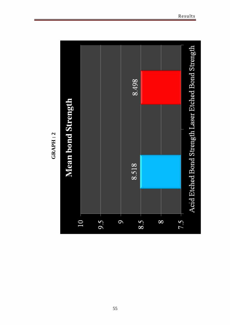
Results
55
GR
AP
H :
2

Discussion
56
DISCUSSION
The aim of this study is to compare the bond strength of
porcelain laminate veneer bonded to acid treated dentin and laser
treated dentin. Most dentin bonding systems rely on some type of
dentin etching and priming to maximize micromechanical adhesion.
Etching enamel affects the prism core and prism periphery. Etching
dentin affects the inter-tubular and peri-tubular dentin, resulting in
enlarging the tubular openings, removin g a large amount of the surface
hydroxyapatite crystals, and leaving an organized network of collagen
fibrils .2 0
Etching of tooth surface can be done either by acid or laser.
BRIEFLY ABOUT COMPONENTS OF LASER UNIT:
Laser unit has active medium, laser resonator and a power
source. The active medium may be gas, solid, liquid, or semi -
conductor. Active medium is contained within an optical enclosure.
Active medium needs to be charged to release photons. The external
source of energy may be electrical, che mical, or flash lamp. The gain
medium is pumped by an external energy source. The gain medium then
emits photons, which bounce back and forth between the reflectors.
Part of the radiation is allowed to exit through an aperture in one of the
reflectors, resulting in the laser beam1 4
(figure : 13).
SPONTANEOUS EMISSION:
When this pumping mechanism pumps energy into the active
medium, the energy is absorbed by the electrons in the peripheral most
layer of the active medium’s atoms. These electrons have gai ned a

Discussion
57
specific amount of energy to reach the next shell farther from the
nucleus, which is at a higher state of energy level. A “population
inversion” can occur when there is more electrons in the higher energy
level than the ground state. The electrons wh ich are in that excited
state spontaneously release that energy in the form of a photon. This is
called spontaneous emission .1 6
EMISSION MODE OF LASERS:
Dental laser devices can emit light energy in two modalities as a
function of time:
(1) Constant / Continuous
(2) Pulsed
Gated-pulse mode
Free-running pulsed mode
1.Continuous-wave mode:
Lasers depend on a beam whose output power is constant over
time and steady when averaged over any longer time periods, with the
very high frequency. The power variation has l ittle or no impact in the
intended application. Such a laser is known as continuous wave. Many
types of lasers can be made to operate in continuous wave mode to
satisfy such an application. The beam is emitted at only one power
level for as long as the operator depresses the foot switch .1 7
2 . Gated-pulse mode:
There are periodic alternations of the laser energy, similar to a
blinking light. This gated-pulse mode is reached by the opening and

Discussion
58
closing of a shutter which is placed in front of the light beam pathway
of a continuous-wave emission. All surgical devices that operate in
continuous wave have this gated -pulse feature. The more advanced
units have computer -controlled shutters that allow for these very short
pulses.1 7
3. Free-running pulsed mode:
It is a true-pulsed mode. This emission is unique in that large
peak energies of laser light are emitted for usually microseconds,
followed by a relatively long time in which the laser is off. These
devices have a rapidly striking flash lamp that pumps the active
medium. With every pulse, high powers are generated. However,
because the pulse duration of this mode is short, the average power that
the t issue experiences is small. Free -running pulsed devices cannot
have a continuous-wave or gated-pulse output .1 7
Contact mode-
Terminal end of fibro optic is placed in direct co ntact of target
tissue and the operator will have a tactile feedback .1 4
Non contact mode :
The hand piece is held away from the tissue and the operator has
to adjust the focus of the beam by varying distance between the hand
piece and target to have the desired effect .1 4
LASER ENERGY AND TISSUE TEMPERATURE:
The principle effect of laser energy is photo thermal (i.e., the
conversion of light energy into heat). This heat effect on tissue

Discussion
59
depends upon the temperature rise and the corresponding reaction of
the interstitial and intracellular water .1 8
TARGET TISSUE EFFECTS IN RELATION TO TEMPERATURE1 8
TISSUE TEMPERATURE OBSERVED EFFECT
37–500C - Hyperthermia
60–700C - Coagulation, protein denaturation
70–800C - Tissue Welding
100–1500C - Vaporization, ablation
>2000C - Carbonization.
EFFECTS OF LASER ON TISSUES :
Laser light has either four different interactions with the target
tissue, depending on the optical properties of that tissue (figure : 14).
They are as follows
ABSORPTION
TRANSMISSION
REFLECTION
SCATTERING
ABSORPTION:
Laser light is converted into effective thermal energy. The
amount of energy that is absorbed by the tissue depends on the tissue
characteristics, such as pigmentation and water content, on the laser
wavelength and emiss ion mode. In general, the shorter wavelengths are
readily absorbed in pigmented tissue and blood elements. Longer

Discussion
60
wavelengths are more interactive with water and hydroxyapatite2 1
(figure : 15).
TRANSMISSION:
Light energy passes freely through the tissue, without interaction
of any kind and has l ittle or no effect . It is an inverse of absorption .2 1
REFLECTION:
Light energy reflects off tissue surface with l ittle or no
absorption and consequently has no effect on tissue. The laser beam
generally becomes more divergent as the distance from the head piece
increases, which become dangerous because the energy is directed to
an unintentional target such as eye. This is major safety concern for
laser operation.2 1
SCATTERING:
Light energy is re-emitted in a random direction and ultimately
absorbed over a greater surface area which produces less intense and
less precisely distributed thermal effect. When laser light emerges from
a laser, it usually enters in the form of pencil thin beam of energy
travelling at speed of light .2 1
This beam travels in straight line unti l it hits something that
reflects or refracts it or unti l it hits something that stops it and absorbs
it energy. The laser beam diverges gradually as it travels away from
laser which means that the beams diameter increases with the distance
between hand piece and target tissue .1 4

Discussion
61
TISSUE CHANGES BY LASER:
There are five important types of biological effects that can
occur once the laser photons enter the tissue: fluorescence,
photothermal, photodisruptive, photochemical, and
photobiomodulation.1 4
Fluorescence happens when actively carious tooth structure is
exposed to the 655 nm visible wavelength and the amount of
fluorescence is related to the size of the lesion, and is useful in
diagnosing incipient carious lesions. e.g., Diagnodent .1 4
Photothermal effects occur when the chromophores absorb the
laser energy and heat is generated. This heat is used to perform work
such as incising tissues or to coagulate the blood. Photothermal
interactions predominate when most soft tissue procedures are
performed with dental lasers. Heat is generated during these procedures
and great care must be taken to avoid thermal damage to the t issues .1 4
Photodisruptive effects: Hard tissues are removed through a
process known as photodisruptive ablation. Short -pulsed laser light
with tremendously high power interact with water in the tissue and
from the hand-piece causing rapid thermal expansion of the water
molecules .1 4
The pulsed Erbium laser ablation efficiency seems to result from
these micro-explosions of overheated tissue water in which their laser
energy is predominantly absorbed. Thus tooth and bone are not

Discussion
62
vaporized but pulverized instead through the photomechanical ablation
process. This shock wave creates the distinct popping sound heard
during erbium laser use. Thermal damage can be prevented when used
properly, especially when the concept of thermal relaxation is
considered.1 4
Photochemical reactions occur when photon energy creates a
chemical reaction. These reactions are utilized in some of the
beneficial effects found in biostimulation. e.g, Photodynamic
therapy.1 4
Photobiomodulation or Biostimulation refers to lasers ability to
speed healing, increase circulation, reduce edema, and minimize pain.
The exact mechanism of these effects is not clear, but i t is theorized
they occur mostly through photochemical and photobiological
interactions within the cellular matrix and mitochondria.
Biostimulation is used dentally to reduce postoperative discomfort and
to treat maladies such as recurrent herpes and aphthous stomatitis. e.g. ,
Low Level Laser therapy.1 4
Thus the primary and beneficial goal of laser ener gy is therefore
primarily due to Absorption of the laser l ight which produces
Incision/Excision, Ablation/Vaporization and Hemostasis/Coagulation.
Now, there has been growing interest in laser application for treating
medical and dental problems.2 2
Thus different laser systems have been
invented (soft & hard tissue lasers).

Discussion
63
It was in 1975 when Rochette described the concept of etching
porcelain with acid and bonding to the tooth, and described a technique
for making ceramic restorations for fractured in cisors without
operative influence, which made the pioneers in veneers to turn
towards porcelain, one of the most popular and attractive materials in
the dental armamentarium and thus was born, the ceramic laminate
veneer. After which, Graham J. Roberts in 1983, clinically evaluated
the technique of mastique acrylic veneers which were bonded to teeth
with composite resins after the acid etch technique .3
EFFECT OF ACID ETCHING ON ENAMEL AND DENTIN :
Acid etching of the enamel appears to improve the retention by
selectively eroding certain hydroxyapatite crystals. The smear layer is
gently removed by the application of acid on the enamel surface.
Microscopic irregularities and acid treated enamel surface energy are
enhanced by removing prismatic and inter-prismatic mineral crystals
and facil itat ing the penetration with the development of resin tags .2 3
(figure : 16)
Phosphoric acid etching is one of the best methods to bond resins
to enamel and dentin. The smear layer is removed by applying acid on
the enamel and dentin surface. In general , 10 -37% ortho-phosphoric
acid is applied to both enamel and dentin .2 3
By simply doubling
etching time from 15 to 30 s, bonding to old dentin resulted in higher
bond strengths that were similar to those found in youn g dentin.2 4

Discussion
64
Despites advances in the chemistry of adhesive systems, dentin
remains a challenging substrate for bonding due to its heterogeneity.
The bonding mechanism of composite resin to acid -etched dentin is
well known. The formation of a hybrid layer and resin tags is essential
to the establishment of a strong bond at the dentin level and may be
achieved by complete dissolution of the smear layer and
demineralization of inter -tubular and peri-tubular dentin by means of
acid etching, resulting in an exposed collagen matrix which is
infiltrated by resin that polymerizes in si tu .2 5
(figure : 17).
The clinical success of porcelain laminate veneers is attributed
to the intimate bond achieved between the restoration and tooth
structure through the luting cement.2 6
Adhesive dental restorative
materials have some major advantages over non adhesive materials. For
example, better adhesion with the tooth surfaces and prevention of
secondary caries thereby preserving the natural tooth structures as
limited cavity preparation is needed.2 7
EFFECT OF LASER ETCHING ON ENAMEL AND DENTIN :
Recently, the use of laser technology has been introduced as an
alternative to tradit ional mechanical rotating instruments in cavity
preparation.2 8
But li ttle is known about the adhesion of resin to laser
irradiated dentin, but it appears that the formation of an inter -diffusion
zone, which is the basis for dentin hybridization in acid -etched dentin,
is unlikely. Instead, laser-treated dentin mostly acquires its bond
strength through the penetration of resin tags into dentinal tubules.

Discussion
65
These resin tag formation shows advantage for only a fraction of the
bond strength in normal hybridized dentin. Some researchers have
explored the use of lasers to modify the surfaces of teeth intentionally
and improve bonding of restorations .2 5
Grinding with rotary instruments is the most commonly used
clinical method for tooth preparation and caries removal. It brings
effectivity and efficiency as well as some inevitable drawbacks, such
as the noise and vibration produced which always make the patients
feel scary about the therapy.2 9
Recently, new techniques were
introduced for caries removal. One of the main advantage in these
techniques is providing minimal damage to tooth structure during
caries removal. Among new techniques, Erbium laser showed
promising results .3 0
Erbium laser has a wide range of advantages, such
as, absence of vibration effect , sound, and stress and reduci ng the
need of local anesthesia .3 1
Currently, laser etching of enamel and dentin surfaces is popular
because of the potential drawbacks of acid etching. Acid etching leads
to chemical changes that can alter the organic matter and decalcify the
organic component. As a result of this demineralization, enamel and
dentin becomes more susceptible to caries attack, which is induced by
plaque accumulation around the bonded composite resin. Recently,
Er:YAG and Er;Cr:YSGG lasers were introduced in dentistry .2 3
(figure:18).

Discussion
66
Due to its beneficial effects, they are used in the hard tissue
treatments. The advantage of an Erbium laser wave is that it is highly
absorbed by water and dental hard tissue. The strong absorption of
water decreases the level of heat throughout tooth preparation. Laser
radiation are better absorbed by water than dental hard tissue, it
reduces the increasing temperature of the tissue during the preparation.
Water reaches the boiling point and causes micro -explosion of the
tooth.2 3
This mechanism breaks up the surrounding tissue into small
fragments and dissipates them at the same time. This micro -explosion
of tooth is so-called a preparation triggered by water. Although most
radiation is highly utilized by water, a certain amount of heat
transmission is unavoidable. Therefore, a water spray is used for
cooling. Lack of adequate water spray causes pulpal damage .2 3
This laser can also be used for etching of enamel and dentin
surfaces for the purpose of bonding the composite resin to enamel an d
dentin surface. Er,Cr:YSGG, which has a high absorption coefficient in
water, enamel and dentin led researchers to explore its use in dentin
conditioning.2 3
Sevil Gurgan et al stated that Enamel etching with the laser at
both energy settings appeared to yield better results than acid etching.
The effect of the Er,Cr:YSGG laser as a dentin -conditioning agent
depends upon multiple parameters. They are energy output, frequen cy,
type of pulse mode, focal distance, irradiation duration, and water

Discussion
67
cooling.3 2
Dentin characteristics created by erbium lasers are irregular
surface, absence of smear layer, with opened dentinal tubules .3 3
Generally, there is variability among the dentin bond strength
values reported by various researchers. This may be attributed to
various testing methods and conditions, the different nature of dentin
substrate, and the composite adhesive used. Although several reports
have compared bond strengths at tained with Er:YAG lasers with
varying results. Few studies have investigated the bond strength
attained with the Er,Cr:YSGG laser .3 2
Visuri et al reported high shear bond strengths of composite to
dentin prepared with an Er:YAG laser using the parameter s of 350 mJ
and 6 Hz. In contrast, Sakakibara et al and Dunn et al used the
parameters of 140 mJ and 30 Hz, and Ceballos et al who used 180 mJ
and 2 Hz, reported decreased bond strength, and Armengol et al and
Kataumi et al found no differences between laser-irradiated and non-
irradiated specimens .3 2
It was postulated that the lased dentin surface yielded an
improved bonding surface due to its scaly and flaky appearance
following laser irradiation. This is caused by the micro -explosions
induced by the laser treatment due to its thermo -mechanical ablation.3 2
The erbium laser initially vaporizes water and other hydrated organic
components of the tissue. On vaporization, internal pressure increases
in the tissue until explosive destruction of the inorgani c components of
the surface occurs. Since intertubular dentin contains more water and

Discussion
68
has a lower mineral content than peritubular dentin, it is selectively
ablated more than the peri -tubular dentin, leaving protruding dentinal
tubules with a cuff -like appearance.3 2
(figure : 19).
This may contribute to an increase in the adhesive surface area.
Opened dentinal tubules and lack of a smear layer are some additional
factors that may enhance bonding to laser -treated dentin. Improved
adhesion to laser-treated dentin may be explained by the mechanical
retention provided by resin tag formation and the infil tration of
adhesive resin into the micro - irregulari ties in lased demineralized
dentin. The highest bond strengths were achieved by laser preparation
and laser etching.3 2
The increase in surface roughness may be one explanation for the
higher dentin bond strengths we found for the laser -treated dentin. This
increased surface roughness allows increased micromechanical
retention at the resin -dentin interface.3 2
Recent improvements in laser technology, and the increased
interest in their potential use for hard tissue applications, warrant
further investigation of the Er,Cr:YSGG laser to prepare teeth for
adhesion with resin-based composites .3 2
According to this present study the bond strength values for both
the groups shows no significant difference. Both have more or l ess
same bond strength values. Laser etching does not make any
statistically significant difference in comparison with acid etching. The

Discussion
69
values obtained shows that acid etching has a wide range of values
varied from 8.1 to 8.9 Mpa. But in laser etching the values are more
consistent, varied from 8.3 to 8.7 Mpa.
Observation of the XY scatter graph of the Acid etched and
Laser etched bond strength shows that acid etching has a wide range of
values varied from 8.1 to 8.9 Mpa, but in laser etching the values are
more consistent, varied from 8.3 to 8.7 Mpa. Also the trend line of
Laser etched bond strength graph is more horizontal than the trend line
of the Acid etched bond strength graph (Graph : 1 & Graph : 2).
The results have indicated that there are no significant
differences in shear bond strengths between the two groups, but as the
values of Laser etched bond strength are more consistent , they can be
more reliable than the Acid etched bond strength values. Furthermore,
it is easy to perform laser etching in patient since it is highly
convenient. Because in acid etching there may be chance of spilling
acid into the adjacent soft tissue, wh ich can cause adverse reactions.
Laser etching is also easy to perform, because there is no usage of
acid. Furthermore, laser etching is more patient friendly.
Acid etching results in chemical changes that can modify the
organic matter and decalcify the organic component. As a result of this
demineralization, enamel and dentin becomes more susceptible to
caries attack, which is induced by plaque accumulation around the
bonded composite resin. So laser etching can be used as an alternative
tool for acid etching.

Conclusion
70
CONCLUSION
Within the limitations of this study, 37% ortho -phosphoric acid
(8.5 Mpa) and laser (8.49 Mpa) treated dentin surfaces showed similar
bond strength values. The differences are not statist ically significant.
But the results of the Laser etched bond strength were more consistent
than Acid etched bond strength. Moreover with recent populari ty and
availabili ty of LASER equipment, etching and restoration can be
accomplished without any need of additional armamentarium and with
better comfort to the patient. Further research needs to be done on the
nature of biological response to laser irradiation in vital teeth
undergoing laser etching.

References
71
REFERENCES
1. Usumez A,Aykent F. Bond strengths of porcelain laminate
veneers to tooth surfaces prepared with acid and Er,Cr:YSGG
laser etching. The Journal of Prosthetic Dentistry. 2003 Jul
31;90(1):24-30.
2. Pahlevan A, Mirzaee M, Yassine E, Omrany LR,Tabatabaee MH,
Kermanshah H,Arami S, Abbasi M. Enamel thickness after
preparation of tooth for porcelain laminate. Journal of Dentist ry
(Tehran, Iran). 2014 Jul;11(4):428 -432.
3. Mathew C A, Mathew S, Karthik K S. A review on ceramic
laminate veneers. Journal of Indian Dental Association. 2010
oct;1(4):33-37.
4. Okamoto M, MineA, Watanabe K, Kawahara D, Yatani H.
Porcelain veneer bonding to dentin and the curing performance
of plasma-arc light with respect to porcelain thickness. Dental
Materials Journal. 2003;22(3):313 -320.
5. Calamia J R, Calamia C S. Porcelain laminate veneers : Reasons
for 25 years success. Dental Cinics of North America. 200 7 Apr
30;51(2):399-417.
6. Gurel G. Porcelain laminate veneers : Minimal tooth preparation
by design. Dental Clinics of North America. 2007 Apr
30;51(2):419-431.

References
72
7. Peumans M, Van Meerbeek B, Lambrechts P, Vanherle G.
Porcelain veneers : A review of the literat ure. Journal of
Dentistry. 2000 Mar 31;28(3):163 -177.
8. Ansari Z J, Fekrazad R, Feizi S, Younessian F, Kalhori K A,
Gutknecht N. The effect of an Er,Cr:YSGG laser on the micro -
shear bond strength of composite to the enamel and dentin of
human permanent teeth . Journal of Lasers in Medical Sciences.
2012 Jul 1;27(4):761-765.
9. Franchi M, Breschi L. Effects of acid etching solutions on human
enamel and dentin. Ouintessence International. 1995 Jun
1;26:431-435.
10.Giray F E, Duzdar L, Oksuz M, Tanboga I. Evaluation of the
Bond Strength of Resin Cements Used to Lute Ceramics on
Laser-Etched Dentin. Photomedicine and Laser Surgery. 2014 Jul
1;32(7):413-421.
11.Lopes G C, Vieira L C, Monteiro S, De Andrada M C, Baratieri
C M. Dentin bonding : Effect of degree of mineralizati on and
acid etching t ime. Operative Dentistry. 2003 Jul 1;28(4):429 -439
12.Martinez-Insua A, Da silva Dominguez L, Rivera F G, Santana -
Penin U A. Differences in bonding to acid -etched or Er:YAG-
laser-treated enamel and dentin surfaces. Journal of Prosthetic
Dentistry. 2000sep 30;84(3):280-288.
13.Turkmen C, Sazak-Ovecoglu H, Gunday M, Gungor G, Meral
Durkan, Oksuz M. Shear bond strength of composite bonded with

References
73
three adhesives to Er,Cr:YSGG laser -prepared enamel.
Quintessence International. 2010 Jun 1;41(6):119 -124.
14.Convissar RA. Principles and practice of laser dentistry. Elsevier Health
Sciences; 2015 Mar 6, p:12-22.
15.Maenosono R M, Bimjunior O, Duarte M A, Palma -Dibb R G,
Wang L, Ishikiriama S K. Diode laser irradiation increases
microtensile bond strength of dentin. Brazilian Oral Research.
2015;29(1):1-5.
16.Acharya S S ,Satyanarayana T S V , Prabhakar R. Lasers in
dentistry- a review. Annals and Essences of Dentistry.
2012;4(4):66-72.
17.Parker S. Introduction, history of lasers and laser l ight
production. British Dental Journal. 2007 Jan 13;202(1):21 -31
18.Coluzzi D J. Lasers for Use in Dental Hygiene. Access
2009;23(1):10-13.
19.Coluzzi DJ, Convissar RA. Atlas of laser applications in dentistry.
Quintessence Publishing Company; 2007, p:2-4.
20.Theodore Roberson, Ha rald Heymann, Ed Swift. Sturdevant’s Art
and Science of Operative Dentistry – 5t h
Edition. Mosby Elsevier;
2012, p:245-251.
21.Parker S. Laser-tissue interaction. British Dental Journal. 2007
Jan 27;202(2):73-81.
22.Usumez S, Orhan M, Usumez A. Laser etching of enamel for
direct bonding with an Er,Cr:YSGG hydrokinetic laser system.

References
74
American Journal of Orthodontics and Dentofacial Orthopedics.
2002 Dec 31;122(6):649-56.
23.Hoshing U A, Patil S, Medha A, Bandekar S D. Comparison of
shear bond strength of composite resin to enamel surface with
laser etching versus acid etching: An in vitro evaluation. Journal
of Conservative Dentistry. 2014 Jul;17(4):320 -324.
24.Lopes G C, Vieira L C, Araujo E, Bruggmann T, Zucco J,
Oliveira G. Effect of dentin age and acid etching time on dentin
bonding. Journal of Adhesive Dentistry. 2011 Apr 1;13(2):139 -
145.
25.Davari A, Sadeghi M, Bakhshi H. Shear bond strength of an
Etch-and-rinse adhesive to Er:YAG laser and/or phosphoric acid -
treated dentin. Journal of Dental Resea rch, Dental Clinics ,
Dental Prospects. 2013;7(2):67-73.
26.Altintas S H, Tak O, Secilmis A, Usumez A. Effect of
provisional cements on shear bond strength of porcelain laminate
veneers. European Journal of Dentistry. 2011 oct;5(4):373 -379.
27.Zafar M S, Ahmed N. The effects of acid etching time on surface
mechanical properties of dental hard tissues. Dental Materials
Journal. 2015;34(3):315–320.
28.Koliniotou-Koumpia E, Kouros P, Zafiriadis L, Koumpia E,
Dionysopoulos P, Karagiannis V. Bonding of adhesives to
Er:YAG laser-treated dentin. European Journal of Dentistry.
2012 Jan;6(1):16-23.

References
75
29.Liu J, Chen H, Ge W, Wang Y, Sun Y, Wang Y, Lu P. A
Roughness Study of Ytterbium-Doped Potassium Yttrium
Tungstate (YB: KYW) Thin-Disk Femtosecond Ablated Dentin.
Journal of Lasers in Medical Sciences. 2014;5(1):32-38.
30.Pahlavan A, Mehmanchi M, Omrani L R, Chiniforush N. Effect
of air abrasion and Erbium-Doped Yttrium Aluminium Garnet
(Er:YAG) laser preparation on shear bond strength of composite
to dentin. Journal of Lasers in Medical Sciences. 2013;4(3):127-
130.
31.Moslemi M, FotouhiArdakani F, Javadi F, KhaliliSadrabad Z,
Shadkar Z, and Shadkar M S . Evaluation of Er,Cr:YSGG Laser
Effect on Microshear Bond Strength of a Self -Adhesive Flowable
Composite in the Dentin of Permanent Molar : An In Vitro Study.
Scientifica. 2016 Jul 14;2016:1 -5.
32.Gurgan S, Kiremitci A, Cakir F Y, Gorucu J, Alpaslan T, Yazici
E, Gutknecht N. Shear bond strength of composite bonded to
Er,Cr:YSGG laser-prepared dentin. Journal of Photomedicine and
Laser surgery. 2008 oct 1;26(5):495-500.
33.Geraldo-Martins V R, Armas-Vega A C, Marques M M. Influence
of etching with Er,Cr:YSGG laser on the microtensile bond
strength of adhesives to dentin. Journal of Oral Laser
Applications. 2010;10:79-86.
Annexure
76
INSTITUTIONAL ETHICS COMMITTEE AND
REVIEW BOARD
ADHIPARASAKTHI DENTAL COLLEGE AND
HOSPITAL
Melmaruvathur, Tamilnadu-603319
MEMBER SECRETARY
Dr.S.Meenakshi, PhD
MEMBERS
Prof.Dr.A.Momon Singh, MD
Prof.Dr.H.Murali, MDS
Dr.Muthuraj, MSc, MPhil, PhD
Prof.Dr.T.Ramakrishnan, MDS
Prof.Dr.T.Vetriselvan, MPharm,
PhD
Prof.Dr.A.Vasanthakumari, MDS
Prof.Dr.N.Venkatesan, MDS
Prof.Dr.K.Vijayalakshmi, MDS
Shri.Balaji, BA, BL
Shri.E.P.Elumalai
CHAIR PERSON
Prof.Dr.K.Rajkumar, BSc,MDS, PhD
This ethical committee has undergone the research protocol
submitted by Dr.J.Raghunathan, Post Graduate Student, Department
of Prosthodontics and crown & bridge under the title
“Comparison Of Bond Strength Of Porcelain Laminate Veneers
Bonded To Laser Treated Dentin And Acid Treated Dentin – An
In Vitro Study” Reference No: 2014-MD-BrI-RIJ-03, under the
guidance of Prof. Dr.A.S.Ramesh MDS., for consideration of
approval to proceed with the study.
This committee has discussed about the material being
involved with the study, the qualification of the investigator, the
present norms and recommendation from the Clinical Research
scientific body and comes to a conclusion that this research protocol
fulfils the specific requirements and the committee authorizes the
proposal.
Date: Member secretary