comparison of black hole sign, satellite sign, and iodine
TRANSCRIPT

Research ArticleComparison of Black Hole Sign, Satellite Sign, and Iodine Sign toPredict Hematoma Expansion in Patients with SpontaneousIntracerebral Hemorrhage
Milind Ratna Shakya,1,2 Fan Fu,1,2 Miao Zhang,1,2 Yi Shan,1,2 Fan Yu,1,2 Shengjun Sun ,3
and Jie Lu 1,2,4
1Department of Radiology, Xuanwu Hospital, Capital Medical University, No. 45 Changchun Street, Xicheng District, Beijing, China2Beijing Key Laboratory of Magnetic Resonance Imaging and Brain Informatics, Xuanwu Hospital, Capital Medical University,No. 45 Changchun Street, Xicheng District, Beijing, China3Neuroradiology Department, Beijing Neurosurgical Institute, Beijing Tiantan Hospital, Capital Medical University,No. 119 Nansihuanxilu, Fengtai District, Beijing, China4Department of Nuclear Medicine, Xuanwu Hospital, Capital Medical University, No. 45 Changchun Street, Xicheng District,Beijing, China
Correspondence should be addressed to Shengjun Sun; [email protected] and Jie Lu; [email protected]
Received 1 May 2020; Revised 2 October 2020; Accepted 21 January 2021; Published 1 February 2021
Academic Editor: Vida Demarin
Copyright © 2021 Milind Ratna Shakya et al. This is an open access article distributed under the Creative Commons AttributionLicense, which permits unrestricted use, distribution, and reproduction in any medium, provided the original work isproperly cited.
Purpose. To discretely and collectively compare black hole sign (BHS) and satellite sign (SS) with recently introduced gemstonespectral imaging-based iodine sign (IS) for predicting hematoma expansion (HE) in spontaneous intracerebral hemorrhage(SICH). Methods. This retrospective study includes 90 patients from 2017 to 2019 who underwent both spectral computedtomography angiography (CTA) as well as noncontrast computed tomography (NCCT) within 6 hours of SICH onset alongwith subsequent follow-up NCCT scanned within 24 hours. We named the presence of any of BHS or SS as any NCCT sign.Two independent reviewers analyzed all the HE predicting signs. Receiver-operator characteristic curve analysis and logisticregression were performed to compare the predictive performance of HE. Results. A total of 61 patients had HE, out of which ISwas seen in 78.7% (48/61) while BHS and SS were seen in 47.5% (29/61) and 41% (25/61), respectively. The area under thecurve for BHS, SS, and IS was 63.4%, 67%, and 82.4%, respectively, while for any NCCT sign was 71.5%. There was nosignificant difference between IS and any NCCT sign (P = 0:108). Multivariate analysis showed IS (odds ratio 68.24; 95% CI11.76-396.00; P < 0:001) and any NCCT sign (odds ratio 19.49; 95% CI 3.99-95.25; P < 0:001) were independent predictors ofHE whereas BHS (odds ratio 0.34; 95% CI 0.01-38.50; P = 0:534) and SS (odds ratio 4.54; 95% CI 0.54-38.50; P = 0:165) had nosignificance. Conclusion. The predictive accuracy of any NCCT sign was better than that of sole BHS and SS. Both any NCCTsign and IS were independent predictors of HE. Although IS had higher predictive accuracy, any NCCT sign may still beregarded as a fair predictor of HE when CTA is not available.
1. Introduction
Spontaneous intracerebral hemorrhage (SICH) accounts for10% to 30% of all strokes worldwide [1]. It is one of the mostdevastating types of stroke, leading to neurological deteriora-tion and case fatality. Owing to its high morbidity and mor-tality rate, the prediction of early hematoma expansion (HE)
is essential [2]. Generally, early HE is seen in up to 20% to30% of patients with SICH [3]. However, previous literaturehas shown up to 73% of patients exert some degree of HE[4–6]. Therefore, reliable imaging tools are required for pre-dicting HE and providing appropriate treatment.
Based on hematoma shape and density, numerous noncon-trast computed tomography- (NCCT-) based HE predicting
HindawiBioMed Research InternationalVolume 2021, Article ID 3919710, 8 pageshttps://doi.org/10.1155/2021/3919710

markers are available [7, 8]. We enrolled one HE predictorfrom each of shape and density group. The black hole sign(BHS) attributes density heterogeneity trait, whereas the satel-lite sign (SS) attributes shape irregularity traits. A computedtomography angiography (CTA) spot sign is the robust markerfor HE prediction. In correspondence with spot sign, both BHSand SS are proven to be the independent predictors for HEwitha significant association [9–12]. The advantage of BHS and SSare its evident visibility in NCCT, which is often available in aclinical setting. Furthermore, it is beneficial in patients withchronic kidney conditions and in patients who are allergic tocontrast medium.
Recently, gemstone spectral imaging- (GSI-) based iodinesign (IS) has also been introduced as a reliable and sensitivemarker for predicting HE [13]. As an assuring scan tech-nique, GSI can efficiently differentiate iodine from the bloodproduct. During active bleeding, its monochromatic imagingcan quantitatively measure the concentration of iodineleaking from the bleeding site. A threshold of iodine concen-tration greater than 7.82 (100μg/ml) has a significant associ-ation with HE [14–16]. However, the correlation betweenGSI-based IS with NCCT-based predicting markers is yet tobe known.
Since the predictive efficiency of BHS and SS has not beentallied with GSI-based IS, this study’s objective is to comparethe predictive performance of NCCT-based BHS and SS withGSI-based IS for prediction of HE in patients with SICH.Besides, we also compared the predictive capability of anyNCCT sign against IS.
2. Materials and Methods
2.1. Study Population. We retrospectively studied 90 consec-utive cases of SICH patients above the age of 18 years. Allpatients enrolled in this study were admitted to Beijing Tian-tan Hospital between November 2017 and December 2019.Informed consent for all the cases was obtained either fromthe patients or from next to kin. The inclusion criteria for thisstudy were (1) patients who underwent NCCT as well ascontrast-enhanced dual-energy spectral computed tomogra-phy angiography (CTA) within 6 hours after onset of symp-toms and (2) follow-up NCCT was scanned within 24 hoursafter the initial scan. Patients were excluded from the study ifthey had SICH history or had secondary SICH caused by anarteriovenous malformation, brain tumor, traumatic braininjury, hemorrhagic transformation of ischemic infarction,moyamoya disease, or anticoagulant induced SICH. Patientswere also excluded if the initial spectral CTA was not donedue to chronic kidney conditions. Patients could not undergoa follow-up CT scan due to emergency surgical interventionof hematoma evacuation, transferred to another center, orwas pronounced dead. The clinical, baseline demographyand radiological variables of all the patients, along withhypertensive history, diabetic history, smoking history, alco-hol abuse, glucose baseline, the National Institutes of HealthStroke Scale (NIHSS) scores, the duration form the onset ofthe symptoms to initial CT scan time, and lab results, werecollected at the emergency department by neurologists.
2.2. Image Acquisition. We used the 64-slice discoveryCT750HD scanner (GE Healthcare, Waukesha, WI) to scanall the patients. Initial NCCT was done by standard single-energy helical mode with 120 kVp, 300mA, 0.6 s gantry rota-tion time, and 0.984 : 1 helical pitch. Subsequent contrast-enhanced CTA was done using dual-energy spectral imagingmode with rapid switching between 80kVp and 140kVp.Scanning parameters were arranged as tube current, 375mA;slice thickness, 5mm; rotation time, 0.6 s/rotation; collimationthickness, 0:625mm× 64; and scan field-of-view, 25 cm. Boththe reconstruction slice thickness and interval were 0.625mm.The contrast agent Iohexol (300mg I/ml; Guerbet, France)was used in the spectral CTA scans according to a patientweight-dependent dose of 0.7ml/kg and an injection rate of6ml/s. Time from contrast bolus injection to CTA acquisitionwas obtained from archived SmartPrep images (GE Health-care). Final follow-up plain CT was obtained within 24 hoursusing the same CT system and parameters.
2.3. Imaging Analysis. Firstly, two independent reviewersblinded to the patients’ clinical data evaluated all the NCCTimages for the presence or absence of BHS and SS from theinitial scan. The BHS (Figure 1(a)) was defined as thehypoattenuated area of a round, oval, or a rod-like shapeencapsulated within the hyperattenuated area, which is notconnected to the adjacent brain tissue, with the difference ofat least 28 Hounsfield units between two densities [9]. TheSS (Figure 1(b)) was defined as a small hemorrhage of maxi-mum transverse diameter, not more than 10mm, which iscompletely detached from the main hemorrhage seen in atleast a single CT slice. The distance between the main hemor-rhage and the detached small hemorrhage should not be morethan 1-20mm. Subarachnoid and intraventricular hemor-rhages were not counted as SS [11]. We named the presenceof any of the BHS or the SS as any NCCT sign. The presenceof any NCCT sign was noted.
Secondly, the reviewers evaluated positive or negative ISfrom the spectral CTA images. The IS was extracted withGSI viewer on standard advantage workstation (AW 4.6;GE Healthcare) of dual-energy spectral CT. Positive IS(Figure 1(c)) was defined as the presence of tiny enhance-ment of ≥1 foci within hematoma with an iodine concentra-tion of >7.82 (100μg/ml) inside the foci, visualized oniodine-based decomposition image [13, 14].
Hematoma volume of initial and follow-up CT scans wasmeasured on separate occasions; the images were random-ized to blind the raters from patients’ information. The loca-tion of hematoma was evaluated and was categorized aseither deep or lobar. The presence or absence of midline shiftwas noted. Intraventricular hematoma extension was notedbut was not included in the volumetric analysis of HE. Hema-toma volume of initial and follow-up CT images was manu-ally calculated using the ABC/2 formula. HE was defined asan absolute growth of hematoma by at least 6ml (follow-upvolume–initial volume), or a relative growth by at least 33%(absolute growth/initial volume) [4, 17].
2.4. Statistical Analysis. All the statistical analyses for thisstudy were performed using SPSS version 26.0 and NCSS
2 BioMed Research International

2019 version 19.0.3 statistical software. The categorical vari-ables were expressed as absolute values (percentages, %),and the continuous variables were expressed as medians
(IQR) or mean ± standard deviation (SD) according to thedistribution of data. An independent-samples t-test was usedfor continuous variables, while Pearson chi-square or Fisher
Table 1: Baseline demographic, clinical, and radiological comparison of patients with and without hematoma expansion.
Entire study (n = 90) Hematoma expansion (n = 61) No hematoma expansion (n = 29) P value
Age, years, mean ± SD 53:88 ± 12:89 52:56 ± 12:85 56:66 ± 12:75 0.160
Gender, male (%) 62 (68.9) 43 (70.5) 19 (65.5) 0.634
Medical history
Hypertension (%) 73 (81.1) 50 (82) 23 (79.3) 0.763
Diabetes mellitus (%) 17 (18.9) 11 (18) 6 (20.7) 0.779
Smoking (%) 56 (62.2) 41 (67.2) 15 (51.7) 0.171
Alcohol consumption (%) 65 (72.2) 45 (73.8) 20 (69) 0.802
Clinical parameters
Glucose, mmol/L, median (IQR) 6.40 (5.49-7.18) 6.40 (5.50-7.30) 6.40 (5.47-6.70) 0.725
Fibrinogen, mean ± SD 2:57 ± 0:606 2:49 ± 0:623 2:73 ± 0:544 0.078
APTT(s), median (IQR) 26.50 (24.30-29.00) 27.10 (25.10-29.50) 25.60 (24.10-27.20) 0.059
INR, median (IQR) 0.93 (0.89-0.98) 0.94 (0.88-0.98) 0.93 (0.89-0.96) 0.450
NIHSS, median (IQR) 9 (5.75-13) 10 (6-14) 7 (5-10) 0.015
Radiological data
Location, deep (%) 72 (80) 50 (82) 22 (75.9) 0.499
IHV, ml, mean ± SD 19:69 ± 17:02 21:85 ± 18:57 15:17 ± 12:26 0.082
IVH, present (%) 24 (26.7) 14 (23) 10 (34.5) 0.248
Midline shift, present (%) 10 (11.1) 7 (11.5) 3 (10.3) 0.873
Onset to CT, hours, median (IQR) 2 (1.5-3.5) 2 (1.5-3.5) 2 (1.5-3.5) 0.782
Black hole signs, present (%) 35 (38.9) 29 (47.5) 6 (17.1) 0.02
Satellite sign, present (%) 27 (30) 25 (41) 2 (6.9) 0.001
Iodine sign, present (%) 52 (57.8) 48 (78.7) 4 (13.8) <0.001Any NCCT sign, present (%) 61 (67.8) 41 (67.21) 20 (68.97) <0.001
IQR: interquartile range; SD: standard deviation; APTT: activated partial prothrombin time; INR: international normalized ratio; NIHSS: National Institutes ofHealth Stroke Scale; IHV: initial hematoma volume; IVH: intraventricular hemorrhage; NCCT: noncontrast computed tomography.
(a) (b)
Mean 9.68std 0.511
(c)
Figure 1: An illustration of hematoma expansion predicting signs. (a) Axial noncontrast computed tomography (NCCT) illustrates blackhole sign described as the hypoattenuated area encapsulated within the area of hyperattenuation, with a difference of at least 28Hounsfield units between two densities. (b) Axial NCCT shows a satellite sign described as the presence of small hemorrhage, which iscompletely detached from the main hemorrhage, observed in at least a single-CT slice. (c) Axial plane on iodine-based decompositionrepresents an iodine sign with a tiny enhanced focus whose iodine concentration is 9.68 100 μg/ml (>7.82 100 μg/ml).
3BioMed Research International
exact test was used for categorical variables in order to verifythe analysis of statistical significance.
Receiver-operating characteristic (ROC) curve analysiswas performed to check the diagnostic accuracy of all the pre-dictive markers for HE. The Z test was performed to comparethe area under the ROC of IS and any NCCT signs. Sensitiv-ity, specificity, positive predictive value, and negative predic-tive value of all predictive markers were calculated. Theassociation between hematoma expansion and all its predic-tive markers was evaluated using univariate and multivariatelogistical regression module. The result of the regressionmodule was presented as odds ratio (OR) and 95% confi-dence intervals (CI). Adjusted variables for multivariateanalysis were selected. A two-tailed P value of <0.05 was con-sidered statistically significant for all tests. Cohen’s kappaanalysis was performed for interreviewer reliability.
3. Results
3.1. Baseline Characteristics. A total of 90 patients wereincluded in this study. The mean age of the patients was53:88 ± 12:89 years, among which 62 (68.9%) were male,and 28 (31.1%) were female. Out of the patients studied, 61(67.8%) of the patients showed HE. Hematomas located indeep and lobar were 72 (80%) and 18 (20%), respectively.The mean initial hematoma volume was 19:69 ± 17:02ml.Patients with HE was more likely to have higher admissionNIHSS score with P = 0:015. On the other hand, no signifi-cant differences in the distribution of age, gender, medicalhistory, glucose level, midline shift, the onset of symptomsto initial CT scan duration, and intraventricular hemorrhagewere observed (Table 1).
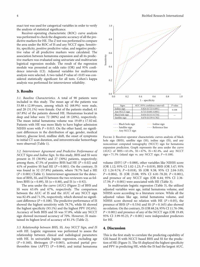
3.2. Interreviewer Agreement and Predictive Performance ofNCCT Signs and Iodine Sign. In this study, BHS and SS werepresent in 35 (38.9%) and 27 (30%) patients, respectively;among them, 47.5% of positive BHS had HE (P = 0:02) and41% of positive SS had HE (P = 0:001). On the contrary, ISwas found in 52 (57.8%) patients, where 78.7% had a HE(P < 0:001) (Table 1). Interreviewer agreement for the detec-tion of BHS, SS, and IS between the two reviewers was as fol-lows BHS (κ = 0:89), SS (κ = 0:88), and IS (κ = 0:92).
The area under the curve (AUC) (Figure 2) of BHS andSS were 63.4% and 67%, respectively. The comparisonbetween the AUC of IS and the AUC of any NCCT signwas 82.4% and 71.5%, respectively, which showed no signifi-cant difference (P = 0:108). The predictive performance of ISshowed the highest sensitivity with 78.7%, while SS showedthe highest specificity (93.1%) and the highest PPV (92.6%).Accuracy of both BHS and SS was 57.8%, while any NCCTsign showed increased accuracy of 70%. However, IS main-tained its highest level of accuracy of 81.1% (Table 2).
3.3. Relationship between BHS, SS, Any NCCT Sign, and ISwith HE. Logistic regression was performed to assess therelationship between clinical and radiological parameterswith HE. In univariable logistic regression, excluding age(P = 0:160), fibrinogen (P = 0:083), activated partial pro-thrombin time (APTT) (P = 0:064), and initial hematoma
volume (IHV) (P = 0:088), other variables like NIHSS score(OR 1.12; 95% CI 1.02-1.23; P = 0:019), BHS (OR 3.47; 95%CI 1.24-9.74; P = 0:018), SS (OR 9.38; 95% CI 2.04-3.05;P = 0:004), IS (OR 23.08; 95% CI 6.81-78.20; P < 0:001),and presence of any NCCT sign (OR 6.44; 95% CI 2.36-17.59; P < 0:001) were associated with HE (Table 3).
In multivariate logistic regression (Table 3), the utilizedadjusted variables were age, initial hematoma volume, andNIHSS score according to a literature review. While all theadjusted values like age, initial hematoma volume, andNIHSS score showed no relation with HE (P > 0:05), thepresence of BHS (P = 0:534) and SS (P = 0:165) also showedno relation. On the contrary, IS (OR 68.24; 95% CI 11.76-396;P < 0:001) and presence of any of the NCCT sign (OR 19.49;95% CI 3.99-95.25; P < 0:001) were independent predictorsfor HE.
4. Discussion
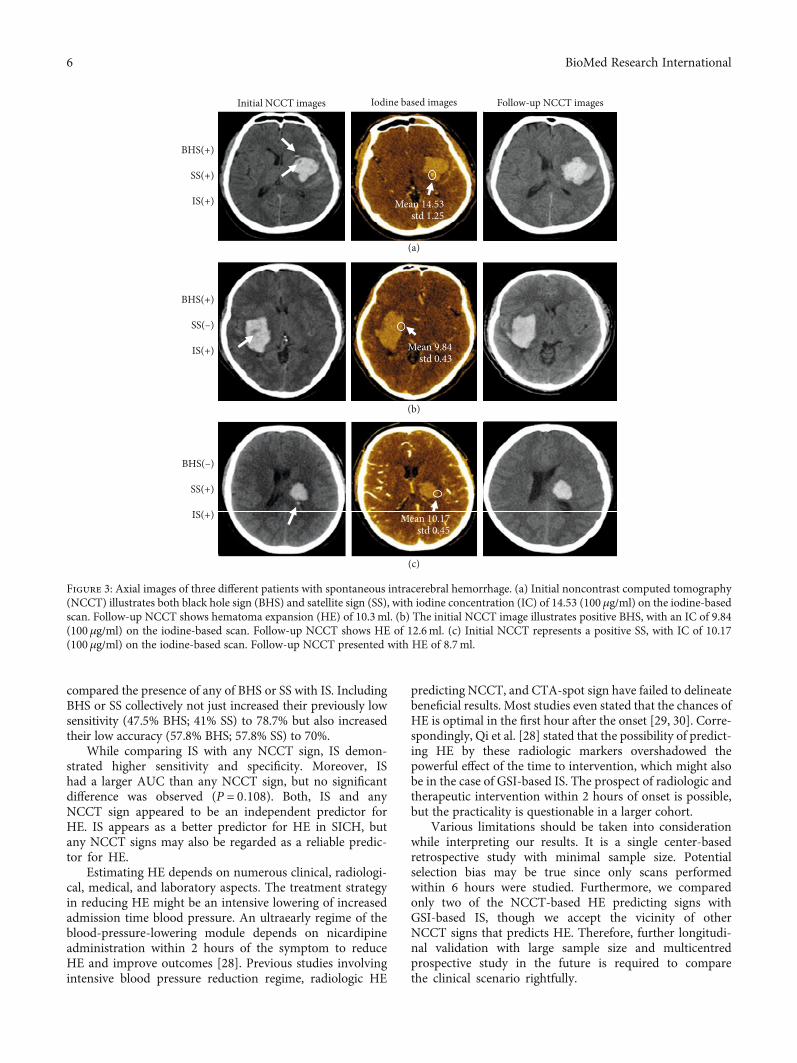
This is the first study to correlate the predicting capability ofGSI-based IS with NCCT-based BHS and SS for the predic-tion of HE (Figure 3). The SS displayed the highest specificityand PPV in predicting HE, while the IS had the largest AUC.
1 – specificity
Sens
itivi
tyIodine sign
Signs AUC (95% confidence interval)
Any NCCT signBlack hole signSatellite sign
0.824 (0.723-0.891)0.715 (0.601-0.801)
0.108
—
P value
0.634 (0.515-0.754)0.670 (0.559-0.782)
Black hole signSatellite signAny NCCT sign
Iodine signReference line
0.0 0.2 0.4 0.6 0.8 1.00.0
0.2
0.4
0.6
0.8
1.0
Figure 2: Receiver-operator characteristic curves analysis of blackhole sign (BHS), satellite sign (SS), iodine sign (IS), and anynoncontrast computed tomography (NCCT) sign for hematomaexpansion prediction. Graph represents the area under the curve(AUC) of BHS = 63:4%, SS = 67%, IS = 82:4%, and any NCCTsign = 71:5% (island sign vs. any NCCT sign; P = 0:108).
4 BioMed Research International

Moreover, the presence of any NCCT signs, i.e., BHS or SS,was better in predicting HE than the presence of a singleNCCT sign. Although GSI-based IS had a higher predictiveaccuracy than any NCCT sign, no statistical significancewas found (P = 0:108). Hence, any NCCT sign is still anacceptable predictor for HE when CTA is unavailable. Wedemonstrated that the presence of any NCCT sign and ISwere independently associated with HE.
Generally, HE was observed in up to 19% to 38% ofpatients with SICH. However, in the phase II trial to confirmHE reduction by recombinant activated factor VII, 70% ofpatients with a HE has been reported [4, 6]. Furthermore,at 1-hour duration after the initial CT scan, 33% of HE werereported, while the addition of 12% of substantial HE couldbe observed in 1-hour to 20-hour duration [5]. This impliesHE proportion may occasionally rise with time. At the sametime, our study shows that 67.8% of patients of SICH had HE.Our analysis also showed (Table 1) parameter like the NIHSSscore (P = 0:015) had a statistical significance. Previousstudies suggest that this parameter is associated HE [17, 18].
In prior studies, it has been demonstrated that there is anassociation of hematoma heterogeneity and HE [19]. TheBHS as a novel sign to determine heterogeneity for the eval-uation of HE was proposed by Li et al. [9] in 2016. It had lowsensitivity (31.9%) and high specificity (94.1%), which is sim-ilar to our result. The low sensitivity may be due to the smallsample size of HE with positive BHS. Pathologically, thehypoattenuated region in NCCT indicates the presence offresh blood, and the hyperattenuated region indicates theblood serum that has been isolated out of the hematoma afterclot formation. Thus, positive BHS suggests the ejection ofblood over a period of time [9, 20].
Additionally, the hematoma irregularity was also inde-pendently associated with hemorrhage growth and poorfunctional outcome [8, 21, 22]. The SS was primarily discov-
ered by Shimoda et al. [11], which is a shape-based indicatorfor predicting HE, and it was said to be present mostly inhematomas with irregular shapes. However, the relationshipbetween shape irregularity and HE was contradicted.According to a study by Barras et al. [7], it was not foundto be an independent predictor for HE. Correspondingly,Takeda et al.’s [23] study also failed to identify the significantassociation between shape irregularity and HE, while theirregular shape of hematoma was found to be independentlyassociated with HE in Blacquiere et al.’s [24] study. The con-tradiction between studies may be due to the unclear defini-tion of irregular shapes, and a higher degree of irregularitymay increase the chances of hematoma growth. Later, Yuet al. [12] suggested that the SS as an independent predictorof HE, and a recent study by Deng et al. [25] revealed the pre-dictive accuracy of the SS of 62% while our study showed rel-atively lower predictive accuracy (57.8%).
Fu et al. [13] first reported the IS as a novel predictor ofHE, which was regarded as early contrast extravasation onongoing bleeding of hematoma in neighboring microvessels[26, 27], although expansion-prone hematoma [21], describedas the presence of at least one of BHS, blend sign, or islandsign, showed good predicting value (OR 28.33; 95% CI12.95-61.98). However, a specific scanning technique of GSIcould efficiently separate iodine and blood via a rapid kVpswitching method. During active blood loss, iodine productleaks from the punctured vessel. GSI monochromatic imagingcan reflect iodine concentration form the leak point [15, 16].Moreover, IS showed higher sensitivity (91.5%) and accuracy(85.7%), which was even higher than that of the previouslyreckoned biomarker for HE prediction, the spot sign [13].However, since CTA is contraindicated in certain conditionsand is not readily available in every facility, NCCT-based pre-dicting signs are essential. Our study not only compared thepredictive capability of individual SS and BHS with IS but also
Table 2: Predictive performance of hematoma predictors.
Sensitivity (%) Specificity (%) PPV (%) NPV (%) Accuracy (%)
Black hole sign 47.5 79.3 82.9 41.8 57.8
Satellite sign 41 93.1 92.6 42.9 57.8
Iodine sign 78.7 86.2 92.3 65.8 81.1
Any NCCT sign 67.2 75.9 85.4 52.4 70
PPV: positive predictive value; NPV: negative predictive value.
Table 3: Univariate and multivariate analysis for hematoma expansion predictors.
VariablesUnivariate Multivariate
OR (95% CI) P value OR (95% CI) P value
Age 0.98 (0.94–1.07) 0.160 0.97 (0.92-1.03) 0.300
Initial hematoma volume 1.03 (0.99–1.06) 0.088 0.96 (0.92-1.00) 0.051
NIHSS 1.12 (1.02–1.23) 0.019 1.13 (0.97-1.33) 0.129
Black hole sign 3.47 (1.24-9.74) 0.018 0.34(0.01-10.23) 0.534
Satellite sign 9.38 (2.04-3.05) 0.004 4.54(0.54-38.50) 0.165
Iodine sign 23.08 (6.81–78.20) <0.001 68.24 (11.76–396.00) <0.001Any NCCT sign 6.44 (2.36–17.59) <0.001 19.49 (3.99–95.25) <0.001OR: odds ratio; CI: confidence interval; NIHSS: National Institutes of Health Stroke Scale; NCCT: noncontrast computed tomography.
5BioMed Research International
compared the presence of any of BHS or SS with IS. IncludingBHS or SS collectively not just increased their previously lowsensitivity (47.5% BHS; 41% SS) to 78.7% but also increasedtheir low accuracy (57.8% BHS; 57.8% SS) to 70%.
While comparing IS with any NCCT sign, IS demon-strated higher sensitivity and specificity. Moreover, IShad a larger AUC than any NCCT sign, but no significantdifference was observed (P = 0:108). Both, IS and anyNCCT sign appeared to be an independent predictor forHE. IS appears as a better predictor for HE in SICH, butany NCCT signs may also be regarded as a reliable predic-tor for HE.
Estimating HE depends on numerous clinical, radiologi-cal, medical, and laboratory aspects. The treatment strategyin reducing HE might be an intensive lowering of increasedadmission time blood pressure. An ultraearly regime of theblood-pressure-lowering module depends on nicardipineadministration within 2 hours of the symptom to reduceHE and improve outcomes [28]. Previous studies involvingintensive blood pressure reduction regime, radiologic HE
predicting NCCT, and CTA-spot sign have failed to delineatebeneficial results. Most studies even stated that the chances ofHE is optimal in the first hour after the onset [29, 30]. Corre-spondingly, Qi et al. [28] stated that the possibility of predict-ing HE by these radiologic markers overshadowed thepowerful effect of the time to intervention, which might alsobe in the case of GSI-based IS. The prospect of radiologic andtherapeutic intervention within 2 hours of onset is possible,but the practicality is questionable in a larger cohort.
Various limitations should be taken into considerationwhile interpreting our results. It is a single center-basedretrospective study with minimal sample size. Potentialselection bias may be true since only scans performedwithin 6 hours were studied. Furthermore, we comparedonly two of the NCCT-based HE predicting signs withGSI-based IS, though we accept the vicinity of otherNCCT signs that predicts HE. Therefore, further longitudi-nal validation with large sample size and multicentredprospective study in the future is required to comparethe clinical scenario rightfully.
Initial NCCT images Iodine based images
(a)
(b)
(c)
Follow-up NCCT images
BHS(+)
SS(+)
IS(+)
BHS(+)
SS(–)
IS(+)
BHS(–)
SS(+)
IS(+)
Mean 14.53std 1.25
Mean 9.84std 0.43
Mean 10.17std 0.45
Figure 3: Axial images of three different patients with spontaneous intracerebral hemorrhage. (a) Initial noncontrast computed tomography(NCCT) illustrates both black hole sign (BHS) and satellite sign (SS), with iodine concentration (IC) of 14.53 (100 μg/ml) on the iodine-basedscan. Follow-up NCCT shows hematoma expansion (HE) of 10.3ml. (b) The initial NCCT image illustrates positive BHS, with an IC of 9.84(100 μg/ml) on the iodine-based scan. Follow-up NCCT shows HE of 12.6ml. (c) Initial NCCT represents a positive SS, with IC of 10.17(100 μg/ml) on the iodine-based scan. Follow-up NCCT presented with HE of 8.7ml.
6 BioMed Research International

5. Conclusion
Our study demonstrates that the presence of GSI-based ISand the presence of any NCCT sign are independent predic-tors of HE. GSI-based IS has a better predictive value for HEwith higher sensitivity and accuracy, but the presence of anyNCCT sign may also be regarded as a reliable predictor. Fur-thermore, BHS and SS are fair predictors and may be consid-ered in settings where spectral imaging is not available orcontraindicated.
Data Availability
The data used to support the findings of this study are avail-able from the corresponding authors upon request.
Disclosure
Fan Fu and Milind Ratna Shakya are the first co-authors inthe article.
Conflicts of Interest
The authors declare that they have no conflict of interest.
Authors’ Contributions
Fan Fu and Milind Ratna Shakya contributed equally to thiswork.
Acknowledgments
This work was supported by the National Natural ScienceFoundation of China (Grant no. 81971614), Capital’s Fundsfor Health Improvement and Research (2018-2-1074), andBeijing Municipal Administration of Hospitals’ Ascent Plan(Code: DFL20190802).
References
[1] A. I. Qureshi, A. D. Mendelow, and D. F. Hanley, “Intracere-bral haemorrhage,” The Lancet, vol. 373, no. 9675, pp. 1632–1644, 2009.
[2] M. L. Flaherty, M. Haverbusch, P. Sekar et al., “Long-termmortality after intracerebral hemorrhage,” Neurology, vol. 66,no. 8, pp. 1182–1186, 2006.
[3] H. B. Brouwers and S. M. Greenberg, “Hematoma expansionfollowing acute intracerebral hemorrhage,” CerebrovascularDiseases, vol. 35, no. 3, pp. 195–201, 2013.
[4] S. A. Mayer, N. C. Brun, K. Begtrup et al., “Efficacy and safetyof recombinant activated factor VII for acute intracerebralhemorrhage,” The New England Journal of Medicine,vol. 358, no. 20, pp. 2127–2137, 2008.
[5] T. Brott, J. Broderick, R. Kothari et al., “Early hemorrhagegrowth in patients with intracerebral hemorrhage,” Stroke,vol. 28, no. 1, pp. 1–5, 1997.
[6] J. C. Hemphill 3rd, S. M. Greenberg, C. S. Anderson et al.,“Guidelines for the management of spontaneous intracerebralhemorrhage,” Stroke, vol. 46, no. 7, pp. 2032–2060, 2015.
[7] C. D. Barras, B. M. Tress, S. Christensen et al., “Density andshape as CT predictors of intracerebral hemorrhage growth,”Stroke, vol. 40, no. 4, pp. 1325–1331, 2009.
[8] C. Delcourt, S. Zhang, H. Arima et al., “Significance of hema-toma shape and density in intracerebral hemorrhage: theintensive blood pressure reduction in acute intracerebral hem-orrhage trial study,” Stroke, vol. 47, no. 5, pp. 1227–1232, 2016.
[9] Q. Li, G. Zhang, X. Xiong et al., “Black hole sign: novel imagingmarker that predicts hematoma growth in patients with intrace-rebral hemorrhage,” Stroke, vol. 47, no. 7, pp. 1777–1781, 2016.
[10] J. Zheng, Z. Yu, R. Guo, H. Li, C. You, and L.Ma, “Meta-analysisof predictive significance of the black hole sign for hematomaexpansion in intracerebral hemorrhage,” World Neurosurgery,vol. 115, pp. e711–e716, 2018.
[11] Y. Shimoda, S. Ohtomo, H. Arai, K. Okada, and T. Tominaga,“Satellite sign: a poor outcome predictor in intracerebralhemorrhage,” Cerebrovascular Diseases, vol. 44, no. 3–4,pp. 105–112, 2017.
[12] Z. Yu, J. Zheng, H. Ali et al., “Significance of satellite sign andspot sign in predicting hematoma expansion in spontaneousintracerebral hemorrhage,” Clinical Neurology and Neurosur-gery, vol. 162, pp. 67–71, 2017.
[13] F. Fu, S. Sun, L. Liu, H. Gu, Y. Su, and Y. Li, “Iodine sign as anovel predictor of hematoma expansion and poor outcomes inprimary intracerebral hemorrhage patients,” Stroke, vol. 49,no. 9, pp. 2074–2080, 2018.
[14] F. Fu, S. Sun, L. Liu, J. Li, Y. Su, and Y. Li, “Iodine concentra-tion: a new, important characteristic of the spot sign that pre-dicts haematoma expansion,” European Radiology, vol. 28,no. 10, pp. 4343–4349, 2018.
[15] M. Karcaaltincaba and A. Aykut, “Dual-energy CT revisitedwith multidetector ct: review of principles and clinical applica-tions,” Diagnostic and Interventional Radiology, vol. 17, no. 3,pp. 181–194, 2010.
[16] N. G. Anderson, A. P. Butler, N. J. Scott et al., “Spectroscopic(multi-energy) CT distinguishes iodine and barium contrastmaterial in MICE,” European Radiology, vol. 20, no. 9,pp. 2126–2134, 2010.
[17] D. Dowlatshahi, A. M. Demchuk, M. L. Flaherty et al., “Defin-ing hematoma expansion in intracerebral hemorrhage: rela-tionship with patient outcomes,” Neurology, vol. 76, no. 14,pp. 1238–1244, 2011.
[18] S. Chen, B. Zhao, W. Wang, L. Shi, C. Reis, and J. Zhang, “Pre-dictors of hematoma expansion predictors after intracerebralhemorrhage,”Oncotarget, vol. 8, no. 51, pp. 89348–89363, 2017.
[19] G. Boulouis, A. Morotti, H. B. Brouwers et al., “Associationbetween hypodensities detected by computed tomographyand hematoma expansion in patients with intracerebral hem-orrhage,” JAMA Neurology, vol. 73, no. 8, p. 961, 2016.
[20] F. Schlunk and S. M. Greenberg, “The pathophysiology ofintracerebral hemorrhage formation and expansion,” Transla-tional Stroke Research, vol. 6, no. 4, pp. 257–263, 2015.
[21] Q. Li, Y.-Q. Shen, X.-F. Xie et al., “Expansion-prone hema-toma: defining a population at high risk of hematoma growthand poor outcome,” Neurocritical Care, vol. 30, no. 3,pp. 601–608, 2019.
[22] Z. Yu, J. Zheng, Z. Xu et al., “Accuracy of shape irregularityand density heterogeneity on noncontrast computed tomogra-phy for predicting hematoma expansion in spontaneous intra-cerebral hemorrhage: a systematic review and meta-analysis,”World Neurosurgery, vol. 108, pp. 347–355, 2017.
7BioMed Research International

[23] R. Takeda, T. Ogura, H. Ooigawa et al., “A practical predictionmodel for early hematoma expansion in spontaneous deepganglionic intracerebral hemorrhage,” Clinical Neurology andNeurosurgery, vol. 115, no. 7, pp. 1028–1031, 2013.
[24] D. Blacquiere, A. M. Demchuk, M. Al-Hazzaa et al., “Intrace-rebral hematoma morphologic appearance on noncontrastcomputed tomography predicts significant hematoma expan-sion,” Stroke, vol. 46, no. 11, pp. 3111–3116, 2015.
[25] L. Deng, G. Zhang, X. Wei et al., “Comparison of satellite signand island sign in predicting hematoma growth and poor out-come in patients with primary intracerebral hemorrhage,”World Neurosurgery, vol. 127, pp. e818–e825, 2019.
[26] D. Dowlatshahi, M. J. Hogan, M. Sharma, G. Stotts,D. Blacquiere, and S. Chakraborty, “Ongoing bleeding in acuteintracerebral haemorrhage,” The Lancet, vol. 381, no. 9861,p. 152, 2013.
[27] C. D. d’Esterre, T. L. Chia, A. Jairath, T. Y. Lee, S. P. Symons,and R. I. Aviv, “Early rate of contrast extravasation in patientswith intracerebral hemorrhage,” American Journal of Neurora-diology, vol. 32, no. 10, pp. 1879–1884, 2011.
[28] Q. Li, A. D. Warren, A. I. Qureshi et al., “Ultra-early bloodpressure reduction attenuates hematoma growth and improvesoutcome in intracerebral hemorrhage,” Annals of Neurology,vol. 88, no. 2, pp. 388–395, 2020.
[29] A. Morotti, G. Boulouis, J. M. Romero et al., “Blood pressurereduction and noncontrast CT markers of intracerebral hem-orrhage expansion,” Neurology, vol. 89, no. 6, pp. 548–554,2017.
[30] A. Morotti, H. B. Brouwers, J. M. Romero et al., “Intensiveblood pressure reduction and spot sign in intracerebral hemor-rhage: a secondary analysis of a randomized clinical trial,”JAMA Neurology, vol. 74, no. 8, pp. 950–960, 2017.
8 BioMed Research International