comparison of articaine and lidocaine used as dental local anesthetics
TRANSCRIPT
1
COMPARISON OF ARTICAINE AND LIDOCAINE USED AS
DENTAL LOCAL ANESTHETICS
by
Ørjan Johansen
Project Thesis 10. semester ( H-99)
May 2004
Section of Dental Pharmacology and Pharmacotherapy, Institute of Clinical Dentistry,
Faculty of Dentistry University of Oslo
2
Page
Contents 2
Preface 3
Introduction 3
The basic properties of articaine and lidocaine 4
Factors affecting local anesthetic action 5
Pharmacokinetics 6
Uptake 6
Distribution 7
Metabolism (Biotransformation) 7
Excretion 8
Pharmacodynamics 8
Clinical comparison of articaine versus lidocaine 9
Safety 9
Efficacy 10
Use in pediatric dentistry 13
Use in geriatric patients 15
Complications 16
Why is articaine delivered as a 4 % solution? 20
Discussion 20
References 22
3
Preface
This paper is submitted as partial fulfillment of the requirements for the degree
Candidatus/Candidata Odontologiae (DDS) by the Faculty of Dentistry, University of Oslo,
Norway.
Introduction
Local anesthesia is an important part of the daily routines for a dentist. In Norway alone a
significant number of carpules (cartridges) of local anesthesia is injected every year. The first
substance that was used for this purpose was cocaine, as far back as in 1884. In 1903, Braun
suggested using adrenaline as a “chemical tourniquet” to prolong the duration of local
anesthetics. In 1904 Einhorn synthesized procaine, an ether anesthesia. In the 1940’s a new
group of local anesthetic compounds, the amides, were introduced. The initial amide local
anesthetic, lidocaine, was synthesized by the Swede chemist Nils Løfgren in 1943. Lidocaine
revolutionized pain control in dentistry worldwide, as it was both more potent and less
allergenic than procaine. In the succeeding years, other amide local anaesthetics (prilocaine in
1953 by Løfgren and Tegner, bupivacaine and mepivacaine in 1957 by A. F. Ekenstam,
etidocaine in 1971 by Takman) were introduced.
These researchers gave the dental practitioner a local anesthetic armamentarium which
provided pulpal anesthesia for periods lasting from 20 minutes (mepivacaine) to as long as
three hours (bupivacaine and etidocaine with adrenaline). In addition, these popular drugs
proved to be more rapid-acting than the older ester-type drugs and, at least from the
perspective of allergenicity, safer. In 1969, Rusching et al. prepared a new drug, articaine. It
differed from the previous amide local anesthetics in that it was derived from thiophene, and
because of that contained a thiophene ring in its molecule in stead of the usual benzene ring. It
was first named carticaine, but its generic name was changed to articaine in 1984. It was
introduced onto the German market in 1969. (Malamed 1997)
For time being, articaine combined with adrenaline 1:100 000 and 1:200 000 has a
market share in Norway of approximately 50 % per January 2004 (personal communication T.
Stjernesund), while lidocaine, prilocaine and mepivacaine constitute the rest. In other
countries like Canada, Italy, France and the Netherlands, articaine is the number one choice,
and in Germany more than 90 percent of the local anesthesia used by dentists is articaine (Isen
2000).
4
The purpose of this article is to present a brief comparison of articaine and lidocaine as used
in dentistry.
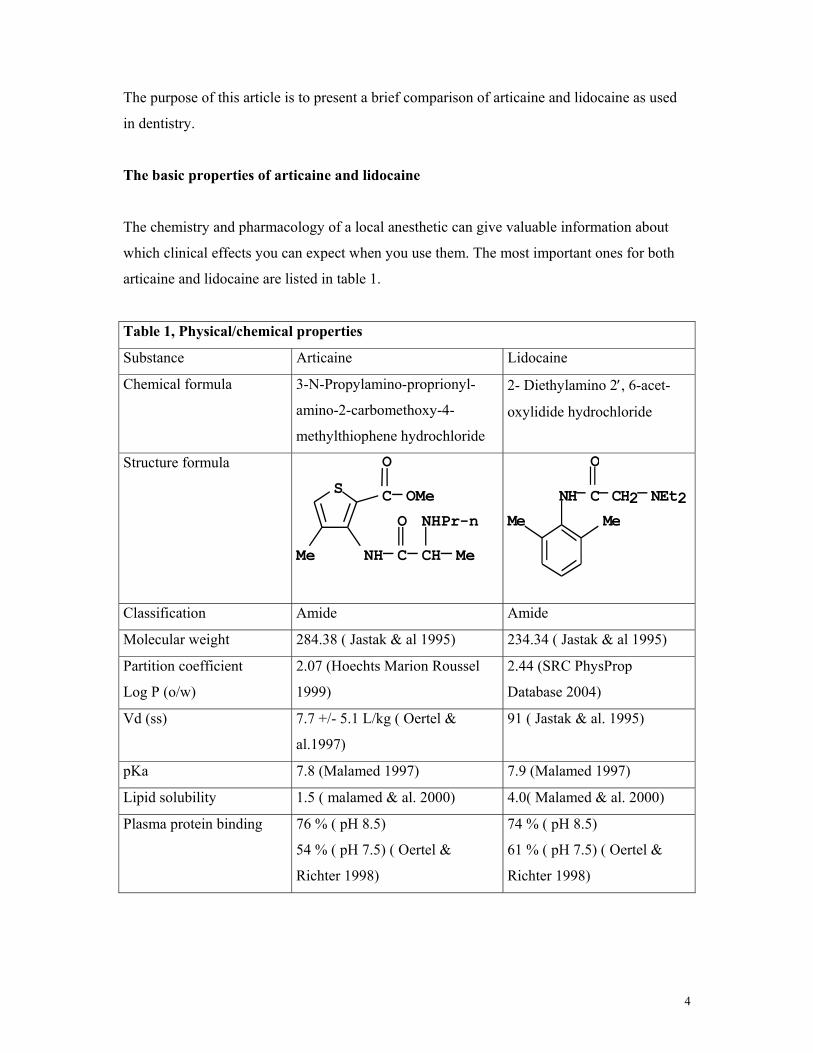
The basic properties of articaine and lidocaine
The chemistry and pharmacology of a local anesthetic can give valuable information about
which clinical effects you can expect when you use them. The most important ones for both
articaine and lidocaine are listed in table 1.
Table 1, Physical/chemical properties
Substance Articaine Lidocaine
Chemical formula 3-N-Propylamino-proprionyl-
amino-2-carbomethoxy-4-
methylthiophene hydrochloride
2- Diethylamino 2′, 6-acet-
oxylidide hydrochloride
Structure formula
NHPr-nMeMe
OMe
NH C CHO
CSO
NEt2MeMe
NH C CH2O
Classification Amide Amide
Molecular weight 284.38 ( Jastak & al 1995) 234.34 ( Jastak & al 1995)
Partition coefficient
Log P (o/w)
2.07 (Hoechts Marion Roussel
1999)
2.44 (SRC PhysProp
Database 2004)
Vd (ss) 7.7 +/- 5.1 L/kg ( Oertel &
al.1997)
91 ( Jastak & al. 1995)
pKa 7.8 (Malamed 1997) 7.9 (Malamed 1997)
Lipid solubility 1.5 ( malamed & al. 2000) 4.0( Malamed & al. 2000)
Plasma protein binding 76 % ( pH 8.5)
54 % ( pH 7.5) ( Oertel &
Richter 1998)
74 % ( pH 8.5)
61 % ( pH 7.5) ( Oertel &
Richter 1998)
5
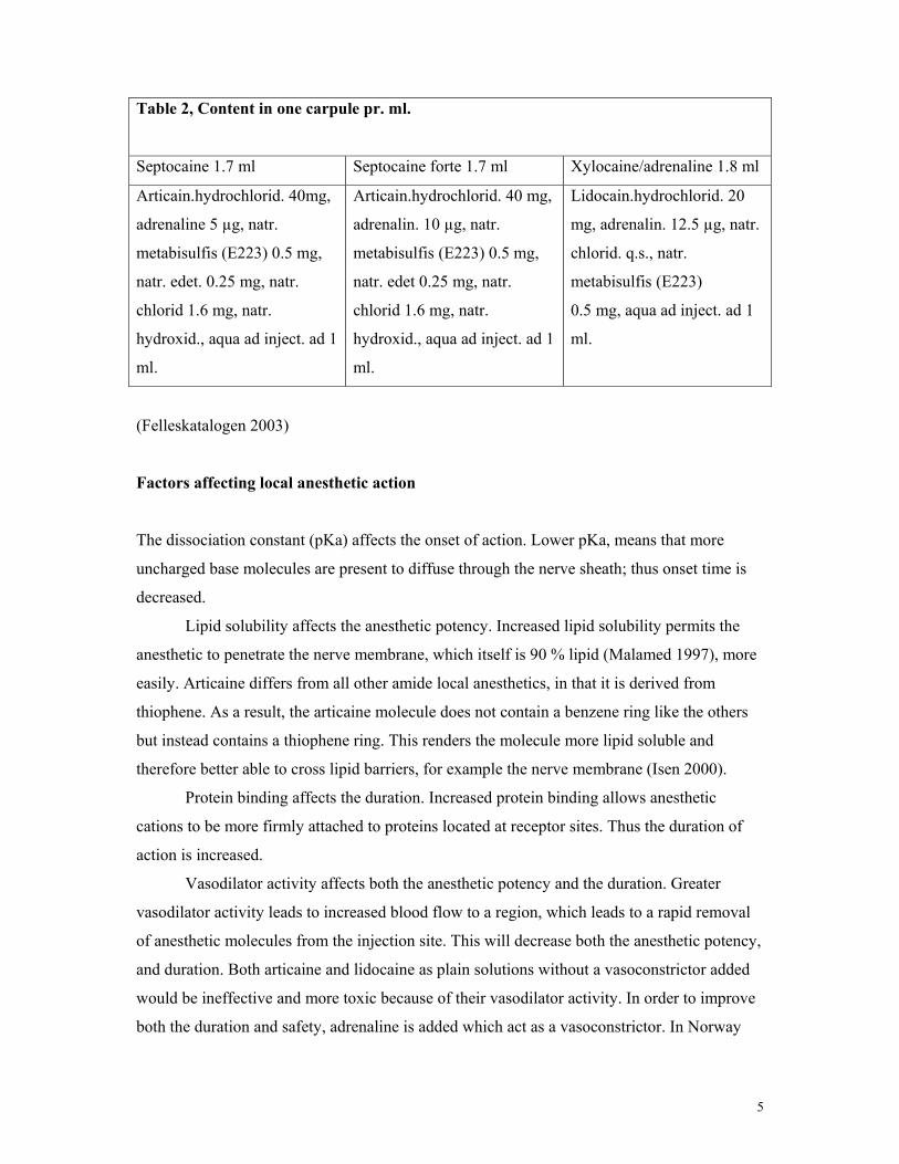
Table 2, Content in one carpule pr. ml.
Septocaine 1.7 ml Septocaine forte 1.7 ml Xylocaine/adrenaline 1.8 ml
Articain.hydrochlorid. 40mg,
adrenaline 5 µg, natr.
metabisulfis (E223) 0.5 mg,
natr. edet. 0.25 mg, natr.
chlorid 1.6 mg, natr.
hydroxid., aqua ad inject. ad 1
ml.
Articain.hydrochlorid. 40 mg,
adrenalin. 10 µg, natr.
metabisulfis (E223) 0.5 mg,
natr. edet 0.25 mg, natr.
chlorid 1.6 mg, natr.
hydroxid., aqua ad inject. ad 1
ml.
Lidocain.hydrochlorid. 20
mg, adrenalin. 12.5 µg, natr.
chlorid. q.s., natr.
metabisulfis (E223)
0.5 mg, aqua ad inject. ad 1
ml.
(Felleskatalogen 2003)
Factors affecting local anesthetic action
The dissociation constant (pKa) affects the onset of action. Lower pKa, means that more
uncharged base molecules are present to diffuse through the nerve sheath; thus onset time is
decreased.
Lipid solubility affects the anesthetic potency. Increased lipid solubility permits the
anesthetic to penetrate the nerve membrane, which itself is 90 % lipid (Malamed 1997), more
easily. Articaine differs from all other amide local anesthetics, in that it is derived from
thiophene. As a result, the articaine molecule does not contain a benzene ring like the others
but instead contains a thiophene ring. This renders the molecule more lipid soluble and
therefore better able to cross lipid barriers, for example the nerve membrane (Isen 2000).
Protein binding affects the duration. Increased protein binding allows anesthetic
cations to be more firmly attached to proteins located at receptor sites. Thus the duration of
action is increased.
Vasodilator activity affects both the anesthetic potency and the duration. Greater
vasodilator activity leads to increased blood flow to a region, which leads to a rapid removal
of anesthetic molecules from the injection site. This will decrease both the anesthetic potency,
and duration. Both articaine and lidocaine as plain solutions without a vasoconstrictor added
would be ineffective and more toxic because of their vasodilator activity. In order to improve
both the duration and safety, adrenaline is added which act as a vasoconstrictor. In Norway
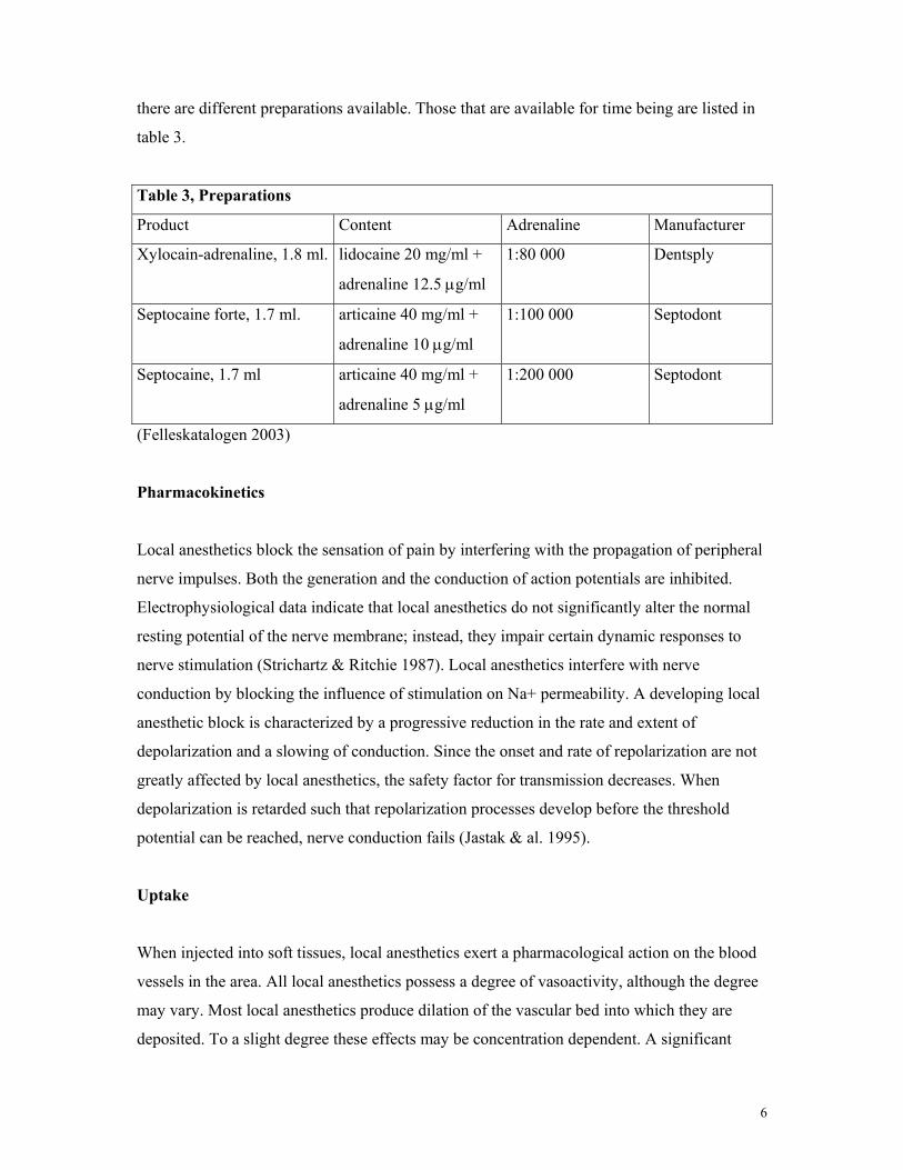
6
there are different preparations available. Those that are available for time being are listed in
table 3.
Table 3, Preparations
Product Content Adrenaline Manufacturer
Xylocain-adrenaline, 1.8 ml. lidocaine 20 mg/ml +
adrenaline 12.5 µg/ml
1:80 000 Dentsply
Septocaine forte, 1.7 ml. articaine 40 mg/ml +
adrenaline 10 µg/ml
1:100 000 Septodont
Septocaine, 1.7 ml articaine 40 mg/ml +
adrenaline 5 µg/ml
1:200 000 Septodont
(Felleskatalogen 2003)
Pharmacokinetics
Local anesthetics block the sensation of pain by interfering with the propagation of peripheral
nerve impulses. Both the generation and the conduction of action potentials are inhibited.
Electrophysiological data indicate that local anesthetics do not significantly alter the normal
resting potential of the nerve membrane; instead, they impair certain dynamic responses to
nerve stimulation (Strichartz & Ritchie 1987). Local anesthetics interfere with nerve
conduction by blocking the influence of stimulation on Na+ permeability. A developing local
anesthetic block is characterized by a progressive reduction in the rate and extent of
depolarization and a slowing of conduction. Since the onset and rate of repolarization are not
greatly affected by local anesthetics, the safety factor for transmission decreases. When
depolarization is retarded such that repolarization processes develop before the threshold
potential can be reached, nerve conduction fails (Jastak & al. 1995).
Uptake
When injected into soft tissues, local anesthetics exert a pharmacological action on the blood
vessels in the area. All local anesthetics possess a degree of vasoactivity, although the degree
may vary. Most local anesthetics produce dilation of the vascular bed into which they are
deposited. To a slight degree these effects may be concentration dependent. A significant
7
effect of vasodilation is an increase in the rate of absorption of the local anesthetic into the
blood, thus decreasing the duration of pain control while increasing the anesthetic blood level
and the potential for overdose. These effects are also related to the vascularity of the injection
site (Malamed 1997). To compensate for this effect, most local anesthetics are manufactured
as solutions containing a vasoconstrictor, for example adrenaline or felypressin.
Distribution
Once absorbed into the blood, local anesthetics are distributed throughout the body to all
tissues. The level of a local anesthetic drug in the blood from which it is distributed to certain
target tissues/organs has a significant bearing on the potential toxicity of the drug. The blood
level of the drug is influenced by the following factors:
• Rate at which the drug is absorbed into the cardiovascular system
• Rate of distribution of the drug from the vascular compartment to the tissues, which is
more rapid in healthy patients.
• Elimination of the drug through metabolic and/or excretory pathways.
The latter two factors act to decrease the blood level of the local anesthetic. The rate at which
a local anesthetic is removed from the blood is described as the elimination half-life of the
drug, t1/2β. Simply stated, the half life is the time required for a 50 % reduction in the blood
level of the drug (Malamed 1997).
Metabolism (Biotransformation)
Metabolism of local anesthetics is important, because the overall toxicity of a drug depends
on a balance between its rate of absorption into the bloodstream at the site of injection and its
rate of removal from the blood the processes of tissue uptake and metabolism. The primary
site of biotransformation of amide drugs is the liver. Liver function and hepatic perfusion
therefore significantly influence the rate of biotransformation of an amide local anesthetic.
Approximately 70 % of the dose of injected lidocaine undergoes biotransformation in patients
with normal liver function. Patients with lower than usual hepatic blood flow (hypotension,
congestive heart failure) or poor liver function (cirrhosis ) are unable to biotransform amide
local anesthetics at a normal rate. This slower than normal biotransformation rate leads to

8
increased anesthetic blood levels and potentially increased toxicity. Significant liver
dysfunction (ASA IV-V) or heart failure (ASA IV-V) represents a relative contraindication to
the administration of amide local anesthetics (Malamed 1997). Articaine differs from other
amide local anesthetics, in that it has an extra ester linkage (COOCH3). 90-95 % is
metabolized in the blood, and only 5-10 % in the liver. This feature is clearly demonstrated
when you compare the half-life (t1/2b) between articaine and lidocaine. The elimination half-
life for lidocaine is 90 min, versus that for articaine is 20 min (Isen 2000). This is the time it
takes to reduce the plasma levels of the drug by 50 %. The major metabolic product of
articaine is articainic acid. It is inactive as a local anesthetic, and systemic toxicity has not
been observed (Oertel & al.1997). This finding is important because an active metabolite may
affect toxicity and may exert undesirable side effects. In comparison, lidocaine has active
metabolites. It is metabolised in the liver by the microsomal P450 enzyme system to
monoethylglyceine and xylidide; xylidide is a local anesthetic and potentially toxic (Malamed
1997).
Excretion
The kidneys are the primary excretory organ for booth the local anaesthetic and its
metabolites. A percentage of a given dose of local anesthetic drug will be excreted unchanged
in the urine, and this varies according to the drug. Patients with significant renal impairment
may be unable to eliminate the parent local anesthetic compound or its major metabolites
from the blood, resulting in slightly elevated blood levels and an increased potential for
toxicity. Thus significant renal disease (ASA IV-VI) represents a relative contraindication to
the administration of local anesthetics. This includes patients undergoing renal dialysis and
those with cronic glomerulonephritis and/or pyelonephritis (Malamed 1997). Articaine is
largely excreted in the urine as the metabolite articaninic acid (64.2 +/- 14.4 %), followed by
articainic glucuronide (13.4 +/- 5.0 %) and the parent drug ( 1.45 +/- 0.77 % ) (Oertel & al.
1997). For lidocaine the excretion is also via the kidneys; less than 10 % unchanged, more
than 80 % various metabolites ( Malamed 1997).
Pharmacodynamics
The potency of articaine is 1.5 times that of lidocaine and 1.9 times that of procaine, and the
toxicity is similar to that of lidocaine and procaine (Malamed 1997). The unintentional
9
intravascular injection of local anesthetic agents in dentistry can occur because of the high
vascularisation of this area. The risk of such an intravascular injection is up to 20 % in
conduction anesthesia of the inferior alveolar nerve (Oertel & al. 1997). The signs and
symptoms are referable to the CNS and cardiovascular system. A comparison between
articaine and lidocaine showed that the signs of CNS toxicity after intravenous administration
of lidocaine were observed more frequently and at a higher degree of severity when compared
with articaine. The cardiovascular parameters did not change (Oertel & al. 1997).
Clinical comparison of articaine versus lidocaine
Safety
Before a new local anesthetic drug can be introduced on the open market, it has to pass
through several different levels of testing and developing. The first step is in vitro studies, and
then we have animal testing and finally clinical testing. Lidocaine, which is a relatively old
local anesthetic, has been studied thoroughly for many years, and has well known effects and
side effects. The safety has also been under substantial investigation for many years. Articaine
is not as old as lidocaine, although it was synthesized back in 1969. It has been used in several
European countries for almost 30 years, and its safety has been well documented by several
different studies.
The unintentional intravascular injection of local anesthetic agents in dentistry can
occur because of high vascularisation in this area. The risk of such an intravascular injection
is up to 20 % in conduction anesthesia of the mandibular nerve (Bartlett 1972). The signs and
symptoms of local anesthetic agent toxicity are referable to the CNS and cardiovascular
system. The initial subjective signs of CNS intoxication are general feeling of
lightheadedness, dizziness, disorientation, drowsiness, anxiety, excitement and visual and
auditory disturbances. Objective signs of early CNS toxicity consist of shivering, muscular
twitching and tremors of the facial muscles. Oertel & al. showed that signs of CNS toxicity
after intravascular injection of lidocaine were observed more frequently and at a higher degree
of severity when compared with articaine (1977). The cardiovascular parameters did not
change (Oertel & al. 1997). It was concluded that an unintentional intravascular injection of
about 80 mg of articaine – the equivalent of 1 cartridge of the commercial 4 % solution – does
not cause toxic effects in healthy patients. This is confirmed by the LD50, which for articaine
10
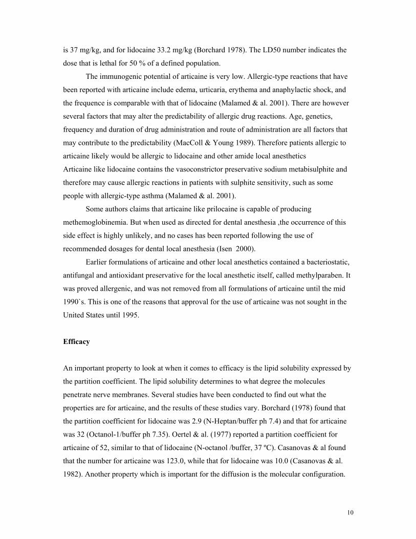
is 37 mg/kg, and for lidocaine 33.2 mg/kg (Borchard 1978). The LD50 number indicates the
dose that is lethal for 50 % of a defined population.
The immunogenic potential of articaine is very low. Allergic-type reactions that have
been reported with articaine include edema, urticaria, erythema and anaphylactic shock, and
the frequence is comparable with that of lidocaine (Malamed & al. 2001). There are however
several factors that may alter the predictability of allergic drug reactions. Age, genetics,
frequency and duration of drug administration and route of administration are all factors that
may contribute to the predictability (MacColl & Young 1989). Therefore patients allergic to
articaine likely would be allergic to lidocaine and other amide local anesthetics
Articaine like lidocaine contains the vasoconstrictor preservative sodium metabisulphite and
therefore may cause allergic reactions in patients with sulphite sensitivity, such as some
people with allergic-type asthma (Malamed & al. 2001).
Some authors claims that articaine like prilocaine is capable of producing
methemoglobinemia. But when used as directed for dental anesthesia ,the occurrence of this
side effect is highly unlikely, and no cases has been reported following the use of
recommended dosages for dental local anesthesia (Isen 2000).
Earlier formulations of articaine and other local anesthetics contained a bacteriostatic,
antifungal and antioxidant preservative for the local anesthetic itself, called methylparaben. It
was proved allergenic, and was not removed from all formulations of articaine until the mid
1990`s. This is one of the reasons that approval for the use of articaine was not sought in the
United States until 1995.
Efficacy
An important property to look at when it comes to efficacy is the lipid solubility expressed by
the partition coefficient. The lipid solubility determines to what degree the molecules
penetrate nerve membranes. Several studies have been conducted to find out what the
properties are for articaine, and the results of these studies vary. Borchard (1978) found that
the partition coefficient for lidocaine was 2.9 (N-Heptan/buffer ph 7.4) and that for articaine
was 32 (Octanol-1/buffer ph 7.35). Oertel & al. (1977) reported a partition coefficient for
articaine of 52, similar to that of lidocaine (N-octanol /buffer, 37 ºC). Casanovas & al found
that the number for articaine was 123.0, while that for lidocaine was 10.0 (Casanovas & al.
1982). Another property which is important for the diffusion is the molecular configuration.
11
Articaine contains a thiophene ring instead of benzene like lidocaine. This gives the molecule
better diffusion properties compared with lidocaine (Casanovas & al. 1982).
One of the reasons why articaine instantly became so popular in many countries was
due to its excellent efficacy. Dentists claimed that they seldom missed with the inferior
alveolar nerve block, and that buccal infiltration in the maxillary arch often was enough
before an extraction of a molar, because of articaine’s bone penetration properties. This
seemingly excellent efficacy is reported from many dentists from around the world, based on
their daily clinical practice.
The next step would therefore be to see if there is any literature to support these
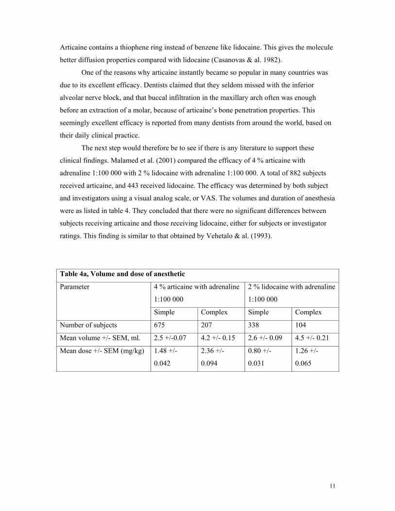
clinical findings. Malamed et al. (2001) compared the efficacy of 4 % articaine with
adrenaline 1:100 000 with 2 % lidocaine with adrenaline 1:100 000. A total of 882 subjects
received articaine, and 443 received lidocaine. The efficacy was determined by both subject
and investigators using a visual analog scale, or VAS. The volumes and duration of anesthesia
were as listed in table 4. They concluded that there were no significant differences between
subjects receiving articaine and those receiving lidocaine, either for subjects or investigator
ratings. This finding is similar to that obtained by Vehetalo & al. (1993).
Table 4a, Volume and dose of anesthetic
4 % articaine with adrenaline
1:100 000
2 % lidocaine with adrenaline
1:100 000
Parameter
Simple Complex Simple Complex
Number of subjects 675 207 338 104
Mean volume +/- SEM, ml. 2.5 +/-0.07 4.2 +/- 0.15 2.6 +/- 0.09 4.5 +/- 0.21
Mean dose +/- SEM (mg/kg) 1.48 +/-
0.042
2.36 +/-
0.094
0.80 +/-
0.031
1.26 +/-
0.065
12
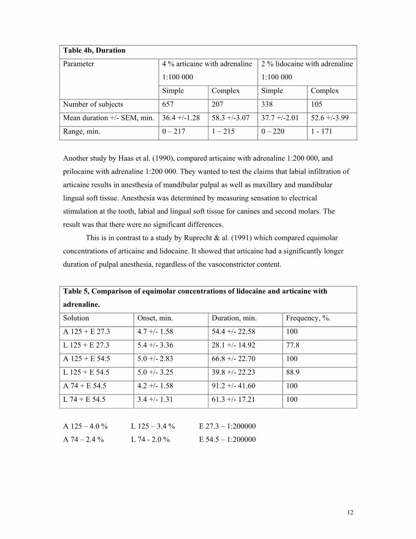
Table 4b, Duration
4 % articaine with adrenaline
1:100 000
2 % lidocaine with adrenaline
1:100 000
Parameter
Simple Complex Simple Complex
Number of subjects 657 207 338 105
Mean duration +/- SEM, min. 36.4 +/-1.28 58.3 +/-3.07 37.7 +/-2.01 52.6 +/-3.99
Range, min. 0 – 217 1 – 215 0 – 220 1 - 171
Another study by Haas et al. (1990), compared articaine with adrenaline 1:200 000, and
prilocaine with adrenaline 1:200 000. They wanted to test the claims that labial infiltration of
articaine results in anesthesia of mandibular pulpal as well as maxillary and mandibular
lingual soft tissue. Anesthesia was determined by measuring sensation to electrical
stimulation at the tooth, labial and lingual soft tissue for canines and second molars. The
result was that there were no significant differences.
This is in contrast to a study by Ruprecht & al. (1991) which compared equimolar
concentrations of articaine and lidocaine. It showed that articaine had a significantly longer
duration of pulpal anesthesia, regardless of the vasoconstrictor content.
Table 5, Comparison of equimolar concentrations of lidocaine and articaine with
adrenaline.
Solution Onset, min. Duration, min. Frequency, %.
A 125 + E 27.3 4.7 +/- 1.58 54.4 +/- 22.58 100
L 125 + E 27.3 5.4 +/- 3.36 28.1 +/- 14.92 77.8
A 125 + E 54.5 5.0 +/- 2.83 66.8 +/- 22.70 100
L 125 + E 54.5 5.0 +/- 3.25 39.8 +/- 22.23 88.9
A 74 + E 54.5 4.2 +/- 1.58 91.2 +/- 41.60 100
L 74 + E 54.5 3.4 +/- 1.31 61.3 +/- 17.21 100
A 125 – 4.0 % L 125 – 3.4 % E 27.3 – 1:200000
A 74 – 2.4 % L 74 - 2.0 % E 54.5 – 1:200000
13
Winther & Nathalang showed that articaine was significantly superior to lidocaine with
respect to frequency, extent and duration of analgesia (1972).
Another important issue is the concentration of adrenaline. The effectiveness of 4 %
articaine associated with 1:100 000 or 1:200 000 adrenaline for inferior alveolar nerve blocks
are the same (Tofoli & al. 2003). This is why 1:200 000 are the recommended concentration
of adrenaline for dental procedures (Jacob 1989), except for those procedures (e.g. surgical
interventions) that requires a larger degree of hemostasis. For these purposes the
recommended concentration is 1:50 000 (Buckley & al. 1984) or 1:80 000 as used in
Scandinavia.
An important issue when comparing two substances as articaine and lidocaine are the
methods used. In order to get statistical significant data, one needs a large enough material
containing a sufficient number of subjects. Many studies fail to show differences because of
this problem. This may be one of the reasons why articaine in different studies tends to be
somewhat more effective than lidocaine, although not significant.
Similar to other amide local anesthetics, articaine blocks sodium channels at a lower
concentration than potassium channels, but lower concentrations of the thiophene derivate
than of the benzene derivates are needed to block the ionic channels. Because of the higher
partition coefficient of lidocaine, it seems that the action on these channels seems not to
follow mere lipid solubility properties of the neutral drug. Differences in the interaction of
local anesthetics with ionic channel proteins might in part be correlated to different binding
properties to plasma proteins, as indicated by a higher affinity of articaine to plasma proteins
as compared to lidocaine (Borchard & Drouin 1980).
Use in pediatric dentistry
In order for a local anesthetic to become popular, it is important that it is useful in a wide
range of situations. Lidocaine has been used for both adults and children for more than five
decades, and pediatric dentistry is for sure an important area.
Articaine should not be used in patients under 4 years of age, because safety and
effectiveness in this group has not yet been seriously investigated (Felleskatalogen 2003),
although 2 studies showed that articaine is likely safe for children under 4 years of age
(Jacobs & al. 1995, Wright & al. 1989). When used in pediatric dentistry, it is important to
remember that articaine is in a 4 % solution, and that maximum dose for children is the same
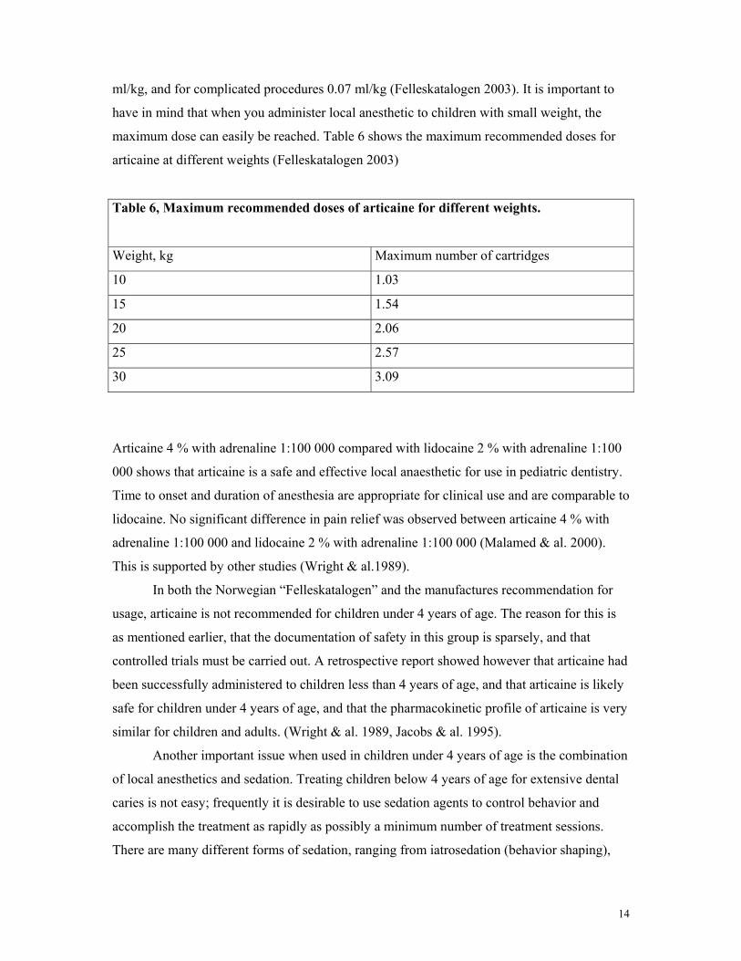
as for adults; 7 mg/kg (0.175 mL/kg). For simple procedures the recommendation is 0.04
14
ml/kg, and for complicated procedures 0.07 ml/kg (Felleskatalogen 2003). It is important to
have in mind that when you administer local anesthetic to children with small weight, the
maximum dose can easily be reached. Table 6 shows the maximum recommended doses for
articaine at different weights (Felleskatalogen 2003)
Table 6, Maximum recommended doses of articaine for different weights.
Weight, kg Maximum number of cartridges
10 1.03
15 1.54
20 2.06
25 2.57
30 3.09
Articaine 4 % with adrenaline 1:100 000 compared with lidocaine 2 % with adrenaline 1:100
000 shows that articaine is a safe and effective local anaesthetic for use in pediatric dentistry.
Time to onset and duration of anesthesia are appropriate for clinical use and are comparable to
lidocaine. No significant difference in pain relief was observed between articaine 4 % with
adrenaline 1:100 000 and lidocaine 2 % with adrenaline 1:100 000 (Malamed & al. 2000).
This is supported by other studies (Wright & al.1989).
In both the Norwegian “Felleskatalogen” and the manufactures recommendation for
usage, articaine is not recommended for children under 4 years of age. The reason for this is
as mentioned earlier, that the documentation of safety in this group is sparsely, and that
controlled trials must be carried out. A retrospective report showed however that articaine had
been successfully administered to children less than 4 years of age, and that articaine is likely
safe for children under 4 years of age, and that the pharmacokinetic profile of articaine is very
similar for children and adults. (Wright & al. 1989, Jacobs & al. 1995).
Another important issue when used in children under 4 years of age is the combination
of local anesthetics and sedation. Treating children below 4 years of age for extensive dental
caries is not easy; frequently it is desirable to use sedation agents to control behavior and
accomplish the treatment as rapidly as possibly a minimum number of treatment sessions.
There are many different forms of sedation, ranging from iatrosedation (behavior shaping),
15
conscious sedation (benzodiazepines) to general anesthesia (inhalation or intravenous) (Koch
& Poulsen 2001). It is important to be careful when you administer local anesthetics to a
sedated child, since there is a higher risk of adverse reactions to occur, and that there are a
direct link between adverse reactions and local anesthesia volumes (Wright & al.1989,
Weaver 1999). When restoring primary mandibular molars, the customary injection is a
mandibular (inferior) dental nerve block. Block anesthesia has some disadvantages for
children. Especially, the lengthy duration of the anesthesia allows for a greater possibility of
postoperative trauma, such as lip or tongue biting. Also, parents must maintain close
supervision while their children are under anesthesia.
Mandibular infiltrations have been a debated area. Some dentists claims that articaine
has better bone penetration, and therefore could produce anesthesia to primary molars of
children. The major obstacle in obtaining anesthesia from infiltrations in the mandible, is the
density and thickness of the bone when compared to maxilla. Articaine possesses properties
that make it suitable for infiltration techniques in the lower jaw, and that this should be the
technique of choice, except for extractions where a nerve block is recommended. Infiltrations
are also more suited for difficult and handicapped children, and it can be safely given to a
reluctant child. Furthermore, the tongue is not affected, thus reducing the risk of postoperative
bites (Dudkiewicz & al. 1997, Wright & al. 1991).
An important issue in obtaining good anesthesia in children is the influence of the
children’s behavior. There is a high relationship between children behaving cooperatively and
comfort during procedures. Children who demonstrate comfort at the time of injection are
likely to exhibit no pain during successive procedures (Wright & al. 1991).
Use in geriatric patients
Aging is associated with physiologic changes which could alter pharmacokinetics of drugs.
Age-related changes in pharmacokinetics affect drug absorption, distribution metabolism, and
elimination. Increase in body fat, decrease in lean body mass, and total body water, changes in
hepatic metabolism, and renal elimination capacity in the elderly are of particular clinical
significance. These changes should be taken into account when choosing drug therapy for
older patients to minimize adverse effects and maximize potential benefits. Because the
median age of the population is steadily increasing, more elderly patients are undergoing
routine dental procedures for which local anesthesia could be required (Oertel & al. 1999).
16
Additionally, pharmacodynamic changes in the elderly can result in greater, or
sometimes even lesser, drug sensitivity than that seen in a young individual. This means that,
in theory, increased or decreased pharmacodynamic sensitivity in the elderly may coexist with
or be independent of, pharmacokinetic changes (Oertel & al. 1999).
Concerning local anesthesia, most of our knowledge of pharmacokinetics is based
almost entirely on investigations in young individuals ( Nordenram & Danielsson 1990). The
only local anesthetic agent that is tested extensively in elderly patients, is lidocaine (Oertel &
al. 1999).
There are no statistical differences in Cmax and Tmax or t1/2 between young and
older individuals (Oertel & al. 1999). This is a very important finding, indicating that there
might be an age-independent plasma esterase function. This is in contrast to other amide local
anesthetics, which is primarily metabolized in the liver, which has an capacity that decreases
with age. There is an significant decrease in CL and Vdss of articaine in elderly, related to the
decrease in lean body mass and increase in body fat (Oertel & al. 1999).
Taken into account that articaine shows an age-independent metabolism, there should
be no reason to change the dosage in elderly patients. But, it is important to remember that
articaine is a highly serum protein bounded drug, and changes in binding to serum are also a
factor that could affect pharmacokinetics in the elderly.
Complications
A wide range of different complications can occur during or after the injection of local
anesthesia. They can be divided into local complications, such as pain on injection, persistent
anesthesia/paresthesia, trismus, hematoma, oedema and facial nerve paralysis, and systemic
complications such as overdoses and allergic reactions.
Paresthesia can be defined as persistent anesthesia (anesthesia well beyond the
expected duration), or altered sensation (tingling or itching) well beyond the expected
duration of anesthesia (Malamed 1997). The definition of paresthesia also includes
hyperesthesia and dysesthesia. Hyperesthesia is defined as increased sensitivity to noxious
stimuli, and dysesthesia as painful sensation to nonnoxious stimuli (Dower 2003). The
symptoms are most commonly associated with mechanical trauma during surgical procedures.
During the administration of anesthesia for a mandibular nerve block, the lingual or inferior
alveolar neurovascular bundle can be traumatized by the sharp needle-tip, the movement of
17
the needle, ekstraneural or intraneural hemorrhage from trauma to the blood vessels, or from
neurotoxic effects of the local anesthetic.
A retrospective analysis of paresthesia after local anaesthetic administration for
nonsurgical dental procedures over a 20-year period, from 1973-1993, was published by Haas
and Lennon in 1995. Paresthesia was defined as numbness or tingling of the mouth or face.
The analysis revealed a higher-than-expected frequency of paresthesia with articaine, based
on the number of cartridges used (2.27 per 1 million injections vs. an expected frequency of
1.20 per 1 million injections). There were no significant differences found with respect to
patient age, patient gender, or needle gauge (Haas & Lennon 1995).
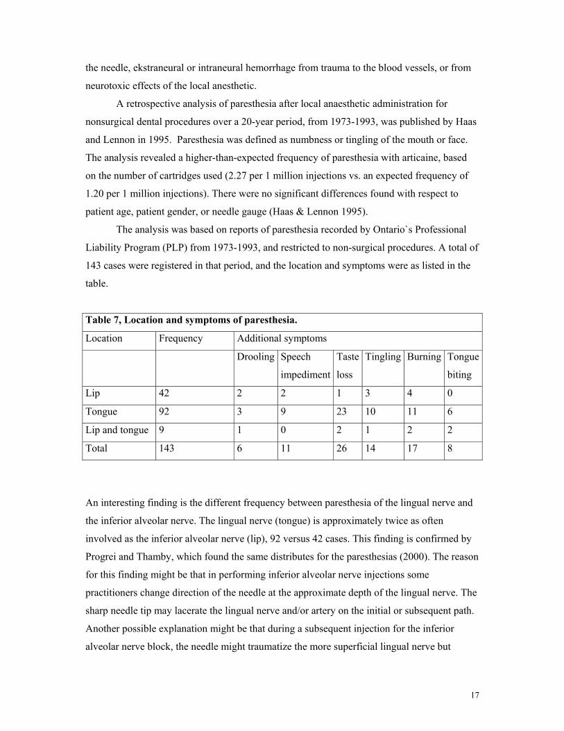
The analysis was based on reports of paresthesia recorded by Ontario`s Professional
Liability Program (PLP) from 1973-1993, and restricted to non-surgical procedures. A total of
143 cases were registered in that period, and the location and symptoms were as listed in the
table.
Table 7, Location and symptoms of paresthesia.
Location Frequency Additional symptoms
Drooling Speech
impediment
Taste
loss
Tingling Burning Tongue
biting
Lip 42 2 2 1 3 4 0
Tongue 92 3 9 23 10 11 6
Lip and tongue 9 1 0 2 1 2 2
Total 143 6 11 26 14 17 8
An interesting finding is the different frequency between paresthesia of the lingual nerve and
the inferior alveolar nerve. The lingual nerve (tongue) is approximately twice as often
involved as the inferior alveolar nerve (lip), 92 versus 42 cases. This finding is confirmed by
Progrei and Thamby, which found the same distributes for the paresthesias (2000). The reason
for this finding might be that in performing inferior alveolar nerve injections some
practitioners change direction of the needle at the approximate depth of the lingual nerve. The
sharp needle tip may lacerate the lingual nerve and/or artery on the initial or subsequent path.
Another possible explanation might be that during a subsequent injection for the inferior
alveolar nerve block, the needle might traumatize the more superficial lingual nerve but
18
without the “electric shock” sensation, because the nerve is usually anesthesized on the initial
attempt (Dower 2003). The cause of the paresthesia may also be combination of
neurotoxicity of the local anesthetic and trauma to the nerve (Kalichman & al. 1993).
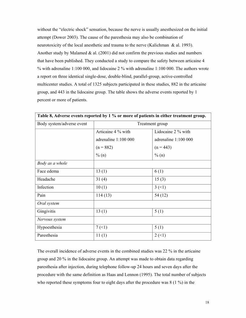
Another study by Malamed & al. (2001) did not confirm the previous studies and numbers
that have been published. They conducted a study to compare the safety between articaine 4
% with adrenaline 1:100 000, and lidocaine 2 % with adrenaline 1:100 000. The authors wrote
a report on three identical single-dose, double-blind, parallel-group, active-controlled
multicenter studies. A total of 1325 subjects participated in these studies, 882 in the articaine
group, and 443 in the lidocaine group. The table shows the adverse events reported by 1
percent or more of patients.
Table 8, Adverse events reported by 1 % or more of patients in either treatment group.
Body system/adverse event Treatment group
Articaine 4 % with
adrenaline 1:100 000
(n = 882)
% (n)
Lidocaine 2 % with
adrenaline 1:100 000
(n = 443)
% (n)
Body as a whole
Face edema 13 (1) 6 (1)
Headache 31 (4) 15 (3)
Infection 10 (1) 3 (<1)
Pain 114 (13) 54 (12)
Oral system
Gingivitis 13 (1) 5 (1)
Nervous system
Hypoesthesia 7 (<1) 5 (1)
Paresthesia 11 (1) 2 (<1)
The overall incidence of adverse events in the combined studies was 22 % in the articaine
group and 20 % in the lidocaine group. An attempt was made to obtain data regarding
paresthesia after injection, during telephone follow-up 24 hours and seven days after the
procedure with the same definition as Haas and Lennon (1995). The total number of subjects
who reported these symptoms four to eight days after the procedure was 8 (1 %) in the
19
articaine group and 5 (1 %) in the lidocaine group. In 5 cases (4 with articaine and 1 with
lidocaine) the symptoms did not begin on the day of study drug administration, suggesting
that they were caused by the procedure rather than the anesthetic. Ultimately most cases of
paresthesia resolve within eight weeks (Malamed 1997).
As can be seen from the text above there is somewhat contradicting numbers regarding
paresthesia after injection of local anesthetics. Why the Haas & Lennon study found that the
numbers of paresthesia for articaine was so high is somewhat uncertain, but some possible
explanations might be considered. First of all the study was based on voluntary reporting from
the dentist in Ontario, Canada. A high degree of subjectivity is involved, and this might
influence on the numbers. Also in 1990 the PLP changed the insurance underwriters, and in
anticipation of the change special notices and requests were sent to all dentists urging them to
report any potential claims. This might have led to a greater number of reported cases.
Second, articaine was introduced onto the Canadian market in 1985. After this, there where an
marketed increase in the reported number of paresthesies. One reasonable explanation for this
finding might be the increased attention that there always will be after the introduction of a
new product onto the market. The dentists that changed to articaine most likely paid more
attention to note any side effects than dentist that used the same local anesthetic that they had
been used for many years.
Another explanation might be that in the Haas & Lennon study, cases where surgery
was involved were excluded. This might hide the numbers of paresthesia actually related to
the drug, and if for instance lidocaine is more used in surgery, this might lower the numbers
of paresthesias related to lidocaine. Another issue is that the Canadian study does not provide
information regarding the duration of paresthesia in the cases reported. It might be that some
cases actually is a result of a somewhat longer duration of the anesthesia, that ultimately
resolved after a relatively short period of time. Since most dentist feel that articaine has a
somewhat better penetration than lidocaine, the technique that they uses have changed. In
stead of inferior alveolar blocks, they infiltrate around the tooth, and hence the risk for
traumatizing the lingual nerve increases.
Although there are no reports demonstrating that the needle used in dental syringes are
large enough to produce a complete severance of the inferior or alveolar or lingual nerves,
simple contact may be sufficient to induce a transient paresthesia (Malamed 1997). The 25-
gauge needle is the largest needle commonly used in dentistry. With an external diameter of
0.45 mm, it is considerable smaller than the lingual nerve, which has been reported to have an
average diameter of 1.86 mm (Kiesselbach & Chamberlain 1984).
20
Articaine is delivered as a 4 % solution in as opposed to lidocaine which is 2 %. It may be
speculated that if there is a toxic local metabolite involved, it may manifest toxicity simply
due to the higher concentration.
Another complication that might occur is paralysis of the oculomotor muscles, leading
to diplopia and even temporary blindness. All such manifestations are transient and disappear
on cessation of the anesthetic effects. In case of paralysis of the extrinsic musculature of the
eye (especially the external rectus muscle), synchronic movement of the eyes becomes
impossible, and diplopia appears. Such complications are possible when performing an
posterior superior alveolar anesthesia. There are many different theories on the mechanisms
behind these opthalmologic complications, and it is likely that the local anesthetic solution
diffuses directly from the pterigomaxillary fossa, through the sphenomaxillary cavity, to the
orbit. This would affect the ciliary ganglion, located between the optic nerve and the external
rectus muscle of the eye (Penarrocha-Diago & Sanchis-Bielsa 2000). The symptoms develop
immediately after the injection of the anesthetic solution and can persist for between 1 minute
and several hours, though they only rarely exceed the duration of the anesthetic effect. Most
of the reported cases in the literature are produced by lidocaine and mepivacaine, but they
may also happen after injections of articaine ( Penarrocha-Diago & Sanchis-Bielsa 2000).
Why is articaine delivered as a 4 % solution?
Articaine is produced as a 4 % local anesthetic solution. This is in contrast to lidocaine which
is a 2 % solution, and similar to prilocaine which also is a 4 % solution. Equal analgesic
efficacy along with lower systemic toxicity (i.e., a wide therapeutic range) allows use of
articaine in higher concentrations than other amide-type local anesthetics (Oertel & al. 1997).
This is advantageous with respect to the required bone penetration, and hence it is possible to
inject smaller volumes, thereby minimizing the injection induced pain.
One possible disadvantage with the higher concentration of the local anesthetic
solution, is that it has been determined that local anesthetic- induced nerve injury is
concentration dependent, with injuries increasing as concentration increases (Dower 2003).
Discussion
As can be seen from the text above, there are many aspects that has to be taken under
consideration when comparing local anesthetic solutions. The quality and validity of data
21
from different studies varies tremendously, and it might be sometimes hard to really spot any
differences. When comparing articaine to lidocaine, I started out with looking at the basic
properties of the drugs. There are relatively few significant differences. One basic difference
is that articaine contains a thiophene ring, compared to the benzene ring of lidocaine (Isen
2000). A number of authors claims that this makes the drug more lipophilic, but this is not
reflected when comparing the n- octanol/Soerensen buffer, which shows varying numbers in
different studies, some favoring articaine and others lidocaine.
Another basic property that differs among the two drugs is that articaine contains an
extra ester linkage. This causes articaine to be hydrolyzed by plasma esterase as well as the
microsomal P450 enzyme system in the liver. This again, causes the half life for articaine to
be approximately 20 minutes, while that for lidocaine is 90 minutes (Isen 2000).
Articaine and lidocaine is marketed in different preparations. Articaine as an 4 %
solution with adrenaline 1:100 000 (Septocaine forte) or 1:200 000 ( Septocaine), and
lidocaine as an 2 % solution with adrenaline 1:80 000. The fact that articaine is delivered as
an 4 % solution, means that when one is calculating the maximum recommended number of
carpules that might be used during one single procedure, it is approximately half as much as
for lidocaine. Given the recommended maximum dose of 7 mg/kg body weight, the maximum
number for articaine would be 7 carpules, while that for lidocaine will be 13.
The fact that articaine can be delivered with an adrenaline concentration of 1:200 000
is important in several situations. For pain control the recommended adrenaline concentration
is 1:200 000, alternatively 1:100 000 where extended pain control is required (Jacob 1989).
When it comes to post-operative pain, adrenaline is one of the major pain inducing factors,
and limiting the concentration from 1:80 000 to 1:160 000 reduces the level of post-operative
pain (Jorkjend 1998). For hemostasis the 1:50 000 dilution of adrenaline is more effective
than less concentrated solutions (Buckley & al 1984), and adrenaline dilutions of 1:50000 and
1:100000 are considerably more effective in restricting surgical blood loss than local
anesthetics without vasoconstrictor additives (Sveen 1979).
For some patients the use of adrenaline in local anesthesia might be dangerous, and
potentially fatal. For instance patients with thyreotoxicosis, heart disease (ASA II-V), and
patients taking drugs as MAO and TCA represents risk patients when using local anesthetics
containing adrenaline (Malamed 1997). When it comes to treatment of these patient groups,
the adrenaline concentration of the anesthetic solution becomes interesting. Articaine 4 %
with adrenaline 1:200 000 might be a better choice in these situations compared to lidocaine 2
% with adrenaline1:80 000.
22
Articaine is delivered as a 4 % solution and lidocaine as a 2 % solution. This again means that
when the same recommendations for the maximum doses are applied, one can inject twice as
many carpules of lidocaine when compared with articaine. This is important to be aware of
during situations where more anesthetics have to be re-injected. But, there is another feature
that has to be taken into account when one is re-injecting anesthetics and is concerned about
the maximum doses, and that is the elimination half-life. As mentioned before, articaine
contains an additional ester group that is quickly hydrolyzed by plasma esterases (Oertel & al.
1997). This gives articaine a elimination half-life of approximately 20 minutes, compared to
that for lidocaine which is approximately 90 minutes. This makes re-injection of articaine
safer, since the majority of that initial dose are metabolized after approximately half an hour,
and the re-injected dose will not be added to the initial one (Isen 2000).
References
Bartlett SZ. Clinical observation on the effect of injections of local anaesthetics preceded by
aspiration. Oral Surg 1972;33:520-525.
Borchard U. Vergleichende pharmacologie der lokalanaesthetica und spezielle pharmakologie
von Carticain. Anasthesiol Intensivmed 1978;113:7-11.
Borchard U, Drouin H. Carticaine: Action of the local anesthetic on myelinated nerve fibres.
Eur J Pharmacol 1980;62:73-79.
Buckley JA, Ciancio SG, McMullen JA. Efficacy of epinephrine concentration in local
aneshesia during periodontal surgery. J Periodontol 1984;55:653-657.
Casanovas AM. Études des relations structures – activé d`une série d` anesthésiques locaux.
Eur J Med Chem - Chim Ther 1982;17:333-337.
Dower JS. A review of paresthesia. Dent Today 2003;22:64-69
Dudkiewicz A, Schwartz S, Laliberté R. Effectiveness of mandibular infiltration in children
using the local anesthetic ultracaine (articaine Hydrochloride). J Can Dent Assoc 1987;1:29-
31.
23
Haas DA, Harper DG, Saso MA, Young ER. Comparison of articaine and prilocaine
anesthesia by infiltration in maxillary and mandibular arches. Anesth Prog 1990;37:230-237.
Haas DA, Lennon D. A 21 year retrospective study of reports of paresthesia following local
anesthetic administration. J Can Dent Assoc 1995;61:319-330.
Isen DA. Articaine: Pharmacology and clinical use of a recently approved local anesthetic.
Dent Today 2000;19:72-77.
Jacob W. Local anaesthesia and vasoconstrictive additional components. Newslett Int Fed
Dent Anesthesiol Soc 1989;2:1-3.
Jacobs W, Ladwig B, Cichon P, Oertel R, Kirch W. Serum levels of articaine 2 % and 4 % in
children. Anesth Prog 1995;42:113-115.
Jastak JT, Yagiela JA, Donaldson D. Local anesthesia of the oral cavity. 1st ed. Philadelphia,
Saunders, 1995.
Jorkjend L. The effect of local anesthetic agents on postoperative pain after periodontal
surgery (thesis). Section of Dental Pharmacology and Pharmacotherapy. Oslo, University of
Oslo, 1998.
Kalichman MW, Moorhouse DF, Powell HC, Myers RR. Relative neural toxicity of local
anesthetics. J Neuropathol 1993;52:234-240.
Kiesselbach JE,Chamberlain JG. Clinical and anatomic observations on the relationship of the
lingual nerve to the mandibular third molar region. J Oral Maxillofac Surg, 1984;42:565-567.
Koch G, Poulsen S. Pediatric dentistry, a clinical approach. 1st ed. Copenhagen, Munksgaard,
2001.
Maccoll S, Young ER. An allergic reaction following injection of local anesthetic: A case
report. J Can Dent 1989;55:981-984.
24
Malamed SF. Handbook of local anaesthesia. 4th ed. St. Louis, Mosby; 1997.
Malamed SF, Gagnon S, Leblanc D. Efficacy of articaine: A new amide local anesthetic. J
Am Dent Assoc 2000;131:635-642.
Malamed SF, Gagnon S, Leblanc D. A comparison between articaine HCL and lidocaine
HCL in pediatric dental patients. Am Acad Ped Dent 2000;22:307-311.
Malamed S F, Gagnon S, Leblanc D. Articaine hydrochloride: a study of the safety of a new
amide local anesthetic. J Am Dent Assoc 2001;132:177-184.
Nordenram A, Danielsson K. Local anaesthesia in elderly patients. An experimental study of
oral infiltration anaesthesia. Swed Dent J 1990;14:19-24.
Oertel R, Ebert U, Rahn R, Kirch W. The effect of age on the pharmacokinetics of the local
anesthetic drug articaine. Regional Anesth Pain Med 1999;24:524-528.
Oertel R, Rahn R, Kirch W. Clinical pharmacokinetics of articaine. Clin Pharmacokinet
1997;33:417-425.
Oertel R, Richter K. Plasma protein binding of the local anaesthetic drug articaine and its
metabolite articainic acid. Pharmazie 1998;53:646-647.
Peñarrocha-Diago M., Sanchis-Bielsa J M. Ophtalmologic complications after intraoral local
anesthesia with articaine. Oral Surg Oral Med Oral Patho 2000;90:21-23.
Progrei M A and Thamby S. Permanent nerve involvement resulting from inferior alveolar
nerve blocks. J Am Dent Assoc 2000;131:901-907.
Ruprecht S, Knoll-Køhler E. Vergleichende Untersuchung äquimolarer Lösungen von
Lidocain und Articain zur Anästhesie. Schweiz Monatsschr Zahnmed 1991;101:1286-1290.
Septocaine Product Monograph, 2003.
25
Strichartz GR, Ritchie JM. The action of local anesthetics on ion channels of excitable tissues.
Handbook of Experimental Pharmacology, Vol.81. Berlin, Springer-Verlag, 1987.
Sveen K. Effect of the addition of a vasoconstrictor to local anesthetic solution on operative
and postoperative bleeding, analgesia, and wound healing. Int J Oral Surg 1979;8:301-306.
Tofoli GR, Ramacciato JC, de Oliviera PC, Volpato MC, Groppo FC, Ranali J. Comparison
of effectiveness of 4 % articaine associated with 1:100 000 or 1:200 000 epinephrine in
inferior alveolar nerve block. Anesth Prog 2003;50:164-168.
Tørisen HM. Felleskatalog over farmasøytiske spesialpreparater markedsført i Norge 2003. 45
utg., Oslo, Felleskatalogen AS, 2003.
Vähätalo K, Antila H, Lehtinen R. Articaine and lidocaine for maxillary infiltration
anesthesia. Anesth Prog 1993;40:114-116.
Weaver J.M. Articaine, a new local anesthetic for american dentists: Will it supercede
lidocaine? Anesth Prog 1999;46:111-112.
Winther JE, Nathalang B. Effectivity of a new local analgesic Hoe 40 045. Scand J Dent Res.
1972;80:272-278.
Wright GZ, Weinberger SJ, Friedman CS, Plotzke OB. The use of articaine local anesthesia in
children under 4 years of. age – A retrospective report. Anesth Prog 1989;36:268-271.
Wright GZ, Weinberger S J, Marti R, Plotzke O. The effectiveness of infiltration anesthesia
in the mandibular primary molar region. Ped Dent 1991;13:278-283.