comparison between primary stage endorectal
DESCRIPTION
Comparison Between Primary Stage EndorectalTRANSCRIPT

Grand Valley State UniversityScholarWorks@GVSU
Masters Theses Graduate Research and Creative Practice
1999
A Comparison Between Primary Stage EndorectalPull-through Versus Two-Stage Surgical Resectionin Hirschsprung's DiseaseTamara M. BengtsonGrand Valley State University
Follow this and additional works at: http://scholarworks.gvsu.edu/theses
This Thesis is brought to you for free and open access by the Graduate Research and Creative Practice at ScholarWorks@GVSU. It has been acceptedfor inclusion in Masters Theses by an authorized administrator of ScholarWorks@GVSU. For more information, please [email protected].
Recommended CitationBengtson, Tamara M., "A Comparison Between Primary Stage Endorectal Pull-through Versus Two-Stage Surgical Resection inHirschsprung's Disease" (1999). Masters Theses. Paper 493.

A COMPARISON BETWEEN PRIMARY STAGE ENDORECTAL PULL-THROUGH VERSUS
TWO-STAGE SURGICAL RESECTION IN HIRSCHSPRUNG’S DISEASE
byTamara M. Bengtson
THESIS
Submitted to the Physician Assistant Studies Program at Grand Valley State University
Allendale, Michigan in partial fulfillment of the requirements
for the degree of
MASTER OF PHYSICIAN ASSISTANT STUDIES1999
Research for this thesis was conducted under the direction of Dr. Daniel Teitelbaum at the University o f Michigan in Ann Arbor, Michigan. This material has been copyrighted.
Any disclosure, copying, or distribution o f the contents of this thesis is strictly prohibited.
THESIS COMMITTEE RESEARCH ADVISOR APPROVAL:
Dr. Terry Bacon-Bagulley
Dr. Neal Uitvlugt
Dr. Alan Davis

A COMPARISON BETWEEN PRIMARY STAGE ENDORECTAL PULL-THROUGH VERSUS TWO-STAGE SURGICAL RESECTION IN HIRSCHSPRUNG’S DISEASE
ABSTRACT
Hirschsprung’s Disease (HD), congenital megacolon, in infants has been historically
treated by a multiple stage surgical process. Initial diagnosis is made by a rectal biopsy
followed by a diverting colostomy, subsequent pull-through procedure and finally a
colostomy takedown. In recent years, a single-stage primary endorectal pull-through
(PERPT) has been advocated. Advantages include a single operation with potentially
equal or fewer complications. Whether a PERPT is superior to a staged procedure is yet
to be proven.
The purpose of this study was to retrospectively compare the incidence of enterocolitis
(EC), the most significant complication o f HD in those who underwent the Soave PERPT
to those who underwent the conventional two-stage surgical treatment for this anomaly.
We hypothesized that the incidence o f EC was less in children who underwent the
Soave PERPT procedure compared to those who underwent the conventional two-stage
surgical treatment for HD.
The incidence of the primary outcome measure (EC) was compared with those who
underwent the two-stage procedure in a historical control utilizing Chi-square analysis.
Secondary outcome measures, which include complications (early and late), stricture
formation (anastomotic and cuff), continence (stool and urinary), fi-equency o f defecation,
soiling, constipation (initial and long-term) and mortality rates, were also reported.

DEDICATION
This work is dedicated to my wonderful daughters, Brittney and Brielle who have
allowed their mother to fulfill her dream. Your love and support in your own little way
over the past few years means more to me than you could ever know. 1 thank God for
your presence in my life.
I would also like to dedicate this work to some very special fiiends and femily who
have given encouragement and support throughout this time-consuming process. Your
understanding and kind words throughout this whole process have been greatly
appreciated.

ACKNOWLEDGEMENTS
The author would like to thank the following people:
1. Committee members for their help and support with special thanks to Dr. Terry
Bacon-Bagulley who has given so much of her time, knowledge, advice and support in
this endeavor.
2. Dr. Neal Uitvlugt, for his time, wisdom and guidance in all aspects o f this paper.
3. Dr. Alan Davis, for his patience with all my questions, knowledge and support.
4. Dr. Justine Ritchie, for her time, wisdom and help in “jump-starting” this project.
5. Dr. Daniel Teitelbaum, for his help and allowing me to be a part o f such a great study.
6. Cook Institute, for all o f their help regarding this research and approving my proposal.
7. Jim Searfoss, for his support and encouragement during the past few years.
8. Family members o f the patients in this study who gave o f their time to answer
questions for this project.
Ill

DEFINITION OF TERMS
Aberrations- 1. abnormal growth or development2. (in genetics) any change in the number or structure o f the chromosome.
Acetylcholine (ACh)- a neurotransmitter substance widely distributed in the body tissues, with a primary function o f mediating the synaptic activity o f the nervous system.
Acetykholine-esterase (Ache)- an enzyme that inactivated the neurotransmitter acetylcholine by hydrolyzing the substance to choline and acetate.
Addiction- compulsive, uncontrollable dependence on a substance, habit, or practice to such a degree that cessation causes severe emotional, mental, or physiologic reactions.
Adrenergic- pertaining to sympathetic nerve fibers o f the autonomic nervous system that use as neurotransmitters epinephrine or epinephrine-like substances.
Aganglionosis megacolon another term for Hirschsprung's disease
Ampulla- a rounded sac-like dilatation o f a duct, canal, or any tubular structure, such as the lacrimal duct, semicircular canal, uterine tube, rectum, or vas deferens.
Anal- o f or pertaining to the anus
Anastomosis- a surgical joining o f two ducts or blood vessels to allow flow fi"om one to another.
Anesthesia- the absence o f normal sensation, especially sensitivity to pain, as induced by anesthetic substance or by hypnosis or as occurs with traumatic or pathophysiologic damage to nerve tissue.
Anomalies- 1. deviation fi*om that what is regarded as normal 2. congenital malformation
Bilious- o f or pertaining to bile
Biopsy- 1. the removal o f a small piece o f living tissue fi-om an organ or other parto f the body for microscopic examination to confirm or establish a diagnosis, estimate prognosis, or follow the course o f the disease 2. the tissue excised for examination.
Bolus- a round mass.
IV

Catecholamine- any one o f a group o f sympathomimetic compounds composed o f a catechol molecule and the aliphatic portion o f an amine. Some catecholamines are produced naturally by the body and fuction as key neurologic chemicals. Some endogenous catecholamines are dopamine, epinephrine, and norepinephrine.
Cardiac- o f or pertaining to the heart.
Caudad- toward the tail or end o f the body, away from the head.
Cerebral- o f or pertaining to the cerebrum
Cholinergic- o f or pertaining to nerve fibers that elaborate acetylcholine at themyoneuronal junctions
Chromosomal Aberration- any change in the structure or any number o f thechromosomes for a given species, which can result in anomalies o f varying severity.
Chromosome- any o f the threadlike structures in the nucleus of a cell that fimction in the transmission o f genetic information.
Colectomy- surgical excision o f part or all o f the colon, performed to treat cancer o f the colon or severe chronic ulcerative colitis.
Colonic- pertaining to the colon
Colostomy- surgical creation o f an artificial anus on the abdominal wall by incising the colon and bringing it out to the sur6ce.
Continence- the ability to control bladder or bowel fimction
Costal- o f or pertaining to the rib.
Craniofacial- pertaining to the cranium and the face.
Defecation- the elimination o f feces from the digestive tract through the rectum
Dehiscence- the separation o f a surgical incision or rupture o f a wound closure.
Dehydration- excessive loss o f water from the body tissues.
Deletion- the loss o f a piece o f chromosome because it has broken away from the genetic material.

Diarrhea- the frequent passage o f loose, watery stools.
DifTerentiation- (in embryology) a process in development in which the unspecializedcells or tissues are systematically modified and altered to achieve specific, and characteristic physical forms, physiologic functions, and chemical properties.
Dilatation- an artificial increase in the diameter o f an opening.
Distention- the state o f being distended or swollen.
Dominant Gene- one that produces a phenotypic effect regardless o f whether its allele is the same or different.
Down’s Syndrome- a congenital condition characterized by varying degrees o f mental retardation and multiple defects.
Emesis- vomit, material expelled from the stomach.
Enterocolitis- an inflammation involving both the large and small intestines.
Excoriation- an injury to the surface o f the skin or other part o f the body, caused by scratching or abrasion.
Extrinsic- pertaining to anything external or originating to anything outside astructure or organism.
Failure to Thrive- the abnormal retardation o f the growth and development o f an infant resulting from conditions that interfere with normal metabolism, appetite and activity.
Fecal- pertaining to the feces
Flexure- a normal bend or curve in a body part such as the colon or the spine.
Ganglion- 1. One o f the nerve cells , chiefly collected in groups outside the central nervous system. 2. a knot or knot-like mass.
Gastroenteritis- inflammation o f the stomach and the intestines accompanying numerous gastrointestinal disorders.
Gene- the biologic unit of genetic material and inheritance.
Genetic- pertaining to genetics or heredity.
VI
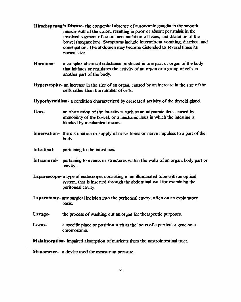
Hirschsprung*» Disease- the congenital absence o f autonomic ganglia in the smooth muscle wall o f the colon, resulting in poor or absent peristalsis in the involved segment o f colon, accumulation o f feces, and dilatation o f the bowel (megacolon). Symptoms include intermittent vomiting, diarrhea, and constipation. The abdomen may become distended to several times its normal size.
Hormone- a complex chemical substance produced in one part or organ o f the bodythat initiates or regulates the activity o f an organ or a group of cells in another part o f the body.
Hypertrophy- an increase in the size o f an organ, caused by an increase in the size of the cells rather than the number of cells.
Hypothyroidism- a condition characterized by decreased activity o f the thyroid gland.
Ileus- an obstruction o f the intestines, such as an adynamic ileus caused byimmobility o f the bowel, or a mechanic ileus in which the intestine is blocked by mechanical means.
Innervation- the distribution or supply o f nerve fibers or nerve impulses to a part of the body.
Intestinal- pertaining to the intestines.
Intram ural- pertaining to events or structures within the walls o f an organ, body part or cavity.
Laparoscope- a type o f endoscope, consisting of an illuminated tube with an optical system, that is inserted through the abdominal wall for examining the peritoneal cavity.
Laparotomy- any surgical incision into the peritoneal cavity, often on an exploratory basis.
Lavage- the process o f washing out an organ for therapeutic purposes.
Locus- a specific place or position such as the locus o f a particular gene on achromosome.
M alabsorption- impaired absorption o f nutrients fi^om the gastrointestinal tract.
M anometer- a device used for measuring pressure.
VII

Meconium- the material that collects in the intestines o f a fetus and forms the first stools o f a newborn.
Meconium Ileus- obstruction o f the small intestine in the newborn caused by impaction o f thick, dry tenacious meconium, usually at or near the ileocecal valve. Symptoms include abdominal distention, vomiting, failure to pass meconium within the first 24 to 48 horns aller birth, and rapid dehydration with associated electrolyte imbalance
Meconium Plug Syndrome- obstruction o f the large intestine in the newborn caused by thick, rubbery meconium that may fill the entire colon and part o f the
terminal ileum. Symptoms include failure to pass meconium within the first 24 to 48 hours after birth, abdominal distention, and vomiting if complete intestinal blockage occurs.
Megacolon- massive, abnormal dilation o f the colon, that may be congenital, toxic, or acquired. Congenital megacolon (Hirschsprung’s Disease) is caused by the absence o f autonomic ganglia in the smooth muscle wall o f the colon.
Monoclonal Antibody (MOAB)- antibodies produced by a hybridoma or antibody- producing cell source for a specific antigen.
M orbidity- an illness or abnormal condition.
Mortality- the condition o f being subject to death.
M utation- an unusual change in genetic material occurring spontaneously or by induction.
Myenteric Plexus- a group o f autonomic nerve fibers and ganglion cells in the muscular coat o f the intestine.
Neural crest- the band o f ectodermally derived cells that lies along the outer surface o f each side o f the neural tube in the early stages o f embryonic development.
Neuroblast- any embryonic cell that develops into a functional neuron; an immature nerve cell.
Neurogepic' pertaining to the formation o f nervous tissue.
Obstruction- something that blocks or clogs, or prevents passage.
Oncogene- a potential cancer-inducing gene.
VIII

Parenteral Nutrition- the administration o f nutrients by a rout other than through thealimentary canal, such as subcutaneously, intravenously, intramuscularly, or intradermally.
Pathogenesis- the source or cause o f an abnormal illness or condition.
Penetrance- a variable factor that modifies basic patterns o f inheritance. It is theregularity with which an inherited trait is manifest in the person who carries the gene.
Perforation- a hole or opening made through the entire thickness o f a membrane, other tissue or material.
Perianal- located around the anus.
Peristalsis- the coordinated, rhythmic, serial contraction o f smooth muscle that forces food through the digestive tract, bile through the bile duct, and urine
through the ureters.
Phenotypic- the complete observable characteristics of an organism or group, including anatomic, physiologic, biochemical, and behavioral traits as determined by the interaction o f both genetic makeup and environmental factors.
Proximal-
Rectum-
Sepsis-
Septicemia-
Sphincter-
Stenosis-
Stool-
nearer to a point o f reference, usually the trunk.
the portion o f the large intestine, about 12 cm long, continuous with the descending colon, just proximal to the anal canal. It follows the sacrococcygeal curve and ends in the anal canal.
infection or contamination.
systemic infection in which pathogens are present in the circulating bloodstream, having spread from an infection in any part o f the body. Characteristically, septicemia causes fever, chill, prostration, pain, headache, nausea, or diarrhea.
a circular band o f muscle fibers that contricts a passage or closes a natural opening in the body.
an abnormal condition characterized by the constriction or narrowing of an opening or passageway in a body structure.
feces.
Stricture- abnormal temporary /permanent narrowing o f the lumen o f a hollow organ.IX

Submucosal- a layer beneath a mucous membrane
Sympathectomy- a surgical interruption o f part o f the sympathetic nerve pathway.

TABLE OF CONTENTS
ABSTRACT.................................................................................................................. i
DEDICATION........................................................................................................... ü
ACKNOW ELDGMENTS.......................................................................................... üi
PREFACE...................................................................................................................... ivDefinition o f Terms............................................................................................. iv
TABLE OF CO N TEN TS........................................................................................... xi
LIST OF T A B LES.......................................................................................................xiv
LIST OF G R A PH S......................................................................................................xv
LIST OF APPENDICES.............................................................................................xvi
CHAPTER PageI. INTRODUCTION...................................................................................... 1
Background to Problem ...................................................................... 1Problem Statement.................................................................................2Purpose ofThis Study........................................................................... 2Hypothesis............................................................................................. 3Si^iificance o f the Problem.................................................................. 3
2. LITERATURE REVIEW .......................................................................... 4Historical Backgroimd........................................................................... 4Etiology.................................................................................................... 5Incidence............................................................................................... 8Pathophysiology................................................................................... 9Clinical Presentation............................................................................. 10Diagnosis............................................................................................... 11Dififerential Diagnosis........................................................................... 13Management.......................................................................................... 13Complications....................................................................................... 17Long-term Clinical Outcome............................................................... 18Conclusion.............................................................................................. 19
XI

3. METHODOLOGY..................................................................................... 21Study Design........................................................................................... 21Study Site................................................................................................. 21Subjects....................................................................................................22Inclusion C riteria.................................................................................... 22Exclusion C riteria................................................................................... 22Primary Outcome Measure.................................................................... 23Secondary Outcome Measures.............................................................. 23Equipment and Instruments................................................................... 23Validity and Reliability........................................................................... 25Procedures................................................................................................ 25Data Analysis............................................................................................27
4. RESULTS.................................................................................................... 29Primary Outcome Measures with Tables and G raphs....................... 29Secondary Outcome Measures with Tables and G raphs................... 42
5. DISCUSSION AND IMPLICATIONS................................................. 57Discussion o f Findings.............................................................................57Limitations...............................................................................................59Suggestions for Future Research...........................................................61Conclusion...............................................................................................62
REFERENCES.............................................................................................................. 63
APPENDIX A -Introductory Letter to the Potential Subjects................................... 66
APPENDIX B - Phone Script o f Verbal Consent....................................................... 68
APPENDIX C - Primary Endorectal Pull-through Parent Phone Interview Form... 70
APPENDIX D - Chart Review Form............................................................................ 73
APPENDIX E - Clinical Grading o f Enterocolitis Form............................................. 79
APPENDIX F - Additional Sheet for Pre-Endorectal Pull-through Enterocolitis ... 81
APPENDIX G - Additional Sheet for Each Readmission............................................. 83
APPENDIX H - Additional Sheet for Each Post Pull-through Enterocolitis 85
APPENDIX 1 - Primary Endorectal Pull-through Computer Data Entry................. 87
XII
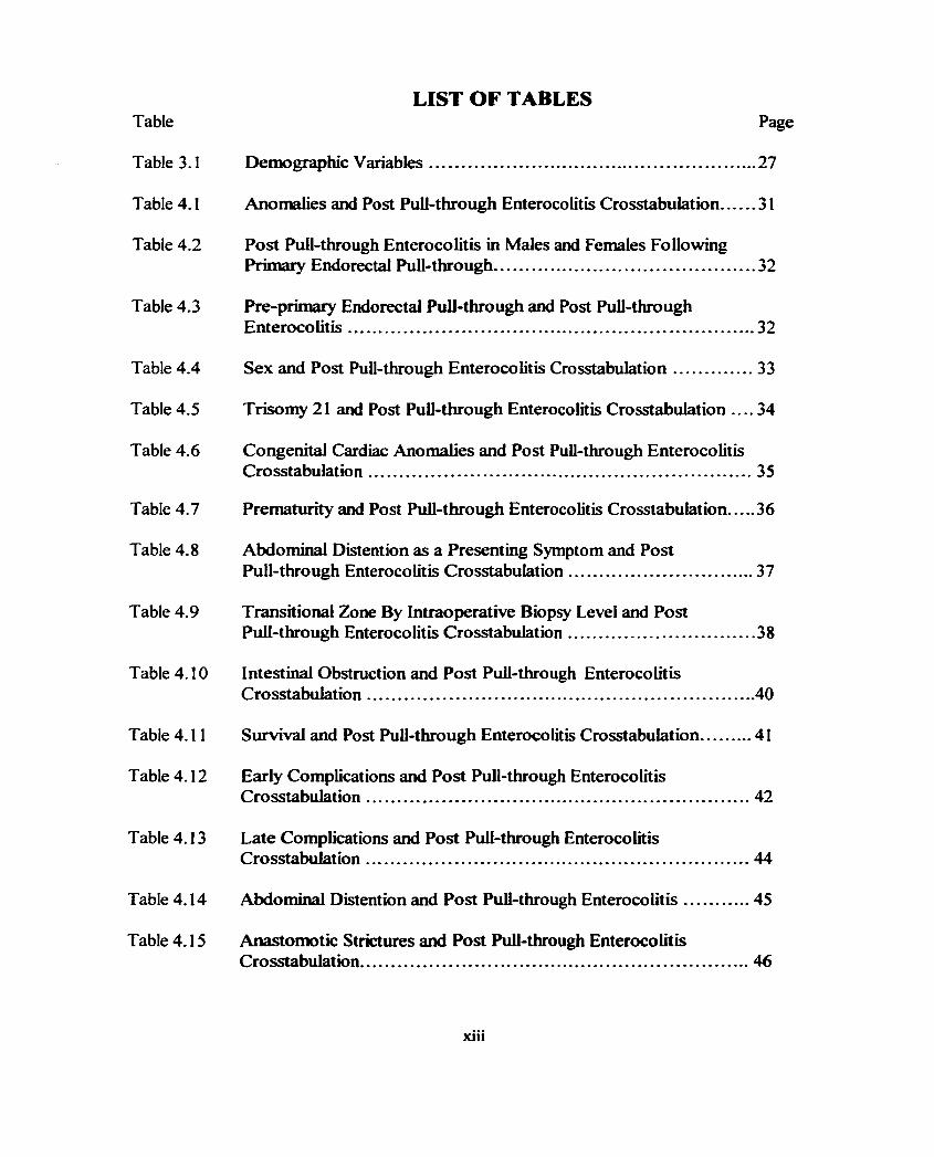
LIST OF TABLESTable Page
Table 3.1 Demographic Variables............................................................................ 27
Table 4.1 Anomalies and Post Pull-through Enterocolitis Crosstabulation..........31
Table 4.2 Post Pull-through Enterocolitis in Males and Females FollowingPrimary Endorectal Pull-through........................................................... 32
Table 4.3 Pre-primary Endorectal Pull-through and Post Pull-throughEnterocolitis............................................................................................ 32
Table 4.4 Sex and Post Pull-through Enterocolitis Crosstabulation.................... 33
Table 4.5 Trisomy 21 and Post Pull-through Enterocolitis Crosstabulation .... 34
Table 4.6 Congenital Cardiac Anomalies and Post Pull-through EnterocolitisCrosstabulation........................................................................................35
Table 4.7 Prematurity and Post Pull-through Enterocolitis Crosstabulation 36
Table 4.8 Abdominal Distention as a Presenting Symptom and PostPull-through Enterocolitis Crosstabulation.......................................... 37
Table 4.9 Transitional Zone By Intraoperative Biopsy Level and PostPull-through Enterocolitis Crosstabulation.......................................... 38
Table 4.10 Intestinal Obstruction and Post Pull-through EnterocolitisCrosstabulation....................................................................................... 40
Table 4.11 Survival and Post Pull-through Enterocolitis Crosstabulation............. 41
Table 4.12 Early Complications and Post Pull-through EnterocolitisCrosstabulation....................................................................................... 42
Table 4.13 Late Complications and Post Pull-through EnterocolitisCrosstabulation....................................................................................... 44
Table 4.14 Abdominal Distention and Post Pull-through Enterocolitis................ 45
Table 4.15 Anastomotic Strictures and Post Pull-through EnterocolitisCrosstabulation.........................................................................................46
X III

Page
Table 4.16 CuflF Stricture and Post Pull-through EnterocolitisCrosstabulation........................................................................................47
Table 4.17 Age o f Stool Continence and Post Pull-through EnterocolitisCrosstabulation........................................................................................ 48
Table 4.18 Age o f Urinary Continence and Post Pull-through EnterocolitisCrosstabulation (Information Obtained In A Phone Interview) 50
Table 4.19 Soiling and Post Pull-through Enterocolitis Crosstabulation............... 52
Table 4.20 Soiling and Post Pull-through Enterocolitis Crosstabulation(Information Obtained In A Phone Interview)..................................... 53
Table 4.21 Constipation and Post Pull-through Crosstabulation............................54
Table 4.22 Constipation and Post Pull-through Enterocolitis Crosstabulation(Information Obtained In A Phone Interview).................................. 55
Table 4.23 Male and Female Mortality Crosstabulation.........................................56
XIV

LIST OF GRAPHS
Graph Page
Graph 4.1 Sex and Post Pull-through Enterocolitis............................................. 33
Graph 4.2 Trisomy 21 and Post Pull-through Enterocolitis.................................. 34
Graph 4.3 Congenital Cardiac Anomalies and Post Pull-through Enterocolitis.. 35
Graph 4.4 Prematurity and Post Pull-through Enterocolitis................................... 36
Graph 4.5 Abdominal Distention as a Presenting Symptom And PostPull-through Enterocolitis....................................................................... 37
Graph 4.6 Transitional Zone By Intraoperative Biopsy Level andPost Pull- through Enterocolitis............................................................ 38
Graph 4.7. Intestinal Obstruction and Post Pull-through Enterocolitis.................. 40
Graph 4.8 Survival and Post Pull-through Enterocolitis......................................... 41
Graph 4.9 Early Complications and Post Pull-through Enterocolitis.....................42
Graph 4.10 Late Complications and Post Pull-through Enterocolitis...................... 44
Graph 4.11 Anastomotic Strictures and Post Pull-through Enterocolitis............... 46
Graph 4.12 CuflF Stricture and Post Pull-through Enterocolitis................................47
Graph 4.13 Soiling and Post Pull-through Enterocolitis........................................... 52
Graph 4.14 Constipation and Post Pull-through Enterocolitis(Information Obtained In A Phone Interview)...................................... 55
Graph 4.15 Male and Female Mortality......................................................................56
XV

LIST OF APPENDICES
Appendix Page
Appendix A Introductory Letter to the Potential Subjects.........................................66
Appendix B Phone Script o f Verbal Consent.............................................................68
Appendix C Primary Endorectal Pull-through Parent Phone InterviewForm...........................................................................................................70
Appendix D Chart Review Form...................................................................................73
Appendix E Clinical Grading o f Enterocolitis Form................................................. 79
Appendix F Additional Sheet for Pre-Endorectal Pull-throughEnterocolitis.............................................................................................. 81
Appendix G Additional Sheet for Each Readmission................................................. 83
Appendix H Additional Sheet for Each Post Pull-through EnterocolitisEpisode...................................................................................................... 85
Appendix 1 Primary Endorectal Pull-through Computer Data EntrySheet...........................................................................................................87
XVI

CHAPTER 1 INTRODUCTION
Background to Problem
Hirschsprung’s disease (HD) is a congenital condition characterized by the absence of
ganglion cells in the submucosal and myenteric plexus o f the distal bowel. This causes a form
of intestinal obstruction. It is thought to be the result o f a failure o f neuronal (ganglion) cells
to migrate fully caudad during embryonic life (Sabiston, 1997).
The fundamental problem with the aganglionic bowel (bowel that lacks nervous tissue) is
its lack o f normal motility. This results in a functional obstruction, and thus to dilatation o f the
normal proximal bowel. This proximal bowel which becomes dilated may do so to the point in
which it is called megacolon (massively enlarged colon). The lack o f ganglion cells also results
in a loss o f the anal sphincter reflex which normally causes relaxation o f the internal sphincter
mechanism in response to rectal stretching (Bishop, 1997). Lack o f this normal reflex, results
in constipation and obstructive symptoms such as bilious emesis, abdominal distention, and
infrequent or delayed defecation (Ashcraft, 1993). The obstructed proximal bowel becomes
inflamed to variable degrees due to stasis. This often results in EC with fever, abdominal
distention, an elevated white blood cell count, protein loss into the bowel and even
perforation. The development o f these adverse events is a major cause o f morbidity and
mortality for these children. It has become a goal for many pediatric surgeons to try to prevent
and aggressively treat EC associated with HD. Thus, advancements in understanding which
surgical interventions may be associated with a higher incidence o f EC post-operatively are
very important. More information is needed to aid in the prevention, diagnosis and treatment
o f children who develop Hirschsprung’s associated EC (HAEC).

Problem Statement
Concomitant with major improvements in the understanding o f the pathophysiology and
genetics o f HD, little advancement has occurred in determining the etiology or prevention of
EC associated with HD. However, the surgical treatment o f HD has continued to evolve.
Procedures have progressed from three stage surgeries to two-stage surgeries. This staged
approach has long been considered the gold standard o f surgical techniques.
Over the past fifteen years a single-stage primary endorectal pull-through (PERPT) has
been developed. Cilley et al noted that larger studies, which focus on the incidence o f both
major and minor complication rates and outcomes, were needed (Cilley et al., 1994). It was
unclear whether the Soave single stage pull-through procedure had a similar clinical outcome
compared to the multi-stage procedure.
Purpose ofThis Studv
The goal o f this study was to look at postoperative complications such as EC
encountered in children who had undergone a Soave single stage (PERPT) compared to
those who had undergone a more conventional two-stage surgical resection for HD. It
was thought that by looking at not only the type o f surgery performed but also the timing
and number o f surgical interventions, that it was possible to identify any statistically
significant benefits o f specific treatment approaches. It was also thought that the
performance o f a PERPT done at an age o f less than two years would be beneficial in
decreasing the incidence o f EC while improving the long-term continence rates in patients
newly diagnosed with HD. This study would provide parents and physicians with more
information regarding the possible outcomes of the difièrent treatment approaches and aid
in the decision making process when one is faced with several treatment choices.

Hypothesis
We hypothesized that: 1. the performance o f a PERPT is associated with a lower incidence
of EC compared to those who have undergone the two-stage surgical intervention for HD.
2. the performance o f a PERPT is associated with a lower incidence o f other complications
compared to those who have undergone the two-stage surgical intervention for HD.
Significance o f the Problem
In the past, standard treatment for HD required many hospitalizations and multiple
operations if a two-stage or three-stage method was used. Cilley, et al noted that there was a
need for long-term follow-up o f these patients who had undergone the single stage procedure
to determine their outcome compared with children who were managed with the staged
approach (Cilley et al., 1994).
A major benefit o f doing the PERPT was the avoidance o f a colostomy, which can often
be time consuming and difficult for parents to care for. The PERPT procedure was considered
more favorable by caregivers as well. Additionally, the added risk o f undergoing anesthesia,
with its potential complications, could be avoided by using the PERPT procedure.
Reporting the rates o f both major and minor complications would hopefully provide
information for the care and treatment o f those newly diagnosed with HD. This knowledge
would also help improve patient survival rates, the level o f comfort, care and
convenience for many patients and families while possibly avoiding the need for a colostomy.
By reducing the number o f surgical interventions, hospitalizations and the incidence o f EC, it
was thought that the saving o f many health care dollars was also a monetary benefit well
worth acknowledging.

CHAPTER 2REVIEW OF LITERATURE AND CONCEPTUAL FRAMEWORK
Historical Background o f Hirschsprung’s Disease
Although there have been a number o f isolated case reports that have been published in the
past, Harold Hirschsprung first described aganglionosis in 18S7. He emphasized the post
mortem findings o f colonic distention and hypertrophy proximal to a smaller, normal-sized
rectum in two inAnts with constipation and abdominal distention since birth (Ashcraft, 1993).
Hirschsprung did not recognize that the cause o f the megacolon was not in the dilated
proximal bowel but in the undilated distal bowel. This oversight contributed to the delay in the
full understanding o f the pathogenesis o f aganglionic megacolon for another sixty years
(Touloukian, 1995).
There were isolated reports o f the histologic absence of ganglion cells in affected
patients in 1901, 1904 and 1920 but it was not until 1948 that the currently understood
clinical and pathological correlation between aganglionosis and incomplete colonic obstruction
finally occurred (Ashcraft, 1993).
The first successful left colectomy and pull-through procedure was done in 1896, yet
therapy in the first five decades o f the twentieth century focused more on both the pathology
associated with the aganglionosis and the more pronounced clinical finding o f abdominal
distention associated with the megacolon. (Ashcraft, 1993).
Pharmacological therapies and surgical sympathectomy were tried as alternative therapies
with irregular results. During this time, little was known about the causes o f megacolon.
Swenson, in the late 1940’s, made the clinical observation that children who had
undergone a colostomy began to improve but soon began to deteriorate with colostomy

closure and improved when the colostomy was re-created. Swenson’s observations
accompanied by his manometric studies and Neuhauser’s radiologic observations at that time
led to the development o f Swenson’s pull-through procedure (Aschcraft, 1993).
With the publication o f their pathologic findings o f aganglionosis in patients with the same
clinical picture in 1948, Zeulzer and Wilson completed the clinical picture which is now
recognized as Hirschsprung’s disease (Aschcraft, 1993).
Since that time, many alternative procedures with regards to diagnosis, testing, treatment
and surgical intervention have been developed as the knowledge concerning this disease
process developed. HD must be looked at as a many faceted disease with an array o f
etiologies, clinical presentations, associated abnormalities, complications, treatment options
and clinical outcomes. A basic review o f the incidence, etiology, pathophysiology, clinical
presentation, diagnosis and treatment o f HD will help set the stage to better understand the
significance o f EC associated HD.
Etioloev o f Hirschsprung’s Disease
The term neurocristopathy, originated by Bolande in 1974, was a description o f lesions
related to aberrations in neural cell growth, migration, and dififerentiation. The proper
migration o f the neural crest tissues during the fourth week o f the fetal development was
noted to be very crucial. These neural crest tissues eventually form the peripheral autonomic
nervous system (Stovrofif, 1995).
Yntema and Hammond in 1954 and Le Douarin and Teillet in 1973 recognized the vagal
crest as the origin o f the enteric nervous system. Okamoto and Ueda, two Japanese pediatric
surgeons, also suggested that the mechanism o f HD was related to the disturbed migration o f
these enteric neurons during embryonic development. (Molenaar, 1995).

HD was thus thought to be caused by an aberration o f the neural crest migration during
fetal development. Enteric ganglion cells mature from neuroblasts derived from the neural
crest during fetal development. These neuroblasts which are first seen adjacent to the pharynx,
migrate caudally during weeks six through eight and ultimately reach the distal rectum in the
twelfth week o f fetal development. (Aschcraft, 1993)
Microsurgical techniques have been used to analyze the development o f the enteric
nervous system and other organs initiated in the vagal crest region. Work is currently being
done in hopes o f determining at which level ablation o f the neural crest produces anomalies
and aganglionosis o f the colon (Molenaar, 1995).
In an immunohistochemical study on the ganglionic and aganglionic segment in HD, Ikawa
et al indicated that the expression of the LI molecule, which plays an important role in cell
adhesion, neural cell migration, and neurite outgrowth, was impaired in the extrinsic nerve
fibers in aganglionic colon. Their findings indicated that the impaired expression o f the LI
molecule might alter neural crest migration and adequate neurite outgrowth, with a resulting
aganglionic segment and abnormal nerve bundles o f extrinsic fibers in HD (Ikawa et al.,
1997).
In 1997, Kusaftika and Puri, found an association between the RET proto-oncogene and
HD. Results o f their study demonstrated that the RET proto-oncogene was a major gene
involved in the development o f HD. They claimed that the RET mRNA level in the
aganglionic bowel specimens o f HD patients was approximately one five hundredth o f that in
normal ganglionic bowel. Decreased RET mRNA expression in the aganglionic bowel
suggests the abnormal development o f neural crest-derived cells in HD (Kusaftika et al.,
1997).
Nitric oxide (NO), an important chemical messenger in the digestive tract, has a relaxing
effect on the smooth muscle o f the bowel. It has been suggested that it could be involved in
gut motility disorders. The initial work by Vanderwinden et al noted that the NO synthase
was not present in the musculature o f the aganglionic segments in patients with HD. These
findings indicated the possible role o f NO synthase deficiency in the pathophysiology o f HD.
(Vanderwinden et al., 1993).
More recently, Hanani et al, in 1995 studied the distribution of the enzyme NADPH
diaphorase (NADPHd) in normal and diseased bowel segments to assess the role o f nerves
that synthesize nitric oxide (NO) in HD. Their recent work has shown that NO has a
protective action on gastrointestinal mucosa, and that a reduction in mucosal NO synthase
(NOS) activity may also have an important immunologic implication in patients with HD
(Hanani et al., 1995).
Current studies associated with this theory done by Kamimura et al are focusing on the
nonadrenergic, noncholinergic (NANG) inhibitory nervous system at the aganglionic segment.
They speculated that the long-segment type cases receive dual nervous inputs, one fi-om the
ganglionic segment and the other fi"om the sacral segment. These findings favored the
hypothesis that the embryogensis o f long-segment-type cases might differ fi-om that o f short-
segment-type cases o f HD (Kamimura, 1997).
Over the years, a large number o f anomalies have been found to be associated with HD.
When a clinical geneticist was involved in the examination o f patients with HD, associated
anomalies were found in 23 % o f the patients with short segment HD and 45 % o f the patients
with long segment HD (defined as those diagnosed with an aganglionic segment proximal to
the sigmoid colon). Molenaar concluded that the high number o f anomalies associated with

increased length o f the aganglionic segment might be related to the pathological involvement
of both the enteric and cardiac crests (Molenaar, 1995). Associated abnormalities were
reported to be 21.2% in one hundred seventy three cases o f HD by Jung et al. in 1995.
Craniofacial, cerebral and cardiac anomalies were found to be predominant and Down’s
syndrome was found to occur mainly in association with the classical short-segment HD
(Molenaar, 1995).
Chromosomal aberrations may also be responsible for the abnormal innervation of the
bowel. In 1992, Martuciello et al described a deletion on the long arm o f chromosome ten in
a newborn patient with total colonic aganglionosis (Martuciello et al., 1992).
Recent studies have emphasized the genetic abnormalities found in familial cases. These
cases appear to be associated particularly with long-segment HD in which there are mutations
o f the RET oncogene. An abnormality o f the endothelin B receptor gene has also been found
in patients with aganglionosis. How this abnormality of this molecule results in aganglionosis
is still unclear (Lebenthal, 1996). A greater understanding o f the phenotypic variances and
mutation patterns in the gene will hopefully better predict outcomes o f surgical procedures
and other modes o f treatment for HD.
Incidence o f Hirschsprung’s Disease
The incidence o f HD was thought to be approximately one in five thousand births. Sex
ratios and inheritance clearly differ between the more common rectosigmoid disease and those
with long-segment disease. Inheritance is thought to be a sex-modified multifactorial trait or
the result o f a recessive gene with low penetrance with the lower risk of an affected sibling at
4 % (Ashcraft, 1993). Longer segment disease, although less fi’equent, is associated with a
decreased sex ratio and an increased sibling risk of approximately

30 % patients with small-bowel transition zones. Inheritance appeared to be compatible with a
dominant gene with incomplete penetrance. As noted earlier, there are other associated
anomalies such as, congenital heart disease, Down’s syndrome (4% to 5 % o f patients with
HD), Smith-Lemli-Ophz and Waardenburg’s syndromes (Ashcraft, 1993).
HD appears to have a definite link with a positive family history for HD. If the first infant
in a family had rectosigmoid involvement, the risk o f a second child being bom with HD is
approximately 6 %. The incidence o f a second child having HD where the first infant had total
colonic aganglionosis is 12 %. Genetic studies support the theory of an abnormal locus on the
tenth chromosome and may explain the increased incidence o f HD iii cases where there is a
positive family history. (Sabiston, 1997).
Current studies on this topic support this theory regarding this genetic component link with
HD and the varying lengths o f aganglionosis. In a sample o f one hundred thirty seven patients
with HD over a twelve year time span, Jung et al., in 1995, observed the male to female ratio
o f 3.6 : 1 and the occurrence o f neonatal HD in seventy cases (51.1%). He observed one
hundred fourteen cases (83.2%) with short-segment and twenty-three cases (16.8%) with
long-segment disease. He also noted a positive family history in four cases (Jung, 1995).
Interestingly, Ryan et al, in 1992, found no association between an increased maternal age and
the occurrence o f HD (Ryan, 1992).
Pathophysiology o f Hirschsprung’s Disease
Aganglionic megacolon or HD, is a neurogenic form o f intestinal obstruction in which
there is an absence o f ganglion cells in the myenteric (Auerbach’s) and submucosal
(Meissners’s) plexuses. In contrast to normally ganglionated bowel, aganglionic bowel has an
increased number o f both cholinergic and adrenergic (Larsson) neryes, a normal tissue content

10
of acetylcholine but an elevated release in response to stimulation, an elevated
acetylcholinesterase concentration, an elevated tissue catecholamine, and a decrease in certain
peripheral nerve fibers. Abnormalities in gut hormones have also been described (Aschcraft,
1993).
The study of Parikh et al, in 1992 demonstrated a quantitative abnormality o f laminin in the
bowel in HD patients supporting the hypothesis that an “abnormal microenvironment” may
also have a role in the pathogenesis o f HD (Parikh et al., 1992).
Clinical Presentation o f Hirschsprung’s Disease
Several patterns o f clinical presentation for HD exist due to the variable length o f bowel
being aflfected. These patterns o f presentation vary in the symptoms as well as in the age o f
the patient at the time o f presentation and initial diagnosis. (Ashcraft, 1993)
In contrast with earlier decades, the diagnosis o f HD is increasingly being made in the
neonatal period. This earlier diagnosis may be due to the increased number o f informed health
care providers, an increase in the number o f neonatologists as well as the development of
easier and quicker biopsy techniques.
The symptoms o f bilious emesis, abdominal distention, and delayed or diminished
fi-equency o f stools has become classic as the presenting symptoms for HD. Swenson noted
that 94 % o f normal-term neonates produce stool within the first 24 hours o f life and in
contrast, 94 % o f neonates with HD do not produce a meconium stool within the first 24
hours o f life (Ashcraft, 1993).
Because not all neonates fully manifest the clinical picture of intestinal obstruction, the
diagnosis may be delayed until infency, particularly until the time that the patient’s diet is
supplemented with cereals and strained foods which result in an increase in stool consistency

I l
at which time the symptoms may appear. Parents may only notice a decrease in stool
frequency, a diminished appetite, and less than expected weight gain. If the infant or child
develops a fever, becomes dehydrated, and experiences diarrhea, it is imperative that a
diagnosis and treatment plan be established as soon as possible. Septicemia and occasional
perforations are associated with the most common cause o f mortality (Ashcraft, 1993).
Diagnosis o f an older patient, although now more infrequently now days because many
children are diagnosed at a much younger age than they were in the past, may be made when a
patient presents with a lifelong history o f infrequent stools, abdominal distention, and poor
nutrition.
On physical exam, vigorous peristalsis may be heard. The patient may present with some
abdominal distention with or without flared costal margins, and a thin abdominal wall. Large
fecal masses may be palpated. Hirschsprung’s patients can be easily distinguished from
patients with functional constipation because they do not have anal pain, bleeding, or an
abnormally large fecal bolus in the rectal ampulla (Aschcraft, 1993).
Diagnosis o f Hirschsprung’s Disease
The diagnosis o f HD can be made with 1. Radiographic studies such as an abdominal series
and / or a barium enema 2. Anorectal monometry 3. Submucosal suction rectal biopsy or fiiU-
thickness rectal biopsy (Rescorla, 1992).
Erect and recumbent abdominal radiographs may demonstrate dilated loops o f bowel. A
barium enema is performed in nearly every suspected case o f HD and may demonstrate an
area o f slightly dilated colon, which may be helpful in determining the location o f the
transition zone (the level of aganglionosis).

12
In newborns, there is no definitive cutofif point indicating the transition zone where the
narrow distal aganglionic rectum or rectosigmoid meets the obstructed dilated normal
proximal colon containing ganglion cells. It may take three to six weeks for the transition zone
to become apparent. The barium enema may look normal in infants with short segment disease
affecting only the rectum and may demonstrate a comma-shaped rectosigmoid, flattened
flexures, and occasionally a microcolon in instances o f total colonic aganglionosis (Sabiston,
1997).
Unlike normal newborns who evacuate the barium enema contrast within ten to eighteen
hours, infants with HD retain the barium for twenty-four to forty-eight hours. Thus it is
important to obtain a delayed abdominal x-ray at twenty-four hours because the transition
zone may be visualized more clearly on this delayed film. In older infants, the transitional zone
may be seen on the initial barium study.
The diagnosis o f HD is then confirmed by obtaining a suction or full-thickness rectal
biopsy where ganglion cells are noted to be absent in the Meissner’s submucosal plexus.
Yamataka et al., in 1992, proposed the use o f an immimohistochemical method for
diagnosing HD using a monoclonal antibody (MAb) 171B5 against synaptic vesicles. Their
findings suggested that Mab 171B5 immunohistochemistry on the lamina propria alone could
differentiate between normal and aganglionic bowel and proposed this method as being
reliable and useful for the detection of HD on a suction rectal biopsy (Yamataka et al., 1992).
In emergent circumstances, a definitive diagnosis can be made on a full-thickness rectal
biopsy that can be evaluated for the absence o r presence o f ganglionated cells in Auerbach’s
myenteric plexus. If no ganglion cells are seen, the diagnosis of HD is confirmed (Sabiston,
1997). Acetylcholinesterase (Ach) staining is also a useful diagnostic tool. Increased Ach

13
staining of neurofibrils is characteristic o f HD. Recent diagnostic advancements have been
made by Kobayashi et al, who in 1995, proposed a modification o f the histochemistry
technique o f Kamovsky and Roots to produce staining o f cholinergic nerve fibers in ten
minutes rather than in two hours, as is the case with the conventional AChE technique. This
provided a quick, simple and reliable method for intraoperative evaluation o f the extent o f the
anganglionic segment (Kobayashi et al., 1995).
Anal monometry, another usefiil diagnostic adjunct, measures the anorectal intraluminal
pressure with a balloon probe connected to a pressure transducer and polygraph recorder. In
infants with HD, this technique usually demonstrates an absent rectoanal inhibitory reflex,
indicating a lack o f relaxation o f the internal sphincter, which is characteristic o f aganglionosis
(Sabiston, 1997). Of the diagnostic methods utilized, it appears that the suction rectal biopsy
is initially the most definitive in making an initial diagnosis of HD.
Difierential Diagnosis o f Hirschsprung’s Disease
The differential diagnosis o f HD includes hypothyroidism, meconium plug syndrome,
colonic neuronal dysplasia, adynamic ileus associated with sepsis, intestinal pseudo
obstruction, and maternal narcotics addiction. These conditions are also associated with
delayed passage o f meconium at birth (Ashcraft, 1993).
Management o f Hirschsprung’s Disease
Decompression
Once the diagnosis o f aganglionosis is established, active intervention is required including
colonic lavage, diversion, or primary pull-through. Colonic lavage, practiced in Europe
decades ago as an alternative to colostomy, is now used as mechanical irrigation. This is
sometimes performed several times daily using a large-bore rectal tube to aid in abdominal

14
decompression. The lavage technique is also thought to be useful in the prevention and
management o f EC.
In the past, surgical colostomy was the initial step in management to relieve obstruction.
This was then followed by a multi-stage surgical resection o f the aganglionic bowel.
Definitive Procedures
Today, one-stage primary pull-through procedures done in the first few weeks o f life are
becoming more popular. Others consider the treatment of choice in the neonatal period to be a
temporary decompressing colostomy at least ten centimeters proximal to the transition zone
followed by biopsies to determine the level o f aganglionosis. Then at six months to one year
of age, a definitive pull-through procedure using the Soave (endorectal) or the modified
Duhamel (retrorectal) is performed in infants with rectosigmoid disease. In cases o f total
aganglionosis, the pull-through procedure may be delayed until eighteen months o f age. Many
pediatricians for total aganglionosis fevor the modified Duhamel. In rare cases of
aganglionosis aflfecting the entire small bowel, an extensive enteromyotomy and myectomy is
advocated by Ziegler and associates. Unfortunately, infants with aganglionosis extending into
the proximal small intestine almost always require long-term total parenteral nutrition (TPN)
to achieve adequate caloric intake and weight gain (Sabiston, 1997).
Cilley et al. (1993) advocated that a primary repair be performed at the time o f diagnosis
and claimed that the modified endorectal pull-through was technically easier in the newborn
than in older children. Cilley also noted that the incidence of postoperative EC was similar to
the 16 % incidence reported in the standard treatment o f HD.
Georgeson et al.(1995) advocated a laparoscopic approach vs. the conventional
laparotomy in the single staged pull-though procedure. The benefits o f this approach are: the

15
avoidance o f a large painful abdominal incision, a more rapid return of the patient’s bowel
hinction, a decreased postoperative recovery time, and a more appealing cosmetic result due
to the smaller size of the incisions for the laparoscopic procedure.
Many different types o f reconstructive procedures and modifications o f these
reconstructive procedures have been performed over the last four to five decades. Success or
failure seemed to depend on the ability o f the surgeon to place bowel that contains ganglion
cells within one centimeter o f the anal verge. Although marked improvement in operative
mortality and fimctional outcome have occurred; there are still complications associated with
each procedure.
The Swenson, Duhamel and Soave / PERPT surgical procedures will be discussed.
Swenson’s Procedure
Swenson’s procedure was the first to address resection o f the distal agang lion ic segment.
Features o f this procedure include careful dissection o f the wall o f the pelvic rectum to protect
the nervi erigentes, followed by eversion o f the native rectum with an oblique single-layer
anastomosis o f the pulled-through colon to the native distal rectal segment, which is then
replaced in the pelvis.
Duhamel’s Procedure
Duhamel’s procedure was designed to avoid dissection anterior to the rectum. It became a
definitive operation in infancy. The original Duhamel procedure was an anastomosis o f the
ganglionated proximal bowel to the closed native rectum at the anal verge. Dilatation o f the
dysfunctional rectum by fecal retention in the blind loop led to Martin’s modification, which
added a proximal suture anastomosis o f anterior native rectum to the pulled-through colon,
followed by the crushing o f the septum with a spur clamp. The result was a rectum o f
16
expanded size with ganglion cells in the posterior half, retention o f native rectum anteriorly,
and avoidance o f the blind pouch with the proximal anastomosis. Surgical stapling devices for
intraoperative anastomosis and division o f the rectal septum are now most commonly used.
With these modifications, Duhamel’s procedure has had wide acceptance for all forms o f
Hirschsprung’s disease with good results and is particularly useful in those with small bowel
transitional zones.
Soave’s / PERPT Procedure
The endorectal pull-through as originally described by Soave and modified by Boley is the
third alternative widely utilized for surgical treatment o f HD. The specific features o f this
operation include an intramural submucosal dissection o f the rectum to a level less than one
centimeter above the verge. After removal o f the mucosa, normal proximal ganglionated
bowel is advanced to the perineum. Excess pulled-through bowel is primarily unanastomosed.
(Ashcraft, 1993).
Comparative results for each o f the three currently utilized procedures are diflScult to
establish clearly. None is without complication. Surgeon experience, bias, and patient
selection may affect the results of comparative surveys o f these surgical procedures; thus it is
truly hard to compare these different surgical approaches.
Complications
There are three major early postoperative complications; enterocolitis, anastomotic leaks
and stenosis or strictures. Additionally, there are late complications, which include stooling
abnormalities such as, constipation, incontinence, and soilage.
17
The incidence o f these complications is quite variable among reported series. Some have
suggested that the incidence o f anastomotic leaks is more frequent in the Swenson's
procedure and stenosis is more common in the endorectal pull-through.
Preoperative Complications
Much o f the mortality in HD is the result o f EC. The mortality with EC may be due to a
delayed diagnosis. Infants who present with EC preoperatively, may be more likely to have
this complication postoperatively following both colostomy and pull-through procedures
(Sabiston, 1997).
Operative mortality has been shown to be greater in Swenson’s procedure and lower in the
modified Duhamel and Soave ’s procedures. Thus there is a rise in the popularity o f the
endorectal pull-through. The effect o f perioperative care, especially in regards to management
o f leaks and sepsis, has improved within the last two decades as well.
The appropriate age or size of the patient for definitive reconstruction has been debated for
decades. Although excellent results have been reported for one-stage endorectal pull-through
without diversion in neonates, some larger collected series suggest significantly greater
mortality and morbidity when reconstruction is performed prior to four months o f age.
Postoperative Complications
Most complications associated with the surgical correction o f HD have been noted in
previous studies. These include enterocolitis, anastomotic stenosis and dehiscence, residual
aganglionosis, small bowel obstruction, perianal excoriation, and long-term malabsorption,
failure to thrive, constipation, and diarrhea.
18
The most pertinent late complication is the development o f enterocolitis. Some authors
believe that postoperative EC occurs in all patients, but may be less in a patient with an
endorectal pull-through (Aschcraft, 1993).
Kobayashi et al (1995) noted that persistent bowel dysfunction was a problem for some
patients. He also noted that EC, constipation and incontinence are the most noted
manifestations o f postoperative bowel dysfunction with the incidence o f EC being estimated at
around 6 to 20 % (Kobayashi et al 1995). Langer and Bimbaum in 1997 also noted that most
children have excellent results after pull-through surgery, but some experience persistent
constipation (Langer et al, 1997).
Many o f the deaths have been observed in infants with Down’s syndrome and in infants
who were less than 4 kg when operated on. For example, Rescorla et al noted that EC was
more common in neonates and children with total colonic aganglionosis (TCA) and Down’s
syndrome. This represented a mortality rate o f 8.5 % for the entire group in the study while
children with Down’s syndrome had a mortality rate o f 26 % (five times that observed in
children with HD without Downs syndrome) (Rescorla, 1992).
Incontinence rates are often poorly reported in many large series. A few generalities can,
however, be made. Incontinence rates are higher in patients with Trisomy 21 and other
syndromes with associated mental retardation. Typical incidences o f incontinence range fi’om
3 to 8 % (as reported in a discussion with Dr. Daniel Teitelbaum, 1998).
Long-term Clinical Outcome
Overall, survival is typically more than 90 %. It is agreed that long term fi)Uow-up is very
important. Incontinence is rare. More than 96 % are usually continent, but soiling may be a
problem in 2 to 3 % o f cases. For those patients experiencing constipation, a high-fiber diet

19
and stool softeners has been found to be helpful and it is noted that many patients symptoms
improve with age (Sabiston, 1997).
Teitelbaum et al in 1997 reported a normalization o f stooling frequency within one year
after surgery in a study on the long-term stooling patterns o f in6nts (n=24) undergoing the
primary endorectal pull-through. It is felt that larger studies are needed to better understand
the long-term clinical outcome for these patients.
Conclusion
In conclusion, the review o f literature shows that the diagnosis, treatment and care o f a
child diagnosed with HD is a very difihcult process. Sources vary on their recommended
treatment approach and thus it is important to look at the complications associated with the
different modes o f treatment to better understand in what areas advances must be made to
better treat these children.
The complications o f EC are o f particular concern due to the morbidity and mortality
associated with it. Many o f these children endure frequent and long hospital stays associated
with this complication. Standard operational definitions o f what constitutes a true case o f EC
versus gastroenteritis may influence the number o f EC case reports between the various
institutions and thus cloud a true statistical significance in evaluating the success o f the
different treatment approaches.
The issue o f continence is also o f great importance to many patients who seek to have a
better quality o f life and thus a closer look at newer data regarding this issue is needed.
More research is also needed to determine at what age the surgical correction is most
beneficial and which surgical methods have the lowest incidence o f EC. Using the single-stage

20
approach at an early age may have some influence on outcomes, such as a decrease in
morbidity and mortality. However, operations at such a young age may adversely efifect long
term stooling patterns (i.e. continence rates). Thus, information from this study may assist
clinicians in the choice o f a safer and more economical approach to the treatment o f HD as
well as provide patients with a better quality o f life.

21
CHAPTER THREE METHODOLOGY
Study Design
This study was a quantitative multi-center study utilizing a retrospective chart review with
a historical control (Elhalaby, 1995) and a telephone interview. This study design was chosen
by the principle researcher to elicit information concerning the postoperative complications
associated with HD following a single stage PERPT. The incidence o f the primary outcome
measure (EC) was then be compared with those who have undergone the two-stage procedure
in a historical control utilizing the T-test and Chi-square analysis. The secondary outcome
measures (stricture rates, continence rates, stooling frequency, mortality rates and infectious
complications) were also be reported.
We hypothesized that: 1. the performance o f a primary endorectal pull-through (PERPT)
was associated with a lower incidence o f EC compared to those who have undergone the two-
stage surgical resection for HD in the historical control group 2. the performance o f a
primary endorectal pull-through (PERPT) was associated with a lower incidence o f other
surgical complications such as incontinence, strictures and constipation compared to those
who have undergone the two-stage surgical resection for HD in the historical control group.
Study site
The primary clinical site is located at Mott Children’s Hospital at the University o f
Michigan. Dr. Daniel Teitelbaum, the director o f pediatric surgery, was the primary researcher
coordinating this study. Three other physicians at their respective clinical sites contributed
data to this research process as well. The clinical sites, which have contributed data for this
study, are as follows:

22
1 Daniel Teitelbaum, MD Mott Children’s Hospital, Box 0245Ann Arbor, Michigan 48109
2 Neil Uitvlugt, MD Spectrum Health Downtown CampusGrand Rapids, Michigan 49503
3 Robert E. Cilley, MD M.S. Hershey Medical Center, PO Box 850Hershey, Pennsylvania 17033
4 Neil Sherman, MD 1135 South Sunset Avenue, Suite 301West Covina, California 1790
Subjects
All patients with HD who have undergone the Soave PERPT surgical procedure between
the dates o f May 1 o f 1987 and September 1** o f 1999 were candidates for the phone
interview and chart review by each clinical site. We initially anticipated an approximate sample
size o f eighty to one hundred charts would be reviewed. There were a total of eight patients
who met all o f the inclusion criteria for this study. The historical control had approximately
one hundred fifteen patients who had undergone the two-stage surgical resection.
Inclusion criteria
Inclusion criteria for this study included: that the child had HD as confirmed by pathology
and underwent the Soave PERPT at less than two years o f age between the dates o f May 1
of 1987 and September 1 o f 1999. This group o f children was then compared to those who
have undergone the two-stage resection process in the historical control.
Exclusion Criteria
Exclusion criteria for this study included: no prior gastrointestinal surgery with either an
ileostomy or a colostomy, children greater than two years o f age at the time of diagnosis with
HD, and surgery prior to May 1**, 1987 or after September 1“, 1999.

23
Primary Outcome Measure
The primary outcome measure which was the incidence of EC in patients undergoing the
Soave PERPT procedure was compared to those who had undergone the two-stage surgical
procedure in the historical control.
Secondary Outcome Measures
Secondary outcome measures, which include complications (early and late), stricture
formation (anastomotic and cuS), continence (stool and urinary), frequency o f defecation,
soiling, constipation (initial and long-term) and mortality rates, were also reported.
Instruments
Research tools involved in this study were as follows: the Introductory Letter to the
Potential Subjects (Appendix A) was sent out to those who meet all o f the inclusion criteria.
This introductory letter was used to introduce the study to the parents o f the potential
candidate, tell the parent what kinds o f questions would be asked in the phone interview and
state the approximate time the researcher would call.
The next tool, the Phone Script o f Verbal Consent (Appendix B), was used for the purpose
o f obtaining informed consent. This Phone Script o f Verbal Consent (Appendix B) was read
over the phone to the parent's o f the potential candidate. This informed the parents o f the
purpose and potential benefits o f this study. It indicated that each parent’s consent would be
obtained by their answer “yes” and the data they provided if they chose to proceed and answer
the questions in the phone questionnaire. It also assured them that their refusal to participate
in the study would have no influence on their child’s present or future care. They were assured
that if the results o f the study were published, the child’s name would not be used and that
their answers would be kept confidential.

24
The Primary Endorectal Pull-through Parent Phone Interview Form (Appendix C) was
used to obtain information in the telephone interview at each clinical site. The patient was
assigned a number on this form, which coincided, with the number placed on the Primary
Endorectal Pull-through Computer Data Entry Sheet (Appendix I). This number then served
as the patient’s identification number between the review forms and the coded data entry
sheets by each clinical site. This patient number was entered and used with the data on
subsequent computer data entry sheets to ensure patient confidentiality.
The process o f coding the phone interview data fi’om the Primary Endorectal Pull-through
Parent Phone Interview Form made the data easier to work with and provided anonymity for
each subject. This information on the Primary Endorectal Pull-through Computer Data Entry
Sheet (Appendix I) was then entered into the Excel spreadsheet at the primary clinical site
(University o f Michigan).
Information fi'om the patient’s hospital charts was obtained using the Chart Review Form
(Appendix D). Other forms used in the chart review process were; Clinical Grading of
Enterocolitis (Appendix E) which aided the chart reviewer in determining the clinical grading
o f enterocolitis to ensure a standardization o f the coding system, another sheet entitled
Additional Sheet for Pre-Endorectal Pull-through Enterocolitis (Appendix F) was used to aid
the primary researcher in identifying those variables which may influence the development o f
Post Pull-through Enterocolitis, an Additional Sheet for Each Readmission (Appendix G) was
also used to aid the primary researcher in the documentation o f post operative complications
in this patient population. Additional Sheet for Each Post Pull-through Enterocolitis Episode
(Appendix H) was used aid the chart reviewer in better defining this primary outcome measure
for instances in which may have suffered repeated bouts of EC. This information was then

25
coded to the Primary Endorectal Pull-through Computer Entry Sheet (Appendix 1). The
information from the chart review, which was coded to these sheets, used a patient
identification number for each subject and provided anonymity for each patient. The
information from the Computer Data Entry Sheets was then entered into the Excel
spreadsheet, which was then analyzed and abstracted on by the co-investigators in Grand
Rapids and the primary clinical site (University o f Michigan).
V aliditv/Reliabilitv
To ensure validity, an adequate number o f infants were recruited to determine if there was
a statistically significant difference in the incidence o f EC between the historical control and
the current study. Information from previous publications on the incidence of EC and other
complications were reviewed to determine the required sample size for this study. The sample
size calculations were based on the assumption that it was not necessary to adjust for
covariance. It was determined that based on the current sample size o f eighty patients, we
would be able to detect a statistical significance.
Procedures
After approval from the appropriate committees had been obtained and confirmed, names
and addresses o f potential subjects were collected by each clinical site based on the inclusion
criteria. Each clinical site conducted a telephone interview and medical record review o f all
subjects. The information obtained by each clinical site was then coded to data entry sheets.
The four clinical sites involved in the study then mailed the data contained on the coded data
entry sheets (Appendix D and Appendix J) to the primary clinical site (University o f Michigan)
where they were analyzed.
26
At the Spectrum Health, Downtown Campus, the names and addresses o f potential
subjects were collected. An Introductory Letter to the Potential Subjects (Appendix A) was
sent out to those who met all o f the inclusion criteria. This introductory letter was used to
introduce the study to the parents o f the potential candidate. This letter informed the parents
as to what kinds o f questions would be asked in the phone interview. It also informed them o f
the approximate time the researcher would be calling.
A Phone Script o f Verbal Consent (Appendix B) was read over the phone to the parent's
o f the potential candidates. This informed the parents o f the purpose and potential benefits of
this study. It indicated that each parent’s consent was the data they provide if they answered
“yes” and chose to proceed with answering the questions in the phone questionnaire. It also
assured them that their refusal to participate in the study questionnaire would have no
influence on their child’s present or future care. They were assured that if the results o f this
study were published, the child’s name would not be used and that their answers would be
kept confidential.
The PERPT Phone Interview Form (Appendix C) was used to obtain information for the
telephone interview at each clinical site. The patient was assigned a number on this form,
which served as their numerical link at each clinical site. Only this number was used to define
the patient’s identities and was entered with their data on subsequent computer data entry
sheets to maintain patient confidentiality.
The medical records were then reviewed by the co-investigators to obtain the data needed
for the study. This information fi'om the patient’s chart was recorded on the Chart Review
Form (Appendix E) and then coded to the Primary Endorectal Pull-through Computer Data
27
Entry Sheet (Appendix J). The coded data entry sheet was then sent to the primary clinical site
to be entered into the Excel spreadsheet for further analysis.
The coded data (Appendix I) that the primary researcher received from each o f the clinical
sites was then consolidated and entered into an Excel spreadsheet.
All names and addresses from the clinical sites will be destroyed following the completion
o f the study so that confidentiality o f the patients will be maintained at all times. All records
will be kept locked to avoid outside access.
Only the designated investigators at each individual clinical site will know the niunerical
link between each patient and their data. This provided a link between the data collecting
forms and the coded data entry sheets. If there were any major problems or inconsistencies,
this allowed the primary researcher to follow-up on this data to determine if there were any
errors in reporting the data to ensure validity and reliability o f the study.
Names and addresses o f the individual participants in this study will be destroyed following
completion o f the study to ensure patient confidentiality.
Data Analvsis
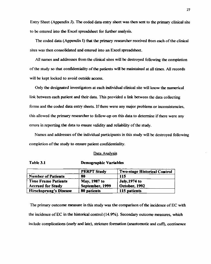
Table 3.1 Demographic Variables
PERPT Study Two-stage Historical ControlNumber o f Patients 80 115Time Frame Patients Accrued for Study
May, 1987 to September, 1999
July,1974 to October, 1992
Hirschsprung’s Disease 80 patients 115 patients
The primary outcome measure in this study was the comparison o f the incidence o f EC with
the incidence o f EC in the historical control (14.9%). Secondary outcome measures, which
include complications (early and late), stricture formation (anastomotic and cufi), continence

28
(stool and urinary), frequency o f defecation, soiling, constipation (initial and long-term) and
mortality rates, were reported.
Associated anomalies were defined as Trisomy 21, congenital cardiac anomalies,
prematurity, congenital gastrointestinal anomalies and other congenital anomalies.
Ordinal data was analyzed using the Mann Whitney U Test. Nominal data was analyzed
using Chi-square or the Fisher’s Exact Test (depending on the sample size). Quantitative data
was analyzed using the T-test. The Binomial Test was used to compare the rate o f EC in the
historical control with the rate o f EC in this study. Logistic Regression was used to detect
predictors for EC. Odds Ratios with a 95% Confidence Interval were calculated for predictor
variables derived from the Log Regression Equation. Significance was assessed at p<0.05.
29
CHAPTER FOUR RESULTS
Comparison o f the Historical Control to the Current Study Population
All o f the patients in this study and the historical control had HD. Patients from the current
study were accrued from May o f 1987 through September o f 1999. The sample from the
historical control was accrued from July 1974 through October o f 1992. Eighty subjects in
this study underwent a primary endorectal pull-through and one hundred and fifteen patients
in the historical control underwent the two-stage surgical procedure.
Results o f the Primary Outcome Measure
The incidence o f EC was compared to the historical control and crosstabulations were
done between EC and other variables o f interest in this study.
Incidence o f EC Compared to the Historical Control
The incidence o f post-operative EC in this study was 41.8% which was significantly higher
than the historical control’s EC rate o f 14.9%.
Binomial Test to Determine the Probability o f Developing EC with a PERPT
Confidence intervals were calculated using the Binomial Test to determine the percentage
o f children with HD undergoing a PERPT who might develop EC. A 95% Confidence Interval
was used. The lower 95% confidence limit for the study sample was 30.8% and the upper
95% confidence limit was 53.4%. This indicates that we are 95% sure that o f all children who
have HD, between approximately 31% to 53% would develop EC if they underwent a PERPT
within the first two years o f life.

30
Logistic Regression to Detect Predictors for EC
Logistic regression was used to detect predictors for EC. Four predictors were found: cuff
stricture, stricture, congenital GI anomalies and perianal excoriation. This information
indicates an association between these variables and EC but does not necessarily indicate that
they cause EC. The data was not recorded in such a way to determine whether the EC
occurred prior to or after the patients experienced one or more o f these complications.
Odds Ratio Estimation to Determine the Risk o f Developing EC
Odds ratio estimation o f risk was done to determine an increased risk o f developing EC
associated with post operative complications such as cuff stricture and strictures. It was
determined that children who have had the PERPT within the first two years o f life have a
10.6 (1.7 to 66.4 Confidence Interval) and 6.6 (1.4 to 37.6) times greater odds o f developing
EC if they have had cuff strictures or strictures respectively as complications post-operatively.
Chi-square Analysis to Determine an Association Between Specific Variables and EC
Chi-square analysis was done to determine if there was any association between specific
variables such as gender, anomalies, complications (early and late) and presenting symptoms
with the development o f EC post-operatively.
Gender
There was no statistical significance found when we looked at gender and the development
o f EC. It was found that 38.6% o f the males (22 /57) in this study and 50% o f the females
(11/ 22) in this study developed EC.
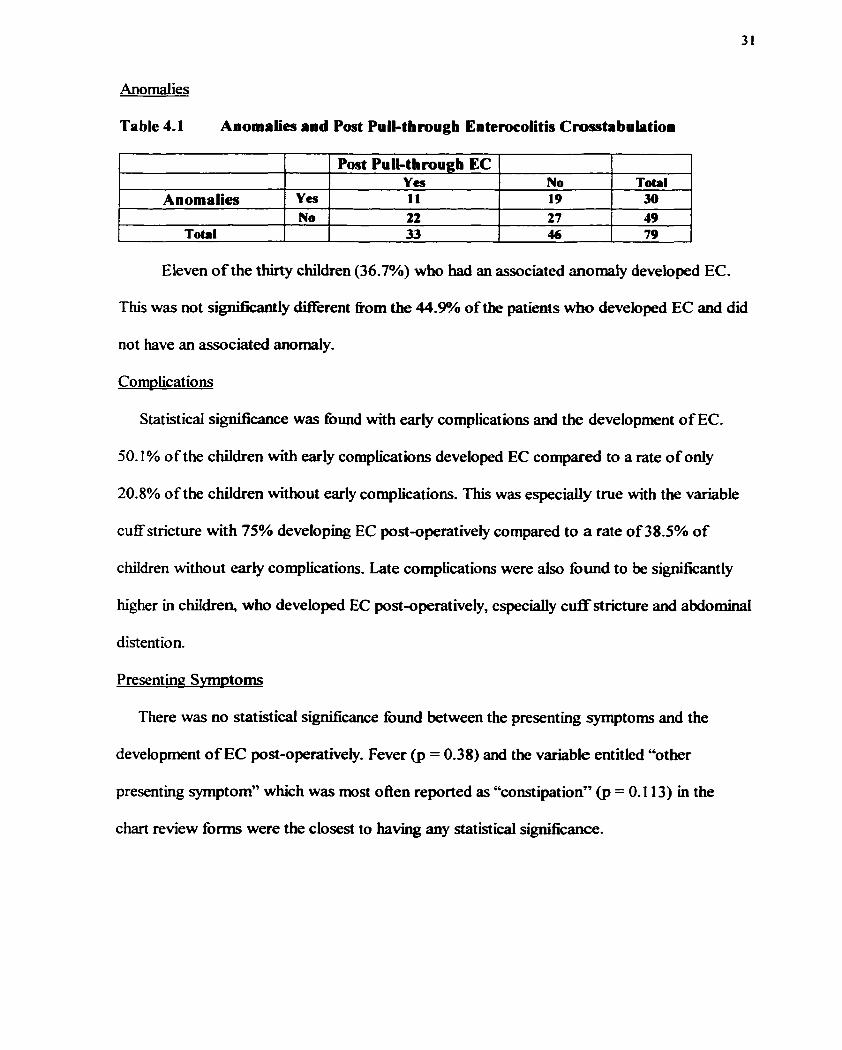
31
Anomalies
Table 4.1 Anomalies and Post Pull-through Enterocolitis Crosstabulation
Post Pu ll-th rough ECYes No Total
Anomalies Yes II 19 30No 22 27 49
Total 33 46 79
Eleven o f the thirty children (36.7%) who had an associated anomaly developed EC.
This was not significantly different from the 44.9% of the patients who developed EC and did
not have an associated anomaly.
Complications
Statistical significance was found with early complications and the development o f EC.
50.1% o f the children with early complications developed EC compared to a rate o f only
20.8% of the children without early complications. This was especially true with the variable
cuff stricture with 75% developing EC post-operatively compared to a rate o f 38.5% of
children without early complications. Late complications were also found to be significantly
higher in children, who developed EC post-operatively, especially cuff stricture and abdominal
distention.
Presenting Symptoms
There was no statistical significance found between the presenting symptoms and the
development o f EC post-operatively. Fever (p = 0.38) and the variable entitled “other
presenting symptom” which was most often reported as “constipation” (p = 0.113) in the
chart review forms were the closest to having any statistical significance.

32
Table 4.2 Post PulMhrough Enterocolitis in Males and FemalesFollowing Primary Endorectal Pull-through
Post Pull- through ECYes No Total
Sex o f Patient Male 22 35 57Female 11 11 22
33 46 79
Eleven o f the twenty-two female patients (50%) developed EC. Twenty-two of the thirty-
five males (38.6%) developed EC. This was not significantly différent. No comparison was
made with children with Trisomy 21 because there were too few patients with Trisomy 21
included in this study to determine any association or significance.
Descriptive Statistics on the Institutional Incidence o f EC
The incidence of EC by institution was:
1. University o f Michigan 25 o f 40 patients developed EC = 62.5%
2. Spectrum 3 o f 13 patients developed EC = 23.1%
3. Penn State 3 o f 13 patients developed EC =23.1%
4. UCLA 2 o f 13 patients developed EC = 15.4%
Table 4.3 Pre-primary Endorectal Pull-through and Post Pull-through Enterocolitis
Pre-PERPT ECYes No Total
Post Pull-through EC Yes 2 30 32No 1 44 45
Total 3 74 77

33
A crosstabulation report o f the frequency o f EC pre-operatively and EC post-operatively
showed that two patients in this study had both pre-PERPT EC and post-PERPT EC (2.6%).
Thirty patients had only post-PERPT EC (39.0%) and only one patient had pre-PERPT EC
without post-PERPT EC (1.3%). There was no statistical significance found to be associated
with these findings.
Table 4.4 Sex and Post Pull-through Enterocolitis Crosstabulation
Post Pull-through ECYes No Total
Sex Male 22 35 57Female 11 11 22
Total 33 46 79
Graph 4.1 Sex and Post Puli-throueh Enterocolitis0Ü -
70-
GO'y*co GO.CO
Q .
o 4 0 'o
aE 3 0 '
20 .
1 0 '
0
P o st Pull-through EC
EZkes
Female
Sex
The table and graph above demonstrate the incidence o f post pull-through EC in male and
female patients in this study. 38.6% o f the males and 50% of the females developed EC Post-
operatively. This was not statistically significant.
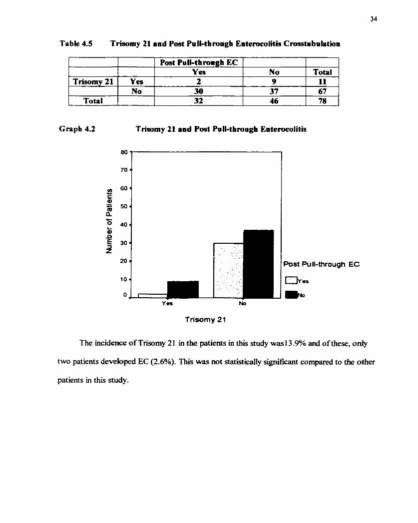
34
Table 4.5 Trisomy 21 and Post PuM-tbrough Enterocolitis Crosstabulation
Post Pull-through ECYes No Total
Trisomy 21 Yes 2 9 11No 30 37 67
Total 32 46 78
Graph 4.2 Trisomy 21 and Post Pull-through Enterocolitis
T riso m y 21
P o s t Pull-through E C
I lY es
The incidence o f Trisomy 21 in the patients in this study was 13.9% and o f these, only
two patients developed EC (2.6%). This was not statistically significant compared to the other
patients in this study.

35
Table 4.6 Coagenital Cardiac Anomalies and Post Pull-throughEnterocolitis Crosstabulation
Post Pull-through ECYes No Total
Congenital Cardiac Anomalies Yes 4 7 11No 29 39 68
Total 33 46 79
Graph 4.3 Congenital Cardiac Anomalies and Post Pull-throughEnterocolitis
P o st P u ll-th rough EC
Y es No
C o n g e n ita l C a rd iac A nom alies
Eleven o f the patients (13.8%) in this study had congenital cardiac anomalies and four
of these patients (5.1%) developed EC post operatively. This incidence was not statistically
significant.

36
Table 4.7 Prematurity and Post Pull-through Enterocolitis Crosstabulation
Post Pull-through ECYes No Total
Prematurity Yes 2 6 8No 31 40 71
Total 33 46 79
Graph 4.4 Prematurity and Post Pull-through Enterocolitis
Prematurity
Post PulM hrough EC
□ v e s
Eight o f the patients (10.1%) in this study were premature and two o f these developed EC
post-operatively (2.5%). The incidence o f this was not statistically significant.

37
Table 4.8 Abdominal Distention as a Presenting Symptom And Post Pull-through Enterocolitis Crosstabulation
Post Pull-through ECYes No Total
Abdominal Distention Yes 30 40 70No 3 5 8
Total 33 45 78
Graph 4.5 Abdominal Distention as a Presenting SymptomAnd Post Pull-through Enterocolitis
Post Pull-through EC
□ Y es
Yes No
Abdominal Distention
The presenting symptom o f abdominal distention was looked at to determine if there was
any statistical significance between this and the development o f EC post-operatively.
Thirty o f the thirty-three patients (90.1%) who developed EC post-operatively also
experienced abdominal distention while (9.1%) did not. No statistical significance was found.

38
Table 4.9 Transitional Zone By Intraoperative Biopsy Level andPost Pull-through Enterocolitis Crosstabulation
Post Pull- through EC
Yes No TotalTransitional Zone by Bx Level Rectum 3 3 6
Rectosigmoid 10 20 30Sigmoid 9 14 23
Descending Colon 2 5 7T ransverse Colon 4 3 7Ascending Colon 3 0 3
Small Bowel 1 0 1Total 32 45 77
Graph 4.6 Transitional Zone By Intraoperative Biopsy Level andPost Pull- through Enterocolitis
I«I
Post Pull-through EC
O e s
Transitional Zone by Biopsy Level

39
Short segment disease was defined as those with the agangiionic segment / transitional
zone found in the rectum, rectosigmoid or sigmoid regions o f the intestine. Long segment
disease was defined as those with the agangiionic segment or transitional zone found in the
descending colon and more proximal bowel. In contrast to Jung’s study, which noted 83.2%
o f their patients with short segment disease and 16.8% o f their patients with long segment
disease, this study showed 76.6% with short segment disease and 23.4% with long segment
disease. (Jung et al., 1995).
The intraoperative biopsy level was also compared with the incidence o f EC post-
operatively. There was no statistical significance shown but there are some trends that may be
best noted when looking at Graph 4.6. For example, the rectosigmoid and sigmoid regions of
the intestine are most commonly found to contain the agangiionic segment o f bowel in HD.
This coincides with findings o f other studies. One might also conclude that o f the patients
involved in this study, those who were diagnosed with longer segment disease experienced a
higher incidence o f EC post-operatively compared to those with shorter segment disease. For
example, 57% o f the patients in this study with the agangiionic portion found in the transverse
colon developed EC post-operatively and 100% o f the patients with the agangiionic portion in
the ascending colon or small intestine developed EC post-operatively.

40
Table 4.10 Intestinal Obstruction and Post Pull-throughEnterocolitis Crosstabulation
Post Pull-through ECYes No Total
Intestinal Obstruction Yes 3 4 7No 30 42 72
Total 33 46 79
Graph 4.7. Intestinal Obstruction and Post Pull-through Enterocolitis
P ost PulMhrough EC
r~TYes
Yes No
Intestinal Obstruction
Seven o f the seventy-eight (8.9%) patients reported on in this study also had an
intestinal obstruction. Three (3.8%) o f these patients developed EC post-operatively. This was
not statistically significant.

Table 4.11 Survival and Post Pull-through Enterocolitis Crosstabulation
41
Post Pull-through ECYes No Total
Survival Alive 31 46 77Dead 0 0 0
Total 31 46 77
Graph 4.8 Survival and Post Pull-through Enterocolitis
Survival
Post Pull-through EC
I lYes
This table and graph demonstrate the incidence o f EC in those who underwent the PERPT
surgical procedure. All the patients who developed EC post-operatively survived and as noted
earlier, one patient died from other medical problems.

42
Early Complications
Table 4.12
Results o f Secondary Outcome Measures
Complications
Early Complications and Post PuH-through Enterocolitis Crosstabulation
Post Pull-through ECYes No Total
Early Complications Yes 28 27 55No 5 19 24
Total 33 46 79
Graph 4.9 Early Complications and Post Pull-through Enterocolitis
SO
TO'
eo<
C2 50'COQ.o 4 0 'o.QE 30'
20'
10'
0
Post Pull-through EC
D v e s
Yes No
Early Complications
The incidence o f early complications (especially cuff stricture) and the development of EC
was found to be statistically significant. Fifty-fiye o f the seyenty-nine patients (69.6%) in this
study deyeloped early complications. Twenty-eight o f the reported seyenty-nine patients
(35.4%) in this study who deyeloped early complications also experienced EC post-
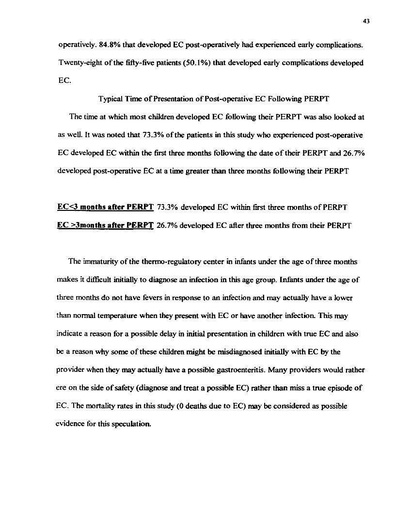
43
operatively. 84.8% that developed EC post-operatively had experienced early complications.
Twenty-eight o f the fifty-five patients (50.1%) that developed early complications developed
EC.
Typical Time o f Presentation o f Post-operative EC Following PERPT
The time at which most children developed EC following their PERPT was also looked at
as well. It was noted that 73.3% of the patients in this study who experienced postoperative
EC developed EC within the first three months following the date o f their PERPT and 26.7%
developed post-operative EC at a time greater than three months following their PERPT
EC<3 months after PERPT 73.3% developed EC within first three months o f PERPT
EC >3months after PERPT 26.7% developed EC after three months fi’om their PERPT
The immaturity o f the thermo regulatory center in infants under the age o f three months
makes it difiScult initially to diagnose an infection in this age group. Infants under the age o f
three months do not have fevers in response to an infection and may actually have a lower
than normal temperature when they present with EC or have another infection. This may
indicate a reason for a possible delay in initial presentation in children with true EC and also
be a reason why some o f these children might be misdiagnosed initially with EC by the
provider when they may actually have a possible gastroenteritis. Many providers would rather
ere on the side o f safety (diagnose and treat a possible EC) rather than miss a true episode o f
EC. The mortality rates in this study (0 deaths due to EC) may be considered as possible
evidence for this speculation.
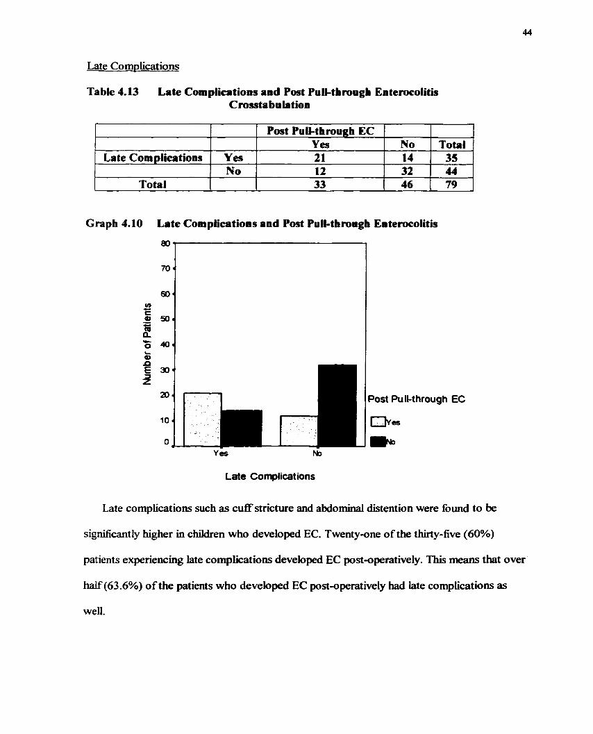
44
Late Complications
Table 4.13 Late Complications and Post Pull-through EnterocolitisC rossta bn lation
Post Pull-through ECYes No Total
Late Complications Yes 21 14 35No 12 32 44
Total 33 46 79
Graph 4.10 Late Complications and Post Pull-through Enterocolitis
80
Post Pull-through EC
I lYes
Yes
Late Complications
Late complications such as cufif stricture and abdominal distention were found to be
significantly higher in children who developed EC. Twenty-one o f the thirty-five (60%)
patients experiencing late complications developed EC post-operatively. This means that over
half (63.6%) o f the patients who developed EC post-operatively had late complications as
well.

45
Abdominal Distention
Table 4.14 Abdominal Distention and Post Pu ll-th rough Enterocolitis
Post Pull-through ECYes No Total
Abdominal Distention Yes 8 0 8No 25 46 71
Total 33 48 79
Eight (100%) o f the patients that developed abdominal distention also developed EC post-
operatively. This was significantly higher than those who did not develop abdominal distention
and did develop EC post-operatively.

46
Stricture Formation
Anastomotic Strictures
Table 4.15 Anastomotic Strictures and Post Pu ll-th rough Enterocolitis Crosstabulation
Post Pull-through ECYes No Total
Anastomotic Stricture Yes 9 3 12No 24 43 67
Total 33 46 79
Graph 4.11 Anastomotic Strictures and Post Pull-through Enterocolitis
80
P ost PulM hrough EC
I lYes
Yes No
A nastom otic Stricture
Strictures were associated with a statistically significant higher incidence o f EC post-
operatively. 75% o f the patients who had anastomotic strictures developed EC post-
operatively.

47
Cuff Strictures
Table 4.16 Cuff Stricture and Post Pull-through EnterocolitisCrosstabulation
Post Pull-through ECYes No Total
CuIT Stricture Yes 11 2 13No 22 44 66
Total 33 46 79
Graph 4.12 Cuff Stricture and Post Pull-through Enterocolitis
Post Pull-through EC
Yes No
Cuff Stricture
Eleven o f the thirteen patients (84.6%) who experienced cuflf strictures developed EC
post-operatively. Only twenty-two o f the sixty-six patients (33.3%) which had not
experienced cuflf strictures developed EC post-operatively.

48
Continence in Males and Females Who Have Had a Primary Endorectal Pull-through
In analyzing continence data, a subset was created from the study sample, which included
all patients that should be continent. Criteria for this subset were defined as all those who
were 4 years (48 months) o f age or continent at the time o f the phone interview.
Stool Continence
Table 4.17 Age o f Stool Continence and Post Pull-through Enterocolitis Crosstabulation
Post Pull-through ECYes No Total
Age o f Stool Continence in Months 12 0 1 118 1 0 124 1 6 727 0 1 130 1 1 234 1 0 136 3 3 638 0 1 142 0 1 148 1 2 360 0 2 2
Total 8 18 26
This table demonstrates that 85% of the patients reported on in this study were stool
continent between twenty-fbur and fbrty-eight months o f age.
The continence evaluation was accomplished using the Parent Phone Interview Form. This
form assisted the primary researcher in eliciting information regarding the patient's current
continence status. A scoring system was used on this form to rank each patient's current
continence status as either: normal (score o f 10), good (score o f 6 to 9), fair (score o f 1 to 5)
or poor (score o f 0).

49
Two different methods were used to look at the medians of these two groups. First, the
total score o f the evaluation was used when we looked at sex. This method showed a median
o f 8 (which is considered “good”) for males and a median o f 9 (which is also considered
“good”) for females. The interquartile range for males was 5.5 to 9 and the interquartile range
for females was 8 to 10. Secondly, the data was broken down into groups by ordinal rank with
the four categories o f normal (4), good (3), fair (2), and poor (1) and numerical equivalents
assigned to each group. The median for males and females was 3 (good). What was
statistically significant was the interquartile range. The interquartile range for males was 2.75
to 3 and the interquartile range for females was 3 to 4. The Mann Whitney U test showed
statistical significance with p<0.05. This indicated that continence rates were “Fair” to
“Good” for the males and “Good” to “Normal” for the females in this study.
Mean Age o f Stool Continence in Males and Females
The mean and standard error were calculated in determining stool continence in months for
both males and female patients involved in this study. The mean age of stool continence in
males was 32.5 ± 2.5 months [x ± SEM] and 34.1 ± 4.4 months in females. This was not
statistically different.
Mean Age o f Stool Continence in Children with a Historv o f EC and No EC
The mean age o f stool continence for children who had developed EC post-operatively
was 32.8 ± 3.2 months and 34.3 ± 3.12 months for those who had not developed EC post-
operatively. This was not statistically different.

50
Urinary Continence
Table 4.18 Age o f Urinary Continence and Post Pull-through EnterocolitisCrosstabulation (Information Obtained in the Phone Interview)
Post Pull-through ECYes No Total
Age o f Urinary Continence in Months 12 1 0 118 1 0 124 0 6 627 0 2 230 2 1 334 1 0 136 2 6 837 0 1 140 0 1 148 1 1 260 1 0 1
Total 9 18 27
Urinary continence, one o f the secondary outcome measures which was looked at
showed that 89% o f the patients in this study were urinary continent between two years
(twenty-four months) and four years (forty-eight months) of age.
Mean Age o f Urinary Continence in Males and Females
The mean and standard error were calculated in determining urinary continence in months
for both male and female patients inyolyed in this study. The mean age o f urinary continence
in males was 30.8 ± 2.3 months and 31.0 ± 3.0 months in females. This was not statistically
different.
Mean Age o f Stool Continence in Children with a History of EC and No EC
The mean age o f stool continence for children who had deyeloped EC post-operatiyely
was 33.3 ± 4.8 months and 31.6 ± 1 .7 months for those who did not deyelop EC
post-operatiyely. This was not statistically different.

51
Frequency o f Defecation
The mean number o f stools per twenty-four hours showed no statistical significance for
the frequency o f defecation between the EC group [1.6 ± 0.2] and the non-EC group [1.3 ±
0.1 ] when we looked at the frequency (p-value o f 0.11 ).
Frequency o f Defecation in Patients with One or Multiple Episodes o f EC
The number o f stools per twenty-four hours was also examined. It was found that children
with pwDst pull-through EC had 3.4 ± 0.5 stools per twenty-four hours and the children
without post pull-through EC had 2.2 ± 0.2 stools per twenty-four hours. Discussions with
clinicians indicate that there is a goal o f the patient having more frequent stools during the
first year to help prevent obstructive type symptoms and possibly the development o f EC.

52
Soiling
Initial FoUow-up o f Soiling
Table 4.19 Soiling and Post Pull-through Enterocolitis Crosstabulation
Post Pull-through ECYes No Total
Soiling Yes 2 0 2No 30 46 76
Total 32 46 78
Graph 4.13 Soiling and Post Pull-through Enterocolitis
80
70
80
® 50
2o « oE 30
20
10
0Yes
Post Pull-through EC
CZ es
Soiling
The table and graph show that the two patients who had problems with soiling had
developed EC while thirty o f the seventy-six patients (39.5%) who did not have problems
with soiling developed EC. None o f the patients defined in the subgroup for continence had
experienced soiling as a late complication.

53
Long-term Follow-up of Soiling
Table 4.20 Soiling and Post Pu ll-th rough Enterocolitis Crosstabulation (Information Obtained In A Phone Interview)
Post Pull-throughECYes No Total
Soiling (Ph. Interview) Clean 3 7 10Occasional 6 8 14
ContinuousIy
2 3 5
Total 11 18 29
Secondary outcome measures such as each patient’s current clinical status regarding his or
her continence outcome was evaluated using the Parent Phone Interview Form. Of the twenty-
nine reported patients, 38% had developed post pull-through EC. The age o f stool continence
was set at four years o f age. A breakdown o f the patient population was done by patient age
at the time o f the phone interview. O f the twenty-nine patients, 34.5% were reportedly clean,
48.3% soiled occasionally, and 17.2% soiled constantly.
Interestingly, 100% o f the patients defined in the subgroup for analysis o f continence did not
experience soiling as a late complication.

54
Constipation
Initial Problems with Constipation
TABLE 4.21 Constipation and Post Pull-through Crosstabulation
ConstipationYes No Total
Post Pull-through EC Yes 12 11 23No 12 21 33
Total 24 32 45
Twenty-four o f forty-five patients (53.3%) experienced constipation post-operatively.
One half o f the patients that experienced constipation also developed EC post-operatively.
There was no statistical significance found between constipation and post-operative EC.
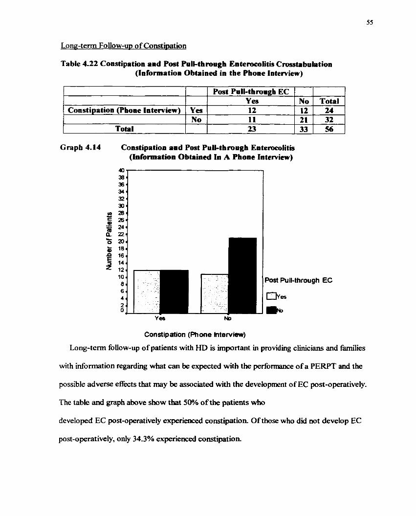
55
Long-term Follow-up o f Constipation
Table 4.22 Constipation and Post Pull-through Enterocolitis Crosstabulation(Information Obtained in the Phone Interview)
Post Pull-through ECYes No Total
Constipation (Phone Interview) Yes 12 12 24No 11 21 32
Total 23 33 56
Graph 4.14 Constipation and Post Pull-through Enterocolitis (Information Obtained In A Phone Interview)
Post PulMhrough EC
□ Y e s
Constipation (P hone interview)
Long-term follow-up o f patients with HD is important in providing clinicians and families
with information regarding what can be expected with the performance o f a PERPT and the
possible adverse effects that may be associated with the development o f EC post-operatively.
The table and graph above show that 50% o f the patients who
developed EC post-operatively experienced constipation. O f those who did not develop EC
post-operatively, only 34.3% experienced constipation.

56
Table 4.23
Mortality
Male and Female Mortality Crosstabuiation
SurvivalAlive Dead Total
Sex Male 56 1 57Female 21 0 21
Total 77 1 78
Graph 4.15 Male and Female M ortality
Survival
□ DeadMWe Femak
Sex
Mortality rates were reported on seventy-eight o f the eighty patients involved in this study.
There was one death (a male child), which was attributed to medical reasons other than EC.
The reported survival rate for patients included in this study was 98.7%.

57
CHAPTER FIVE DISCUSSION AND IMPLICATONS
Discussion o f Findings
The results o f this study suggest that the performance o f a PERPT is not associated with a
lower incidence o f EC compared to those who have undergone the two-stage surgical
resection for HD in the historical control group.
The incidence o f EC in the two-stage historical control group was 14.9%. This study
found the incidence o f EC to be 41.8% in those who underwent the PERPT.
Logistic regression was used to check for predictors o f EC. Four predictors were found;
cuff stricture, anastomotic stricture, congenital GI anomalies, and perianal excoriation. Of
these, cuff stricture and anastomotic stricture were found to be significant. This information
indicates an association between these variables and EC but does not necessarily indicate that
they cause EC.
Chi-square analysis was done to determine if there was any association between specific
variables such as gender, anomalies, complications (early and late) and presenting symptoms
with the development of EC post-operatively. The variable, which proved to be statistically
significant, was “complications”. These complications were defined as either being “early
complications” (occurring less than 60 days post-operatively) or as “late complications”
(occurring at or greater than 60 days post-operatively) complications. 50.1% o f the children
with early complications developed EC. This was especially true with the variable cuff
stricture with 75% developing EC post-operatively (probability level o f 0.01). Late
complications were also found to be significantly higher in children, who developed EC post-
operatively, especially cuff stricture and abdominal distention.
58
The incidence o f early complications (especially cu£f stricture) and the development o f EC
was found to be statistically significant. Fifty-five o f the seventy-nine patients (69.6%) in this
study developed early complications. Twenty-eight o f the fifty-five patients (50.1%) that
developed early complications developed EC.
Late complications such as cuff* stricture and abdominal distention were found to be
significantly higher in children who developed EC. Twenty-one o f the thirty-five (60%)
patients experiencing late complications developed EC post-operatively. This means that over
half (63.6%) o f the patients who developed EC post-operatively had late complications as
well. All eight (100%) o f the patients that developed abdominal distention also developed EC
post-operatively. This was significantly higher than those who did not develop abdominal
distention and did develop EC post-operatively.
Strictures were associated with a statistically significant higher incidence o f EC post-
operatively. EC is felt to be secondary to stool stasis and bacterial overgrowth. The increased
resistance to normal passage o f stool secondary to stricture formation may lead to EC. Only
twenty-two o f the sixty-six patients (33.3%) which had not experienced cuflF strictures
developed EC post-operatively.
73.3% o f the patients in this study who experienced post-operative EC developed EC
within the first three months following the date o f their PERPT and 26.7% developed post
operative EC at a time greater than three months following their PERPT. Most o f the patients
in this study were less than three months o f age at the time o f their PERPT. The immaturity of
the thermo-regulatory center in infiuits under the age o f three months may make it difficult
initially to diagnose an infection in this age group.

59
85% o f the patients reported on in this study were stool continent between twenty-four
and forty-eight months o f age. What was statistically significant was the interquartile range.
This indicated that continence rates were “Fair” to “Good” for the males and “Good” to
“Normal” for the females in this study.
In regards to the institutional incidence o f EC, it was noted that the current study
demonstrated a significantly higher incidence o f EC in the PERPT patients with an incidence
o f 41.8% versus the 14.9% incidence o f EC in the historical control.
While these percentages appear dramatically different, it is o f interest to note that the EC rates
at clinical site. The primary clinical site had a 62.5% incidence o f EC while the three remaining
contributing sites had much lower EC rates, ranging fi"om 15.4% to 23.1%. These incidences
are much closer to the value o f the historical control.
With regards to the long-term outcome in the PERPT patients, the PERPT patients did not
have a significant difference in the secondary outcome measures o f soiling and constipation
rates.
Limitations
There were several limitations in the design o f this study. First to be noted was the manner
in which the data was collected. Collection o f the data by only one person would have been
preferred. Since there were four clinical sites spread across the United States, this was a
problem that could only have been solved by sending one person to each clinical she to collect
the data. Monetary constraints did not allow for that to be done in this study. The different
investigators at each clinical she hoped to solve this problem by following the same forms for
the phone interview, chart review and coded data entry sheets in the acquisition of data. In
reviewing the data points, it was noted that if a considerable difference between two data

60
collection sites was found, a review o f the collection process at that particular site along with
a review o f any questionable patient information would be done. The primary clinical site
proposed to re-abstract the data if a major discrepancy was noted.
As noted earlier, standard operational definitions o f what constitutes a true case o f EC
versus a gastroenteritis may influence the number o f EC cases reported between the various
institutions and thus could cloud a true statistical significance in evaluating the success of the
diSerent treatment approaches.
The length o f time over which this study spanned was also a limitation to this study.
This problem was unavoidable due to the incidence o f HD and the specific number of patients
who met all o f the inclusion and exclusion criteria for this study. Unfortunately we were
forced to evaluate information in which there are inherently different levels o f care. It was
noted by the primary clinical site that if trends were found, there would be a stratification of
data analysis by time. If there was a significant difference in the primary and secondary
outcome measures, a stratification o f data analysis was to be done by age o f the patient at the
time of surgical intervention.
The fact that this was a retrospective study was also a significant limitation to this study.
This meant that great care was to be taken in the interpretation o f the data.
Use o f the Excel spreadsheet was very helpful, yet was a potential problem if data was
incorrectly entered. Great care was taken in the process o f data entry. It was hoped that the
use o f only one person in the processing o f the data fi’om the coded data entry sheets into the
Excel spreadsheet would minimize any problems that might arise.
There were many limitations in the use o f a historical control There was no way to ensure
the historical control’s accuracy because we were comparing our data to data that had been

61
abstracted at an earlier time by another person and their tools for abstraction o f the data may
have been different than those used in this study.
Prospective analysis would be difGcult and would take a large span o f time and monetary
funds to accomplish. Interesting factors derived from this study may warrant a prospective
study in specific areas in the study of HD in the future.
There were specific problems encountered when comparing the patient population that
underwent the primary endorectal pull-through to those who underwent the two-stage
endorectal pull-through procedure. This made more difGcult to compare these two
populations but we felt that these factors were take taken into consideration when doing the
abstraction and analysis o f the data presented. With such factors taken into account and
adjusted for, it was our belief that the data would show whether or not children with HD
would benefit from a single stage surgical intervention.
Suggestions for Future Research
Larger, prospective studies if possible would be helpful in the future. If parents, at the time
o f diagnosis, would agree to participate in extended studies which specifically monitor the
incidence o f EC and continence outcomes in children bom with HD, certain benefits from
particular surgical approaches and treatment may be found to be beneficial.
Since the patients in this study all underwent the Soave endorectal pull-through, it may be
o f interest to note the incidence o f EC in a larger study (200 to 300 patients). It may also be
helpful to compare children who have undergone the Soave to those who have undergone the
Duhamel procedure to look at the incidence o f EC, surgical complications, morbidity and
mortality as well as long-term outcomes in stricuture formation and continence in a large
study.

62
If a large prospective study o f this sort is not feasible, then closer follow-up o f existing
patients with a standardization o f diagnosis, care and the documentation in reporting EC and
other complications between several participating clinical sites would be helpful.
The incidence o f maternal diabetes was a factor that was noted in many chart reviews.
There may be a link between maternal diabetes meliitus and the development o f HD. Future
studies which include this variable may provide some insight regarding this possible link.
Conclusion
The researchers have concluded that the incidence o f Hirschsprung’s associated EC
is significantly greater in the patients who have had the PERPT compared to the historical
control. This reported greater incidence o f EC may be partly due to a lower thresh-hold in
making the diagnosis o f EC at each of the clinical sites involved in this study. In addition, an
increased level o f awareness in recent years may account for the more fi*equent diagnosis o f
EC. The performance o f a PERPT may still be a good option for some infents bom with HD.
Secondary outcome measures demonstrated a cotrqjarable outcome in areas such as
soiling, constipation and continence. The higher incidence of EC in this study did not appear
to have any impact on the survival rate.
Since 73.3% o f the patients in this study who experienced postoperative EC developed
EC within the first three months following the date o f their PERPT, it may be o f benefit for
these children to be placed on a regimen o f prophylactic antibiotics following their PERPT
until their thermo-regulatory centers mature.
O f most significance to the femilies is the avoidance o f the added risk associated with
another surgical procedure, months o f inconvenience and the added healthcare cost o f the
colostomy in the two-stage procedure.

63
REFERENCES
Ashcraft, K.W., Holder, T.M. (Eds.)- (1993). Pediatric Surgery. (2" ed ). Philadelphia: W.B. Saunders Co., 358-359, 364-366.
Bishop, W.P., Bishop, W.P., (1997). Hirschsprung’s Disease. University o f Iowa:Web Site: http://gutl.peds.uiowa.edu/HIRSCHPR/index.htm.
Gilley, R.E., Statter, M B., Hirschl, R.B., Coran, A.G.(1993). Definitive treatment o f Hirschsprung’s disease in the newborn with a one-stage procedure. Surgerv.555.
Elhalaby, E.A. (1995) Enterocolitis associated with Hirschsprung’s disease: a clinical histopathological correlative study. Journal o f Pediatric Surgerv.30:t7) 1023-6.
Elhalaby E.A. (1995). Enterocolitis associated with Hirschsprung’s disease: a clinical- radiological characterization based on 168 patients. Journal o f Pediatric Surgerv. 30:( 1)76-83.
Georgeson, K.E., Fuenfer, M.M., Hardin, W.D., (1995). Primary laparoscopic pull- through for Hirschsprung’s disease in inAnts and children. Journal o f Pediatric Surgerv. 301(7)1017.
Hanani, M., Louzon, V., Udassin, R. Freund, H R., Karmeli, F., Rachmilewitz, D. (1995). Nitric oxide containing nerves in bowel segments o f patients with Hirschsprung’s disease. Journal o f Pediatric Sureerv.30:f6)822.
Ikawa, H., Kawano, H., Takeda, Y., Masuyama, H., Watanabe K, Endo M,Yokoyama J, Kitajima M, Uyermura K, Kawamura K. (1997). Impaired expression of neural cell adhesion molecule LI in the extrinsic nerve fibers in Hirschsprung’s disease. Journal of Pediatric Surgerv. 32:(4)542.
Jung, P.M. (1995). Hirschsprung’s disease: one surgeon’s experience in one institution. Journal o f Pediatric Surgerv. 30:(5)649.
Kamimura, T., Kubota, M., Suita, S., (1997). Functional innervation o f the aganglionic segment in Hirschspnmg’s disease—a comparison o f the short- and long-segment type.Journal of Pediatric Surgerv. 32:(51677.
Kobayashi, H., Hirakawa, H., Surana, R., O’Briain, D.S., Puri, P. (1995). Intestinal neuronal dysplasia is a possible cause o f persistent bowel symptoms after pull-through operation for Hirschsprung’s disease. Journal o f Pediatric Surgerv. 30:(2)253.
Kobayashi, H., Wang, Y., Hirakawa, H., O’Briain, D.S., Puri, P. (1995).Intraoperative evaluation o f extent o f aganglionosis by rapid acetylcholinesterase histochemical technique. Journal of Pediatric Surgerv. 30:248.

64
Kusaflika T, Puri P. (1997). Altered RET gene mRNA expression in Hirschsprung’s disease. Journal o f Pediatric Surgerv. 32:(2)600.
Langer, J.C., Bimbaum, E. (1997). Preliminary experience with intersphincteric botulinum toxin for persistent constipation after pull-through for Hirschsprung’s disease. Journal o f Pediatric Surgerv. 32:(7)1060-1061.
Langer, J.C., Fitzgerald, P.O., Winthrop, A.L. et al. (1996). One-stage versus two- stage Soave pull-through for Hirschsprung’s disease in the first year o f life. Journal o f Pediatric Surgerv. 31:33-37.
Lebenthal E.(1996). Pediatric Clinics o f North America: Pediatric Gastroenteroloev 11.43:(4)427.
Martucciello,G., BicoccM, M.P., Dodero, P. (1992). Total colonic aganglionosis associated with interstitial deletion o f the long arm o f chromosome 10. Pediatric Surgerv International. 7:308-310.
Martucciello, P., Favre, A., Takahashi, M., Jasoniu, V. (1995). Immunohistochemical localization o f RET protein in hirschsprung's disease. Journal o f Pediatric Surgerv. 30:0)433.
Molenaar, J.C. (1995). Pathogenetic aspects o f Hirschsprung’s Disease. British Journal o f Surgerv. 82:145-147.
O'Neill, J.A., Rowe, M.L, Grosfeld, J.L., Fonkalsrud, E.W., Coran, A.G. (Eds.). (1998). Pediatric Surgerv. (5th ed.). St. Louis: Mosby Year Book, Inc.
Parikh, O.K., Tam, P., VanVelzen, D., Edgar, D. (1992). Abnormalities in the distribution o f laminin and collagen type IV in hirschsprung's disease. Gastroenterology: (102)1241.
Rescorla, F.J., Morrison, S.M., Engles, D., West, K.W., Grosfeld, J.L. (1992). Hirschsprung's disease: evaluation o f mortality and long-term fimction in 260 cases. Archives o f Surgerv. 127: (8)935.
Ryan, E.T., Ecker, J.L., Christakis, N.A., Folkman, J. (1992). Hirschsprung’s disease: associated abnormalities and demography. Journal o f Pediatric Surgerv. 27: (1)76.
Sabiston, D.S. (Eds ). (1997). Textbook o f Surgerv. (15th ed.) Philadelphia: W.B. Saunders Co., 1243.
Strovroflf, M., Kykew, P., Teague, W.G. (1995). The complete spectrum o f neurocristopathy in an infant with congenital hypoventilation, hirschsprung’s disease and neuroblastoma. Journal o f Pediatric Surgerv. 30: (8)1220.

65
Swenson, O. (1989) My early experience with Hirschsprung’s Disease. Journal o f Pediatric Surgerv. 24: (8) 840.
Swenson, O. (1996) Early history o f the therapy o f Hirschsprung’s Disease: Facts and personal observations over fifty years. Journal o f Pediatric Surgerv. 31: 1005.
Tehelbaum, D.H., Drongowski, R.A., Chamberlain, J.N., Coran, A.G., (1997). Longterm stooling patterns in infants undergoing primary endorectal pull-through for Hirschsprung’s disease. Journal o f Pediatric Surgerv. 32: (7) 1049,1052.
Touloukian, R.J., (1995). Pediatric surgery between 1860 and 1900. Journal o f Pediatric Surgerv. 30: (7)914.
Vanderwinden, J.M., DeLaet, M.H., Schifihnann, S.N. (1993). Nitric oxide synthase distribution in the enteric nervous system o f Hirschsprung’s disease. Gastroenteroloev 105:(4)969-973.
Yamataka, A., Nagaoka, I., Miyano, T., Yanai, T., Fujimoto, T., Okazaki, T., (1995). Quantitative analysis o f neuronal innervation in the aganglionic bowel of patients with Hirschsprung’s disease. Journal o f Pediatric Surgerv. 30:(21260-262.
Yamataka, A., Ohshiro, K., Kobayashi, H., Fujiwara, T., Sunagawa, M., Miyano, T. (1997). Intestinal pacemaker C-KIT+ cells and synapses in allied Hirschspnmg’s disease. Journal o f Pediatric Surgerv. 32:(711069. 1071.

6 6
APPENDIX A
INTRODUCTORY LETTER TO POTENTIAL SUBJECTS
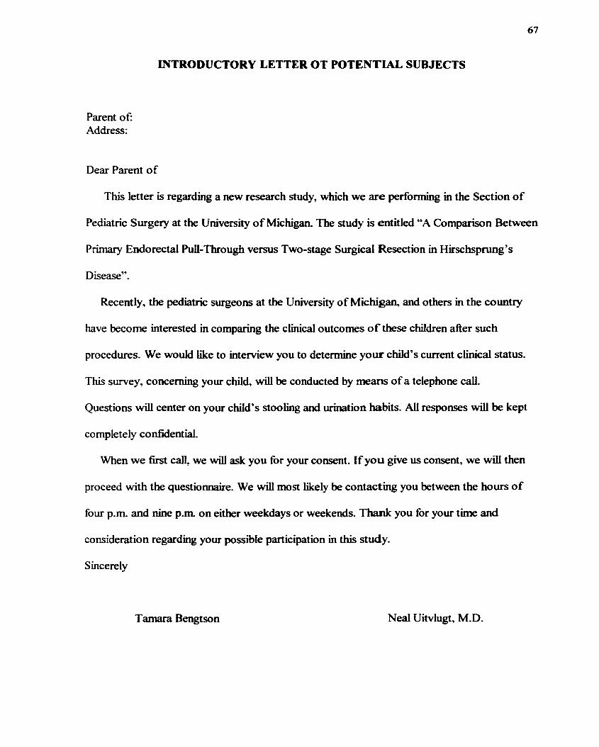
67
INTRODUCTORY LETTER OT POTENTIAL SUBJECTS
Parent of: Address:
Dear Parent o f
This letter is regarding a new research study, which we are performing in the Section o f
Pediatric Surgery at the University o f Michigan. The study is entitled “A Comparison Between
Primary Endorectal Pull-Through versus Two-stage Surgical Resection in Hirschsprung’s
Disease”.
Recently, the pediatric surgeons at the University o f Michigan, and others in the coimtry
have become interested in comparing the clinical outcomes o f these children after such
procedures. We would like to interview you to determine your child’s current clinical status.
This survey, concerning your child, will be conducted by means o f a telephone call.
Questions will center on your child’s stooling and urination habits. All responses will be kept
completely confidential.
When we first call, we will ask you for your consent. If you give us consent, we will then
proceed with the questionnaire. We will most likely be contacting you between the hours o f
four p.m. and nine p.m. on either weekdays or weekends. Thank you for your time and
consideration regarding your possible participation in this study.
Sincerely
Tamara Bengtson Neal Uitvlugt, M.D.

68
a 1*Wn d i x b
PHONE SC R H ^ tfP VERBAL CONSENT

6 9
PHONE SCRIPT OF VERBAL CONSENT
Hello, my name is . I am a Grand Valley State University Physician Assistant
Studies student working on a clinical research study with Dr. Daniel Teitelbaum at the
University o f Michigan and Dr. Neil Uitvlugt, a pediatric surgeon at the Spectrum Downtown
Campus in Grand Rapids, Michigan.
You are one o f approximately eighty people I am hoping to interview. 1 am phoning
because we are looking for information related to your child’s treatment for Hirschsprung’s
Disease. I would like to ask you a few questions. Information from this study will hopefully
help to improve the care and help benefit other children bom with Hirschsprung’s Disease in
the future.
If you choose not to participate in this study, your choice will have no influence on your
child’s present or future care. If the results o f this study are published, your child’s name will
not be used and your answers will be kept confidential.
If you have any questions regarding my role in this study, please feel free to contact
Professor Paul Huizenga, Chair o f the Human Research Review Committee at Grand Valley
State University. The phone number is 616-895-2472.
Do you mind if I ask you a few questions?
(If the response is yes, go to survey)

70
APPENDIX C
PRIMARY ENDORECTAL PULL-THROUGH
PARENT PHONE INTERVIEW FORM

71
PRIMARY ENDORECTAL PULL-THROUGH
PARENT PHONE INTERVIEW FORM
(Over the Last Six Months)
Name (Last, First)__________________________________________________________Hospital:__________________________________________ Birth Date:___ / ____ / Patient Number: Interview Date: / /
Continence Evaluation (Circle one score fo r each category)
Frequency o f Defecation ScoreNormal (1 -2 /day) 2Often (3-5 / day) 1Abnormal (> 6 / day) 0
Stool ConsistencyFormed 2Loose 1Liquid 0
Soiling (small amounts of feces staining the underwear)Clean (never stained) 2Occasional (> 0 but < 6 / week) 1
(during diarrhea, flatulence, with mucous leakage)Permanent Soiling (every day) 0
Urgency PeriodNormal (minutes) 2Short (seconds) 1None 0
Requires Diapers (Incontinence = regular loss o f solidfeces)None 2Occasionally (< 1 / week) 1Continuously (> 1 / week) 0
Total Score (addpoints in all categories): ___________
Scoring:Normal = 10 pointsGood = 6 to 9 pointsFair = 1 to 5 pointsPoor = 0 points
Notes:

72
Stool Frequency / 24 Hours at Time of Telephone Interview:____________________
Age o f Stooi-continence:________________________________ (year +/- month o f age)
Constipation:^e^5 than three spontaneous bowel movements per week or painful bowel movements)
(Please circle correct response) Yes or No
Approximate Number o f Constipative Occurrences:
(Please circle correct response) Weekly Monthly
Interval Between Occurrences:
(Please circle correct response)
Treatment o f Constipation:
Drug Treatment:I . D rug:_________
< 3 months or > 3 months
Length o f treatment:Dosage: ________ / day
/ week / month
2. Drug: Dosage:Length o f treatment:
________ /day/ week / month
3. Drug: Dosage:Length o f treatment:_____________________
Enemas: Yes or No Type:______________________________
Anal Dilatation: Yes or No If yes (circle one):
Finger Bougie Both None
Frequency / day:_________ or Frequency / month:_____
Age of Urinary Continence:________________________(year + / - month o f age)
Disorders o f Mictu ration: Yes or No If yes (circle one):
Daytime urinary dribbling Yes (constant / occasional) No
Nocturnal enuresis Yes (constant / occasional) No
_______ / day/ week / month

73
APPENDIX D
CHART REVIEW FORM

74
CHART REVIEW FORM
H ospital:________________________Name: (last, first)__________________Sex: Male FemaleRace: White Black Asian HispanicBirth Date:___ /__/ __Birth Weight: _________ (kg)First Admit Date:__/ __ / __Diagnosis Date:__/ __ / __ICU Days:____________ (days)
Patient Number:
Gestational A ge:___________First Discharge D ate:__/ __ / __ERPT Date:__/ __ / __Weight at ERPT:_________(kg)
Associated Anomalies and Confounding Factors
Trisomy 21 Cong. Cardiac Others Gl
Yes No Yes No Yes No
PrematurityOthers:If Yes, Type:
Yes No
Major Presenting Signs and Symptoms (circle one for each)'.
Vomiting Yes No Abdominal Distention Yes NoDiarrhea Yes No Fever (>100F) Yes NoLethargy Yes No Rectal Bleeding Yes NoDelay in Passage o f Meconium (circle one):
< 24 hours 24-48 hoursOthers:
> 48 hours
Radiology at Presentation:
Abdominal X-ray: Yes NoResults : NormalIleus Yes NoPerforation Yes NoCut-oflfSign Yes No
Barium Enema: Yes NoResults: Normal
Length o f Aganglionic Segment byultrashort rectosigmoidcolonic total intestinal
Retention o f Barium at 24 Hours:Yes No
Abnormal
Abnormal(circle one) long colonic unknown
total

75
Rectal Biopsy:SuctionResults:
Yes No {^If yes, circle one)FuU-thickness BothNormal Abnormal
None
Manometric Study: Yes No {.Ifyes, circle one)Results: Normal Abnormal
Pre-ERPT enterocolitis (EC) Yes No Date:___ / /(explosive diarrhea, abdom inal distention, fever (>JOOF), lethargy, shock)
Enterocolitis Treatm entAntibiotics Yes NoAdmitted Yes NoOstomy for EC Yes No
Number o f D ays:____Admission Date: / /Discharge Date: __/ ___/
*For each episode defined as outpatient or inpatient visit fo r Pre-ERPT EC, place information on attached sheet entitled Appendix F Additional Sheet fo r Pre-Endorectal Pull-through Enterocolitis
Intraoperative Findings
Transitional Zone Seen Grossly:Level o f Biopsy (Circle one):
Rectal Recto-sigmoidDescending Colon Transverse ColonSmall Bowel
Yes No
SigmoidAscending Colon
Proximal Dilatation at Time o f Initial Operation: Mild (<2cm) Moderate (2-4cm)Did this cause any problems?Problems:
Yes No { I f yes, circle one)Massive (>4cm)Yes No { I f yes, please explain)
Anatomic Abnormalities:Specific anatomic abnormalities:
Yes No { I f yes, please specify)
Intraoperative Complications Yes NoCuflfTear: Yes NoTension o f the pull-through segment: Yes NoPoor blood supply o f the pull-through segment: Yes NoOther complications:____________________________________

76
I Postoperative Course (operation day = day 0)
Wound healing problems: Yes NoPost-operative antibiotics: Yes No Number o f Days:Post operative TPN: Yes No Number o f Days:Enteral feeding started on post-operative day number:_____________Post operative procedures: Yes No { I f yes, please explain)
Re-exploration Yes No Date:__/ / Reason:________________________________________________Other procedures:________________________________________
Réadmissions
Readmission: Yes NoDate o f readmission:__/ __ / __Reason for readmission:____________________Enterocolitis: Yes NoMalnutrition: Yes No { I f yes. please explain)Weight at readmission (kg):____________ ____Surgical complication (specify):_____Other cause for readmission (specify):
**Use additional copies under Appendix G entitled Additional Sheet fo r Each Readmission fo r each time a patient was readm itted
Outcome
A=Early Complication (day 0 to day 60post-op)Yes No
Anastomotic dehiscence Yes NoAnastomotic stricture Yes NoPelvic or intraabdominal infection Yes NoCuflf abscess Yes NoPerianal fistula Yes NoIleus (delayed bowelfunction>5days) Yes NoPerianal excoriation Yes No
{ I f yes, circle a ll that apply)

77
B=Late complication (>day 61): Yes No (J f yes, circle a ll thaiRetraction o f pull-through Yes NoDid this require repeated:
Dilatations Yes NoStricturoplasty Yes NoRedo-ERPT Yes No
Distention Yes NoCuff Stricture Yes NoDid this require repeated:
Dilatations Yes NoStricturoplasty Yes NoRedo-ERPT Yes No
Perianal excoriations Yes NoIntestinal obstruction Yes NoRectal prolapse Yes NoSoiling Yes NoConstipation Yes No
Long-term medication (more than a couple o f weeks)Laxatives: Yes NoDependence: Always Sometimes
Frequency per month:
Prokinetic agent: Yes NoDependence: Always Sometimes
Frequency per month:
Rectal washout: Yes NoDependence: Always Sometimes
Frequency per month:
Colonic irrigation: Yes NoDependence: Always Sometimes
Frequency per month:
SurvivalAlive: Yes No Cause of Death:
Date of last follow-up: / /

78
Post Pull-through Enterocolitis Episodes *(use one sheet Appendix H entitled Additional Sheet fo r Each Post Pull-through Enterocolitis Episode fo r each episode o f enterocolitis):
Clinical Grade: 1 2 3*(See attached Appendix E to assist in grading each episode o f enterocolitis)
Treatment
A= Outpatient Yes No Date: / /
B=In patient Yes No {ifyes, please f i l l in)Admission D ate:__/ __ / __Discharge D ate :__/ __ / __
1. Non-operative Yes No ( I f yes, circle all that apply)
Observation (no treatment) Yes NoDecompression Yes No
(e.g. rectal tube, finger or bougie dilatation, enemas)IV Fluid Yes No { I f yes, circle all that apply)
Regular Yes No Saline or Ringer’s LactateTPN Yes No # o f days:
Antibiotics Yes No If yes, (circle one)Oral or Parenteral
Operative Yes No { I f yes, circle all that apply)
Dilatation under anesthesia Yes NoSphincterotomy Yes NoSphincterectomy Yes NoOstomy Yes NoRedo-puUthrough Yes No

79
APPENDIX E
CLINICAL GRADING OF ENTEROCOLITIS FORM

80
CLINICAL GRADING OF ENTEROCOLITIS FORM
1. Mild explosive diarrheaMild to moderate abdominal extension No significant systemic manifestation
2. Moderately explosive diarrhea Moderate to severe abdominal distentionAssociated with mild to moderate systemic manifestation (e.g. fever and tachycardia)
3. Explosive diarrhea Marked abdominal distention Shock or impending shock

81
APPENDIX F
ADDITIONAL SHEET FOR PRE ENDORECTAL
PULL-THROUGH ENTEROCOLITIS

82
ADDITIONAL SHEET FOR PRE-ENDORECTAL
PULL-THROUGH ENTEROCOLITIS
Pre-ERPT EnterocolitisEnterocolitis (EC) - [explosive diarrhea, abdominal distention, fever (>100 F), lethargy, shock]
Hospital:Name: (last, first)
Patient Number:
Pre-ERPT EC: Yes No Date o f Pre-ERPT EC: / /Enterocolitis treatment:
Antibiotics; Yes No Number o f Davs:Admitted: Yes No Admission Date: / /
Discharge Date: / /Ostomy for EC: Yes No

83
APPENDIX G
ADDITIONAL SHEET FOR EACH RE ADMISSION
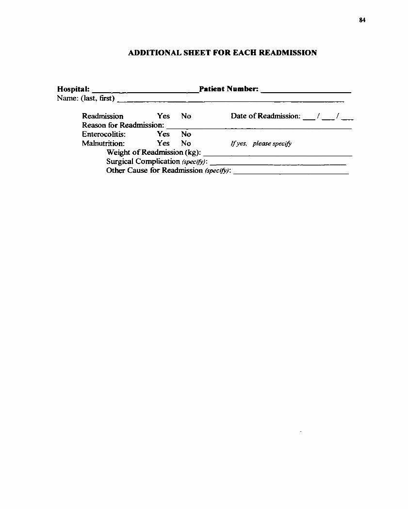
84
ADDITIONAL SHEET FOR EACH RE ADMISSION
H ospital:______Name: (last, first)
Patient Number:
Readmission YesReason for Readmission: _ Enterocolitis:Malnutrition;
No Date o f Readmission:
Yes No Yes No
Weight o f Readmission (kg):_______Surgical Complication (specify):____________
Other Cause for Readmission (specify):
I f yes, please specify

8S
APPENDIX H
ADDITIONAL SHEET FOR EACH POST PULL-THROUGH
ENTEROCOLITIS EPISODE

86
ADDITIONAL SHEET FOR EACH POST PULL-THROUGH
ENTEROCOLITIS EPISODE
Hospital:_____________________________Patient Number:Name: (last, first)_____________________________________
Clinical Grade: 1 2 3(See Appendix E entitled Clinical Grading o f Enterocolitis Form to assist in grading
o f each episode o f enterocolitis)
Treatment:A= Outpatient Yes No Date:___ / ____/ ___
B= Inpatient Yes No Admission D ate:____/ ____/Discharge D ate:___/ ____/
1. Non-operative Yes No (If yes, please circle a ll that apply)Observation (no treatment): Yes NoDecompression: Yes No
(e.g. rectal tube, fin g er or Bougie dilatation or enemas)IV Fluid: Yes No
Regular (Saline, Ringer’s Lactate) Yes NoTPN Yes NoIf yes, number o f days of IV fluid:_______________
Antibiotics: Yes NoIf yes, please circle: Oral Parenteral
2. Operative Yes No (If yes, please circle a ll that apply)
Dilatation under anesthesia Sphincterotomy Sphincterectomy Ostomy

87
APPENDIX I
COM PUTER DATA ENTRY SHEET
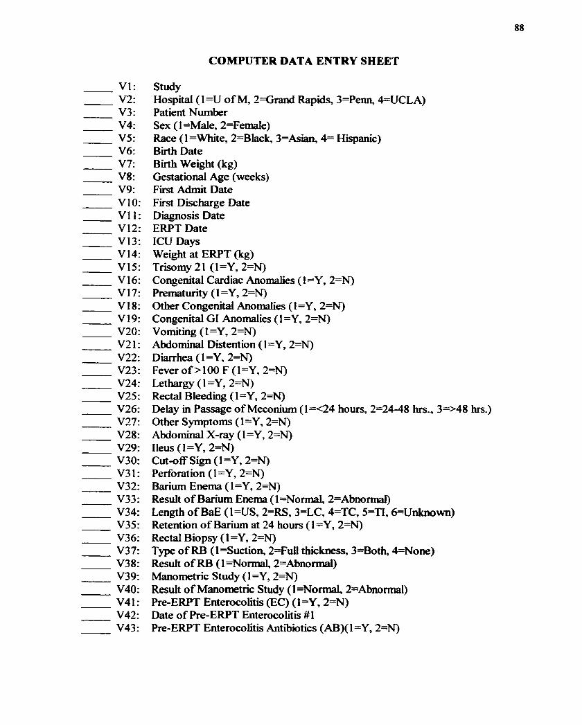
88
COMPUTER DATA ENTRY SHEET
VI: StudyV2: Hospital (1=U o f M, 2=Grand Rapids, 3=Penn, 4=UCLA)V3: Patient NumberV4: Sex ( 1 =Male, 2=Female)V5: Race (l=White, 2=Black, 3=Asian, 4= Hispanic)V6: Birth DateV7: Birth Weight (kg)V8: Gestational Age (weeks)V9: First Admit DateVIO: First Discharge DateVI I: Diagnosis DateV12: ERPT DateV I3: ICUDaysV 14: Weight at ERPT (kg)V15: Trisomy 21 (1=Y, 2=N)V 16: Congenital Cardiac Anomalies ( 1 =Y, 2=N)V 17 : Prematurity ( 1 = Y, 2=N)V I8: Other Congenital Anomalies ( 1=Y, 2=N)V 19: Congenital G1 Anomalies ( 1 =Y, 2=N)V20: Vomiting (1=Y, 2=N)V21 : Abdominal Distention ( 1 = Y, 2=N)V22: Diarrhea (1=Y, 2=N)V23: Fever o f >100 F (1=Y, 2=N)V24: Lethargy (1=Y, 2=N)V25: Rectal Bleeding (1=Y, 2=N)V26: Delay in Passage o f Meconium (1=<24 hours, 2=24-48 hrs., 3=>48 hrs.)V27: Other Symptoms (1=Y, 2=N)V28: Abdominal X-ray (1=Y, 2=N)V29: Ileus (1=Y, 2=N)V30: Cut-off Sign ( 1 =Y, 2=N)V31 : Perforation ( 1 =Y, 2=N)V32: Barium Enema (1=Y, 2=N)V33 : Result o f Barium Enema ( 1 =NormaL 2=Abnormal)V34: Length o f BaE (1=US, 2=RS, 3=LC, 4=TC, 5=T1, 6=Unknown)V35: Retention o f Barium at 24 hours (1=Y, 2=N)V36: Rectal Biopsy ( 1 =Y, 2=N)V37: Type o f RB (l=Suction, 2=FuU thickness, 3=Both, 4=None)V38: Result o f RB (l=Normal, 2=Abnormal)V39: Manometric Study (1=Y, 2=N)V40: Result o f Manometric Study (l=NormaL 2=Abnormal)V41 : Pre-ERPT Enterocolitis (EC) ( 1 = Y, 2=N)V42: Date o f Pre-ERPT Enterocolitis #1V43: Pre-ERPT Enterocolitis Antibiotics (AB)(1=Y, 2=N)
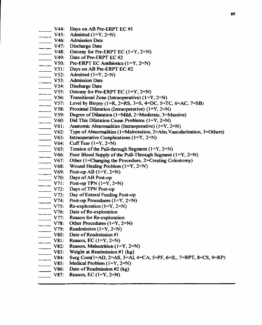
89
V44: Days on AB Pre-ERPT EC #1V45 : Admitted ( 1 = Y, 2=N)V46: Admission DateV47: Discharge DateV48: Ostomy for Pre-ERPT EC (1=Y, 2=N)V49: Date o f Pre-ERPT EC #2V50: Pre-ERPT EC Antibiotics ( 1 =Y, 2=N)V51 : Days on AB Pre-ERPT EC #2V52: Admitted ( 1 =Y, 2=N)V53: Admission DateV54: Discharge DateV55: Ostomy for Pre-ERPT EC (l=Y , 2=N)V56: Transitional Zone (Intraoperative) (1=Y, 2=N)V57: Level by Biopsy (1=R, 2=RS, 3=S, 4=DC, 5=TC, 6=AC, 7=SB)V58: Proximal Dilatation (Intraoperative) (1=Y, 2=N)V59: Degree o f Dilatation (l=Mild, 2=Moderate, 3=Massive)V60: Did This Dilatation Cause Problems (1=Y, 2=N)V61 : Anatomic Abnormalities (Intraoperative) ( 1 = Y, 2=N)V62: Type o f Abnormalities (I=Malrotation, 2=Abn.Vascularization, 3=Others)V63 : Intraoperative Complications ( 1 =Y, 2=N)V64: Cuff Tear ( 1 =Y, 2=N)V65: Tension o f the Pull-through Segment (1=Y, 2=N)V66: Poor Blood Supply o f the Pull-Through Segment (1=Y, 2=N)V67: Other (l=Changing the Procedure, 2=Creating Colostomy)V68: Wound Healing Problem (1=Y, 2=N)V69: Post-op AB (1=Y, 2=N)V70: Days o f AB Post-opV71 : Post-op TPN ( 1 = Y, 2=N)V72; Days o f TPN Post-opV73: Day o f Enteral Feeding Post-opV74: Post-op Procedures (1=Y, 2=N)V75: Re-exploration ( 1 = Y, 2=N)V76: Date o f Re-explorationV77: Reason for Re-explorationV78: Other Procedures (1=Y, 2=N)V79: Readmission (1=Y, 2=N)V80: Date o f Readmission #1V81 : Reason, EC ( 1 = Y, 2=N)V82; Reason, Malnutrition (1=Y, 2=N)V83: Weight at Readmission #1 (kg)V84: Surg Com(l=AD, 2=AS, 3=A1, 4=CA, 5=PF, 6=IL, 7=RPT, 8=CS, 9=RP)V85: Medical Problem (1=Y, 2=N)V86: Date o f Readmission #2 (kg)V87: Reason, EC ( 1 =Y, 2=N)
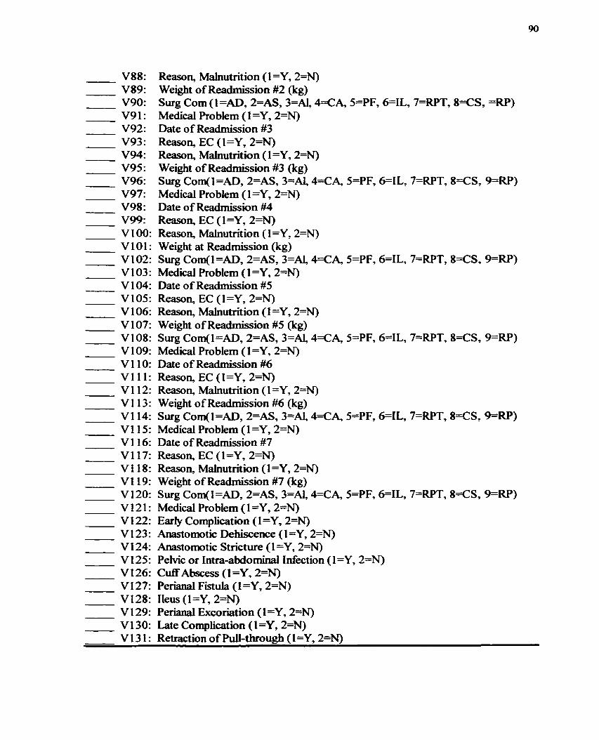
90
V88: Reason, Malnutrition (1=Y, 2=N)V89: Weight o f Readmission #2 (kg)V90: Surg Com (l=AD, 2=AS, 3=A1, 4=CA, 5=PF, 6=IL, 7=RPT, 8=CS, =RP) V 91 : Medical Problem ( 1 =Y, 2=N)V92 : Date o f Readmission #3 V93: Reason, EC (1=Y, 2=N)V94: Reason, Malnutrition ( 1 =Y, 2=N)V95: Weight o f Readmission #3 (kg)V96: Surg Com(l=AD, 2=AS, 3=A1, 4=CA, 5=PF, 6=IL, 7=RPT, 8=CS, 9=RP)V97: Medical Problem (1=Y, 2=N)V98: Date o f Readmission #4 V99: Reason, EC ( 1 =Y, 2=N)V I00: Reason, Malnutrition (1=Y, 2=N)V 101 : Weight at Readmission (kg)V I02: Surg Com(l=AD, 2=AS, 3=A1, 4=CA, 5=PF, 6=IL, 7=RPT, 8=CS, 9=RP) V 103 : Medical Problem ( 1 =Y, 2=N)V 104: Date o f Readmission #5 V105: Reason, EC (1=Y, 2=N)V 106: Reason, Malnutrition ( 1 =Y, 2=N)V 107: Weight o f Readmission #5 (kg)V108: Surg Com(l=AD, 2=AS, 3=A1, 4=CA, 5=PF, 6=IL, 7=RPT, 8=CS, 9=RP) V 109: Medical Problem ( 1 =Y, 2=N)V I 10: Date o f Readmission #6 V I 11: Reason, EC (1=Y, 2=N)V I12: Reason, Malnutrition (1=Y, 2=N)V I 13: Weight o f Readmission #6 (kg)V I 14: Surg Com(l=AD, 2=AS, 3=A1,4=CA, 5=PF, 6=IL, 7=RPT, 8=CS, 9=RP) V I15: Medical Problem ( 1 = Y, 2=N)V I 16: Date o f Readmission #7 V I17: Reason, EC (1=Y, 2=N)V I 18: Reason, Malnutrition (1=Y, 2=N)V I 19: Weight o f Readmission #7 (kg)V I20: Surg Com(l=AD, 2=AS, 3=A1, 4=CA, 5=PF, 6=IL, 7=RPT, 8=CS, 9=RP) V 121 : Medical Problem ( 1 = Y, 2=N)V 122: Early Complication ( 1 =Y, 2=N)V I23: Anastomotic Dehiscence (1=Y, 2=N)V I24: Anastomotic Stricture (1=Y, 2=N)V I25: Pelvic or Intra-abdominal Infection (1=Y, 2=N)V 126: CuflF Abscess ( 1 =Y, 2=N)V127: Perianal Fistula (1=Y, 2=N)V I28: Ileus (1=Y,2=N)V 129: Perianal Excoriation ( 1 =Y, 2=N)V I30: Late Conq)lication (1=Y, 2=N)V I31 : Retraction o f Pull-through (1=Y, 2=N) _____________________________

91
V I32: Treatment, Dilatations (1=Y, 2=N)V I33: Treatment, Strictureplasty (1=Y, 2=N)V I34: Treatment, Redo-ERPT (1=Y, 2=N)V I35: Distention (1=Y, 2=N)V 136: Cuflf Stricture ( 1 =Y, 2=N)V137: Treatment, Dilatations (1=Y, 2=N)V138: Treatment, Strictureplasty (1=Y, 2=N)V139: Treatment, Redo-ERPT (1=Y, 2=N)V I40: Perianal Excoriation (1=Y, 2=N)V 141 : Intestinal Obstruction ( 1 = Y, 2=N)V I42: Rectal Prolapse (1=Y, 2=N)V143: Soiling (1=Y, 2=N)V144: Constipation (1=Y, 2=N)V145: Laxatives (1=Y, 2=N)V I46: Dependence on Laxatives (l=Always, 2=Sometimes)V 147: Frequency o f Laxatives (per month)V148: Prokinetic Agent (1=Y, 2=N)V I49: Dependency on Prokinetic Agent (l=Always, 2=Sometimes) V I50: Frequency o f Prokinetic Agent (per month)V 151 : Rectal Wash-out ( 1 = Y, 2=N)V I52: Dependency on Rectal Wash-out (l=Always, 2-Sometimes) V I53: Frequency o f Rectal Wash-out (per month)V I54: Colonic Irrigation (1=Y, 2=N)V I55: Dependency on Colonic Irrigation (l=AIways, 2=Sometimes) V 156: Frequency o f Colonic Irrigation (per month)V157: Survival (l=Alive, 2=Dead)V I58: Date o f DeathV I59: Cause o f Death (l=Surgical Complication, 2=Others)V I60: Date o f Last Follow-up V 161 : Post Pull-through EC ( 1 = Y, 2=N)V I62: Grade o f Post PuU-through EC #1 (1=1, 2=2, 3=3)V I63: Treatment, Outpatient (1=Y, 2=N)V I64: Date o f the Outpatient Treatment V165: Treatment, Inpatient (1=Y, 2=N)V I66: Admission Date V I67: Discharge DateV I68: Treatment, Non-operative (1=Y, 2=N)V I69: Treatment, Observation (no treatment) (1=Y, 2=N)V I70: Treatment, Decompression (1=Y, 2=N)V 171 : IV Fluid ( 1 =Regular, 2=TPN)V I72: Days on TPN V 173 : Antibiotics ( 1 =Y, 2=N)V I74: Antibiotic Route (l=Oral, 2=Parenteral)V175: Operative Treatment (1=Y, 2=N)V I76: Operative Treatment, Dilatation Under Anesthesia (1=Y, 2=N)
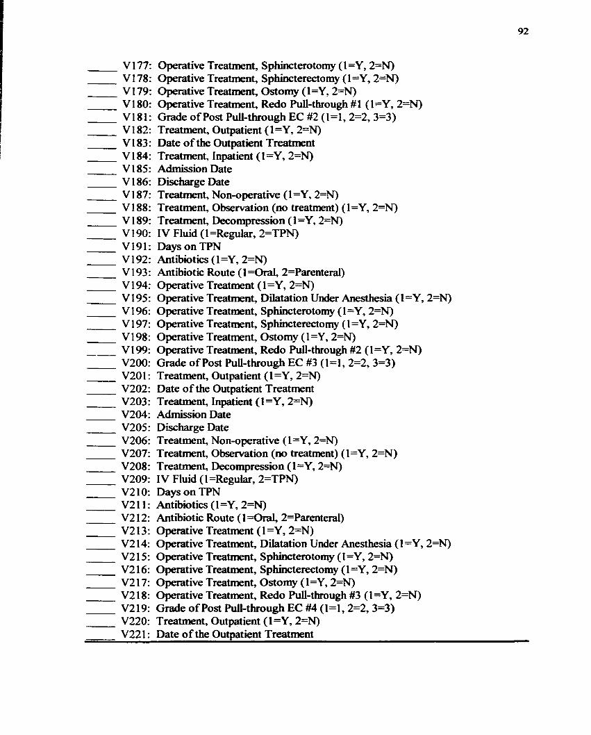
92
V177: Operative Treatment, Sphincterotomy (1=Y, 2=N)V178: Operative Treatment, Sphincterectomy (1=Y, 2=N)V179: Operative Treatment, Ostomy (1=Y, 2=N)V I80: Operative Treatment, Redo Pull-through#! (1=Y, 2=N)V I81: Grade o f Post Pull-through EC #2 (1=1, 2=2, 3=3)V I82: Treatment, Outpatient (1=Y, 2=N)V I83: Date o f the Outpatient Treatment V I84: Treatment, Inpatient (1=Y, 2=N)V I85: Admission Date V 186: Discharge DateV I87: Treatment, Non-operative (1=Y, 2=N)V I88: Treatment, Observation (no treatment) (1=Y, 2=N)V 189: Treatment, Decompression ( 1 =Y, 2=N)V190: IV Fluid ( 1 =Regular, 2=TPN)V191: Days on TPNV I92: Antibiotics (1=Y, 2=N)V I93: Antibiotic Route (l=Oral, 2=Parenteral)V I94: Operative Treatment (1=Y, 2=N)V195: Operative Treatment, Dilatation Under Anesthesia (1=Y, 2=N) V I96: Operative Treatment, Sphincterotomy (1=Y, 2=N)V I97: Operative Treatment, Sphincterectomy (1=Y, 2=N)V I98: Operative Treatment, Ostomy (1=Y, 2=N)V I99: Operative Treatment, Redo Pull-through #2 (1=Y, 2=N)V200: Grade o f Post Pull-through EC #3 (1=1, 2=2, 3=3)V201: Treatment, Outpatient (1=Y, 2=N)V202: Date o f the Outpatient Treatment V203: Treatment, Inpatient (1=Y, 2=N)V204: Admission Date V205: Discharge DateV206: Treatment, Non-operative (1=Y, 2=N)V207: Treatment, Observation (no treatment) (1=Y, 2=N)V208: Treatment, Decompression (1=Y, 2=N)V209: IV Fluid (l=Regular, 2=TPN)V210: Days on TPNV211 : Antibiotics (1=Y, 2=N)V212: Antibiotic Route (l=Oral, 2=Parenteral)V213 : Operative Treatment ( 1 =Y, 2=N)V214: Operative Treatment, Dilatation Under Anesthesia (1=Y, 2=N) V215: Operative Treatment, Sphincterotomy (1=Y, 2=N)V216 : Operative Treatment, Sphincterectomy ( 1 = Y, 2=N)V217: Operative Treatment, Ostomy (1=Y, 2=N)V218: Operative Treatment, Redo Pull-through #3 (1=Y, 2=N)V219: Grade o f Post Pull-through EC #4 (1=1, 2=2, 3=3)V220: Treatment, Outpatient (1=Y, 2=N)V221 : Date o f the Outpatient Treatment______________

93
V222: Treatment, Inpatient (1=Y, 2=N)V223: Admission Date V224: Discharge DateV225: Treatment, Non-operative (1=Y, 2=N)V226: Treatment, Observation (no treatment) (1=Y, 2=N)V227: Treatment, Decompression (1=Y, 2=N)V228: IV Fluid (l=ReguIar,2=TPN)V229: Days on TPN V230: Antibiotics (I=Y, 2=N)V231 : Antibiotic Route (l=Oral, 2=ParenteraI)V232; Operative Treatment (1=Y, 2=N)V233: Operative Treatment, Dilatation Under Anesthesia (l=Y, 2=N) V234: Operative Treatment, Sphincterotony (1=Y, 2=N)V235: Operative Treatment, Sphincterectomy (l=Y , 2=N)V236: Operative Treatment, Ostomy (1=Y, 2=N)V237: Operative Treatment, Redo Pull-through #4 (1=Y, 2=N)V238: Grade o f Post Pull-through EC #5 (1=1, 2=2, 3=3)V239: Treatment, Outpatient (l=Y, 2=N)V240: Date of the Outpatient Treatment V241: Treatment, Inpatient (I=Y, 2=N)V242: Admission Date V243 Discharge DateV244: Treatment, Non-operative (I=Y, 2=N)V245: Treatment, Observation (no treatment) (1=Y, 2=N)V246: Treatment, Decompression (1=Y, 2=N)V247: IV Fluid (l=Regular, 2=TPN)V248: Days on TPN V249: Antibiotics (1=Y, 2=N)V250: Antibiotic Route (l=Oral, 2=ParenteraI)V251 Operative Treatment ( 1 = Y, 2=N)V252: Operative Treatment, Dilatation Under Anesthesia (1=Y, 2=N) V253: Operative Treatment, Sphincterotomy (I=Y, 2=N)V254: Operative Treatment, Sphincterectomy (1=Y, 2=N)V255: Operative Treatment, Ostomy (1=Y, 2=N)V256: Operative Treatment, Redo Pull-through #5 (1=Y, 2=N)V257: Grade o f Post Pull-through EC #6 (1=1, 2=2, 3=3)V258: Treatment, Outpatient (1=Y, 2=N)V259: Date of the Outpatient Treatment V260: Treatment, Inpatient (I=Y, 2=N)V261: Admission Date V262: Discharge DateV263 Treatment, Non-operative (1=Y, 2=N)V264: Treatment, Observation (no treatment) (I=Y, 2=N)V265: Treatment, Decompression (I=Y, 2=N)V266: IV Fluid (l=Regular, 2=TPN)____________________________
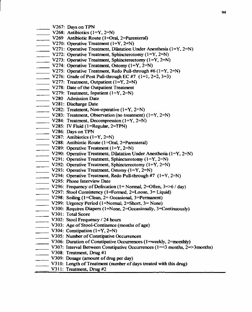
94
V267: Days on TPN V268: Antibiotics (1=Y, 2=N)V269 Antibiotic Route (l=Oral, 2=ParenteraI)V270: Operative Treatment (1=Y, 2=N)V271 : Operative Treatment, Dilatation Under Anesthesia (I=Y, 2=N)V272: Operative Treatment, Sphincterotomy (1=Y, 2=N)V273: Operative Treatment, Sphincterectomy (1=Y, 2=N)V274: Operative Treatment, Ostomy (1=Y, 2=N)V275: Operative Treatment, Redo Pull-through #6 (1=Y, 2=N)V276: Grade o f Post Pull-through EC #7 (1=1, 2=2, 3=3)V277: Treatment, Outpatient (1=Y, 2=N)V278: Date o f the Outpatient Treatment V279: Treatment, Inpatient (1=Y, 2=N)V280 Admission Date V281: Discharge DateV282: Treatment, Non-operative (1=Y, 2=N)V283: Treatment, Observation (no treatment) (1=Y, 2=N)V284: Treatment, Decompression (1=Y, 2=N)V285: IV Fluid (l=Regular, 2=TPN)V286: Days on TPN V287: Antibiotics (1=Y, 2=N)V288: Antibiotic Route (l=Oral, 2=Parenteral)V289: Operative Treatment (1=Y, 2=N)V290: Operative Treatment, Dilatation Under Anesthesia (1=Y, 2=N)V291 : Operative Treatment, Sphincterotomy (1=Y, 2=N)V292: Operative Treatment, Sphincterectomy (1=Y, 2=N)V293: Operative Treatment, Ostomy (1=Y, 2=N)V294: Operative Treatment, Redo Pull-through #7 (1=Y, 2=N)V295: Phone Interview DateV296: Frequency o f Defecation (1= Normal, 2=Often, 3=>6 / day)V297: Stool Consistency (l=Formed, 2=Loose, 3= Liquid)V298: Soiling (l=Clean, 2= Occasional, 3=Permanent)V299; Urgency Period (l=Normal, 2=Short, 3= None)V300: Requires Diapers (l=None, 2=Occasionally, 3=Continuously)V301: Total ScoreV302: Stool Frequency / 24 hoursV303: Age o f Stool-Continence (months o f age)V304: Constipation (1=Y, 2=N)V305: Number o f Constipative OccurrencesV306: Duration o f Constipative Occurrences (l=weekly, 2=monthly)V307: Interval Between Constipative Occurrences (1=<3 months, 2=>3months)V308: Treatment, Drug #1V309: Dosage (amount o f drug per day)V310: Length o f Treatment (number o f days treated with this drug)V311: Treatment, Drug #2 ______________ _________
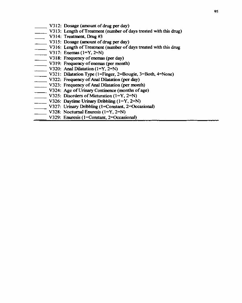
95
V312: Dosage (amount of drug per day)V313: Length o f Treatment (number o f days treated with this drug)V314: Treatment, Drug #3V315: Dosage (amount of drug per day)V316: Length o f Treatment (number o f days treated with this drug V317: Enemas ( 1 =Y, 2=N)V318: Frequency o f enemas (per day)V319: Frequency o f enemas (per month)V320: Anal Dilatation (1=Y, 2=N)V321 : Dilatation Type (l=Finger, 2=Bougie, 3=Both, 4=None) V322: Frequency o f Anal Dilatation (per day)V323: Frequency o f Anal Dilatation (per month)V324: Age o f Urinary Continence (months o f age)V325: Disorders o f Micturation (1=Y, 2=N)V326: Daytime Urinary Dribbling (1=Y, 2=N)V327: Urinary Dribbling (l=Constant, 2=Occasional)V328: Nocturnal Enuresis (1=Y, 2=N)V329: Enuresis ( 1 =Constant, 2=Occasional)