comparative treatment planning in radiotherapy and...
TRANSCRIPT

ACTAUNIVERSITATISUPSALIENSISUPPSALA2006
Digital Comprehensive Summaries of Uppsala Dissertationsfrom the Faculty of Medicine 117
Comparative Treatment Planningin Radiotherapy and Clinical Impactof Proton Relative BiologicalEffectiveness
JONAS JOHANSSON
ISSN 1651-6206ISBN 91-554-6184-Xurn:nbn:se:uu:diva-6593


To my family


List of publications
This thesis is based on the following papers, referred to in the text by their Roman numerals.
I. Johansson J, Isacsson U, Lindman H, Montelius A, Glimelius B. “Node-positive left-sided breast cancer patients after breast-conserving surgery: potential outcomes of radiotherapy modalities and techniques.” Radiother Oncol. 2002 Nov;65(2):89-98.
II. Johansson J, Blomquist E, Montelius A, Isacsson U, Glimelius B. “Poten-tial outcomes of modalities and techniques in radiotherapy for patients with hypopharyngeal carcinoma.” Radiother Oncol. 2004 Aug;72(2):129-38.
III. Tilly N, Johansson J, Isacsson U, Medin J, Blomquist E, Grusell E and Glimelius B, “The influence of RBE variations in clinical proton beams” Phys Med Biol. 2005 June 21;50(12):2765-77.
IV. Johansson J, Montelius A, Glimelius B, “Treatment planning compari-son of 3 dimensional-conformal radiotherapy, intensity modulated radiother-apy and proton beam radiotherapy for locally advanced rectal cancer” Manu-script.
I. “Reprinted with permission from Elsevier, Copyright (2002).” II. “Reprinted with permission from Elsevier, Copyright (2004).” III. “Reprinted with permission from IOP Publishing Ltd Copyright(2005).
http://www.iop.org/journals/pmb

Contents
Introduction.....................................................................................................9
Irradiation modalities and techniques ...........................................................10 Target volumes and risk organs................................................................10 3D-CRT and intensity modulated radiotherapy .......................................10 The potential of protons (and ions) ..........................................................13 Radiobiology of protons (and ions)..........................................................15 Clinical experience with IMRT and protons (and ions) ...........................17
Aims of the thesis..........................................................................................19
Treatment Planning Comparisons.................................................................20 Treatment planning systems.....................................................................28 Design of treatment planning comparisons ..............................................29
General considerations.........................................................................29 Physical dose-volume evaluation ........................................................29 Modelling treatment outcome with dose-response models..................30
Results and discussion ..................................................................................32 Left-sided node positive breast cancer (Paper I) ......................................32 Hypopharyngeal carcinoma (Paper II) .....................................................34 Locally advanced rectal cancer (Paper IV) ..............................................37 Potential RBE effects in proton treatment planning (Paper III) ...............39
Future perspectives .......................................................................................42
Conclusions...................................................................................................45
Acknowledgements.......................................................................................47
References:....................................................................................................49
Abbreviations
RT Radiation therapy CTRT Chemo-radiotherapy
3D-CRT 3 dimensional conformal radiotherapy IMRT Intensity-modulated photon radiotherapy MLC Multi-leaf collimator MIMiC Multivane intensity-modulating collimator DAO Direct aperture optimisation
IMPT Intensity modulated proton radiotherapy SOBP Spread out Bragg peak LET Linear energy transfer RBE Relative biological effect
GTV Gross tumour volume CTV Clinical target volume PTV Planning target volume OAR Organs at risk DVH Dose volume histogram CI Conformity index TCP Tumour control probability NTCP Normal tissue complication probability EUD Equivalent uniform dose
CNS Central nervous system AVM Arteriovenous malformation IMN Internal mammary nodes
CT Computed tomography MRI Magnetic resonance imaging PET Positron emission tomography

9
Introduction
The development and implementation of new irradiation techniques in radio-therapy (RT) is presently a fast growing field due to the availability of so-phisticated modalities such as intensity modulated radiotherapy with photons (IMRT), protons and light ions. At the same time treatment planning, meth-ods for fixation of the patient, compensation for patient movements and im-age-guided RT are developing. The dose distributions that can be obtained with protons, light ions and IMRT are generally superior to those obtained with three dimensional conformal radiotherapy (3D-CRT) techniques with photons, i.e. the techniques presently used as standard treatments at most sites world-wide. All improvements aim at delivering radiation dose more selectively and conformably adapted to the tumour volume so that high doses in volumes outside the tumour volume can be decreased, thereby spar-ing normal tissues. Alternatively, the improved selectivity can be used to increase the tumour dose and increase the tumour control while maintaining acceptable complication rates in the surrounding normal tissues. To take full advantage of the improved dose distributions using the new techniques, frac-tionation schedules and combinations with sensitising drugs must also be optimised.
Although the availability of improved treatment techniques with photons, protons and light ions increases, the important question remains whether or not the clinical outcome for the patients will be sufficiently improved. Long-term clinical experience using these more sophisticated techniques is still rare and higher treatment efficacy of sufficient extent to motivate the in-creased costs is still to be proven. In the absence of clinical evidence sup-porting new treatments, the outcome from a patient treatment could be simu-lated by treatment planning comparisons. The comparisons are then evalu-ated using physical dose measures and by applying dose-response models. The latter can estimate tumour control probability (TCP) and normal tissue complication probability (NTCP). From the results of dose-response model-ling and early clinical data, it is possible to get at least an idea of the poten-tial improvements that can be achieved. Sensitivity analyses can reveal the robustness of the estimates. An estimate of the potential number of patients eligible for treatment with IMRT, protons and light ions can also be ob-tained, as recently done for protons [60] and light ions [10,107,170].

10
Irradiation modalities and techniques
Target volumes and risk organs Three-dimensional imaging is fundamental for modern radiotherapy treat-ment planning. Different imaging techniques, such as computerised tomo-graphy (CT), magnetic resonance imaging (MRI) and positron emission to-mography (PET), provide a basis for definition of targets and risk organs as well as accurate dose calculations. In clinical practice a few important ana-tomical volumes are defined as prerequisites for treatment planning [83,84]. The primary tumour volume with the highest tumour cell density is defined as the Gross Tumour Volume (GTV). The Clinical Target Volume (CTV) is a volume, which includes the GTV, and in addition, a margin containing assumed subclinical, i.e. non-detectable disease. To account for organ movements and errors in patient set-up, a margin is applied around the CTV creating a Planning Target Volume (PTV). The dose delivered to the PTV represents the dose delivered to the CTV and ensures that the prescribed dose is delivered to the CTV [83,84]. This implies that the objective for the PTV also is the prescription dose. Normal tissues significantly affected by their radio-sensitivity, and thereby influencing the prescribed dose to the CTV, are defined as Organs at Risk (OAR).
3D-CRT and intensity modulated radiotherapy A 3D-CRT treatment plan is based on 3D imaging in the delineation of target volumes and OARs, and is delivered by combining a number of beams from different directions to irradiate the PTV. The aperture for each beam is shaped by a multileaf collimator (MLC) to be conformed with the projected contour of the PTV. The fluence of such a beam can be optimised using e.g. wedges or physical modulators (compensation filters). This optimisation is made manually by varying beam energy and beam weight or by applying beam modulators such as wedge filters. The constraints for a 3D-CRT plan-ning are generally stated in treatment protocols or individually set for each patient by the radiation oncologist.
Modern delivery techniques, using photons, may be summarised in the term intensity modulated radiotherapy (IMRT) [2,215,216]. IMRT with as-sociated computerised optimisation of fluence profiles is a technology that

11
has been developed dramatically since the end of the 1980s [18,24,29,217]. To obtain the desired dose distribution to the target and at the same time spare specific normal tissues, the fluence profiles generally have to be non-uniform. An interactive computer optimisation procedure calculates, for all the selected beam directions, the fluence profiles necessary to obtain the desired dose distribution within the patient. The optimisation algorithm uses dose or dose-volume objectives entered interactively in the calculation of the dose-distribution. Typical dose objectives are given as min/max dose (Gy) to the entire PTV or OARs, whereas dose-volume objectives are given as min/max dose (Gy) to partial volumes of a PTV or an OAR. Importance factors can be assigned to PTVs and OARs in order to improve the optimised result. Objective functions used in the optimisation process always provide results that correspond to the user-defined input in terms of the objectives. Therefore, user interaction, providing new improved objectives and impor-tance factors, is required to obtain a clinically feasible treatment plan.
A few different types of algorithms are usually used in the optimisation of IMRT plans. In the optimisation problem, the objective function is to be minimised. A commonly used class of optimisation algorithms is gradient based. These algorithms search the local neighbourhood for a minimum value of the objective function, and could end up in a local minimum instead a global minimum. However, it has been shown that many commonly em-ployed objective functions are convex, i.e. only have one minimum, and therefore the gradient methods are effective for optimisation [41]. Gradient based algorithms are commonly employed in commercial treatment planning systems, such as Helax TMS [70,71], Oncentra Masterplan with optimisation module from Raysearch Laboratories [81,126] and Konrad [169]. Another type of algorithm frequently used for optimisation of dose distributions is simulated annealing [217]. These algorithms are suitable also for non-convex problems. The simulated annealing optimisation was implemented in the first commercial treatment-planning optimisation program, CORVUS (NOMOS Corporation).
Two main IMRT delivery techniques can be recognised, viz. cone beam IMRT and tomotherapy. Cone beam IMRT uses standard C-arm linear ac-celerators equipped with MLCs. One technique, called step and shoot, builds up the fluence profiles from multiple static fields from each beam direction. Sliding window is a technique where dynamic movements of the MLC are used to obtain the fluence profiles from each direction. Theoretically, it of-fers higher resolution in obtained fluence profiles and a faster realisation, although the calculations of the movements of the MLC are more complex.
Tomotherapy can be described as slice or fan beam IMRT. With serial tomotherapy, one slice of the tumour is treated at a time with a series of gan-try angles up to a full rotation after which the patient is moved to treat the next slice. Serial tomotherapy was the first commercially available system for IMRT, using the multivane intensity-modulating collimator (MIMiC) as

12
an add-on to a conventional accelerator. MIMiC was used together with the CORVUS treatment planning system for integrated optimisation (NOMOS Corporation). MIMiC provides a fan beam divided into two thin slices, which can be fluence modulated during the treatment. Inspired by the spiral CT technology, Mackie [138] introduced spiral tomotherapy with a rotating linac using a similar collimator as the MIMiC. The main difference between serial and spiral tomotherapy is that in spiral tomotherapy the patient is moved continuously by the couch while being irradiated with the rotating linac, which gives a spiral irradiation pattern.
The non-uniform fluence profiles from each beam direction calculated with the optimisation techniques are delivered by using different settings for the MLCs. Thus, the fluence profiles must be converted into machine pa-rameters (segmentation). There are numerous algorithms published to per-form this conversion, e.g. [36,199,201]. However, an important objective in treatment planning is adequate dose coverage to the target volumes, which in most cases means homogenous dose within the target volume. In the seg-mentation process, the dose distributions may be degraded. To overcome this degradation, also the MLC settings can be directly involved in the optimisa-tion process e.g. [81,190]. This is called direct aperture optimisation (DAO), and this approach is now becoming available in treatment planning systems. As an example both these methods were used to optimise a breast cancer case with exactly the same beam configuration and objectives. The dose volume histograms (DVHs) are shown for the respective technique in Figure 1. In this specific case, a significant difference with respect to homogeneity between the dose distributions was obtained in favour of the DAO technique. Using fluence optimisation with segmentation, acceptable plans may still be obtained although the DAO method appears to be more robust.
Figure 1. DVHs for a) PTV and heart and b) left and right lung using either direct aperture optimisation (solid lines) or segmentation of optimised fluence profiles (dotted lines). The are the objectives set for the optimisation algorithm.

13
The potential of protons (and ions1)In 1946, Robert Wilson suggested that protons could be used for medical purposes [226]. In 1954, the first proton beam (340 MeV) was used to irradi-ate the pituitary gland at the Lawrence Berkeley Laboratory, California, USA. Between 1954 and 1992, 2054 patients were treated, the first 30 with protons and the remaining ones with helium ions [118]. This centre was fol-lowed in the 1950s and 60s by other centres: the Gustav Werner Institute (GWI), Uppsala, Sweden, the Harvard Cyclotron Laboratory, Boston Massa-chusetts USA, Dubna and Institute of Theoretical and Experimental Physics (ITEP), Moscow, Russia. The proton beam from the synchrocyclotron at theGWI was developed both for neurosurgery [111,115] and for large-field proton treatments [44,63,108,203]. The first medically dedicated proton beam facility was built at Loma Linda University in California, USA in 1990 [195]. As of July 2005, there are 23 active proton centres and 3 centres using C ions [192]. In total 43000 patients have been treated with protons and 4500 patients with light or heavy ions Figure 2. Currently a number of facili-ties are planned, 15 with protons only, 4 combining protons and ions, and one with C-Ar ions.
Figure 2. The total number of patients treated with heavy charged particles. (Total number treated , protons , other heavy charged particles ).
1 In this thesis, particles with atomic number Z = 2-6 will be considered as light ions and Z 7 as heavy ions.

14
The positively charged hydrogen ions or protons deposit energy in matter by Coulomb interactions with electrons and nuclei, by bremsstrahlung energy loss, and by nuclear reactions. These interactions lead to a slowing down of the primary protons. The dominant effect of energy loss is Coulomb interac-tion with the atomic electrons. As the protons penetrate into the material the absorbed dose increases gradually and near the end of the range the dose increases rapidly (shaded light grey curve in figure 3). This peak in the dose distribution was discovered by Bragg [22] and bears his name. The absorbed dose is proportional to the energy deposition per unit path length, i. e. the stopping power. The stopping power increases as the proton energy de-creases until very little energy remains. The dose distribution has a sharp fall-off near the maximum range, as all protons have approximately the same range due to the small fluctuations in energy loss over the path (range strag-gling). As the protons are much heavier (approximately 1835 times) than the electrons, the multiple scattering by the atomic electrons will lead only to small lateral deviations along the path. Proton beams will therefore have relatively sharp penumbras. The penumbra will increase with depth and at 15-20 cm in tissue, it will be comparable to those of high-energy photon beams.
The mono-energetic proton beam extracted from the accelerator is a nar-row pencil beam with a sharp Bragg-peak. This beam cannot provide a uni-form dose to a target of any significant size. Therefore, different methods to spread out the Bragg peak both laterally and in depth are implemented. The simplest method to spread out the beam laterally to a useful size is to use high atomic number scattering foils, the so called passive scattering method [65,108,142]. A proton beam passing through a single scattering foil shows a Gaussian fluence profile [65,142]. Therefore, to achieve larger field sizes, techniques using double scattering foils are used [17,26].
An alternative method to passive scattering is the use of active scanning of the narrow proton pencil beam by means of magnetic deflection. The use of scanning to achieve large uniform treatment fields was first implemented by the Uppsala group, where scanning magnets produced diamond patterns of variable size to cover the treatment field [109]. The narrow proton beam can also be scanned in a raster pattern or in concentric rings to produce uni-form fields of variable size [96,172]. Dynamic scanning minimises energy losses and the maximum useful range is preserved.
The sharp Bragg-peak of the proton beam must be modulated in range in order to give a homogeneous dose to an extended volume. In a passively spread out beam this is done by using a rotating stepped absorber [103], a ridge filter [97] or a spiral ridge filter [99,100,104]. The range is shifted in discrete steps and an almost flat extended spread out Bragg-peak (SOBP) can be obtained. In Figure 3, the Bragg peak of a 173 MeV proton beam has been modulated in 10 steps to create a uniform dose over the last 54 mm of the depth dose curve. This technique for passive modulation can only pro-

15
duce a uniform range modulation (constant SOBP) throughout the entire portal, and the extent of the SOBP is determined by the maximum size of the target.
In Figure 3, a mono-energetic and range modulated proton depth dose dis-tributions are compared to a 6 MV photon beam. Photons interact with mat-ter in rare single events and the photon depth dose curve is characterised by an exponential decrease with depth. The figure shows that photons give much higher dose both on the entrance and exit sides of the target (repre-sented by the SOBP) as indicated with dark shaded areas. The proton ener-gies required to reach any target location in the human body would be in the range of approximately 70-250 MeV corresponding to a range in depth of approximately 4-35 cm. The range for a passively scattered therapy beam is adapted to the distal end of the target volume by applying a range compensa-tor or bolus. This filter is machined such that the maximum range is con-formed to the shape of the distal surface of the target.
With actively scanned pencil beams it is possible to deliver the beam even more selectively than with passively scattered beams. With a scanned pencil beam, the modification of the Bragg-peak is accomplished by varying the absorber thickness or energy of the incident beam over the beam portal. The use of scanned beams to vary the range modulation allows the high dose region of the SOBP to be conformed to both the distal and proximal target extension [27,33,127]. This 3D computer controlled pencil beam scanning is, in analogy with photon IMRT, called intensity modulated proton therapy or IMPT. Depending on the time structure of the proton beam from the accel-erator, IMPT can be delivered in different ways [127]. The dose from the pencil beam can be deposited as a sequence of static applications (discrete spot scanning) [95,167] or as a continuous scanning of the pencil beam, on a raster pattern, throughout equal-range layers (raster scanning) [82,132].
Radiobiology of protons (and ions) The biological effects of a proton beam are close to those of photons. This implies that most of the knowledge from photon therapy can be applied also to protons, with respect to treatment protocols. However, protons have a slightly higher relative biological effectiveness (RBE) compared to photons and electrons and in general, a clinical RBE of 1.1 (with 60Co used as refer-ence) is applied over the entire SOBP [8,68]. As the proton energy decreases at the end of the range, the linear energy transfer (LET) increases, and thus also the RBE. Gerweck and Kozin [59] reviewed RBE data from several studies and found RBE values between 1.1 and 1.6 with a tendency that RBE increases with depth of the beam. Early researchers also made similar obser-vations [45,110]. Figure 4 shows the results of a RBE corrected depth dose curve, where also the calculated RBE is shown as a function of proton range.

16
The calculation was made by averaging the LET at each depth for all the individual Bragg peaks contributing to the dose. An effect of the RBE cor-rection is that the effective range of the beam increases by a few millimetres, which may be important in certain applications. The RBE corrected dose was calculated using the linear-quadratic cell survival model with tissue specific parameters for cell survival as a function of LET (Paper III). Un-certainties in the in vitro and the clinical in vivo data are still large, but until these are resolved, a clinical RBE of 1.1 is used [68].
Figure 3. Central-axis depth dose curves for one mono-energetic 173 MeV proton beam (light grey), one spread out Bragg peak based on the mono-energetic 173 MeV proton beam and 6 MV photon beam (dark grey).
Compared to protons, light ions have an even higher LET and RBE in the Bragg peak region while the RBE is still fairly low in the entrance region [28]. The heavy ions have not only higher RBE in the Bragg peak but also, in the entrance region of the beam which can introduce too much normal tissue damage [28]. Well oxygenated tumours are less radio-resistant than hypoxic tumours and an argument for light ions compared to protons is that the treatment of tumours containing many hypoxic cells would be improved [25].

17
Figure 4. Central-axis depth dose curves for one spread out Bragg peak built from the mono-energetic 173 MeV proton beam and one corrected for RBE (lower solid line not corrected for RBE, upper solid line corrected for RBE), grey indicate exces-sive dose). The RBE correction for the corresponding 173 MeV proton beam is also shown (dashed lines).
Clinical experience with IMRT and protons (and ions) The clinical outcome of IMRT has so far mainly been reported from early clinical trials, and no definite trials directly revealing superiority over 3D-CRT have been published. The studies have typically concerned the feasibil-ity of the techniques, investigating toxicity and presenting only preliminary, however promising, results of efficacy. The most common tumour sites so far treated with IMRT have been various tumours in the head and neck re-gion [7,20,40,43,76,112,154,165,228], and prostate cancer [40,90,228]. Also lung cancer [14,73,188], gynaecologic malignancies [14,73,188] and miscel-laneous cancer sites [79,141,186] have been treated with IMRT. The experi-ence using IMRT has been summarised in many recent reviews e.g. [12].
For protons and ions, several studies have been published providing re-sults concerning local control, survival and toxicity. The situation for pro-tons is otherwise similar to that of IMRT, i.e. there are no randomised trials, with only one exception, for prostate cancer [233]. Skull-base chordomas and chondrosarcomas have been treated with doses between 60-79 CGy with protons [80,157,176,220] and carbon ions [187]. Local control is generally

18
higher for chondrosarcomas (90-100%) compared to chordomas (76-87.5%) [80,157,176,187,220]. For meningeomas, high tumour control and an overall survival between 90-100% have been reported [66,67,156,219]. In the treat-ment of arteriovenous malformations in the brain, the relations between dose and volume are very important, and protons may provide particular benefits for the larger malformations not possible to optimally treat with any other method [119,191,202,223]. Protons have also been extensively used in the treatment of intraocular melanomas, particularly when large and centrally located, due to the favourable dose distribution of the proton beam [42,120,155]. There are also several other tumour types investigated using both protons and carbon ions, e.g. lung cancer [16,31,105,149,152] and pros-tate cancer [88,194,233]. An overview of studies using charged particles, particularly protons was recently published in a special issue of Acta On-cologica (Vol 44, No 8, 2005).

19
Aims of the thesis
The overall aim of this thesis is to investigate the potential clinical advan-tages of various techniques and modalities in radiotherapy. A primary aim is to investigate the potential advantages of protons and IMRT compared to conventional 3D-CRT. The comparisons are based on calculated physical dose distributions for the respective technique. Whenever possible, dose-response models have been used to predict the outcome of patient treat-ments. Three tumour locations have been selected in the comparisons, left-sided node positive breast cancer, hypopharyngeal carcinoma, and rectal cancer. In all locations, locally advanced tumours were selected. A secon-dary aim has been to investigate the potential clinical relevance of the slightly increased relative biological effectiveness at the end of the proton range.

20
Treatment Planning Comparisons
Most of the early clinical trials investigating the use of IMRT, protons, and other ions show encouraging results both regarding efficacy and toxicity. Large investments in new facilities, and knowledge of these new techniques should be based upon proven superiority of one technique compared to oth-ers. In the long term perspective large prospective and controlled clinical trials are needed for evidence based assessment of the various techniques and modalities. As the availability of IMRT, proton and ion beam therapy increases, it will be easier to initiate the necessary clinical studies comparing the different techniques. To initiate such clinical studies, an indication of the potential magnitude of benefit from a certain technique is valuable when designing the study. To correctly design a clinical trial, the difference in the expected outcome of the different techniques must be approximated in either absolute or relative terms. However, the number of facilities (IMRT, protons or ions) available for treating large series of patients required to claim the superiority of one technique over another is still limited. It could be dis-cussed why conclusive evidence from trials is still lacking; despite over 40000 patients have been treated with charged particles. This is not unique for charged particles, but is also seen for many other sophisticated new treatment modalities [139]. The investments in new technology today are sometimes more based on encouraging results from preliminary evidence than solid evidence from trials. However, concerning radiation therapy, model treatment planning studies contribute to the knowledge about poten-tial gains. In treatment planning studies, patient treatments are compared using calculated dose distributions for the various techniques.
Treatment planning comparisons have also been used to compare differ-ent techniques and modalities in attempts to predict differences in treatment outcome for a patient. The studies have been comparing dose volume statis-tics for tumours/targets and OARs. Most treatment planning comparisons are made using calculated dose distributions for various techniques and the re-sults generally show superiority for protons and light ions compared to IMRT. Some of the treatment planning studies have also included attempts to predict the outcome by using dose-response models that give an estimate of TCP and NTCP. In this thesis, both comparisons of dose-distributions and dose-response models have been used to compare protons with IMRT and protons and IMRT with 3D-CRT. Table 1 shows a list of published treatment planning comparisons, where protons have been included as one part of the

21
comparison. There is a large variety in how the studies were performed. The early studies compared 3D-CRT (or a conventionally given standard treat-ment) and protons, neither IMRT nor IMPT were then available. Several of the newer comparisons have included both IMRT and IMPT, but only com-parably few studies have estimated the possible clinical outcome using dose-response models. The number of patients included in the studies also varies significantly, actually, most of them only included one patient.

Table 1. Comparative treatment planning studies (Reproduced from [60], with permission)
Photons Protons Comments Reference Year Tumour type Number of patientsplanned 3D-CRT IMXT Passive Scanned
Suit et al [205] 1988 Cervical cancer 1 X X Better dose distributions with improved local control, less toxicity
Brown et al [30] 1989 Nasopharynx 2 X X Better dose distributions with improved local control, less toxicity
Urie + Gotein [211] 1989 Chordoma/ chondrosarcoma
12 X X X Variably (intensity) modulated protons reduce dose to normal tissues (integral dose by 3 – 12%-units) compared to fixed (SOBP) protons, however, the largest difference was between protons and photons (2 patients)
Austin-Seymour et al [9]
1990 Skull base 1 X X Less dose to OARs, e.g. the optic nerve
Austin-Seymour et al [9]
1990 Prostate 1 X X Less dose to OARs
Tatsuzaki et al [209] 1991 Rectum 1 X X Reduced dose tom small bowel using protons
Archambeau et al [5]
1992 Thalamic pediatric astrocytoma
1 X X Improved dose distribution, lower normal brain dose, higher tumour dose possible

Gademann & Wan-nenmacher [55]
1992 Pediatric retroperito-neal tumours
1 X X Better dose localization, less second cancers
Levin [117] 1992 Para-aortic nodes, cervix cancer
1 X X Higher doses could be reached using protons, improved tumour control by 10 – 20%
Miralbell et al [144] 1992 Maxillary sinus 1 X X Less dose to OARs using a proton boost
Slater et al [196] 1992 Tonsil 2 X X Superior dose distributions, higher tumour doses, less doses to OARs (chiefly mandible parotic glands)
Smit [197] 1992 Cervical cancer 1 X X Higher doses (by 20%) could be reached using protons, 40% increase in tumour control
Tatsuzaki et al [208] 1992 Glioblastoma 1 X X Less dose to non-target brain using protons
Wambersie et al [212]
1992 Pediatric brain tumours
3 X X Less dose to non-target brain using protons
Miralbell & Urie [148]
1993 Large AVM 1 X X Less dose to non-target brain, brain stem and optic chiasm using protons
Lee et al [114] 1994 Prostate 12 X X Distinctly reduced rectal NTCP using protons in one-third of the cases, minimal gain in the remaining
Isacsson et al [87] 1996 Rectum 6 X X At 5% NTCP any organ, TCP is increased by 14%-units with protons

Isacsson et al [85] 1997 Ewing/paraspinal 1 X X At 1% NTCP in spinal cord, TCP in increased by 5%-units
Miralbell et al [145] 1997 Medulloblastoma-supratentorial target
1 X X X Better sparing normal tissues with protons and IMXT compared to conventional with less IQ-reduction
Miralbell et al [147] 1997 Medulloblastoma-spina techa target
1 X X X Decreased dose to all critical organs using protons
Sandison et al [179] 1997 Chest wall 1 X X Less lung dose using protons
Isacsson et al [86] 1998 Oesophagus 5 X X At 5% NTCP in any organ TCP is increased by 20%-units (from 2 to 25%) with protons
Verhey et al [222] 1998 CNS 5 X X Less dose to normal brain
Fuss et al [53] 1999 Optic nerve, gliomas 7 X X CI 2.9 photons, 2.3 protons, larger differences in larger tumours
Glimelius et al [61] 1999 Sacral chordoma 1 X X Lower doses to rectum and urinary bladder using one proton beam compared to 3D-CRT photons
Lee et al [113] 1999 Lung 13 X X More patients could be treated to higher tumour doses using protons compared to any photon technique
Lomax [127] 1999 Nasopharynx 1 X X Intensity modulation show advantages when few beams are used

Lomax et al [129] 1999 Various 9 X X X Reduced medium to low dose for protons compared to IMXT
Fuss et al [54] 2000 Pediatric optic nerve glioma
7 X X Reduced NTCPs, likely clinically significant for cognitive impairment
Lin et al [122] 2000 CNS, pediatric fossa 9 X X Protons result in increased normal tissue sparing, e.g. the cochlea (25% of dose compared to 75% of prescribed dose)
Miralbell et al [143] 2000 Orbital and paraorbi-tal
4 X X Similar PTV coverage, lower integral doses to OARs (x1.5-1.9), predicted NTCPs (severe late tox) similarly low
Oelfke + Bortfeld [158]
2000 - X X X IMPT advantages to SOBP protons and IMXT in a theo-retical study, integral dose 30% lower using IMPT vs SOBP, a factor 2-3 vs IMXT
Paulino et al [166] 2000 Medulloblastoma 5 X X Lower doses to all risk organs
Smith et al [198] 2000 Multiple sites 10+ X X X X Improved clinical outcomes at all sites, reduced NTCPs/higher TCPs
Zurlo et al [234] 2000 Pancreas/biliary 4 X X X Protons allowed delivery of planned dose in all patients, not or barely possible with photons
Baumert et al [11] 2001 CNS 7 X X For complex PTV shapes and when PTV close to critical organs, protons yield better dose distributions than photons for SRT
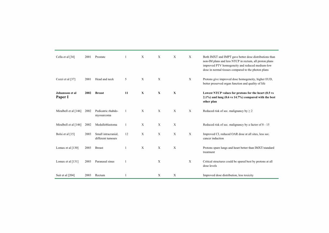
Cella et al [34] 2001 Prostate 1 X X X X Both IMXT and IMPT gave better dose distributions than non-IM plans and less NTCP in rectum, all proton plans improved PTV homogeneity and reduced medium-low dose in normal tissues compared to the photon plans
Cozzi et al [37] 2001 Head and neck 5 X X X Protons give improved dose homogeneity, higher EUD, better preserved organ function and quality of life
Johansson et al Paper I
2002 Breast 11 X X X Lowest NTCP values for protons for the heart (0.5 vs 2.1%) and lung (0.6 vs 14.7%) compared with the best other plan
Miralbell et al [146] 2002 Pedicatric rhabdo-myosarcoma
1 X X X X Reduced risk of sec. malignancy by 2
Miralbell et al [146] 2002 Medulloblastoma 1 X X X Reduced risk of sec. malignancy by a factor of 8 - 15
Bolsi et al [15] 2003 Small intracranial, different tumours
12 X X X X Improved CI, reduced OAR dose at all sites, less sec. cancer induction
Lomax et al [130] 2003 Breast 1 X X X Protons spare lungs and heart better than IMXT/standard treatment
Lomax et al [131] 2003 Paranasal sinus 1 X X Critical structures could be spared best by protons at all dose levels
Suit et al [204] 2003 Rectum 1 X X Improved dose distribution, less toxicity

Johansson et al Paper II
2004 Hypopharynx 5 X X X Protons give lower non-target doses compared to 3D-CRT/IMXT. NTCP parotid glands 40 – 43% protons, 51 – 65% IMXT, 93+% 3D-CRT
Mock et al [150] 2004 Paranasal sinus 5 X X X Similar CI but reduced doses to OAR (by 60%) and inte-gral doses using protons
St Clair et al [200] 2004 Medulloblastoma 1 X X X Substantial normal tissue sparing, e.g. to the cochleas and the heart
Weber et al [221] 2004 Paraspinal sarc 5 X X Similar conformity, reduced integral dose to OARs, dose escalation to 93 CGE possible with protons
Yoch + Tarbell [231]
2004 Pediatric, CNS 2 X X X Better dose homogeneity and conformity
Krengli et al [106] 2005 Retinoblastoma 3 X X Protons can achieve significant lens sparing and reduced risk of second malignancies
Mu et al [153] 2005 Medulloblastoma 5 X X X Risk second cancer conv RT 18%, IMXT 28%, IMPT 4%
Johansson et al Paper IV
2006 Rectal Cancer 16 X X X 50% reduction of NTCP using IMXT or protons. If the small bowel is a serial organ, 50% reduction using IMXT and 90% using protons if it is more parallel

28
Treatment planning systems The main treatment planning system (TPS) used in this thesis is the Helax-TMS (Treatment Management System, NUCLETRON, Uppsala, Sweden). The system provides algorithms for photons, electrons and protons, making it suitable for treatment planning comparisons. The dose calculation algo-rithms in the TPS have been described in detail elsewhere [4,177,178]. For the proton calculations in Paper I, II, and III, the 180 MeV clinical beam at the T Svedberg laboratory has been used. Monte Carlo calculated proton beams with higher energies (200 and 230 MeV) were also introduced into the TPS [101] since the range of the 180 MeV proton beam was not suffi-cient for the most deep-seated targets (Paper IV).
A gradient based optimisation algorithm for IMRT is available in the TPS with various modifications in different versions of TMS [70,71]. These algo-rithms were used in Papers I and II. The optimised fluence profiles from Helax-TMS IMRT are segmented into MLC settings a predefined number of times during the optimisation and this may influence the dose distribution negatively. In Paper IV Oncentra Masterplan 3.0 (NUCLETRON) with an IMRT algorithm from Raysearch Laboratories was used [81,126]. This algo-rithm uses a direct aperture optimisation (DAO).
In Helax TMS two different proton algorithms are available, a depth penetration algorithm and a pencil beam kernel algorithm [177,178]. The differences between the algorithms are in the accuracy and the calculation time. The depth penetration algorithm is fast and therefore suitable for inter-active treatment planning, where beam direction and proton range are opti-mised manually. The dose prediction accuracy of depth penetration algo-rithms is limited, particularly in the vicinity of heterogeneities [74,168,182,184]. For the passively scattered proton beams, a range modula-tor with suitable SOBP is applied to cover the entire target with homogenous dose for each beam direction. The planning of the scanned beam means, firstly, a calculation of range compensation filters, defining the maximum range of the proton beam, and, secondly, a calculation of the modulation range which varies over the beam portal allowing the SOBP to be conformed to both the distal and proximal target extension.
The calculated range compensation filter often needs manual adjustments. For the final dose calculation the more accurate pencil kernel algorithm is used [177,178]. Several other similar pencil beam kernels algorithms have been published [74,168,182,184,207]. For target volumes with complicated shapes the planning becomes an iterative process where the plans must be evaluated with slightly modified range compensation filters. Full 3D IMPT is not available in Helax-TMS, and therefore it has not been used in any of the comparisons in this thesis.

29
Design of treatment planning comparisons General considerations In this thesis, the treatment planning studies have had the following design:
1. In all studies, protons and IMRT were compared to conventional plan-ning (3D-CRT) used in the clinic today. The objectives were the same concerning dose homogeneity, i.e. as homogenous dose as possible to the targets.
2. Targets/tumours for several patients were planned to simulate the ana-tomical variability between patients with the same disease. Five or more patients were used in the investigations.
3. The evaluations included physical dose-volume information for the tu-mour and relevant risk volumes.
4. Dose response models were used to investigate the potential clinical outcome of the treatment. For all studies NTCP and, for one study, TCP was calculated. Since input parameters of these models suffer from un-certainties, different approaches for sensitivity analyses were made both for the TCP and for the NTCP calculations.
Physical dose-volume evaluation In all treatment planning studies, the dose volume histograms for the plan-ning target volume (PTV) were used to describe the dose given to the tu-mour. ICRU 62 [84] recommends that dose distributions are normalised to a reference point within the PTV to ensure a precise dose delivery. This is not always suitable for treatment planning comparisons, particularly if the dose distributions are inhomogeneous, which is the case for the IMRT plans. Therefore, the mean dose to the PTV was used as normalisation in the com-parisons.
One way of evaluating the dose coverage in target volumes or dose bur-dens to OARs is to calculate volumes receiving more dose than a specified dose level. Such volumes can be extracted from DVHs. These volumes are typically denoted as Vdose and the volumes and dose levels can be given ei-ther in absolute or in relative terms. Other measures that can be calculated from DVHs are e.g. mean and integral doses, and efficiency factors that sometimes can be useful for comparisons. Significant maximum and mini-mum doses should, however, be explored directly in the dose distributions since information on position and extent of each maximum or minimum will be lost in a DVH. Several conformity indices have also been suggested [84,102,214] as a measure of how well a dose distribution is shaped around the PTV.

30
Modelling treatment outcome with dose-response models Several models for calculating tumour control have been proposed (e.g. [23,123,137,218]). The basic model used in Paper I, II, III and IV used to determine the tumour and normal tissue response was the linear Poisson model or the equivalent linear quadratic Poisson model [123]. This model was selected since input parameter data have recently become available for many risk organs. The probability for controlling a tumour or induce a cer-tain injury to an organ is given by
)2lnln(50/ eDndeeeDP (1)
The tumour control probability for a heterogeneous tumour can then be cal-culated from:
N
i
vi
iDPTCP )( (2)
The normal tissue complication probability according to the relative seriality model [94] is defined as
sN
i
si DPNTCP
1
)(11 (3)
where D50 is defined as the dose for which the response is 50%, and is de-fined as the maximum normalised value of the dose response gradient and s is a relative seriality parameter describing the structure of the organs (serial or parallel). The parameters D50, and s are determined for a fixed fraction size, usually 2 Gy per fraction. The dose input to these models then requires fraction sizes equivalent to 2 Gy. Therefore, every dose step in a DVH must be recalculated to represent a 2 Gy equivalent dose. The main approach was to use every single dose step as input to the models. The consequence of this is that TCP values were calculated as a worst case value, since the lowest TCP value determines the level of tumour control (Paper II). The uncertain-ties in the model parameters may be large, and therefore sensitivity analyses of TCP and NTCP calculations were made in selected cases to investigate to what extent the parameter uncertainties will influence the predicted outcome for different treatment techniques.
In the rectal cancer Paper (IV) the main OAR is the small bowel. No up-dated parameter data for the small bowel have been published recently. Ga-gliardi et al [57] derived a set of parameter data for D50, , and s for cardiac

31
mortality based on clinical data. In that study clinical data for a given risk was available, but the input DVHs were not. The approach used then was to simulate a number of model patients with the treatment technique used and derive new DVHs to be used for modelling. The parameter values for D50, ,and s values could then be derived using the average DVH of the model pa-tients and the risk for cardiac mortality as input to the dose response model. Other methods use DVHs from individual patient treatments and each pa-tient is followed to obtain specific set of parameters for a selected endpoint [56].
Here a simple model is described to obtain approximate values for D50, ,and s for the linear quadratic Poisson dose response model from a set of model patients using their average DVH. The idea is to derive D50 from rele-vant data of , s, and / . Common values of are usually between 1 and 4, and s is defined by the structure of the organ and must be between 0 (paral-lel) and 1 (serial). / is either usually low (3 Gy) for late responding tissue while for acute responding tissues it is high (10 Gy). Within these limits, a parameter space can be derived for D50 with corresponding values for , and s for specific clinical endpoints with an associated NTCP value. This method was applied in Paper IV to derive D50 as a function of and s for the acute endpoint (diarrhoea) and for the late endpoint (obstruction, ileus), see Figure 5. In these two cases, D50 is nearly constant independent of the value of .Evaluating NTCP for various treatment techniques should then be made for the entire parameter space.
Figure 5. Parameter space for late and acute toxicity for rectal cancer. D50 plotted as a function of and s.

32
Results and discussion
Left-sided node positive breast cancer (Paper I)Loco-regional radiotherapy after mastectomy or breast-conserving cancer surgery decreases loco-regional recurrence rates with a factor three or more [1], and result in a small but definite increase in survival. After radiotherapy, the risk for lung toxicity is about 10% [125], and it correlates well with a dose level of 20 Gy to 30% of the volume. The dose to the heart and the lungs varies depending on how many of the internal mammary nodes (IMN) are included in the target volume, i.e. how far distally along the chain the target extends [181]. Early studies showed a markedly increased risk of se-vere cardiac mortality, up to 7% after 10 years [57], whereas later studies have shown less risk [160,161]. Loco-regional RT of breast cancer where also the IMN are included in the target volume is complex with difficulties to spare volumes of lung and heart and requires advanced techniques to achieve satisfactory dose distributions [91,124]. Several authors have sug-gested tangential plan optimisation with the objective to obtain a more ho-mogenous dose distribution [35,75,98,171]. One report deals with clinical implementation of IMRT for breast cancer [38]. Sandison suggested a proton arc technique aiming at minimising dose to lung tissue [179]. Three planning comparisons between protons and photons for breast cancer have been per-formed. One study, reported by Fogliata et al [47], did not include the lymph nodes, whereas the two other studies did (including the IMNs) (Paper I and [130]). They concluded that it is possible to achieve the same target homo-geneity or spare the surrounding tissues to about the same extent with either of the techniques, but that only protons could result in both a homogeneous dose within PTV and low doses to surrounding tissues.
In Paper I, eleven consecutive left-sided stage II (UICC stage T1–2 N1–2 M0) breast cancer patients were included in the comparison. The target vol-ume included the breast parenchyma, and the lymph nodes in the supraclavi-cular fossa, the axilla and the parasternal region (IMN). The treatment plan-ning was performed with four different techniques, one tangential (3D-CRT) and one patched technique with photons and electrons (3D-CRT), IMRT and protons. All plans in Paper I including IMRT were made with Helax-TMS. The dose given to the tumour volumes was assumed to be appropriate for controlling the disease and the same TCP can thus be expected for all the techniques. The difference between the plans is mainly due to a reduction of dose to the left lung and the heart. The results, in terms of NTCP (Table 2),

33
clearly show an advantage using protons as compared to the other tech-niques.
During the past few years, IMRT has been tuned to yield even better treatment plans than was possible to achieve in Paper I. Therefore, all IMRT plans have been replanned in Oncentra Masterplan 3.0 (OM3.0) using DAO (Figure 6). The main objectives for the lung and heart were taken from the IMRT algorithm comparison by Fogliata [48] which had a similar target definition, including also IMN, but a stronger homogeneity constraint was set for the PTV. In order to push the more powerful OM3.0 optimiser to its limits, other objectives than those used with the old TMS optimiser, had to be used. The results from the OM3.0 optimisation are similar to the results reported by Fogliata [48].
In the new IMRT optimisation, a more homogeneous dose distribution is achieved, and less dose is given to both the lung and heart. The calculated risk for cardiac mortality decreased to nearly 1% and the risk for radiation induced pneumonitis decreased from 28.2% to 2.5% by using OM3.0 com-pared to the old TMS optimisation (Table 2).
Figure 6. Two IMRT treatment plans optimised with TMS 5.0 (left) and OM3.0 (right). Note that dose to the left lung is much lower using the OM3.0 optimised plan.
Breast cancer is a large group of patients and the size of the subgroup that would require these complex treatments in order to have very low risks of late toxicity can still be considerable. Comparing the different techniques, protons appear superior with very low NTCP values for both the heart and lungs. However, the differences in NTCP are small when comparing with a new and more powerful IMRT optimiser. Some patients may still benefit sufficiently from protons compared to IMRT. This fraction can not be esti-mated with any certainty. Other factors may also be of relevance with pro-tons, such as a higher skin dose or a lower integral dose with less risk of late radiation–induced secondary malignancies.

34
Table 2. NTCP values for heart and lung for the 4 techniques used in Paper I and a new improved IMRT technique. Abbreviations: PTV = planning target volume; IMRT = intensity-modulated radiotherapy, V105, V95, V90, = partial volume (%) receiving more than 105, 95, 90 dose. Daverage = average dose in volume, NTCP = normal tissue complication probability.
Protons IMRTTMS Patched Tangential IMRTOM3.0
PTV V90 (%) 97.5 ± 1.7 93.3 ± 1.6 92.7 ± 1.7 98.5 ± 0.9 98.7 ± 0.8 V95 (%) 94.0 ± 2.7 85.9 ± 2.5 84.4 ± 2.7 93.2 ± 3.2 92.2 ± 4.1 V105 (%) 8.6 ± 8.3 22.5 ± 3.5 24.7 ± 5.7 11.1 ± 4.7 10.5 ± 4.6 Heart NTCP 0.5 ± 0.5 2.2 ± 1.7 2.1 ± 1.1 6.7 ± 2.7 1.3 ± 0.4 V35Gy (%) 1.4 ± 1.0 11.0 ± 7.6 10.5 ± 4.6 19.3 ± 6.6 9.0 ± 2.3 V45Gy (%) 0.8 ± 0.8 2.2 ± 2.2 2.3 ± 2.0 13.1 ± 6.1 0.2 ± 0.1 Lung NTCP 0.6 ± 0.8 28.2 ± 9.7 14.7 ± 10.2 28.3 ± 15.9 2.5 ± 0.6 V20Gy (%) 20.7 ± 9.1 65.6 ± 9.2 45.5 ± 9.0 49.1 ± 8.9 22.9 ± 0.7 V25Gy (%) 17.4 ± 7.9 55.9 ± 9.4 40.0 ± 8.9 47.3 ± 9.7 18.6 ± 0.7 Daverage (Gy) 9.8 ± 3.8 27.5 ± 2.8 21.7 ± 4.0 24.5 ± 4.5 17.2 ± 0.7
Hypopharyngeal carcinoma (Paper II) For patients with locally advanced head and neck (H&N) cancers stage T3-4N0, treated with a standard schedule of 70 Gy given in 35 fractions, a local control of about 40-50% is obtained after 2 years [52,78,159]. A local recur-rence at the primary site is a common reason for failure [92] and much effort is spent to increase local control. Several studies have shown an increase in local control and disease specific survival by using accelerated or hyperfrac-tionated treatment schedules. There is, however, little difference in overall survival between the different schedules [232]. The randomised trials have used 3D-CRT planning to investigate the effects of the altered regimens. The use of moderately accelerated or hyperfractionated schedules have not intro-duced more late toxicity [52,78,159], whereas more aggressive regimens have resulted in unacceptable late toxicity [89]. The risk for acute toxicity, particularly confluent mucositis, is generally higher in accelerated treatments as compared to conventional treatments, which limits the use of these treat-ments [52,78,159,193].
H&N tumours often have complex shape and several organs are at risk, which means that tumours in the H&N region are sites of interest for IMRT. With modern treatment techniques, it is meaningful to be more precise in the target definitions. For example, one department reported a significant de-crease in target volumes after a few years use of IMRT [189]. A wide varia-tion between departments has also been reported for a tonsil cancer case [77]. However, new recommendations for delineation of tumour and nodal
35
regions of the H&N are under development [64,116]. The target volumes in Paper II were delineated as if the treatments were designed for 3D-CRT. They were not updated for more conformal techniques [64,116], but still representative of advanced hypopharyngeal cancer target volumes. The PTV was divided into three volumes, where PTV1 was the primary tumour and PTV2 and 3 were the right and left side lymph node volumes, respectively.
In the H&N region, the prescribed total dose is higher for the primary tu-mour than for the lymph-node targets. With 3D-CRT techniques, the extra dose to the primary tumour is given as a boost. For conformal techniques such as IMRT or protons, another strategy has been proposed, the simultane-ously integrated boost (SIB). This means that the primary target (PTV1) is given a relatively high dose per fraction, while the targets with subclinical involvement (PTV2-3) receive a lower, but sufficient dose per fraction. Sev-eral fractionation schedules have been suggested [32,39,112,151,230]. To control subclinical disease in the lymph nodes, the prescription dose should be about 50 Gy in 2 Gy fractions, but slightly lower fraction doses can be used with maintained beneficial effects [227]. A dose of 1.8 Gy per fraction has been suggested [151,229]. This altered fractionation schedule results in a dose escalation which appears to be clinically feasible [32,112]. and suitable for both IMRT and protons. However, acute and late effects of such a frac-tionation must be investigated further.
Several treatment planning studies comparing dose distributions for vari-ous H&N tumour sites have been performed for different techniques. One study included only protons [127], whereas protons have been compared with 3D-CRT and IMRT in the others [37,129,131,150]. These studies gen-erally show that proton beams result in improved dose distributions, particu-larly regarding volumes receiving low and inter-mediate doses. In Paper II,calculations of NTCP and TCP have also been performed.
In Paper II, treatment plans were prepared for five patients using 3D-CRT (standard fractionation 2 Gy per fraction with 35 fractions), IMRT, and protons (2.39 Gy per fraction with 30 fractions). The resulting dose distribu-tions were used to quantify the potential gains according to dose-response models. Paper II shows that there is no significant difference in target cov-erage between IMRT and protons for the PTVs. The main difference is seen in the dose to the non-target tissues, where protons give lower doses.
By increasing the fraction dose from 2 Gy given in 35 fractions to 2.39 Gy given in 30 fractions, it is possible to increase TCP from 38% to 55%. A further increase in dose to the primary tumour would theoretically give an even larger gain in TCP. In the comparison between IMRT and protons, the fractionation schedule was the same and TCP values of about 55% were found for both techniques. In Figure 7, the reliability of these values was tested in a sensitivity analysis. The ratio between the average TCP for the 3D-CRT plans on the one hand and the IMRT and proton plans on the other

36
hand was nearly independent of D50, while the ratio increased slightly with increasing .
NTCP values were calculated for the parotid glands using parameter val-ues from Schilstra [183]. The NTCP values for the parotid glands favour the proton and IMRT plans compared to the conventional plans. However, there is a large individual spread in the NTCP for the parotid glands and it is diffi-cult to find a general superiority for either protons or IMRT based upon this treatment planning study. NTCP values varied between 3-91% for the proton plans and between 22-99% for the IMRT plans, whereas they were well above 90% for all the conventional plans. The NTCP values for the parotid glands will depend on clinical decisions regarding target delineation and the type of technique used. In this study, the parotid glands were spared so that the CTV was delineated side by side with the parotids. When adding the PTV margin, it partly covered the parotid glands, which means that part of the parotid gland received full dose.
IMRT and protons provided sufficient doses, according to the prescrip-tion, to the target volumes, whereas the 3D-CRT plan did not, using a slightly lower dose prescription. IMRT and protons also improved the spar-ing of the OARs compared to 3D-CRT. The results from this, and other stud-ies, indicate that IMRT and protons offer more flexibility in the treatment planning than 3D-CRT. The sparing of OARs with IMRT and protons pro-vides, at the same time, a possibility for dose escalation to the target volume. This could result in increased local tumour control and survival compared to standard 3D-CRT techniques. The 3D-CRT technique could also spare non-target tissue, but only because of insufficient lymph node target coverage. Both IMRT and protons give approximately the same coverage of the target volume, although there is a difference in the low doses to non-target vol-umes, favouring protons.
In a comparison between different IMRT optimisers, TMS gave higher doses to normal tissues compared to two other systems. Thus, it could be argued that IMRT could probably do slightly better than was achieved in Paper II [46]. However, protons could also achieve even lower doses to non-target tissues, e.g. using IMPT.

37
Figure 7. Average TCP for various values of the parameters D50 and for the pri-mary tumour , the proton plan (solid line), the IMRT plan (dotted line), and the 3D-CRT plan (dashed line). varied between 1.5–4 with D50 = 72 Gy and D50 between 66–74 Gy with = 2.
Locally advanced rectal cancer (Paper IV) In the treatment of advanced rectal cancer, T3-T4, N0/+, less severe acute toxicity in the small bowel has been reported for pre-operative (27%) com-pared to post-operative radiotherapy (40%) with doses up to 50.4 Gy in combination with 5FU [180]. In phase II trials exploring other drugs in addi-tion to 5FU, grade 3-4 toxicity has been reported in between 8-40% of the patients [6,174]. Radiotherapy alone gives a very low risk for severe toxic-ity, and this is also true for cytotoxic drugs alone in the doses possible to give with radiotherapy, whereas it appears as if chemoradiotherapy (CTRT) gives higher risks. Therefore, since CTRT is more efficient than radiother-apy alone [19,21,58], it is of importance to further minimise the irradiated volume of the small bowel to minimise both acute and late effects and, at the same time, allow further attempts to intensify the chemotherapy. The small bowel is the critical OAR in rectal cancer irradiation. Several authors have suggested that it is the highest doses in the small bowel that needs to be de-creased [51,175]. This would imply a serial structure of the small bowel according to the dose-response models.
A few comparative treatment planning studies have been made for rectal cancer [87,209]. One of them investigated passively scattered proton beams with a conventional radiotherapy treatment [87]. In that study, including patients with in-extirpable rectal cancers, the aim was to maximise the TCP, using the objective to obtain 5% NTCP in any risk-organ. The results indi-cated a 14% gain in TCP.
In Paper IV treatment plans were made for 16 patients with T3-T4, N+ rectal cancer were done. In the study, the main objective was to decrease the dose given to the small bowel, since acute toxicity in the small bowel is of

38
immediate concern using CTRT, but also the bladder and femural heads were spared in the optimisation process. The V95 of small bowel decreased by 60-70% using protons or IMRT compared to 3D-CRT. In the dose-response modelling, NTCP values were evaluated using the derived values of D50 with the associated values of and s. It is not obvious if the structural behaviour of the small bowel is serial (s=1) or more parallel (s = 0.1). There-fore, both these alternatives were evaluated. If the small bowel is serial, NTCP for acute toxicity show that it could potentially be decreased by 50% using protons and slightly less using IMRT (Fig. 8a). If the small bowel is more parallel the late toxicity would also be roughly 50% lower for IMRT but significantly lower (about 90%) for protons (Fig. 8b).
Figure 8a. NTCP as a function of and s for acute toxicity (diarrhoea) for protons, IMRT and 3D-CRT.

39
Figure 8 b. NTCP as a function and s for late toxicity (obstruction, ileus) for pro-tons, IMRT and 3D-CRT.
Potential RBE effects in proton treatment planning (Paper III) In the clinical setting, most centres take RBE into account by applying a constant RBE value of 1.1 over the entire SOBP. Paganetti [162] reported RBE values both for in vitro and in vivo systems and concluded that an RBE value of 1.1 can be used clinically. However, there is experimental evidence from in vitro studies, that RBE is not a constant value over depth. In a multi-centre comparison [69], RBE values for intestinal crypt regeneration in mice were investigated. In the middle of the SOBP, the RBE value ranged be-tween 1.08 and 1.18 and it was always 5-10% greater at the end of the SOBP compared to the middle part.
Depending on the irradiation technique used, the distribution of LET can vary considerably [224,225]. In a scanned 3D proton beam, the LET distri-bution would be nearly homogenous, while for distal edge tracking tech-niques, the border of the target volume would have higher LET values com-pared to the centre of the target [224,225]. A variable RBE correction based on the LET distribution for each beam should be applied.

40
In Paper III implications of RBE effects have been considered for a case of hypopharyngeal carcinoma. A treatment plan was made with three proton beams directed such that the distal penumbra was positioned just in front of the spinal cord. Passively scattered beams were used and these were modu-lated in range to obtain a fixed extent of the SOBP. Three target volumes are outlined, the primary target (CTV1), and two lymph node targets (CTV2 and 3) (Figure 9 a). Three beams were applied with the beam angles 0 , 45 and 315 (Figure 9 b). These plan results in multiple Bragg peaks with a high LET close to the spinal cord, i.e. with the potential risk of an increased bio-logical effect close to the spinal cord (Figure 9 b). In the study, the physical dose without RBE correction was compared to the dose calculated with ei-ther a constant (RBE ~ 1.1) or a variable RBE correction. The variable RBE correction was made using the Linear Quadratic model [50] for cell survival with the modification that and depends on LET.
CTV2-3
CTV1A B
Figure 9. a) Hypopharynx cancer defining CTV1 and CTV2-3, and spinal cord. b) The three beam directions, and the physical dose distribution.
In figure 10, the DVHs for the CTV1, CTV2-3 and the spinal cord are presented. Normally the DVHs would be presented for PTV, but the RBE calculations are based upon tissue specific parameters, the CTV would better represent the tissues of interest. It can be debated whether or not the margin belongs to the target or to the normal tissue. For the spinal cord, it can be seen that the effective dose is increased by a factor 1.5, but still well below any critical dose levels. This indicates that the safety margin to the spinal cord has been sufficient.
The DVH for the CTV shows that the effective dose is increased by on an average a factor of 1.2 for the variable RBE correction. The plans using physical dose, dose with constant RBE, and dose with variable RBE, give
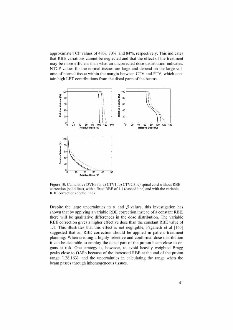
41
approximate TCP values of 48%, 70%, and 84%, respectively. This indicates that RBE variations cannot be neglected and that the effect of the treatment may be more efficient than what an uncorrected dose distribution indicates. NTCP values for the normal tissues are large and depend on the large vol-ume of normal tissue within the margin between CTV and PTV, which con-tain high LET contributions from the distal parts of the beams.
Figure 10. Cumulative DVHs for a) CTV1, b) CTV2,3, c) spinal cord without RBE correction (solid line), with a fixed RBE of 1.1 (dashed line) and with the variable RBE correction (dotted line)
Despite the large uncertainties in and values, this investigation has shown that by applying a variable RBE correction instead of a constant RBE, there will be qualitative differences in the dose distribution. The variable RBE correction gives a higher effective dose than the constant RBE value of 1.1. This illustrates that this effect is not negligible, Paganetti et al [163] suggested that an RBE correction should be applied in patient treatment planning. When creating a highly selective and conformal dose distribution it can be desirable to employ the distal part of the proton beam close to or-gans at risk. One strategy is, however, to avoid heavily weighted Bragg peaks close to OARs because of the increased RBE at the end of the proton range [128,163], and the uncertainties in calculating the range when the beam passes through inhomogeneous tissues.
42
Future perspectives
It has been estimated that radiotherapy contributes to cure of at least 20% of newly diagnosed cancer patients [13,139]. In Sweden, the total number of patients that receive radiotherapy have also increased in recent years, from 32% in 1992 to 47% in 2001 [173]. With the advances in other treatment modalities, particularly in medical oncology with new cytotoxic and biologi-cal drugs, it has also been estimated that the relative importance of radiation therapy may actually increase [72,139]. Better possibilities to eradicate spread tumour cells with drugs will actually increase the demand for more effective local treatments with less adverse effects. More conformed radia-tion therapy is a promising modality that may fulfil many of these demands. Further developments in radiation oncology is to be expected in many areas, as e.g. discussed in the Swedish Cancer Society Radiation Therapy Investi-gation [139].
The attempts to rank and quantify the gains that can be reached with new developments in radiation oncology, studied in this thesis, should be looked upon in these perspectives.
To achieve an increase in anti-tumour efficacy, contributions from several different radiation technologies are required. At present, much research and technology development is devoted to refinement and implementation of techniques such as IMRT, protons and light ion therapy, improving the physical distribution of the dose. In addition, light ions may have biological advantages [25,210]. Of great potential relevance is also better imaging techniques, not only anatomical (CT, MRI) but also functional (MRI, PET) that will improve the staging of the tumour and the delineation of the target volumes [49]. With more accurate target delineation than generally has been the case in the past, conformed radiotherapy will be even better appreciated.
With the introduction of IMRT, it can be expected that photon radiother-apy will certainly be improved. Although the basic principles of IMRT were defined already in the 1980s [18,24,29,217], the introduction into clinical routine has been rather slow but it is definitely increasing. A survey in the USA showed that 32% of the radiation oncologists uses IMRT [140], and most of them had begun to use IMRT after year 2000. In the group, who did not yet have IMRT, most of them were planning to introduce it within very few years. Economical aspects have likely contributed to its popularity in e.g. the USA [12,164].

43
IMRT has the potential to improve the outcome of radiotherapy to prop-erly selected patient groups, although the benefits have been debated [93,185,216]. In the absence of results from prospective, preferably random-ised trials, there is only a limited knowledge of what groups of patients that will benefit the most. Several comparative model treatment planning studies, including the three presented in this thesis, give some indications of the magnitude of the gains that can motivate investments in more advanced equipment and know-how.
Are the gains so marked that IMRT, solely based upon the indirect evi-dence we presently have, immediately should be a new standard? If this is the case, at what tumour sites? Concerning the three sites explored in this thesis, it is not apparent that the gains are sufficient considering the potential disadvantages of IMRT, e.g. the increased induction of secondary cancers [146,153]. If the calculated magnitude of the gain in TCP and the reduction in NTCP of salivary glands in the hypopharynx cancer case and of small bowel toxicity in the rectal cancer case, are true, a randomised study includ-ing a couple of hundred patients will be sufficient. In breast cancer, many more patients will be necessary, and it could be questioned if such a study is realistic. The very long delay before a study would give any results also pre-cludes a randomised study in breast cancer patients, even if only those at high risk to get late adverse effects from the conventional radiotherapy are included.
An increase of costs for investment follows with each step of improve-ment of the dose distribution, or from 3D-CRT to IMRT, to protons, IMPT, and light ions, etc. There are considerable investment costs for proton ther-apy and probably even more so for light ion therapy.
The greatest difference between IMRT and protons is significantly lower doses to surrounding normal structures by protons, which can be used either to decrease toxicity, or to give higher doses to the tumour target with im-proved tumour control. The three examples studied here together with the many other model studies performed up to now (see Table 1) tell that pro-tons for many tumour sites will be sufficiently better to motivate investments in such facilities. These are also the conclusions reached by a Swedish Pro-ton Therapy Investigation [3], although the main conclusion from the inves-tigation was that the gains appear sufficient to invest in a facility for clinical research. The facility should be designed and dimensioned so that clinical trials could be run within a reasonable time. Other groups [10,107,170] have also reached similar conclusions concerning investments in particle therapy. The number of planned new facilities for proton (and light ion) therapy is an expression of this increased knowledge, although other aspects are likely also prevailing.
Development and implementation of IMPT will further improve the tech-nology, with a possibly even greater efficacy compared to the “conven-tional” protons [127]. Therefore, the technology of scanned beams should be

44
further developed. However, the magnitude of the gain going from regular proton beam therapy to IMPT is unknown, although a few model studies have been performed with even lower doses to the surrounding tissues [15,34,127,146,158,198].
Is then proton therapy too expensive? Based on the results from the clini-cal studies performed, and the treatment planning comparisons, it has been estimated that for, e.g. breast cancer, medulloblastoma, prostate cancer and head and neck cancer, the additional costs are reasonable if the patients are selected properly [134-136]. Lundkvist et al based their calculations on the potential number of patients that could be eligible for proton therapy in Sweden [60]. Goitein and Jermann [62] calculated the costs for protons compared with IMRT and found a relative cost of 1.3-2.4 depending on how the investment costs in the facility are handled. The increased costs relate not only to the accelerators but also to the beam lines, gantries and buildings, and personnel. Lievens discussed the cost of protons [121], and concluded that the cost of proton therapy is in the order of what society can accept for a new treatment (either RT, new drugs or surgical techniques), and therefore acceptable.
Further improvement using light ion therapy could results in even more selectively conformed dose distributions, making use of an even sharper lateral penumbra and a higher RBE within the SOBP [25,213]. The tail from ligth ion fragments may however make the depth dose distribution less ad-vantageous. It has been estimated that the cost per fraction for light ion ther-apy will be 3 times higher compared to protons [210]. Although it is claimed that using hypo-fractionation, the cost per patient can be decreased signifi-cantly [25,206]. However, before utilising such fractionation schedules, first they must be carefully evaluated in clinical trials.
The improved dose distributions from IMRT, protons, particularly IMPT, and light ions create conditions conducive for therapies with combinations of cytotoxic and targeted drugs. Radiotherapy in these combinations will dra-matically increase the risk for normal tissue toxicity. Therefore, the impor-tance of conformal radiation will likely increase once we get more knowl-edge of the efficacy of newer radiation-drug combinations.

45
Conclusions
From treatment planning studies such as the ones performed in this thesis, it is possible to identify not only the technique that provides the best outcome but also to quantify the improvement.
Techniques using protons and IMRT have major advantages compared to 3D-CRT plans in delivering the dose more conformally. The studies show that IMRT and protons are more flexible compared to 3D-CRT and the two methods obtain equally good results in shaping the dose to the target volume. Since protons spare volumes of normal tissue better than IMRT, because of steeper dose gradients close to the target volume, protons also offer a poten-tial for dose escalations, which may improve local tumour control. The greatest possibility of dose-escalation with protons was, among the tumours investigated here, shown for patients with hypopharyngeal cancer (PaperII).
Both protons and IMRT can for left-sided node-positive breast cancer (Paper I) decrease the risk for cardiac mortality from 6.7% with a tangential 3D-CRT technique to 0.5% and 1.3%, respectively. The risk for radiation induced pneumonitis can also be reduced significantly, to 0.6% and 2.5% for protons and IMRT, respectively, compared to 14.7% for the best 3D-CRT technique. These new, more complicated and expensive techniques may be of relevance for certain patients at greater risk for late complications.
For hypopharyngeal carcinoma, protons and IMRT provide more selec-tive treatment plans than the 3D-CRT plan (Paper II). A simultaneous boost technique, with protons and IMRT, allows potential dose-escalation, with an increase in TCP of 17%. The NTCP values for the parotid glands were also reduced significantly using either of the new techniques. IMRT can be of value for many patients with head and neck cancers such as hypopharyngeal cancer, and once proton beams are generally more available, these sites should be clinically explored.
In locally advanced rectal cancer (Paper IV), both acute and late toxicity from the small bowel, the main OAR can be reduced by approximately 50%, using either protons or IMRT, if the small bowel is considered serial. Pro-tons are even more favourable if the small bowel is parallel while the reduc-tion with IMRT remains at approximately the same level. Also other tissues around the rectum and adjacent nodes can be spared with the new tech-niques. Both of them may allow further intensification of CTRT combina-tions, having the potential to improve treatment results.

46
With a variable RBE correction of proton treatment plans, including LET dependence and tissue specific parameters, significant differences were found in the corrected dose distributions compared to the uncorrected distri-bution (Paper III). The relevance of these differences must be further inves-tigated, particularly since the RBE correction depends on tissue specific parameters. The possibility to directly account for RBE should at least be included in the treatment planning systems. Intensity modulated protons should include RBE in the optimisation process to take advantage of the variable RBE in the proton beam, unless future studies show that these vari-able corrections are clinically insignificant at most or all major tumour sites.
In this thesis, three treatment planning studies have shown a large poten-tial for both protons and IMRT compared to the treatment presently used at most radiation departments, 3D-CRT. The estimates of potential improve-ments in clinical outcome, either in terms of decreased NTCP or increased TCP, are more favourable for protons compared to IMRT. In the absence of results from large clinical trials, decisions about investments in new technol-ogy will rely on treatment planning studies. These should be accompanied by studies analysing cost-benefits. The results of this thesis show that in-creased use of both IMRT and protons can be supported, although the poten-tial for protons is stronger. Thus, together with a health economical study performed in parallel [133], investments in proton facilities with a capacity and willingness to treat many patients in clinical trials can be supported.

47
Acknowledgements
I wish to express my sincere appreciation to all persons and institutions, who have contributed and encouraged me in making this thesis a reality:
My main supervisor Professor Bengt Glimelius, for skillful scientific super-vision, and for all the time spent reading my manuscripts.
My supervisor, Anders Montelius, for encouraging support and introduction to clinical radiotherapy physics.
My co-authors Ulf Isacsson, Erik Blomquist, Nina Tilly, Henrik Lind-man, Joakim Medin, Erik Grusell, Anders Montelius, and Bengt Glimelius for important contributions to this study.
The section of Oncology at Uppsala University, Professor Ingela Turesson,for giving me the opportunity to do my thesis in a stimulating environment. Didde Simonsson-Westerström and Inger Hjertström-Öst, for all the help with the administrative work.
All colleagues at the proton-therapy project in Uppsala, Ulf Isacsson, Nina Tilly, Stefan Lorin, Peter Kimstrand, Erik Grusell, Erik Blomquist, for creating a stimulating research environment. Ulf for introducing me to the proton treatment planning and the discussions about IMRT optimisation, and Nina for providing data needed and always answering new questions.
Elizabeth Morhed and Carina Öberg-Kreuger for valuable help with treatment planning and collecting patient material.
Kellie Russell, Anders Gustafsson (Nucletron Scandinavia) and Anders Murman (Raysearch Laboratories) for providing all the new modules and data for the treatment planning systems.
The entire staff at the Department of Hospital Physics, for creating a posi-tive and friendly atmosphere!

48
Per Kjäll, Jürgen Arndt, Håkan Nordström and Ola Eriksson at ELEKTA for encouraging these studies.
My mother Inga-lill, and brother Per, encouraging support!
Annika and Elisia, for love, joy and friendship, supporting me whenever I need it!
This thesis has been supported by grants from the Swedish Cancer Societyand Lions Cancer Foundation at the Akademiska Sjukhuset in Uppsala.

49
References:
[1] Favourable and unfavourable effects on long-term survival of radiotherapy for early breast cancer: an overview of the randomised trials. Early Breast Cancer Trialists' Collaborative Group. Lancet 2000;355:1757-70.
[2] Intensity-modulated radiotherapy: current status and issues of interest. Int J Radiat Oncol Biol Phys 2001;51:880-914.
[3] Swedish Proton Therapy Investigation. Acta Oncol 2005;44. [4] Ahnesjo A, Saxner M, Trepp A. A pencil beam model for photon dose calculation.
Med Phys 1992;19:263-73. [5] Archambeau JO, Slater JD, Slater JM, Tangeman R. Role for proton beam irradia-
tion in treatment of pediatric CNS malignancies. Int J Radiat Oncol Biol Phys 1992;22:287-94.
[6] Arnold D, Siewczynski R, Schmoll H-J. Chemotherapy in rectal cancer. Eur J Can-cer Suppl 2005;3:389-400.
[7] Arthur DW, Vicini FA, Kuske RR, Wazer DE, Nag S. Accelerated partial breast irradiation: an updated report from the American Brachytherapy Society. Brachy-therapy 2003;2:124-30.
[8] Austin-Seymour M, Munzenrider JE, Goitein M et al. Progress in low-LET heavy particle therapy: intracranial and paracranial tumors and uveal melanomas. Radiat Res Suppl 1985;8:S219-26.
[9] Austin-Seymour M, Urie M, Munzenrider J et al. Considerations in fractionated proton radiation therapy: clinical potential and results. Radiother Oncol 1990;17:29-35.
[10] Baron MH, Pommier P, Favrel V et al. A "One-day survey": As a reliable estima-tion of the potential recruitment for proton- and carbon- ion therapy in France. Ra-diother Oncol 2004;73:S15-S17.
[11] Baumert BG, Lomax AJ, Miltchev V, Davis JB. A comparison of dose distributions of proton and photon beams in stereotactic conformal radiotherapy of brain lesions. Int J Radiat Oncol Biol Phys 2001;49:1439-49.
[12] Bentzen SM. Radiation therapy: intensity modulated, image guided, biologically optimized and evidence based. Radiother Oncol 2005;77:227-30.
[13] Bentzen SM, Heeren G, Cottier B et al. Towards evidence-based guidelines for radiotherapy infrastructure and staffing needs in Europe: the ESTRO QUARTS pro-ject. Radiother Oncol 2005;75:355-365.
[14] Biancia CD, Yorke E, Chui CS et al. Comparison of end normal inspiration and expiration for gated intensity modulated radiation therapy (IMRT) of lung cancer. Radiother Oncol 2005;75:149-56.
[15] Bolsi A, Fogliata A, Cozzi L. Radiotherapy of small intracranial tumours with different advanced techniques using photon and proton beams: a treatment planning study. Radiother Oncol 2003;68:1-14.

50
[16] Bonnet RB, Bush D, Cheek GA et al. Effects of proton and combined proton/photon beam radiation on pulmonary function in patients with resectable but medically in-operable non-small cell lung cancer. Chest 2001;120:1803-10.
[17] Bonnett DE. Current developments in proton therapy: a review. Phys Med Biol 1993;38:1371-92.
[18] Bortfeld T, Burkelbach J, Boesecke R, Schlegel W. Methods of image reconstruc-tion from projections applied to conformation radiotherapy. Phys Med Biol 1990;35:1423-34.
[19] Bosset JF. [Rectal cancer: preoperative chemoradiation, a new standard]. Cancer Radiother 2005;9:137-9.
[20] Braaksma MM, Wijers OB, van Sornsen de Koste JR et al. Optimisation of confor-mal radiation therapy by intensity modulation: cancer of the larynx and salivary gland function. Radiother Oncol 2003;66:291-302.
[21] Braendengen M, Tveit K, Berglund Å et al. 612 POSTER A randomized phase III study (LARCS) comparing preoperative radiotherapy alone versus chemoradiother-apy in non-resectable rectal cancer. Eur J Cancer Suppl 2005;3:172.
[22] Bragg W. Studies in radioactivity. London, 1912. [23] Brahme A. Dosimetric precision requirements in radiation therapy. Acta Radiol
Oncol 1984;23:379-91. [24] Brahme A. Optimization of stationary and moving beam radiation therapy tech-
niques. Radiother Oncol 1988;12:129-40. [25] Brahme A. Recent advances in light ion radiation therapy. Int J Radiat Oncol Biol
Phys 2004;58:603-616. [26] Brahme A. On the optimal choice of scattering foils for electron therapy. Royal
Institute of Technology Report TRITA-EPP 72-17 1972. [27] Brahme A, Kallman P, Lind BK. Optimization of proton and heavy ion therapy
using an adaptive inversion algorithm. Radiother Oncol 1989;15:189-97. [28] Brahme A, Lewensohn R, Ringborg U et al. Design of a centre for biologically
optimised light ion therapy in Stockholm. Nucl Instrum Meth B 2001;184:569-588. [29] Brahme A, Roos JE, Lax I. Solution of an integral equation encountered in rotation
therapy. Phys Med Biol 1982;27:1221-9. [30] Brown AP, Urie MM, Chisin R, Suit HD. Proton therapy for carcinoma of the
nasopharynx: a study in comparative treatment planning. Int J Radiat Oncol Biol Phys 1989;16:1607-14.
[31] Bush DA, Slater JD, Bonnet R et al. Proton-beam radiotherapy for early-stage lung cancer. Chest 1999;116:1313-9.
[32] Butler EB, Teh BS, Grant WH, 3rd et al. Smart (simultaneous modulated acceler-ated radiation therapy) boost: a new accelerated fractionation schedule for the treatment of head and neck cancer with intensity modulated radiotherapy. Int J Ra-diat Oncol Biol Phys 1999;45:21-32.
[33] Carlsson AK, Andreo P, Brahme A. Monte Carlo and analytical calculation of proton pencil beams for computerized treatment plan optimization. Phys Med Biol 1997;42:1033-53.
[34] Cella L, Lomax A, Miralbell R. Potential role of intensity modulated proton beams in prostate cancer radiotherapy. Int J Radiat Oncol Biol Phys 2001;49:217-23.
[35] Cho BC, Hurkmans CW, Damen EM, Zijp LJ, Mijnheer BJ. Intensity modulated versus non-intensity modulated radiotherapy in the treatment of the left breast and

51
upper internal mammary lymph node chain: a comparative planning study. Radio-ther Oncol 2002;62:127-36.
[36] Convery DJ, Webb S. Generation of discrete beam-intensity modulation by dy-namic multileaf collimation under minimum leaf separation constraints. Phys Med Biol 1998;43:2521-38.
[37] Cozzi L, Fogliata A, Lomax A, Bolsi A. A treatment planning comparison of 3D conformal therapy, intensity modulated photon therapy and proton therapy for treatment of advanced head and neck tumours. Radiother Oncol 2001;61:287-97.
[38] Cozzi L, Fogliata A, Nicolini G, Bernier J. Clinical experience in breast irradiation with intensity modulated photon beams. Acta Oncol 2005;44:467-74.
[39] Dawson LA, Anzai Y, Marsh L et al. Patterns of local-regional recurrence following parotid-sparing conformal and segmental intensity-modulated radiotherapy for head and neck cancer. Int J Radiat Oncol Biol Phys 2000;46:1117-26.
[40] de Arruda FF, Puri DR, Zhung J et al. Intensity-modulated radiation therapy for the treatment of oropharyngeal carcinoma: The Memorial Sloan-Kettering Cancer Cen-ter experience. Int J Radiat Oncol Biol Phys 2006;64:363-73.
[41] Deasy JO. Multiple local minima in radiotherapy optimization problems with dose-volume constraints. Med Phys 1997;24:1157-61.
[42] Egger E, Zografos L, Schalenbourg A et al. Eye retention after proton beam radio-therapy for uveal melanoma. Int J Radiat Oncol Biol Phys 2003;55:867-80.
[43] Eisbruch A, Schwartz M, Rasch C et al. Dysphagia and aspiration after chemoradio-therapy for head-and-neck cancer: which anatomic structures are affected and can they be spared by IMRT? Int J Radiat Oncol Biol Phys 2004;60:1425-39.
[44] Falkmer S, Fors B, Larsson B et al. Pilot study on proton irradiation of human carcinoma. Acta Radiol 1962;58:33-51.
[45] Falkmer S, Larsson B, Stenson S. Effects of single dose proton irradiation of normal skin and Vx2 carcinoma in rabbit ears: a comparative investigation with protons and roentgen rays. Acta Radiol 1959;52:217-34.
[46] Fogliata A, Bolsi A, Cozzi L. Comparative analysis of intensity modulation inverse planning modules of three commercial treatment planning systems applied to head and neck tumour model. Radiother Oncol 2003;66:29-40.
[47] Fogliata A, Bolsi A, Cozzi L. Critical appraisal of treatment techniques based on conventional photon beams, intensity modulated photon beams and proton beams for therapy of intact breast. Radiother Oncol 2002;62:137-145.
[48] Fogliata A, Nicolini G, Alber M et al. IMRT for breast. a planning study. Radiother Oncol 2005;76:300-10.
[49] Forssell-Aronsson E, Kjellen E, Mattsson S, Hellström M, Swedish cancer society investigation group. Medical imaging for improved tumour characterization, de-lineation and treatment verification. Acta Oncol 2002;41:604-14.
[50] Fowler JF. The linear-quadratic formula and progress in fractionated radiotherapy. Br J Radiol 1989;62:679-94.
[51] Frykholm GJ, Isacsson U, Nygard K et al. Preoperative radiotherapy in rectal carci-noma--aspects of acute adverse effects and radiation technique. Int J Radiat Oncol Biol Phys 1996;35:1039-48.
[52] Fu KK, Pajak TF, Trotti A et al. A Radiation Therapy Oncology Group (RTOG) phase III randomized study to compare hyperfractionation and two variants of ac-celerated fractionation to standard fractionation radiotherapy for head and neck
52
squamous cell carcinomas: first report of RTOG 9003. Int J Radiat Oncol Biol Phys 2000;48:7-16.
[53] Fuss M, Hug EB, Schaefer RA et al. Proton radiation therapy (PRT) for pediatric optic pathway gliomas: comparison with 3D planned conventional photons and a standard photon technique. Int J Radiat Oncol Biol Phys 1999;45:1117-26.
[54] Fuss M, Poljanc K, Miller DW et al. Normal tissue complication probability (NTCP) calculations as a means to compare proton and photon plans and evaluation of clinical appropriateness of calculated values. Int J Cancer 2000;90:351-8.
[55] Gademann G, Wannenmacher M. Charged particle therapy to pediatric tumors of the retroperitoneal region: a possible indication. Int J Radiat Oncol Biol Phys 1992;22:375-81.
[56] Gagliardi G, Bjohle J, Lax I et al. Radiation pneumonitis after breast cancer irradia-tion: analysis of the complication probability using the relative seriality model. Int J Radiat Oncol Biol Phys 2000;46:373-81.
[57] Gagliardi G, Lax I, Ottolenghi A, Rutqvist LE. Long-term cardiac mortality after radiotherapy of breast cancer--application of the relative seriality model. Br J Ra-diol 1996;69:839-46.
[58] Gerard J, Bonnetain F, Conroy T et al. Preoperative (preop) radiotherapy (RT) + 5 FU/folinic acid (FA) in T3-4 rectal cancers : results of the FFCD 9203 randomized trial. ASCO Annual Meeting 2005 2005:Abstract 3504.
[59] Gerweck LE, Kozin SV. Relative biological effectiveness of proton beams in clini-cal therapy. Radiother Oncol 1999;50:135-42.
[60] Glimelius B, Ask A, Bjelkengren G et al. Number of patients potentially eligible for proton therapy. Acta Oncol 2005;44:836-49.
[61] Glimelius B, Isacsson U, Blomquist E et al. Potential gains using high-energy pro-tons for therapy of malignant tumours. Acta Oncol 1999;38:137-45.
[62] Goitein M, Jermann M. The Relative Costs of Proton and X-ray Radiation Therapy. Clin Oncol (R Coll Radiol) 2003;15:S37-S50.
[63] Graffman S, Brahme A, Larsson B. Proton radiotherapy with the Uppsala cyclotron. Experience and plans. Strahlentherapie 1985;161:764-70.
[64] Gregoire V, Levendag P, Ang KK et al. CT-based delineation of lymph node levels and related CTVs in the node-negative neck: DAHANCA, EORTC, GORTEC, NCIC,RTOG consensus guidelines. Radiother Oncol 2003;69:227-36.
[65] Grusell E, Montelius A, Brahme A, Rikner G, Russell K. A general solution to charged particle beam flattening using an optimized dual-scattering-foil technique, with application to proton therapy beams. Phys Med Biol 1994;39:2201-2216.
[66] Gudjonsson O, Blomquist E, Lilja A et al. Evaluation of the effect of high-energy proton irradiation treatment on meningiomas by means of 11C-L-methionine PET. Eur J Nucl Med 2000;27:1793-9.
[67] Gudjonsson O, Blomquist E, Nyberg G et al. Stereotactic irradiation of skull base meningiomas with high energy protons. Acta Neurochir (Wien) 1999;141:933-40.
[68] Gueulette J, Bohm L, Slabbert JP et al. Proton relative biological effectiveness (RBE) for survival in mice after thoracic irradiation with fractionated doses. Int J Radiat Oncol Biol Phys 2000;47:1051-8.
[69] Gueulette J, Octave-Prignot M, De Costera BM, Wambersie A, Gregoire V. Intesti-nal crypt regeneration in mice: a biological system for quality assurance in non-conventional radiation therapy. Radiother Oncol 2004;73 Suppl 2:S148-54.

53
[70] Gustafsson A, Lind BK, Brahme A. A generalized pencil beam algorithm for opti-mization of radiation therapy. Med Phys 1994;21:343-56.
[71] Gustafsson A, Lind BK, Svensson R, Brahme A. Simultaneous optimization of dynamic multileaf collimation and scanning patterns or compensation filters using a generalized pencil beam algorithm. Med Phys 1995;22:1141-56.
[72] Hellman S. Technology, biology and traffic. Acta Oncol 2001;40:679-81. [73] Holloway CL, Robinson D, Murray B et al. Results of a phase I study to dose esca-
late using intensity modulated radiotherapy guided by combined PET/CT imaging with induction chemotherapy for patients with non-small cell lung cancer. Radiother Oncol 2004;73:285-7.
[74] Hong L, Goitein M, Bucciolini M et al. A pencil beam algorithm for proton dose calculations. Phys Med Biol 1996;41:1305-30.
[75] Hong L, Hunt M, Chui C et al. Intensity-modulated tangential beam irradiation of the intact breast. Int J Radiat Oncol Biol Phys 1999;44:1155-64.
[76] Hong TS, Tome WA, Chappell RJ et al. The impact of daily setup variations on head-and-neck intensity-modulated radiation therapy. Int J Radiat Oncol Biol Phys 2005;61:779-88.
[77] Hong TS, Tome WA, Chappell RJ, Harari PM. Variations in target delineation for head and neck IMRT: An international multi-institutional study. Int J Radiat Oncol Biol Phys 2004;60:S157-8.
[78] Horiot JC, Bontemps P, van den Bogaert W et al. Accelerated fractionation (AF) compared to conventional fractionation (CF) improves loco-regional control in the radiotherapy of advanced head and neck cancers: results of the EORTC 22851 ran-domized trial. Radiother Oncol 1997;44:111-21.
[79] Huang E, Teh BS, Strother DR et al. Intensity-modulated radiation therapy for pediatric medulloblastoma: early report on the reduction of ototoxicity. Int J Radiat Oncol Biol Phys 2002;52:599-605.
[80] Hug EB, Loredo LN, Slater JD et al. Proton radiation therapy for chordomas and chondrosarcomas of the skull base. J Neurosurg 1999;91:432-9.
[81] Hårdemark B, Liander A, Rehbinder H, Lof J. Direct machine parameter optimiza-tion with RayMachine® in Pinnacle3®. 2003.
[82] IBA. http://www.iba-worldwide.com.[83] ICRU50. Prescribing, recording and reporting photon beam therapy. Bethesda,
1993.[84] ICRU62. Prescribing, recording and reporting photon beam therapy (Supplement to
ICRU Report 50). Bethesda, 1999. [85] Isacsson U, Hagberg H, Johansson KA et al. Potential advantages of protons over
conventional radiation beams for paraspinal tumours. Radiother Oncol 1997;45:63-70.
[86] Isacsson U, Lennernas B, Grusell E et al. Comparative treatment planning between proton and x-ray therapy in esophageal cancer. Int J Radiat Oncol Biol Phys 1998;41:441-50.
[87] Isacsson U, Montelius A, Jung B, Glimelius B. Comparative treatment planning between proton and X-ray therapy in locally advanced rectal cancer. Radiother On-col 1996;41:263-72.
[88] Ishikawa H, Tsujii H, Kamada T et al. A phase II trial using carbon ion radiotherapy (C-ion RT) for prostate cancer. ASCO Annual Meeting 2005 2005:Abstract 3504.

54
[89] Jackson SM, Weir LM, Hay JH, Tsang VH, Durham JS. A randomised trial of accelerated versus conventional radiotherapy in head and neck cancer. Radiother Oncol 1997;43:39-46.
[90] Jani AB, Su A, Milano MT. Intensity-modulated versus conventional pelvic radio-therapy for prostate cancer: analysis of acute toxicity. Urology 2006;67:147-51.
[91] Jansson T, Lindman H, Nygard K et al. Radiotherapy of breast cancer after breast-conserving surgery: an improved technique using mixed electron-photon beams with a multileaf collimator. Radiother Oncol 1998;46:83-9.
[92] Johansen LV, Grau C, Overgaard J. Hypopharyngeal squamous cell carcinoma--treatment results in 138 consecutively admitted patients. Acta Oncol 2000;39:529-36.
[93] Johansson KA, Mattsson S, Brahme A et al. Radiation therapy dose delivery. Acta Oncol 2003;42:85-91.
[94] Kallman P, Agren A, Brahme A. Tumour and normal tissue responses to fraction-ated non-uniform dose delivery. Int J Radiat Biol 1992;62:249-62.
[95] Kanai T, Kawachi K, Kumamoto Y et al. Spot scanning system for proton radio-therapy. Med Phys 1980;7:365-9.
[96] Kanai T, Kawachi K, Matsuzawa H, Inada T. Broad beam three-dimensional irra-diation for proton radiotherapy. Med Phys 1983;10:344-6.
[97] Karlsson BG. [Methods for Calculating and Obtaining Some Favorable Dosage Distributions for Deep Therapy with High Energy Protons.]. Strahlentherapie 1964;124:481-92.
[98] Kestin LL, Sharpe MB, Frazier RC et al. Intensity modulation to improve dose uniformity with tangential breast radiotherapy: initial clinical experience. Int J Ra-diat Oncol Biol Phys 2000;48:1559-68.
[99] Khoroshkov VS, Berejev VM, Zolotov VA, Luk'iashin VE, Shimchuk GG. The spiral ridge filter. Institute of Theoretical and Experimental Physics ITEP report 86-149 1986.
[100] Khoroshkov VS, Breev VM, Zolotov VA, Luk'iashin VE, Shimchuk GG. [Spiral comb filter]. Med Radiol (Mosk) 1987;32:76-80.
[101] Kimstrand P, Russell K, Johansson J. Monte carlo calculation of proton depth dose distributions and Implentation of data into Helax TMS. Personal communication 2004.
[102] Knoos T, Kristensen I, Nilsson P. Volumetric and dosimetric evaluation of radiation treatment plans: radiation conformity index. Int J Radiat Oncol Biol Phys 1998;42:1169-76.
[103] Koehler AM, Schneider RJ, Sisterson JM. Range modulators for protons and heavy ions. Nuclear Instruments and Methods 1975:437.
[104] Kostjuchenko V, Nichiporov D, Luckjashin V. A compact ridge filter for spread out Bragg peak production in pulsed proton clinical beams. Med Phys 2001;28:1427-30.
[105] Koto M, Miyamoto T, Yamamoto N et al. Local control and recurrence of stage I non-small cell lung cancer after carbon ion radiotherapy. Radiother Oncol 2004;71:147-56.
[106] Krengli M, Hug EB, Adams JA et al. Proton radiation therapy for retinoblastoma: comparison of various intraocular tumor locations and beam arrangements. Int J Radiat Oncol Biol Phys 2005;61:583-93.
55
[107] Krengli M, Orecchia R. Medical aspects of the national centre for oncological had-rontherapy (CNAO -- Centro nazionale adroterapla oncologica) in Italy. Radiother Oncol 2004;73:S21-S23.
[108] Larsson B. Radiological properties of beams of high-energy protons. Radiat Res Suppl 1967;7:304-11.
[109] Larsson B. Pre-therapeutic physical experiments with high energy protons. Br J Radiol 1961;34:143-51.
[110] Larsson B, Kihlman BA. Chromosome aberrations following irradiation with high-energy protons and their secondary radiation: a study of dose distribution and bio-logical efficiency using root-tips of Vicia faba and Allium cepa. Int J Radiat Biol 1960;2:8-19.
[111] Larsson B, Leksell L, Rexed B et al. The high-energy proton beam as a neurosurgi-cal tool. Nature 1958;182:1222-3.
[112] Lauve A, Morris M, Schmidt-Ullrich R et al. Simultaneous integrated boost inten-sity-modulated radiotherapy for locally advanced head-and-neck squamous cell car-cinomas: II--clinical results. Int J Radiat Oncol Biol Phys 2004;60:374-87.
[113] Lee CH, Tait D, Nahum AE, Webb S. Comparison of proton therapy and conformal X-ray therapy in non-small cell lung cancer (NSCLC). Br J Radiol 1999;72:1078-84.
[114] Lee M, Wynne C, Webb S, Nahum AE, Dearnaley D. A comparison of proton and megavoltage X-ray treatment planning for prostate cancer. Radiother Oncol 1994;33:239-53.
[115] Leksell L, Larsson B, Andersson B et al. Lesions in the depth of the brain produced by a beam of high energy protons. Acta Radiol 1960;54:251-64.
[116] Levendag P, Braaksma M, Coche E et al. Rotterdam and Brussels CT-based neck nodal delineation compared with the surgical levels as defined by the American Academy of Otolaryngology-Head and Neck Surgery. Int J Radiat Oncol Biol Phys 2004;58:113-23.
[117] Levin CV. Potential for gain in the use of proton beam boost to the para-aortic lymph nodes in carcinoma of the cervix. Int J Radiat Oncol Biol Phys 1992;22:355-9.
[118] Levy RP, Fabrikant JI, Frankel KA et al. Heavy-charged-particle radiosurgery of the pituitary gland: clinical results of 840 patients. Stereotact Funct Neurosurg 1991;57:22-35.
[119] Levy RP, Schulte RW, Slater JD, Miller DW, Slater JM. Stereotactic radiosurgery--the role of charged particles. Acta Oncol 1999;38:165-9.
[120] Li W, Gragoudas ES, Egan KM. Tumor basal area and metastatic death after proton beam irradiation for choroidal melanoma. Arch Ophthalmol 2003;121:68-72.
[121] Lievens Y, den Bogaert WV. Proton beam therapy: Too expensive to become true? Radiother Oncol 2005;75:131-133.
[122] Lin R, Hug EB, Schaefer RA et al. Conformal proton radiation therapy of the poste-rior fossa: a study comparing protons with three-dimensional planned photons in limiting dose to auditory structures. Int J Radiat Oncol Biol Phys 2000;48:1219-26.
[123] Lind BK, Mavroidis P, Hyodynmaa S, Kappas C. Optimization of the dose level for a given treatment plan to maximize the complication-free tumor cure. Acta Oncol 1999;38:787-98.

56
[124] Lind PA, Rosfors S, Wennberg B et al. Pulmonary function following adjuvant chemotherapy and radiotherapy for breast cancer and the issue of three-dimensional treatment planning. Radiother Oncol 1998;49:245-54.
[125] Lind PA, Wennberg B, Gagliardi G, Fornander T. Pulmonary complications follow-ing different radiotherapy techniques for breast cancer, and the association to irradi-ated lung volume and dose. Breast Cancer Res Treat 2001;68:199-210.
[126] Lof J, Rehbinder H. RaySearch White Paper - Inverse Planning Optimization with RayOptimizer in Pinnacle3., 2002.
[127] Lomax A. Intensity modulation methods for proton radiotherapy. Phys Med Biol 1999;44:185-205.
[128] Lomax AJ, Boehringer T, Coray A et al. Intensity modulated proton therapy: a clinical example. Med Phys 2001;28:317-24.
[129] Lomax AJ, Bortfeld T, Goitein G et al. A treatment planning inter-comparison of proton and intensity modulated photon radiotherapy. Radiother Oncol 1999;51:257-71.
[130] Lomax AJ, Cella L, Weber D, Kurtz JM, Miralbell R. Potential role of intensity-modulated photons and protons in the treatment of the breast and regional nodes. Int J Radiat Oncol Biol Phys 2003;55:785-92.
[131] Lomax AJ, Goitein M, Adams J. Intensity modulation in radiotherapy: photons versus protons in the paranasal sinus. Radiother Oncol 2003;66:11-8.
[132] Lorin S, Grusell E, Tilly N et al. Development of a compact proton scanning system in Uppsala with a moveable second magnet. Phys Med Biol 2000;45:1151-63.
[133] Lundkvist J. The role of economic evaluations in health care decision making, Ph.D. Thesis. Karolinska Institutet, 2005.
[134] Lundkvist J, Ekman M, Ericsson SR et al. Economic evaluation of proton radiation therapy in the treatment of breast cancer. Radiother Oncol 2005;75:179-85.
[135] Lundkvist J, Ekman M, Ericsson SR, Jonsson B, Glimelius B. Proton therapy of cancer: potential clinical advantages and cost-effectiveness. Acta Oncol 2005;44:850-61.
[136] Lundkvist J, Ekman M, Ericsson SR, Jonsson B, Glimelius B. Cost-effectiveness of proton radiation in the treatment of childhood medulloblastoma. Cancer 2005;103:793-801.
[137] Lyman JT. Complication probability as assessed from dose-volume histograms. Radiat Res Suppl 1985;8:S13-9.
[138] Mackie TR, Holmes T, Swerdloff S et al. Tomotherapy: a new concept for the delivery of dynamic conformal radiotherapy. Med Phys 1993;20:1709-19.
[139] Mattsson S, Brahme A, Carlsson J et al. Swedish Cancer Society radiation therapy research investigation. Acta Oncol 2002;41:596-603.
[140] Mell LK, Roeske JC, Mundt AJ. A survey of intensity-modulated radiation therapy use in the United States. Cancer 2003;98:204-11.
[141] Milano MT, Chmura SJ, Garofalo MC et al. Intensity-modulated radiotherapy in treatment of pancreatic and bile duct malignancies: toxicity and clinical outcome. Int J Radiat Oncol Biol Phys 2004;59:445-53.
[142] Miller DW. A review of proton beam radiation therapy. Med Phys 1995;22:1943-54.

57
[143] Miralbell R, Cella L, Weber D, Lomax A. Optimizing radiotherapy of orbital and paraorbital tumors: intensity-modulated X-ray beams vs. intensity-modulated proton beams. Int J Radiat Oncol Biol Phys 2000;47:1111-9.
[144] Miralbell R, Crowell C, Suit HD. Potential improvement of three dimension treat-ment planning and proton therapy in the outcome of maxillary sinus cancer. Int J Radiat Oncol Biol Phys 1992;22:305-10.
[145] Miralbell R, Lomax A, Bortfeld T, Rouzaud M, Carrie C. Potential role of proton therapy in the treatment of pediatric medulloblastoma/primitive neuroectodermal tumors: reduction of the supratentorial target volume. Int J Radiat Oncol Biol Phys 1997;38:477-84.
[146] Miralbell R, Lomax A, Cella L, Schneider U. Potential reduction of the incidence of radiation-induced second cancers by using proton beams in the treatment of pediat-ric tumors. Int J Radiat Oncol Biol Phys 2002;54:824.
[147] Miralbell R, Lomax A, Russo M. Potential role of proton therapy in the treatment of pediatric medulloblastoma/primitive neuro-ectodermal tumors: spinal theca irradia-tion. Int J Radiat Oncol Biol Phys 1997;38:805-11.
[148] Miralbell R, Urie M. Potential improvement of three dimension treatment planning and proton beams in fractionated radiotherapy of large cerebral arteriovenous mal-formations. Int J Radiat Oncol Biol Phys 1993;25:353-8.
[149] Miyamoto T, Yamamoto N, Nishimura H et al. Carbon ion radiotherapy for stage I non-small cell lung cancer. Radiother Oncol 2003;66:127-40.
[150] Mock U, Georg D, Bogner J, Auberger T, Potter R. Treatment planning comparison of conventional, 3D conformal, and intensity-modulated photon (IMRT) and proton therapy for paranasal sinus carcinoma. Int J Radiat Oncol Biol Phys 2004;58:147-54.
[151] Mohan R, Wu Q, Manning M, Schmidt-Ullrich R. Radiobiological considerations in the design of fractionation strategies for intensity-modulated radiation therapy of head and neck cancers. Int J Radiat Oncol Biol Phys 2000;46:619-30.
[152] Moyers MF, Miller DW, Bush DA, Slater JD. Methodologies and tools for proton beam design for lung tumors. Int J Radiat Oncol Biol Phys 2001;49:1429-38.
[153] Mu X, Bjork-Eriksson T, Nill S et al. Does electron and proton therapy reduce the risk of radiation induced cancer after spinal irradiation for childhood medulloblas-toma? A comparative treatment planning study. Acta Oncol 2005;44:554-62.
[154] Munter MW, Thilmann C, Hof H et al. Stereotactic intensity modulated radiation therapy and inverse treatment planning for tumors of the head and neck region: clinical implementation of the step and shoot approach and first clinical results. Ra-diother Oncol 2003;66:313-21.
[155] Naeser P, Blomquist E, Montelius A, Thoumas KA. Proton irradiation of malignant uveal melanoma. A five year follow-up of patients treated in Uppsala, Sweden. Ups J Med Sci 1998;103:203-11.
[156] Noel G, Bollet MA, Calugaru V et al. Functional outcome of patients with benign meningioma treated by 3D conformal irradiation with a combination of photons and protons. Int J Radiat Oncol Biol Phys 2005;62:1412-22.
[157] Noel G, Habrand JL, Mammar H et al. Combination of photon and proton radiation therapy for chordomas and chondrosarcomas of the skull base: the Centre de Pro-tontherapie D'Orsay experience. Int J Radiat Oncol Biol Phys 2001;51:392-8.

58
[158] Oelfke U, Bortfeld T. Intensity modulated radiotherapy with charged particle beams: studies of inverse treatment planning for rotation therapy. Med Phys 2000;27:1246-57.
[159] Overgaard J, Hansen HS, Specht L et al. Five compared with six fractions per week of conventional radiotherapy of squamous-cell carcinoma of head and neck: DA-HANCA 6 and 7 randomised controlled trial. Lancet 2003;362:933-40.
[160] Overgaard M, Hansen PS, Overgaard J et al. Postoperative radiotherapy in high-risk premenopausal women with breast cancer who receive adjuvant chemotherapy. Danish Breast Cancer Cooperative Group 82b Trial. N Engl J Med 1997;337:949-55.
[161] Overgaard M, Jensen MB, Overgaard J et al. Postoperative radiotherapy in high-risk postmenopausal breast-cancer patients given adjuvant tamoxifen: Danish Breast Cancer Cooperative Group DBCG 82c randomised trial. Lancet 1999;353:1641-8.
[162] Paganetti H, Niemierko A, Ancukiewicz M et al. Relative biological effectiveness (RBE) values for proton beam therapy. Int J Radiat Oncol Biol Phys 2002;53:407-21.
[163] Paganetti H, Olko P, Kobus H et al. Calculation of relative biological effectiveness for proton beams using biological weighting functions. Int J Radiat Oncol Biol Phys 1997;37:719-29.
[164] Paliwal BR. "IMRT may be used to excess because of its higher reimbursement from Medicare". Med Phys 2004;31:1-2.
[165] Parliament MB, Scrimger RA, Anderson SG et al. Preservation of oral health-related quality of life and salivary flow rates after inverse-planned intensity- modu-lated radiotherapy (IMRT) for head-and-neck cancer. Int J Radiat Oncol Biol Phys 2004;58:663-73.
[166] Paulino AC, Wen BC, Brown CK et al. Late effects in children treated with radia-tion therapy for Wilms' tumor. Int J Radiat Oncol Biol Phys 2000;46:1239-46.
[167] Pedroni E, Bacher R, Blattmann H et al. The 200-MeV proton therapy project at the Paul Scherrer Institute: conceptual design and practical realization. Med Phys 1995;22:37-53.
[168] Petti PL. Differential-pencil-beam dose calculations for charged particles. Med Phys 1992;19:137-49.
[169] Preiser K, Bortfeld T, Hartwig K, Schlegel W, Stein J, editors. A new program for inverse radiotherapy planning. Madison, WI: Medical physics publishing, 1997.
[170] Ramona M, Ulrike M, Robert J et al. Epidemiological aspects of hadron therapy: A prospective nationwide study of the Austrian project MedAustron and the Austrian Society of Radiooncology (OEGRO). Radiother Oncol 2004;73:S24-S28.
[171] Remouchamps VM, Vicini FA, Sharpe MB et al. Significant reductions in heart and lung doses using deep inspiration breath hold with active breathing control and in-tensity-modulated radiation therapy for patients treated with locoregional breast ir-radiation. Int J Radiat Oncol Biol Phys 2003;55:392-406.
[172] Renner TR, Chu WT. Wobbler facility for biomedical experiments. Med Phys 1987;14:825-34.
[173] Ringborg U, Bergqvist D, Brorsson B et al. The Swedish Council on Technology Assessment in Health Care (SBU) systematic overview of radiotherapy for cancer including a prospective survey of radiotherapy practice in Sweden 2001--summary and conclusions. Acta Oncol 2003;42:357-65.

59
[174] Rodel C, Grabenbauer GG, Papadopoulos T et al. Phase I/II trial of capecitabine, oxaliplatin, and radiation for rectal cancer. J Clin Oncol 2003;21:3098-104.
[175] Roeske JC, Bonta D, Mell LK, Lujan AE, Mundt AJ. A dosimetric analysis of acute gastrointestinal toxicity in women receiving intensity-modulated whole-pelvic ra-diation therapy. Radiother Oncol 2003;69:201-7.
[176] Rosenberg AE, Nielsen GP, Keel SB et al. Chondrosarcoma of the base of the skull: a clinicopathologic study of 200 cases with emphasis on its distinction from chor-doma. Am J Surg Pathol 1999;23:1370-8.
[177] Russell KR, Grusell E, Montelius A. Dose calculations in proton beams: range straggling corrections and energy scaling. Phys Med Biol 1995;40:1031-43.
[178] Russell KR, Isacsson U, Saxner M et al. Implementation of pencil kernel and depth penetration algorithms for treatment planning of proton beams. Phys Med Biol 2000;45:9-27.
[179] Sandison GA, Papiez E, Bloch C, Morphis J. Phantom assessment of lung dose from proton arc therapy. Int J Radiat Oncol Biol Phys 1997;38:891-7.
[180] Sauer R, Becker H, Hohenberger W et al. Preoperative versus postoperative chemo-radiotherapy for rectal cancer. N Engl J Med 2004;351:1731-40.
[181] Sautter-Bihl ML, Hultenschmidt B, Melcher U, Ulmer HU. Radiotherapy of internal mammary lymph nodes in breast cancer. Principle considerations on the basis of do-simetric data. Strahlenther Onkol 2002;178:18-24.
[182] Schaffner B, Pedroni E, Lomax A. Dose calculation models for proton treatment planning using a dynamic beam delivery system: an attempt to include density het-erogeneity effects in the analytical dose calculation. Phys Med Biol 1999;44:27-41.
[183] Schilstra C, Meertens H. Calculation of the uncertainty in complication probability for various dose-response models, applied to the parotid gland. Int J Radiat Oncol Biol Phys 2001;50:147-58.
[184] Schneider U, Schaffner B, Lomax T, Pedroni E, Tourovsky A. A technique for calculating range spectra of charged particle beams distal to thick inhomogeneities. Med Phys 1998;25:457-63.
[185] Schulz RJ, Kagan AR. On the role of intensity-modulated radiation therapy in radia-tion oncology. Med Phys 2002;29:1473-82.
[186] Schulz-Ertner D, Nikoghosyan A, Jakel O et al. Feasibility and toxicity of combined photon and carbon ion radiotherapy for locally advanced adenoid cystic carcinomas. Int J Radiat Oncol Biol Phys 2003;56:391-8.
[187] Schulz-Ertner D, Nikoghosyan A, Thilmann C et al. Carbon ion radiotherapy for chordomas and low-grade chondrosarcomas of the skull base. Results in 67 patients. Strahlenther Onkol 2003;179:598-605.
[188] Schwartz DL, Montgomery RB, Yueh B et al. Phase I and initial phase II results from a trial investigating weekly docetaxel and carboplatin given neoadjuvantly and then concurrently with concomitant boost radiotherapy for locally advanced squam-ous cell carcinoma of the head and neck. Cancer 2005;103:2534-43.
[189] Shah HK, Tome WA, Bentzen SM et al. Evolution in H&N IMRT Practice and Early Assessment of Outcome at a Single Academic Center. Int J Radiat Oncol Biol Phys 2005;63:S360-1.
[190] Shepard DM, Earl MA, Li XA, Naqvi S, Yu C. Direct aperture optimization: a turnkey solution for step-and-shoot IMRT. Med Phys 2002;29:1007-18.

60
[191] Silander H, Pellettieri L, Enblad P et al. Fractionated, stereotactic proton beam treatment of cerebral arteriovenous malformations. Acta Neurol Scand 2004;109:85-90.
[192] Sisterson J. Particles PTCOG Newsletter. Particles PTCOG Newsletter 2005. [193] Skladowski K, Maciejewski B, Golen M et al. Randomized clinical trial on 7-day-
continuous accelerated irradiation (CAIR) of head and neck cancer - report on 3-year tumour control and normal tissue toxicity. Radiother Oncol 2000;55:101-10.
[194] Slater JD, Rossi CJ, Jr., Yonemoto LT et al. Proton therapy for prostate cancer: the initial Loma Linda University experience. Int J Radiat Oncol Biol Phys 2004;59:348-52.
[195] Slater JM, Archambeau JO, Miller DW et al. The proton treatment center at Loma Linda University Medical Center: rationale for and description of its development. Int J Radiat Oncol Biol Phys 1992;22:383-9.
[196] Slater JM, Slater JD, Archambeau JO. Carcinoma of the tonsillar region: potential for use of proton beam therapy. Int J Radiat Oncol Biol Phys 1992;22:311-9.
[197] Smit BM. Prospects for proton therapy in carcinoma of the cervix. Int J Radiat Oncol Biol Phys 1992;22:349-53.
[198] Smith AR, Loeffler JS, Adams JA, Lomax AJ, Niemierko A. The potential for proton therapy to improve clinical outcomes: Comparisons of proton and x-ray treatment plans for the purpose of tumor dose escalation and/or reduction of treat-ment-related morbidity. Int J Radiat Oncol Biol Phys 2000;48:338.
[199] Spirou SV, Chui CS. Generation of arbitrary intensity profiles by dynamic jaws or multileaf collimators. Med Phys 1994;21:1031-41.
[200] St Clair WH, Adams JA, Bues M et al. Advantage of protons compared to conven-tional X-ray or IMRT in the treatment of a pediatric patient with medulloblastoma. Int J Radiat Oncol Biol Phys 2004;58:727-34.
[201] Stein J, Bortfeld T, Dorschel B, Schlegel W. Dynamic X-ray compensation for conformal radiotherapy by means of multi-leaf collimation. Radiother Oncol 1994;32:163-73.
[202] Steinberg GK, Fabrikant JI, Marks MP et al. Stereotactic heavy-charged-particle Bragg-peak radiation for intracranial arteriovenous malformations. N Engl J Med 1990;323:96-101.
[203] Sténson S. Effects of high-energy protons on healthy organs and malignant tumours. Including a study of radiation protection by local hypoxia, Doctoral dissertation. Uppsala University, 1969.
[204] Suit H, Goldberg S, Niemierko A et al. Proton beams to replace photon beams in radical dose treatments. Acta Oncol 2003;42:800-8.
[205] Suit HD, Becht J, Leong J et al. Potential for improvement in radiation therapy. Int J Radiat Oncol Biol Phys 1988;14:777-86.
[206] Svensson H, Ringborg U, Naslund I, Brahme A. Development of light ion therapy at the Karolinska Hospital and Institute. Radiother Oncol 2004;73 Suppl 2:S206-10.
[207] Szymanowski H, Oelfke U. Two-dimensional pencil beam scaling: an improved proton dose algorithm for heterogeneous media. Phys Med Biol 2002;47:3313-30.
[208] Tatsuzaki H, Urie MM, Linggood R. Comparative treatment planning: proton vs. x-ray beams against glioblastoma multiforme. Int J Radiat Oncol Biol Phys 1992;22:265-73.

61
[209] Tatsuzaki H, Urie MM, Willett CG. 3-D comparative study of proton vs. x-ray radiation therapy for rectal cancer. Int J Radiat Oncol Biol Phys 1992;22:369-74.
[210] Turesson I, Johansson KA, Mattsson S. The potential of proton and light ion beams in radiotherapy. Acta Oncol 2003;42:107-14.
[211] Urie MM, Goitein M. Variable versus fixed modulation of proton beams for treat-ments in the cranium. Med Phys 1989;16:593-601.
[212] Wambersie A, Gregroire V, Brucher JM. Potential clinical gain of proton (and heavy ion) beams for brain tumors in children. Int J Radiat Oncol Biol Phys 1992;22:275-86.
[213] Wambersie A, Hendry J, Gueulette J et al. Radiobiological rationale and patient selection for high-LET radiation in cancer therapy. Radiother Oncol 2004;73 Suppl 2:S1-14.
[214] van't Riet A, Mak AC, Moerland MA, Elders LH, van der Zee W. A conformation number to quantify the degree of conformality in brachytherapy and external beam irradiation: application to the prostate. Int J Radiat Oncol Biol Phys 1997;37:731-6.
[215] Webb S. The physical basis of IMRT and inverse planning. Br J Radiol 2003;76:678-89.
[216] Webb S. Contemporary IMRT, 2004. [217] Webb S. Optimisation of conformal radiotherapy dose distributions by simulated
annealing. Phys Med Biol 1989;34:1349-70. [218] Webb S, Nahum AE. A model for calculating tumour control probability in radio-
therapy including the effects of inhomogeneous distributions of dose and clono-genic cell density. Phys Med Biol 1993;38:653-66.
[219] Weber DC, Lomax AJ, Rutz HP et al. Spot-scanning proton radiation therapy for recurrent, residual or untreated intracranial meningiomas. Radiother Oncol 2004;71:251-8.
[220] Weber DC, Rutz HP, Pedroni ES et al. Results of spot-scanning proton radiation therapy for chordoma and chondrosarcoma of the skull base: the Paul Scherrer Insti-tut experience. Int J Radiat Oncol Biol Phys 2005;63:401-9.
[221] Weber DC, Trofimov AV, Delaney TF, Bortfeld T. A treatment planning compari-son of intensity modulated photon and proton therapy for paraspinal sarcomas. Int J Radiat Oncol Biol Phys 2004;58:1596-606.
[222] Verhey LJ, Smith V, Serago CF. Comparison of radiosurgery treatment modalities based on physical dose distributions. Int J Radiat Oncol Biol Phys 1998;40:497-505.
[223] Vernimmen FJ, Slabbert JP, Wilson JA, Fredericks S, Melvill R. Stereotactic proton beam therapy for intracranial arteriovenous malformations. Int J Radiat Oncol Biol Phys 2005;62:44-52.
[224] Wilkens JJ. Evaluation of Radiobiological Effects in Intensity Modulated Proton Therapy:New Strategies for Inverse Treatment Planning, Ph.D. Thesis. University of Heidelberg, Germany, 2004.
[225] Wilkens JJ, Oelfke U. Optimization of radiobiological effects in intensity modu-lated proton therapy. Med Phys 2005;32:455-65.
[226] Wilson R. Radiological Use of Fast Protons. Radiology 1946:487-91. [227] Withers HR, Peters LJ, Taylor JM. Dose-response relationship for radiation therapy
of subclinical disease. Int J Radiat Oncol Biol Phys 1995;31:353-9.

62
[228] Wolden SL, Chen WC, Pfister DG et al. Intensity-modulated radiation therapy (IMRT) for nasopharynx cancer: Update of the Memorial Sloan-Kettering experi-ence. Int J Radiat Oncol Biol Phys 2006;64:57-62.
[229] Wu Q, Manning M, Schmidt-Ullrich R, Mohan R. The potential for sparing of parotids and escalation of biologically effective dose with intensity-modulated ra-diation treatments of head and neck cancers: a treatment design study. Int J Radiat Oncol Biol Phys 2000;46:195-205.
[230] Wu Q, Mohan R, Morris M, Lauve A, Schmidt-Ullrich R. Simultaneous integrated boost intensity-modulated radiotherapy for locally advanced head-and-neck squam-ous cell carcinomas. I: dosimetric results. Int J Radiat Oncol Biol Phys 2003;56:573-85.
[231] Yock TI, Tarbell NJ. Technology insight: Proton beam radiotherapy for treatment in pediatric brain tumors. Nat Clin Pract Oncol 2004;1:97-103.
[232] Zackrisson B, Mercke C, Strander H, Wennerberg J, Cavallin-Stahl E. A systematic overview of radiation therapy effects in head and neck cancer. Acta Oncol 2003;42:443-61.
[233] Zietman AL, DeSilvio ML, Slater JD et al. Comparison of conventional-dose vs high-dose conformal radiation therapy in clinically localized adenocarcinoma of the prostate: a randomized controlled trial. Jama 2005;294:1233-9.
[234] Zurlo A, Lomax A, Hoess A et al. The role of proton therapy in the treatment of large irradiation volumes: a comparative planning study of pancreatic and biliary tumors. Int J Radiat Oncol Biol Phys 2000;48:277-88.


Acta Universitatis UpsaliensisDigital Comprehensive Summaries of Uppsala Dissertationsfrom the Faculty of Medicine 117
Editor: The Dean of the Faculty of Medicine
A doctoral dissertation from the Faculty of Medicine, UppsalaUniversity, is usually a summary of a number of papers. A fewcopies of the complete dissertation are kept at major Swedishresearch libraries, while the summary alone is distributedinternationally through the series Digital ComprehensiveSummaries of Uppsala Dissertations from the Faculty ofMedicine. (Prior to January, 2005, the series was publishedunder the title “Comprehensive Summaries of UppsalaDissertations from the Faculty of Medicine”.)
Distribution: publications.uu.seurn:nbn:se:uu:diva-6593
ACTAUNIVERSITATISUPSALIENSISUPPSALA2006