comparative study of the molecular mechanism of action of
TRANSCRIPT
Comparative study of the molecular mechanism of action
of the synthetic progestins, Medroxyprogesterone acetate
and Norethisterone acetate
Donita Jean Africander
Dissertation presented in fulfilment of the requirements for the degree PhD in Biochemistry at the University of Stellenbosch
Promoter: Prof. Janet P. Hapgood
Co-promoter: Prof. Ann Louw
March 2010

ii
DECLARATION
_____________________________________________________
By submitting this dissertation electronically, I declare that the entirety of the
work contained therein is my own, original work, that I am the owner of the
copyright thereof (unless to the extent explicitly otherwise stated) and that I
have not previously in its entirety or in part submitted it for obtaining any
qualification.
Signature: Date: March 2010
Copyright © 2010 Stellenbosch University
All rights reserved

iii
ABSTRACT ____________________________________________________ Medroxyprogesterone acetate (MPA) and norethisterone (NET) and its derivatives (norethisterone enanthate (NET-EN); norethisterone acetate (NET-A)), are used by millions of women as contraceptives and in hormone replacement therapy (HRT). Although both progestins are widely used, very little is known about their mechanism of action at the molecular level. In this thesis, the differential regulation of gene expression and molecular mechanism of action via different steroid receptors by these synthetic progestons, as compared to progesterone (Prog) was investigated in human cell lines. In the first part of the study, the effect of Prog, MPA and NET-A on the expression of endogenous cytokine genes was investigated in two epithelial cell lines of the human female genital tract, Ect1/E6E7 (an ectocervical cell line) and Vk2/E6E7 (a vaginal cell line). Quantitative realtime RT-PCR (QPCR) showed ligand-specific and cell-specific regulation of the interleukin (IL)-6, IL-8 and RANTES (Regulated-upon-Activation, Normal T cell Expressed and Secreted) genes with Prog, MPA and NET-A. Moreover, the repression of the TNFα-induced RANTES gene by MPA in the Ect1/E6E7 cell line was found to be mediated by the androgen receptor (AR). The second part of the study focused on elucidating the androgenic activities of these two progestins, in comparison to Prog. Competitive binding in whole cells revealed that Prog, MPA and NET-A have a similar binding affinity for the hAR as the natural androgen dihydrotestosterone (DHT). Both transactivation and transrepression transcriptional assays demonstrate that, unlike Prog, MPA and NET-A are efficacious AR agonists, with activities comparable to DHT. Using a mammalian two-hydrid assay, it was shown that MPA and NET-A exert their androgenic actions by different mechanisms. NET-A, like DHT and other well-characterised androgens, induces the ligand-dependent interaction between the NH2- and COOH-terminal domains (N/C-interaction) of the AR independent of promoter-context, while MPA does this in a promoter-dependent manner. In the third part of this study, competitive binding revealed that MPA and NET-A have a similar binding affinity to each other, but about a 100-fold lower affinity than Prog for the human mineralocorticoid receptor (hMR), while RU486 has an even lower affinity for the hMR. Promoter-reporter assays showed that MPA, NET-A and RU486 are all antagonists of the hMR, but unlike Prog, they have weak antagonistic activity. However, on the endogenous MR-regulated Orm-1 (α-glycolytic protein or orosomucoid-1) gene expressed in a rat cardiomyocyte cell line, NET-A and RU486, but not MPA, has similar antagonistic activity as Prog. This study is the first to show that, NET-A and RU486, but not MPA, can dissociate between transrepression and transactivation via the hMR. Taken together, these results show that natural Prog and the synthetic progestins, MPA and NET-A display differential promoter-, cell- and receptor-specific effects on gene expression. Furthermore they may have important implications for cervicovaginal immune function, cardiovascular and other physiological functions.

iv
OPSOMMING _____________________________________________________ Medroksieprogesteroon asetaat (MPA), noretisteroon (NET) en derivate daarvan (noretisteroon enantaat (NET-EN); noretisteroon asetaat (NET-A), word deur miljoene vroue gebruik as voorbehoedmiddels en vir hormoon vervangingsterapie (HVT). Tenspyte daarvan dat beide hierdie progestiene algemeen gebruik word, is min bekend oor hulle meganisme van werking op molekulêre vlak. In hierdie proefskrif word die differensiële regulering van geenuitdrukking asook die molekulêre meganisme van werking deur middel van steroïedreseptore van beide hierdie sintetiese progestiene, ondersoek, en vergelyk met progesteroon (Prog), in menslike sellyne. In die eerste deel van die studie is die effek van Prog, MPA en NET-A op die uitdrukking van endogene sitokinien gene ondersoek in twee epiteel sellyne van die menslike vroulike geslagskanaal, Ect1/E6E7 (‘n ektoservikale sellyn) en Vk2/E6E7 (‘n vaginale sellyn). Kwantitatiewe intydse RT-PKR het ligand-spesifieke en sel-spesifieke regulering van interleukien (IL)-6, IL-8 en RANTES (Regulering-na-Aktivering, Normale T-sel Uitgedrukte en Afgeskei) gene getoon met Prog, MPA en NET-A. Verder is gevind dat die onderdrukking van die TNF-α-geïnduseerde RANTES geen deur MPA in die Ect1/E6E7 sellyn bemiddel word deur die androgeen reseptor (AR). Die tweede deel van die studie het gefokus op die toeligting van die androgeniese aktiwiteit van die twee progestiene in vergelyking met Prog. Kompeterende binding in volselle het getoon dat Prog, MPA en NET-A ‘n soortelyke bindings affiniteit vir die menslike AR as die natuurlike androgeen dehidrotestosteroon (DHT) vir die menslike AR het. Beide transaktiverings en transonderdrukkings transkripsionele analieses toon dat, anders as Prog, MPA en NET-A effektiewe AR agoniste is met aktiwiteite wat vergelykbaar is met die van DHT. Deur die gebruik van ‘n soogdier twee-hibried toets, kon gewys word dat MPA en NET-A hul androgeniese effekte uitoefen deur verskillende meganismes. NET-A, soos DHT en ander goed gekarakteriseerde androgene, induseer die ligand-afhanklike interaksie tussen die NH2- en COOH-terminale domeine (N/C-interaksie) van die AR, onafhanklik van die promoter-konteks. MPA, aan die ander kant, doen dit op ‘n promoter-afhanklike manier. In die derde deel van die studie het kompeterende binding getoon dat MPA en NET-A soortelyke relatiewe bindings affiniteite vir die menslike mineralokortikoïed reseptor (hMR) het, maar dat hierdie affiniteit ongeveer 100-voud laer is as die van Prog en dat die affiniteit van RU486 vir hMR selfs nog laer is. Promoter-rapporteerder toetse het getoon dat MPA, NET-A en RU486 almal antagoniste van die hMR is, maar anders as Prog, is hierdie ‘n swak antagonistiese aktiwiteit. Nietemin, op die endogene MR-gereguleerde Orm-1 (α-glikolitiese proteïen of orosomukoïed-1) geen, uitgedruk in ‘n rot kardiomiosiet sellyn, het NET-A en RU486, maar nie MPA nie, ‘n soortgelyke antagonistiese aktiwiteit as Prog. Hierdie studie is die eerste om te wys dat NET-A en RU486, maar nie MPA nie, kan onderskei tussen transrepressie en transaktivering deur middel van die hMR. Samevattend toon die resultate dat natuurlike Prog en die sintetiese progestiene, MPA en NET-A, ‘n differentiële promoter-, sel- en reseptor-spesifieke effek op geenuitdrukking het. Verder mag die resultate belangrike implikasies vir servikovaginale immuunfunksie, asook kardiovaskulêre en ander fisiologiese funksies, inhou.

v
To my family…

vi
LIST OF ABBREVIATIONS _____________________________________________________
ANOVA analysis of variance
AF-1 activation function-1
AF-2 activation function-2
Ald aldosterone
AP-1 Activator Protein-1
AR androgen receptor
ARE(s) androgen response element(s)
ATCC American Type Culture Collection
BMD bone mineral density
bp base-pair
CBG corticosteroid-binding globulin
cDNA complementary DNA
CEE / MPA conjugated equine estrogen / medroxyprogesterone
acetate
CHD coronary heart disease
Cort cortisol
C-terminal carboxy-(COOH-) terminal
CVD cardiovascular disease
DBD DNA-binding domain
DEPC diethylpyrocarbonate
Dex Dexamethasone
DHT dihydrotestosterone

vii
DMEM Dulbecco’s Modified Eagle Medium
DMPA depot medroxyprogesterone acetate
DHP 5 -dihydroprogesterone
DNA deoxyribonucleic acid
E2 17β-estradiol
ECL enhanced chemiluminescence
EGF epidermal growth factor
ER estrogen receptor
ERK extracellular signal-regulated kinase
ES endometrial stromal
FCS fetal calf serum
FRET fluorescence resonance energy transfer
FSH follicle-stimulating hormone
GCs glucocorticoids
GR glucocorticoid receptor
GRE(s) glucocorticoid response element
HESCs human endometrial stromal cells
HIV human immunodeficiency virus
HPA hypothalamic-pituitary-adrenal
HPG hypothalamic-pituitary-gonadal
HPLC high-performance liquid chromatography
HPV human papillomavirus
HRE(s) hormone response element(s)
HRT hormone replacement therapy
Hsp(s) heat-shock protein(s)

viii
HSV-1 herpes simplex virus-1
HSV-2 herpes simplex virus-2
HT/HRT hormone replacement therapy
5-HT 5-hydroxytryptamine
HUVECs human umbilical vein endothelial cells
IL-1 interleukin -1
IL-2 interleukin -2
IL-6 interleukin -6
IL-8 interleukin -8
IgG immunoglobulin G
iNOS nitric oxide synthase
JNK Jun N-terminal kinase
LH luteinizing hormone
LNCaP lymph node carcinoma of the prostate
MAPK mitogen-activated protein kinase
MAPKK MAPK kinase
MEK MAPK kinase
MIB mibolerone
MR mineralocorticoid receptor
M-MLV Moloney Murine Leukemia Virus
MMTV mouse mammary tumor virus
MPA medroxyprogesterone acetate
MRE(s) mineralocorticoid response element(s)
mRNA messenger ribonucleic acid
N-CoR nuclear receptor co-repressor

ix
NET norethisterone / norethindrone
NET-A norethisterone / norethindrone acetate
NET-EN norethisterone / norethindrone enanthate
NFĸB nuclear factor kappa-B
NOS nitric oxide synthase
nGRE negative glucocorticoid response element
NTD amino (NH2)-teminal domain
N/C-interaction interaction between the N- and C-terminal domains
OHF hydroxyflutamide
Orm-1 α-acidic glycoprotein or oricomucosoid
PAGE polyacrylamide gel electrophoresis
PAI-1 plasminogen activator inhibitor-1
PBMCs peripheral blood mononuclear cells
PBS phosphate-buffered saline
PHA phytohemaglutinin
PMA phorbol-myristate acetate
PR progesterone receptor
Prog progesterone
PTHrP parathyroid hormone related peptide
RANTES Regulated-upon-Activation, normal T cell Expressed and
Secreted
R1881 methyltrienolone
R5020 promegestone
RT-PCR reverse transcription-polymerase chain reaction
RU486 mifepristone

x
SDS sodium dodecyl sulphate
SEM standard error of the mean
SHBG sex hormone binding globulin
SHIV simian-human immunodeficiency virus
SRC-1 steroid receptor co-activator-1
STI sexually transmitted infection
THP 3 ,5 -tetrahydroprogesterone
TNF-α tumor necrosis factor-alpha
TPA tetradecanoyl phorbol acetate
WHI Women’s Health Initiative
WHIMS Women’s Health Initiative Memory Study
WHO World Health Organization
WISDOM Women’s International Study of Long Duration Oestrogen
after the Menopause

xi
ACKNOWLEDGEMENTS _____________________________________________________
I would like to express my sincere gratitude to everyone who has made this
thesis possible and especially to:
My supervisor, mentor and friend, Prof Janet Hapgood: Thank you so much
for your guidance, patience, support, and for being a role model to me. I have
learnt so much from you, not only about science, but also how to juggle being
a student, having a fulltime academic post and being a mom. You remain my
inspiration.
My husband, Nolan: Words cannot express the gratitude and love I feel for
you. It wasn’t always easy, especially when you started your MBA studies, but
you were, and remain, a constant pillar of strength to me. Without your love,
patience and support, this would not have been possible.
My daughters, Nicole and Robyn, who had so little of me in your lives as a
result of this PhD: I know you did not always understand when mommy had to
work long hours, and I’m sorry for all the things I’ve missed, but I hope that
someday you will appreciate and respect the decisions I have made. Know
this, you girls and daddy are the light of my life and I will always love you.
My parents: Thank you for providing me with every opportunity in life and for
all your support, your love and your belief in my abilities. And a very special
thanks for not only encouraging both Nolan and I in our endeavors, but for
always being there.

xii
My brother, Paul, and the other very special people in my life Karin and Geoff,
who were “parents” to our kids during the final phases of writing up; Nicky,
without whom I would not have survived in the final days; Alan, Bev, Des,
Chris, Selwyn, Deliah, Yvette, Heidi, Barbie, Valmae, Carol, aunty Sandra,
Glenda, Lee-Anne, Alet, Ryan, Melissa, and last but not least, Tats: Thank
you very much for all your support and love.
My co-supervisor, colleague and friend, Prof. Ann Louw: for your continual
support and encouragement, especially during times of disbelief. I am
eternally grateful to you.
Carmen Langeveldt, laboratory manager and friend. Thank you for the
maintenance of our cell cultures, and of course for being my lunchtime buddy!
Present and past members of the Hapgood lab, especially Dominique, Hanél,
Wilmie, Nicky, Chanel, Andrea and Kate: Thank you, thank you, thank you, for
all the moans and groans about experiments not working, for brainstorming
new ideas, but most of all for being such good friends.
My colleagues, the students and friends in the Biochemistry Department,
especially Ralie, Lynne, Welma, Renate, Dewald, Steven, and Koch, a HUGE
thank you for keeping me sane.
Thank you also to the University of Stellenbosch, National Research
Foundation of South Africa and the Medical Research Council, for providing
research funding.

xiii
THESIS OUTLINE _____________________________________________________
This thesis consists of five chapters. Chapters 1, 2, 3 and 4 are written up in
manuscript format. Chapters 2, 3 and 4 include a brief introduction to the
specific aims of the particular studies, report and discuss the undertaken
experiments and the results obtained. These chapters will shortly be
submitted for publication. The references for all the chapters are included in
one section following Chapter 5.
1. Chapter 1: Literature review. This chapter gives a detailed overview of
the relevant knowledge currently available in the literature, with a
particular focus on directly comparing the molecular mechanism of
action of medroxyprogesterone acetate (MPA) and norethisterone
enanthate (NET-EN)/norethisterone acetate (NET-A). The review was
written by the candidate. Nicolette Verhoog also works on the progestin
project, and contributed in the exchange of information and ideas.
Dominique Koubovec previously reviewed the topic in her PhD thesis
and this material was used as a guideline for the current review.
2. Chapter 2: Differential regulation of endogenous pro-inflammatory
cytokine genes by MPA and NET-A in cell lines of the female
genital tract. This chapter contains the results of a study investigating
and comparing the regulation of endogenous cytokine genes by Prog,
MPA and NET-A in Ect1/E6E7 (human ectocervical) and Vk2/E6E7
(human vaginal) cell lines. All experiments were performed by the
candidate, except for the maintenance of the cell lines, which was
performed by Carmen Langeveldt.

xiv
3. Chapter 3: A comparative study of the androgenic properties of
progesterone and the synthetic progestins, medroxyprogesterone
acetate (MPA) and norethisterone acetate (NET-A). This chapter
includes the results of a study into the molecular mechanism of action
of MPA and NET-A, as compared to Prog, via the hAR in the COS-1
cell line. All experiments were performed by the candidate, except for
the maintenance of the COS-1 monkey kidney cell line, which was
performed by Carmen Langeveldt.
4. Chapter 4: Investigating the anti-mineralocorticoid properties of
synthetic progestins used in hormone replacement therapy. This
chapter reports on the findings of a study investigating whether MPA
and NET-A, like Prog, can act via the MR. This study was performed in
COS-1 (monkey kidney) as well as H9C2 cell lines (rat
cardiomyocytes). All experiments were performed by the candidate,
except for the maintenance of the cell lines, which was performed by
Carmen Langeveldt.
5. Chapter 5: Conclusions and Future Perspectives. In this final
chapter, the results of the overall study are discussed and conclusions
drawn in the context of the larger body of work.
The appendices (A-D), found at the back of the thesis, include data not
shown, but referred to as supplementary material within the manuscripts, as
well as additional results not included in the chapters and definitions.

xv
The literature review presented in Chapter 1 will be submitted to Endocrine
Reviews. The manuscript presented in Chapter 2 will be submitted to
Contraception, while the manuscripts presented in Chapter 3 and Chapter 4
will be submitted to the Journal of Steroid Biochemistry and Molecular Biology
and Molecular and Cellular Endocrinology, respectively.
Consistent with manuscript format, the collective term “we” and “our” is often
used in the thesis. However, all the experimental work was performed by the
candidate, barring the figure in Appendix B5 which was performed by Tamzin
Tanner (MSc thesis) previously from the Hapgood laboratory.

xvi
TABLE OF CONTENTS
_____________________________________________________
LIST OF ABBREVIATIONS viii
THESIS OUTLINE xv
CHAPTER 1
REVIEWING THE MOLECULAR MECHANISM OF ACTION OF
MEDROXYPROGESTERONE ACETATE AND
NORETHISTERONE ENANTHATE/ACETATE 1
Abstract 2
1.1 Introduction 3
1.2 Therapeutic applications 7
1.2.1 Contraception 7
1.2.1.1 Female contraception 7
1.2.1.2 Male contraception 9
1.2.2 Hormone replacement therapy 10
1.2.3 Other applications 13
1.3 Physiological effects of MPA and NET-EN/NET-A/NET 14
1.3.1 Reproduction 14
1.3.2 Adrenal function 18
1.3.3 Skeletal function 20
1.3.4 Brain function 24
1.3.5 Reproductive cancers 27
1.3.5.1 Breast cancer 27

xvii
1.3.5.2 Endometrial, ovarian and cerviCal cancers 31
1.3.6 Cardiovascular function 33
1.3.7 Immune function 41
1.4 Mechanism of action 48
1.4.1 Serum binding globulins 49
1.4.2 Steroid receptors 52
1.4.3 Effects of MPA and NET on target genes via the: 56
1.4.3.1 Progesterone receptor (PR) 56
1.4.3.2 Glucocorticoid receptor (GR) 62
1.4.3.3 Androgen receptor (AR) 68
1.4.3.4 Mineralocorticoid receptor (MR) 73
1.4.3.5 Estrogen receptor (ER) 74
1.5 Conclusion 75
HYPOTHESES AND AIMS 78
CHAPTER 2
DIFFERENTIAL REGULATION OF ENDOGENOUS PRO-
INFLAMMATORY CYTOKINE GENES BY MPA AND NET-A IN
CELL LINES OF THE FEMALE GENITAL TRACT 80
Abstract 81
Introduction 83
Materials and methods 86
xviii
Inducing compounds 86
Cell culture 87
Plasmids 88
Isolation of total RNA and realtime quantitative RT-PCR (QPCR)
analysis of representative genes 88
Western blotting 89
Whole cell binding assays to determine steroid receptor contentt in
the Ect1/E6E7 and Vk2/E6E7 cell line 90
Luciferase reporter assays 91
Data manipulation and statistical analysis 92
Results 92
MPA and NET-A, unlike Prog, exhibit differential patterns of gene
regulation on pro-inflammatory chemokine 92
The PR, AR and GR are expressed in both ectocervical and vaginal
cell lines 93
Receptor-specific antagonists indicate a role for the AR in the
downregulation of the RANTES pro-inflammatory chemokine gene by
MPA in the ectocervical cell line 98
Only the GR was transcriptionally active in promoter-reporter
Transactivation assays in both ectocervical and vaginal cell lines 100
Discussion 105

xix
CHAPTER 3
A COMPARATIVE STUDY OF THE ANDROGENIC
PROPERTIES OF PROGESTERONE AND THE SYNTHETIC
PROGESTINS, MEDROXYPROGESTERONE ACETATE (MPA)
AND NORETHISTERONE ACETATE (NET-A) 112
Abstract 113
Introduction 114
Materials and methods 120
Inducing compounds 120
Plasmids 120
Cell culture 121
Whole cell binding assays 121
Transient transfection assays 123
Mammalian two-hybrid assays 124
Data manipulation and statistical analysis 124
Results 125
MPA and NET-A have a similar binding affinity for the AR 125
MPA and NET-A display androgen agonist activity that is similar to
that of DHT for transactivation 126
MPA, but not NET-A, induces the ligand-dependent interaction
between the amino- and carboxyl-terminals of the androgen receptor
in a promoter-dependent manner 132
MPA and NET-A display similar androgenic properties for transrepression 137
xx
Discussion 142
CHAPTER 4
INVESTIGATING THE ANTI-MINERALOCORTICOID
PROPERTIES OF SYNTHETIC PROGESTINS USED IN
HORMONE REPLACEMENT THERAPY 152
Abstract 153
Introduction 155
Materials and methods 159
Plasmids 159
Inducing compounds 160
Cell culture 160
Whole cell binding assays 161
Luciferase reporter assays 162
Mammalian two-hybrid assays 164
Isolation of total RNA and realtime quantitative RT-PCR
analysis of representative genes 164
Western blotting 166
Data manipulation and statistical analysis 166
Results 167
MPA and NET-A have a similar binding affinity for the MR 167
Unlike Prog, MPA and NET-A display weak MR antagonist activity
and no mineralocorticoid agonist activity for transactivation 169
Unlike MPA and NET-A, Prog and Dex induce the

xxi
ligand-dependent interaction between the amino- and carboxyl-terminals
of the mineralocorticoid receptor 172
MPA and NET-A display dissimilar mineralocorticoid properties
for transrepression on the AP-1 promoter 175
Unlike, MPA Prog, NET-A and RU486 do inhibit the aldosterone-
induced upregulation of the endogenous Orm-1 gene 179
Discussion 183
CHAPTER 5
CONCLUSIONS AND FUTURE PERSPECTIVES 194
REFERENCES 218
APPENDIX A: DATA NOT INCLUDED IN CHAPTER 2 269
A1: Hydroxyflutamide does not inhibit the effects of Prog, MPA or NET-A on
RANTES gene expression in the human vaginal cell line (Vk2/E6E7) 270
APPENDIX B: DATA NOT INCLUDED IN CHAPTER 3 271
B1: Optimisation of [3H]-MIB concentration for the determination of Kd or Ki
values of ligands for overexpressed hAR. 272
B2: Time course to establish equilibrium time for binding of 0.2 nM [3H]-MIB to
overexpressed hAR. 273
B3: Transrepression assay in COS-1 cells in the absence and presence of
overexpressed hAR. 274
B4: MPA and NET-A lack AR antagonist activity. 275

xxii
B5: MPA, but not NET-A, antagonizes the DHT-induced N/C-interaction of the
AR. 276
APPENDIX C: DATA NOT INCLUDED IN CHAPTER 4 277
C1: Optimisation of [3H]-Ald concentration for the determination of Kd or Ki
values of ligands via overexpressed hMR. 278
C2: Time course to establish equilibrium time for binding of 0.2 nM [3H]-Ald to
overexpressed hMR. 279
C3: Transrepression assay in COS-1 cells in the absence and presence of
overexpressed hR. 280
C4: Prog displays weak partial agonist activity for transactivation via
overexpressed MR in COS-1 cells 281
C5: Similar induction of the MR N/C-interaction by aldosterone and cortisol.
282
C6: Antagonist activity of NET-A via the hGR in COS-1 cells. 283
APPENDIX D: DEFINITIONS AND EXTRA DATA NOT
INCLUDED IN CHAPTERS 284
D1: Binding parameters and calculations 285
D2: Pharmacological definitions 288
D3: Transactivation in COS-1 cells in the absence and presence of
overexpressed MR, AR or GR 290
D4: Effects of Dex, MPA and NET-A on IL-6 protein production in human
monocytes 291

1
Chapter 1
_____________________________________________________
Reviewing the molecular mechanism of action of
medroxyprogesterone acetate and norethisterone enanthate/acetate
Donita Africander1, Nicolette Verhoog*, Dominique Koubovec1 and Janet Hapgood*
1Department of Biochemistry, University of Stellenbosch, Private Bag X1, Matieland,
7602, South Africa.
* Department of Molecular and Cell Biology, University of Cape Town, Private Bag,
Rondebosch, 7700, South Africa.
Manuscript in preparation for submission to Endocrine Reviews.

2
Abstract
Medroxyprogesterone acetate (MPA) and norethisterone (NET) and its derivatives
(norethisterone enanthate (NET-EN); norethisterone acetate (NET-A)), are used by
millions of women as contraceptives and in hormone replacement therapy (HRT). In
addition, both MPA and NET-acetate (NET-A) are used in cancer therapy and in the
treatment of gynaecological disorders such as endometriosis and premenstrual
dysphoria. Although both progestins are widely used, very little is known about their
mechanism of action at the molecular level. The importance of investigating these
mechanisms, as compared to those of progesterone (Prog), has recently been
highlighted by clinical evidence showing that MPA increases the risk of the
development of breast cancer and coronary heart disease in HRT users. In addition,
use of MPA as a contraceptive has also been shown to increase viral shedding,
which raises concern as to its impact on the spread of viral diseases. There are
currently no reviews in the literature other than one of our own, comparing the
mechanism of action of MPA and NET-A or NET-EN (Hapgood et al., 2004). Here we
review the physiological effects of these two progestins, as well as their regulation of
and/or binding to serum-binding proteins and steroidogenic enzymes. In addition, as
it is known that both MPA and NET can bind not only to the progesterone receptor,
but also to the glucocorticoid, androgen, mineralocorticoid, and possibly the estrogen
receptors, it is plausible that MPA and NET exert therapeutic actions as well as side-
effects via some of these receptors. We thus also review the molecular mechanism of
action of both MPA and NET via each of the above steroid receptors on various
target genes.

3
1.1 Introduction
Progestins are a class of synthetically developed compounds. Their development
was based on similarity of biological actions to that of the endogenous ovarian
hormone progesterone, which plays a pivotal role in female reproduction. These
progestins have many therapeutic applications in female reproductive medicine, and
are used instead of progesterone because of their longer biological half-life (Speroff,
1996). A wide variety of progestins are available, that in addition to their common
progestogenic effects, exhibit a range of biological effects that differ not only from
each other, but also from that of progesterone (Schindler et al., 2003). The synthetic
progestins, medroxyprogesterone acetate (MPA) and norethisterone enanthate
(NET-EN), are two highly effective injectable progestogen-only contraceptives that
have been available in many countries for over 40 years (Westhoff, 2003), including
South Africa. In fact, in South Africa, MPA and NET-EN are the most commonly used
contraceptives (Draper et al., 2006). These progestins are not only used as
contraceptive agents, but also in hormone replacement therapy (HRT) (also referred
to as hormone therapy (HT)), as well as a number of other non-contraceptive
therapies.
Progestins are chemically derived from parent compounds such as testosterone
resulting in the 19-nortestosterone derivatives or from progesterone, resulting in the
17-hydroxy (17-OH) progesterone derivatives and 19-norprogesterone derivatives.
MPA (6α-methyl-17-acetoxy pregn-4-ene-3, 20-dione) (also referred to as Depo-
Provera®, depot medroxyprogesterone acetate (DMPA) or Petogen®, the latter
locally manufactured in South Africa) is a 17-OH progesterone derivative (21-carbon
series steroid) containing the pregnane nucleus, while NET-EN (17α-ethynyl-17β-

4
heptanoyloxy-4-estren-3-one) (also referred to as either norethindrone enanthate,
norethisterone enanthate or Nur-Isterate®) is a 19-nortestosterone derivative (19-
carbon series steroid) containing the androstane nucleus (structures depicted in
Figure 1). Due to the aforementioned structures, MPA is often referred to as a true
progestin, while NET-EN, which retains its androgenic activity, is referred to as an
androgenic progestin (Darney, 1995).
As with most drugs, a number of side-effects, some more severe than others, have
been reported with the clinical use of MPA and NET. Interestingly, the World Health
Organization (WHO) classifies MPA and NET-EN in the same category of medical
eligibility, and makes no distinction between the two with regard to their side-effects
or contra-indications (WHO, 2004). Similarly, Haider and Darney (2007) recently
reported that NET-EN has the same mechanism of action as MPA in terms of
contraceptive action and efficacy, with the same advantages and disadvantages. The
advantages refered to the convenient and effective contraceptive method, the fact
that it can be used by women with contra-indications to estrogen, as well as some
therapeutic benefits, while the disadvantages refer to the side-effect profile. Similarly,
the Cochrane comparative review on the contraceptive effectivity, reversibility and
side-effects of MPA and NET-EN, also reported that MPA and NET-EN are similar,
barring the slower return to fertility with MPA (Draper et al., 2006). While clinical
studies have not identified significant differences between the side-effect profile of
MPA and NET in patients, given the enormous spectrum of possible side-effects, and
the specific circumstances under which these may manifest, it is possible that
differences may yet be identified, especially given the differences in their activity
recently identified at a cellular level (Koubovec et al., 2005; Sasagawa et al., 2008).

5
Interestingly, these studies observed differences in mechanism of action not only
between MPA and NET, but also as compared to Prog. These observed differences
in activity are a matter of concern, especially since these progestins are usually
reported to act in a similar manner. A number of factors may account for the
differential effects of MPA vs. NET, such as their differences in molecular structure,
metabolism, bio-availibility, and binding affinities to different steroid receptors or
receptor isoforms (Stanczyk et al., 2003; Schindler et al., 2003). It is thus clear that
additional comparative studies between these two progestins, relative to each other
and Prog, are needed at the cellular level. The objective of the present review is thus
to highlight the differences between MPA and NET-EN/NET-A1, as compared to
Prog, in terms of (1) therapeutic applications, observed side-effects and physiological
effects, (2) their interaction with serum proteins, and (3) their mechanism of action via
different steroid receptors.
1 NET-EN is a derivative of NET used in injectable contraception; NET-A is the acetate ester of NET and is used
in oral contraception or HRT; the derivatives, NET-EN and NET-A, have to be metabolically converted to NET
in order to become biologically active. At times, NET will be used generically to include all derivatives.
6
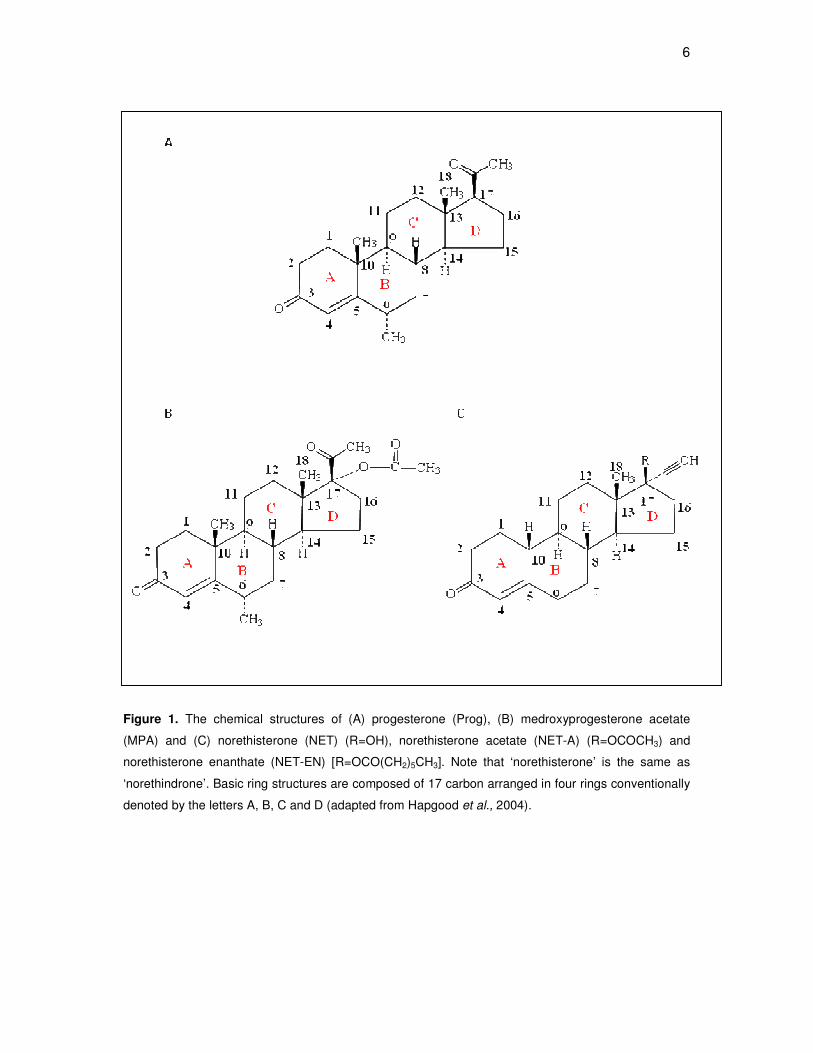
Figure 1. The chemical structures of (A) progesterone (Prog), (B) medroxyprogesterone acetate
(MPA) and (C) norethisterone (NET) (R=OH), norethisterone acetate (NET-A) (R=OCOCH3) and
norethisterone enanthate (NET-EN) [R=OCO(CH2)5CH3]. Note that ‘norethisterone’ is the same as
‘norethindrone’. Basic ring structures are composed of 17 carbon arranged in four rings conventionally
denoted by the letters A, B, C and D (adapted from Hapgood et al., 2004).

7
1.2 Therapeutic applications
1.2.1 Contraception
1.2.1.1 Female contraception
When used as injectable contraceptives in women, both MPA and NET-EN
formulations are administered as intramuscular injections, however, they differ in
dosage and frequency of administration. MPA is administered as a 150 mg aqueous
suspension every three months (Mishell, 1996), whereas NET-EN is administered as
a 200 mg oily suspension every two months (Garza-Flores et al., 1991). Interestingly,
a new formulation of MPA, at a 30% lower dose (104 mg), has recently been
approved in the United States for subcutaneous administration every 3 months (Jain
et al., 2004), and is referred to as Depo-Sub Q.
After injection, MPA is fairly stable and is itself the active contraceptive compound
(Speroff, 1996), whereas NET-EN and NET-A are hydrolysed to
norethindrone/norethisterone (NET) and other metabolites, which together have
contraceptive action (Stanczyk and Roy, 1990). Women receiving the 150 mg
intramuscular injection of MPA, typically have serum concentrations of about 2.6-3.9
nM for the duration of contraceptive treatment (Mathrubutham and Fotherby, 1981;
Mishell, 1996), while the 200 mg dose of NET-EN has been reported to result in
serum concentrations of about 1.5-59 nM (Fotherby et al., 1983).
MPA mediates its contraceptive action by inhibiting the secretion of the pituitary
gonadotropins, luteinizing hormone (LH) and follicle-stimulating hormone (FSH),
thereby preventing follicular maturation and ovulation (Mishell, 1996; Kaunitz, 2000;
Greydanus et al., 2001). MPA also alters the endometrial lining and reduces

8
glycogen secretion, thus preventing a blastocyst from entering the endometrial cavity
(Mishell, 1996). In addition, MPA thickens the cervical mucus, which interferes with
sperm penetration into the uterus (Greydanus et al., 2001). Although NET-EN has
also been shown to block ovulation, (Bhowmik and Mukherjea, 1988), its primary
contraceptive action involves altering the content of cervical mucus thus preventing
sperm movement into the uterine cavity (Bhowmik and Mukherjea, 1987). MPA and
NET-EN therefore have multiple sites of action, and are thus both highly effective
contraceptive agents in women.
Despite the effectiveness of MPA and NET-EN in preventing pregnancy, there are
several side-effects associated with their use. The side-effect profile of both these
progestins includes irregular bleeding, amenorrhea, breast tenderness, headaches,
weight gain, acne and vaginal discharge (Kaunitz, 2000; Greydanus et al., 2001;
Benagiano et al., 1978; Darney, 1995; Tyler, 1970; Schwallie, 1976; El-Mahgoub and
Karim, 1972; Westhoff, 2003; Haider and Darney, 2007; Spencer et al., 2009).
Interestingly, in a community-based cross-sectional household survey to determine
perceived side-effects of MPA and NET-EN in KwaZulu-Natal in South Africa, many
women reported vaginal wetness as a side-effect (Smit et al., 2002). It is unclear
whether this vaginal wetness is the same as the previously reported side-effect of
vaginal discharge. In addition, side-effects such as dizziness, fatigue, bloating of the
abdomen or breasts, behavioural changes, reduced libido and decreased bone
mineral density (BMD) have also been reported for MPA (Kaunitz, 2000; Greydanus
et al., 2001). Although the side-effect profile of NET-EN is not as well defined as that
of MPA, it is assumed to be similar to, but less severe than MPA, with a more rapid
return to fertility after termination of treatment (Benagiano et al., 1978; Fraser and
9
Weisberg 1982; Koetsawang, 1991, Draper et al., 2006). This difference in time
before return to fertility has been challenged by Bigrigg and co-workers (1999), as
they report that there is in fact, no statistically significant delay in return to fertility by
MPA users.
1.2.1.2 Male contraception
Male contraception involves the administration of synthetic analogues of
testosterone, which functions as a contraceptive by suppressing the secretion of the
gonadotropins, LH and FSH, from the pituitary (Amory and Bremner, 1998; Morse et
al., 1973). The suppression of LH and FSH deprives the testes of the stimulatory
signals required for spermatogenesis, leading to decreased sperm counts and
reversible infertility in most men. However, the administration of testosterone
derivatives alone does not completely suppress sperm production in all men (Amory
and Bremner, 1998; McLachlan et al., 2002). For this reason, recent research has
been investigating the use of testosterone analogues in combination with progestins.
Testosterone esters, combined with injections of MPA or NET-EN, show severe
suppression of spermatogenesis, due to the synergistic suppression of gonadotropin
levels (Kamischke et al., 2000a; Kamischke et al., 2000b; Turner et al., 2003;
reviewed by Nieschlag et al., 2003; Gu et al., 2004, reviewed by Amory, 2008). Thus,
combined treatment with testosterone and progestins, holds promise for an effective
male contraceptive. However, as observed in women, the use of synthetic progestins
causes certain side-effects in men. For example, the use of NET-EN in male
contraception has been shown to decrease the levels of high-density lipoprotein
(HDL) and lipoprotein, which may lead to negative effects on the cardiovascular
system (Zitzmann et al., 2002). Current research in male hormonal contraception, as
10
with female contraception, is thus focusing on producing an effective contraceptive
agent, while minimizing side-effects.
1.2.2 Hormone replacement therapy
Hormone replacement therapy (HRT/HT) is commonly prescribed to alleviate
symptoms experienced by women after menopause. These symptoms, including hot
flushes, urogenital atrophy, bone loss and vaginal dryness, are due to a decrease in
estrogen levels (Hickey et al., 2005; Greendale, 1999). HRT includes administration
of either estrogen alone, or estrogen combined with a progestin, such as MPA or
NET-EN (Greendale, 1999). The latter treatment is used for menopausal women with
an intact uterus so as to counteract the proliferative effects of estrogen on the uterine
epithelium, thereby preventing estrogen-induced endometrial hyperplasia (Gambrell
Jr et al., 1980; Taitel and Kafrissen, 1995; Brunelli et al., 1996; Palacios et al., 2006).
The progestin may be administered either continuously (every day) or sequentially
(for a part of each month). It has been reported that, in the longterm, continuous
therapy may be more protective against endometrial hyperplasia than sequential
therapy (reviewed by Lethaby et al., 2004).
Whether MPA or NET is used as the progestin of choice for HRT differs
internationally. In the United States, the most commonly used progestin is MPA,
generally combined with conjugated equine estrogens (CEE) in formulations for oral
administration (Newcomb et al., 2002). Similarly, in France, MPA or cyproterone
acetate is mainly used (Fournier et al., 2005). In contrast, only a small percentage of
women in the United Kingdom and Northern Europe use MPA (≤ 20%) (Beral, 2003;
Magnusson et al., 1999; Jernstrom et al., 2003; Stahlberg et al., 2004), while the

11
majority use NET-A or other 19-nortestosterone-derivatives (Campagnoli et al., 2005;
Fournier et al., 2005).
Originally, the dose of MPA employed in HRT was a sequential dosage of 10 mg/day
for about 11 days per month, but subsequently the dose has been reduced to 2.5 to 5
mg/day (Brunelli et al., 1996; Van de Weijer, 2007; Archer and Pickar, 2000). HRT
doses of NET range from about 0.35 to 2.1 mg/day (Taitel and Kafrissen, 1995).
Women receiving the Activelle HRT regime (0.5 mg NET-A, 1 mg estradiol) are
reported to have peak serum concentrations of NET ranging between 3.64 and 17.7
nM (Activelle package insert reg. no. 33/21.8.2/0532, Novo Nordisk Inc.), while
serum levels of MPA for HRT range between 0.02 and 0.2 nM (Ghatge et al., 2005).
Side-effects of MPA and NET used in HRT include changes in the levels of lipids and
lipoproteins, as well as adverse effects on vasomotion, which may increase
cardiovascular risk in postmenopausal women (Sitruk-Ware, 2000). In addition, MPA
and NET have been implicated in increased risk of breast cancer development (Riis
et al., 2002; Stahlberg et al., 2003). Although these side-effects have long been
recognized, it had always been assumed that the benefits of HRT for
postmenopausal women outweighed the risks. However, the Women’s Health
Initiative (WHI) trial in the USA of a combined estrogen and progestin (MPA) HRT
regime in healthy postmenopausal women highlighted several side-effects such as
increased risk of breast cancer, coronary heart disease (CHD), venous
thromboembolism, stroke and dementia (Rossouw et al., 2002). In addition, data by
investigators working on the same trial (estrogen plus MPA) also suggested an
increase in the risk for ovarian cancer (Anderson et al., 2003). These side-effects
12
were deemed so severe that the trial was terminated two years earlier than planned.
This caused much confusion and alarm amongst HRT users, and as a result many
postmenopausal women have stopped using HRT (Ettinger et al., 2004). It is
noteworthy that a similar trial on the use of estrogen alone also indicated an
increased risk of stroke, but no increase in breast cancer risk or cardiovascular
disease (CVD) (Anderson et al., 2004), thus implicating the MPA component in
breast cancer and CVD side-effects. The Women’s International Study of Long
Duration Oestrogen after the Menopause (WISDOM) investigation (Vickers et al.,
2007) was prematurely stopped following publication of the results of the WHI study.
Their results were consistent with the WHI study in indicating increased
cardiovascular and thromboembolic risk when HRT (estrogen plus MPA) was started
a considerable time after menopause (Vickers et al., 2007). The ‘Million Women
Study’ found that both MPA and NET substantially increased the risk of breast cancer
in long-term HRT users (Beral; 2003). Taken together, results from these studies
indicate that usage of MPA may result in increased risk of breast cancer. In addition,
all studies except the Million Women Study which did not investigate cardiovascular
effects, indicate that MPA may have increased risk of CVD. NET usage has also
been implicated, in the Million Women Study, in an increase of breast cancer risk.
The controversy surrounding the risk/benefit ratio of progestins in HRT has resulted
in a trend towards prescription of HRT with lower doses of progestin, and different
routes of administration such as gels, sprays, vaginal rings, intrauterine systems or
transdermal patches (Nath and Sitruk-Ware, 2009). In this way, the potentially
harmful effects of estrogen on the endometrium are still counteracted by MPA, while
the progestin-induced side-effects on the breast and heart may possibly be

13
minimized (Sitruk-Ware, 2007). However, the safety of these new systems (low
dosage, parenterally administered) vs. the old (higher dosage, oral therapy) needs to
be evaluated.
1.2.3 Other applications
In addition to the use of MPA and NET-EN/NET-A/NET in contraception and HRT,
they are also used in a number of other therapeutic applications. MPA is used in the
treatment of gynaecological disorders such as dysmenorrhea, menorrhagia
(excessively heavy menstrual bleeding), ovulatory pain, pain associated with ovarian
disease, premenstrual dysphoria, perimenopausal symptoms (Kaunitz, 1998) and
endometriosis, a complex disorder causing pelvic pain and infertility (Irahara et al.,
2001; Harrison and Barry-Kinsella, 2000; Muneyyirci-Delale and Karacan, 1998;
Vercellini et al., 2003). NET can also be used in the treatment of endometriosis
(Vercellini et al., 2003), and the dosage used for both MPA and NET is about 50-100
mg/day (Harrison and Barry-Kinsella, 2000; Telimaa et al., 1989). MPA has also
been associated with haematological improvement in women with sickle cell disease
(Grimes, 1999), as well as reduced seizure frequency in women with seizure
disorders (Kaunitz, 2000). A recent study, however, observed increased seizure
occurrences with the use of CEE/MPA in HRT, suggesting that MPA may not be the
optimal progestin for HRT in postmenopausal women with epilepsy (Harden et al.,
2006).
In cancer therapy, MPA is used at very high doses (Etienne et al., 1992; Yamashita
et al., 1996), typically between 500 and 1500 mg/day taken orally for about 12 weeks
(Blossey et al., 1984). At these high cancer therapy doses, serum levels of MPA are

14
approximately 0.14-1.7 µM (Thigpen et al., 1999; Focan et al., 2001), and potent
glucocorticoid-like side-effects such as inhibition of adrenal function (Blossey et al.,
1984; Papaleo et al., 1984; Lang et al., 1990) and immunosuppression (Yamashita et
al., 1996; Mallmann et al., 1990; Scambia et al., 1988), have been observed.
Finally, MPA is also prescribed for mentally handicapped women who have
menstrual hygiene problems (Elkins et al., 1986). Furthermore it is also used in the
treatment of deviant sexual behaviors in men such as pedophilia, exhibitionism,
transvestism, and voyeurism (Kravitz et al., 1995; Bradford, 1999). Although NET is
not as widely used as MPA, with the exception of use for HRT and contraception, it
has been used in the treatment of acne (Zouboulis and Piquero-Martin, 2003) and in
treating gastrointestinal symptoms of women with colorectal endometriosis (Ferrero
et al., 2009).
1.3 Physiological effects of MPA and NET-EN/NET-A/NET
1.3.1 Reproduction
Hypothalamic gonadotropin-releasing hormone (GnRH) is the key hormone
responsible for regulating reproduction. It is secreted by the hypothalamus and
travels via the blood to the anterior pituitary where it binds to the GnRH receptor on
the cell surface of gonadotrope cells. Intracellular signal transduction pathways are
subsequently activated, stimulating the synthesis and release of the gonadotropins,
LH and FSH. These gonadotropins then enter the systemic circulation to regulate
gonadal function, including steroidogenesis and gametogenesis. MPA mediates its
contraceptive action by inhibiting the secretion of LH and FSH, thereby preventing
15
follicular maturation and preventing ovulation (Mishell, 1996; Kaunitz, 2000). The
inhibition of the gonadotropins results in suppression of ovarian estradiol production.
Although the effect of MPA and NET on GnRH synthesis and release has not yet to
our knowledge been determined, a few studies have investigated the effects of MPA
and to a lesser extent NET, on LH, FSH and steroid hormone levels, in an attempt to
fully understand the contraceptive mechanism of action of these two progestins. An
early study showed that MPA and NET-EN inhibits the mid-cycle surge of FSH and
LH, but that the release of these gonadotropins continues at luteal phase levels
(Mishell et al., 1977; Franchimont et al., 1970). Another early study measured the
peripheral blood levels of LH, FSH, and estradiol after intramuscular injection of a
contraceptive dose of MPA in normal women (Jeppsson and Johansson, 1976). The
levels of all three hormones remained in the range of the early follicular phase of a
normal menstrual cycle (low levels) and ovulation was suppressed due to
suppression of the LH peak. Interestingly, no suppression of basal LH and FSH
levels was reported in any of the women, an effect which likely contributes to the lack
of menopausal-like symptoms in women receiving contraceptive doses of MPA. In
another study using contraceptive doses, normal menstruating women showed a
decline in plasma levels of estradiol, progesterone and 17α-hydroxyprogesterone to
early follicular phase levels sixteen days after MPA administration (Aedo et al.,
1981). In addition, no LH surge or ovulation was detected in patients receiving 1 mg
of NET over a 5-day period in in vitro fertilisation studies (Letterie, 2000).
For male contraception, MPA or NET-EN, combined with testosterone esters, show
severe suppression of spermatogenesis, due to the synergistic suppression of

16
gonadotropin (LH and FSH) levels (Kamischke et al., 2000; Turner et al., 2003;
reviewed by Nieschlag et al., 2003; Gu et al., 2004; reviewed by Amory 2008).
Similarly, it has been shown that MPA effectively suppresses spermatogenesis by
inhibiting testosterone and gonadotropin production in rats (Lobl et al., 1983;
Flickinger, 1977).
MPA and to a much lesser extent NET, has been shown to influence expression of a
number of genes involved in reproductive functions. Examples of such genes include
tissue factor (TF) (Krikun et al., 2000), decidual cell-expressed plasminogen activator
inhibitor-1 (PAI-1) (Lockwood, 2001), transforming growth factor-β (TGF-β), (Arici et
al., 1996b), vascular endothelial growth factor (VEGF) (Sugino et al., 2001), c-fos
and prolactin (PRL) (Reis et al., 1999), and the metalloproteinases (MMPs) (Bruner-
Tran et al., 2006). Tissue factor, a cell membrane-bound glycoprotein, is responsible
for the initiation of hemostasis during implantation and placentation, and is
associated with decidualisation (differentiation) in the uterus. Decidualisation is an
adaptation of the uterus to enable implantation of the embryo, and may occur as a
result of hormonal contraception. MPA at 100 nM was shown to significantly enhance
TF gene transcription in human endometrial stromal cells (HESCs) (the progenitors of
decidual cells) (Krikun et al., 2000). Furthermore, decidual cell-expressed PAI-1 plays
a role in preventing haemorrhage during human pregnancy implantation. MPA, in the
absence and presence of estradiol, was shown to enhance PAI-1 expression in
HESCs (Schatz and Lockwood, 1993; Schatz et al., 1995; Lockwood, 2001). It is
noteworthy that estradiol is ineffective alone, but enhances the MPA-mediated
effects.

17
Another MPA-regulated gene involved in endometrial functions is transforming
growth factor beta (TGF-β), which is believed to play a role in the predecidualisation
of HESCs and in the completion of decidualisation after blastocyst implantation.
Treatment of cultured HESCs with 1 nM MPA resulted in reduced levels of TGF-β3
mRNA, and a small increase in TGF-β1 mRNA levels (Arici et al., 1996b). In contrast,
in endometrial samples from women having received higher doses of MPA (10
mg/day), TGF-β3 expression was enhanced, with no observable change in TGF-β1
levels (Reis et al., 2002). On the other hand, in a comparative study of MPA and NET
in MCF-7 breast cancer cell lines, MPA did not affect TGF-β2 and TGF-β3 mRNA
levels, whereas a highly significant decrease was observed with NET (Jeng and
Jordan, 1991). Collectively, the data by Reis et al. (2002) and Jeng and Jordan
(1991), suggest that MPA acts in a cell-specific manner, and moreover suggest that
MPA and TGF-β3 may together mediate endometrial differentiation.
VEGF and its receptors play important roles in implantation and maintenance of
pregnancy. Expression of VEGF and one of its receptors, kinase insert domain-
containing region (KDR), was significantly increased by MPA (1 µM) and estrogen
(10 nM) in human endometrial stromal cells isolated from proliferative phase
endometrium (Sugino et al., 2001). Another study in human endometrium showed
that MPA inhibited c-fos gene expression, and enhanced the expression of PRL
(Reis et al., 1999). The authors suggested that inducing similar c-fos and PRL
expression levels to those in secretory endometrium may be the mechanism by
which MPA exerts its anti-proliferative effects. In addition, MPA, NET-A and Prog
were shown to differentially regulate pro-matrix metalloproteinase (pro-MMP)-3 and
pro-MMP-7 protein expression in stromal cells isolated from normal endometrial

18
tissue donors and endometriosis patients, in the absence and presence of the pro-
inflammatory cytokine IL-1α (Bruner-Tran et al., 2006). MMP expression is crucial for
endometrial growth and remodeling, but failure to suppress MMPs may impair
implantation and promote the development of endometriosis (Bruner et al., 1997;
Osteen et al., 2005). MPA and Prog suppressed pro-MMP-3 and pro-MMP-7 in both
healthy tissue donors and endometriosis patients regardless of IL-1α challenge, while
NET-A could do so only in normal cells and in the absence of IL-1α challenge. The
fact that NET could not suppress these MMP’s in the presence of IL-1α induced
inflammation, or in endometriosis (inflammatory disease) patients (Podgaec et al.,
2007), suggests that NET is a weaker anti-inflammatory agent as compared to MPA
and natural Prog, and thus may not be an optimal treatment for women with
endometriosis.
1.3.2 Adrenal function and steroidogenesis
Surprisingly little research appears to have been carried out in humans on the effects
of MPA on adrenal function, and to our knowledge, there is only one report on the
effects of NET (Amatayakul et al., 1988).
Jones and co-workers (1974) reported lowered baseline plasma cortisol levels in
contraceptive users of MPA. Similarly, a later study also showed that administration
of a single dose of MPA resulted in a slight but significant reduction of cortisol in
normal menstruating women (Aedo et al., 1981). However, healthy, non-lactating
Thai women who received long-term MPA and NET treatment were found to have no
significant change in adrenal function as measured by cortisol levels (Amatayakul et
al., 1988).
19
At higher doses (up to 1500 mg orally per day), MPA has been shown to cause
significant inhibition of adrenal function (Hellman et al., 1976; Blossey et al., 1984;
Papaleo et al., 1984; Lang et al., 1990), which may be attributed to its glucocorticoid
activity (van Veelen et al., 1985). Furthermore, a study evaluating the adrenal
function of postmenopausal breast cancer patients treated with MPA (300 mg),
reported no difference in adrenocorticotropic hormone (ACTH) levels, but significantly
lower cortisol, androstenedione and dehydroepiandrosterone sulphate (DHEA-S)
levels, when compared to a control group (van Veelen et al., 1984). Androstenedione
is the main precursor of estrogens in postmenopausal women and this reduction in
its levels could be the cause of hypoestrogenism induced by MPA. Earlier studies,
however, observed reduction in both ACTH and cortisol levels by MPA (Matthews et
al., 1970; Hellman et al., 1976). In addition, MPA used in the treatment of abnormal
sexual behaviors in males (100 to 1000 mg weekly by intramuscular injection)
significantly reduced mean serum concentrations of total testosterone and cortisol
(Guay, 2008).
MPA directly inhibits multiple steps in human sex steroid biosynthesis. Studies using
cultured rodent Leydig cells and testicular homogenates showed that MPA (1 µM)
inhibited the activities of three key enzymes involved in steroidogenesis: 17α-
hydroxylase (P450c17), 3β-hydroxysteroid dehydrogenase/D5-D4-isomerase
(3βHSD) and 17β-hydroxysteroid dehydrogenase (17βHSD) (Barbieri and Ryan,
1980). These enzymes are responsible for the synthesis of estradiol and Prog, in the
ovary, in response to LH and FSH. Prog is synthesized from pregnenolone by the
3βHSD, while estradiol is synthesized from testosterone by aromatase, or from
estrone by 17β-HSD. Furthermore, Prog is a precursor for testosterone via the action
20
of P450c17 and 17β-HSD. In a study evaluating the effect of MPA as a substrate for,
or inhibitor of the enzymes mediating the early steps common to both human adrenal
and gonadal steroidogenesis, namely cholesterol side-chain cleavage enzyme
(P450scc), 17α-hydroxylase/17,20-lyase (P450c17) and type II 3βHSD (3βHSDII),
MPA showed no effect on P450c17 or P450scc, whereas it competitively inhibited
3βHSDII (Lee et al.,1999). Since 3βHSDI shares 93.5% amino acid identity with
3βHSDII (Rhéaume et al., 1991), it is likely that 3βHSDI may also be inhibited by
MPA. Since MPA is structurally similar to 17-hydroxyprogesterone, the mechanism
by which MPA inhibits 3βHSD is likely to be product inhibition.
In summary, most studies on the effect of MPA on adrenal function focus on high
doses such as those used in cancer therapy. More research on the effects of lower
doses of MPA, and particularly of NET, on adrenal function is thus necessary for a
better understanding of side-effects of contraceptive and HRT doses.
1.3.3 Skeletal function
Bone mineral density (BMD) is an important indicator of skeletal health in
postmenopausal women. A number of studies have reported that long-term
contraceptive use of MPA has a negative effect on BMD (refs), although the
mechanism is poorly understood. However, it has been postulated that this occurs as
a consequence of estrogen deficiency, which is induced by MPA due to inhibition of
secretion of the pituitary gonadotropins by MPA (Jeppsson et al., 1982). Indeed, in a
study by Cundy et al. (2003), it was shown that supplemental estrogen therapy
arrested MPA-related bone loss in premenopausal women treated for a minimum of
two years with MPA and with a below average baseline lumbar spine BMD. Similarly,

21
in another randomized trial comparing estrogen supplementation with placebo among
adolescents, increases in BMD were observed in the group receiving estrogen
supplements, and decreases in the placebo group (Cromer et al., 2005). Notably, the
decrease in bone density tends to be most significant in women who start MPA use
at an early age, and in those whose duration of use exceeds 15 years (Cundy et al.,
1991; Cromer et al., 1996; Cundy et al., 1998; Paiva et al., 1998; Gbolade et al.,
1998; Scholes et al., 1999; Tang et al., 1999; Cundy et al., 2003; Cromer et al.,
2005). These findings are significant as the peak bone mass attained during
adolescence is one of the primary determinants of osteoporosis risk in post-
menopausal women. Thus, the main reason for concern for women using MPA in
adolescence is the potential risk for future osteoporosis and osteoporotic fractures.
A substantial number of studies evaluating the potential association between MPA
usage and changes in BMD, indicate decreases in BMD among MPA users (Cundy
et al., 1998; Cundy et al., 1994; Scholes et al., 2002; Scholes et al., 2004; Berenson
et al., 2004; Clark et al., 2004; Busen et al., 2003; Cromer et al., 1996; Lara-Torre et
al., 2004; Cromer et al., 2004; Scholes et al., 2005). In contrast, a few studies have
showed positive effects of MPA on BMD. For example, premenopausal women with
amenorrhea or abnormal menstrual cycles treated with MPA (10 mg/day for 10 days
per month) were shown to have improved spinal bone density (Prior et al., 1994).
However, a study investigating the effect of MPA (20 mg/day) on BMD in
postmenopausal women showed that MPA therapy could not arrest spinal bone loss.
However, when postmenopausal women were treated with MPA (10 mg/day)
combined with estrogen (0.3 mg/ day), bone loss was reduced (Gallagher et al.,
1991). Similarly, results from the Postmenopausal Estrogen/Progestin Interventions

22
trial (PEPI) showed that postmenopausal women receiving estrogens (0.625 mg/day)
in combination with MPA (10 mg/day for 12 days/month) exhibited an increase in
bone mass (Writing Group for the PEPI trial, 1996). In addition, results from the
Women’s Health Initiative trial demonstrated that conjugated equine estrogen (CEE)
(0.625 mg/day) plus MPA (2.5 mg/day) increased bone BMD and reduced the risk of
fracture in postmenopausal women (Cauley et al., 2003). Furthermore, a cross-
sectional study among postmenopausal women showed that the mean BMD of past
users of MPA (median length of use ~3 years), was comparable to non-users (Orr-
Walker et al., 1998), indicating that bone loss occurring with MPA use is reversible.
Similarly, a study following up on adolescent users of MPA following discontinuation,
showed a significant increase in BMD (Scholes et al., 2005). Consistent with these
latter two studies, three prospective studies indicated that BMD tended towards
baseline values following MPA discontinuation in women of all ages (Clark et al.,
2004; Clark et al., 2006; Kaunitz et al., 2006). In addition, recovery in BMD was seen
as early as 24 weeks after cessation of therapy, and the BMD in past MPA users was
similar to that in nonusers, 2-3 years after discontinuation of contraceptive injections
(Rosenberg et al., 2007). Taken together, most studies have found that women lose
BMD while using MPA, but regain it after discontinuation of MPA use.
Studies on the effects of NET on BMD are limited. Oral administration of
contraceptive doses of NET (0.35 mg/day) was reported to protect against loss of
bone mass in breast-feeding women (Caird et al., 1994). In addition, clinical studies
have shown that an oral contraceptive containing 20-35 µg/day of ethinyl estradiol in
combination with NET resulted in the optimal bone-sparing effect in premenopausal
women (DeCherney, 1996). Similarly, women receiving a NET-containing oral

23
contraceptive pill showed a 2.33% gain in BMD (Berenson et al., 2001). Furthermore,
the effect of NET in HRT has also been investigated and appears to be controversial.
NET-A has been reported to have positive effects on postmenopausal bone
metabolism, and has been shown to increase bone mass more than alendronate, an
effective candidate for both the prevention and treatment of osteoporosis (Riis et al.,
2002). Similarly, in the review by Taitel and Kafrissen (1995), a number of studies
reported increased bone mass and BMD in postmenopausal women treated with
estradiol and NET-A. In addition, NET (5 mg/day) for 4 months was also shown to
prevent bone loss in postmenopausal osteoporosis by decreasing bone turnover
(Horowitz et al., 1993). Conversely, another study was unable to show a consistent
increase in markers of bone formation in postmenopausal women treated with 5
mg/day NET-A for 9 weeks (Onobrakpeya et al., 2001), indicating that NETA does
not have short-term anabolic effects on bone.
In contrast to the effects seen with the majority of studies on the oral contraceptive
formulation of NET, limited data with injectable NET-EN indicate a negative effect on
BMD in adolescent (Beksinska et al., 2007; Beksinska et al., 2009) and adult users
(Rosenburg et al., 2007). Consistent with the reversal of negative BMD effects
following the discontinuation of injectable MPA use, the recovery of BMD was also
seen when usage of injectable NET was stopped (Rosenburg et al., 2007; Beksinska
et al., 2009). However, according to Sarfati and de Vernejoul (2009), bone loss does
not occur with injectable NET, and NET exerts anabolic effects on bone. These
authors also report that bone formation with NET may reflect peripheral conversion to
ethinylestradiol or direct androgenic effects on bone tissue. In addition, Ishida et al.
(2008) reported that postmenopausal women who have been diagnosed with

24
osteoporosis, should preferably use NET-A, rather than MPA, in HRT so as to
prevent the possibility of fractures. It has been speculated that the positive effect of
NET-A on BMD may be attributed to its androgenic activity (Sarfati and de Vernejoul,
2009), and/or its lack of glucocorticoid activity (Ishida et al., 2008). The latter would
be consistent with the proposal that bone loss associated with MPA, at contraceptive
doses or higher, is due to its glucocorticoid activity (Ishida and Heersche, 2002).
In summary, studies to date provide sufficient evidence to support a link between
MPA usage and reduction of BMD, although the benefits of MPA as a contraceptive
may outweigh the risk of decreased BMD. Whether NET has such negative effects
on BMD is controversial. Nevertheless, concern still remains for adolescent users, as
maximal BMD is obtained during adolescence. Further studies are thus needed to
establish whether, after stopping the use of MPA and NET as injectable
contraceptives, BMD remains at lower levels long-term, and how the risk of future
fractures is affected. Additional research is also needed to address the role of MPA
vs. NET on bone metabolism in HRT users. Finally, many factors, such as dosage,
age of onset of usage, duration of usage, peak BMD at adolescence, and even the
level of exercise, may contribute to the effect of the progestins on BMD. Thus, these
factors need to be considered in future research assessing the impact of MPA vs.
NET on BMD.
1.3.4 Brain function
The role of Prog or synthetic progestins in the brain has recently been under much
scrutiny, after a number of studies reported differences in neurological effects of Prog
and MPA in cell culture models (Nilsen and Brinton, 2002a; Nilsen and Brinton,

25
2002b; Nilsen and Brinton, 2003). Both Prog and MPA can cross the blood-brain
barrier (Lanthier and Patwardhan, 1986; Skatrud et al., 1978), while Prog is also
synthesized in the brain de novo (Baulieu et al., 2001). Two clinical trials showed that
MPA abrogated beneficial effects of longterm estrogen replacement therapy on
cognitive function (Ohkura et al., 1995; Rice et al., 2000). Similarly, analysis from the
Women’s Health Initiative Memory Study (WHIMS) clinical trial reported that estrogen
plus progestin (MPA) therapy did not improve cognitive function in older women, and
resulted in a doubling of the diagnosis of Alzheimer’s disease (Rapp et al., 2003).
This finding could possibly be explained by the results of the study by Nilsen and
Brinton (2002a), which suggest that MPA may counteract the beneficial effects of
estrogen on cognitive function and prevention of Alzheimer’s disease.The authors
showed that 17β-estradiol and Prog, but not MPA, protect hippocampal neurons
against glutamate neurotoxicity. In addition, unlike MPA, progesterone results in
nuclear translocation of mitogen-activated protein kinase (MAPK) in hippocampal
neurons, which appears to be linked to the neuroprotective profile of progesterone
(Nilsen and Brinton, 2003). In addition, a study in rats reports that Prog, but not MPA,
is neuroprotective in vivo (Ciriza et al., 2006). In contrast to MPA, Prog is
metabolised to 5α-dihydroprogesterone (DHP), which is subsequently reduced to 3α,
5α-tetrahydroprogesterone (THP), in the nervous system. The authors show that the
observed neuroprotective effects of Prog are due to these metabolites. Prog can also
be metabolised to allopregnenalone (AP) (3α-5α–tetrahydroprogesterone), which is a
potent modulator of the GABAA receptor, and also plays a role in neuroprotection
(Hosie et al., 2006). Most progestins used in HRT, like MPA and NET, contain the
typical steroidal backbone (Figure 1). The potential thus exists for MPA and NET to
be metabolised into neuroactive steroids that may modulate the GABAA receptor.

26
Winneker et al. (2003) used rat behavioural models to assess the potential GABAA
receptor activity of MPA and NET. Both these progestins were shown to produce
some anxiolytic-like effects, supporting the concept of their metabolism into
neuroactive steroids (Pluchino et al., 2009). However, a recent study indicated that
unlike Prog, MPA could not downregulate the mRNA expression of the α4 subunit of
GABAA receptors in the CA1 hippocampus of female rats (Pazol et al., 2009).
Changes in the α4 subunit expression in the CA1 hippocampus have important
implications for the regulation of anxiety (Shen et al., 2005). Further clinical studies
are needed to establish the eventual functional consequences of the differential
regulation of gene expression by Prog, MPA and NET.
In summary, the above data indicates opposing effects of Prog and MPA on
neuroprotection. Thus it is evident that MPA cannot be used as a simple substitute
for Prog in the brain. Indeed, the WHIMS clinical trial found that estrogen combined
with MPA showed an increased risk for dementia in women aged 65 and older
(Shumaker et al., 2003). On the other hand, a recent study in primary cultures of
cortical neurons indicates that Prog and MPA exert similar protective effects on
neurons, and that the effects depend strictly on the previous hormonal status of the
neurons and timing of exposure (Mannella et al., 2009). In addition, the authors
showed that the activation of nitric oxide synthase (NOS) is critical for the
neuroprotective effects by Prog and MPA. Finally, there is paucity in the research on
the effects of NET on neuroendocrine function, which needs to be addressed
considering current knowledge that the type of progestin is likely to be critical for
achieving therapeutic benefits, preventing neurodegenerative disease and sustaining
cognitive function throughout the menopausal years. Clearly, additional research is

27
needed to explore the neurobiology of progesterone vs. different synthetic
progestins.
1.3.5 Reproductive cancers
1.3.5.1 Breast cancer
There is evidence in the literature to suggest that the use of MPA as a contraceptive
does not increase breast cancer risk (reviewed by Kaunitz, 1996; Dillis and
Schreiman, 2003). Based on comparative mammograms of two patients while using
injectable MPA vs. after discontinuation, Dillis and Schreiman (2003) suggested that
MPA has a suppressive effect on breast density in women receiving contraceptive
injections. In contrast, recent clinical evidence suggests that MPA in HRT use
increases the risk of breast cancer (2003) suggested a suppressive effect of MPA on
breast density in women receiving contraceptive injections. In contrast, recent clinical
evidence suggests that MPA in HRT use increases the risk of breast cancer
(Rossouw et al., 2002; Beral, 2003). This raises the question of whether the addition
of MPA, or NET, to estrogen replacement therapy is more harmful than beneficial.
Indeed, although the use of NET in HRT has been implicated in increased risk of
breast cancer in clinical studies (Beral, 2003; Colditz, 2005), it has also been
reported to significantly inhibit breast cancer cell proliferation in vitro (Seeger et al.,
2003). Thus reports on the effects of progestins at a cellular level do not concur with
studies conducted at a systemic level.
The above-mentioned trials confirmed results from numerous observational studies
demonstrating that MPA, and NET are associated with an increase in breast cancer
risk (de Lignières, 2002; Newcomb et al., 2002; Lee et al., 2005; Ross et al., 2000;

28
Ewertz et al., 2005). In the study by Newcomb and co-workers (2002),
postmenopausal women using estrogen-only HRT showed a significant increase in
breast cancer risk, and this risk was increased by the addition of MPA. Furthermore,
Ross et al. (2000) compared the risk for breast cancer in women on combined
estrogen and MPA therapy, to women on estrogen alone therapy, and demonstrated
that the use of MPA increased the risk for breast cancer compared with estrogen
alone. Observations from a population-based cohort study (Ewertz et al., 2005), and
a meta-analysis of 61 studies (Lee et al., 2005), showed that both MPA and NET
were associated with increased risk of breast cancer, with increased duration of use
of MPA and NET in HRT.
In a study in the MCF-7 breast cancer cell line, MPA was shown to have no effect on
TGF-β2 and TGF-β3 mRNA levels, whereas a dramatic decrease was observed with
NET (Jeng and Jordan, 1991). This effect seen with NET was accompanied by cell
growth stimulation, which suggested that the differential regulation of TGF-β
expression by NET might be partly responsible for the growth stimulation induced by
NET. These results are consistent with a role for NET in contraception that might
facilitate the development of breast cancer. In a study in a normal human breast
epithelial cell line (MCF10A), the effects of Prog, MPA and NET on proliferation and
apoptosis were investigated. The results showed that MPA significantly increased the
growth factor-induced stimulation of MCF10A cells, while Prog and NET had no
effect, thus implying an enhanced proliferative effect by MPA (Krämer et al., 2006). In
a human cancerous breast cancer cell line (HCC1500) however, MPA and NET
enhanced the initial growth factor-induced proliferative effect, while Prog had no
effect in these cells. Collectively, these data indicate differences between Prog, MPA

29
and NET in terms of breast cancer risk in the same in vitro model, and moreover
raises the concern that MPA may increase the mitotic rate of normal epithelial breast
(Seeger and Mueck, 2008).
MPA and NET may also enhance the conversion of weaker endogenous estrogens
into more potent estrogens (Coldham et al., 1990; Seeger et al., 2000; Campagnoli et
al., 2005; Xu et al., 2007), which could potentially contribute to their carcinogenic
effects. For example, Seeger et al. (2000) showed that NET-A, used by
postmenopausal women for HRT, may promote the formation of the genotoxic
estrogen metabolite 16-hydroxyestrone. In human breast cancer cell lines, MPA but
not NET was shown to stimulate the conversion of inactive estrone sulfate into active
estrone by stimulating sulfatase, as well as increasing 17-beta-hydroxysteroid
reductase activity (Xu et al., 2007). Other studies demonstrated an increase in the
activity of 17-beta-hydroxysteroid reductase by both MPA and NET (Campagnoli et
al., 2005; Coldham et al., 1990). Increased activity of this enzyme may increase the
intracellular production of more potent estrogens, and possibly increase breast
cancer risk.
MPA has also been implicated in breast cancer cell migration and invasion (Birrell et
al., 2007; Fu et al., 2008]. In the study by Fu et al. (2008), both MPA and Prog, alone
or in combination with 17β-estradiol (E2), increased breast cancer cell migration and
invasion by a mechanism involving the activation of the actin-binding protein moesin,
and the initiation of actin remodelling. The authors suggest that MPA may play a role
in the development of PR+ breast cancer by changing the ability of cancer cells to

30
interact with the extracellular environment, and ultimately their ability to move or
invade the surrounding environment.
On the other hand, results from some in vitro studies investigating the effects of MPA
and NET on breast cancer, do not correlate with the recently available clinical
evidence suggesting an increased risk of breast cancer for HRT users. MPA has
been shown to significantly decrease parathyroid hormone related peptide (PTHrP)
expression in breast tissue (Sugimoto et al., 1999). The level of PTHrP expressed in
breast cancer tissue is closely associated with the incidence of bone metastasis. This
result is consistent with the therapeutic effect of MPA when used as second line
hormonal therapy for the treatment of metastatic breast cancer. Similarly, MPA has
been shown to induce the expression of the metastasis suppressor gene, Nm23-H1,
in a metastatic breast cancer cell line, thereby decreasing metastatic colonization
(Palmieri et al., 2005).
MPA also inhibits the E2-stimulated growth of the MCF-7 cell line (Lippert et al., 2001;
Schoonen et al., 1995), while another study MCF-7 cells showed that both MPA and
NET displayed significant inhibition of cell proliferation (Seeger et al., 2003). The
above in vitro studies, in contrast to recent clinical evidence for MPA and NET, seem
to indicate that both these progestins may reduce the risk of breast cancer. Cyclin
D1, which is required for cell cycle progression in G1, has been implicated in the
pathogenesis of breast cancer. When the T47-D breast cancer cell line was treated
with MPA, increased expression of cyclin D1 mRNA and protein was observed after
24 hours, while a decrease was observed after 72 hours (Thuneke et al., 2000). In
contrast to the recent clinical evidence, the authors suggest that long-term MPA
31
administration may therefore have an inhibitory influence on the proliferative activity
of breast cancer cells.
Taken together, the evidence in the literature indicates that the role of MPA in breast
cancer development is not straightforward. At high dosages such as those in cancer
therapy, MPA has anti-proliferative effects on breast cancer cells, elucidating its
therapeutic effects in the treatment of metastatic cancer. In contrast, the role of MPA
in breast cancer development becomes blurred when used for contraception and
HRT. Some studies indicate that MPA enhances the development of breast cancer
(Ganzina et al., 1979, Etienne et al., 1992; Bentel et al., 1999; Hofseth et al., 1999;
Thuneke et al., 2000; Cavalli et al., 1984), while others show that does not increase
breast cancer risk (reviewed by Kaunitz, 1996; Dillis and Schreiman, 2003; Palmieri
et al., 2005; Lippert et al., 2001; Schoonen et al., 1995; Thuneke et al., 2000).
Similarly, NET has also been shown to significantly inhibit cell proliferation at a
concentration range of 0.01 nM to 10 µM (Seeger et al., 2003), but also to increase
breast cancer proliferation at HRT doses (Millions women’s study collaborators).
Further investigation is therefore required to determine the conditions under which
MPA or NET (in contraceptive or HRT use) facilitates the development of breast
cancer in women, in order to minimize the risk to users.
1.3.5.2 Endometrial, ovarian and cervical cancers
The use of MPA as an injectable contraceptive is associated with an 80% reduction
of endometrial adenocarcinoma (Kaunitz, 1996). Further in vitro evidence for the
protective effect of MPA on the endometrium involves a study on regularly cycling
women treated with MPA (10 mg/day), which showed suppression of c-fos

32
expression in the human endometrial samples (Reis et al., 1999). The proto-
oncogene, c-fos is one of the putative mediators of estrogen-induced endometrial
proliferation.
A number of studies have been undertaken in cell lines and tissue samples to further
determine the effects and mechanisms of action of MPA and NET on uterine
(endometrial) cancer. Consistent with the clinical evidence suggesting no deleterious
effect of MPA on the development of uterine cancers, MPA, unlike estradiol, did not
increase the expression of PD-ECGF, an angiogenic factor, in the Ishikawa cell line
(Aoki et al., 2003). Similarly, NET combined with estrogen in HRT has been reported
to protect against the hyperplastic effects of estrogen treatment on the endometrium
(Riis et al., 2002).
Although NET in HRT use has been shown to have protective effects on the
endometrium, its A-ring (figure 1) reduced derivatives were able to significantly
increase mRNA levels of c-fos in ovariectomized rats. These results indicate that
NET administration might indirectly induce estrogenic effects through the action of its
5α-dihydro and 3β, 5α-tetrahydro derivatives, an activity that may facilitate the
development of uterine cancer (Mendoza-Rodriguez et al., 1999). In contrast to the
above study, and consistent with its protective effects on the endometrium, in vitro
studies showed upregulation of Wnt-7a gene expression by NET-A in normal
estrogen treated endometrial epithelial cells. This upregulation of Wnt-7a is thought
to be associated with the antineoplastic effects of progestins on the endometrium
(Oehler et al., 2002). Taken together, the in vitro studies are consistent with the
clinical evidence that neither MPA nor NET increase the risk of endometrial cancer.

33
However, the study by Mendoza-Rodriguez et al. (1999) suggests that NET
metabolites may increase the risk of endometrial cancer under certain conditions.
The use of MPA as contraceptive has been reported to have no effect the risk of
epithelial ovarian cancer or cervical neoplasia (Kaunitz, 1996, Shapiro et al., 2003).
In contrast, recent evidence suggests that MPA in HRT use increases the risk of
ovarian cancers (Anderson et al., 2003). To our knowledge, nothing is known on the
effects of contraceptive or HRT doses of NET on the development of ovarian and
cervical cancers. Further investigation is therefore required to determine whether
NET (in contraceptive or HRT use) facilitates the development of these reproductive
cancers in women.
1.3.6 Cardiovascular function
Cardiovascular disease (CVD) is the main cause of death in both men and women of
the developed world (ESHRE Capri Workshop Group, 2006). Pre-menopausal
women are considered more protected from CVD than men due to the presence of
endogenous estrogen. When estrogen levels start declining after the onset of
menopause, the relative cardioprotective effect by estrogen also wanes. In a study by
Bairey Merz et al. (2003), it was shown that disruption of the ovulatory cycle in
premenopausal women, indicated by estrogen deficiency and hypothalamic
dysfunction, is associated with an increased risk of coronary atherosclerosis and
adverse cardiovascular events (Bairey Merz et al., 2003). Furthermore, lipid and
lipoprotein profiles have been shown to deteriorate following menopause, resulting in
increased risk of coronary heart disease (CHD), which is known to be reversed by
estrogen therapy (Dubey et al., 2005).

34
Synthetic progestins, including MPA and NET, have been shown to negate the
cardioprotective effects of estrogen, in addition to producing negative cardiovascular
effects when used alone (Farish et al., 1986; LaRosa, 1994; Kim et al., 1994; Levine
et al., 1996; Adams et al., 1997; Register et al., 1998; Chen et al., 1998; Spencer et
al., 2000; Mishra et al., 2005; Jeanes et al., 2006, Fernandes et al., 2008).
Consistent with this, recent randomised clinical trials have suggested that estrogen
plus MPA in HRT regimes does not confer cardiac protection, and may increase the
risk of coronary heart disease in postmenopausal women (Rossouw et al., 2002;
Manson et al., 2003). In contrast, a trial on estrogen alone did not show increased
risk of CVD (Women’s health initiative steering committee, 2004).
The PEPI study, which compared a variety of cardiovascular effects, including lipid
effects, of usage of both MPA and Prog in combination with CEE, indicated that all
regimens were associated with clinically significant improvements in lipoprotein
levels, but that many of estrogen’s beneficial effects on HDL-cholesterol (HDL-C)
were reversed with the addition of MPA, but not Prog (Writing group for the PEPI trial,
1995). These results are consistent with other studies, showing that MPA, and also
NET, reduces HDL levels (Ottosson et al., 1985; Hirvonen et al., 1981; Saarikoski et
al., 1990; Miller et al., 1991), while progesterone either maintains or increases
estrogen’s positive lipid and HDL effects (Ottosson et al., 1985; Saarikoski et al.,
1990). Ottosson et al. (1985) compared the lipid effects in menopausal women
receiving estrogen combined with MPA or Prog, and demonstrated that those
receiving estrogen combined with MPA had a significant reduction in HDL and HDL
subfraction 2, while no significant changes were seen in those women receiving
estrogen combined with Prog. Furthermore, a randomized trial which compared the

35
lipid effects in women using Prog and NET, indicated that women using NET had a
significantly decreased HDL, whereas those using Prog showed no decrease in HDL.
In addition to the effects of Prog, MPA and NET on lipids, their effects on other risk
factors of CVD such as atherosclerosis, vasoconstriction/vasodilation, and
carbohydrate metabolism, have also been examined. The lack of correct endothelial
function (endothelial dysfunction) is the initial event in the development of
atherosclerosis and an early marker of cardiovascular disease (CVD) (Abdu et al.,
2001). It starts with injury of the vascular endothelium and results in an increase of
monocytes and macrophages in the subendothelium by increasing the expression of
cellular adhesion molecules such as intercellular adhesion molecule (ICAM-1),
vascular and cell adhesion molecule-1 (VCAM-1) and E-selectin, as well as
increased production of chemokines like monocyte attracting protein (MCP-1)
(Seeger et al., 2007). Consequently, smooth muscle cells migrate and proliferate
resulting in remodeling of the vascular wall. When a further buildup of inflammatory
cells and lipids occurs, atherosclerotic plaques form. When these plaques rupture, it
may cause myocardial infarction or stroke. A number of studies have shown that
unlike Prog, which inhibits atherosclerotic plaque formation, MPA promotes plaque
formation and also prevents the plaque-inhibiting and lipid-lowering actions of
estrogen (Levine et al., 1996; Adams et al., 1997; Register et al., 1998).
Both MPA and NET-A have been shown to have deleterious effects on the
vasculature by dose dependently increasing the expression of the adhesion
molecules, ICAM-1 and VCAM-1, in human umbilical venous endothelial cells
(HUVECs) (Tatsumi et al., 2002). Similarly, a study in human vascular endothelial

36
cells, showed that unlike Prog, MPA did not inhibit the expression of VCAM-1 (Otsuki
et al., 2001). As already mentioned, monocyte adhesion to endothelial cells is an
important initial event at the onset of atherosclerosis, thus the above data suggests
that MPA and NET may contribute unfavourably to this disease (Tatsumi et al.,
2002). Furthermore, treatment of vascular smooth muscle cells with MPA, in contrast
to NET, upregulated PAR-1 expression, which markedly potentiated the vascular
procoagulent effects of thrombin, an effect thought to be due to the glucocorticoid-like
activity of MPA (Herkert et al., 2001). In addition, long-term administration of MPA to
ovariectomised rats increased PAR-1 protein levels in the arterial wall, which resulted
in increased responsiveness of isolated aortic rings to thrombin (Herkert et al., 2001).
Interestingly however, MPA unlike NET antagonised the estradiol-induced significant
reduction of MCP-1 in human female coronary endothelial cell cultures (Mueck et al.,
2002). Taken together, a large number of studies indicate that unlike Prog, MPA and
NET-A have adverse effects on early stages of atherosclerosis, while the study by
Mueck et al. (2002) infers that MPA and NET-A may differ in their effects, and that
NET-A may be protective under some conditions.
Two recent studies have also indicated that MPA promotes endothelial dysfunction
(Meendering et al., 2008; Lizarelli et al., 2009), which could subsequently lead to
increased stroke risk. Meendering et al. (2008) demonstrated that MPA counteracts
the beneficial effects of estradiol on endothelium-dependent vasodilation in young
women. In particular, their data suggests that the decrease of the vasoconstrictor,
endothelin-1, by estradiol is reversed by MPA. A study in isolated human endothelial
cells as well as in ovariectomized rat aortas, demonstrated that MPA has no effect on
endothelial nitric oxide (NO) production, while Prog increases its production

37
(Simoncini et al., 2004). Production of NO, a vasodilatory agent, is essential for
normal vascular endothelial functioning, and impaired synthesis of NO would result in
atherosclerosis (Yasa and Türkseven, 2005). Thus, the study by Simoncini et al.,
2004, implies that Prog would have beneficial effects on the vasculature, while MPA
would not. In addition, that study showed that MPA inhibits the expression of
leukocyte adhesion molecules and moreover, when MPA was used in combination
with estradiol, it hinders the favorable estradiol signaling, while Prog enhances the
effects of estradiol. Overall, these results highlight the fact that Prog and MPA have
distinct biological effects, with MPA generally having a more negative effect on the
vascular system.
A number of studies have shown that coronary artery spasm, which increases the
risk for heart attack and stroke, is reduced with the use of estrogen and/or Prog,
while the addition of MPA to estrogen has the opposite effect, resulting in
vasoconstriction, thus increasing the risk for ischemic heart disease (Miyagawa et al.,
1997; Minshall et al., 1998; Mishra et al., 2005). For example, Miyagawa and co-
workers (1997) showed that primates pretreated with estradiol combined with Prog,
did not experience vasospasm, while all of those treated with MPA showed
significant vasospasm. Similarly, Mishra et al. (2005) recently also showed that Prog
protected against coronary hyperreactivity, while MPA had the opposite effect and
induced coronary constriction. A study investigating the effects of MPA and NET-A in
an animal model of the human menopause (ovariectomised New Zealand white
rabbits), indicated that potassium and endothelin-1 induced higher contractile
responses in the distal coronary artery from animals treated with MPA compared to
animals treated with NET-A (Pedersen et al., 2004). Furthermore, another study by

38
this group, investigating vascular function in coronary arteries of hyperlipidemic
rabbits, showed that when estradiol is combined with MPA or NET-A, the beneficial
effects of estradiol on NO and Ca2+-mediated vascular reactivity are negated by
MPA, but not by NET-A (Pedersen et al., 2006). Taken together, the results of the
two latter studies show adverse effects with MPA but not NET-A, clearly indicating
that MPA and NET-A cause different vascular effects, emphasizing that the choice of
progestin is crucial in HRT.
An increase in insulin resistance and subsequent deterioration in glucose tolerance
(associated with menopause) could result in type 2 diabetes as well as CVD. MPA
and NET-A have been shown to significantly increase insulin resistance (Wagner et
al., 1996; Godsland et al., 1992; Christodoulakos et al., 2005; 1993; Spencer et al.,
2000; Fernandes et al., 2008) when compared with estrogen and progesterone
(Godsland et al., 1993; Spencer et al., 2000). Christodoulakos et al. (2005),
comparing insulin resistance in postmenopausal women receiving different regimens
of HRT, demonstrated that estrogen combined with NET-A showed no association
with insulin resistance, while when estrogen was combined with MPA, increased
insulin resistance was found. Similarly, a placebo-controlled trial showed that a 6-
month treatment with estrogen and NET-A, did not affect insulin sensitivity (Walker et
al., 2001). Conversely, Fernandes et al. (2008) showed that in postmenopausal
women receiving HRT, estrogen alone caused an improvement insulin resistance,
while when combined with NET-A, this positive effect was no longer seen. Taken
together, these data indicate that MPA may worsen glucose tolerance causing insulin
resistance, thereby increasing risk of CVD. However, the effects of NET are

39
controversial, and more research is needed to establish the effect of NET on glucose
metabolism.
Hypertension is one of the greatest risk factors of CVD (Mueck and Seeger, 2004).
When used in HRT, synthetic progestins, such as MPA and NET-A, that are reported
to lack the anti-mineralocorticoid activity of Prog (Winneker et al., 2003; Palacios et
al., 2006), could lead to cardiovascular complications, as they are not able to
antagonize the salt-retaining and blood pressure raising effects of estrogens.
Estrogen acts on the renin-angiotensin-aldosterone system (RAAS), leading to
increased synthesis of angiotensinogen, which subsequently increases aldosterone
levels thereby promoting sodium and water retention (Oelkers, 1996). Thus estrogen
indirectly leads to increases in weight and blood pressure. On the other hand, Prog is
a potent antagonist of aldosterone (Wambach et al., 1979; Rafestin-Oblin et al.,
1992; Quinkler et al., 2002) via binding to the mineralocorticoid receptor (MR). Thus
Prog, acting via the MR, will cause increased sodium excretion and prevent sodium
retention, conferring potential blood pressure benefits. As MPA and NET-A are
unable to antagonize the salt-retaining and blood pressure raising effects of
estrogens, it would lead to weight gain and increased blood pressure, which may
subsequently lead to cardiovascular complications.
Consistent with the above effects of Prog on the RAAS, Rylance et al. 1985 observed
no changes in blood pressure with progesterone in normotensive postmenopausal
women, but a slight reduction in blood pressure was observed in hypertensive
women. In contrast, NET-A in combination with estradiol in HRT has been reported to
result in increases in blood pressure (Activelle package insert reg. no.
40
33/21.8.2/0532, Novo Nordisk). Similarly, Rosano et al. (2001) showed that estradiol
associated with a high dose of NET-A (10 mg) increased blood pressure in
hypertensive postmenopausal women. In another study, NET-A in HRT was
associated with an increased risk of stroke among hypertensive but not normotensive
women. The authors thus suggested that hypertensive postmenopausal women
should not use HRT regimes containing NET-A (Lokkegaard et al., 2003). A study in
ovariectomized female Wistar rats treated with Ald and salt to induce renal injury,
showed that estradiol in combination with MPA, increased the development of kidney
injury, sodium retention and increased blood pressure (Arias-Loza et al., 2009). In
contrast to the risk for HRT users, contraceptive usage of MPA or NET-EN is
reported to have a relatively minor effect on blood pressure. The Cochrane
comparative review on these two progestins reported similar relatively small changes
in blood pressure, which are not clinically relevant, for both MPA and NET-EN
(Draper et al., 2006).
In contrast to the studies showing negative effects of MPA on CVD, and variable
effects for NET (negative and positive), a study in human vascular coronary cells
demonstrated that estrogen in combination with MPA and NET significantly reduced
the inflammatory markers, monocyte chemoattractant protein-1 (MCP-1), E-Selectin
and ICAM-1, as well as the plaque marker PAI-1 (Seeger et al., 2007), suggesting
that MPA and NET can also positively influence the vasculature. PAI-1, synthesized
by the endothelium, is an independent risk factor for the development of CVD and a
promoter of thrombosis after plaque rupture. Similarly, a prospective randomized
study investigating the pro-coagulant and fibrinolytic activity of HRT showed that
estrogen combined with MPA or NET, reduced PAI-1 levels (Taner et al., 2006). MPA

41
and NET also enhanced the positive effect of estradiol on the precursor of matrix
metalloproteinase-1 human female coronary endothelial cell cultures (Mueck et al.,
2002). Consistent with positive effects on the vasculature with MPA usage, but in
contrast to previous studies, a recent study showed that MPA combined with
estrogen does not impair the improvement in flow-mediated dilatation in resistance
arteries of postmenopausal women, and that MPA alone has no effect (Kublickiene et
al., 2008). These data suggest that use of MPA or NET, either alone or in
combination with estrogen, has a positive effect on the function of resistance arteries,
and may prevent endothelial dysfunction.
Understanding the effects of MPA and NET on cardiovascular mechanisms and how
they translate into clinical risk, together with the current epidemiological knowledge,
should thus be considered when prescribing the optimal progestin for HRT,
especially in women susceptible to cardiovascular disease.
1.3.7 Immune function
The possibility that MPA, and to a lesser extent NET, can exert immunosuppressive
effects on a physiological level was raised many years ago by Hulka et al. (1965). In
their study in rabbits, they observed that MPA treatment (5 mg daily) suppressed
circulating antibody production to the same extent as observed with cortisol treatment
(50 mg twice a week). Similarly, MPA was shown to suppress the primary humoral
antibody response in rabbits and also to extend survival of dogs with renal allografts
as well as survival of rabbits with skin allografts (Turcotte et al., 1968). In the guinea
pig, MPA decreased expression of splenic macrophage Fcgamma receptors, which
play a critical role in host defence against infection (Gomez et al., 1998). In contrast,
42
MPA was shown to enhance antibody production in mice in vivo and in vitro in
cultures of splenocytes, bone marrow cells or lymph node cells (Vermeulen et al.,
2001).
Surprisingly, little research appears to have been carried out in humans on the
effects of contraceptive doses of MPA on immune function. Two early findings show
that MPA increases cell-mediated immune reactivity in the skin (Gerretsen et al.,
1979) and has a profound effect on the efferent phase of the immune response
(Gerretsen et al., 1980). However, MPA was found to compromise cell-mediated
immune status in Bangladeshi women receiving these compounds as injectable
contraceptives (Majumder et al., 1987). Doses of MPA used in HRT also modulate
human immune function, selectively affecting various immune cell subsets (Brunelli et
al., 1996, Malarkey et al., 1997). Furthermore, MPA has been shown to have anti-
inflammatory activity, by decreasing the levels of the plasma proinflammatory
markers, C-reactive protein and serum amyloid A protein, that are increased by
estrogen, in postmenopausal women using the CEE/MPA HRT treatment (Wakatsuki
et al., 2002). In addition, MPA used together with estrogen, has been shown to
downregulate release of the pro-inflammatiry cytokines interleukin-2 (IL-2) and
interferon gamma (IFN-γ) by phytohemaglutinin (PHA)-stimulated peripheral blood
mononuclear cells (PBMCs) isolated from postmenopausal women using HRT
therapy (Stopińska-Głuszak et al., 2006). The authors propose that the use of
estrogen combined with MPA in HRT may affect cell-mediated immunity which could
lead to the development of neoplastic disease.

43
At higher doses (up to 1500 mg orally per day), MPA has been shown to cause
significant immunosuppression in patients (Brunelli et al., 1996; Malarkey et al.,
1997; Yamashita et al., 1996; Mallmann et al., 1990; Scambia et al., 1988;
Kurebayashi et al., 2003; Naglieri et al., 2002). Mallman et al. (1990) observed
decreased T cell numbers in breast cancer patients receiving high doses of MPA,
while another group showed that MPA could inhibit lymphocyte proliferation in vitro
(Kontula et al., 1983). Furthermore, MPA treatment of PBMCs from cancer patients
was shown to significantly reduce the proliferative response to PHA (Mantovani et al.,
1997). Similarly, MPA at 100 ng/ml was shown to exert a significant inhibitory effect
on lymphocyte response to mitogens in healthy volunteers (Corsini and Puppo, 1982-
1983). In contrast, Grönroos and Eskola (1984) showed no effect of MPA on
lymphocyte proliferation, or in the proportion of T and B cells in endometrial cancer
patients. Furthermore, MPA at 0.2 µg/ml has been shown to decrease the levels of
IL-1β, IL-6, tumor necrosis factor-alpha (TNFα) and serotonin (5-HT) produced in
culture by PHA-stimulated PBMCs from cancer patients (Mantovani et al., 1997).
Moreover, treatment of serotonin-induced myometrial smooth muscle cells with MPA
resulted in a marked decrease in the expression of IL-1α mRNA and protein (Lan et
al., 1999). Similarly, IL-1 and IL-6 were strongly suppressed by MPA (≥ 100 nM), but
not Prog, in normal human lymphocytes (Bamberger et al., 1999). Koubovec et al.
(2005) showed, using promoter-reporter assays, that MPA elicited more potent
transrepression of the IL-8 promoter than Prog, while NET-A only repressed the
reporter by 22% at a concentration of 10 µM. Furthermore, studies have shown that 1
µM MPA could inhibit IL-8 production in endometrial explants and chorion cells (Kelly
et al., 1994), as well as β-chemokine production in choriodecidual cells (Kelly et al.,

44
1997). In contrast Arici et al. (1996a) found that 100 nM MPA increased the
expression of IL-8 mRNA in HESCs.
Interestingly, MPA is widely used to facilitate infection of sexually transmitted
diseases in animal models. Kaushic et al. (2003) showed that MPA treatment (up to 2
mg/mouse) changed susceptibility and local immune responses to genital herpes
simplex virus type 2 (HSV-2) infections. Similarly, longer exposure to MPA lead to
poor innate and adaptive immune responses to HSV-2 that failed to protect mice
from subsequent genital challenges (Gillgrass et al., 2003). Progesterone implants,
which could mimic hormonally based contraceptives like MPA, were shown to thin the
vaginal epithelium and enhanced SIV vaginal infection in monkeys (Cohen, 1996;
Marx et al., 1996). Interestingly, a recent animal study showed increased
susceptibility to vaginal simian-human immunodeficiency virus (SHIV) transmission
and suppression of the antiviral cellular immune response in SHIV-infected rhesus
macaques administered the injectable MPA contraceptive (Trunova et al., 2006),
indicating an immune- rather than transmission-based mechanism. In addition, the
use of MPA at contraceptive doses has been associated with increased acquisition of
cervical chlamydial and gonococcal infections (Morrison et al., 2004). This increase
could be related to significant changes observed in the vaginal intraepithelial
leukocyte population of MPA contraceptive users, which was thought to reflect
altered local immune capacity (Ildgruben et al., 2003). In contrast, in a retrospective
cohort study among HIV-infected women, MPA contraceptive use did not appear to
enhance the risk of sexually transmitted infection (STI) acquisition (Overton et al.,
2008). With the exception of the latter study, the above-mentioned studies suggest
that MPA may exert a negative influence on local defence mechanisms in the human

45
female genital tract, which is the primary route for heterosexual transmission of HIV.
During male-to-female transmission, it is thought that virus in the semen enters the
stratified squamous epithelium of the vagina or the columnar epithelium of the
endocervix to infect cells within or below the epithelium (reviewed in Pope and
Haase, 2003). It is not clear which cells are the first to interact with, and to be
infected with the virus, whether environmental factors in the reproductive tract tissues
may influence the infection, or whether there are local immune-system responses
directed at the virus. Understanding these processes is critical, particularly for
developing effective prophylactic microbiocides and vaccines (Boggiano and Littman,
2007).
When used as a contraceptive, MPA was found to increase HIV viral shedding
(Mostad et al., 1997) and HSV cervical shedding in HIV-infected women (Mostad et
al., 2000). Although Mostad et al. (1997) did not investigate the molecular
mechanism of these effects, they postulate that the effects may be mediated by
factors such as direct effects on the virus, effects on local genital tract physiology or
effects on immune modulation of viral replication, or a combination of these effects.
Similarly, MPA used as a contraceptive causes a modest but significant increase in
shedding of HIV-1 DNA in humans (Wang et al., 2004). Moreover, MPA users
infected with HIV have more viral variants and higher viral loads than women who are
not on hormonal contraception (Lavreys et al., 2004). Female sex workers in Kenya
who used MPA were found to have an increased incidence of HIV-1 infection (Martin
et al., 1998). Taken together, the above studies raise concern, as they suggest that
MPA exerts not only systemic but also local immunosuppressive side-effects in vivo,
46
and when used as injectable contraception has deleterious effects on HIV viral
shedding.
In contrast to the above studies, recent data suggest that MPA as a contraceptive
does not increase the risk of HIV infectability in women (Myer et al., 2007;
Kleinschmidt et al., 2007; Morrison et al., 2007). Although the results from the study
by Morrison et al. (2007), called the Hormonal Contraception and Risk of HIV
Acquisition (HC-HIV) study, found no overall increased risk of HIV acquisition among
MPA users in Uganda and Zimbabwe, they observed a link between HIV, MPA and
herpes simplex virus-2 (HSV-2). They indicated that whether or not women were
infected with HSV-2, was a determining factor for whether women using MPA were at
risk of HIV infection. They reported that women using MPA only had increased risk of
HIV infection if they were not already infected with HSV-2 (Morrison et al., 2007),
indicating that HSV-2 infection appears to protect against HIV infection in MPA users.
This result is surprising, as there is some evidence that genital tract shedding of HSV
increases rather than decreases HIV acquisition and also other STIs (Ramaswamy et
al., 2007; Corey et al., 2007). However, whether MPA influences susceptibility to HIV
remains a controversial issue.
In contrast to what is known for MPA, very little is known about the effects of NET on
immune function. Early studies in animal models on the effects NET (5 mg daily) on
the immune system reported significantly prolonged survival of skin allografts and a
moderate suppression of antibody production (Hulka et al., 1965). Similary to MPA,
NET also decreases splenic macrophage Fcgamma receptor expression in the
guinea pig, thereby impairing the clearance of IgG-coated erythrocytes (Gomez et al.,

47
1998). Furthermore, in humans, NET like MPA compromised cell-mediated immune
status in Bangladeshi women receiving these compounds as injectable
contraceptives (Majumder et al., 1987). In a study in men, contraceptive doses of
NET-EN upregulated IL-6 (proinflammatory cytokine), while downregulating IL-10
(anti-inflammatory cytokine), implying an inflammatory profile in healthy men
(Zitzmann et al., 2005). In a study in healthy postmenopausal women however, NET-
A in combination with estrogen, similarly to estrogen only therapy, had no effect on
IL-6 levels (Žegura et al., 2006). Whether or not NET is associated with an increased
risk of HIV/HSV acquisition and shedding and viral load and viral diversity remains to
be determined. In this regard, two recent studies in a cohort of South African women,
showed no association between risk of HIV infection and NET-EN at contraceptive
doses (Myer et al., 2007; Kleinschmidt et al., 2007).
To date, very little is known about the molecular mechanisms of action of MPA and
NET on immune function, in particular the target cells, target genes and dose
responses. In this regard, MPA has been reported to modulate transcription of a
number of genes via the glucocorticoid receptor (GR), such as IL-2 in normal human
lymphocytes (Bamberger et al., 1999), IL-6 and IL-8 in mouse fibroblast cells
(Koubovec et al., 2004) and the nm-23 tumor suppressor gene in breast cancer cells
(Ouatas et al., 2003). In contrast, MPA’s suppression of the RANTES gene in
endometrial cells was progesterone receptor (PR)-mediated (Zhao et al., 2002).
Thus, the above results suggest that the immune suppression manifested by MPA is
mostly mediated by its glucocorticoid rather than progestogenic actions (Kurebayashi
et al., 2003; Koubovec et al., 2004). Consistent with this idea is the finding that MPA,
but not Prog, possesses potent anti-inflammatory properties that contribute to its
48
ability to prevent inflammation-induced pre-term birth in mice (Elovitz and Wang,
2004), indicating that MPA has more pronounced immunosuppressive effects than
Prog.
Considering the above data, it is clear that more research on the effects of MPA and
NET on target genes involved in immune function in specific target cells, is needed at
the molecular level.
1.4 Mechanism of action
There are many unanswered questions about the relative actions of MPA and NET at
the cellular level. It is known, however, that both these progestins bind to, as well as
regulate, serum levels of steroid-binding proteins such as corticosteroid-binding
globulin (CBG) (Pugeat et al., 1981; Misao et al., 1998a; van der Vange et al., 1990)
and sex hormone-binding globulin (SHBG) (Pugeat et al., 1981; Misao et al., 1998b;
Rodriguez-Aleman et al., 2000; Onobrakpeya et al., 2001; Fotherby, 1988; Biglia et
al., 2003; Ylikorkala et al., 2003). This means that they could possibly alter serum
concentrations, and thus the bioavailability, of endogenous steroids to various target
tissues. Moreover, it is known that the intracellular effects of MPA and NET, are
mediated via binding to not only the progesterone receptor (PR), but also other
steroid receptors such as the GR, MR, androgen (AR), and possibly the estrogen
(ER) receptor, thereby regulating an array of genes. In the sections below, we briefly
review what is known at the molecular level about the effects of MPA and NET on
serum-binding proteins, followed by a detailed review on the known effects of MPA
and NET via the different members of the steroid receptor family.
49
1.4.1 Serum binding globulins
When steroids enter the bloodstream, they interact with several serum-binding
proteins, and their biological activity depends on the ratio of bound to free steroid.
Steroids that are bound to serum-binding proteins are unavailable to tissues,
whereas steroids that are unbound are free to diffuse from the blood into the cells of
target tissues where they either get metabolised or interact with specific steroid
receptors. CBG and SHBG are low abundance serum-binding proteins with high
steroid-binding affinity and specificity (Hammond, 2002). They are the main
transporter proteins that carry steroid hormones in the plasma. SHBG preferentially
binds to testosterone, while CBG preferentially binds to cortisol (Pugeat et al., 1981).
Once MPA and NET-EN reach the bloodstream, they demonstrate very different
binding properties towards these serum proteins, resulting in different effects on their
free (bioavailable) concentrations and those of endogenous steroids which compete
for binding to serum proteins (Schindler, 2003; Pugeat et al., 1981).
CBG is a glycoprotein synthesized mainly in the liver (Scrocchi et al., 1993), with high
affinity for glucocorticoids and progestins, and low affinity for androgens, estrogens
and mineralocorticoids (Breuner and Orchinik, 2002). It is the major transport protein
for natural glucocorticoids (GCs) in all mammalian species including humans
(Hammond, 1990). The main function of CBG is to transport and regulate the
bioavailability of GCs to a range of target tissues (Hammond, 1990). In this way, CBG
may be protecting tissues against potentially harmful effects of elevated GCs
(Breuner and Orchinik, 2002). CBG has also been shown to influence metabolic
clearance rates of GCs as the greater the fraction of bound hormone, the slower the
metabolic clearance (Siiteri et al., 1982). Although CBG is reported to have high

50
binding affinity for progestins, Schindler et al. (2003) have previously reported that
MPA has no binding affinity for CBG. Similarly, a recent study observed very low
relative binding affinities (RBAs) of Prog, MPA, as well as NET, for human CBG (less
than 1%, compared to 100% for cortisol) (Sasagawa et al., 2008). This result for MPA
and NET-A is in agreement with an earlier study showing that these progestins could
not compete with cortisol for binding to CBG (Ottosson et al., 1985). In contrast to the
study by Sasagawa et al. (2008), Ottosson et al. (1985) showed that Prog could
compete with cortisol for binding to CBG. Thus there are a number of inconsistencies
in the literature, which should be addressed by further research.
Little is known on the effects of MPA or NET on CBG gene regulation. One study
showed that MPA at 1 µM suppressed CBG mRNA expression in an endometrial
cancer cell line (Misao et al., 1998a), which could influence bioavailability of GCs to
target tissues. In contrast, oral contraceptive doses of NET resulted in an increase in
serum CBG levels in healthy women (van der Vange et al., 1990).
SHBG is a homodimeric glycoprotein produced by hepatocytes (Jänne et al., 1998)
that binds testosterone and estradiol with high affinity (Hammond, 2002). Its primary
function is to transport these two biologically active steroids in the blood, thus playing
an important role in regulating the levels of free fraction of testosterone and estradiol
available to target cells (Siiteri et al., 1982). Furthermore, several reports have shown
that SHBG can exit the blood circulation and enter the extravascular compartments
of various tissues (Bordin and Petra, 1980; Sinnecker et al., 1988). This may regulate
the local access of testosterone and estradiol to target cells that are not in the
immediate vicinity of the blood supply.
51
MPA, NET and NET-A have been shown to influence SHBG levels in a number of
studies. In the Ishikawa cell line (human endometrial cancer cells), low MPA
concentrations (0.1 nM) added together with estradiol (10 nM) were found to increase
SHBG mRNA expression, whereas the addition of high concentrations (1-10 µM) of
MPA with or without estradiol (10 nM) suppressed it (Misao et al., 1998b). Similarly,
serum SHBG concentrations investigated in postmenopausal women decreased 58%
with a daily treatment of 60 mg MPA for two weeks (Saaresranta et al., 2000). This
decrease in SHBG concentrations in response to MPA is significant, as low levels of
circulating SHBG may predict diabetes and cardiovascular diseases (Pugeat et al.,
1995; Hajamor et al., 2003). In contrast, serum SHBG levels were significantly
increased in postmenopausal women treated with MPA at 2.5- to 10 mg daily in
combination with 2 mg per day estradiol valerate (Rodriguez-Aleman et al., 2000).
Furthermore, postmenopausal women using the CEE/MPA regimen for HRT
exhibited 2.5-fold higher SHBG levels, with a consequent corresponding 3.5-fold
lower free androgen level, compared to non-users (Christodoulakos et al., 2005).
Postmenopausal women treated with NET-A (5 mg/day) for 9 weeks showed a
significant decrease in SHBG levels (Onobrakpeya et al., 2001). Similarly, increasing
concentrations of NET antagonised the stimulatory effect of estrogen and resulted in
a reduction of serum SHBG concentrations (Fotherby, 1988). However, in another
study of postmenopausal women receiving E2/NET-A this regimen increased SHBG
levels and decreased free testosterone (Dören et al., 2001). Consistent with this
study, SHBG levels increased in osteoporotic elderly women receiving 1 mg NET-A
in combination with 2 mg estradiol over a period of one year (Ylikorkala et al., 2003).

52
In contrast to these studies showing either decreased or increased SHBG levels,
some studies have shown no variations in serum levels of SHBG in postmenopausal
women receiving NET-A in combination with estradiol (Biglia et al., 2003;
Christodoulakos et al., 2005).
In addition to regulating SHBG levels, NET, to a greater extent than MPA, has been
shown to bind to and displace [3H]-testosterone from SHBG (Pugeat et al., 1981;
Fotherby, 1988; Darney, 1995; Activelle package insert reg. no. 33/21.8.2/0532,
Novo Nordisk). The results from a recent study showing RBAs of NET for human
SHBG as 19.5%, and less than 1% for both MPA and Prog, relative to testosterone =
100%, are consistent with this (Sasagawa et al., 2008). This may result in the
displacement of endogenous steroids from SHBG such as testosterone and
estrogen, which may increase their bioavailability to target tissues.
1.4.2 Steroid receptors
The effects of steroid hormones are mediated through binding to intracellular steroid
receptors, a subfamily of the nuclear receptor superfamily. The steroid receptor
family comprises the PR, GR, MR, AR, and ER. These receptors are hormone-
activated transcription factors that share a high level of similarity with regards to their
structure (Figure 2), as well as their mechanism of action. The receptors all contain a
variable amino-terminal transactivation domain (NTD), followed by the well conserved
DNA-binding domain (DBD) and the moderately conserved carboxy-terminal domain,
containing the ligand-binding domain (LBD) (Mangelsdorf et al., 1995). Furthermore,
they have two major transcriptional activation domains i.e. activation function 1 (AF-
1) and activation function 2 (AF-2) located in the NTD and LBD, respectively. Co-

53
activators bind to the AF-2 site, and are thus important for the induction of
transcriptional activity of the receptor, while the AF-1 site is required for optimal
transcriptional activity. In the absence of hormone, steroid receptors are mostly
associated with heat-shock proteins.
Figure 2. A schematic representation of the general structural and functional domains of steroid
hormone receptors.
Although all steroid receptors function by essentially identical mechanisms, their
target genes and also target tissues, may differ (McKenna and O’Malley, 2002). The
inactive receptor is activated by hormone binding after which the hormone receptor-
complex translocates to the cell nucleus where it binds to specific DNA sequences,
called hormone response elements (HREs), in the promoter regions of target genes,
and subsequently regulates gene expression (reviewed in Evans, 1988 and Beato,
1989). For example, the GR binds to HREs called glucocorticoid response elements
(GREs). When receptors bind directly to the DNA of target genes, expression of
specific target genes is modulated by activating (transactivation) or repressing
(transrepression) transcription. Transrepression can take place without direct DNA
binding by the receptor but rather via protein-protein interactions with other
transcription factors such as nuclear factor-kappa B (NFκB) or activator protein-1
Ligand-binding domain (AF-2)
DNA-binding domain
N-terminal domain (AF-1)
NH2- -COO-

54
(AP-1) (McEwan et al., 1997; Webster et al., 1999). By far the best-studied in terms
of transrepression is the GR (De Bosscher et al., 2003; Koubovec et al., 2005). GCs
may inhibit other genes by transcriptional cross-talk where the GR mutually interferes
with other signalling pathways such as those involving AP-1, NFκB, C/EBP, CREB,
p53 and Smad (De Bosscher et al., 2003). This may require direct or indirect binding
of the GR monomer to these transcription factors, and as this effect does not require
direct binding of the GR to DNA, the term “tethering GRE” is often used to describe
these elements (Newton, 2000). Many genes involved in the inflammatory response
such as cytokines and chemokines have been reported to be repressed in this way
(De Bosscher et al., 2003).
Many genes contain GREs in their promoters, including lipocortin-1 (also known as
annexin I), p11/calpactin binding protein (Flower and Rothwell, 1994; Pitzalis et al.,
2002; Yao et al., 1999), tyrosine aminotransferase (TAT) (Sassi et al., 1998), and the
mouse mammary tumour virus (MMTV) gene (Scheidereit et al., 1983). The
consensus sequence for GRE binding is the palindromic 15bp sequence
GGTACAnnnTGTTCT, where n is any nucleotide (Adcock, 2000). This element may
also mediate induction by progesterone, androgens and mineralocorticoids, via the
PR, AR and MR, respectively (Beato, 1989).
Much less is known about the mechanisms by which the AR, PR and MR repress
genes, but given the similar mechanism of action established thus far for
transactivation, it is likely that these receptors repress genes by mechanisms similar
to those of the well-studied GR. Indeed, the AR and PR have also been shown to
mediate transrepression of genes via the same mechanism as the GR by tethering to
55
other transcription factors. For example, NFκB can be antagonised by the PR
(Kalkhoven et al., 1996) and the AR (Palvimo et al., 1996; Bellido et al., 1995),
resulting in repression of genes. This antagonism involves direct protein-protein
interactions between the steroid receptors and NFκB (Palvimo et al., 1996;
Kalkhoven et al., 1996). Not much is known about the transrepression of genes via
the MR. Early work by Pearce and Yamamoto (1993) showed that the MR could not
repress AP-1 activity in response to aldosterone (Ald), while a later report suggested
that the MR could exert repressive effects on NFĸB (Liden et al., 1997).
The prediction of the physiological effects of MPA and NET is not straightforward.
These effects may be influenced by the fact that different cells have different levels,
as well as different isoforms, of steroid receptors. A cell’s response to a particular
progestin may thus differ depending on which receptor or receptor isoform it binds. In
addition, receptor density in a specific cell has also been reported to determine the
biological character (agonist or antagonist) of steroids, as well as the transcriptional
activity (transactivation versus transrepression) of steroids (Zhao et al., 2003).
Another level of complexity is the presence of plasma membrane steroid receptors
that signal by rapid non-genomic mechanisms and crosstalk between various
signalling pathways (Turgeon et al., 2004). For these reasons, recent research has
begun to address the cell-specific effects of MPA and NET. In the following section,
we will thus review what is known of the mechanism of action of MPA and NET via
steroid receptors, and their subsequent effects on target genes.

56
1.4.3 Effects of MPA and NET on target genes via the:
1.4.3.1 Progesterone receptor (PR)
The physiological effects of progestins in target tissues are assumed to be mediated
by binding to the classic intracellular PR. Indeed, both MPA and NET have been
shown to bind to the PR in a number of studies (MacLaughlin and Richardson, 1979;
Bergink et al., 1983; Feil and Bardin, 1979; Zhang et al., 2000; Selman et al., 1996;
Bergink et al., 1983; Deckers et al., 2000; Chavez et al., 1985; Kontula et al., 1975),
with almost equal affinity (Kontula et al., 1975; Bergink et al., 1983). It is noteworthy
that although Bergink and co-workers (1983) observed similar relative binding
affinities for both MPA and NET for the PR (44- and 38% respectively, relative to
100% binding by the synthetic progestin, Org 2058) in cytosolic fractions of the MCF-
7 breast cancer cell line, MPA had a higher binding affinity (65%) for the PR than
NET (41%) relative to 100% binding by Org 2058, in intact MCF-7 cells. It is unclear
from the above data whether the relative binding activity was determined from Kd
values or from EC50 curves.
There are two functional isoforms of the PR, PR-A and PR-B, which are transcribed
from two promoters of a single gene (Kastner et al., 1990). The ratios of the
individual isoforms vary in reproductive tissues (Shyamala et al., 1990), and they
have different physiological functions. Considering that MPA is a progesterone
derivative and NET-EN, a testosterone derivative, it is likely that they may have
different binding affinities for the PR-A and PR-B isoforms, as well as different
biological activities depending on the target cell. In normal human breast tissue, the
ratio of the PR isoforms, PR-A:PR-B is approximately 1:1, while in a large percentage
of breast cancer cells, this ratio is changed, and constitutes a risk for breast cancer

57
(Mote et al., 2002). In a study evaluating the effects of CEE and MPA on the
expression of PR-A and PR-B in breast tissue of castrated female monkeys, it was
found that CEE combined with MPA decreased PR-A and increased PR-B levels,
while treatment with conjugated estrogen alone increased the expression of both PR
isoforms (Isaksson et al., 2003). Interestingly, MPA alone did not affect the levels of
either isoform. These findings suggest that MPA, when used together with estrogen
in HRT, upregulates PR-B, which is responsible for both proliferation and
differentiation of the mammary tissue (Sitruk-Ware and Plu-Bureau, 2004). This
alteration of the normal PR-A:PR-B ratio has also been shown in another study
(Vereide et al., 2006), and may be a putative mechanism by which MPA, and
possibly NET, when used together with estrogen in HRT, increase the risk for breast
cancer.
MPA has also been shown to be a PR agonist for transactivation via synthetic HRE
sequences as well as via natural promoter-reporter constructs for several target
genes involved in different physiological processes such as tumour development
(Bamberger et al., 1999). In a study in Ishikawa endometrial adenocarcinoma cells
co-transfected with either hPRA or hPRB, MPA has been shown to increase vascular
endothelial growth factor (VEGF) promoter construct activity via three functional
progesterone response elements (PREs) identified in the VEGF promoter (Mueller et
al., 2003). These results raise the possibility that increased angiogenesis in response
to progestins may play a role in cell growth or metastasis in some human tumours
(Hyder et al., 1998). However, the role of MPA in cancer development is
controversial. MPA effectively inhibited estrogen-induced growth of the T47D cell
line, and also inhibited growth of the Ishikawa endometrial cancer cell line stably

58
transfected with PR-B in a dose-dependent manner, irrespective of the presence or
absence of estrogen in the culture medium. Neither the parental Ishikawa cell line,
which is PR negative, nor the cell line stably transfected with PR-A, showed growth
inhibition during culture in the presence of MPA. When the PR-B cell line was
cultured with MPA in the presence of RU486 (antiprogestin), the MPA-induced
growth inhibition was impaired (Smid-Koopman et al., 2003). In contrast, MPA
induces human breast cancer cell proliferation by increasing cyclin D1 promoter
activity via PR-B, but not PR-A (Saitoh et al., 2005). As the cyclin D1 promoter does
not have progesterone-response element-related sequences, this appears to be a
nongenomic mechanism which entails activation of the PI3K/Akt/NFκB signalling
cascade (Saitoh et al., 2005).
A study comparing the transcriptional effects of MPA and Prog in human breast
cancer cell lines expressing either PR-A or PR-B, transiently transfected with a PRE2-
luciferase construct, indicated that MPA and Prog were equally effective through both
PR-isoforms (Ghatge et al., 2005). Interestingly, when co-transfecting exogenous
PR-isoforms individually with the PRE2-luciferase construct, MPA had lower
transcriptional activity than Prog, at all doses, via both PR-A and PR-B. In contrast,
expression profiling of endogenous PR-regulated genes comparing Prog and MPA, in
the wild type T47Dco breast cancer cell line stably expressing equimolar levels of
PR-A and PR-B, indicated that MPA and Prog have similar transcriptional activity
(Ghatge et al., 2005).
MPA has been shown in many studies to play a significant role in various endometrial
functions via the PR. MPA was shown to increase the promoter activity of insulin-like

59
growth factor binding proteins-1 (IGFBP-1) in HESCs co-transfected with either hPR-
A or hPR-B. IGFBP’s regulate insulin-like growth factor action, which is believed to
play a role in endometrial differentiation. In contrast, IGFBP3 (the major binding
protein of IGF-1) is downregulated by MPA in the Ishikawa endometrial cell line
stably transfected with the PR-B, but not in the parental Ishikawa cells or those stably
transfected with PR-A (Smid-Koopman et al., 2003). Relatively high plasma IGF-1
and low IGFBP3 levels have been independently associated with an increased risk of
prostate-, breast- (in premenopausal women), colorectal epithelium-, and possibly
lung cancer development (Renehan et al., 2005). It thus stands to reason that as the
levels of IGFBP3 decrease, the amount of freely available IGF-1 consequently
increases, resulting in an increased risk of cancer development.
MPA has also been shown to increase the promoter activity of a deleted glycodelin-A
(GdA) promoter-reporter construct in the HEC-1B cell line (a human endometrial
adenocarcinoma cell line originally derived from the glandular component of the
endometrium) via the PR. The timing of expression of GdA in the glandular cells of
the human endometrium is critical for the regulation of fertilisation, as it potentially
inhibits fertilisation but enhances implantation (Gao et al., 2001). Thus, this increase
in GdA expression by MPA may possibly participate in its contraceptive mechanism
of action. Furthermore, MPA at 10 mg/day also significantly enhanced decidual
prolactin (PRL) gene expression (mRNA and protein) in the human endometrium in
vivo (Reis et al., 1999). Similar results were obtained in HESCs, where MPA was
shown to enhance PRL gene activation in the presence of elevated intracellular
cAMP levels (Brosens et al., 1999). PRL is a marker of decidualisation, which is a
critical process in the preparation of the uterus for blastocyst implantation and which
60
involves stromal cell differentiation. This synergy between MPA and cAMP on PRL
expression was postulated to be dependent upon cellular levels of PR, and facilitated
by transient expression of steroid receptor co-activator 1e (SRC-1e) but not SRC-1a.
An additional study revealed an increase in STAT5 in response to cAMP/MPA
treatment, which enhanced PRL expression in human endometrial stromal (ES) cells
(Mak et al., 2002). Furthermore, MPA acts via the PR to suppress functional long
form leptin receptor (OB-R (L)) mRNA expression by 50% in human organ-cultured
proliferative endometrial samples, as indicated by the inhibition of MPA-induced
suppression by the addition of the PR antagonist, RU486 (Koshiba et al., 2001). MPA
may therefore alter the sensitivity of the endometrium to leptin, which is involved in
the stimulation of reproductive functions and possibly in early development.
NET has also been shown to have agonist activity for transactivation via the PR on
synthetic and natural promoter constructs of genes involved in various physiological
functions. NET was shown to induce a PRE2-TATA-CAT reporter vector transiently
transfected in the CV-1 cell line stably transfected with the rabbit PR, and this activity
was inhibited by RU486 (Pasapera et al., 2001). In the CHO cell line transfected with
the MMTV-luciferase reporter, NET showed about 12% transactivation activity
relative to 100% agonist activity of Org 2058 (Schoonen et al., 2000; Deckers et al.,
2000). As was observed with MPA, NET also increased VEGF release into the media
of cultured T47-D breast cancer cells (Hyder et al.,1998), an effect likely mediated via
the three functional PRE elements located in the promoter (Mueller et al.,2003).
Furthermore, NET (like MPA) was shown to increase the promoter activity of IGFBP-
1 in endometrial cells co-transfected with hPRA via PRE sites present in the promoter
(Gao et al., 2000). A study using Affymetrix GeneChip U133A expression arrays,

61
demonstrated that the effects of MPA, NET-A and Prog on PR-regulated gene
expression in the PR-positive T47Dco breast cancer cell line, were similar to each
other (Bray et al., 2005). Similarly, a recent study reported EC50 values for MPA, NET
and Prog for the PR in the COS-1 cell line transiently co-transfected with PRE2-tk-
luc, as 0.1, 0.2 and 0.4-0.8 nM, respectively (Sasagawa et al., 2008). Furthermore, in
the CHO cell line stably transfected with either human PR-A or PR-B, and a MMTV-
luciferase reporter, Dijkema and co-workers (1998) reported similar rank order of
MPA, NET-A and Prog for both the PR-A and PR-B. Upon closer inspection of their
data however, it appears that the rank order for potency for PR-A is MPA > NET-A >
Prog, while for PR-B, MPA = NET-A > Prog.
Both MPA and NET-A were shown to dose-dependently (0.1 to 10 nM) increase
expression of two markers of vascular inflammation, intracellular adhesion molecule-
1 (ICAM-1) and vascular cell adhesion molecule-1 (VCAM-1) (Tatsumi et al., 2002).
This increase was moderately blocked by the addition of RU486, but not by
hydroxyflutamide, an AR antagonist, and the authors conclude that this stimulation
was mediated to some extent via the PR. Considering that RU486 is also an
antagonist of the GR it may be that the GR also plays a role. However, the authors
do not comment on such a possibility. In human ES cells, prolonged MPA exposure
(100 nM for 8 days) was shown to repress the transcription and protein levels of
RANTES (Regulated-upon-Activation, Normal T cell Expressed and Secreted) via the
PR (Zhao et al., 2002). RANTES is a critical chemokine in the pathogenesis of
endometriosis and has been suggested to play an early role in the inflammatory
response (Khorram et al., 1993; Hornung et al., 1997)

62
Although the above studies shed some light on the mechanism of action of MPA and
NET via the PR on various physiological functions, further pharmacological
investigation into the relative binding affinity, relative agonist potency and mechanism
of action of MPA and NET for both transactivation and transrepression via PR-A and
PR-B are required.
1.4.3.2 Glucocorticoid receptor (GR)
A large number of studies have indicated that MPA interacts with the GR (Bojar et al.,
1979; Winneker and Parsons, 1981; Teulings et al., 1980; Kontula et al., 1983;
Selman et al., 1996; Bamberger et al., 1999; Simoncini et al., 2004; Koubovec et al.,
2004; Koubovec et al., 2005; Ronacher et al., 2009). One study, in human
mononuclear leukocytes, found that MPA displays considerable binding affinity
towards the GR (42%, relative to 100% binding by the synthetic glucocorticoid (GC)
Dexamethasone (Dex), calculated from EC50 curves at the 50% competition level)
(Kontula et al., 1983). Interestingly, the relative binding affinity of the naturally
occurring ligand, cortisol, was significantly lower (25%) than that of MPA. Similar
results were obtained in human renal carcinoma cells (Bojar et al., 1979) and human
breast cancer cells (Teulings et al., 1980). In addition, our recent study in the COS-1
monkey kidney cell line transfected with the hGR indicated a relative binding affinity
of 19 nM for MPA, compared to 14 nM for Dex (Ronacher et al., 2009). Kontula and
co-workers also obtained a Kd value (31 nM), which is a more accurate measure of
binding affinity, for MPA binding to the GR in human mononuclear leukocytes, which
was shown to be only 3 times higher than that of Dex (Kd = 10 nM) (Kontula et
al.,1983). An additional study by our group revealed a Ki value of 10.8 nM for MPA,
only about 2.5 times higher than the Kd of 4.2 nM for Dex , for the hGR in a human

63
lung carcinoma cell line (A549) expressing endogenous GR (Koubovec et al., 2005).
Furthermore, MPA’s high affinity for the GR was also demonstrated in the beagle
dog, with a calculated Ki value of 3.7 nM for MPA, which was shown to be 4-5 times
higher than that of Dex (Selman et al., 1996). In another study, the affinity of the rat
GR for MPA (Kd = 5.0 nM) was observed to be similar to that of Dex (Kd = 4.1 nM)
(Winneker and Parsons, 1981).
In contrast to MPA, NET has been shown to bind with very low affinity to the hGR
(0.1% relative to 100% binding by Dex, calculated from EC50 curves) and was also
unable to displace [3H]-MPA from the human GR in human mononuclear leukocytes
(Kontula et al., 1983). These results are consistent with a later study in a human
leukemic IM-9 cell line, indicating binding activity of NET towards the human GR to
be below 1% relative to Dex = 100% (Schoonen et al., 2000). Our results obtained
from a study in the COS-1 cell line transfected with a human GR expression vector
also indicate that NET has a low affinity for the GR (RBA for NET=1688 nM, Dex=14
nM) (Ronacher et al., 2009). In addition, we have directly compared the relative
binding affinities of MPA and NET-A to each other, and to Prog, for the GR in the
A549 cell line, and subsequently determined precise Ki values for MPA (10.8 nM),
NET (270 nM) and Prog (215 nM) (Koubovec et al., 2005).
Since many of the side-effects of conventional GCs used to treat inflammatory or
autoimmune diseases may be attributed to transactivation of GRE-driven promoters
(Imai et al., 1993; Brasier and Li, 1996), an important goal of pharmacological and
clinical research has been to identify GCs that discriminate between transactivation
and transrepression. The transrepressive effect of MPA, and to a much lesser extent
NET, on in vitro functions of human lymphocytes via the GR has previously been

64
investigated. Results indicate that MPA (at 2.5x10⁻⁷ M) acts as a dissociated GC,
since it efficiently (same extent as Dex) transrepresses phorbol-ester-stimulated IL-2
and IL-6 transcription, but only minimally activates transcription via GREs
(Bamberger et al., 1999). These observed effects of MPA are likely to be mediated
by the GR, as Dex, the synthetic GR ligand, showed similar effects, and moreover
human lymphocytes express GR, very low levels of AR, and no PR. In addition, MPA
was also shown to repress transcription of the IL-2 promoter construct stimulated with
tetradecanoyl phorbol acetate (TPA) and ionomycin, via co-transfected GR in the
Jurkat T-lymphocyte cell line (Bamberger et al., 1999). Furthermore, the dose-
dependent (10 nM to 10 µM) decrease in the secretion and mRNA expression of IL-6
by MPA in a human thyroid cancer cell line (KTC-2 cells), was also suggested to be
GR-mediated (Kurebayashi et al., 2003).
Although NET has previously been shown to display no transactivation activity
towards the GR in the leukemic IM-9 cell line (Schoonen et al., 2000), it had not been
established whether it displays dissociative GC properties until the work by our
group. We pharmacologically characterised the effects of MPA compared to NET and
Prog on GR-mediated gene regulation for both transrepression and transactivation in
the human embryonic kidney (HEK293) cell line stably transfected with a rat GR
expression vector, and transiently transfected with a glucocorticoid response element
(GRE)-driven reporter construct or with an IL-8 promoter-reporter construct
(Koubovec et al., 2005). In contrast to the minimal transcriptional activity observed
via GREs in human lymphocytes, our study showed that MPA displayed
transcriptional activity via GREs. In fact, MPA displayed much greater glucocorticoid
agonist potency than Prog for both transactivation (MPA: EC50 = 7.2 nM; Prog: EC50
65
= 280 nM) and transrepression (MPA: EC50 = 2.7 nM; Prog: EC50 = 26 nM). In
contrast to MPA, NET-A did not transactivate even at 10 µM, and only marginally
(22%) transrepressed the IL-8 reporter at this concentration. We observed similar
results in a COS-1 cell system, transiently transfected with hGR and a GRE-driven
reporter construct (pTAT-GRE-E1b) or an AP-1 or NFκB promoter–reporter construct,
respectively (Ronacher et al., 2009). In this system, NET-A was shown to antagonize
GR-mediated transactivation (Appendix C6). Interestingly, results from another recent
study in the COS-1 cell line co-transfected with the GR and the PRE2-tk-luc
promoter-reporter, corroborates the results indicating that MPA is an agonist for
transactivation via the GR (EC50 = 23.3 nM is about 11.5 times higher than the 2 nM
observed by Ronacher et al., 2009), but this study did not show antagonist activity for
NET up to 3 x 10⁻⁶M (Sasagawa et al., 2008).
Consistent with agonist activity of MPA via the GR as discussed above, there are
several reports of GR-mediated MPA effects on target genes in different tissues and
cells. It has been suggested that MPA, but not Prog, can exert negative effects in
kidney cells via the GR. MPA increased α-subunit of epithelial Na channel (α-ENaC)
mRNA levels in the kidney cortex in vivo (in adrenalectomized mice), while MPA, but
not Prog, dose dependently increased endogenous α-ENaC, as well as serum and
glucocorticoid-regulated kinase 1 (sgk1) mRNA expression via the GR in two mouse
cortical collecting duct (CCD) cell lines, M-1 and MDCK-C7 (Thomas et al.,2006). In
addition, MPA was unable to stimulate an α-ENaC promoter-reporter construct where
the GRE had been mutated, indicating that the stimulation of α-ENaC is mediated via
the GRE in its 5’-flanking region. Furthermore, the dose-dependent (10 nM to 1µM)
elevation of Nm23-H1 (metastasis suppressor) protein expression by MPA in

66
metastatic human breast carcinoma cells via the GR was thought to involve GREs
(Ouatas et al., 2003). Recently, it was shown that MPA downregulates endothelial
nitric oxide synthase (eNOS) mRNA expression and the formation of nitric oxide
levels in human umbilical vein endothelial cells (HUVECs) via the GR (Zerr-Fouineau
et al., 2007), thereby reducing the anti-aggregatory effect of endothelial cells.
Several studies indicate that the GR may play a role in the effects of MPA on the
immune system. MPA, unlike NET, has been shown to induce GC-like effects
including inhibition of the proliferative responses to the T-cell mitogens concanavalin
A and phytohaemagglutinin (Kontula et al., 1983). We have also previously
demonstrated that the GR partly mediates MPA repression of IL-6 and IL-8 promoter-
reporter activity at the transcriptional level, most likely via interference with NFκB and
AP-1 transcription factors, in murine fibrosarcoma L929sA (Koubovec et al.,2004).
Since GCs impact on virtually every aspect of the immune and inflammatory
response (Spangelo and Gorospe, 1995; Galon et al., 2002) MPA, and possibly NET,
may exert side-effects due to their GC-like repressive action on target genes involved
in immune function, other than those reviewed above.
In addition to genes involved in immune function, several genes involved in
reproduction are negatively regulated by GCs, and may also be targets for MPA,
such as the genes for the α-subunit of glycoprotein hormones (Chatterjee et al.,
1991) and PRL (Sakai et al., 1988). Furthermore, physiological concentrations of
GCs have been proposed to be essential and optimal for osteoblast proliferation and
differentiation. It has recently been proposed that bone loss associated with MPA
administration, at contraceptive doses or higher, is caused by decreased osteoblast

67
differentiation as a likely result of MPA occupying the GR, since increasing GR
occupancy beyond that reached at normal GC concentrations reduces osteoblast
differentiation (Ishida and Heersche, 2002).
Taken together, MPA, unlike NET-A and Prog, has high affinity for the GR (Koubovec
et al., 2005; Ronacher et al., 2009). MPA represses cytokine genes in vitro
(Koubovec et al., 2004, Bamberger et al., 1999) and displays greater GR agonist
potency and efficacy than progesterone or NET-A for both transactivation and
transrepression of synthetic promoter-reporter constructs (Koubovec et al,. 2005). In
addition, MPA protected the GR from partial trypsin digestion in vitro to a much
greater extent than NET-A or Prog at saturating concentrations, suggesting that the
differences in biological activity of the progestins are not only due to differences in
their affinity for the GR, but also due to the induction of different conformational
changes in the liganded-GR (Koubovec et al., 2005). Furthermore, the findings that
GR levels can determine whether MPA acts as a GR agonist or antagonist in some
systems, and can also influence its dissociative properties (extent of transactivation
versus transrepression activity), could have major implications for the physiological
effects of MPA, and maybe even NET-A, via the GR (Bamberger et al., 1999, Zhao et
al., 2003). Finally, the glucocorticoid-like actions of MPA may cause side-effects in
women using MPA for contraception, as well as other reproductive therapies via
transactivation and transrepression of GC-responsive target genes, and in particular
may compromise immune function via transrepression of cytokine genes. The above-
mentioned data all indicate that MPA and NET-A display very different GC-like
properties compared to each other and to Prog, confirming that all progestins are not
the same as each other, and very rarely the same as Prog.

68
1.4.3.3 Androgen Receptor (AR)
As for the GR, a multitude of studies show that MPA binds to and is an agonist for
transactivation via the AR (MacLaughlin and Richardson, 1979; Teulings et al., 1980;
Perez-Palacios et al., 1981a; Perez-Palacios et al., 1981b; Perez-Palacios et al.,
1983; Bergink et al., 1983; Hackenberg et al., 1993; Bentel et al., 1999; Bamberger
et al., 1999; Kemppainen et al., 1999). In the study by Perez-Palacios and co-
workers (1983), the Kd of MPA for the cytosolic AR from rat pituitary and
hypothalamus was determined as 1.7 nM and 2.9 nM, respectively. Similarly, the Kd
of MPA for the AR in MFM-223 human mammary carcinoma cells was determined as
3.6 nM, compared with a Kd of 0.18 nM for the natural androgen dihydrotestosterone
(DHT) (Hackenberg et al., 1993). In addition, NET has also been shown to bind to
and have agonist activity for transactivation via the AR (Perez-Palacios et al., 1981a;
Bergink et al., 1983; Chavez et al., 1985; Bergink et al., 1985; Schoonen et al., 2000;
Deckers et al., 2000). However, in two different studies (Schoonen et al., 2000;
Deckers et al., 2000), the relative binding and relative agonist activity values for
transactivation of NET for the AR were shown to be low: 3.2% and 1.1% of DHT
(100%), respectively. Similarly, the Kd of NET for the AR was determined as 19 nM
(8.1% relative binding compared to 100% binding by the synthetic androgen, R1881)
in rat prostrate cytosol (Chavez et al., 1985). Furthermore, when comparing MPA and
NET, both progestins showed similar relative binding affinities (8- and 7%
respectively, relative to 100% binding for DHT) for the endogenously expressed AR
in intact human breast cancer (MCF-7) cells, yet in cytosol fractions of the same cells
MPA showed a higher binding affinity (29%) relative to NET (17%) for the AR
(Bergink et al., 1985). It is unclear from some of the above data whether the relative
binding activity was determined from equilibrium dissociation constant (Kd or Ki)

69
values or from relative binding affinity (RBA) values (EC50 values). This is relevant as
Kd or Ki values are a constant value for a particular receptor, that cannot be
influenced by different experimental conditions, and thus represent a more accurate
measure of relative binding affinity of a ligand for its receptor than EC50 values. In
contrast, the EC50 value is a relative number, which is influenced by receptor levels
and radiolabeled ligand concentration (further explanation in Appendix D1). The
possibility also exists that the results reported by Bergink et al. (1985) may be
inaccurate, as these cells express steroid receptors other than the AR, to which
these progestins may bind.
Several reports in the literature show that MPA acts as an agonist for transactivation
via the AR on several different synthetic reporter genes as well as on endogenous
genes. For example, MPA (at 0.1 nM) was shown to induce transcriptional activation
of a MMTV-luciferase reporter construct in the CV-1 cell line via the AR. However,
100-fold more MPA was required to achieve the same agonist activity as DHT, and
the synthetic AR agonists, mibolerone and R1881. Similarly, the androgenic activity
of MPA was also confirmed in the HeLa cervical carcinoma cell line transiently co-
transfected with an AR expression vector and two different promoter-reporter
constructs (PRE2-luc and MMTV-luc) (Ghatge et al., 2005). In another study, MPA (at
2.5 x 10⁻⁷ M) was reported to transactivate a GRE-driven reporter construct, via co-
transfected AR in the Jurkat T lymphoma cell line (Bamberger et al., 1999). NET, as
well as its A-ring reduced derivative, 5α-NET, have also been shown to have AR-
mediated transcriptional activation in the HeLa cervical cell line transiently
transfected with an AR expression vector and a PRE-E1b-CAT reporter plasmid
(Garcia-Becerra et al., 2004). Interestingly, 5α-NET displayed a four-fold higher

70
androgenic activity than NET. Recently, a study has investigated agonist activity of
MPA, NET and Prog for transactivation in the COS-1 cell line transiently co-
transfected with the AR and PRE2-tk-luc, and indicate EC50 values for agonist activity
of MPA and NET of 56.8 and 19.6 nM, respectively, while no activity was observed
for Prog (at 3 µM) (Sasagawa et al., 2008). These investigators also looked at the
antagonist activity of MPA, NET and Prog for transactivation in the COS-1 cell line
transiently co-transfected with the AR and PRE2-tk-luc, and indicated that, unlike
Prog, neither MPA nor NET antagonized DHT-mediated transcription via the AR (up
to 3 µM) (Sasagawa et al., 2008).
Despite the established agonist activity of MPA, the mechanism whereby MPA acts
as an AR agonist appears to differ from that for DHT. Kemppainen et al. (1999)
investigated the mechanism of AR activation by MPA, using a mammalian two-hybrid
assay in the CHO cell line, and showed that MPA (up to 1 µM) failed to promote the
AR NH2-terminal and carboxy-terminal (N/C) interaction, which is usually induced by
high-affinity agonists and believed to be inhibited by antagonists. Although not an AR
antagonist, MPA was shown to be a potent antagonist of the DHT-induced AR N/C-
interaction in the same cells. In contrast, NET was able to induce the AR N/C-
interaction, and did not antagonize the DHT-induced AR N/C-interaction (Tamzin
Tanner in Hapgood laboratory, MSc thesis). In addition, partial proteolytic digestion of
in vitro translated AR, demonstrated that both NET and 5α-NET, induced a similar
conformational change to each other, and to DHT, in the AR (Garcia-Becerra et al.,
2004). Recently, Birrell and co-workers (2007), formulated a molecular model in
which MPA-bound AR forces the displacement of a critical amino acid residue,
Phe874, thereby disrupting the well-defined AF-2 cleft observed for the DHT-bound

71
AR, taking on an atypical ligand binding domain structure distinct from that mediated
by DHT. The authors thus hypothesized that this may explain divergent effects of
MPA and DHT on AR-regulated gene expression. Indeed, a recent comparative gene
expression profiling study in human breast cancer cell lines showed that MPA and
DHT regulated distinct subsets of genes (Ghatge et al., 2005). However, they also
showed that, unlike Prog, MPA at physiological (10 nM) and pharmacological (1 µM)
doses, is as potent as DHT in regulating AR-mediated gene expression. In
agreement with its agonist activity for transactivation, MPA has been reported to act
as an agonist for transrepression via the AR. For example, Bamberger et al. (1999)
showed that MPA (at 2.5 x 10⁻⁷ M) transrepressed an IL-2 promoter-reporter
construct stimulated with TPA and ionomycin, via co-transfected AR in the Jurkat T
lymphoma cell line.
In agreement with results showing that MPA acts as an AR agonist, there is evidence
that several physiological effects of MPA administered to patients are mediated via
the AR. Clinical studies indicate that the response of breast tumours to high-dose
MPA therapy is dependent on the expression of the AR (Birrell et al., 1995). Further
evidence that MPA acts via the AR in breast cancer cells has been provided by
studies indicating inhibition of proliferation of AR-positive, but not AR-negative, cells
by MPA, and the reversal of this inhibition with AR-specific antagonists (Hackenberg
et al., 1993; Bentel et al., 1999). This argument for MPA effects being mediated by
the AR, was strengthened by the study of Ghatge et al. (2005) showing that, in
contrast to Prog, MPA is as potent as DHT in regulating AR-mediated gene
expression in human breast cancer cell lines. In addition, the inability of some tumors
in advanced breast cancer to respond to MPA therapy is related to reduced AR levels

72
or impaired AR function (Buchanan et al., 2005). Moreover, a recent study in an
epithelial ovarian cancer cell line expressing endogenous AR indicated that MPA is
more effective than DHT, in increasing the invasive potential of these cells via the AR
(Gogoi et al, 2008). Consistent with the above-mentioned studies, an earlier study
suggested that the significant inhibition of serum gonadotropin levels in
postmenopausal women who had received either MPA or NET-EN, in contrast to
Prog, was due to their actions via the AR (Perez-Palacios et al., 1981a). Additionally,
it is postulated that the androgenic properties of MPA are responsible for the
favourable decrease in the levels of the plasma proinflammatory markers, C-reactive
protein and serum amyloid A protein, increased by estrogen, in postmenopausal
women using the CEE/MPA HRT treatment (Wakatsuki et al., 2002).
In contrast to the data available for MPA, much less is known about the biological
activity of NET via the AR. Although the recent study by Sasagawa et al. (2008) has
now characterized MPA and NET pharmacologically in terms of potency for
transactivation, further work is still required to pharmacologically characterise effects
of MPA vs. NET vs. Prog, in terms of potency and/or efficacy, on AR-mediated gene
regulation, for transactivation and transrepression of various target genes in the
same system. Furthermore, no direct comparison of the relative equilibrium
dissociation constants of the two progestins has been reported. Further studies are
therefore required to determine precise Ki values for both MPA and NET for the AR,
as well as studies to characterise the mechanisms and effects of MPA vs. NET vs.
Prog on AR-mediated gene regulation for both transrepression and transactivation of
various target genes in the same system.

73
1.4.3.4 Mineralocorticoid Receptor (MR)
Prog has a high affinity for the MR, displaying weak partial agonist2, transactivation
activity, but potent antagonist3 activity for Ald, via the MR (Wambach et al., 1979;
Rafestin-Oblin et al., 1992; Quinkler et al., 2002). In contrast, both MPA and NET-A
are reported to bind weakly to the cytoplasmic MR in rat kidney (Wambach et al.,
1979), and their agonist activity for transactivation via the MR, as well as antagonist
activity for Ald via the MR, is controversial. The RBA’s for the MR were obtained from
EC50 curves and determined as 4% for MPA and <1% for both NET and NET-A,
compared with 100% binding by Prog. A number of studies report that MPA and NET
do not display any agonist or antagonist properties via the MR (Winneker et al., 2003
and references therein, Palacios et al., 2006). In contrast, Sasagawa et al. (2008)
showed that although MPA and NET have no agonist activity via the transiently
transfected MR co-transfected with PRE2-tk-luc in the COS-1 cell line, both MPA and
NET were able to antagonize Ald-mediated transcription via the MR, albeit to a much
lesser extent than Prog. The difference in the antagonistic activity of MPA and NET
relative to Prog via the MR may have serious implications for post-menopausal
women using estrogen/progestin treatment for HRT, as they would not be able to
antagonize the transcriptional activity of Ald via the MR, thus leading to side-effects
such as weight gain and increased blood pressure. Since high blood pressure is a
cardiovascular disease risk factor, using synthetic progestins, such as MPA and
NET-A, that lack anti-mineralocorticoid activity in HRT, could lead to cardiovascular
2 A drug or ligand that elicits a response less than the maximal response of the full agonist (See Appendix B1).
3 A drug or ligand that does not elicit a response itself, but inhibits agonist-mediated responses (See Appendix
B1).

74
complications. In addition, it has not yet been established whether these progestins
can transrepress target genes via the MR. Further work is thus required to
characterise the effects of MPA vs. NET on MR-mediated gene regulation for both
transrepression and transactivation of various target genes in the same system.
Findings of such studies may result in an improved understanding of the relative
effects of MPA and NET on blood pressure and in cardiovascular disease, as has
been highlighted by the drive to find new progestins, such as trimegestone (Winneker
et al., 2003) and drospirenone (Elger et al., 2003, Palacios et al. ,2006), with anti-
mineralocorticoid activity.
1.4.3.5 Estrogen Receptor (ER)
Evidence for activity of both MPA and NET via the estrogen receptor is contradictory.
Two functional ERs, ERα and ERβ, transcribed from different genes, have been
identified (Nilsson et al., 2001). Both MPA and NET-A have previously been reported
to bind estrogen receptors both in vivo (in the rat uterus) and in vitro (Di Carlo et al.,
1983). Contrary to these studies, two other studies showed that MPA does not bind
to the ER (Teulings et al., 1980; Bergink et al., 1983), while another study indicated
that it does not have any estrogenic effects (Markiewicz and Gurpide, 1994). Reports
on the estrogenic activity of NET are similarly contradictory. NET has been reported
to have no binding or transactivation activities via the ER (Schoonen et al., 2000;
Bergink et al., 1983). However, in other studies NET (Markiewicz and Gurpide, 1994)
and its A-ring reduced metabolites (Mendoza-Rodriguez et al., 1999) have been
reported to show intrinsic estrogenic activity. Furthermore, a 5α-reduced metabolite
of NET (3β, 5α-NET), has been shown to selectively activate ERα at low
concentrations, while ERβ agonistic activity was observed only at high concentrations

75
(1 µM) (Larrea et al., 2001). Interestingly, a recent study in the COS-1 cell line
transiently transfected with the ERα or ERβ expression vectors and a synthetic
promoter-reporter construct (ERE3-tk-luc), revealed that NET, but not MPA or Prog,
showed preferential agonistic activity towards ERα (EC50 = 39.3 nM), and also
minimal agonistic activity towards ERβ (EC50 = 1097.2 nM) (Sasagawa et al., 2008).
In summary, data on the activity of MPA and NET via the ER is controversial. The
differences in the results of the various studies may be due to different extents of
metabolism of these progestins in different cells. Thus it may be that MPA and NET
themselves do not bind to the respective ER isoforms, but that their metabolites do.
However, it is hard to discriminate between the binding of the test compound vs. its
metabolites. Further research is thus needed to determine the mechanisms and
possible clinical implications of progestin activity via ER isoforms.
1.5 Conclusion
The synthetic progestins, MPA, NET-EN and NET-A, are widely used by millions of
women in contraception and HRT. Although both progestins have been shown to
exert a range of side-effects when used in vivo, little is known about their mechanism
of action at the cellular level. The importance of investigating the mechanism of
action of synthetic progestins at the molecular level, is highlighted by recent clinical
evidence showing that MPA and NET increase the risk of the development of breast
cancer in HRT users (Rossouw et al., 2002, Millions Women’s study collaborators). In
contrast, Prog has been shown to have anti-proliferative, and anti-estrogenic effects
on breast tissue. Whether MPA and NET, either alone or in combination with
estrogen in HRT, have beneficial or deleterious cardiovascular effects, is still
controversial. Certainly, the results of the WHI trial, and a number of other studies

76
(Rossouw et al., 2002; Register et al., 1998; Manson et al., 2003; Meendering et al.,
2008), seem to suggest that MPA is not cardioprotective. In contrast, some studies
suggest that MPA does not have any detrimental cardiovascular effects (Taner et al.,
2006; Seeger et al., 2007). Similarly, a number of studies have indicated that NET
has harmful cardiovascular effects (Rosano et al., 2001; Lokkegard et al., 2003;
Fernandes et al., 2008), while other studies show that it does not (Mueck et al., 2002;
Pedersen et al., 2004; Pedersen et al., 2006, Seeger et al., 2007). On the other
hand, Prog has not been shown to have adverse cardiovascular effects (Ottoson et
al., 1985; Saarikoski et al., 1990; Simoncini et al., 2004; Mishra et al., 2005). These
conflicting data highlight a need for further research to clarify the role of MPA and
NET on breast cancer and CVD markers at the cellular level.
MPA used as contraception has also been shown to increase viral shedding (Mostad
et al., 1997), which raises concern as to its impact on the spread of viral diseases.
Several clinical studies have shown no link between MPA or NET-EN usage and HIV
infection (Myer et al., 2007; Kleinschmidt et al., 2007). However numerous reports
suggest there is a link between progestins and HIV infection (Martin et al., 1998,
Lavreys et al., 2004; Morrison et al., 2007), highlighting the ongoing controversy in
this area. More research is needed on the clinical effects, and mechanisms of action
of these progestins, particularly with respect to HIV and other sexually transmitted
infections (STIs). While MPA and NET are known to have varying effects on systemic
immune function (reviewed above), their effect on local immune function in the
cervicovaginal tract is likely to be most pertinent to unraveling their relative effects on
early STIs. Clearly more research is required to investigate the relative effects of
synthetic progestins on immune function in the reproductive tract.

77
As Prog and these synthetic progestins have different chemical structures, they may
have different effects on target cells, depending on their dose, route of
administration, bioavailability, effects on endogenous steroid metabolism in the target
cell, their affinity for different steroid receptors or receptor isoforms, as well as the
availability of steroid-responsive promoters and cofactors. Surprisingly, there is a
paucity of research addressing the above-mentioned factors, particularly the relative
binding affinity and relative agonist potency and efficacy (relative to each other and to
Prog) of MPA and NET-EN (and NET-A) for transrepression and transactivation via
various steroid receptors as well as their effects on intracellular signalling pathways,
in the same system. Our recent work highlights the importance of such investigations
as we have shown that MPA displays greater GR agonist potency and efficacy than
either Prog or NET-A for both transactivation and transrepression of synthetic
promoter-reporter constructs, and displays a higher affinity than Prog or NET-A for
the GR (Koubovec et al., 2005). Furthermore, their interactions with serum-binding
proteins as well as their effects on steroidogenesis require further investigation.
These studies would be important in defining differences in mechanism of action of
various synthetic progestins, and hence facilitate an informed choice of progestins,
by women and their physicians, for reproductive intervention and therapy.

78
HYPOTHESES AND AIMS
From the literature it is clear that Prog, MPA and NET-A differ in their structure,
metabolites, bioavailability, affinity for different steroid receptors, and ultimately in
their biological activity.
To this end, the primary hypothesis of this thesis was that MPA and NET-A differ at
least in some respects in their biological activity towards the hAR and hMR as
compared to each other and Prog. Furthermore, it was hypothesized that MPA and
NET-A would differ in their regulation of cytokine genes in the lower female genital
tract. More specifically it was hypothesized that:
1) MPA and NET-A (as compared to Prog), differentially regulate the cytokine and
chemokine genes, in a cell-specific manner, in cervicovaginal epithelial cells;
2) different steroid receptors are responsible for the differential regulation of these
genes;
3) unlike Prog, MPA and NET-A are potent androgens;
4) MPA and NET-A have weaker anti-MR activity than Prog;
5) the aldosterone-induced upregulation of the Orm-1 (α-glycolytic protein or
orosomucoid-1) gene via the hMR, is antagonized by progesterone, but not by
MPA and NET-A;

79
The aims of this project were three-fold:
Firstly, the regulation of TNFα-induced cytokine and chemokine genes, specifically
IL-6, IL-8 and RANTES, in response to the endogenous female hormone, Prog, and
the two synthetic progestins, MPA and NET-A, was investigated. These studies were
performed in two immortalized cell lines from the lower female genital tract namely,
Ect1/E6E7 (human ectocervical epithelial cell line) and Vk2/E6E7 (human vaginal
epithelial cell line). Thereafter, the hypothesis that different steroid receptors are
mediating the differential regulation of these genes, was tested by re-evaluating the
expression of the genes when incubating cells with the progestins, in the presence
and absence of the androgen receptor-specific antagonist hydroxyflutamide and
RU486, the PR and GR antagonist.
Secondly, using the COS-1 monkey kidney cell line, the mechanism of action of MPA
and NET-A were directly compared to each other and Prog, via overexpressed
human androgen receptor (hAR) and human mineralocorticoid receptor (hMR),
respectively. Relative dissociation constants (Ki values) were determined for the
progestins for each receptor, as well as the AR- and MR-mediated transcriptional
activation and repression by MPA and NET-A in the COS-1 cell line.
Thirdly, the MR-mediated transcriptional regulation of a gene that is a marker of
cardiovascular disease (CVD), Orm-1, was investigated in a rat cardiomyocyte cell
line, with a particular focus on the antagonistic role of MPA and NET-A, as compared
to Prog.

80
Chapter 2
_____________________________________________________
Differential regulation of endogenous pro-inflammatory cytokine
genes by MPA and NET-A in cell lines of the female genital tract
Donita Africander1 and Janet Hapgood*
1Department of Biochemistry, University of Stellenbosch, Private Bag X1, Matieland,
7602, South Africa.
* Department of Molecular and Cell Biology, University of Cape Town, Private Bag,
Rondebosch, 7700, South Africa.
Manuscript in preparation for submission to Contraception.
81
Abstract
Medroxyprogesterone acetate (MPA) and norethisterone (NET) and its derivatives
are widely used in female reproductive therapy, but little is known about their
mechanism of action at the cellular level. MPA used as a contraceptive, has been
shown to increase HIV (Mostad et al., 1997) and HSV cervical shedding in HIV-
infected women (Mostad et al., 2000). Mucosal innate immune factors as well as
proinflammatory cytokines are associated with elevated levels of HIV-1 shedding in
the female genital tract (Gumbi et al., 2008). Although use of MPA as a
contraceptive has been implicated in effects on local immune function (IIdgruben et
al., 2003), not much is known about the effects of MPA and NET-A in the female
genital tract. In this study, we thus investigated the regulation of cytokine genes by
MPA and NET-A, as compared to the endogenous hormone, progesterone (Prog), on
mucosal immunity in an in vitro cell culture model of the female cervicovaginal
environment, by comparing their effects on the regulation of the endogenous pro-
inflammatory cytokine/chemokine genes, interleukin (IL)-6, IL-8 and RANTES
(Regulated on Activation, Normal T cell Expressed and Secreted). Two epithelial cell
lines generated from normal human vaginal (Vk2/E6E7) and ectocervical
(Ect1/E6E7) cells, immortalized by expression of the E6 and E7 genes of human
papillomavirus type 16 (Fichorova et al., 1997), were used as model systems. Our
study is the first to show that ligand-specific and cell-specific regulation of IL-6, IL-8
and RANTES gene expression occurs with Prog, MPA and NET-A in the
cervicovaginal environment. Moreover, we show that the repression of the TNFα-
induced RANTES gene by MPA in the Ect1/E6E7 cell line is mediated by the
androgen receptor. Collectively, these data demonstrate that different anatomical
sites of the female genital tract respond differently to Prog and the synthetic

82
progestins, possibly due to interaction with different steroid receptors. This may have
important implications for women’s risk of susceptibility to infections.
.

83
Introduction
The mucosal surface of the lower female genital tract (cervicovaginal environment) is
a complex system that provides a barrier against pathogens. Epithelial cells lining the
cervicovaginal mucosa are the point of entry for many viral, bacterial, and parasitic
infections (Fichorova and Anderson, 1999). Therefore, these cells have features
enabling them to combat infections, such as the expression of hormone receptors,
hormone-dependent immune functions, and expression of genital tract-specific
defensins (Press et al., 1986; Kutteh et al., 1998; Quayle et al., 1998). The
cervicovaginal epithelial cells constitutively express a wide variety of pro- and anti-
inflammatory mediators, such as the cytokines, interleukin (IL)-1, IL-6, IL7,
macrophage colony-stimulating factor, transforming growth factor beta, and the
chemoattractant cytokines, IL-8 and RANTES (Regulated-upon-Activation, Normal T
cell Expressed and Secreted), which are upregulated in response to tumor necrosis
factor (TNF)-α (Fichorova and Anderson, 1999; Fichorova et al., 2001a). These cells
thus execute a vital role in the innate and acquired immune systems present at the
mucosal surfaces.
IL-8 and RANTES (also termed CCL5) are chemotactic cytokines, or chemokines,
involved in the early inflammatory response by recruiting specific leukocytes,
particularly macrophages, to sites of ongoing inflammation and injury, while IL-6 is
responsible for neutrophil priming to chemotactic factors (Kunkel et al., 1997). Thus,
the cytokine milieu in the cervicovaginal mucosa is an important determinant of
resistance and susceptibility to infections. Interestingly, increased levels of the
cytokines IL-1, TNF-α, IL-6 and the chemokines RANTES, macrophage inflammatory
protein (MIP)-1α and MIP-1β in cervicovaginal secretions, have been associated with

84
human immunodeficiency virus (HIV)-1 infection and bacterial vaginosis (Fichorova et
al., 2001b).
The transmission of- and susceptibility to infections in women may be better
understood if factors affecting the immune response in the vagina and cervix are
more clearly defined. Research in animal models and in women indicates that local
and regional immune responses can influence the outcome of vaginal challenge with
microbial pathogens (Rakasz and Lynch, 2002). Sex hormones are examples of
factors that have been shown to influence susceptibility and disease predisposition to
many genital tract infections (Brabin, 2002). Furthermore, there are indications that
women using antibiotics, corticosteroids (immunosuppressive therapy), oral
contraceptives and hormone replacement therapy, are more susceptible to fungal
vaginal infections (Spinillo et al., 1995; Geiger and Foxman 1996; Magliani et al.,
2002).
Medroxyprogesterone acetate (MPA) used as contraceptive, has been shown to
increase HIV (Mostad et al., 1997) and HSV cervical shedding in HIV-infected
women (Mostad et al., 2000). Although Mostad and co-workers did not investigate
the molecular mechanism of these effects, they postulate that the effects may be
mediated by factors such as direct effects on the virus, effects on local genital tract
physiology or effects on immune modulation of viral replication, or a combination of
these effects (Mostad et al., 1997). Interestingly, a recent animal study showed that
MPA increased susceptibility to vaginal simian-human immunodeficiency virus (SHIV)
transmission and suppressed the antiviral cellular immune response in SHIV-infected
rhesus macaques (Trunova et al., 2006), indicating an immune- rather than

85
transmission-based mechanism. In another animal study, it was shown that MPA
treatment at contraceptive doses rendered mice 100-fold more susceptible to genital
HSV-2 infection compared to untreated mice (Kaushic et al., 2003). In addition, the
use of MPA has been associated with increased acquisition of cervical chlamydial
and gonococcal infections (Morrison et al., 2004).
At the molecular level, MPA has been reported to modulate transcription of a number
of genes via the glucocorticoid receptor (GR). For example, IL-2 in normal human
lymphocytes (Bamberger et al., 1999), IL-6 and IL-8 in a mouse fibroblast cell line
(Koubovec et al., 2004) and the nm-23 tumor suppressor gene in a breast cancer cell
line (Ouatas et al., 2003). In contrast, MPA’s suppression of the RANTES gene in
endometrial cells was progesterone receptor (PR)-mediated (Zhao et al., 2002).
Furthermore, MPA has been shown to regulate a number of genes via both the PR
and the androgen receptor (AR) in human breast cancer cell lines (Ghatge et al.,
2005). It is thus apparent that even though MPA and NET were developed for the
similarity of their biological actions to those of progesterone (mediating their effects
by binding to the PR), they can also initiate a diverse range of biological effects by
cross-reacting with other members of the steroid receptor family such as the GR, the
AR and possibly the mineralocorticoid receptor (MR) (Bentel et al., 1999; Philibert et
al., 1999; Hapgood et al., 2004; Koubovec et al., 2005).
An important question is thus whether, and to what extent, clinical doses of MPA, and
also NET-A, regulate known pro-inflammatory mediators such as IL-6, IL-8 and
RANTES, in the cervicovaginal mucosa. Understanding the mechanisms of this
regulation and the receptors involved, would further our understanding of differential

86
gene regulation by different progestins and assist in the design of new progestins
with fewer side effects. Investigating these mechanisms at a site relevant to
infections, such as the cervicovaginal environment, is likely to be relevant to mucosal
immunity. Factors that affect immunity in the cervicovaginal environment may be
important determinants of transmission risk of pathogens such viruses, and
understanding these factors may shed light on molecular events occurring during
infections. In the light of the above, our strategy was to investigate the effects of MPA
and NET-A relative to Prog on mucosal immunity in an in vitro cell culture model of
the female cervicovaginal environment, by comparing their effects on the regulation
of the endogenous pro-inflammatory cytokine/chemokine genes IL-6, IL-8 and
RANTES. Two epithelial cell lines generated from normal human vaginal (Vk2/E6E7)
and ectocervical (Ect1/E6E7) cells, immortalized by expression of the E6 and E7
genes of human papillomavirus type 16 (Fichorova et al., 1997), were used as model
systems.
Materials and methods
Inducing compounds
4-pregnene-3, 20-dione (progesterone; Prog), 6α-methyl-17α-hydroxy-progesterone
acetate (medroxyprogesterone acetate; MPA), 17α-ethynyl-19-nortestosterone 17β-
acetate (norethindrone acetate; NET-A), 5α-androstan-17β-ol-3-one
(dihydrotestosterone; DHT), 11β-(4-dimethylamino)phenyl-17β-hydroxy-17-(1-
propynyl)estra-4,9-dien-3-one (mifepristone; RU486), 11β,16α-9-fluoro-11,17,21-
trihydroxy-16-methylpregna-1,4-diene-3,20-dione (dexamethasone; Dex), 11β,21-
dihydroxy-3,20-dioxo-4-pregnen-18-al (aldosterone; Ald), and recombinant TNF-α
were obtained from Sigma-Aldrich, South Africa. NET-A, the acetate, was used as it

87
is soluble in water compared with the insoluble ester of NET-EN. In vivo, both NET-
EN and NET-A undergo hydrolysis and are converted to NET and its metabolites
(Stanczyk and Roy, 1990). Hydroxyflutamide (OHF) was obtained from Dr C. Tendler
(Schering Plough Research Institute, USA). Unlabelled 17β-hydroxy-7α,17α-
dimethylestr-4-en-3-one (mibolerone; MIB), [3H]-MIB (76.8 Ci/mmol), unlabelled
R5020, 3[H]-R5020 (84.6 Ci/mmol) and [3H]-Ald (87.9 Ci/mmol) were purchased from
PerkinElmer Life and Analytical Science, South Africa. [3H]-Dex (89 Ci/mmol) was
purchased from AEC-Amersham (South Africa). All unlabelled test compounds were
dissolved in absolute ethanol and stored at -20oC. These compounds were then
added to serum-free culturing medium such that the final concentration of ethanol
was 0.1%. Control incubations (no test compounds) were performed in the presence
of 0.1% ethanol.
Cell culture
Ect1/E6E7 and Vk2/E6E7 cell lines (human ectocervical and vaginal epithelial cell
lines, respectively) were purchased from American Type Culture Collection (ATCC),
USA and maintained in keratinocyte serum-free medium (GibcoBRL, Paisley, UK)
supplemented with 50 µg/ml bovine pituitary extract (GibcoBRL, Paisley, UK), 0.1
ng/ml epidermal growth factor (GibcoBRL, Paisley, UK), 0.4 mM CaCl2, 50 IU/ml
penicillin and 50 µg/ml streptomycin (GibcoBRL, Paisley, UK). Cultures were
maintained in 75 cm2 culture flasks (Greiner Bio-One International, Austria) at 37°C,
in an atmosphere of 90% humidity and 5% CO2. All cultures were regularly tested for
mycoplasma infection by means of Hoechst staining (Freshney RI, 1987), and only
mycoplasma-negative cell lines were used in experiments.

88
Plasmids
Plasmids expressing the human mineralocorticoid receptor, pRShMR, and the
human glucocorticoid receptor, pRS-hGRα were a kind gift from Prof. R. Evans
(Howard Hughes Medical Institute, La Jolla, USA). A plasmid expressing the human
androgen receptor, pSVARo (Brinkmann et al., 1989) was obtained from Frank
Claessens, (University of Leuven, Leuven, Belgium). The plasmid expressing the
human progesterone receptor, isoform B, pSG5hPR-B (Kastner et al., 1990), was
obtained from Stoney Simons Jr (NIH, Bethesda, USA). The pGL2basic empty vector
was obtained from Promega, Madison, USA.
Isolation of total RNA and realtime quantitative RT-PCR (QPCR) analysis of
representative genes
Ect1/E6E7 and Vk2/E6E7 cell lines were maintained as described above. Cells were
induced with 0.02 µg/µl TNF-α and 1 µM of the test compounds, in the absence and
presence of 10 µM receptor-specific antagonists, for 24 hours. Total RNA was
isolated from cells using Tri-reagent (Sigma-Aldrich, South Africa) according to the
manufacturer’s instructions. The integrity of the RNA (presence of intact 18S and 28S
ribosomal bands) was confirmed by denaturing agarose gel electrophoresis. Total
RNA (1 µg) was reverse transcribed using the Transcriptor First Strand cDNA
synthesis kit (Roche Applied Science, South Africa) according to the manufacturer’s
instructions. Real-time PCR was performed by using a Light-Cycler, rapid thermal
cycler system (Roche Applied Science, South Africa) according to the manufacturer’s
instructions. Nucleotides, TaqDNA polymerase, and buffer used in the reaction were
those included in the LightCycler-FastStart DNA MasterPLUS SYBR Green I system
(Roche Diagnostics, South Africa). Agarose gel electrophoresis and melting curve
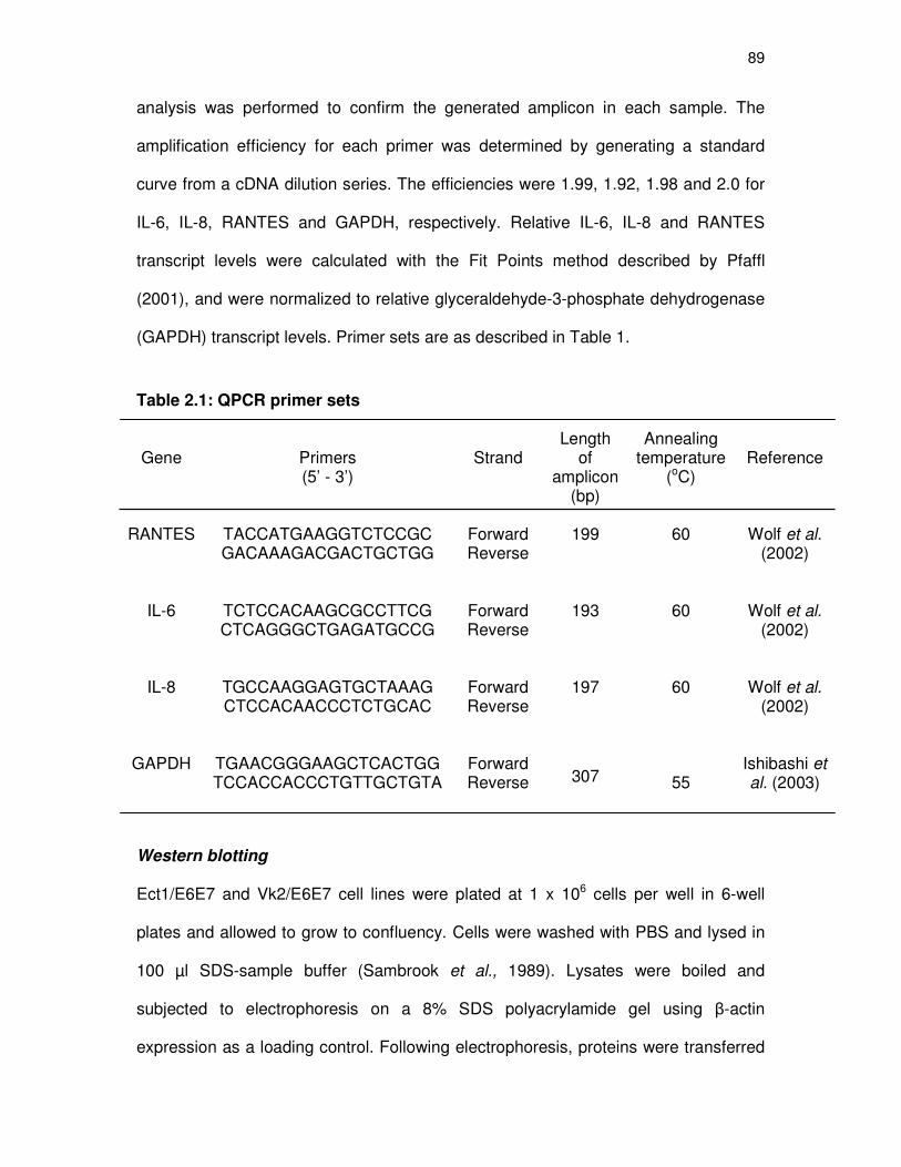
89
analysis was performed to confirm the generated amplicon in each sample. The
amplification efficiency for each primer was determined by generating a standard
curve from a cDNA dilution series. The efficiencies were 1.99, 1.92, 1.98 and 2.0 for
IL-6, IL-8, RANTES and GAPDH, respectively. Relative IL-6, IL-8 and RANTES
transcript levels were calculated with the Fit Points method described by Pfaffl
(2001), and were normalized to relative glyceraldehyde-3-phosphate dehydrogenase
(GAPDH) transcript levels. Primer sets are as described in Table 1.
Table 2.1: QPCR primer sets
Gene
Primers (5’ - 3’)
Strand
Length of
amplicon (bp)
Annealing temperature
(oC)
Reference
RANTES
TACCATGAAGGTCTCCGC GACAAAGACGACTGCTGG
Forward Reverse
199
60
Wolf et al.
(2002)
IL-6
TCTCCACAAGCGCCTTCG CTCAGGGCTGAGATGCCG
Forward Reverse
193
60
Wolf et al.
(2002)
IL-8
TGCCAAGGAGTGCTAAAG CTCCACAACCCTCTGCAC
Forward Reverse
197
60
Wolf et al.
(2002)
GAPDH
TGAACGGGAAGCTCACTGG TCCACCACCCTGTTGCTGTA
Forward Reverse
307
55
Ishibashi et al. (2003)
Western blotting
Ect1/E6E7 and Vk2/E6E7 cell lines were plated at 1 x 10⁶ cells per well in 6-well
plates and allowed to grow to confluency. Cells were washed with PBS and lysed in
100 µl SDS-sample buffer (Sambrook et al., 1989). Lysates were boiled and
subjected to electrophoresis on a 8% SDS polyacrylamide gel using β-actin
expression as a loading control. Following electrophoresis, proteins were transferred

90
to nitrocellulose membranes. Blots were probed with an anti-AR (H-280, 1:1000),
anti-GR (H300, 1:3000), anti-PR-B (sc-811, 1:500), anti-PR-AB (sc-810, 1:500) or
anti-MR (H300, 1:1000) antibodies from Santa Cruz Biotechnology Inc. (Santa Cruz,
CA, USA), as well as the anti-β-actin (1:1500) antibody from Cell Signalling
Technology, Massachusetts, USA, for 16 hours at 4oC. Blots were subsequently
incubated with horseradish peroxidase (HRP)-conjugated goat anti-rabbit secondary
antibody (1:10000, NA934VS, Amersham, South Africa) for 1 hour. Proteins were
visualized using enhanced chemiluminescence (ECL) from Amersham, followed by
exposure to Hyperfilm MP high performance autoradiography film (Amersham, South
Africa).
Whole cell binding assays to determine steroid receptor content in the
Ect1/E6E7 and Vk2/E6E7 cell lines
Competitive whole cell binding assays were performed essentially as described by
Bamberger et al. (1995), with the following modifications. Ect1/E6E7 and Vk2/E6E7
cell lines were maintained as described above. Cells were seeded into 12-well tissue
culture plates (Nunc, Denmark) at 2 x 10⁵ cells per well. On day 3, the cells were
washed with phosphate-buffered saline (PBS) and incubated for 2 hours at 37°C with
10 nM 3[H]-MIB (76.8 Ci/mmol) for AR, 3[H]-Ald (87.9 Ci/mmol) for MR, 3[H]-R5020
(84.6 Ci/mmol) for PR (3[H]-MIB, 3[H]-Ald, and 3[H]-R5020 all from PerkinElmer Life
and Analytical Science, South Africa) or 3[H]-Dex (89 Ci/mmol) for GR (AEC-
Amersham, South Africa), in the absence (total binding) and presence of 10 µM
unlabelled MIB, aldosterone, R5020, or Dex (non-specific binding), respectively.
Working on ice at 4oC, cells were washed three times with ice-cold 1X PBS
containing 0.2% (w/v) bovine serum albumin (BSA) for 15 min. Cells were then lysed

91
with 200 µl reporter lysis buffer (Promega, Madison, USA). Total binding was
determined by scintillation counting as counts per minute (cpm), while specific
binding was determined by subtracting non-specific binding from total binding.
Protein concentrations were determined using the Bradford protein assay method
(Bradford, 1976). Specific binding was normalized to the protein concentration. The
reported values are averages of at least three independent assays, with each
condition performed in triplicate. The receptor numbers in ƒmol/mg protein were
determined as follows:
The specific activity of the tritium-labelled test compound (Ci/mmol) was converted to
disintegrations per minute (dpm) per mmol, by multiplying by a factor of 2.22 x 10¹² (1
Curie equals 2.22 x 10¹² dpm). The dpm was multiplied by the counting efficiency to
get cpm per mmole, and divided by 1012 to get cpm/ƒmol. Counting efficiency was
40% in our system. The specific activity was expressed as cpm/ƒmol. Dividing the
specific binding cpm value by this value gives specific binding in ƒmol, which was
divided by the protein content of the sample in mg, to yield the number of binding
sites in ƒmol/mg protein.
Luciferase reporter assays
For transactivation assays, the Ect1/E6E7 and Vk2/E6E7 cells were maintained as
above and seeded into 24-well tissue culture plates at 5 x 10⁴ cells per well. On day
2, cells in each well were transiently transfected with 330 ng of the pTAT-GRE-E1b-
luc construct and 33 ng of the expression vector for the hMR, hGR, hAR, hPR-B or
the empty vector pGL2basic, using the FuGENE6 transfection reagent (Roche
Molecular Biochemicals, South Africa) in accordance with the manufacturer’s
instructions. On day 3, the cells were washed with PBS and induced with 10 µM of
92
either Ald, Dex, MIB, or R5020. After 24 hours, the cells were lysed with reporter lysis
buffer (Promega, Madison, USA). Luciferase activity in the lysate was measured
using the Luciferase Assay System (Promega, Madison, USA) and a Veritas
microplate luminometer (Turner Biosystems, Sunnyvale CA, USA). The values
obtained were normalised to expression of β-gal, which was measured using the
GalactoStar Assay Kit from Tropix (Bedford MA, USA). The reported values are
averages of at least three independent experiments, with each condition performed in
triplicate.
Data manipulation and statistical analysis
The Graph Pad Prism software was used for data manipulations, graphical
representations, and statistical analysis. One-way ANOVA analysis of variance and
Bonferroni posttests (compares all pairs of columns) were used for statistical
analysis. Statistical significance of differences is indicated by *, ** or ***, to indicate
p<0.05, p<0.01 or p<0.001, respectively, whereas no statistical significance (ns) is
indicated by p>0.05. The letters a,b,c etc. are also used to denote statistically
significant differences, where all those values which differ significantly from others,
are assigned a different letter.
Results
MPA and NET-A, unlike Prog, exhibit differential patterns of gene regulation on
pro-inflammatory chemokines
Human Ect1/E6E7 and Vk2/E6E7 cell lines were treated for 24 hours with 0.02 µg/µl
TNF-α and 1 µM Prog, MPA or NET-A, followed by QPCR analysis for expression of
the IL-6, IL-8 and RANTES genes, respectively. In both the Ect1/E6E7 and Vk2/E6E7

93
cell lines, results show that unlike Prog which upregulates IL-6 gene expression, both
MPA and NET-A have no effect (figure 2.1A and 2.1B). In contrast, MPA and NET-A
differentially regulate both the IL-8 (figure 2.2A) and RANTES (figure 2.3A) genes in
the Ect1/E6E7 cell line. The results show that MPA, like Prog upregulates IL-8, (10.3-
vs. 12-fold, respectively), while NET-A upregulates the gene to a significantly lower
extent (2.8-fold) (figure 2.2A). Although not statistically different, Prog and NET-A
seemed to upregulate the IL-8 gene in the Vk2/E6E7 cell line to a similar extent,
while MPA showed the greatest level of upregulation (figure 2.2B). Unlike Prog which
upregulates RANTES gene expression in the Ect1/E6E7 cell line, MPA
downregulates the RANTES gene expression, while NET-A has no effect (figure
2.3A). Conversely, both MPA and NET-A, like Prog, upregulate the expression of the
RANTES gene in the Vk2/E6E7 cell line (figure 2.3B). Taken together, MPA and
NET-A exhibit differential patterns of gene regulation on expression of the IL-8 and
RANTES genes, as compared to Prog, in a promoter- and cell- specific manner. In
addition, unlike Prog, MPA and NET-A do not upregulate IL-6 gene expression.
The PR, AR and GR are expressed in both ectocervical and vaginal cell lines
As these progestins are known to interact with the PR, GR, AR and possibly the MR
(Philibert et al., 1999), we hypothesized that the differential regulation of genes by
MPA and NET-A, particularly on the RANTES gene, was due to their action via
different steroid receptors, or differing activities via the same steroid receptor. To test
this hypothesis, we firstly investigated which members of the steroid receptor family
were expressed in these cell lines using a combination of Western blotting (figure
2.4A) and whole cell binding (figure 2.4B and 2.4C).

94
Figure 2.1: Effect of MPA and NET-A on the TNF-α-induced expression of the IL-6 gene in
human ectocervical and vaginal epithelial cell lines. (A) Human Ect1/E6E7- and (B) Vk2/E6E7 cell
lines were incubated with 0.02 µg/µl TNF-α in the presence of 0.1% EtOH or 1 µM Prog, MPA or NET-
A for 24 hours. Total RNA was extracted, cDNA synthesized and expression levels of IL-6 and
GAPDH mRNA were determined by QPCR. Results shown (ratio of the specific target gene/GAPDH
gene) are the averages (±SEM) of at least three independent experiments.
EtOH Prog MPA NET-A0
1
2
3b
aa
a
1 µµµµM test compound
Rati
o I
L6/G
AP
DH
exp
ressio
n
EtOH Prog MPA NET-A0
1
2
3
4
5
1 µµµµM test compound
b
a
a
a
Rati
o I
L6/G
AP
DH
exp
ressio
n
(A) ECTOCERVICAL CELLS
(B) VAGINAL CELLS
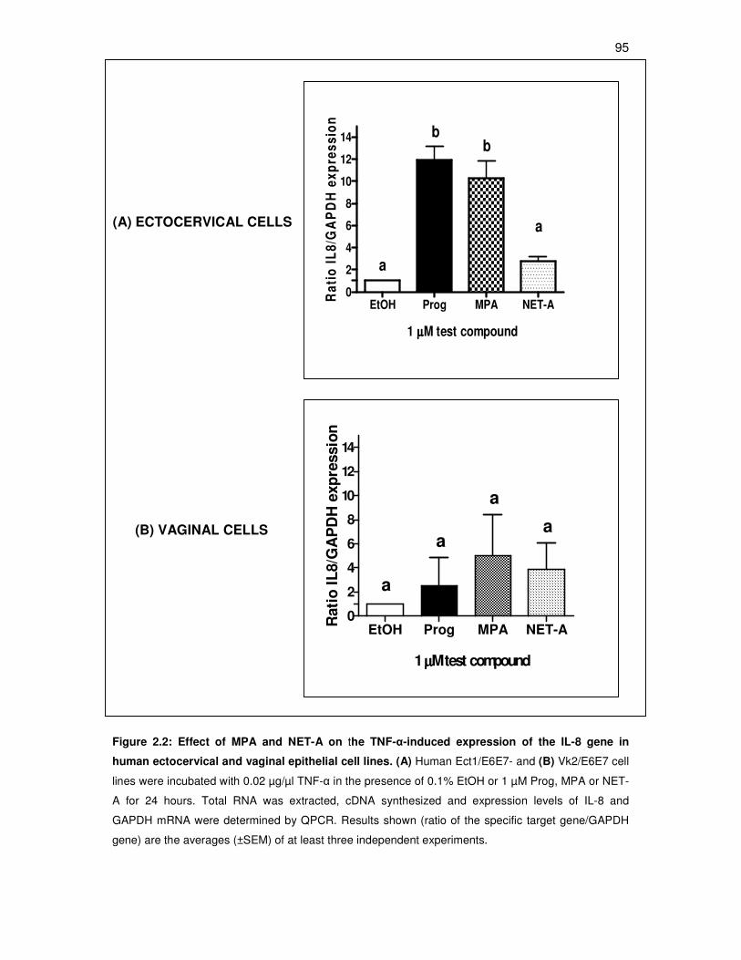
95
Figure 2.2: Effect of MPA and NET-A on the TNF-α-induced expression of the IL-8 gene in
human ectocervical and vaginal epithelial cell lines. (A) Human Ect1/E6E7- and (B) Vk2/E6E7 cell
lines were incubated with 0.02 µg/µl TNF-α in the presence of 0.1% EtOH or 1 µM Prog, MPA or NET-
A for 24 hours. Total RNA was extracted, cDNA synthesized and expression levels of IL-8 and
GAPDH mRNA were determined by QPCR. Results shown (ratio of the specific target gene/GAPDH
gene) are the averages (±SEM) of at least three independent experiments.
(A) ECTOCERVICAL CELLS
(B) VAGINAL CELLS
EtOH Prog MPA NET-A0
2
4
6
8
10
12
14
1 µµµµM test compound
b
a
b
a
Ra
tio
IL
8/G
AP
DH
ex
pre
ss
ion
EtOH Prog MPA NET-A0
2
4
6
8
10
12
14
1µµµµM test compound
a
a
a
a
Rati
o I
L8/G
AP
DH
exp
ressio
n

96
Figure 2.3: Effect of MPA and NET-A on the TNF-α-induced expression of the RANTES gene in
human ectocervical and vaginal epithelial cell lines. (A) Human Ect1/E6E7- and (B) Vk2/E6E7 cell
lines were incubated with 0.02 µg/µl TNF-α in the presence of 0.1% EtOH or 1 µM Prog, MPA or NET-
A for 24 hours. Total RNA was extracted, cDNA synthesized and expression levels of RANTES and
GAPDH mRNA were determined by QPCR. Results shown (ratio of the specific target gene/GAPDH
gene) are the averages (±SEM) of at least three independent experiments.
(A) ECTOCERVICAL CELLS
(B) VAGINAL CELLS
EtOH Prog MPA NET-A0
1
2
3
4 b
a
c
a
1µµµµM test compoundR
ati
o R
AN
TE
S/G
AP
DH
ex
pre
ss
ion
EtOH Prog MPA NET-A0
1
2
3 b
a
a,ba,b
1µµµµM test compound
Rati
o R
AN
TE
S/G
AP
DH
exp
ressio
n

97
Whole cell binding results showed that the GR, AR, and PR, are expressed in both
these cell lines, while relatively very low amounts of MR are expressed only in the
Vk2/E6E7 cell line (figure 2.4C). Western blot analysis using an antibody specific for
the MR (H-300), confirmed the absence of the MR in the Ect1/E6E7 cells (data not
shown-Renate Louw, MSc student). However, the low levels of MR in the Vk2/E6E7
cell line observed in the whole cell binding assay, could not be detected using
Western blotting (data not shown-Renate Louw, MSc student).
We subsequently determined the endogenous steroid receptor levels in both cell
lines. The results show that there is significantly (p<0.001) more PR than AR or GR
in the Ect1/E6E7 andVk2/E6E7 cell lines, respectively. Moreover, the levels of AR vs.
GR are similar to each other in both cell lines (p>0.05), (figure 2.4B and 2.4C). The
expression of the GR and AR in both cell lines was confirmed by Western blot
analysis using antibodies specific for the GR and AR (figure 2.4A). As the GR is
ubiquitously expressed (reviewed in Gross and Cidlowski, 2008), it is difficult to find a
cell line that does not show low levels of GR. As expected, we detected low levels of
endogenous GR in the untransfected COS-1 cell line (negative control). There are
two functional isoforms of the PR, PR-A and PR-B (Kastner et al., 1990; Kraus and
Katzenellenbogen, 1993). Western blot analysis using an antibody specific for the
PR-B isoform showed that both these cell types did not express PR-B. We were
unable to optimize the commercial antibody detecting the PR-A isoform (anti-PR-AB
(sc-810), Santa Cruz Biotechnology Inc. (Santa Cruz, CA, USA)), and thus could not
determine by Western blotting whether the PR-A isoform is expressed in these cell
lines. Taken together, the results showing the expression of the PR, AR, and GR
isoforms in both the Ect1/E6E7 and Vk2/E6E7 cell lines, support our hypothesis that

98
the differential effects of MPA and NET-A could be a consequence of their action via
different steroid receptors.
Receptor-specific antagonists indicate a role for the AR in the downregulation
of the RANTES pro-inflammatory chemokine gene by MPA in the ectocervical
cell line
Having determined that these cell lines mostly express AR, GR and PR, our strategy
was to determine which of these steroid receptors mediate the differential effects of
MPA and NET-A on the RANTES genes in the Ect1/E6E7 cell line. Although
differential effects were also observed for MPA and NET-A on the expression of the
IL-8 gene, it was decided to continue the investigation only for the RANTES gene, as
MPA and NET-A exhibited differential effects, not only to each, but also to Prog on
this gene. For this reason, Ect1/E6E7 cells were incubated with 0.02 µg/ml TNF-α
and 1 µM MPA or NET-A, in the absence and presence of 10 µM hydroxyflutamide
(OHF), the classical androgen antagonist or mifepristone (RU486), the PR and GR
antagonist. The addition of OHF prevented the inhibition by MPA, indicating that, in
this cell line, the repression by MPA on the RANTES gene is mediated by the AR
(figure 2.7A).

99
~110 kD AR
~97 kD GR
Vk2/E
6E
7
Ect1
/E6E
7
-ve
ctl
+ve
ctl
~116 kD PR-B
PR AR GR MR0
1
2
3
4
5
80
100
120
140
a
bb
cSte
roid
recep
tor
co
nte
nt
fmo
l/m
g p
rote
in
PR AR GR MR0
1
2
3
4
5100
120
140
160
180
200
a
b
b
c
Ste
roid
re
ce
pto
r c
on
ten
tfm
ol/m
g p
rote
in
Figure 2.4 legend on next page
(B)
(C)
Ectocervical cells
Vaginal cells
(A)

100
Figure 2.4: PR, AR and GR are expressed in human ectocervical and vaginal epithelial cell
lines. (A) Whole cell extracts were prepared from human Ect1/E6E7 and Vk2/E6E7 cell lines,
untransfected COS-1 cell line (negative control (-ve ctl)), COS-1 cell line transfected with pRS-hGRα,
pSVARo, or pSG5hPR-B expression vectors (positive controls (+ve ctl)). Equal amounts of protein (20
µg) were analyzed by Western blotting with the GR or AR or PR-B antibodies. (B) Human Ect1/E6E7-
and (C) Vk2/E6E7 cell lines were incubated with 10 nM [3H]-R5020 or [
3H]-Dex or [
3H]-MIB or [
3H]-
Ald in the absence (total binding) and presence of 10 µM unlabelled (non-specific binding) R5020 or
Dex or MIB or Ald, respectively. Specific binding (total binding minus non-specific binding) is plotted.
Results shown are the averages (±SEM) of at least three independent experiments with each
condition performed in triplicate. The number of binding sites in ƒmol/mg protein was calculated from
the specific activity of the radiolabelled ligands and the specific binding cpm value, as described in the
materials and methods section.
Only the GR was transcriptionally active in promoter-reporter transactivation
assays in both ectocervical and vaginal cell lines
Having established that the Ect1/E6E7 and Vk2 cell lines both express PR, GR, AR,
and very low levels of MR in the Vk2/E6E7 cell line, we next determined whether
these receptors are transcriptionally active by transiently transfecting these cells with
a glucocorticoid response element (GRE)-driven reporter construct containing two
copies of the rat tyrosine amino transferase (TAT) GRE. The cells were
subsequently exposed to 10 µM of the receptor-specific agonist Dex for the GR, MIB
for the AR, R5020 for the PR and Ald for the MR. The results show that only the GR
is transcriptionally active in the Ect1/E6E7 (figure 2.5) and Vk2/E6E7 (figure 2.6) cell
lines. However, this does not mean that the other endogenously expressed receptors
are not biologically active in other assays. Results from transrepression experiments
via a synthetic NFκB-promoter have indicated transcriptionally active GR and AR, but
not PR in the ectocervical cells (data not shown-Renate Louw, MSc student). In
addition, it has previously been shown that the potency of a ligand for transactication
via a GRE is less than the potency for transrepression via NFκB (Ronacher et al.,
2009). Thus the other endogenous receptors may still be active on other synthetic

101
and endogenous genes, such as in transrepression assays. These results with
transfected receptors do however also show that the agonist used are active in these
cell lines.
Consistent with this result, dihydrotestosterone (DHT), the natural androgen, also
repressed the RANTES gene in this cell line via the AR (figure 2.7A). Interestingly,
the addition of OHF did not significantly inhibit the effects of Prog or NET-A,
indicating that the effects of these compounds on the RANTES gene are not
mediated via the AR. A similar experiment with OHF in the Vk2/E6E7 cell line
indicated that the AR is not involved in any of the responses by Prog, MPA or NET-A
on the RANTES gene (Appendix A1).
RU486 did not inhibit the effects mediated by the positive controls: dexamethasone
(Dex), a synthetic GR agonist, or promegestone (R5020), a synthetic PR agonist
(figure 2.7B), indicating that RU486 is not working as a GR or PR antagonist in the
Ect1/E6E7 cell line, but rather as an agonist. It has previously been reported that
RU486 may act as an agonist in some cells and as an antagonist in others
(Bamberger and Chrousos, 1995). The fact that the expression of the RANTES gene
is enhanced when RU486 is added together with Prog, MPA or NET-A, is difficult to
interpret. Taken together, conclusions cannot be drawn as to the role of the GR or
PR in mediating the repression of the TNFα-induced RANTES gene by MPA. Using a
PR antagonist, like Org31710, which has little anti-glucocorticoid activity compared to
RU486 (Kloosterboer et al., 1994), or a GR-specific antagonist like DO6 (Miner et al.,
2003), was not possible since these antagonists are not commercially available.

102
Figure 2.5: Transactivation of a transiently transfected GRE-containing promoter-reporter
construct via endogenous or expressed GR, AR, PR, or MR. Ect1/E6E7 cells (5x10⁴ cells per well
in a 24-well plate) were transiently transfected with 330 ng pTAT-GRE2-Elb-luc and 33 ng pRS-hGRα,
pSVARo, pSG5hPRB , pRS-hMR or pGL2basic (empty vector control) as indicated, with Fugene 6
transfection reagent according to the manufacturer’s instructions. Twenty-four hrs after transfection
cells were induced for 24 hrs with vehicle (ethanol (EtoH)), 10 µM Dex, MIB, R5020 or Ald (receptor-
agonists).
EtOH Dex EtOH Dex0
2
4
6
8
10
-GR +GR
Fo
ld i
nd
ucti
on
(L
uc)
±± ±±S
EM
(n
=3)
EtOH MIB EtOH MIB0
1
2
3
4
5
80
100
120
140
-AR +AR
Fo
ld i
nd
ucti
on
(Lu
c)
±± ±±S
EM
(n
=3)
EtOH R5020 EtOH R50200
1
2
3
4
5100
150
200
250
300
-PR +PR
Fo
ld i
nd
ucti
on
(L
uc)
±± ±±S
EM
(n
=3)
EtOH Ald EtOH Ald0
1
2
3
4
525
30
35
40
45
50
-MR +MR
Fo
ld i
nd
ucti
on
(Lu
c)
±± ±±S
EM
(n
=3)
(A) (B)
(C) (D)
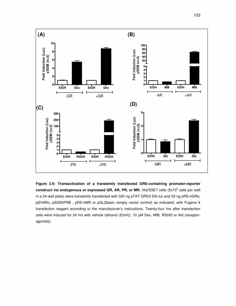
103
Figure 2.6: Transactivation of a transiently transfected GRE-containing promoter-reporter
construct via endogenous or expressed GR, AR, PR, or MR. Vk2/E6E7 cells (5x10⁴ cells per well
in a 24-well plate) were transiently transfected with 330 ng pTAT-GRE2-Elb-luc and 33 ng pRS-hGRα,
pSVARo, pSG5hPRB , pRS-hMR or pGL2basic (empty vector control) as indicated, with Fugene 6
transfection reagent according to the manufacturer’s instructions. Twenty-four hrs after transfection
cells were induced for 24 hrs with vehicle (ethanol (EtoH)), 10 µM Dex, MIB, R5020 or Ald (receptor-
agonists).
(A) (B)
(C) (D)
EtOH Dex EtOH Dex0
2
4
6
8
10
-GR +GR
Fo
ld i
nd
uc
tio
n (
Lu
c)
±± ±±S
EM
(n
=3
)
EtOH MIB EtOH MIB0
1
2
3
4
5
20
40
60
80
100
-AR +AR
Fo
ld i
nd
uc
tio
n (
Lu
c)
±± ±±S
EM
(n
=3
)
EtOH R5020 EtOH R50200
1
2
3
4
5
100
200
300
-PR +PR
Fo
ld i
nd
uc
tio
n (
Lu
c)
±± ±±S
EM
(n
=3
)
EtOH Ald EtOH Ald0
1
2
3
-MR +MR
Fo
ld i
nd
uc
tio
n (
Lu
c)
±± ±±S
EM
(n
=3)

104
Figure 2.7: Effect of the androgen receptor antagonist (OHF) (A) or
glucocorticoid/progesterone receptor antagonist (RU486) (B) on the MPA inhibition of the TNF-
α-induced RANTES gene. The human Ect1/E6E7 cell line was incubated with 0.02 µg/µl TNF-α in the
presence of 1 µM Prog, MPA, NET-A, DHT, DEX, or R5020, and absence or presence of 10 µM (A)
hydroxyflutamide (OHF) or (B) RU486 for 24 hours. Total RNA was extracted, cDNA synthesized, and
expression levels of RANTES and GAPDH mRNA were determined by QPCR. Results shown (ratio of
the specific target gene/GAPDH gene) are the averages (±SEM) of at least two independent
experiments. Open bars are compounds and vehicle only, while the black bars are compounds plus
antagonist.
(B)
EtOH Prog MPA NET DHT0
1
2
3
4
OHF
w/o OHFR
ati
o R
AN
TE
S/G
AP
DH
exp
ressio
n**
**
*
ns
ns(A)
EtOH Prog MPA NET DEX R50200
1
2
3
4
10
20
30
RU486
w/o RU486
Rati
o R
AN
TE
S/G
AP
DH
expre
ssio
n

105
DISCUSSION
Inflammation of the lower human female genital tract increases susceptibility to viral
infections such as human immunodeficiency virus (HIV) (Kreiss et al., 1994; Hart et
al., 1999) and human papilloma virus (HPV) (Castle et al., 2000). In addition,
excessive release of proinflammatory cytokines may alter the mucosal immune
function (Alfano and Poli, 2005). Thus, understanding factors that may influence the
local mucosal immune response, such as endogenous hormones or hormonal
contraception, is crucial, especially since the cervicovaginal mucosa is the primary
site of HIV-1 infection during male to female transmission (reviewed in Pope and
Haase, 2003). Although, the synthetic progestins medroxyprogesterone acetate
(MPA) and norethindrone acetate (NET-A) are widely used in both contraception and
hormone replacement therapy (HRT), little is known about their effects on the
cervicovaginal environment, and whether they interfere with local epithelial immune
function. In this study we investigated the effects of MPA, NET-A and Prog on two
immortalized, morphologically and functionally different epithelial cell lines, one from
a normal human ectocervix and the other from a normal human vagina (Fichorova et
al., 1999). Although immortalized, the authors report that the morphological and
immunocytochemical properties of these cell lines resemble not only their tissues of
origin, but also those of primary cultures (Fichorova et al., 1997). Notably, this is not
the case for the HeLa cervical cell line, which is probably the most frequently used
cell line derived from the human female lower genital tract mucosa (Fichorova et al.,
1997). The Ect1/E6E7 and Vk2/E6E7 cell lines express characteristics of stratified
squamous nonkeratinizing epithelia, and constitutively express IL-8, while IL-6 and
RANTES are produced when the cell lines are stimulated with either TNFα or
interferon gamma (IFNγ) (Fichorova and Anderson, 1999).

106
Prog increases the expression of IL-6, IL-8 and RANTES, in both the Ect1/E6E7 and
Vk2/E6E7 cell lines, while NET-A had little to no effect. We found that Prog
upregulated the TNFα-induced proinflammatory cytokine interleukin (IL)-6 in both cell
lines, while MPA and NET-A had no effect (figure 2.1A and 2.1B). This is inconsistent
with previous studies that have shown the repression of IL-6 by MPA in other cell
types (Mantovani et al., 1997; Koubovec et al., 2004). Similarly, NET-A has
previously been shown to either upregulate (Zitzmann et al., 2005) or downregulate
(Wessel Kriek, MSc thesis) IL-6. In contrast, NET-A in combination with estrogen, like
estrogen only, had no effect on IL-6 levels in healthy postmenopausal women
(Žegura et al., 2006). In the Ect1/E6E7 cell line, the IL-8 gene was upregulated by
Prog, MPA and NET-A, albeit to different extents, and statistical analysis showed that
while the extent of upregulation by Prog and MPA is similar (p>0.05), NET-A’s
upregulation is significantly different from both Prog (p<0.001) and MPA (p<0.01)
upregulation (figure 2.2A). A similar pattern was observed for the compounds on the
IL-8 gene in the Vk2/E6E7 cell line (figure 2.2B). However, due to variability in the
extent of the response between experiments, when the results from different
independent experiments were combined, the changes in IL-8 mRNA levels were not
statistically significant. However, these results follow a similar trend between
experiments, and are most likely physiologically relevant. Consistent with our results,
both Prog and MPA have previously been shown to upregulate IL-8 mRNA levels in
human endometrial stromal cells (Arici et al., 1996). As Prog has previously been
shown to have a protective role in the vagina (Beaver, 1960), our results showing
that MPA and NET-A are similar to Prog in upregulating IL-8, albeit to different
extents, in the Ect1/E6E7 and Vk2/E6E7 cell lines, could indicate that these

107
progestins may provide an enhanced local defense mechanism against infections in
the vaginal mucosa.
MPA substantially and significantly downregulates the expression of the
proinflammatory RANTES gene in the Ect1/E6E7 cell line, (figure 2.3A), while it is
upregulated by MPA in the Vk2/E6E7 cell line (figure 2.3B). Thus, the same
compound, MPA, exhibits differing effects on the same gene (RANTES), in different
cell types, indicating cell-specific effects of MPA. In addition, NET-A had no
statistically significant effect on the RANTES gene in both cell types, indicating
differential regulation of RANTES by the two synthetic progestins thought to have
similar biological activity. Trying to understand the physiological implications of this
result on the RANTES gene is not easy. In epithelial cells, RANTES recruits immune
cells into the reproductive tract, in this manner contributing to inflammation. Inhibition
of RANTES by MPA would thus be expected to suppress inflammation, thereby
blocking infiltration of immune cells (Cho et al., 2006). This lack of recruitment of
immune cells into the reproductive tract may have implications for women using
MPA. In terms of HIV-1 infection, which has been associated with increased
RANTES in cervicovaginal secretions (Behbahani et al., 2007), these implications
may be positive in terms of less HIV-infection due to decreased inflammation
(Cummins et al., 2006). However, women using MPA in contraception and HRT are
also at risk of other infections, and thus the suppression of RANTES may prevent the
mounting of an effective response to combat these infections in the ectocervical
environment. In addition, it is important to remember that the defense function in the
cervicovaginal environment is dependent on number of regulatory factors in different
anatomical sites, with a constant release of pro- and anti-inflammatory mediators.

108
Thus the observed effects of MPA on RANTES should be considered in the light of
the fact that there may be a balance between pro- and anti-inflammatory molecules
produced in the cervicovaginal environment. It would be of interest to look at the
expression of RANTES in cervicovaginal lavage samples from healthy-, and HIV-1
infected- women using MPA and NET as injectable contraception, as compared to
women not using these contraceptives.
In summary, the general trend that was observed for the effect of Prog, MPA and
NET-A on endogenous cytokine genes in the cervicovaginal environment, is that
Prog upregulates the expression of IL-6, IL-8 and RANTES, while NET-A either
upregulates these genes, or has no effect. In contrast, MPA either has no effect (e.g.
IL-6 gene), or upregulates (e.g. IL-8 gene), or downregulates (e.g. RANTES gene)
expression of these genes. Furthermore, Prog shows regulation different to both
MPA and NET-A on the RANTES gene in the Ect1/E6E7 cell line (figure 2.3A), as
well as on the IL-6 gene in both the Ect1/E6E7 and Vk2/E6E7 cell lines (figure 2.1A
and 2.1B). Taken together, these results show that cytokine genes in the
cervicovaginal environment are regulated in a ligand-specific and cell-specific
manner by Prog, MPA and NET-A.
We hypothesized that the ligand-specific and cell-specific effects regulation of
cytokine genes in the cervicovaginal environment are due to ligands acting via
different steroid receptors, or differing activities via the same steroid receptor. Using
a combination of whole cell binding and Western blotting, we showed that the PR, AR
and GR are expressed in both cell lines (figure 2.4A-C). Whole cell binding
experiments showed that the amount of GR and AR in the Ect1/E6E7 and Vk2/E6E7

109
cell lines is similar (figure 2.4B and 2.4C). Since the whole cell binding experiments
showed the presence of a large amount of PR, one would expect that the progestins
would predominantly act via this receptor. However, we did not observe any PR-
mediated effects in our study. In fact, the potent synthetic PR agonist, R5020,
displayed no activity for transactivation or transrepression via the PR. A possible
explanation for this intriguing result may be explained by the expression levels of
different PR isoforms. We were unable to detect the PR-B isoform in any of the cell
lines, and thus we postulate that it is the transcriptionally less active PR-A isoform
that is present in these cell lines. Indeed, variation in the expression of PR-A and PR-
B has previously been reported in the eutopic endometrium during the menstrual
cycle (Mangal et al., 1997), with no PR-B detectable during the secretory and early
proliferative phases. Similarly, Attia et al. (2000) did not detect any PR-B protein in
endometriotic tissue, and they ascribe the clinically observed resistance of
endometriosis to treatment with progestins, to this absence of PR-B, and presence of
PR-A.
We next determined which steroid receptor(s) are involved in the response of MPA
on the RANTES gene in the Ect1/E6E7 cell line, by using receptor-specific
antagonists. As we had shown that this cell line expresses AR, GR and PR (figure
2.4B), we looked at the expression of the RANTES gene in the presence of OHF, the
AR-specific antagonist, as well as RU486, the PR and GR antagonist. The results
show that in the Ect1/E6E7 cell line, RU486 behaves as an agonist, rather than an
antagonist (figure 2.7B). RU486 had previously been shown to act as both an agonist
and as an antagonist, depending on receptor density (Zhao et al., 2003).
Furthermore, it has been reported that RU486 is a partial agonist for both PR and GR
110
when selective coregulators are recruited (Jackson et al., 1997; Schulz et al., 2002).
This approach of using RU486 as a receptor antagonist in this cell line is thus limited.
Interestingly, the results using OHF showed that the repression of MPA on the
RANTES gene was lifted when the AR is antagonised, revealing a possible role for
the AR (figure 2.7A). The fact that OHF could lift the repression of MPA on the
RANTES gene to similar levels as the control (EtOH), suggests that the PR, despite
being the most abundant receptor, as well as the GR, are not involved in the MPA-
mediated downregulation of the RANTES gene in the Ect1/E6E7 cell line.
Considering that both MPA and NET-A have previously been reported to have
androgenic properties (Bentel et al., 1999; Teulings et al., 1980; Bamberger et al.,
1999; Kemppainen et al., 1999; Hackenberg et al., 1993; Perez-Palacios et al., 1983;
Perez-Palacios et al., 1981a; Perez-Palacios et al., 1981b; Bergink et al., 1983;
Schoonen et al., 2000; Deckers et al., 2000), it was surprising that NET-A did not act
via the AR in this cell line. However, as it is known that NET-A can be metabolized
(Stanczyk and Roy, 1990), the possibility exists that NET-A is metabolized in this cell
line, and that the metabolite(s), are less efficacious AR agonists than NET-A itself.
In summary, results from our study are the first to indicate not only differential
regulation of the cytokine genes by Prog, MPA and NET-A, but also cell-specific
effects. Moreover, we showed that the effect of MPA on the RANTES gene in the
Ect1/E6E7 cell line is AR-mediated.
Acknowledgements
We thank Carmen Langeveldt for technical support. This work was supported by
grants to JPH and DA from the Medical Research Council (MRC) and the National

111
Research Foundation (NRF) in South Africa, and Stellenbosch University. Any
opinion, findings and conclusions or recommendations expressed in this material are
those of the author(s) and therefore the NRF does not accept any liability in regard
thereto.

112
Chapter 3
_____________________________________________________
A comparative study of the androgenic properties of progesterone
and the synthetic progestins, medroxyprogesterone acetate (MPA)
and norethisterone acetate (NET-A)
Donita Africander1 and Janet Hapgood1*
1Department of Biochemistry, University of Stellenbosch, Private Bag X1, Matieland,
7602, South Africa.
* Current address: Department of Molecular and Cell Biology, University of Cape
Town, Private Bag, Rondebosch, 7700, South Africa.
Manuscript in preparation for submission to Journal of Steroid Biochemistry and
Molecular Biology.

113
Abstract
Medroxyprogesterone acetate (MPA) and norethisterone enanthate (NET-EN) are
used by millions of women as contraceptives and in hormone replacement therapy
(HRT). Surprisingly, even though both progestins are widely used, very little is known
about their mechanism of action at the molecular level. The importance of
investigating these mechanisms, and comparing them to those of progesterone
(Prog), has recently been highlighted by clinical evidence showing that MPA and
NET increase the risk of the development of breast cancer in HRT users. MPA is also
implicated in increased risk of coronary heart disease in HRT users. The aim of this
study was to investigate and compare the androgenic activities of these two clinically
important progestins. Competitive binding in whole cells revealed that Prog, MPA and
NET-A have a similar binding affinity for the androgen receptor (AR) to that of the
natural androgen dihydrotestosterone (DHT) (Ki’s for DHT, Prog, MPA and NET-A
are 29.4, 36.6, 19.4 and 21.9 nM, respectively). Moreover, in both transactivation and
transrepression transcriptional assays we demonstrate that, unlike Prog, MPA and
NET-A are efficacious AR agonists, with activities comparable to DHT. The finding
that both MPA and NET-A have strong androgenic effects when compared to
endogenous Prog, may have important implications for women using these
progestins in HRT or contraception, as it may lead to side-effects such as weight
gain, lipid profile modifications or increased breast cancer risk. Furthermore, we show
that MPA and NET-A exert their androgenic actions by different mechanisms, as
NET-A, like DHT and other well-characterised androgens, induces the ligand-
dependent interaction between the NH2- and COOH-terminal domains (N/C-
interaction) of the AR independent of promoter-context, while MPA does this in a
promoter-dependent manner.

114
Introduction
The synthetic progestins, medroxyprogesterone acetate (MPA) and norethisterone
enanthate (NET-EN) are widely used in contraception and in hormone replacement
therapy (HRT). MPA itself is the major progestogenic compound, while NET-EN and
norethisterone acetate (NET-A) are hydrolysed to norethisterone (NET) and its
metabolites, which together have progestogenic action (Stanczyk and Roy, 1990).
The Women’s Health Initiative (WHI) trial showed healthy postmenopausal women
using combined estrogen and MPA therapy in HRT have an increased risk of breast
cancer and cardiovascular complications (Rossouw et al., 2002). A similar trial on the
use of estrogen alone was only terminated two years later and its analysis, although
indicating an increased risk for stroke, showed no increase in breast cancer risk
(Anderson et al., 2004). MPA and NET have both been shown to substantially
increase the risk of breast cancer in longterm HRT users in the “Million Women
study” (Beral, 2003). Surprisingly, very little information is available about the
molecular mechanism of action of MPA and NET, in particular their comparative
properties (Hapgood et al., 2004).
Notably, a few studies have recently started investigating these mechanisms, but
focus mostly on MPA, and not NET-A. MPA has been reported to modulate
transcription of a number of genes via the glucocorticoid receptor (GR), such as IL-2
in normal human lymphocytes (Bamberger et al., 1999), IL-6 and IL-8 in a mouse
fibroblast cell line (Koubovec et al., 2004) and the nm-23 tumor suppressor gene in a
breast cancer cell line (Ouatas et al., 2003). Interestingly, MPA has a greater affinity
than NET-A for the GR, and acts as a partial agonist for transactivation (Koubovec et
al., 2005), while NET-A is a GR antagonist (Appendix C6). This demonstrates that
115
MPA and NET do not have the same activity, nor do they act the same as Prog, via
the GR. It is thus apparent that even though MPA and NET were developed so that
their biological actions mimicked that of progesterone (mediating their effects by
binding to the PR), they can also initiate a diverse range of biological effects by
cross-reacting with other members of the steroid receptor family such as the GR and
possibly the mineralocorticoid receptor (MR) (Hapgood et al., 2004; Koubovec et al.,
2005; Philibert et al., 1999). In this way, MPA and NET may exert side-effects via any
of these receptors, such as the observed immunosuppressive effects of MPA via the
GR. This highlights the importance of understanding the interactions of these
compounds with different steroid receptors.
Both MPA and NET-A have previously been reported to have androgenic properties
(Bentel et al., 1999; Teulings et al., 1980; Bamberger et al., 1999; Kemppainen et al.,
1999; Hackenberg et al., 1993; Perez-Palacios et al., 1983; Perez-Palacios et al.,
1981a; Perez-Palacios et al., 1981b; Bergink et al., 1983; Schoonen et al., 2000;
Deckers et al., 2000; Bergink et al., 1983; Bergink et al., 1985). In addition, MPA has
been shown to regulate a number of genes via the AR in human breast cancer cell
lines (Ghatge et al., 2005). Our own work in human ectocervical cells (Ect/E6E7),
indicates involvement of the AR in the downregulation of the proinflammatory
RANTES (Regulated-upon-Activation, normal T cell Expressed and Secreted) gene
by MPA (Chapter 2). Little research has been done to directly compare the relative
androgenic activities of MPA and NET-A, as compared to the activity of Prog, and the
precise mechanisms involved.
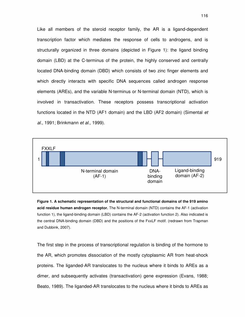
116
Like all members of the steroid receptor family, the AR is a ligand-dependent
transcription factor which mediates the response of cells to androgens, and is
structurally organized in three domains (depicted in Figure 1): the ligand binding
domain (LBD) at the C-terminus of the protein, the highly conserved and centrally
located DNA-binding domain (DBD) which consists of two zinc finger elements and
which directly interacts with specific DNA sequences called androgen response
elements (AREs), and the variable N-terminus or N-terminal domain (NTD), which is
involved in transactivation. These receptors possess transcriptional activation
functions located in the NTD (AF1 domain) and the LBD (AF2 domain) (Simental et
al., 1991; Brinkmann et al., 1999).
Figure 1. A schematic representation of the structural and functional domains of the 919 amino
acid residue human androgen receptor. The N-terminal domain (NTD) contains the AF-1 (activation
function 1), the ligand-binding domain (LBD) contains the AF-2 (activation function 2). Also indicated is
the central DNA-binding domain (DBD) and the positions of the FxxLF motif. (redrawn from Trapman
and Dubbink, 2007).
The first step in the process of transcriptional regulation is binding of the hormone to
the AR, which promotes dissociation of the mostly cytoplasmic AR from heat-shock
proteins. The liganded-AR translocates to the nucleus where it binds to AREs as a
dimer, and subsequently activates (transactivation) gene expression (Evans, 1988;
Beato, 1989). The liganded-AR translocates to the nucleus where it binds to AREs as
DNA-binding domain
FXXLF
1 919
N-terminal domain (AF-1)
Ligand-binding domain (AF-2)

117
a dimer, and subsequently activates gene expression (transactivation) (Evans, 1988;
Beato, 1989). The liganded-AR can also cause repression of gene expression
(transrepression). This involves the ligand-activated AR binding via protein-protein
interactions to promoter-bound transcription factors such as nuclear factor-kappa B
(NFκB) and activator protein-1 (AP-1). Furthermore, in the presence of a classical AR
agonist, the transactivation capacity of the AR is mediated primarily by an interaction
between the N-terminal transactivation domain (AF-1) and the AF-2 cleft in the C-
terminal LBD. Studies to further reveal the subdomains mediating this so-called “N/C
interaction” implicate the FXXLF motif, 23FQNLF27, in the NTD (He et al., 2000). In
addition, the N-terminal WXXLF binding motif, 433WHTLF437, has also been reported
to bind to the AF-2 domain, thereby contributing to the N/C-interaction (He et al.,
2000). This N/C interaction takes place both within one molecule (intramolecular),
and between two AR molecules (intermolecular) (Schaufele et al., 2005).
Interestingly, it has previously been shown that both Prog and MPA cannot induce
the AR N/C-interaction (Kemppainen et al., 1999), but to the best of our knowledge,
this has not been investigated for NET.
In this study we investigated the mechanism of action of MPA and NET-A via the AR
and compared it to that of Prog, by evaluating and comparing binding affinities for the
AR. Although a number of studies have looked at the relative binding affinities of
MPA and NET-A to the AR, the data from these studies may be misleading as many
performed their experiments in cell lines that express, not only endogenous AR, but
also other steroid receptors to which these compounds can bind (Hackenberg et al.,
1993; Zhao et al., 2000). Thus, we performed whole cell binding assays in the COS-1
cell line, that have negligible levels of endogenous steroid receptors, and determined
118
the Kd’s of Prog, MPA and NET-A for the AR. In addition, we compared the
transcriptional activation and repression of these compounds via the AR, as well as
their ability to induce the N/C-interaction of the AR. The androgenic properties were
compared with those of the synthetic androgens, mibolerone and R1881, the native
ligand, 5α-dihydrotestosterone (DHT), and the synthetic progestin, mifepristone
(RU486), a derivative of norethindrone, that has previously been shown to have
androgenic and anti-androgenic properties (Kemppainen et al., 1992; Song et al.,
2004) (refer to figure 2 for the chemical structures of the androgens, progesterone,
and the synthetic progestins).

119
Figure 2. The chemical structures of natural (endogenous) and synthetic androgens (white boxes) and
progestins (light grey boxes). The natural androgen, dihydrotestosterone (DHT); synthetic androgens,
mibolerone (MIB) and methyltrienolone (R1881); natural progesterone (Prog), synthetic progestins
medroxyprogesterone acetate (MPA), norethisterone enanthate (NET-EN) and mifepristone (RU486).
Adapted from Ronacher et al., 2009.
Dihydrotestosterone Mibolerone Metyltrienolonee
Progesterone MPA NET-EN
RU486

120
Materials and methods
Inducing compounds
5α-androstan-17β-ol-3-one (dihydrotestosterone; DHT), 11β-(4-dimethylamino)
phenyl-17β-hydroxy-17-(1-propynyl)estra-4,9-dien-3-one) (mifepristone; RU486), 4-
pregnene-3, 20-dione (progesterone; Prog), 6α-methyl-17α-hydroxy-progesterone
acetate (medroxyprogesterone acetate; MPA), 17α-ethynyl-19-nortestosterone 17β-
acetate (norethindrone acetate; NET-A), 11β,16α-9-fluoro-11,17,21-trihydroxy-16-
methylpregna-1,4-diene-3,20-dione (Dexamethasone; Dex), recombinant TNF-α and
phorbol 12-myristate-13-acetate (PMA) were obtained from Sigma-Aldrich, South
Africa. NET-A, the acetate, was used as it is soluble in water compared with the
insoluble ester NET-EN. In vivo, both NET-EN and NET-A undergo hydrolysis and
are converted to NET and its metabolites (Stanczyk and Roy, 1990).
Hydroxyflutamide (OHF) was obtained from Dr C. Tendler (Schering Plough
Research Institute, USA). Unlabelled 17β-hydroxy-7α, 17α-dimethylestra-4-en-3-one
(mibolerone; MIB), 17β-17-hydroxy-17-methyl-estra-4,9,11-trien-3-one
(methyltrienolone; R1881) and [3H]-Mibolerone ([3H]-MIB) (76.8 Ci/mmol) were
purchased from (PerkinElmer Life and Analytical Science, South Africa). All
unlabelled test compounds were dissolved in absolute ethanol and stored at -20oC.
These compounds were then added to serum-free culturing medium such that the
final concentration of ethanol was 0.1%. Control incubations (no test compounds)
were performed in the presence of 0.1% ethanol.
Plasmids
The plasmid pTAT-GRE-E1b-luc, driven by the E1b promoter that contains two
copies of the rat TAT-GRE, has been described previously (Sui et al., 1999). The

121
cytomegalovirus (CMV)-driven-β-galactosidase expression vector (pCMV-β-gal) was
obtained from Prof. G. Haegeman (University of Gent, Belgium). The 5xNFκB-luc and
7xAP-1-luc plasmids were from Stratagene (Houston, Texas, USA). A plasmid
expressing the human androgen receptor, pSVARo (Brinkmann et al., 1989), the
human AR (hAR) DBD-LBD expression vector, pSG5-hAR(DBD-LBD) (Alen et al.,
1999), the hAR NTD-VP16 fusion protein expression vector, pSNATCH-II(hAR-NTD)
(Alen et al., 1999), as well as the reporter constructs for selective androgen response
elements (AREs), 4xSC-ARE1.2 and 4xSLP-HRE2 (Schauwaers et al., 2007), were
obtained from Prof. Frank Claessens (University of Leuven, Leuven, Belgium).
Cell culture
COS-1 cell lines (monkey kidney cells) were purchased from American Type Culture
Collection (ATCC), USA and maintained in high glucose (4.5 g/ml) Dulbecco’s
Modified Eagle’s Medium (DMEM) (Sigma-Aldrich, South Africa), supplemented with
10% fetal calf serum (FCS) (Delta Bioproducts, Johannesburg, South Africa), 50
IU/ml penicillin and 50 µg/ml streptomycin (GibcoBRL, Paisley, UK). Cultures were
maintained in 75 cm2 culture flasks (Greiner Bio-One International, Austria) at 37°C,
in an atmosphere of 90% humidity and 5% CO2. All cultures were regularly tested for
mycoplasma infection by means of Hoechst staining (Freshney RI, 1987), and only
mycoplasma-negative cell lines were used in experiments.
Whole cell binding assays
Competitive whole cell binding assays were performed essentially as described by
Bamberger et al. (1995), with the following modifications. The COS-1 cell line was
maintained in DMEM as described above. Cells were seeded into 24-well tissue

122
culture plates (Nunc, Denmark) at 1 x 10⁵ cells per well. On day 2, cells were
transfected with 0.375 µg pSVARo expression vector and 0.0375 µg of the pCMV-β-
gal expression vector (Stratagene, Houston, Texas, USA), using the FuGENE6
transfection reagent (Roche Molecular Biochemicals, South Africa) according to the
manufacturer’s instructions. On day 3, the cells were washed with phosphate-
buffered saline (PBS) and incubated for 16 hours (time taken to reach equilibrium;
Appendix B2) at 37°C with 0.2 nM [3H]-MIB (PerkinElmer Life and Analytical Science,
South Africa) in the absence and presence of increasing concentrations of unlabelled
MIB, R1881, DHT, Prog, MPA, NET-A or RU486. Working on ice at 4oC, cells were
washed three times with ice-cold 1X PBS containing 0.2% (w/v) bovine serum
albumin (BSA) for 15 min. Cells were then lysed with 200 µl reporter lysis buffer
(Promega, Madison, USA). Total binding ([3H]-MIB in the absence of added
unlabelled ligand) was determined by scintillation counting and expressed as 100%.
Specific bound [3H]-MIB was calculated as the difference between total and non-
specific binding, the latter determined by incubating cells in the presence of [3H]-MIB
plus 10 µM unlabelled MIB and expressed as a relative % of total binding. The β-
galactosidase chemiluminescent reporter gene assay system (Tropix Inc., U.S.A.)
was used to determine β-galactosidase activity, to correct for differences in
transfection efficiencies. Binding data were analyzed using GraphPad Prism
software, with nonlinear regression and assuming competitive binding to one class of
binding sites. Ki values for DHT, R1881, Prog, MPA, NET-A and RU486 were
determined from the heterologous displacement curves using the EC50s, Kd for MIB
and concentration of radiolabelled MIB, according to the method of Cheng and
Prusoff (1973). The reported values are averages of at least three independent
assays, with each condition performed in triplicate.

123
Transient transfection assays
For transactivation assays, the COS-1 cell line was maintained as above and seeded
into 96-well tissue culture plates (Nunc, Denmark) at 1 x 10⁴ cells per well. On day 2,
cells in each well were transfected with 100 ng of the appropriate luciferase reporter
constructs, 10 ng of the appropriate expression vectors for either a human steroid
receptor or relevant fusion proteins, and 10 ng of the pCMV-β-gal expression vector
(Stratagene, Houston, Texas, USA), using the FuGENE6 transfection reagent
(Roche Molecular Biochemicals, South Africa) in accordance with the manufacturer’s
instructions. On day 3, cells were washed with PBS and induced with increasing
concentrations of MIB, R1881, DHT, Prog, MPA, NET-A or RU486. After 24 hours,
cells were lysed with 25 µl reporter lysis buffer (Promega, Madison, USA). Luciferase
activity in the lysate was measured using the Luciferase Assay System (Promega,
Madison, USA) and a Veritas microplate luminometer (Turner Biosystems, Sunnyvale
CA, USA). The values obtained were normalised to expression of β-galactosidase,
which was measured using the GalactoStar Assay Kit from Tropix (Bedford MA,
USA).
For transrepression assays, the COS-1 cell line was seeded into 24-well plates at a
density of 5 x 10⁴ cells per well. The following day the cells were transfected with
0.045 µg of pSVARo, 0.09 µg of 5xNFκB-luc or 7xAP-1-luc plasmids, and 0.0225 µg
of pCMV-β-Gal using the FuGENE6 transfection reagent (Roche Molecular
Biochemicals) in accordance with the manufacturer’s instructions. Twenty four hours
after transfection the cells were washed with PBS and incubated with serum-free
medium containing 10 ng/ml PMA and 1 µM of the test compounds. Luciferase
activity in the lysate was measured as described above. The values obtained were

124
normalised to expression of β-galactosidase, which was measured using the
GalactoStar Assay Kit from Tropix (Bedford MA, USA). The reported values are
averages of at least three independent experiments, with each condition performed in
triplicate. Where normalising to β-galactosidase is absent, transfection efficiency was
previously determined to be highly reproducible. The reported values are averages of
at least three independent experiments, with each condition performed in triplicate.
Mammalian two-hybrid assays
The COS-1 cell line was cultured in DMEM as described above. The cells were
plated at a density of 5 x 10⁴ cells/well in 24-well plates. Cells were transfected using
FuGENE6 transfection reagent (Roche Molecular Biochemicals, South Africa) in
accordance with the manufacturer’s instructions. A total of 0.868 µg DNA was
transfected per well as follows: 0.067 µg pSNATCH II-hAR (hAR-NTD), 0.067 µg of
pSG5-hAR (DBD-LBD), 0.067 µg of pCMVβ-gal and 0.667 µg of pTAT-GRE-E1b-luc
or 4xSC ARE1.2-luc or 4xSLP HRE2-luc, respectively. Twenty four hours after
transfection the cells were washed with PBS and incubated for 24 hours with serum-
free medium containing increasing concentrations of the test compounds. Luciferase
activity in the lysate was measured as described above.
Data manipulation and statistical analysis
The Graph Pad Prism software was used for data manipulations, graphical
representations, and statistical analysis. One-way ANOVA analysis of variance and
Bonferroni (compares all pairs of columns) posttests were used for statistical
analysis. Non-linear regression and one site competition were used in whole cell
binding assays, whereas non-linear regression and sigmoidal dose response were
125
used in transactivation (antagonist) experiments. Statistical significance of
differences is indicated by *, ** or ***, to indicate p<0.05, p<0.01 or p<0.001,
respectively, whereas no statistical significance is indicated by p>0.05. The letters
a,b,c etc. are also used to denote statistically significant differences, where all those
values which differ significantly from others, are assigned a different letter.
Results
MPA and NET-A have a similar binding affinity for the AR
The COS-1 cell line was transiently transfected with a full-length hAR expression
vector, and in order to obtain accurate Kd and Ki values, a range of experiments were
first performed to establish two important parameters, namely concentration of
radiolabelled ligand to use and time to reach equilibrium. An explanation for the
relevance of these parameters can be found in Appendix D1. An appropriate
concentration of [3H]-mibolerone ([3H]-MIB), in the range two to ten times lower than
the EC50, was established as 0.2 nM (Appendix B1), and the incubation time required
for equilibrium to be reached for 0.2 nM [3H]-MIB binding to the AR was determined
as sixteen hours (Appendix B2). Homologous/heterologous curves with unlabelled
steroids show that R1881, DHT, Prog, MPA, NET-A and RU486 are able to compete
with [3H]-MIB for binding to the AR (figure 3). The curves for all the test compounds
are consistent with competitive binding to the same site as mibolerone (MIB). These
homologous/heterologous displacement curves were analysed and the Kd value
determined for MIB, while Ki values were determined for R1881, DHT, Prog, MPA,
NET-A and RU486 (Table 1). Both the synthetic androgens, MIB (Kd = 0.38 ± 0.15
nM) and R1881 (0.217 ± 0.62 nM), have similar affinity for the AR, which is
significantly higher than that of DHT, MPA and NET-A Prog (p<0.01). The results
126
indicate that MPA (Ki = 19.4 ± 0.04 nM), NET-A (Ki = 21.9 ± 0.10 nM) and Prog (36.6
± 0.20 nM) bind to the AR with a similar affinity as that of the natural androgen DHT
(Ki = 29.4 ± 0.01 nM). DHT showed a 77-fold lower affinity for the AR than MIB
whereas MPA and NET-A displayed a 51-fold and 57-fold lower affinity than MIB,
respectively (Table 1). Interestingly, RU486 has a higher affinity for the AR than the
other progestins, and the natural androgen, DHT. It displays an approximately 10-fold
lower affinity than MIB and R1881, compared to about a 100-fold lower affinity shown
by the other ligands in this study.
MPA and NET-A display androgen agonist activity that is similar to that of DHT
for transactivation
The finding that both Prog, MPA and NET-A bind to the AR with the same affinity as
the natural androgen, DHT, raised the question whether once bound, these three
ligands could activate the AR in a manner similar to DHT. We thus investigated the
relative agonist and antagonist potency and efficacy of the test compounds for
transcriptional regulation via the classical androgen response element (ARE). Due to
the high degree of structural and functional conservation within the DBD of steroid
receptors, most steroid receptors can bind the same DNA response element
(reviewed in Beato, 1989). The classical ARE thus also serves as a response
element for the GR, PR and MR (reviewed in Beato, 1989), and is then termed
glucocorticoid response element (GRE), progesterone response element (PRE) and
mineralocorticoid response element (MRE), respectively. Thus, the COS-1 cell line
was transiently transfected with a glucocorticoid response element (GRE)-driven
reporter construct containing two copies of the rat TAT-GRE-luc (a promoter-reporter
construct containing a simple promoter linked to the luciferase reporter gene, and

127
referred to as the “classical” ARE), a full-length hAR expression vector, as well as the
pCMV-β-gal expression vector. The cells were subsequently exposed to increasing
amounts of MIB, R1881, DHT, Prog, MPA, NET-A or RU486. Figure 4A clearly
indicates that MPA and NET-A have similar, and relatively strong, agonist potencies
compared to each other, and to the natural androgen DHT, for the AR. Although the
levels of maximal induction displayed by DHT appear to be higher than that of MPA
and NET-A, this difference is not statistically significant (p>0.05), indicating that these
compounds probably have a similar efficacy for agonist activity via the hAR. The
transcriptional activity of these compounds in the absence of transfected receptor
was negligible (Appendix D3). The potencies and efficacies of these compounds for
the AR are summarised in Table 2. Although Prog has a similar binding affinity as
DHT, MPA and NET-A, it displays relatively weak agonist activity (EC50 = 601.2 nM,
with maximal induction of ~34%). Similarly, despite the high binding affinity of RU486,
it displays very weak agonist activity. In addition, we confirmed the previously
reported AR antagonist activity of RU486 (Kemppainen et al., 1992; Song et al.,
2004), and Prog (Fuhrmann et al., 1996; Kemppainen et al., 1999) (figure 4B). The
determined antagonist potency (EC50 values) for RU486 (29.5 nM ± 0.12) and Prog
(12.3 nM ± 0.17) were not significantly different (p>0.05). Thus, although Prog, MPA
and NET-A bind to the AR with similar binding affinity, MPA and NET-A differ from
Prog in their transcriptional activity.

128
Figure 3: MPA and NET-A both compete with [3H]-mibolerone ([
3H]-MIB) for binding to the
human androgen receptor. The COS-1 cell line was transiently transfected with the pSVARo and
pCMV-β-gal expression vectors. Twenty-four hours later the cells were incubated with 0.2 nM [3H]-MIB
in the absence and presence of increasing concentrations of either unlabelled mibolerone (MIB) (�),
R1881 (▼), DHT (▲), Prog (♦), MPA (●), NET-A (○) or RU486 (◊) for 16 hours. Results are plotted as
% specific binding where total specific binding of [3H]-MIB in the absence of unlabelled ligand is set as
100% and binding of unlabelled ligand is set as a % binding relative to MIB, after normalization for
transfection efficiency with β-galactosidase levels. Competition for binding is illustrated by the total
percentage of [3H]-mibolerone bound to the hAR. Result shown is representative of three independent
experiments with each condition performed in triplicate (±SEM).
-13 -12 -11 -10 -9 -8 -7 -6 -5 -40
50
100
150MIB
Prog
RU486
DHT
MPANET-A
R1881
log [unlabelled competitor] M
% s
pecif
ic b
ind
ing
(cp
m/ ββ ββ
-gal)

129
Table 1: Relative binding affinities of the ligands for the AR (Kd / Ki values)a
Ligand Kd or Ki (M) ± SEM Fold differences
MIB 3.83 x 10⁻¹⁰ ± 0.15 1
R1881 2.17 x 10⁻¹⁰ ± 0.62 0.57
DHT 2.94 x 10⁻⁸ ± 0.01 76.76
Prog 3.66 x 10⁻⁸ ± 0.20 95.56
MPA 1.94 x 10⁻⁸ ± 0.04 50.65
NET-A 2.19 x 10⁻⁸ ± 0.10 57.18
RU486
4.66 x 10⁻⁹ ± 0.55 12.17
a Binding data of three independent experiments were analyzed using GraphPad Prism software, with
nonlinear regression and assuming competitive binding to one class of binding sites. Ki values ± SEM
for R1881, DHT, Prog, MPA, NET-A and RU486 were determined from the heterologous displacement
curves using the EC50s, Kd ± SEM for MIB, and concentration of radiolabelled MIB, according to the
method of Cheng and Prusoff (1973) that is described in Appendix D1. Fold differences calculated by
setting the Kd for MIB in M as 1, and expressing the Ki in M for the other ligands as a fraction thereof.
Statistical analysis of the Kd / Ki values using Bonferroni (compares all pairs of columns) posttests,
indicated the following: MIB vs. DHT (p< 0.01); MIB vs. DHT, MPA, NET-A (p< 0.01); MIB vs. Prog
(p<0.001); MIB vs. RU486, R1881 (p>0.05); DHT vs. Prog, MPA, NET-A, RU486 (p>0.05); DHT vs.
R1881 (p<0.01); MPA vs. NET-A (p>0.05); MPA, NET-A vs. RU486 )p>0.05); MPA, NET-A vs. R1881
(p<0.01); Prog vs. MPA, NET-A, RU486 (p>0.05); Prog vs. R1881 (p<0.01); RU486 vs. R1881
(p>0.05).

130
A Agonist activity
-14 -13 -12 -11 -10 -9 -8 -7 -6 -5 -40
50
100
150MIBR1881DHTProgMPANET-ARU486
log [test compound] M
Rela
tive l
ucif
era
se a
cti
vit
y±± ±±
SE
M
(10
-5M
Mib
= 1
00
%)
B Antagonist activity
Figure legend on next page.
-12 -11 -10 -9 -8 -7 -6 -5 -40
50
100
150
Prog
RU486
log [test compound] M
Rela
tive l
ucif
era
se a
cti
vit
y±± ±±
SE
M
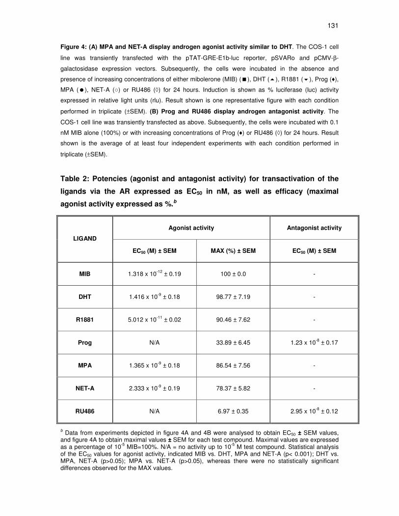
131
Figure 4: (A) MPA and NET-A display androgen agonist activity similar to DHT. The COS-1 cell
line was transiently transfected with the pTAT-GRE-E1b-luc reporter, pSVARo and pCMV-β-
galactosidase expression vectors. Subsequently, the cells were incubated in the absence and
presence of increasing concentrations of either mibolerone (MIB) (�), DHT (�), R1881 (�), Prog (♦),
MPA (�), NET-A (○) or RU486 (◊) for 24 hours. Induction is shown as % luciferase (luc) activity
expressed in relative light units (rlu). Result shown is one representative figure with each condition
performed in triplicate (±SEM). (B) Prog and RU486 display androgen antagonist activity. The
COS-1 cell line was transiently transfected as above. Subsequently, the cells were incubated with 0.1
nM MIB alone (100%) or with increasing concentrations of Prog (♦) or RU486 (◊) for 24 hours. Result
shown is the average of at least four independent experiments with each condition performed in
triplicate (±SEM).
Table 2: Potencies (agonist and antagonist activity) for transactivation of the
ligands via the AR expressed as EC50 in nM, as well as efficacy (maximal
agonist activity expressed as %.b
b Data from experiments depicted in figure 4A and 4B were analysed to obtain EC50 ± SEM values, and figure 4A to obtain maximal values ± SEM for each test compound. Maximal values are expressed as a percentage of 10-5 MIB=100%. N/A = no activity up to 10-5 M test compound. Statistical analysis of the EC50 values for agonist activity, indicated MIB vs. DHT, MPA and NET-A (p< 0.001); DHT vs. MPA, NET-A (p>0.05); MPA vs. NET-A (p>0.05), whereas there were no statistically significant differences observed for the MAX values.
Agonist activity Antagonist activity
LIGAND
EC50 (M) ± SEM MAX (%) ± SEM EC50 (M) ± SEM
MIB 1.318 x 10⁻¹² ± 0.19 100 ± 0.0 -
DHT 1.416 x 10⁻⁹ ± 0.18 98.77 ± 7.19 -
R1881 5.012 x 10⁻¹¹ ± 0.02 90.46 ± 7.62 -
Prog N/A 33.89 ± 6.45 1.23 x 10⁻⁸ ± 0.17
MPA 1.365 x 10⁻⁹ ± 0.18 86.54 ± 7.56 -
NET-A 2.333 x 10⁻⁹ ± 0.19 78.37 ± 5.82 -
RU486 N/A 6.97 ± 0.35 2.95 x 10⁻⁸ ± 0.12
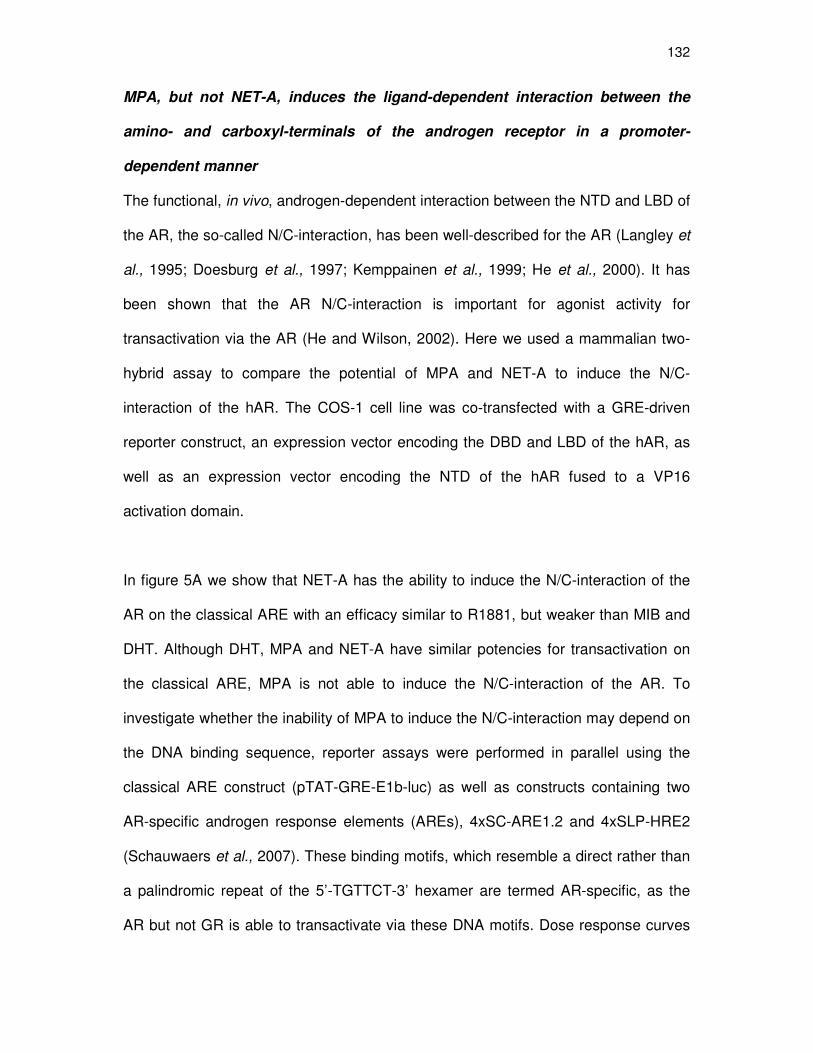
132
MPA, but not NET-A, induces the ligand-dependent interaction between the
amino- and carboxyl-terminals of the androgen receptor in a promoter-
dependent manner
The functional, in vivo, androgen-dependent interaction between the NTD and LBD of
the AR, the so-called N/C-interaction, has been well-described for the AR (Langley et
al., 1995; Doesburg et al., 1997; Kemppainen et al., 1999; He et al., 2000). It has
been shown that the AR N/C-interaction is important for agonist activity for
transactivation via the AR (He and Wilson, 2002). Here we used a mammalian two-
hybrid assay to compare the potential of MPA and NET-A to induce the N/C-
interaction of the hAR. The COS-1 cell line was co-transfected with a GRE-driven
reporter construct, an expression vector encoding the DBD and LBD of the hAR, as
well as an expression vector encoding the NTD of the hAR fused to a VP16
activation domain.
In figure 5A we show that NET-A has the ability to induce the N/C-interaction of the
AR on the classical ARE with an efficacy similar to R1881, but weaker than MIB and
DHT. Although DHT, MPA and NET-A have similar potencies for transactivation on
the classical ARE, MPA is not able to induce the N/C-interaction of the AR. To
investigate whether the inability of MPA to induce the N/C-interaction may depend on
the DNA binding sequence, reporter assays were performed in parallel using the
classical ARE construct (pTAT-GRE-E1b-luc) as well as constructs containing two
AR-specific androgen response elements (AREs), 4xSC-ARE1.2 and 4xSLP-HRE2
(Schauwaers et al., 2007). These binding motifs, which resemble a direct rather than
a palindromic repeat of the 5’-TGTTCT-3’ hexamer are termed AR-specific, as the
AR but not GR is able to transactivate via these DNA motifs. Dose response curves
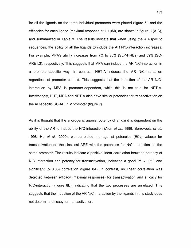
133
for all the ligands on the three individual promoters were plotted (figure 5), and the
efficacies for each ligand (maximal response at 10 µM), are shown in figure 6 (A-C),
and summarized in Table 3. The results indicate that when using the AR-specific
sequences, the ability of all the ligands to induce the AR N/C-interaction increases.
For example, MPA’s ability increases from 7% to 36% (SLP-HRE2) and 59% (SC-
ARE1.2), respectively. This suggests that MPA can induce the AR N/C-interaction in
a promoter-specific way. In contrast, NET-A induces the AR N/C-interaction
regardless of promoter context. This suggests that the induction of the AR N/C-
interaction by MPA is promoter-dependent, while this is not true for NET-A.
Interestingly, DHT, MPA and NET-A also have similar potencies for transactivation on
the AR-specific SC-ARE1.2 promoter (figure 7).
As it is thought that the androgenic agonist potency of a ligand is dependent on the
ability of the AR to induce the N/C-interaction (Alen et al., 1999; Berrevoets et al.,
1998, He et al., 2000), we correlated the agonist potencies (EC50 values) for
transactivation on the classical ARE with the potencies for N/C-interaction on the
same promoter. The results indicate a positive linear correlation between potency of
N/C interaction and potency for transactivation, indicating a good (r2 > 0.59) and
significant (p<0.05) correlation (figure 8A). In contrast, no linear correlation was
detected between efficacy (maximal responses) for transactivation and efficacy for
N/C-interaction (figure 8B), indicating that the two processes are unrelated. This
suggests that the induction of the AR N/C interaction by the ligands in this study does
not determine efficacy for transactivation.
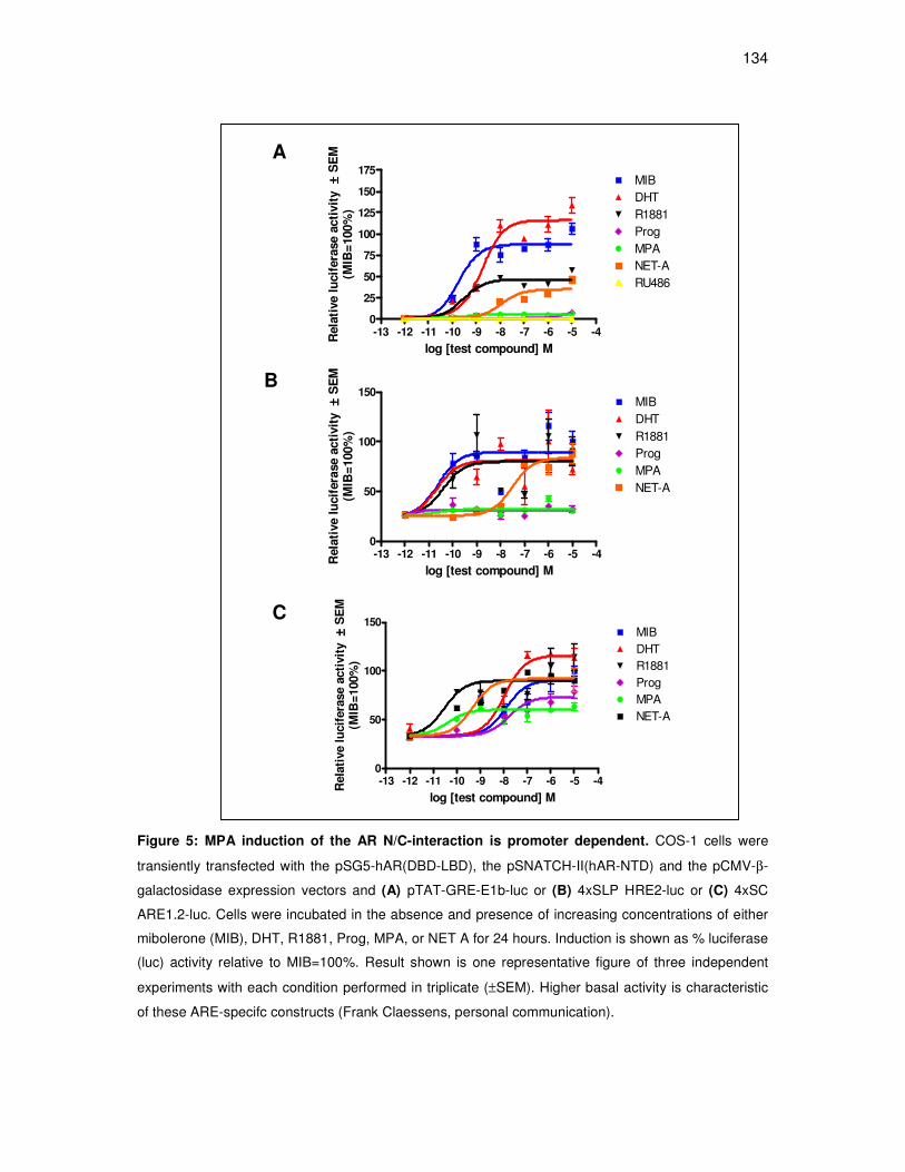
134
Figure 5: MPA induction of the AR N/C-interaction is promoter dependent. COS-1 cells were
transiently transfected with the pSG5-hAR(DBD-LBD), the pSNATCH-II(hAR-NTD) and the pCMV-β-
galactosidase expression vectors and (A) pTAT-GRE-E1b-luc or (B) 4xSLP HRE2-luc or (C) 4xSC
ARE1.2-luc. Cells were incubated in the absence and presence of increasing concentrations of either
mibolerone (MIB), DHT, R1881, Prog, MPA, or NET A for 24 hours. Induction is shown as % luciferase
(luc) activity relative to MIB=100%. Result shown is one representative figure of three independent
experiments with each condition performed in triplicate (±SEM). Higher basal activity is characteristic
of these ARE-specifc constructs (Frank Claessens, personal communication).
-13 -12 -11 -10 -9 -8 -7 -6 -5 -40
25
50
75
100
125
150
175MIBDHTR1881ProgMPANET-ARU486
log [test compound] M
Rela
tive l
ucif
era
se a
cti
vit
y±± ±±
SE
M(M
IB=
100%
)
A
-13 -12 -11 -10 -9 -8 -7 -6 -5 -40
50
100
150MIBDHTR1881ProgMPANET-A
log [test compound] M
Rela
tive l
ucif
era
se a
cti
vit
y±± ±±
SE
M(M
IB=
100%
)
B
-13 -12 -11 -10 -9 -8 -7 -6 -5 -40
50
100
150MIBDHTR1881ProgMPANET-A
log [test compound] M
Rela
tive l
ucif
era
se a
cti
vit
y±± ±±
SE
M(M
IB=
100%
)
C

135
Figure 6: In contrast to DHT and NET-A, MPA does not induce the ligand-dependent N/C-
interaction of the hAR on a pTAT-GRE. The COS-1 cell line was transiently transfected with the
pSG5-hAR(DBD-LBD), the pSNATCH-II(hAR-NTD) and the pCMV-β-galactosidase expression vectors
and (A) pTAT-GRE-E1b-luc or (B) 4xSC ARE1.2-luc or (C) 4xSLP HRE2-luc. Cells were subsequently
exposed to 10 µM of MIB, DHT, R1881, Prog, MPA, NET-A or RU486 for 24 hours. Results are
averages of at least three independent experiments with each condition performed in triplicate
(±SEM).
Mib
DHTPro
gM
PA
NET-A
RU486
R1881
0
50
100
150
10µµµµM test compound
a
b
c c
d
c
dM
ax
ima
l in
du
cti
on
±± ±±S
EM
n=
3-4
(Mib
=1
00
%)
Mib
DHT
Pro
gM
PA
NET-A
RU48
6
R18
81
0
50
100
150
200
10 µµµµM test compound
a
a
a,b
b bb
b,c
Maxim
al in
duction
±± ±±S
EM
n=3-4
(Mib
=100%
)
(B) SLP-HRE2
Mib
DHT
Prog
MPA
NET-A
RU48
6
R18
81
0
50
100
150
200
10 µµµµM test compound
a
a
a
a
a
a
a
Maxim
al
ind
ucti
on
±± ±±S
EM
n=
3-4
(Mib
=100%
)
(C) SC-ARE1.2
(A) pTAT-GRE

136
Table 3: Maximal AR N/C-induction (at 10 µµµµM of each ligand).c
cRelative responses for AR transactivation on pTAT-GRE-E1b-luc and for induction of AR N/C-
interaction on pTAT-GRE-E1b-luc or 4xSLP-HRE2 or 4xSC-ARE 1.2-luc reporter plasmids at 10 µM
ligand, were determined in the transiently transfected COS-1 cell line. Values were obtained from at
least three independent experiments, each performed in triplicate, after which the ± SEM was
calculated for each ligand. MIB activity was taken as 100% and all other values are expressed as a %
relative to this.
MAXIMAL N/C-INDUCTION (%) ± SEM
LIGAND (Classical)
pTAT-GRE-E1b
(AR-selective)
4xSLP-HRE2
(AR-selective)
4xSC-ARE1.2
MIB
100 ± 0.0
100 ± 0.0
100 ± 0.0
DHT
128.70 ± 7.72
136.0 ± 18.37
147.2 ± 1.54
R1881
47.97 ± 7.42
74.6 ± 5.31
105.9 ± 22.73
Prog
13.51 ± 2.0
34.41 ± 1.19
78.77 ± 19.2
MPA
7.19 ± 0.10
35.89 ± 3.30
58.82 ± 11.58
NET-A
41.6 ± 4.10
57.59 ± 4.62
88.73 ± 25.48
RU486
0.81 ± 0.16
16.35 ± 3.67
62.19 ± 14.03

137
-14 -13 -12 -11 -10 -9 -8 -7 -6 -5 -40
20
40
60
80
100MIBDHTProgMPANET-ARU486
Log [Test compound] M
Lu
cif
era
se a
cti
vit
y
(RL
U)
±± ±± S
EM
(n
=3)
Figure 7: MPA and NET-A display androgen agonist activity similar to DHT on an AR-specific
ARE. COS-1 cells were transiently transfected with the 4xSC ARE1.2-luc reporter, pSVARo and
pCMV-β-galactosidase expression vectors. Subsequently, the cells were incubated in the absence and
presence of increasing concentrations of either mibolerone (MIB) (�), DHT (�), Prog (♦), MPA (�),
RU486 (◊), or NET-A (○) for 24 hours. Induction is shown as % luciferase (luc) activity expressed in
relative light units (rlu). Result shown is one representative figure with each condition performed in
triplicate (±SEM).
MPA and NET-A display similar androgenic properties for transrepression.
Having shown that both MPA and NET-A can transactivate an ARE-containing
promoter via the AR, albeit via a different mechanism, we were interested in
comparing the androgenic properties of MPA and NET-A for transrepression. We
thus evaluated the transrepressive ability of MPA and NET-A via the AR by
transfecting the COS-1 cell line with luciferase reporter plasmids linked downstream
of either five copies of a nuclear factor kappa-B (NFĸB) site or seven copies of an
activator protein-1 (AP-1) site, both in the absence and presence of a transfected AR
expression vector (Appendix B3). Cells were stimulated with phorbol 12-myristate 13-
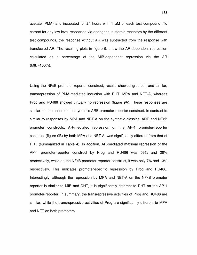
138
acetate (PMA) and incubated for 24 hours with 1 µM of each test compound. To
correct for any low level responses via endogenous steroid receptors by the different
test compounds, the response without AR was subtracted from the response with
transfected AR. The resulting plots in figure 9, show the AR-dependent repression
calculated as a percentage of the MIB-dependent repression via the AR
(MIB=100%).
Using the NFĸB promoter-reporter construct, results showed greatest, and similar,
transrepression of PMA-mediated induction with DHT, MPA and NET-A, whereas
Prog and RU486 showed virtually no repression (figure 9A). These responses are
similar to those seen on the synthetic ARE promoter-reporter construct. In contrast to
similar to responses by MPA and NET-A on the synthetic classical ARE and NFĸB
promoter constructs, AR-mediated repression on the AP-1 promoter-reporter
construct (figure 9B) by both MPA and NET-A, was significantly different from that of
DHT (summarized in Table 4). In addition, AR-mediated maximal repression of the
AP-1 promoter-reporter construct by Prog and RU486 was 59% and 38%
respectively, while on the NFĸB promoter-reporter construct, it was only 7% and 13%
respectively. This indicates promoter-specific repression by Prog and RU486.
Interestingly, although the repression by MPA and NET-A on the NFκB promoter
reporter is similar to MIB and DHT, it is significantly different to DHT on the AP-1
promoter-reporter. In summary, the transrepressive activities of Prog and RU486 are
similar, while the transrepressive activities of Prog are significantly different to MPA
and NET on both promoters.

139
-14 -12 -10 -8 -6 -4-15
-10
-5
0MibR1881DHTProgMPANET-ARU486
Potency (NC-interaction)
Po
ten
cy (
Tra
nsacti
vati
on
)
Figure 8: (A) Correlation analysis of potency for GRE transactivation versus GRE N/C-
interaction. Relative potencies for transactivation (Table 2) were compared to relative potencies for
N/C-interaction as determined by mammalian two hybrid assay (figure 5A) using Pearson correlations.
Results showed p<0.05 and r2=0.59. (B) Correlation analysis for efficacy for GRE transactivation
versus GRE N/C- interaction. Relative efficacies for transactivation (Table 2) were compared to
relative efficacies for N/C-interaction as determined by mammalian two hybrid assay (figure 5A) using
Pearson correlations. Results showed no statistical significance and r2=0.49.
A
B
0 50 100 1500
50
100
150MibR1881DHTProgMPANET-ARU486
Efficacy (NC-interaction)
Eff
icacy (
Tra
nsacti
vati
on
)

140
A NFĸB
MIB
DHTPro
gM
PA
Net-A
RU486
0
50
100
150
a
b
aaa
b
1µµµµM test compound
Re
lati
ve
re
pre
ss
ion
via
NF
κκ κκB
±± ±±S
EM
(M
IB=
10
0%
)
B AP-1
MIB
DHT
Prog
MPA
Net
-A
RU48
6
0
50
100
150
a
a
b
cc
b
1µµµµM test compound
Rela
tive r
ep
ressio
n v
ia A
P-1
±± ±±S
EM
(M
IB=
100%
)
Figure 9 legend on next page.

141
Figure 9: Transrepression activity via the hAR. The COS-1 cell line was transiently transfected with
pSVARo, pCMV-β-galactosidase expression vectors and (A) NFĸB- or (B) AP-1-containing promoter-
luciferase reporter constructs. Subsequently, the cells were stimulated with PMA and incubated with 1
µM of either MIB, DHT, Prog, MPA, NET-A or RU486 for 24 hours. Once the repressive abilities of the
test compounds were determined, in the absence and presence of transfected AR (Appendix B3),
repression in the absence of AR was subtracted from the repression in the presence of transfected
AR. The resulting plots show repression of the individual ligands calculated as a percentage of
repression by MIB (100%), thus indicate the transrepression dependent on the expression of AR.
Result shown is the average of three independent experiments with each condition performed in
triplicate (±SEM).
Table 4: Relative efficacies for transactivation and transrepression via the AR
at 1 µM ligandd
Ligand
GRE (from Table 2)
Relative response
NFĸB
Relative response
AP-1
Relative response
MIB
100 100 100
DHT
108 ± 11 99 ± 12 108 ± 12
Prog
13 ± 2 7 ± 4 59 ± 8
MPA
78 ± 3 84 ± 10 79 ± 3
NET-A
73 ± 7 85 ± 16 87 ± 4
RU486
12 ± 2 13 ± 9 38 ± 12
d Relative responses for transactivation via pTAT-GRE-E1b-luc and for transrepression via either
NFĸB or AP-1-luc reporter plasmids at 1 µM ligand, were determined in a transiently transfected COS-
1 cell line. Values were obtained from at least three independent experiments, each performed in
triplicate, after which the ± SEM was calculated for each ligand. MIB activity was taken as 100% and
all other values are expressed as a % relative to this.

142
DISCUSSION
MPA and NET-A bind with similar relatively affinity as DHT to the hAR
To the best of our knowledge, a detailed comparison of the relative equilibrium
dissociation constants, and relative efficacy for transactivation and transrepression,
by Prog, MPA and NET-A (or NET) for the AR, in the same system, has not been
reported previously. We show that these compounds bind to the same site as that of
MIB, and that Prog, MPA and NET-A bind to the AR with a similar affinity to that of
DHT, the endogenous ligand of the AR (figure 3), and that in the context of this series
of experiments, there is no significant difference in the binding affinities of DHT, Prog,
MPA and NET-A for the hAR (Table 1). Moreover, we determined accurate
equilibrium dissociation constants for these compounds.
The determined Kd / Ki of MIB and R1881 for the AR in our system (Kd / Ki = 0.383
and 0.22 nM, respectively), is similar to that reported in a previous study for MIB
(0.53 nM; Schilling et al., 1984) and R1881 (0.6 nM; Bonne and Raynaud, 1975).
Consistent with our results, a study by Kemppainen et al. (1999), also in the COS-1
cell line, showed that DHT and MPA have similar relative binding affinities. We also
demonstrate, in accordance with Kemppainen and co-workers, that Prog binds with a
similar relative binding affinity to the AR as DHT and MPA (Kemppainen et al., 1999).
In addition, a previous report in intact MCF-7 cells, showing that MPA and NET bind
with a similar relative affinity to the endogenously expressed AR (Bergink et al., 1983
and references therein), is in agreement with our results. In contrast, a study by
Hackenberg et al. (1993) determined a Kd of MPA for the AR as 3.6 nM in the MFM-
223 human mammary cancer cell line compared with a Kd of 0.18 nM for
dihydrotestosterone (DHT). In addition, the Kd / Ki values reported in the literature for
143
DHT of 0.5 nM (Zhao et al., 2000) differ from the values that we determined for DHT
(Ki = 29.4 nM). As the cell lines used in the studies by Hackenberg et al. (1993) and
Zhao et al. (2000), breast- and prostate cancer cells, respectively, also express
steroid receptors other than the AR, to which these progestins may bind, the
possibility exists that the results reported in these studies may be inaccurate. Thus,
to determine accurate Kd values of different ligands, it is best to use a system like the
one in the present study, where the AR was overexpressed in COS-1 cells, with
negligible levels of endogenous steroid receptors present. This allows a direct
comparison of the relative binding affinities of ligands for the AR within a system
where only the AR is present. Thus, the Ki-values of these compounds for the AR
reported in the present study may better reflect the relative affinities of these
compounds.
To our knowledge, this study is the first to establish accurate Ki values for MPA, NET-
A and RU486 towards the human AR. Interestingly, RU486 has a higher affinity for
the AR than all the other progestins used in our study, including DHT, but lower than
MIB and R1881. Song et al. (2004) previously reported that the relative binding
affinities of R1881 and RU486 differ by a factor of 12, and by 32 from data from
Kemppainen et al. (2002). Our result indicates a factor of 22, which is similar to the
result of Kemppainen et al. (2002). Taken together, our findings show that Prog and
the synthetic progestins, MPA, NET-A and RU486, all bind the AR with relatively high
affinity.

144
Unlike Prog, MPA and NET-A are potent agonists for the AR
We subsequently demonstrated that, although Prog, MPA, NET-A and DHT bound
the AR with similar binding affinities, Prog showed only weak partial agonist activity
(figure 4A). In contrast, both MPA and NET-A are potent and efficacious AR agonists,
with activity comparable to that of DHT (figure 4A). Notably, DHT, MPA and NET-A
have a lower potency and efficacy in comparison to MIB. Kemppainen et al., 1999,
showed that 100-fold more MPA is required to achieve the same agonist activity as
DHT, MIB and R1881. This AR agonist activity was determined in the CV-1 cell line
transiently transfected with a mouse mammary tumor virus (MMTV)-luciferase
reporter and full-length hAR expression vectors. In a separate study, using the
Chinese hamster ovary (CHO) cell line stably transfected with the MMTV-luciferase
reporter and hAR expression vectors, the relative agonistic activity of NET for the AR
was found to be only about 1% of that of DHT (Deckers et al., 2000). Thus, in these
two aforementioned studies, due to the fact that MPA and NET both required 100-
fold greater concentrations to achieve similar agonist activity as DHT, MPA was
described as a weak AR agonist whereas NET was said to have low androgenic
activity. The discrepancies between our results and the aforementioned studies could
be attributed to the different systems in which the agonist activities were measured. If
this is the case, then contributing factors could be the cell line used and/or the
sequence or context of the hormone response element used in the reporter
construct. In addition, a very recent study also performed in the COS-1 cell line using
a PRE2-tk-luc construct, reported EC50 values for Prog, MPA and NET-A of
approximately 3000, 56.8 and 19.6 nM, respectively (Sasagawa et al., 2008), while
we determined values of 601, 1.37 and 2.33 nM, respectively.

145
In accordance with our results indicating that MPA and DHT are equally potent AR
agonists, a comparative study by Ghatge and co-workers (2005) recently showed
that MPA is as potent as DHT in regulating AR-mediated gene expression in human
breast cancer cell lines (Ghatge et al., 2005). Furthermore, we demonstrate that Prog
and RU486 are potent antagonists of MIB induced AR-mediated transcription (figure
4B), with minimal agonist activity (figure 4A). Similarly, Prog (Kemppainen et al.,
1999; Sasagawa et al., 2008) and RU486 (Song et al., 2004) have previously been
reported to antagonize DHT-induced AR- mediated transcription. In contrast, MPA
and NET-A did not antagonize MIB-induced AR-mediated transcription. Overall,
these results indicate that Prog displays weak androgenic activity, while the synthetic
progestins, MPA and NET-A, display potent androgenic activities similar to the
natural androgen, DHT. In addition, Prog has anti-androgenic activities, while the
synthetic progestins do not. Interestingly, the synthetic progestin, RU486, has similar
activities as Prog.
MPA and NET-A activate the AR by different mechanisms
A number of parameters contribute to the potency of a ligand such as its affinity for
the receptor, rate of nuclear translocation, elicited receptor conformation, stabilisation
of receptor-ligand complex, the recruitment of coregulators and transcription
machinery, as well as DNA binding and rate of ligand dissociation. As we had already
addressed the binding of ligands to the AR, we next investigated the effect of all the
ligands in our study on another parameter. Considering that the N/C-interaction of the
AR is well-described, we decided to investigate the N/C-interaction of the AR in
response to ligand, as a measure of receptor conformation and stabilisation. To the
best of our knowledge, the capability of MPA (Kemppainnen et al., 1999), but not

146
NET-A, to induce the N/C-interaction of the AR has been previously investigated. Our
results indicate that MPA, in contrast to NET-A and DHT, does not induce the N/C-
interaction on a classical ARE-promoter (figure 6A), suggesting that MPA and NET-A
activate the AR by different mechanisms. This difference between MPA and NET in
inducing the AR N/C-interaction was first observed by Tamzin Tanner in the Hapgood
laboratory (MSc thesis). These results suggest that the conformation of the AR
attained by the N/C-interaction is not imperative to render the receptor
transcriptionally active, since MPA still has the ability to activate an androgen-
responsive reporter. When comparing the ability of the two synthetic progestins to
induce the AR N/C-interaction, it would appear that NET-A displays behaviour that is
more characteristic of classical strong androgen agonists. An explanation for this
difference in induction of AR N/C-interaction, could be the fact that MPA and NET-A
are structurally different, with MPA being a 21-carbon series steroid, and therefore
considered a true progestin, whereas NET-A is a 19-carbon series steroid structurally
more closely related to the androgens. This difference in the structures could
possibly account for the more typical androgenic profile of NET-A.
The crystal structure analysis of the AR bound to DHT showed a conformational
arrangement of the ligand binding domain (LBD), and the formation of the conserved
cleft, AF-2, which is critical for interaction with coregulators (Sack et al., 2001).
Recently, in a review by Birrell and co-workers (Birrell et al., 2007), a molecular
model was presented, in which MPA forces the displacement of a critical amino acid
residue, Phe874, in the AR, thereby disrupting this well-defined AF-2 cleft observed
for the DHT-bound receptor. The authors hypothesized that this could cause MPA
and DHT to have differing effects on AR-regulated gene expression. A recent study

147
of AR-regulated genes in breast cancer cell lines supports this hypothesis (Ghatge et
al., 2005). This raises the possibility that NET-A-bound AR may have a similar
conformation as the DHT-bound AR, and that MPA and NET-A, could therefore have
divergent effects on AR-regulated genes.
The ability of a compound to bind to the AR but not induce the N/C-interaction has
previously been reported for the AR antagonists hydroxyflutamide and cyproterone
acetate (Langley et al., 1995; Kemppainen et al., 1999). In accordance with these
previously reported results, the two antagonists in our system, Prog and RU486 (see
figure 8B), also did not induce the N/C-interaction of the AR (Kemppainen et al.,
1999; Song et al., 2004). Consistent with the previous finding that the degree of
agonist potency at low physiological concentrations parallels the ability of a
compound to induce the N/C-interaction (Kemppainen et al., 1999), we show that
potency for transactivation via a synthetic GRE correlates with the potency for the
induction of N/C-interaction (r2 = 0.59; p<0.05) for most of the ligands (figure 8A),
barring MPA. Correlation of efficacy for transactivation versus efficacy for induction of
N/C-interaction, showed no statistical significance (r2 = 0.49). In addition, it was
previously reported that MPA is a potent antagonist of a DHT-induced N/C-interaction
(Kemppainen et al., 1999; Birrell et al., 2007). This result was corroborated in our lab
(MSc thesis of Tamzin Tanner), while we found that NET-A could not antagonize the
DHT-induced N/C-interaction (Appendix B5), highlighting a potentially significant
difference in the mechanism of action of the two progestins. Altogether, the findings
indicate that Prog, MPA and NET-A activate the AR by different mechanisms.
Notably, the mechanism of AR activation by RU486 is comparable to Prog.

148
Induction of the AR N/C-interaction is promoter-dependent
The steroid receptor family has a high degree of structural and functional
conservation. Thus, it is not surprising that they can recognize virtually the same
DNA motifs, e.g. some androgen response elements (AREs), also function as
glucocorticoid response elements (GREs) or progesterone response elements
(PREs), or vice versa (Evans, 1988; Beato, 1989; Mangelsdorf et al., 1995).
However, the AR can also interact with binding motifs that resemble direct rather
than palindromic repeats of the 5’-TGTTCT-3’-hexamer. These motifs are termed AR-
specific, and have been described in several androgen-responsive genes, such as
the sc-ARE1.2 in the far upstream enhancer of the human secretory component (SC)
gene (Haelens et al., 1999), and the SLP-HRE2 from the upstream enhancer of the
mouse sex-limited (SLP) gene (Adler et al., 1991). In addition, it has been shown that
the AR N/C interaction is important for AR activation of the promoters of some
androgen-regulated genes, but not others (He et al., 2002). We thus hypothesized
that the inability of MPA to induce the N/C-interaction is dependent on the nature of
the response element i.e. the DNA binding sequence. To this end, we tested whether
MPA could induce the ligand-dependent N/C-interaction using two androgen-
selective AREs. We observed an overall increase in the ability of all ligands to
increase the AR N/C-interaction on both promoters. These results support our
hypothesis that the induction of the N/C-interaction by MPA is promoter-dependent
(figure 5A-5C and figure 6A-C). He and co-workers (2002b) have previously shown
that the AR N/C-interaction is important for some androgen-regulated promoters, but
not others. Our finding that NET-A shows similar potency and efficacy to MPA in
inducing transcription via the AR, but has a different effect on the N/C-interaction,
even on selective AREs, suggests that the N/C-interaction is not essential for

149
transactivation activity on these synthetic promoters. This has previously been
suggested by other groups (Shen et al., 2005). Other factors such as the dissociation
of heat-shock proteins, nuclear translocation, DNA binding affinity and rate of ligand
dissociation may also play a role in the transactivation activity.
MPA and NET-A display similar androgenic properties for transrepression on
synthetic promoters
Many steroid receptors have been suggested to exert inhibitory effects on
inflammatory response genes (transrepression) through direct interactions with
transcription factors such as NFĸB and AP-1, thereby attenuating inflammatory
responses. NFĸB and AP-1 have been shown to be crucial for the regulation of the
inflammatory process through their direct activation of proinflammatory cytokines and
chemokines (Karin, 2009; Xiao and Ghosh, 2005). By far the most best-studied in
terms of transrepression is the GR (De Bosscher et al., 2003; Koubovec et al., 2005),
but transrepression has also been reported for the ER (reviewed by Pascual and
Glass, 2006). Interestingly, not much is known about transrepression mediated via
the AR. On these grounds, we investigated the comparative effects of all the ligands
on transrepression via both NFĸB and AP-1 on synthetic promoter-reporter
constructs.
The results of the transrepression assays (at 1 µM of each ligand) (figure 9A and 9B)
as well as the effects observed for transactivation (figure 4A), are summarized in
Table 4. Similar relative activity of the ligands on all three promoters was observed.
Notably, both the weak partial agonist/potent antagonists, Prog and RU486, could
transrepress more effectively via the AP-1 promoter than via NFκB. This increase in
150
transrepression on AP-1 via the AR, at least for Prog, was also observed for this
ligand on the same promoter via the GR (Ronacher et al., 2009). Briefly, unlike Prog,
the transrepressive activities of MPA and NET-A via the AR is similar to the natural
androgen, DHT.
Conclusion
Although Prog, MPA and NET-A are structurally similar, relatively minor differences
can cause profound alterations in their biochemical activity (Darney, 1995). The
findings of our study highlight the fact that, although these compounds are designed
to mimic the biological activity of natural Prog, they do not act similarly to Prog at the
molecular level. Moreover, the ability of MPA and NET-A to exert similar effects
through the AR, but via different mechanisms, implies that such compounds could
then also exhibit very different effects via different steroid receptors in some target
tissue. Additionally, MPA could also exert different effects on endogenous genes via
the AR, in the same cell. Indeed, we have observed such differential effects by MPA
and NET-A on the RANTES gene in a human ectocervical cell line (Ect1/E6E7)
(Chapter 2).
This finding that MPA acts differently to NET-A, and moreover does not mimic the
effects of Prog, may have important implications for women’s health. By elucidating
the mechanisms by which MPA and NET-A exert their effects, we could facilitate
improved drug design for contraceptive and HRT agents, as well as help to unravel
molecular mechanisms by which the compounds exert their side effects.

151
Acknowledgements
We thank Carmen Langeveldt for technical support. We furthermore thank Prof.
Frank Claessens for donation of the ARE-specific plasmids as well as the AR
constructs used in the mammalian two-hybrid assays. This work was supported by
grants to JPH and DA from the Medical Research Council (MRC) and the National
Research Foundation (NRF) in South Africa, and Stellenbosch University. Any
opinion, findings and conclusions or recommendations expressed in this material are
those of the author(s) and therefore the NRF does not accept any liability in regard
thereto.
152
Chapter 4
_____________________________________________________
Investigating the anti-mineralocorticoid properties of synthetic
progestins used in hormone replacement therapy
Donita Africander and Janet P. Hapgood*
Department of Biochemistry, University of Stellenbosch, 7600 Stellenbosch, South
Africa
*Department of Molecular and Cell Biology, University of Cape Town, Private Bag,
Rondebosch, 7700, South Africa
Manuscript in preparation for submission to Molecular and Cellular Endocrinology.

153
Abstract
Progesterone (Prog) has a high affinity for the mineralocorticoid receptor (MR) and is
a potent MR antagonist. The synthetic progestins, medroxyprogesterone acetate
(MPA) and norethisterone derivatives, widely used as progestin-only injectable
contraceptives and in hormone replacement therapy (HRT), have been reported to
lack this anti-mineralocorticoid activity. This suggests that when they are used in
combination with estrogen during HRT, they are unable to antagonize the salt-
retaining effects of estrogen, which may lead to weight gain and increased blood
pressure. As the latter are causes of cardiovascular disease, this may explain the
mechanism for the observed adverse cardiovascular outcomes associated with
estrogen plus progestin therapy. In this study, we compared the binding affinity of
Prog for the MR with that of MPA and NET-A, as well as with the known
glucocorticoid and progesterone receptor antagonist, RU486. We also compared the
relative antagonist potencies of the ligands for transactivation on a synthetic
GRE/MRE via the MR, as well as their ability to transrepress MR-mediated NFĸB and
AP-1 promoter-reporter genes. In addition, the effect on an endogenous gene, Orm-
1, shown to contain a GRE/MRE, was investigated. Competitive binding revealed that
MPA and NET-A have similar binding affinities, but that their affinity is a 100-fold
lower than that of Prog for the human MR (Kd of 0.153 nM for aldosterone (Ald) and
Ki’s of 1.69, 197, and 229 nM for Prog, MPA and NET-A, respectively). Interestingly,
RU486 has a very low affinity for the hMR (Ki of 2980 nM). Furthermore, on a
synthetic mineralocorticoid response element (MRE)-containing promoter, we
showed that MPA, NET-A and RU486 are all antagonists of the hMR, but that unlike
Prog, they have weak antagonistic activity. However, on the endogenous MRE-
containing Orm-1 (α-glycolytic protein or orosomucoid-1) gene expressed in a rat

154
cardiomyocyte cell line, NET-A, but not MPA, has similar antagonistic activity as
Prog, suggesting that NET-A may be partially cardioprotective while MPA is not.
Interestingly, RU486 also has similar antagonist activity as Prog on the Orm-1 gene.
In addition, we highlight the underappreciated fact that dexamethasone (Dex), the so-
called GR-specific agonist, is a partial agonist for MR transactivation, and show for
the first time, that it is a full agonist for MR transrepression. Consistent with agonist-
induced interactions between the N- and C termini of steroid receptors (N/C-
interaction), our findings show that efficacy for transactivation via a synthetic GRE
correlates with the efficacy for the induction of MR N/C-interaction (r2 = 0.9179;
p<0.01), indicating that there is a relationship between transactivation and MR N/C-
interaction. Moreover, this study is the first to show that NET-A and RU486, but not
MPA, can dissociate between transrepression (AP-1 promoter-reporter) and
transactivation (GRE promoter-reporter) via the MR. These differences indicate that
NET-A and RU486 may be promising therapeutics for the treatment of MR-mediated
inflammation.

155
INTRODUCTION
Synthetic progestins have been used for decades in female reproductive health.
Although used in the treatment of a wide range of gynaecological disorders such as
endometriosis, the two most frequent uses of progestins are for contraception, and
for hormone replacement therapy (HRT) in postmenopausal women. These
compounds are used to mimic the actions of the endogenous hormone progesterone
(Prog), but have a longer plasma half-life and higher bio-availability than Prog
(Speroff, 1996). Medroxyprogesterone acetate (MPA) and norethisterone enanthate
(NET-EN) are two widely used progestins which are chemically derived from 17α-
hydroxy-progesterone and from 19-nortestosterone derivatives, respectively. MPA
itself, rather than its metabolites, is the major progestogenic compound, while NET-
EN and norethisterone acetate (NET-A) are hydrolysed to norethisterone (NET) and
its metabolites, which together have progestogenic action (Stanczyk and Roy, 1990).
Both MPA and NET are reported to have side-effects for users. The side-effect profile
when used as contraception includes irregular bleeding patterns, breast tenderness,
headache, dizziness, and weight gain (Haider and Darney, 2007; Nur-Isterate
package insert reg. no. J/21.8.2/136, Shering (Pty) Ltd). Recent studies suggest that
MPA and NET used in HRT may increase the incidence of breast cancer (Rossouw
et al., 2002; Beral, 2003). In addition to an increased risk of breast cancer, the
Women’s Health Initiative (WHI) study on combined estrogen and MPA used in
healthy postmenopausal women also showed increased risk of cardiovascular
complications (Rossouw et al., 2002). As these progestins are designed to mimic the
actions of progesterone, it is assumed that their biological effects, like progesterone,
are mediated by the progesterone receptor (PR). However, these progestins cross-

156
react with other members of the steroid receptor family and it is speculated that the
observed side-effects are mediated in this way.
The PR, androgen receptor (AR), glucocorticoid receptor (GR), estrogen receptor
(ER) and mineralocorticoid receptor (MR) constitute the steroid receptor family. They
are ligand-dependent transcription factors that display a high level of similarity with
regards to their structure and mechanism of action, and they regulate gene
expression of target genes by activating (transactivation) or repressing
(transrepression) transcription (Evans, 1988; Beato, 1989). In general, for
transactivation, the liganded-receptor translocates to the nucleus and binds hormone
response elements (HREs) to activated genes, while during transrepression, the
liganded-receptor binds to other transcription factors such as NFĸB and AP-1,
thereby inhibiting gene activation (Evans, 1988; Beato, 1989). Both MPA and NET-A
have been shown to have similar affinities for the AR, and are both potent agonists
for transactivation like dihydrotestosterone (DHT), the native androgen (Chapter 3).
Interestingly, MPA has a greater affinity than NET-A for the GR, and acts as a partial
agonist for transactivation via the GR (Koubovec et al., 2005), while NET-A is a GR
antagonist (supplementary material in Appendix C6). Interestingly, the activity of
MPA and NET via the GR and AR differs from that of Prog, in that Prog is a weak
partial agonist for the GR, and an antagonist for the AR. Collectively, this
demonstrates that MPA and NET do not have the same activity, nor do they act the
same as Prog, via different steroid receptors.
Prog has a high affinity for the MR, displaying weak agonist transactivation activity,
but potent antagonist activity (Wambach et al., 1979; Rafestin-Oblin et al., 1992;

157
Quinkler et al., 2002). In contrast, both MPA and NET-A have been reported to bind
to the MR with low relative affinity and do not display any agonist or antagonist
properties towards the MR (Winneker et al., 2003, Palacios et al., 2006). The
difference in the activity of MPA and NET as compared to Prog, via the MR, may
have serious implications for post-menopausal women using estrogen/progestin
treatment for HRT. This is because the progestin used in HRT should be able to
antagonize the salt-retaining and blood pressure raising effects of estrogens.
Estrogen acts on the renin-angiotensin-aldosterone system (RAAS), leading to
increased synthesis of angiotensinogen, which subsequently increases aldosterone
levels thereby promoting sodium and water retention (Oelkers, 1996). Thus estrogen
indirectly leads to increases in weight and blood pressure. Thus, if progestins used in
combined HRT lack anti-mineralocorticoid activity, it could lead to side-effects such
as weight gain and an increase in blood pressure. Since high blood pressure is a
cardiovascular disease risk factor, using synthetic progestins, such as MPA and
NET-A, that lack anti-mineralocorticoid activity in HRT, could lead to cardiovascular
complications, as observed in the WHI study (Rossouw et al., 2002). However, the
role of progestins such as MPA and NET-A, in cardiovascular disease remains poorly
defined.
Recently, Fejes-Tóth and Náray-Fejes-Tóth (2007) showed that Aldosterone (Ald),
the natural MR ligand, regulates the expression of several genes that may contribute
to the development of cardiovascular damage. Furthermore, it is known that Ald
plays an important role in hypertension and heart failure, and MR antagonists are
frequently prescribed to block MR activity in hypertensive patients (Pitt et al., 1999;
Pitt et al., 2003). Thus, blocking Ald regulation of these genes by MR antagonists

158
could alleviate cardiovascular damage, and may be beneficial to patients with chronic
heart failure. We were particularly interested in the Orm-1 (α-glycolytic protein or
orosomucoid-1), an inflammation/acute phase-related gene, whose increased
expression is associated with increased risk of cardiovascular disease (Engström et
al., 2003). The promoter of Orm-1 contains a functional glucocorticoid response
element/mineralocorticoid response element (GRE/MRE) (Klein et al., 1988). It has
been reported that the effect of Ald on Orm-1 is direct, mediated by the binding of the
liganded-MR to MREs in the promoter region (Fejes-Tóth and Náray- Fejes-Tóth,
2007).
In the current study, we determined the equilibrium constants (Kd or Ki’s) for the MR
of Ald, Prog, MPA, NET-A, as well as RU486, the known PR and GR antagonist, and
probable MR antagonist (www.nursa.org/10.1621/datasets.02001). We also
compared the relative antagonist potencies of the ligands for transactivation on a
synthetic GRE/MRE, as well as their ability to transrepress NFĸB and AP-1 promoter-
reporter genes via the MR. To further investigate the anti-mineralocorticoid activity of
these compounds, we used rat cardiomyocytes transiently transfected with MR
(Fejes-Tóth and Náray- Fejes-Tóth, 2007), and measured the expression of the
endogenous Orm-1 gene in response to Ald, in the absence and presence of the
synthetic progestins. Specifically, we hypothesized that MPA and NET-A, unlike
Prog, would not antagonize the Ald-mediated MR transactivation.

159
MATERIALS AND METHODS
Plasmids
The plasmid, pTAT-GRE-E1b-luc, driven by the E1b promoter containing two copies
of the rat TAT-GRE, has been described previously (Sui et al., 1999). The
cytomegalovirus (CMV)-driven-β-galactosidase expression vector (pCMV-β-gal) was
obtained from Prof. G. Haegeman (University of Gent, Belgium). The 5xNFκB-luc and
7xAP-1-luc plasmids were from Stratagene (Houston, Texas, USA). A plasmid
expressing the human mineralocorticoid receptor, pRShMR, was a kind gift from Prof.
R. Evans and previously described (Arriza et al., 1987). Plasmids expressing human
glucocorticoid receptor, pRS-hGRα were a kind gift from Prof. R. Evans (Howard
Hughes Medical Institute, La Jolla, USA). A plasmid expressing the human androgen
receptor, pSVARo (Brinkmann et al., 1989) was obtained from Frank Claessens,
(University of Leuven, Leuven, Belgium). The plasmid expressing the human
progesterone receptor, isoform B, pSG5hPR-B (Kastner et al., 1990), was obtained
from Stoney Simons Jr (NIH, Bethesda, USA). The GAL4-MRC expression vector
and the VP16-MRNT fusion protein expression vectors (provided by Prof’s Peter
Fuller (Prince Henry’s Institute of Medical Research, Victoria, Australia) and Fraser
Rogerson (University of Melbourne Department of Paediatrics, and Murdoch
Childrens Research Institute, Royal Children's Hospital, Victoria, Australia), were
previously described (Rogerson and Fuller, 2003). The reporter vector, pG5-luc,
which contains the luciferase gene driven by a GAL4 responsive promoter, was
obtained from Prof Douglas Stocco (Texas Tech University Health Sciences Center,
Texas, USA). The pGL2basic empty vector was obtained from Promega, Madison,
USA.

160
Inducing compounds
11β,21-Dihydroxy-3,20-dioxo-4-pregnen-18-al (Aldosterone; Ald), 11β-(4-
Dimethylamino)phenyl-17β-hydroxy-17-(1-propynyl)estra-4,9-dien-3-one
(mifepristone; RU486), 7α-acetylthio-3-oxo-17α-pregn-4-ene-21,17-carbolactone
(spironolactone), 4-pregnene-3, 20-dione (progesterone; Prog), 6α-methyl-17α-
hydroxy-progesterone acetate (medroxyprogesterone acetate; MPA), 17α-ethynyl-
19-nortestosterone 17β-acetate (norethindrone acetate; NET-A), (11β,16α)-9-fluoro-
11,17,21-trihydroxy-16-methylpregna-1,4-diene-3,20-dione (Dexamethasone; Dex)
and phorbol 12-myristate 13-acetate (PMA) were obtained from Sigma-Aldrich, South
Africa. NET-A, the acetate, was used as it is soluble in water compared with the
insoluble ester NET-EN. In vivo, both NET-EN and NET-A undergo hydrolysis and
are converted to NET and its metabolites (Stanczyk and Roy, 1990). [3H]-Aldosterone
([3H]-Ald) (87.9 Ci/mmol), unlabelled 17β-hydroxy-7α,17α-dimethylestr-4-en-3-one
(mibolerone; MIB), and unlabelled R5020 were purchased from PerkinElmer Life and
Analytical Science, South Africa. All unlabelled test compounds were dissolved in
absolute ethanol and stored at -20oC. These compounds were then added to serum-
free culturing medium such that the final concentration of ethanol was 0.1%. Control
incubations (no test compounds) were performed in the presence of 0.1% ethanol.
Cell culture
COS-1 cell lines (monkey kidney cells) were maintained in high glucose (4.5 g/ml)
Dulbecco’s Modified Eagle’s Medium (DMEM) (Sigma-Aldrich, South Africa). Medium
was supplemented with 10% fetal calf serum (FCS) (Delta Bioproducts, South Africa),
50 IU/ml penicillin and 50 µg/ml streptomycin (GibcoBRL, Paisley, UK). The H9C2
cell line (rat cardiomyocyte cells) was a kind gift from Dr. Rob Smith (Physiology
161
Department, University of Stellenbosch, South Africa) and was maintained in high
glucose (4.5 g/ml) DMEM (Sigma-Aldrich, South Africa). Medium was supplemented
with 10% FCS (Delta Bioproducts, South Africa), 50 IU/ml penicillin and 50 µg/ml
streptomycin (GibcoBRL, Paisley, UK). All cultures were maintained in 75 cm2 culture
flasks (Greiner, Bio-One International, Austria) at 37°C, in an atmosphere of 90%
humidity and 5% CO2, and were regularly tested for mycoplasma infection by means
of Hoechst staining (Freshney, 1987), and only mycoplasma-negative cell lines were
used in experiments.
Whole cell binding assays
Competitive whole cell binding assays were performed essentially as described by
Bamberger et al. (1995), with the following modifications. The COS-1 cell line was
maintained in DMEM as described. Cells were seeded into 24-well tissue culture
plates at 1 x 10⁵ cells per well. On day 2, cells were transfected with 0.375 µg
pRShMR expression vector and 0.0375 µg of the pCMV-β-gal expression vector
(Stratagene, Houston, Texas, USA), using the FuGENE6 transfection reagent
(Roche Molecular Biochemicals, South Africa) according to the manufacturer’s
instructions. On day 3, the cells were washed with phosphate-buffered saline (PBS)
and incubated with 0.2 nM [3H]-Ald (PerkinElmer Life and Analytical Science, South
Africa) in the absence (total binding) or presence of increasing concentrations of
unlabelled Ald, Prog, MPA, NET-A or RU486 for 16 hours (time taken to reach
equilibrium; Appendix C2) at 37°C. Working on ice at 4oC, cells were washed three
times with ice-cold 1X PBS containing 0.2% (w/v) bovine serum albumin (BSA) for 15
min. Cells were then lysed with 200 µl reporter lysis buffer (Promega, Madison,
USA). Total binding ([3H]-Ald in the absence of added unlabelled ligand) was

162
determined by scintillation counting and expressed as 100%. Specific bound [3H]-Ald
was calculated as the difference between total and non-specific binding, the latter
determined by incubating cells in the presence of [3H]-Ald plus 10 µM unlabelled Ald
and expressed as a relative % of total binding. The β-galactosidase
chemiluminescent reporter gene assay system (Tropix Inc., U.S.A.) was used to
determine β-galactosidase (β-gal) activity, to correct for differences in transfection
efficiencies. Binding data were analyzed using GraphPad Prism software, with
nonlinear regression and assuming competitive binding to one class of binding sites.
Ki values for Prog, MPA, NET-A and RU486 were determined from the heterologous
displacement curves using the EC50s, Kd for Ald, and concentration of radiolabelled
Ald, according to the method of Cheng and Prusoff (1973) and described in Appendix
D1. The reported values are averages of at least three independent assays, with
each condition performed in triplicate.
Luciferase reporter assays
For transactivation assays in the COS-1 cell line: COS-1 cells were maintained as
above and seeded into 96-well tissue culture plates at 1 x 10⁴ cells per well. On day
2, cells in each well were transiently transfected with 50 ng of the pTAT-GRE-E1b-luc
construct, 5 ng of the expression vector for the human mineralocorticoid receptor
(pRShMR), and 5 ng of the pCMV-β-gal expression vector, using the FuGENE6
transfection reagent (Roche Molecular Biochemicals, South Africa) in accordance
with the manufacturer’s instructions. On day 3, the cells were washed with PBS and
induced with 1 or 10 µM of either Ald, Prog, MPA, NET-A, RU486 or Dex to
investigate agonist activity, while for antagonist activity, the cells were incubated with
1 nM Ald in the absence and presence of increasing concentrations of Prog, MPA,

163
NET-A or RU486. After 24 hours, the cells were lysed with reporter lysis buffer
(Promega, Madison, USA). Luciferase activity in the lysate was measured using the
Luciferase Assay System (Promega, Madison, USA) and a Veritas microplate
luminometer (Turner Biosystems, Sunnyvale CA, USA). The values obtained were
normalised to expression of β-gal, which was measured using the GalactoStar Assay
Kit from Tropix (Bedford MA, USA).
For transactivation assays in the H9C2 cell line: H9C2 cells were maintained as
above and seeded into 24-well tissue culture plates at 5 x 10⁴ cells per well. On day
2, cells in each well were transiently transfected with 330 ng of the pTAT-GRE-E1b-
luc construct and 33 ng of the expression vector for the hMR, hGR, hAR, hPR-B or
the empty vector pGL2basic, using the FuGENE6 transfection reagent (Roche
Molecular Biochemicals, South Africa) in accordance with the manufacturer’s
instructions. On day 3, the cells were washed with PBS and induced with 10 µM of
either Ald, Dex, MIB, or R5020. After 24 hours, the cells were lysed with reporter lysis
buffer (Promega, Madison, USA). Luciferase activity in the lysate was measured
using the Luciferase Assay System (Promega, Madison, USA) and a Veritas
microplate luminometer (Turner Biosystems, Sunnyvale CA, USA).
For transrepression assays, the COS-1 cell line was seeded into 24-well tissue
culture plates at a density of 5 x 10⁴ cells per well. The following day the cells were
transfected with 0.045 µg of pRShMR, 0.09 µg of 5xNFκB-luc or 7xAP-1-luc plasmids
respectively, and 0.0225 µg of pCMV-β-Gal using the FuGENE6 transfection reagent
(Roche Molecular Biochemicals, South Africa) according to the manufacturer’s
instructions. Twenty four hours after transfection the cells were washed with PBS and

164
incubated with serum-free medium containing 10 ng/ml PMA and 1 µM of each test
compound. Luciferase activity in the lysate was measured as described above. The
values obtained were normalised to expression of β-gal, which was measured using
the GalactoStar Assay Kit from Tropix (Bedford MA, USA). Where normalising to B-
galactosidase is not performed, transfection efficiency was previously determined to
be highly reproducible. The reported values are averages of at least three
independent experiments, with each condition performed in triplicate.
Mammalian two-hybrid assays
The mammalian two-hybrid assay was performed in the COS-1 cell line. The cells
were maintained as described, and then plated in 24-well tissue culture plates at a
density of 5 x 10⁴ cells per well. On day 2, the cells were transfected with 0.1 µg/well
GAL4-MRC, 0.5 µg/well VP16-MRNT and 0.5 µg/well pG5-luc, using the FuGENE6
transfection reagent (Roche Molecular Biochemicals, South Africa) according to the
manufacturer’s instructions. Twenty four hours after transfection, cells were induced
with 1 or 10 µM Ald, Prog, MPA, NET-A, RU486 or Dex for 24 hours. Luciferase
activity in the lysate was measured as described above. The reported values are
averages of at least three independent experiments, with each condition performed in
triplicate. The values obtained were normalised to expression of β-gal, which was
measured using the GalactoStar Assay Kit from Tropix (Bedford MA, USA).
Isolation of total RNA and real-time quantitative RT-PCR analysis of
representative genes
The H9C2 cell line was maintained as described above. Cells were seeded into 12-
well plates, transiently tranfected with the MR, and subsequenlty grown in the

165
absence of FCS for 48 h before treatment with test compounds. Cells were induced
with 1 nM Ald in the absence and presence of 1 µM Prog, MPA, NET-A, RU486 or
Spironolactone for 24 hours. Total RNA was isolated from cells using Tri-reagent
(Sigma-Aldrich, South Africa) according to the manufacturer’s instructions. The
integrity of the RNA (presence of intact 18S and 28S ribosomal bands) was
confirmed by agarose gel electrophoresis. Total RNA (1 µg) was reverse transcribed
using the Transcriptor First Strand cDNA synthesis kit (Roche Applied Science,
South Africa) according to the manufacturer’s instructions. Real-time PCR was
performed by using a Light-Cycler, rapid thermal cycler system (Roche Applied
Science, South Africa) according to the manufacturer’s instructions. Nucleotides,
TaqDNA polymerase, and buffer used in the reaction were those included in the
LightCycler-FastStart DNA MasterPLUS SYBR Green I system (Roche Diagnostics,
South Africa). β-actin was used as an internal control. Primer sets are as described in
Table 1. The thermal cycling parameters were: initial denaturation at 95oC for 3 min,
followed by 40 cycles at 95oC for 15 sec and 57oC for 30 sec.
Table 1: QPCR primer sets (Orm-1 and β-actin sequences were obtained from Prof. Anikó Náray-Fejes-Tóth) (Fejes-Tóth and Náray- Fejes-Tóth, 2007).
Gene Primers Strand Annealing temperature
(oC)
Orm-1
TGCCCATTTGATAGTGCTGAAG CAGGCTGTCAAAGATGTGGGCA
Forward Reverse
60
β-actin
TTGTAACCAACTGGGACGATATGG GATCTTGATCTTCATGGTGCTAGG
Forward Reverse
60

166
Western blotting
H9C2 cell lines were plated at 1 x 10⁶ cells per well in 6-well plates and allowed to
grow to confluency. Cells were washed with PBS and lysed in 100 µl SDS-sample
buffer (Sambrook et al., 1989). Lysates were boiled and subjected to electrophoresis
on a 8% SDS polyacrylamide gel using β-actin expression as a loading control.
Following electrophoresis, proteins were transferred to nitrocellulose membranes.
Blots were probed with an anti-MR (H300, 1:1000), anti-GR (H300, 1:3000), anti-AR
(H-280, 1:1000), or anti-PR-AB (sc-810, 1:500) antibodies from Santa Cruz
Biotechnology Inc. (Santa Cruz, CA, USA), as well as the anti-β-actin (1:1500)
antibody from Cell Signalling Technology, Massachusetts, USA, for 16 hours at 4 oC.
Blots were subsequently incubated with horseradish peroxidase (HRP)-conjugated
goat anti-rabbit secondary antibody (1:10000, NA934VS, Amersham, South Africa)
for 1 hour. Proteins were visualized using enhanced chemiluminescence (ECL) from
Amersham, followed by exposure to Hyperfilm MP high performance autoradiography
film (Amersham, South Africa).
Data manipulation and statistical analysis
The Graph Pad Prism software was used for data manipulations, graphical
representations, and statistical analysis. One-way ANOVA analysis of variance and
Bonferroni (compares all pairs of columns) posttests were used for statistical
analysis. Non-linear regression and one site competition were used in whole cell
binding assays, whereas non-linear regression and sigmoidal dose response were
used in transactivation (antagonist) experiments. Statistical significance of
differences is indicated by *, ** or ***, to indicate p<0.05, p<0.01 or p<0.001,
respectively, whereas no statistical significance is indicated by p>0.05. The letters

167
a,b,c etc. are also used to denote statistically significant differences, where all those
values which differ significantly from others, are assigned a different letter. Fractional
occupancy of the MR for each compound was calculated using the equation:
Fractional occupancy = [ligand] / ([ligand]+Ki), using the Ki values summarized in
Table 2.
RESULTS
MPA and NET-A have a similar binding affinity for the MR
Competitive whole cell binding assays in the COS-1 cell line transiently transfected
with a full-length human mineralocorticoid receptor (hMR) expression vector, were
performed to determine Kd and Ki values. In order to obtain accurate Kd and Ki values
using homologous and heterologous displacement curves, respectively, a range of
experiments was first performed to establish an appropriate concentration of
radiolabelled ligand and the time to reach equilibrium. An appropriate concentration
of [3H]-Ald, in the range two to ten times lower than the EC50, was established as 0.2
nM (discussed in Appendix D1), and the incubation time required for equilibrium to be
reached for 0.2 nM [3H]-Ald binding to the MR, was determined as sixteen hours
(discussed in Appendix D1).
Homologous/heterologous curves with unlabelled steroids were then obtained and
results show that MPA and NET-A bind to the MR with a similar affinity, but
approximately 100-fold lower than that of Prog and 1000-fold lower than that of the
natural mineralocorticoid, Ald (figure 1). Interestingly, RU486 also binds the MR but
with an approximately 20 000-fold lower affinity than Ald. The curves for each
competitor steroid indicate competitive binding to the same site as Ald (R2 values for

168
each MPA, NET-A, Prog and RU486 were 0.9453, 0.9452, 0.9837 and 0.9431
respectively, for one site competitive binding curve). The Kd for Ald and Ki values for
Prog, MPA, NET-A and RU486 are summarised in the Table 2. The Ki for Prog was
determined as 1.69 nM, which is similar to a previously reported value of 1.2 nM
(Quinkler et al., 2002).
-13 -12 -11 -10 -9 -8 -7 -6 -5 -40
50
100
150AldProgMPANET-ARU486Dex
log [unlabelled competitor] M
% s
pecif
ic b
ind
ing
(cp
m/ ββ ββ
-gal)
Figure 1: MPA and NET-A both compete with [3H]-Ald for binding to the human
mineralocorticoid receptor. The COS-1 cell line was transiently transfected with the pRShMR and
pCMV-β-galactosidase expression vectors.Twenty-four hours later the cells were incubated with 0.2
nM [3H]-Ald in the absence or presence of increasing concentrations of either unlabelled Ald (�), Prog
(♦), MPA (●), NET-A (▼), RU486 (◊) or Dex (▲) for 16 hours. Results are plotted as % specific binding
where total specific binding of [3H]-Ald in the absence of unlabelled ligand is set as 100% and binding
of unlabelled ligand is set as a % binding relative to Ald, after normalization for transfection efficiency
with β-galactosidase levels. Result shown is representative of three independent experiments with
each condition performed in triplicate (±SEM).

169
Table 2: Relative binding affinities of the ligands for the MR (Kd or Ki values)a
Test compound Kd or Ki (M) ± SEM
Fold differences
Ald
1.53 x 10⁻¹⁰ ± 0.04
1
Prog
1.69 x 10⁻⁹ ± 0.07
11.05
MPA
1.97 x 10⁻⁷ ± 0.04
1287.58
NET-A
2.29 x 10⁻⁷ ± 0.07
1496.73
RU486
2.98 x 10⁻⁶ ± 0.08
19477.12
Dex
5.14 x 10-9 ± 0.07
33.59
a Binding data of three independent experiments were analyzed using GraphPad Prism software, with
nonlinear regression and assuming competitive binding to one class of binding sites. Ki values ± SEM
for Prog, MPA, NET-A, RU486 and Dex were determined from the heterologous displacement curves
using the EC50s, Kd ± SEM for Ald, and concentration of radiolabelled Ald, according to the method of
Cheng and Prusoff (1973) that is described in Appendix D1. Fold differences calculated by setting the
Kd for MIB in M as 1, and expressing the Ki in M for the other ligands as a fraction thereof. Statistical
analysis of the Kd / Ki values, indicated the following: Ald vs. Prog, MPA, NET-A, RU486, Dex (p<
0.001); Prog vs. MPA, NET-A, RU486, Dex (p< 0.001); MPA, NET-A vs. RU486, Dex (p<0.001); MPA
vs. NET-A (p>0.05); Prog vs. Dex (p<0.05).
Unlike Prog, MPA and NET-A display weak MR antagonist activity and no
mineralocorticoid agonist activity for transactivation
Having shown that MPA, NET-A and RU486 bind to the hMR, albeit with low affinity,
we directly compared their mineralocorticoid properties by transiently transfecting the
COS-1 cell line with a glucocorticoid response element (GRE)/mineralocorticoid
response element (MRE)-driven reporter construct containing two copies of the rat
TAT GRE, a hMR expression vector and the pCMV-β-gal expression vector. The

170
cells were subsequently exposed to either 1 or 10 µM Ald, Prog, MPA, NET-A,
RU486 or Dex. The results clearly show that 10 µM MPA, NET-A and RU486, unlike
10 µM Prog, do not display agonist activity for the MR (figure 2A). A similar profile
was observed at 1 µM of each compound (Appendix C4). The transcriptional activity
of these compounds in the absence of transfected receptor was negligible (Appendix
D3). Interestingly, Dex, the well-described potent GR-specific agonist (Honer et al.,
2003; Yu et al., 2004), displays partial agonist activity of about 37% of maximal
activity relative to Ald via the MR, showing that Dex is a partial agonist for
transactivation via the MR. Whole cell binding experiments showed that Dex binds to
the MR with a significantly higher affinity than Prog (Table 2).
In order to obtain additional information about the pharmacological profiles of these
compounds, we investigated their antagonist activity by transfecting the COS -1 cell
line as above, and incubating the cells with 1 nM Ald in the absence or presence of
increasing concentrations of Prog, MPA, NET-A and RU486 (figure 2B). These
results indicate that although, MPA, NET-A and RU486 can similarly antagonise the
effects of Ald, they are much less potent than Prog (approximately 87-216 fold less
potent). We determined potencies (EC50 values) for antagonist activity of 3.1, 310,
270 and 670 nM for Prog, MPA, NET-A and RU486, respectively. Interestingly, our
EC50 value for antagonist activity of Prog (3.1 nM) indicates that Prog is a more
potent at antagonizing the effects of Ald than the well-known MR antagonist
spironolactone (EC50 of 14 nM) (reviewed in Lu et al., 2006). Whether Dex has MR
antagonist activity was not investigated.
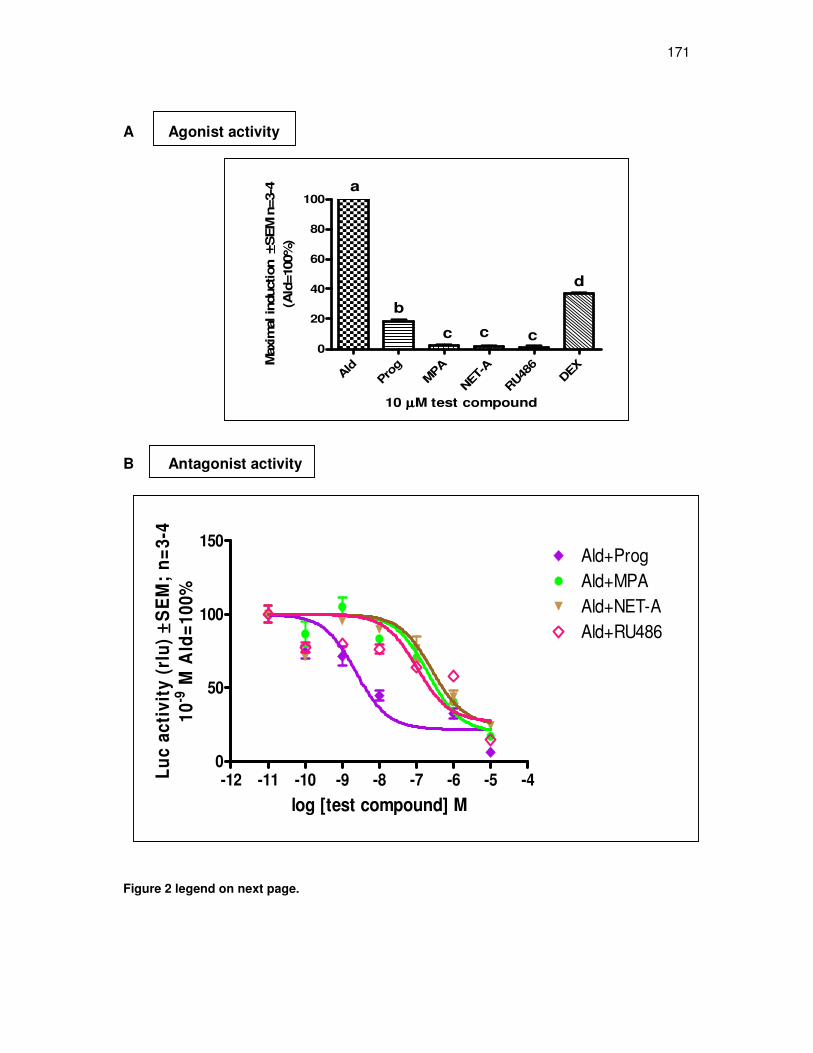
171
A Agonist activity
B Antagonist activity
-12 -11 -10 -9 -8 -7 -6 -5 -40
50
100
150
Ald+MPAAld+Prog
Ald+NET-AAld+RU486
log [test compound] M
Lu
c a
cti
vit
y (
rlu
)±± ±±
SE
M;
n=
3-4
10
-9 M
Ald
=1
00
%
Figure 2 legend on next page.
Ald
Pro
g
MPA
NET-
A
RU48
6DEX
0
20
40
60
80
100
10 µµµµM test compound
a
b
c cc
dM
axim
al in
duction
±± ±±SEM
n=3-4
(Ald
=100%
)
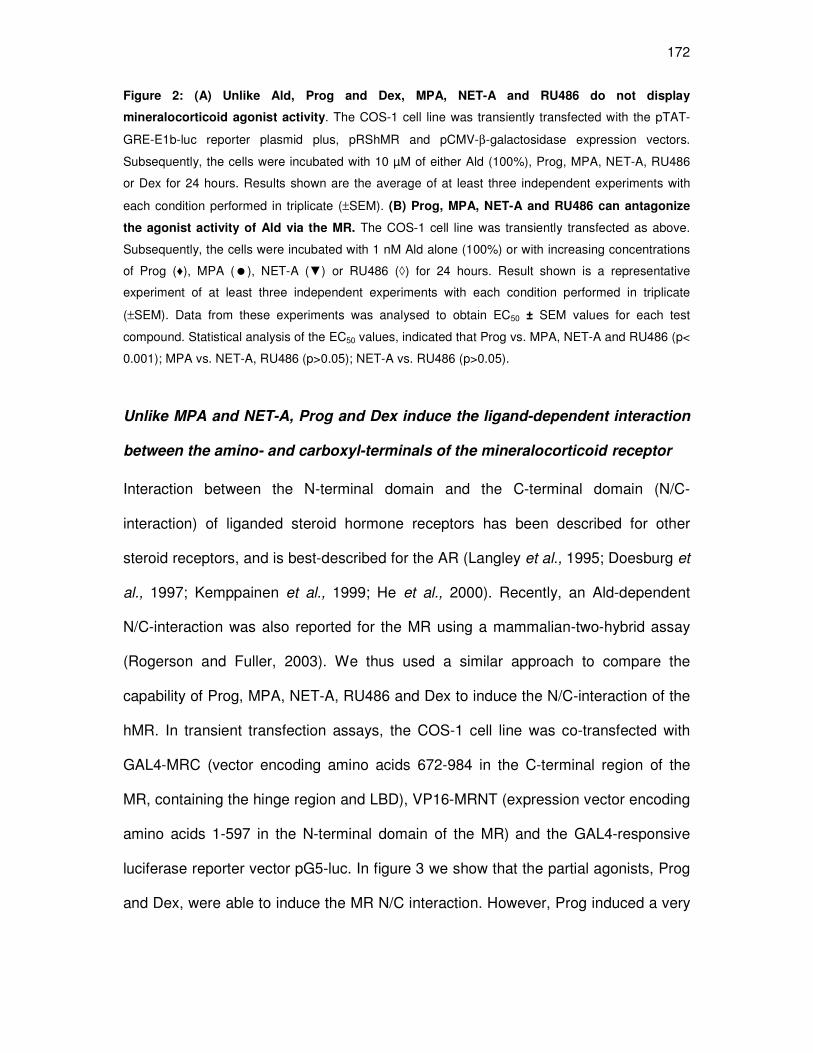
172
Figure 2: (A) Unlike Ald, Prog and Dex, MPA, NET-A and RU486 do not display
mineralocorticoid agonist activity. The COS-1 cell line was transiently transfected with the pTAT-
GRE-E1b-luc reporter plasmid plus, pRShMR and pCMV-β-galactosidase expression vectors.
Subsequently, the cells were incubated with 10 µM of either Ald (100%), Prog, MPA, NET-A, RU486
or Dex for 24 hours. Results shown are the average of at least three independent experiments with
each condition performed in triplicate (±SEM). (B) Prog, MPA, NET-A and RU486 can antagonize
the agonist activity of Ald via the MR. The COS-1 cell line was transiently transfected as above.
Subsequently, the cells were incubated with 1 nM Ald alone (100%) or with increasing concentrations
of Prog (♦), MPA (�), NET-A (▼) or RU486 (◊) for 24 hours. Result shown is a representative
experiment of at least three independent experiments with each condition performed in triplicate
(±SEM). Data from these experiments was analysed to obtain EC50 ± SEM values for each test
compound. Statistical analysis of the EC50 values, indicated that Prog vs. MPA, NET-A and RU486 (p<
0.001); MPA vs. NET-A, RU486 (p>0.05); NET-A vs. RU486 (p>0.05).
Unlike MPA and NET-A, Prog and Dex induce the ligand-dependent interaction
between the amino- and carboxyl-terminals of the mineralocorticoid receptor
Interaction between the N-terminal domain and the C-terminal domain (N/C-
interaction) of liganded steroid hormone receptors has been described for other
steroid receptors, and is best-described for the AR (Langley et al., 1995; Doesburg et
al., 1997; Kemppainen et al., 1999; He et al., 2000). Recently, an Ald-dependent
N/C-interaction was also reported for the MR using a mammalian-two-hybrid assay
(Rogerson and Fuller, 2003). We thus used a similar approach to compare the
capability of Prog, MPA, NET-A, RU486 and Dex to induce the N/C-interaction of the
hMR. In transient transfection assays, the COS-1 cell line was co-transfected with
GAL4-MRC (vector encoding amino acids 672-984 in the C-terminal region of the
MR, containing the hinge region and LBD), VP16-MRNT (expression vector encoding
amino acids 1-597 in the N-terminal domain of the MR) and the GAL4-responsive
luciferase reporter vector pG5-luc. In figure 3 we show that the partial agonists, Prog
and Dex, were able to induce the MR N/C interaction. However, Prog induced a very

173
weak N/C-interaction of about 5% (compared to 100% for Ald), while the observed
transactivation activity was about 20% (figure 2A).
Figure 3: In contrast to Ald, Prog and Dex, MPA, NET-A and RU486 do not induce the N/C-
interaction of the hMR. The COS-1 cell line was transiently transfected with the GAL4-MRC and
VP16-MRNT expression vectors, as well as the GAL4-responsive luciferase reporter vector pG5-luc.
The cells were exposed to 10 µM Ald (100%), Prog, MPA, NET-A, RU486 or Dex for 24 hours. Results
are averages of at least three independent experiments with each condition performed in triplicate
(±SEM).
Interestingly, no N/C-interaction was observed for Prog at 1 µM (data not shown),
even though it had transactivation activity of 6% at this concentration (Appendix C4).
In contrast, Dex could induce the MR N/C-interaction at 1 and 10 µM (data not
shown; figure 3). Nevertheless, the transactivation efficacy of the group of
compounds (maximal induction at 10 µM ligand) showed a good (r2=0.9179), and
significant (p<0.01) correlation with their efficacy for inducing the N/C-interaction
EtO
HAld
Pro
gM
PA
NET-A
RU48
6DEX
02468
10125060708090
100110
a,b
aa a
b
a,b
a
10µµµµM test compound
Maxim
al
ind
ucti
on
±± ±±S
EM
n=
3
(Ald
=100%
)

174
(figure 4). The effect of a given ligand depends on the fraction of receptors that are in
fact occupied by the specific ligand. Thus, fractional occupancy describes the relative
receptor occupancy for any ligand at equilibrium, as a function of the concentration of
ligand and the Kd of the particular ligand. In Table 3, it is shown that 10 µM Ald, Prog
and Dex saturates the MR, while MPA and NET-A both have a fractional occupancy
of ~ 98%. In contrast, only 77% of the MR is occupied at 10 µM RU486.
Figure 4: Correlation analysis of efficacy for transactivation versus N/C- interaction. Relative
efficacies for transactivation (figure 2A) were compared to relative efficacies for N/C-interaction as
determined by mammalian two hybrid assay (figure 3) using Pearson correlations. Results showed
statistical significance (p<0.01) and r2 = 0.9179. *Strictly, it is not correct to do correlation analysis on
data that has only one point with a great efficacy, whilst the other points are either 0 or close to 0.
-50 0 50 100 150
50
100
150Ald
Prog
MPA
NET-ARU486
DEX
Efficacy (N/C-interaction)
Eff
icacy (
Tra
nsacti
vati
on
)
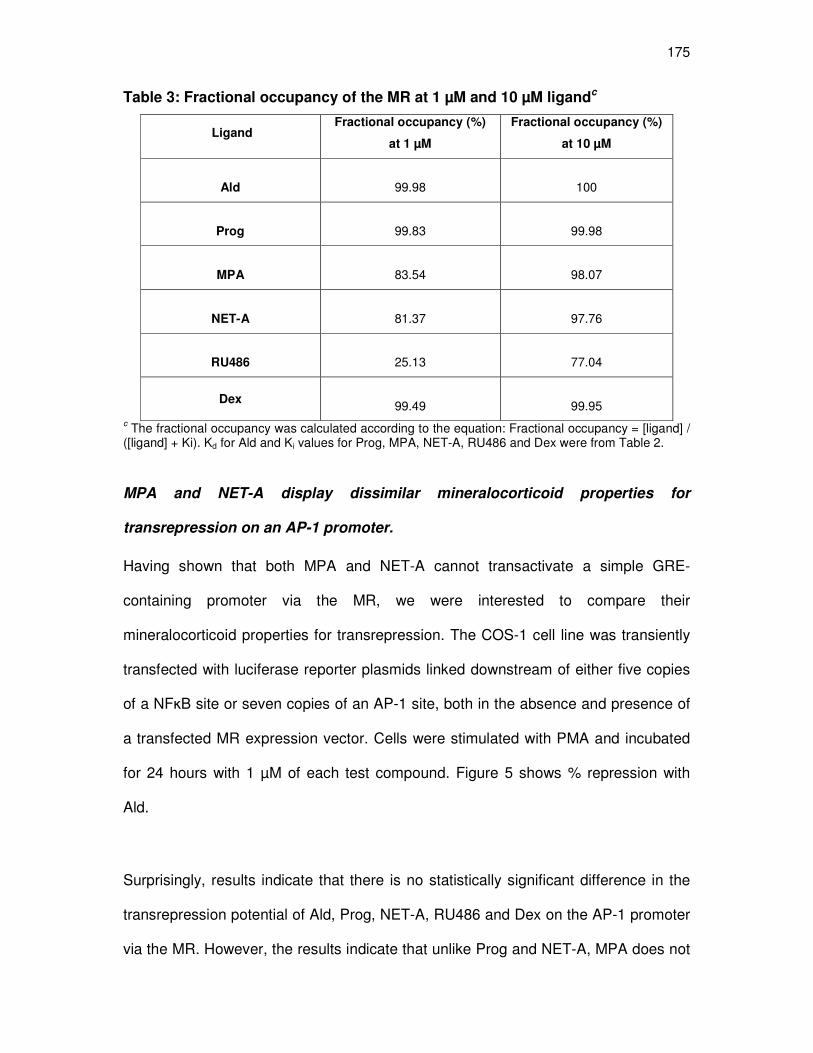
175
Table 3: Fractional occupancy of the MR at 1 µM and 10 µM ligandc
Ligand Fractional occupancy (%)
at 1 µM
Fractional occupancy (%)
at 10 µM
Ald
99.98
100
Prog
99.83
99.98
MPA
83.54
98.07
NET-A
81.37
97.76
RU486
25.13
77.04
Dex
99.49
99.95 c The fractional occupancy was calculated according to the equation: Fractional occupancy = [ligand] / ([ligand] + Ki). Kd for Ald and Ki values for Prog, MPA, NET-A, RU486 and Dex were from Table 2. MPA and NET-A display dissimilar mineralocorticoid properties for
transrepression on an AP-1 promoter.
Having shown that both MPA and NET-A cannot transactivate a simple GRE-
containing promoter via the MR, we were interested to compare their
mineralocorticoid properties for transrepression. The COS-1 cell line was transiently
transfected with luciferase reporter plasmids linked downstream of either five copies
of a NFĸB site or seven copies of an AP-1 site, both in the absence and presence of
a transfected MR expression vector. Cells were stimulated with PMA and incubated
for 24 hours with 1 µM of each test compound. Figure 5 shows % repression with
Ald.
Surprisingly, results indicate that there is no statistically significant difference in the
transrepression potential of Ald, Prog, NET-A, RU486 and Dex on the AP-1 promoter
via the MR. However, the results indicate that unlike Prog and NET-A, MPA does not
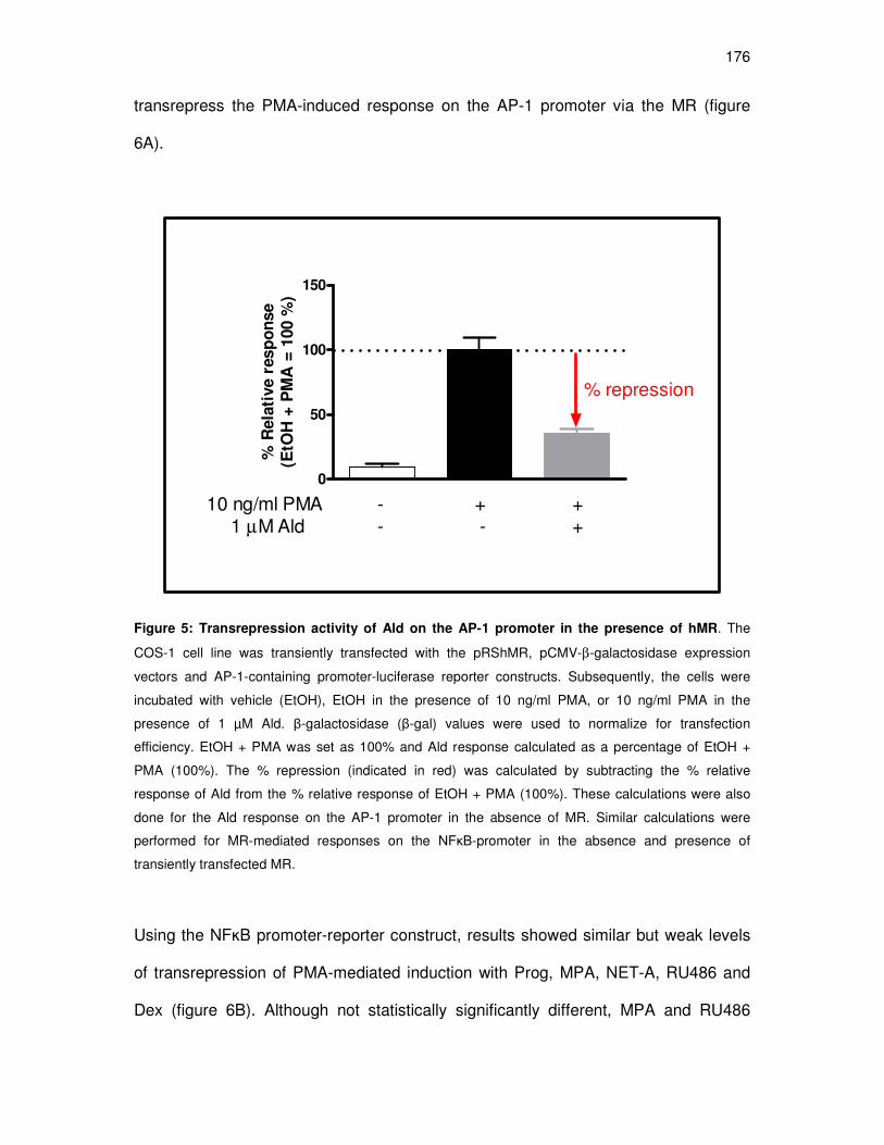
176
transrepress the PMA-induced response on the AP-1 promoter via the MR (figure
6A).
0
50
100
150
10 ng/ml PMA - + +1 µM Ald - - +
% repression
% R
ela
tive r
esp
on
se
(EtO
H +
PM
A =
100 %
)
Figure 5: Transrepression activity of Ald on the AP-1 promoter in the presence of hMR. The
COS-1 cell line was transiently transfected with the pRShMR, pCMV-β-galactosidase expression
vectors and AP-1-containing promoter-luciferase reporter constructs. Subsequently, the cells were
incubated with vehicle (EtOH), EtOH in the presence of 10 ng/ml PMA, or 10 ng/ml PMA in the
presence of 1 µM Ald. β-galactosidase (β-gal) values were used to normalize for transfection
efficiency. EtOH + PMA was set as 100% and Ald response calculated as a percentage of EtOH +
PMA (100%). The % repression (indicated in red) was calculated by subtracting the % relative
response of Ald from the % relative response of EtOH + PMA (100%). These calculations were also
done for the Ald response on the AP-1 promoter in the absence of MR. Similar calculations were
performed for MR-mediated responses on the NFκB-promoter in the absence and presence of
transiently transfected MR.
Using the NFĸB promoter-reporter construct, results showed similar but weak levels
of transrepression of PMA-mediated induction with Prog, MPA, NET-A, RU486 and
Dex (figure 6B). Although not statistically significantly different, MPA and RU486

177
appear to repress the NFĸB promoter to a slightly better extent (26% and 23%,
respectively) than Prog and NET-A (12% and 5%, respectively). When comparing the
transrepression activity of the different ligands on the AP-1 versus the NFĸB
promoter-reporter, Prog, NET-A and RU486 all exhibit greater transrepression via
AP-1 (51%, 74%, 60%, respectively) than via the NFĸB promoter (12%, 5%, 23%,
respectively). The activities of the ligands (at 1 µM) on the three synthetic promoter
constructs are summarized in Table 4. Given that MPA and NET-A bind to the MR
with similar affinities, and that neither is an agonist for transactivation (GRE), it is
noteworthy that they act differently on the AP-1 promoter.
The transrepression/transactivation (AP-1/GRE) ratios for all the ligands were
calculated, and are summarized in Table 5 in the discussion. Transrepression
induced by NET-A for example was 74% of that caused by Ald (100%), while NET-A-
mediated transactivation is only 1.6%. The transrepression/transactivation ratio for
NET-A is thus 46.3 (74% / 1.6%). It should be noted that at 1 µM of the ligands, the
fractional occupancy for Ald, Prog and Dex are ~ 100%, while for MPA and NET-A it
is ~84% and 81%, respectively (Table 3). Given that the fractional occupancy of 1 µM
RU486 is only 25%, the 60% percent MR-mediated repression with RU486 on the
AP-1 promoter is surprising, and difficult to explain. It may indicate that the affinity of
a ligand for the MR, does not necessarily determine the efficacy of the ligand for
transrepression.

178
A AP-1
Ald
Prog
MPA
NET-A
RU48
6Dex
0
50
100
150
a
b
a
aa
a
1µµµµM test compound
Rela
tive r
ep
ressio
n v
ia A
P-1
±± ±±S
EM
(A
ld=
100%
)
B NFĸB
Ald
Prog
MPA
NET-A
RU48
6Dex
0
50
100
150
a
b b b
b
1µµµµM test compound
Rela
tive r
ep
ressio
n v
ia N
Fκκ κκB
±± ±±S
EM
(A
ld=
100%
)
b
Figure 6 legend on next page.

179
Figure 6: Transrepression activity via the hMR. The COS-1 cell line was transiently transfected with
the pRShMR, pCMV-β-galactosidase expression vectors and (A) NFĸB- or (B) AP-1-containing
promoter-luciferase reporter constructs. Subsequently, the cells were stimulated with 10 ng/ml PMA
and incubated with 1 µM Ald, Prog, MPA, NET-A, RU486 or Dex for 24 hours. β-gal values were used
to normalize for transfection efficiency. Once the repressive abilities of the test compounds were
determined, in the absence and presence of transfected MR (Appendix C4), repression in the absence
of MR was subtracted from the repression in the presence of transfected MR. The % repression for the
other ligands was expressed as a % of Ald response which was set at 100%. The result shown is the
average of three independent experiments with each condition performed in triplicate (±SEM).
Unlike MPA, Prog, NET-A and RU486 do inhibit the aldosterone-induced
upregulation of the endogenous Orm-1 gene
To investigate whether the results observed on a synthetic promoter could be
mimicked on an endogenous promoter, we transiently transfected a rat
cardiomyocyte cell line (H9C2) with the full-length hMR or hGR expression vector.
Western blot analysis confirmed the expression of the transfected MR and GR in
these cells, as well as endogenous expressed MR and GR (figure 7A and 7B). To
determine whether the endogenous MR and GR, as well as the overexpressed
receptors are transcriptionally active, we assessed their transactivation potential in
the presence of receptor agonists, using a promoter-reporter assay. The results in
figure 7C-F indicate that the endogenous MR and GR, as well as the overexpressed
MR, GR, PR and AR, are transcriptionally active. Consistent with the lack of
transcrptional activity for endogenous PR and AR, Western blot analysis did not
detect endogenous AR or PR protein levels in these cells (data not shown).
We focussed our study on the inflammation/acute phase-related gene, Orm-1, known
to be associated with increased risk of cardiovascular disease (Engström et al.,
2003), and previously reported to be upregulated by 1 nM Ald in a H9C2 cell line
stably expressing the MR, via the suggested mechanism of binding of the liganded

180
MR to a MRE (Fejes-Tóth and Náray-Fejes-Tóth, 2007). In these cells, the
investigators showed a 1000-fold induction of the Orm-1 gene by 1 nM Ald at 24 h,
whereas we observed only a 6-fold induction at this time point (figure 8A and 8B).
Although not significantly different to the control, MPA caused a 4.4-fold upregulation
of the Orm-1 gene, while the other ligands displayed a 2-fold upregulation (figure 8A).
We also investigated the antagonist activity of the progestins on the Ald-induced
expression of Orm-1, by incubating the parent H9C2 cell line transiently transfected
with the MR, with 1 nM Ald in the presence of 1 µM of each progestin, as well as the
known MR antagonist, spironolactone, as a positive control. Our result on a synthetic
GRE showing that Prog is a potent MR antagonist (EC50 = 3.1 nM) was corroborated
on the endogenous Orm-1 gene. Although MPA, NET-A and RU486 showed a weak
but similar antagonist potency for the MR on the synthetic promoter (EC50 of 310, 270
and 670 nM, respectively), they acted differently on the endogenous promoter of the
Orm-1 gene (figure 8B). Interestingly, NET-A and RU486 antagonized the Ald-
induced upregulation of the Orm-1 gene to similar extents as Prog. In contrast, MPA
did not antagonize the upregulation of the Ald-induced Orm-1 gene (figure 8B). In
fact, although not significant, MPA seemed to enhance the upregulation of the Ald-
induced Orm-1 gene (figure 8B).
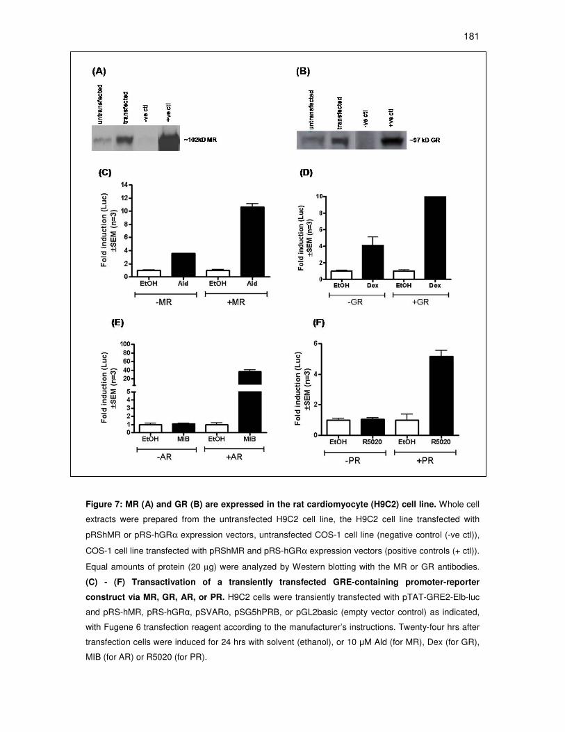
181
Figure 7: MR (A) and GR (B) are expressed in the rat cardiomyocyte (H9C2) cell line. Whole cell
extracts were prepared from the untransfected H9C2 cell line, the H9C2 cell line transfected with
pRShMR or pRS-hGRα expression vectors, untransfected COS-1 cell line (negative control (-ve ctl)),
COS-1 cell line transfected with pRShMR and pRS-hGRα expression vectors (positive controls (+ ctl)).
Equal amounts of protein (20 µg) were analyzed by Western blotting with the MR or GR antibodies.
(C) - (F) Transactivation of a transiently transfected GRE-containing promoter-reporter
construct via MR, GR, AR, or PR. H9C2 cells were transiently transfected with pTAT-GRE2-Elb-luc
and pRS-hMR, pRS-hGRα, pSVARo, pSG5hPRB, or pGL2basic (empty vector control) as indicated,
with Fugene 6 transfection reagent according to the manufacturer’s instructions. Twenty-four hrs after
transfection cells were induced for 24 hrs with solvent (ethanol), or 10 µM Ald (for MR), Dex (for GR),
MIB (for AR) or R5020 (for PR).

182
EtOH
1 nM
Ald
Prog
MPA
NET
RU48
6
0
2
4
6
8
1 µµµµM test compound
a
Rati
o O
rm-1
/ββ ββ
-acti
nexp
ressio
na
a
aa
b
0
2
4
6
8
10
12
14
1 nM Aldosterone
1 µµµµM Prog
1 µµµµM MPA
1 µµµµM NET-A
1 µµµµM RU486
1 µµµµM Spironolactone
-
-
-
-
-
+
-
-
-
-
+ + + + -
+
+
+
+
+
-
-
-
-
-
-
- -
-
-
-
-
--
- -
-
-
-
- - -
+
+
-
-
-
-
Rati
o O
rm-1
/ββ ββ
-acti
nexp
ressio
n
a
b
b
a a aa
a
Figure 8 legend on next page.
A
B

183
Figure 8: (A) Agonist and (B) antagonist activity of Prog, MPA, NET-A and RU486 on the
endogenous MRE-containing Orm-1 gene in the rat cardiomyocyte cell line transiently
transfected with the MR. (A) Ald and MPA upregulate the Orm-gene to similar extents. Cells
were incubated with 1nM Ald or 1 µM Prog, MPA, NET-A, or RU486, for 24 hours. (B) Prog, NET-A
and RU486 antagonise the aldosterone-induced upregulation of the Orm-1 gene. Cells were
incubated with 1 nM Ald in the absence and presence of 1 µM Prog, MPA, NET-A, RU486 or
Spironolactone. Total RNA was extracted, cDNA synthesized and expression levels of Orm-1 and β-
actin mRNA were determined by QPCR. Ratios of the Orm-1/β-actin gene were calculated using the Fit Points method described by Pfaffl (2001). Results shown are the averages of three independent
experiments.
DISCUSSION
For women with an intact uterus, the inclusion of progestin in continuous combined
HRT is needed to counteract the proliferative effects of estrogen on the uterine
endometrium, thereby decreasing the risk of cancer associated with unopposed
estrogen therapy (Gambrell Jr et al., 1980; Palacios et al., 2006). However, the
estrogen component in HRT can also cause water and sodium-retention mediated
via the RAAS (Oelkers, 1996), leading to weight gain and increased blood pressure.
Since hypertension (high blood pressure) increases cardiovascular risk in both men
and women (Calhoun and Oparil, 1997), there may be an association between the
use of menopausal HRT and an elevated risk for cardiovascular disease.
Endogenous progesterone, however, has anti-MR effects on the RAAS (Oelkers,
1996; Oelkers, 2004), thereby counterbalancing the estrogen-related water and
sodium-retention and providing positive effects on body weight and also blood
pressure. Thus, progestins used in HRT should ideally mimic these anti-
mineralocorticoid properties.

184
The MR belongs to the family of steroid hormone receptors, comprising structurally
similar proteins such as the GR, AR, PR and ER. In general, they are ligand-
activated transcription factors that comprise four different domains namely the highly
variable NH2-terminal domain (NTD), the highly conserved and centrally located
DNA-binding domain (DBD), which directly interacts with the DNA response
elements, the hinge region and the COOH-terminal (C-terminal) domain. The C-
terminal domain contains the ligand binding domain (LBD) to which the ligand binds.
Furthermore, these receptors also possess transcriptional activation functions
located in the NTD (AF1 domain) and the LBD (AF2 domain). Mineralocorticoids (or
ligands binding to the MR) bind to the cytoplasmic MR in target tissues. Upon ligand
binding, the receptor translocates to the nucleus where it regulates gene expression
(Evans, 1988; Beato, 1989). An increase in gene transcription or positive regulation
of target genes is termed transactivation, and occurs when ligand-activated MR binds
to specific DNA sequences, called mineralocorticoid response elements (MREs) in
the promoter regions of specific genes, resulting in gene activation (Evans, 1988;
Beato, 1989). The steroid receptor family can also negatively regulate genes
(transrepression), and this is mediated via the ligand-activated receptor not directly
binding to DNA but via protein-protein interactions with other transcription factors
such as nuclear factor-kappa B (NFĸB) and activator protein-1 (AP-1) (Lu et al.,
2006). AP-1 can be composed of either homodimers or heterodimers between
members of the Jun and Fos families. This mechanism of transrepression is well-
described for the GR, but as yet the MR has not been shown to modulate AP-1- and
NFκB-induced transcription by direct protein-protein interactions.
185
One of the aims of the present study was to evaluate the interactions with the MR of
two synthetic progestins, MPA and NET-A, frequently prescribed for HRT and to
provide a comparative biochemical profile of these progestins, as well as
progesterone (the natural hormone), in the same model system. Although MPA and
NET-A have been used clinically for more than 40 years (Westhoff, 2003), their
apparent Ki values for the human MR in the same model system, have not been
established. Furthermore, to our knowledge, our study is the first to investigate their
ability to induce the MR N/C interaction, as well as their activity for transrepression of
transcription via the MR.
The Kd for Ald binding to the hMR determined in our system (0.153 nM) is similar to
previously reported Kd’s of ± 0.8-3 nM (Arriza et al., 1987; Lombes et al., 1994; Lim-
Tio et al., 1997). We established Ki’s for Prog, MPA and NET-A of 1.69, 197 and 229
nM, respectively (Table 1). In addition, we determined a Ki for RU486 (mifepristone),
a known antagonist of the PR, GR and AR (Spitz and Bardin, 1993; Song et al.,
2004), of 2980 nM. RU486 has previously been reported to have no affinity for the
MR (Leonhardt and Edwards, 2002). When comparing the binding affinities of MPA
and NET-A as percentages relative to the native ligand Ald (100%), our values for
MPA and NET-A (0.078% and 0.067%, respectively), are slightly lower than
previously reported values of 3.1% and 2.7%, respectively (Philibert et al., 1999;
Winneker et al., 2003 ). Furthermore, we show, as have others have (Arriza et al.,
1987; Rupprecht et al., 1993b; Grossmann et al., 2004), that the potent synthetic GR
agonist, Dex, also binds the MR with relatively high affinity, although it does not
activate the MR with the same potency as it does the GR (Arriza et al., 1987;
Rupprecht et al., 1993b; reviewed in Rogerson et al., 2003; Grossmann et al., 2004).

186
In addition, we determined the agonist and antagonist properties of MPA and NET-A
for transactivation via an MRE using promoter-reporter assays in the COS-1 cell line.
Our results show that, while both MPA and NET-A are devoid of mineralocorticoid
agonist activity via an MRE, Prog has very weak partial agonist activity at 10 µM
(approximately 20% activity) (figure 2A), and only about 6% at 1 µM (Appendix C4).
In contrast, a previous study reported agonist activity of Prog for transactivation via
the MR as 27% at 1 µM (Quinkler et al., 2002). The discrepancy between our results
and that study could be due to the different promoter and cell lines used, as they
used a synthetic reporter gene containing an MMTV promoter in the CV-1 cell line,
while we used the pTAT-GRE-E1b-luc vector in COS-1 cells. However, the observed
differences could also be attributed to differences in MR levels. It has previously
been shown that receptor levels affect the potency and efficacy of full GR agonists,
partial agonists and antagonists (Zhao et al., 2003). Interestingly, a very recent study
showed no agonist activity for up to 3 µM Prog in the COS-1 cell line co-transfected
with a MR-expression vector and PRE2tk-luc promoter-reporter (Sasagawa et al.,
2008), which is more consistent with our results.
To further investigate agonist potential of ligands via steroid receptors, much
attention has focussed on agonist-induced interactions between the N- and C termini,
an interaction termed the N/C-interaction. This N/C-interaction is best characterized
for the AR and involves binding of the AF2 region of the LBD with specific sequences
in the N-terminal domain (He et al., 2000), and has been shown to play a major role
in AR transcriptional activity (Kemppainen et al., 1999; He et al., 1999; He et al.,
2000). An Ald-dependent N/C-interaction was also recently shown for the MR
187
(Rogerson and Fuller, 2003). Although the other physiological ligand of the MR,
cortisol (Cort), has a higher potency than Ald in transactivation assays (Rogerson et
al., 1999), a MR N/C-interaction was only weakly induced in its presence (Rogerson
and Fuller, 2003). The authors hypothesize that their results showing that Ald, but not
Cort, can induce the MR N/C-interaction, may explain why Ald and Cort have
differential effects in some non-epithelial cells. However, that study only compared
Ald and Cort up to 100 nM. At a concentration of 10 µM Ald and Cort, we find similar
induction of the N/C-interaction by both ligands (supplementary material in Appendix
C5). In addition, Prog, a very weak partial agonist for the MR in our system, could
weakly induce the N/C-interaction at a concentration of 10 µM (figure 3), but not 1 µM
(data not shown). Similarly to Rogerson and Fuller (2003) who showed no N/C-
interaction for the MR antagonist spironolactone, our results indicate that the
antagonists in our system, that do not display partial agonist activity, such as MPA,
NET-A and RU486, could not promote an N/C-interaction (figure 3). We show that
efficacy for transactivation via a synthetic GRE correlates with the efficacy for the
induction of N/C-interaction (r2 = 0.9179; p<0.01) (figure 4), indicating that there is a
good relationship between transactivation and MR N/C-interaction.
We determined considerable anti-mineralocorticoid activity of Prog (EC50 = 3.1 nM),
which is in agreement with previously reported EC50 values in the range 2-10 nM
(Rupprecht et al., 1993; Auzou et al., 2000; Geller et al., 2000), but lower than a
recent study where the EC50 for Prog was determined as 11.4-18.1 nM (Sasagawa et
al., 2008). The molecular basis of Prog MR antagonistic activity still needs to be
researched, but it is speculated that it may be due to the destabilization of helix 12 of
the MR binding domain (Elger et al., 2003). Surprisingly, although it has previously

188
been reported that MPA, NET-A and RU486 have no MR antagonist activity
(Winneker et al., 2003 and references therein, reviewed by Rowlands 2003, Palacios
et al., 2006; Zhang et al., 2006), we showed MR antagonist activity of these ligands,
albeit to a lesser extent than for Prog (figure 2B). We determined potencies (EC50
values) of 310, 270 and 670 nM for MPA, NET-A and RU486, respectively.
Interestingly, in accordance with our results, a recent study also showed MR
antagonist activity for MPA and NET-A, but reported much lower potencies (1108.6
and 1100.9 nM for MPA and NET-A, respectively) (Sasagawa et al., 2008). The EC50
values obtained in our study are higher than the peak serum level reached with HRT
(MPA = 0.2 nM and NET-A = 17.7 nM) (Ghatge et al., 2005; Activelle package insert
reg. no. 33/21.8.2/0532, Novo Nordisk Inc.) or contraception (MPA = 3.9 nM; NET-A
= 59 nM) (Mishell, 1996; Fotherby et al., 1983), but the possibility that higher
concentrations may be present in vivo in target tissues compared with concentrations
that are usually measured in the blood, should not be excluded.
In general, not much is known about transrepression mediated via the MR. Early
work by Pearce and Yamamoto (1993) showed that the MR could not repress AP-1
activity in response to Ald. In contrast to that study, we have shown that Ald can
repress AP-1 activity via the MR. In agreement with the study by Liden et al. (1997)
suggesting that the MR can repress transactivation of NFĸB, we also show that Ald
can repress NFĸB activity via the MR. We investigated the comparative effects of all
the ligands on transrepression via the MR on both NFĸB and AP-1 promoter
constructs. The results of the transrepression assays (at 1 µM) (figure 6A and 6B) as
well as the effects observed for transactivation (figure 2A), are summarized in Table

189
4. Figure 6B indicates that none of the ligands, except Ald, could significantly
transrepress the NFĸB site via the MR.
Table 4: Relative responses for transactivation and transrepression via the MR
at 1 µM ligandc
Ligand
GRE
Relative response
(%)
NFĸB
Relative response
(%)
AP-1
Relative response
(%)
Ald
100
100
100
Prog
19 ± 0.7
12 ± 11.3
51 ± 17.1
MPA
2 ± 0.7
26 ± 6.3
1.8 ± 1.8
NET-A
1.6 ± 0.8
5 ± 4.0
74 ± 3.7
RU486
1.3 ± 1.0
23 ± 12.8
60 ± 14.2
Dex
27 ± 0.2
21 ± 16.0
103 ± 5.9 c Relative responses for transactivation via pTAT-GRE-E1b-luc and for transrepression via either
NFĸB or AP-1-luc reporter plasmids at 1 µM ligand, were determined in the transiently transfected
COS-1 cell line. Results are averages of at least three independent experiments, each performed in
triplicate. Ald activity was taken as 100% and all other values are expressed as a % thereof.
One of the most important and novel findings of our study, is that both NET-A and
RU486 can transrepress the PMA-induced response on the AP-1 promoter via the
MR, without exhibiting agonist activity for MR transactivation via a GRE. MPA, on the
other hand, cannot transrepress the PMA-induced response on the AP-1 promoter
via the MR, or induce agonist activity for MR transactivation via a GRE. Ligands that
can transrepress, but not transactivate, are referred to as dissociated ligands, and
are well described for the GR (Vaysierre et al., 1997; De Bosscher et al., 2005). The
transrepression by the ligands in this study is consistent with a MR-dependent

190
mechanism as the repression without transfected MR, was subtracted from the
repression with transfected MR. However, elucidation of the mechanism behind this
dissociative activity will require additional studies. Interestingly, Vaysierre et al.
(1997) reported a similar ability for synthetic glucocorticoids that could differentiate
between transactivation and AP-1 transrepression via the GR. Similarly, we show
that while Dex is a partial agonist for MR transactivation on a GRE promoter, it is a
full agonist for transrepression on the AP-1 promoter via the MR. An AP-1
transrepression/transactivation ratio was calculated for all the ligands (Table 5). NET-
A and RU486 had an AP-1 transrepression/transactivation ratio of 46.3 and 46.2,
respectively compared to 1.0 for Ald, indicating that NET-A and RU486 did not
transactivate a GRE via the MR, but was able to repress AP-1 transactivation via the
MR. As the fractional occupancy of the MR at 1 µM is only 25%, it is intriguing that
RU486 could repress the AP-1 activity by 60%. This result may indicate that the
affinity of a ligand for the MR, does not necessarily determine the efficacy of the
ligand for transrepression, as has been shown for GR ligands via both NFĸB and AP-
1 (Ronacher et al., 2009). In contrast, MPA could not transactivate a GRE or repress
AP-1 activity via the MR. The transrepression/transactivation ratio for MPA was thus
0.9 indicating that MPA does not distinguish between transactivation and
transrepression via the MR. In fact, MPA does not display any mineralocorticoid
agonist properties. This ratio for Prog and Dex was 2.7 and 3.8, respectively,
indicating that these ligands are better at transrepression than transactivation. MR
actions via these transcription factors, NFĸB and AP-1, may play an important role in
inflammation, typically associated with MR-mediated cardiovascular injury.

191
It has been reported that MR antagonism protects the cardiovascular system from
damaging inflammation (Fejes-Tóth and Náray-Fejes-Tóth, 2007). For this reason,
we next determined whether MPA, NET-A and RU486 could antagonize Ald-induced
effects on a risk factor of cardiovascular disease. Using a rat cardiomyocyte cell line
transiently transfected with the MR as a model for cardiovascular disease, we
investigated the antagonistic properties MPA, NET-A and RU486, on the expression
of the endogenous Ald-responsive gene, Orm-1. This gene is involved in
inflammation, and elevated levels are considered a cardiovascular risk factor
(Engström et al., 2003). Interestingly, patients with metabolic syndrome have
elevated plasma Orm-1 levels, which may account for the increased prevalence of
cardiovascular problems in this syndrome. Although MPA, NET-A and RU486 acted
as weak antagonists of the MR in a GRE-reporter assay, we showed that NET-A and
RU486, but not MPA, inhibit the effect of Ald on the expression of the endogenous
MRE-containing gene, Orm-1, to a similar extent as Prog. MPA increased the
expression of Orm-1 (figure 8A and 8B). However, the H9C2 cell line also expresses
the GR (figure 7B) and it is known that glucocorticoids can induce Orm-1 (Vannice et
al., 1984). Taking this into account, together with the fact that we show no MR
agonist properties for MPA in a GRE-reporter assay, it is probable that MPA, known
to have glucocorticoid agonist properties at a concentration of 1 µM (Koubovec et al.,
2004; Koubovec et al., 2005; Ronacher et al., 2009), would preferentially act via the
GR, and it is thus likely that the agonist activity of MPA on the endogenous Orm-1
gene is mediated by the GR rather than the MR.
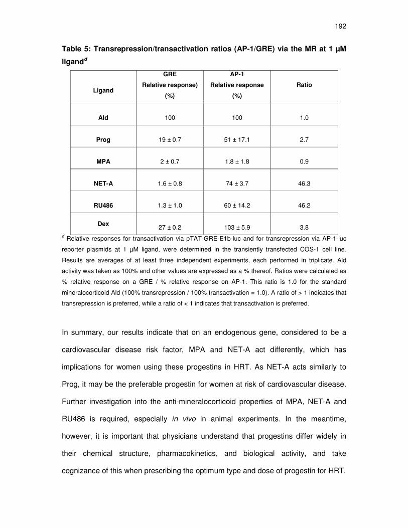
192
Table 5: Transrepression/transactivation ratios (AP-1/GRE) via the MR at 1 µM
ligandd
Ligand
GRE
Relative response)
(%)
AP-1
Relative response
(%)
Ratio
Ald
100
100
1.0
Prog
19 ± 0.7
51 ± 17.1
2.7
MPA
2 ± 0.7
1.8 ± 1.8
0.9
NET-A
1.6 ± 0.8
74 ± 3.7
46.3
RU486
1.3 ± 1.0
60 ± 14.2
46.2
Dex
27 ± 0.2
103 ± 5.9
3.8 d Relative responses for transactivation via pTAT-GRE-E1b-luc and for transrepression via AP-1-luc
reporter plasmids at 1 µM ligand, were determined in the transiently transfected COS-1 cell line.
Results are averages of at least three independent experiments, each performed in triplicate. Ald
activity was taken as 100% and other values are expressed as a % thereof. Ratios were calculated as
% relative response on a GRE / % relative response on AP-1. This ratio is 1.0 for the standard
mineralocorticoid Ald (100% transrepression / 100% transactivation = 1.0). A ratio of > 1 indicates that
transrepression is preferred, while a ratio of < 1 indicates that transactivation is preferred.
In summary, our results indicate that on an endogenous gene, considered to be a
cardiovascular disease risk factor, MPA and NET-A act differently, which has
implications for women using these progestins in HRT. As NET-A acts similarly to
Prog, it may be the preferable progestin for women at risk of cardiovascular disease.
Further investigation into the anti-mineralocorticoid properties of MPA, NET-A and
RU486 is required, especially in vivo in animal experiments. In the meantime,
however, it is important that physicians understand that progestins differ widely in
their chemical structure, pharmacokinetics, and biological activity, and take
cognizance of this when prescribing the optimum type and dose of progestin for HRT.

193
Acknowledgements
We thank Carmen Langeveldt for technical support. We furthermore thank Prof’s.
Rogerson and Fuller for the donation of MR constructs used in the mammalian two-
hybrid assays. This work was supported by grants to JPH and DA from the Medical
Research Council (MRC) and the National Research Foundation (NRF) in South
Africa, and Stellenbosch University. Any opinion, findings and conclusions or
recommendations expressed in this material are those of the author(s) and therefore
the NRF does not accept any liability in regard thereto.

194
Chapter 5
_____________________________________________________
CONCLUSIONS AND FUTURE PERSPECTIVES
195
5. CONCLUDING DISCUSSION
Although MPA and NET-A are widely used in female reproductive therapy, not much
is known about their mechanism of action at the molecular level. Clinical evidence
indicating that the use of MPA and NET-A in HRT increases the risk of breast cancer
(Rossouw et al., 2002; Beral, 2003), and also that MPA increases the risk of CVD,
has highlighted the importance of investigating these mechanisms. In addition to
these studies implicating MPA in effects on cardiovascular function and breast
cancer in HRT users, use of MPA as a contraceptive has also been implicated in
effects on local immune function (Ildgruben et al., 2003). Little is known about the
effects of MPA and NET-A, as compared to Prog, in the cervicovaginal environment,
and whether they interfere with local epithelial immune function. In the first part of this
thesis (Chapter 2), we thus investigated the regulation of cytokine genes by MPA and
NET-A, as compared to the endogenous hormone Prog, in Ect1/E6E7 and Vk2/E6E7
cell lines with a view to understanding the possible steroid receptor mediated effects
of these synthetic progestins on immunity in the female genital tract. In addition,
although it is known that both MPA and NET-A can bind the PR, GR, AR and MR, no
thorough comparison exists on the binding affinity of MPA and NET-A, as compared
to Prog, towards the AR and MR. Moreover, an accurate pharmacological
characterisation of the relative efficacy and potency of the two progestins on gene
regulation via these receptors is lacking. In the second and third part of the
experimental work in this thesis (Chapter 3 and 4), we thus examined the mechanism
of action of MPA and NET-A, relative to Prog, at the molecular level via
overexpressed AR and MR in the COS-1 cell line. Additionally, we investigated the
MR-mediated transcriptional regulation of the endogenous Orm-1 gene, a marker of

196
CVD, in a rat cardiomyocyte cell line, with a particular focus on the antagonistic role
of MPA and NET-A, as compared to Prog.
5.1 Comparing the regulation of endogenous pro-inflammatory
cytokine genes, by MPA and NET-A, in the female reproductive tract
In Chapter 2, specific genital tract cell types for the induction or upregulation of
protective molecules were investigated. As a model for cervicovaginal mucosal
immunity, we used epithelial cell lines derived from the female ectocervical and
vaginal mucosa, and showed that ligand-specific and cell-specific regulation of the
pro-inflammatory cytokine (IL-6) and chemokines (IL-8 and RANTES) gene
expression occurs by Prog, MPA and NET-A. Chemokines initiate the immune
response by controlling leukocyte migration and lymphocyte development. All three
cytokines have been shown to be expressed and secreted by Ect/E6E7 and
Vk2/E6E7 cell lines in response to activation by TNFα (Fichorova et al., 1999). Many
inflammatory response genes containing NFĸB response elements, like the IL-6, IL-8
and RANTES genes, are upregulated in response to proinflammatory cues initiated
by cytokines like TNFα. IL-8 and RANTES are involved in the early inflammatory
response, by recruiting specific leukocytes to sites of ongoing inflammation, while IL-
6 is responsible for neutrophil priming to chemotactic factors (Kunkel et al., 1997).
These specific cytokines were chosen because they have previously been shown to
be repressed by MPA in other cell types. For example, MPA has previously been
shown to repress IL-6 and IL-8 promoter–reporter constructs at the transcriptional
level (Koubovec et al., 2004), as well as the RANTES gene in endometrial cells
(Zhao et al., 2002). Moreover, IL-6, IL-8 and RANTES have been reported to have
distinct roles in HIV-1 pathogenesis (Fichorova et al., 2004).
197
Results showed that unlike Prog which upregulated TNFα-stimulated IL-6 gene
expression in both the ectocervical and vaginal cell lines, MPA and NET-A had no
effect. This is inconsistent with previous studies that have shown the repression of IL-
6 by MPA in other cell types (Mantovani et al., 1997; Koubovec et al., 2004; Wessel
Kriek, MSc thesis). Similarly, NET-A has previously been shown to either upregulate
(Zitzmann et al., 2005) or downregulate (Wessel Kriek, MSc thesis) IL-6. In contrast,
NET-A in combination with estrogen, like estrogen only, had no effect on IL-6 levels
in healthy postmenopausal women (Žegura et al., 2006).
In contrast to the observed effects on the IL-6 gene, we show that MPA and NET-A
upregulate the TNFα-induced IL-8 gene expression to different extents in both cell
types, indicating pro-inflammatory activity of these progestins which is similar to that
of Prog. Similarly, Prog and MPA have previously been shown to upregulate IL-8
mRNA levels in human endometrial stromal cells (Arici et al., 1996). As IL-8 is a
chemotactic factor for neutrophils and lymphocytes, the local concentration of IL-8 is
important in determining whether either neutrophils or lymphocytes are primarily
recruited. Prog has previously been shown to induce a neutrophil response in the
vagina of germ-free mice, even in the absence of bacterial infection (Beaver, 1960),
indicating a protective role for Prog in the vagina. Thus, our results demonstrating
upregulation of IL-8 by natural Prog, MPA and NET-A, may indicate that in these cell
lines, the synthetic progestins are similar to Prog, and that this may provide an
enhanced local defense mechanism against infections in the vaginal mucosa.
Interestingly, results indicated differential regulation of the TNFα-induced RANTES
gene by MPA, NET-A and Prog in the Ect1/E6E7 cells, but similar regulation in the

198
Vk2/E6E7 cell line. As RANTES is a pro-inflammatory chemokine, the suppression of
the RANTES gene by MPA indicates that MPA is anti-inflammatory. In epithelial cells,
RANTES is responsible for recruiting immune cells into the reproductive tract (Kunkel
et al., 1997) and thus inhibition of RANTES would suppress inflammation, thereby
blocking infiltration of immune cells. As the transmission of viruses such as HIV, HSV
and HPV occurs at the cervicovaginal environment (Tjiong et al., 2001), a lack of
recruitment of immune cells into the reproductive tract may have negative
implications for women using MPA, as they would not be able to effectively combat
infections in the ectocervical environment. Consistent with this result, injectable
hormonal contraceptive usage has previously been associated with increased risk of
cervical cancer due to HPV (Castle et al., 2005). In contrast, a South African study
investigating the link between use of MPA and NET-A as progestin-only injectable
contraceptives, and the risk for cervical cancer due to HPV, indicated that these
progestins did not contribute to the risk of cervical cancer (Shapiro et al., 2003).
Downregulation of RANTES gene expression by MPA has previously been reported
in endometrial stromal cells (Zhao et al., 2002), and the PR was shown to mediate
the effect. In addition, MPA has previously been shown to repress the pro-
inflammatory chemokine, IL-8, via the GR (Koubovec et al., 2004). We thus
investigated which steroid receptor is mediating the effects of MPA on RANTES in
the Ect1/E6E7 cell line, by using receptor-specific antagonists. Our results using
RU486, the antagonist for the PR and GR, was inconclusive as RU486 behaved as
an agonist in this cell line. We were unable to exclude the involvement of the GR and
PR, particularly the PR-A isoform, which is a weak activator of transactivation, but a
strong activator of transrepression (Kalkhoven et al., 1996). However, when using the
199
AR-specific antagonist, hydroxyflutamide, we showed that the repression of the
RANTES gene by MPA is mediated by the AR. Consistent with this result, the
androgenic properties of MPA have previously been proposed to be responsible for
the anti-inflammatory effects of MPA on plasma inflammatory markers, such as C-
reactive protein and serum amyloid A protein (Wakatsuki et al., 2002).
Our study is the first to show ligand-specific and cell-specific effects on cytokine and
chemokine genes in the cervicovaginal environment. In addition, we show that the
observed repression of the TNFα-induced RANTES gene by MPA in the Ect1/E6E7
cell line is mediated by the AR. Considering that NET-A was shown to have similar
androgenic properties to MPA for transactivation and transrepression of synthetic
promoters (see Chapter 3), it was surprising that NET-A did not act via the AR on the
endogenous RANTES promoter, that contains NFκB-binding sites, in the Ect1/E6E7
cell line. This difference indicates that the repression of the endogenous RANTES
gene is more complex than repression on a synthetic NFĸB-promoter, and possibly
involves more factors than just NFκB tethering. It is thus possible that there are other
regulators or elements determining the response of NET-A on the endogenous gene,
which are not present in the promoter-reporter construct used. Furthermore, as it is
known that NET-A can be metabolized (Stanczyk and Roy 1990), the possibility thus
exists that NET-A is metabolized in this cell line, and that the metabolite(s), are less
efficacious AR agonists than NET-A itself. This could be investigated in future, by
transfecting an ARE-luc or NFκB-luc into the Ect1/E6E7 cells, and determining
whether NET-A can transactivate or transrepress via the AR.

200
In addition, the repression of RANTES by MPA via the AR was not observed in the
Vk2/E6E7 cell line, indicating that MPA acts in a cell-specific manner. As the levels of
AR in the Ect1/E6E7 and Vk2/E6E7 cell lines are similar, it is unlikely that these
differences in responses are due to different levels of steroid receptors. It may be
due to other factors such as different extent of metabolism or even different levels of
coregulators. Using whole cell binding experiments, we showed that the amount of
GR and AR in the Ect1/E6E7 cells line are similar to those in the Vk2/E6E7 cell line.
The expression of AR in the human vagina and cervix has previously been reported
(Ruizeveld de Winter et al., 1991), and it is known that the GR is ubiquitously
expressed (reviewed in Gross and Cidlowski, 2008). A further complexity is the
affinity of MPA and NET-A for the different steroid receptors, in particular the GR
versus the AR, as there appear to be similar levels of these receptors in the
Ect1/E6E7 cell line. MPA has a much higher affinity for the GR than NET-A (10.8 vs.
270 nM, respectively) (Koubovec et al., 2005), while both MPA and NET-A have
similar affinities for the AR (19.4 and 27.9 nM, respectively). Thus, in a cell where the
AR and GR are expressed equally, it is assumed that MPA can act via either the GR
or AR as its affinity for these receptors is similar. However, one would then assume
that NET-A would act via the AR, which is not the case in this study.
Nevertheless, this study is the first to show that MPA suppresses the RANTES gene
via the AR in a human ectocervical cell line. Further work is necessary to assess the
impact of this novel finding. Moreover, the finding that MPA acts differently to NET-A,
and does not mimic the effects of Prog, has important implications for women’s
health.

201
Finally, the immunoregulatory milieu of the genital mucosa is important in determining
the response to antigenic challenge. Mucosal innate immune factors as well as
proinflammatory cytokines are associated with elevated levels of HIV-1 shedding in
the female genital tract (Gumbi et al., 2008). During male-to-female transmission, it is
thought that virus in the semen enters the stratified squamous epithelium of the
vagina or the columnar epithelium of the endocervix to infect cells within or below the
epithelium (reviewed in Pope and Haase, 2003). It is not clear which cells are the first
to interact with, and to be infected with the virus, whether environmental factors in the
reproductive tract tissues may influence the infection, or whether there are local
immune-system responses directed at the virus. Thus, a greater understanding of the
immune responses at these different anatomical locations in the genital tract, and
how MPA and NET used as contraceptives and in HRT regulate genes relevant to
the cervicovaginal mucosal immune response, may have implications for the
development of efficacious topical microbicide products for the prevention of sexually
transmitted HIV-1 infection, or for the development of HIV-1 vaccines. This may thus
also have important implications for women’s risk of susceptibility to infections. In
addition, novel avenues may be identified both to protect against potential pathogens
and to improve the quality of women’s reproductive health.
5. 2. Comparing the androgenic properties of Prog, MPA and NET-A
in the COS-1 cell line
In the second part of this thesis (Chapter 3), for the first time, equilibrium binding
studies in the COS-1 cell line enabled the calculation of precise Ki values of Prog,
MPA, NET-A and RU486 for the AR. RU486 was included as it is a synthetic
progestin previously reported to bind the AR (Song et al., 2004). The COS-1 cell line

202
does not contain significant levels of endogenous steroid receptors and is thus an
appropriate model cell line for comparing the activities of ligands via a specific
transiently transfected receptor. Although saturation binding experiments are the
conventional way in which Kd’s are determined, we determined Kd and Ki values by
using the alternate method of homologous/heterologous competitive binding
experiments, using the Cheng-Prusoff equation (Cheng and Prusoff, 1973). We
optimized the concentration of 3H-[MIB] (two to ten times lower than the EC50) to use,
and found 0.2 nM to be optimal. The time for 0.2 nM 3H-[MIB] to reach binding
equilibrium was determined as 16 hours.
The results showed that Prog, MPA, NET-A and RU486 were able to compete with
MIB for binding to the AR. Prog, MPA and NET-A displayed similar binding affinity to
each other (Ki = 36.6 nM, 19.4 nM and 21.9 nM respectively), and the natural
androgen DHT (Ki = 29.4 nM). MPA and NET have previously been shown to bind to
the endogenously expressed AR in the MCF-7 breast cancer cell line with a similar
relative affinity to each other, albeit with a much lower relative binding affinity than
that found in our study (Bergink et al., 1983 and references therein). In addition, our
results for Prog, MPA and DHT are consistent with a previous study in the COS-1 cell
line (Kemppainen et al., 1999). Previous studies have reported that RU486 has low
binding affinity for the AR in rat prostate cytosols (Philibert et al., 1999; Cabeza et al.,
2007). Interestingly, the study by Cabeza and co-workers (2007) shows a 23-fold
lower relative binding affinity of RU486 compared to MIB. Indeed, in our study when
compared to MIB, RU486 has a 12-fold lower affinity, which is in the same order of
magnitude as the results by Cabeza et al. (2007). However, when compared to the

203
natural androgen DHT, RU486 (synthetic progestin) has a 6-fold higher binding
affinity for the hAR.
We demonstrated that unlike Prog, MPA and NET-A are potent and efficacious AR
agonists, with activity comparable to that of DHT. Furthermore, consistent with other
studies, we show that Prog (Kemppainen et al., 1999; Sasagawa et al., 2008) and
RU486 are potent antagonists (Song et al., 2004; Cabeza et al., 2007) with minimal
agonist activity. We show that in contrast to DHT and NET-A, MPA does not induce
the N/C-interaction of the AR on the classical ARE, which also serves as a response
element for the GR, PR and MR (reviewed in Beato, 1989). In addition, we show that
MPA can induce the N/C-interaction on two selective AREs, suggesting a promoter-
dependent induction of the AR N/C interaction by MPA. Indeed, it has been shown
that the AR N/C interaction is important for AR activation of some androgen-
regulated promoters, but not others (He et al., 2002). However, we cannot exclude
the possibility that the reason for these quantitative differences in the ability of MPA
to induce the N/C interaction via the promoter-reporter-constructs used in this study
is due to different expression levels of the promter-reporter constructs or the AR.
Taken together, these results suggest that MPA and NET-A activate the AR by
different mechanisms on the classical ARE, but by similar mechanisms on selective
AREs.
It is well-known that the anti-inflammatory effects of glucocorticoids are mediated by
the GR interacting with transcription factors, such as NFĸB and AP-1, thereby
repressing the activity of the transcription factor and the target genes (De Bosscher
et al., 2003). Considering that the AR and GR belong to the same superfamily and
204
are closely related, the question arose whether the AR could transrepress NFĸB and
AP-1 containing genes in a similar manner. Indeed all the AR agonists used in this
study could transrepress NFĸB and AP-1 synthetic promoter-reporter constructs via
the AR. In addition, a comparison of the effects of all the ligands on transactivation
on the classical ARE versus transrepression on both NFĸB and AP-1 synthetic
promoter-reporter constructs, showed similar activity of the ligands on all three
promoters. Interestingly, the RANTES gene promoter has four potential NFĸB
response elements (Lebovic et al., 2001), thus the possibility exists that the AR-
mediated repression of the RANTES gene by MPA (Chapter 2) occurs via NFĸB.
In summary, our results indicate that the synthetic progestins, MPA and NET-A, but
not natural Prog, display binding affinities and biological activity comparable to the
native androgenic ligand, DHT, via the AR. It has been proposed that the androgenic
effects of MPA may reverse the protective effects of endogenous androgen signaling
in the breast (Birrell et al., 2007). Our results showing similar potent androgenic
activity of MPA and NET-A suggest that NET-A would do the same. Indeed, both
MPA and NET were shown to substantially increase the risk of breast cancer in long-
term HRT in the ‘Million Women Study’ (Beral, 2003).
5. 3. Comparing the anti-mineralocorticoid properties of Prog, MPA
and NET-A in the COS-1 and cardiomyocyte cell lines
In the third part of this thesis (Chapter 4), we used the same model system (COS-1
cell line) to evaluate MR activity induced by MPA and NET-A, relative to Prog. The
results from our study are the first to determine accurate apparent Ki values for MPA,
NET-A and RU486 towards the human MR in COS-1. As in Chapter 3, we first

205
optimized the appropriate concentration of 3H-[Ald] to use and the time needed to
attain binding equilibrium. The Ki’s values established for Prog, MPA and NET-A
indicate that MPA and NET-A bind to the MR with similar affinity, but with a
significantly lower affinity than Prog (1288-fold, 1497-fold and 11-fold compared to
Ald). In addition, the binding affinity of RU486 was 19 477-fold lower than Ald.
Previous reports on whether RU486 binds the MR or not are contradictory, as it has
been reported to have no affinity for the MR (Leonhardt and Edwards, 2002), while
on the Nuclear Receptor Signaling Atlas webpage it is classified as an MR
antagonist, the Ki of which still needs to be curated
(www.nursa.org/10.1621/datasets.02001). In addition, we show that the synthetic GR
agonist Dex also binds the MR with relatively high affinity (7-fold less compared to
Ald). Although this is not a novel result (Arriza et al., 1987; Rupprecht et al., 1993b;
Grossmann et al., 2004), it is one that is under-appreciated in the current literature,
which refers to Dex as a GR-specific ligand. In fact, in rabbit reticulocyte lysates, Dex
has previously been shown to have a similar relative binding affinity as Ald (Hellal-
Levy et al., 1999). Thus, experiments performed in cell lines expressing both
endogenous GR and MR, should not exclude a role for the MR on the basis that Dex
is GR-specific.
Our results show that, while both MPA and NET-A are devoid of mineralocorticoid
agonist activity via an MRE, Prog has very weak partial agonist activity at 10 µM.
Other in vitro studies have demonstrated agonist activity of 1 µM Prog for
transactivation via the MR (Quinkler et al., 2002), while one recent study showed no
agonist activity up to 3 µM (Sasagawa et al., 2008). Consistent with previous reports

206
(Hellal-Levy et al. 1999; Grossmann et al., 2004), Dex showed partial agonist activity
for transactivation via the hMR.
As investigated and discussed in Chapter 3, studies have shown that the ligand-
induced N/C-interaction of the AR plays a major role in AR transcriptional activity
(Kemppainen et al., 1999; He et al., 1999; He et al., 2000). Similarly, an Ald-
dependent N/C-interaction has been shown for the MR (Rogerson and Fuller, 2003).
When investigating the ability of the ligands used in this study to induce the MR N/C
interaction, we show that both of the partial agonists for transactivation, Prog and
Dex, could marginally induce the N/C-interaction at 10 µM. Consistent with previous
results for the MR antagonist spironolactone (Rogerson and Fuller, 2003), we found
no N/C-interaction for MPA, NET-A and RU486, the antagonists in our system. Our
findings show that efficacy for transactivation via a synthetic GRE correlates with the
efficacy for the induction of N/C-interaction on the same GRE, indicating that the MR
N/C-interaction is important for MR transcriptional activity (r2=0.9179; p<0.01). When
investigating the MR antagonist activity of the ligands, we show that MPA, NET-A
and RU486 are weak antagonists (EC50 values of 310 nM, 270 nM, and 670 nM,
respectively) relative to the reported values for the well-known MR antagonist
spironolactone (EC50 of 14 nM) (reviewed in Lu et al., 2006). This is in contrast to
previous reports showing no MR antagonist activity (Winneker et al., 2003 and
references therein, reviewed by Rowlands 2003, Palacios et al., 2006; Zhang et al.,
2006).
MR antagonism has been reprted to protect the cardiovascular system from
damaging inflammation (Fejes-Tóth and Náray-Fejes-Tóth, 2007). Orm-1, an Ald-

207
responsive gene, is involved in inflammation, and high levels are associated with
increased risk of CVD (Engström et al., 2003). Our results investigating the
antagonist properties of MPA, NET-A and RU486, relative to Prog, on the expression
of the endogenous Orm-1 gene in a rat cardiomyocyte cell line transiently transfected
with the MR, showed that Prog, NET-A and RU486 inhibited the Ald-induced
upregulation on Orm-1 mRNA expression. In contrast, MPA increased the expression
of the Ald-induced upregulated Orm-1 gene. Moreover, when examining the
expression of endogenous Orm-1, an MRE-containing gene, in response to Prog,
MPA, NET-A and RU486 on their own, we showed that, in contrast to MPA Prog,
NET-A and RU486 could not increase Orm-1 expression significantly. However, since
we show that the H9C2 cell line also expresses endogenous GR, it may be that MPA,
at a concentration of 1 µM, would act preferentially via the GR. Thus we cannot
exclude the possibility that the agonist activity of MPA on the endogenous Orm-1
gene (figure 8A) is mediated, at least in part, by the GR. Future studies to delineate
the role of the GR would include studies in the presence of a GR antagonist and/or
knockdown of the GR by siRNA technology.
Given that MR antagonism is known to protect the cardiovascular system from
damaging inflammation, it is interesting to see that, not only does MPA not
antagonize the Ald-effects on Orm-1, but it may increase its expression. These
results may provide an explanation for the observed adverse effects of MPA on CVD
in the WHI study. On the other hand, NET-A partially inhibits the Ald-upregulated
Orm-1 expression, indicating that NET-A could have cardioprotective properties.
208
Little is known about MR-mediated transrepression of genes, and similarly to the AR
discussed in Chapter 3, this receptor is very closely related to the GR with a similar
mechanism of action. Our findings show that only the natural ligand, Ald, and none of
the progestins tested, could significantly elicit MR transrepression via NFκB. In
contrast, this study demonstrates that Prog, NET-A and RU486 cause
transrepression via AP-1 tethering. In addition, we show that even though MPA and
NET-A are similar in most assays, NET-A but not MPA, can transrepress the PMA-
induced response on the AP-1 promoter via the MR. The
transrepression/transactivation ratio for MPA is 0.9, which is similar to that of Ald
used as the standard (transrepression / transactivation = 1.0) indicating that MPA
cannot dissociate between transrepression and transactivation. In fact, under these
experimental conditions, MPA is not an agonist for transactivation or transrepression
via the MR. In contrast, to our knowledge, our study is the first to show that NET-A
and RU486 can discriminate between transrepression and transactivation
(transrepression/transactivation ratio for is 46.3 and 46.2, respectively) via the MR.
For the GR, such compounds that can dissociate between transrepression and
transactivation mechanisms are called dissociated glucocorticoids. These
compounds are thought to retain the beneficial anti-inflammatory activities
(transrepression mechanism) without the negative side-effects (transactivation
mechanism) (Vayssière et al., 1997). Thus NET-A and RU486 may be referred to as
dissociated mineralocorticoids. This result indicates that NET-A and RU486 may be
promising agents for the treatment of certain inflammatory conditions, such as the
inflammation associated with MR-mediated cardiovascular injury. However,
elucidation of the mechanism behind this dissociative activity will require additional
studies.

209
Taken together, the results from this study indicate that MPA and NET-A act
differently to each other and Prog for transactivation and transrepression of synthetic
promoters (GRE vs. AP-1) via overexpressed hMR in COS-1 cells. In addition,
differential effects are also observed on an endogenous GRE/MRE-containing gene
considered to be a cardiovascular disease risk factor. Although these results
elucidate only a single step in the complex pathophysiological process of CVD, it may
have implications for women using these progestins in reproductive therapy. As NET-
A antagonizes the Orm-1 gene to a similar extent as Prog, it may offer
cardioprotection, and thus be the preferable progestin for women at risk of
cardiovascular disease. Similarly, the dissociative mineralocorticoid property of NET-
A, makes it a candidate for therapeutic use that would combat inflammation, while
displaying few side-effects. However, it has been reported that progestins displaying
high androgenic potency (as shown for MPA and NET-A in Chapter 3), may not
protect women against heart attacks (Sitruk-Ware, 2000).
Collectively, the results from our study show that sometimes MPA and NET-A act in a
similar manner, but different from Prog. For example, MPA and NET’s agonist
transactivation and transrepression activity via expressed AR in COS-1 cells
(Chapter 3), their lack of regulation of the IL-6 gene in the Ect1/E6E7 and Vk2/E6E7
cell lines (Chapter 2), as well as their lower binding affinity for the MR, lack of agonist
activity, and weaker antagonistic activity on a synthetic MRE promoter (Chapter 4),
are similar, but different from Prog. However, sometimes they act like Prog, for
example in having similar binding affinities to the AR (Chapter 3), and in upregulating
the IL-8 and RANTES genes in the Vk2/E6E7 cell line (Chapter 2). Sometimes MPA
and NET-A act differently from each other, and from Prog. For example, in the
210
regulation of the endogenous RANTES gene in the Ect1/E6E7 cell line (Chapter 2).
Finally, there are circumstances under which MPA and NET differ in their actions, but
one of them displays actions similar to that of Prog. These circumstances include
similar activities of Prog and NET-A, but not MPA, for transrepression on the AP-1
promoter via the MR (Chapter 4), similar regulation of the IL-8 gene by Prog and
MPA, but not NET, in the Ect1/E6E7 cells (Chapter 2), and similar antagonist
activities by Prog and NET-A, but not MPA, on the Orm-1 gene (Chapter 4).
Our results clearly suggest that making an informed decision about the specific
progestin to use clinically, is not a trivial matter. Finally, it is thus important that
physicians understand that progestins differ widely in their chemical structure,
pharmacokinetics, and biological activity, and take cognizance of this when
prescribing the optimum type and dose of progestin for contraception or HRT.
211
5.4 Future Perspectives
In the first part of this thesis (Chapter 2), the differential regulation of pro-
inflammatory cytokines by MPA and NET-A, as well as cell-specific effects were
demonstrated, to our knowledge, for the first time in human ectocervical and vaginal
epithelial cell lines. Previous reports indicate that different cells may respond
differently to a particular ligand, depending on several factors, such as the relative
levels and different isoforms of different steroid receptors, and the receptor
conformation induced by the ligand which subsequently affects the recruitment of co-
regulators (co-activators and co-repressors) (Turgeon et al., 2004; reviewed by
Hapgood et al., 2004).
To gain insight into the differential and cell-specific effects of MPA and NET-A, a
number of factors may thus be addressed. We have already determined the relative
expression levels of PR, GR, AR, and ER (Renate Louw and Dewald Noeth, MSc
students) in these cell lines. Moreover, it would be interesting to determine PR
isoform is expressed in these cell lines, and whether they might be involved in any of
the observed responses, as these isoforms are known to be transcriptionally different
(Shyamala et al., 1990; Kastner, et al., 1990).
We hypothesized that the differential effects of MPA and NET-A on the RANTES
gene is as a result of the ligands acting via different steroid receptors. Although
experiments using the AR antagonist, hydroxyflutamide indicate a role for the AR in
mediating the repression of RANTES in the Ect1/E6E7 cell line, the possible role for
the other steroid receptors, particularly the GR, has not been excluded. Thus these
possibilities remain to be investigated. Specific steroid receptors may be knocked

212
down by using siRNA technology, in the quest to determine which steroid receptors
play a role, and to what extent, in the responses of MPA and NET-A on the IL-8 and
RANTES genes in the Ect1/E6E7 versus the Vk2/E6E7 cell lines. Furthermore, as
MPA and NET-A are also used in combination with estrogen in HRT, it would be of
interest to determine not only the direct effects of MPA and NET-A on these genes,
but also their modulation of the effect of estrogen.
In addition, we have compared the Ki for MPA and NET-A via overexpressed AR in
the COS-1 cell line (Chapter 3). It would be interesting to perform binding
experiments in the Ect1/E6E7 and Vk2/E6E7 epithelial cell lines, and compare the Ki
values for MPA and NET-A via endogenous AR, GR or PR, so as to provide insights
into the relationship between binding parameters and biological activity. Furthermore,
the co-regulator (co-factor and co-repressor) expression and/or recruitment leading
to gene transcription may be different in the Ect1/E6E7 versus Vk2/E6E7 cell lines.
The expression levels can be investigated by preparing whole cell lysates from both
cell lines, and assessing the levels of co-regulators such as SRC-1, GRIP-1, p300,
NCor and Smrt using Western blotting, while the differential recruitment of co-
regulators to promoters could be determined by chromatin immunoprecipitation
(ChIP) assays.
In the literature, there is a paucity of data thoroughly comparing the binding affinities
of MPA and NET-A via specific steroid receptors. To date, only one study has
addressed this, specifically for the GR, and performed a thorough pharmacological
characterization of the relative efficacy and potency of MPA versus NET-A on gene
regulation (Koubovec et al., 2005). Our study has now addressed this for the AR and

213
MR (Chapters 3 and 4). However, it would also be interesting to do these
experiments for the PR isoforms, as well as isoforms of the GR, AR and MR. Work in
this area has already been started in our laboratory, showing that both MPA and
NET-A have a similar affinity for overexpressed human PR-A and PR-B in COS-1
cells (Renate Louw, BSc(Hons)). Further investigation into their PR isoform-specific
agonist and antagonist activity are currently in progress.
The AR N/C-interaction was initially shown to be crucial for transactivation
(Kemppainen et al., 1999). However, recent evidence shows that the AR N/C-
interaction enhances AR transactivation activity but that transactivation can proceed
without N/C-interaction (Shen et al., 2005). Indeed, we have also shown a disparity
between the MPA-induced AR N/C-interaction and AR transactivation activity in the
presence of MPA. In addition, it has recently been shown that the p160 co-activator,
GRIP-1, mediates the DHT-induced AR N/C-interaction (Shen et al., 2005). Thus, to
provide insight into the reason for the lack of N/C-interaction on a classical ARE by
MPA, but not NET-A, it would be interesting to determine whether GRIP-1 is involved
in AR N/C-interaction and AR transactivation in the presence of MPA and NET-A. It is
likely that GRIP-1 would also mediate NET-A induced N/C-interaction, while it would
not interact with MPA-bound AR.
We characterized the interaction between the N-terminal domain of the AR and the
ligand-bound ligand binding domain in vitro using a mammalian two-hybrid assay. A
recent study however used the quantitative fluorescence resonance energy transfer
(FRET) assay to investigate AR N/C-interaction in living cells, and showed that the
N/C-interaction is initiated rapidly following the addition of DHT, before transport to

214
the nucleus (Schaufele et al., 2005). To confirm our results obtained with the
mammalian two-hybrid assay, we could perform similar experiments to look at the
conformation of the AR in the presence of MPA and NET-A, using ARs double
tagged at the N and C termini with the FRET couple cyano fluorescent protein (CFP)
and yellow fluorescent protein (YFP).
A limitation of our study may be that we used only promoter-reporter constructs and
overexpressed AR to investigate the mechanisms of transcriptional regulation of
MPA and NET-A via the AR. However, our results for the repression by MPA on the
RANTES gene via the AR suggest that the effects seen with synthetic promoter-
reporter genes are reflected on endogenous genes. In contrast, the result with NET
on the endogenous RANTES gene differs to the effect found with synthetic promoter-
reporters, and it is thus possible that promoter-reporters may not reflect the
regulation on full-length endogenous promoters in the context of the native chromatin
structure. However, a study by Ghatge and co-workers (2005), showing that MPA
and DHT are equally potent in regulating endogenous genes in breast cancer cell
lines, supports our findings showing similar potent androgenic activity of MPA and
DHT in the COS-1 cell line transiently transfected with an AR expression vector and
promoter-reporter constructs. We thus conclude that the approach of using promoter-
reporter constructs, as well as using the COS-1 cell line, devoid of steroid receptors,
are appropriate methods for directly comparing the mechanism of action of MPA and
NET via different steroid receptors. This approach is also useful as cells expressing
physiologically relevant genes are often found in cells that contain several different
steroid receptors, making it difficult to identify which receptor is mediating a specific
response. However, it would still be interesting to demonstrate the relevance of our

215
results obtained with transfected promoter-reporter constructs by verifying these
results on an endogenous promoter, particularly for NET-A.
In the third part of this thesis (Chapter 4), we show that MPA, NET-A and RU486 are
devoid of MR agonist activity for transactivation and are weak MR antagonists. For
the GR it has been shown that receptor density can change the biocharacter of MPA
and RU486 (Zhao et al., 2003). At high receptor density, both MPA and RU486
behaved as full agonists for transrepression, while at low receptor density, the
ligands behaved as full antagonists. It would thus be interesting to see whether MPA,
NET-A and RU486, can also change their agonist versus antagonist activities for
both transactivation and transrepression at varying concentrations of MR, and also
other steroid receptors. Furthermore, as we only determined the efficacy of ligands
for MR transrepression on AP-1, it would be useful to also determine the potency of
these ligands.
In addition, the MR N/C-interaction was shown only for the agonists used in this
study. It is thus possible that disruption of the N/C-interaction is one mechanism by
which antagonists inhibit activation of the MR. It would be interesting to see whether
MPA, NET-A and RU486 can antagonize the Ald-induced MR N/C-interaction. Our
results on the expression of the endogenous Orm-1 gene were determined in H9C2
cells with transiently transfected MR. Furthermore, the hypothesis that the MR plays
a role in the antagonism of NET-A and RU486 may be strengthened by knocking
down the MR using siRNA technology. Similar experiments may also be performed to
exclude the role of the GR.
216
Finally, the studies with overexpressed AR and MR, as well as synthetic promoters,
in the COS-1 cell line shed light on the putative role of steroid receptors in mediating
the responses of MPA and NET-A in different cell types, such as the ectocervical and
vaginal epithelial cell lines, as well as the cardiomyocyte cell line.
5.5 Conclusion
In summary, the results presented in this thesis show that in agreement with our
hypothesis Prog, MPA and NET-A differentially regulate IL-8 and RANTES gene
expression in the female genital tract, in a cell-specific manner. The differential
effects of the progestins as compared to each other and Prog, was most striking on
the RANTES gene. Prog upregulated TNFα-induced RANTES gene expression,
NET-A had no effect, and MPA repressed this gene. This result implies that the use
of MPA in vivo may either exert local immunosuppressive side-effects, or positive
effects in terms of inflammation and HIV, while NET-A and natural Prog, will not
exhibit these effects. Interestingly, both MPA and NET-A act differently to Prog,
which had previously been shown to have a protective role in the vagina. This could
in fact indicate that both MPA and NET-A may impact on the risk of susceptibility to
infections in the cervicovaginal environment. It was further shown that the repression
of TNFα-induced RANTES gene expression by MPA in the ectocervical cell line is
mediated by the AR. In contrast, the effect of MPA in the vaginal cell line was not
mediated by the AR. As hypothesized, the results investigating the activity of MPA
and NET-A via overexpressed AR in COS-1 cells, show that both these synthetic
progestins, unlike Prog, are potent androgens. This suggests that women using
these progestins therapeutically could have androgenic side-effects such as negative
impact on fetal or neonatal development when contraceptives are inappropriately

217
used (Kemppainen et al., 1999) or effects on hepatic endocrine function in HRT
users (Nugent et al., 2003). In addition, the use of these potent androgens may
reverse the protective effects of natural androgens in breast tissue, leading to
increased risk of breast cancer in women, as was seen in the ‘Million Women Study’
(Million women study collaborators, 2003). Furthermore, our results show that both
MPA and NET-A are weak partial antagonists of Ald-mediated MR transactivation on
a synthetic GRE promoter. However, MPA, unlike Prog and NET-A, cannot
antagonize the Ald-upregulated expression of the endogenous GRE-containing Orm-
1 gene. This may have adverse implications for cardiovascular side-effects when
MPA is clinically used. Moreover, our results indicate that MPA on its own may
directly enhance the expression of genes that are cardiovascular risk markers. The
combined results from this thesis, together with the existing physiological and
molecular information available on MPA and NET-A, relative to natural Prog, may
improve our understanding of the physiological outcomes of these progestins in vivo.

218
References
Abdu, T. A., Neary, R., Elhadd, T. A., Akber, M., Clayton, R. N. 2001. Coronary risk in
growth hormone deficient hypopituitary adults: increased predicted risk is due largely
to lipid profile abnormalities. Clin Endocrinol (Oxf) 55, 209-216.
Adams, M.R., Register, T.C., Golden, D.L., Wagner, J.D., Williams, J.K. 1997.
Medroxyprogesterone acetate antagonizes inhibitory effects of conjugated equine
estrogens on coronary artery atherosclerosis. Arteriosclerosis, thrombosis, and
vascular biology 17, no. 1: 217-21.
Adcock, I.M. 2000. Molecular mechanisms of glucocorticosteroid actions. Pulm.
Pharmacol. Ther. 13, 115-26.
Aedo, A.R., Landgren, B.M., Diczfalusy, E. 1981. Studies on ovarian and adrenal
steroids at different phases of the menstrual cycle. Contraception 24, 117-135.
Affandi, B. 2002. Long-acting progestogens. Best Practice Res. Clin. Obstet.
Gynaecol. 16, 169-179.
Alen, P., Claessens, F., Verhoeven, G., Rombauts, W., Peeters, B. 1999. The
androgen receptor amino-terminal domain plays a key role in p160 coactivator-
stimulated gene transcription. Mol. Cell. Biol. 19, 6085-6097.
Amatayakul, K., Petpoo, W., Ratanawananukul, N., Tanthayaphinant, O.,
Tovanabutra, S., Suriyanon, V. 1988. A study of adrenal corticol function and its
reserve activity in long-acting injectable contraceptive users. Contraception 37, 483-
492.
Amory, J., Bremner, W. 1998. The use of testosterone as a male contraceptive.
Bailliere's Clinical Endocrinology and Metabolism, 12(3).
Amory, J. 2008. Progress and prospects in male hormonal contraception. Curr Opin
Endocrinol Diabetes Obes, 15(3), 255-260.

219
Anderson, R.E., Stein, A.L., Paulson, R.J., Stanczyk, F.Z., Vijod, A.G., Lobo, R.A.
1990. Effects of norethindrone on gonadotropin and ovarian steroid secretion when
used for cycle programming during in vitro fertilization. Fertil. Steril. 54, 96-101.
Anderson, G.L., Judd, H.L., Kaunitz, A.M., Barad, D.H., Beresford, S.A., Pettinger,
M., Liu, J., McNeeley, S.G., Lopez, A.M. 2003. Effects of estrogen plus progestin on
gynecologic cancers and associated diagnostic procedures: the Women's Health
Initiative randomized trial. JAMA. 290, 1739-48.
Anderson, G. L., Limacher, M., Assaf, A. R., Bassford, T., Beresford, S. A., Black, H.,
Bonds, D., Brunner, R., Brzyski, R., Caan, B., et al., 2004. Effects of conjugated
equine estrogen in postmenopausal women with hysterectomy: the Women's Health
Initiative randomized controlled trial. Jama 291, 1701-1712.
Aoki, I., Fujimoto, J., Tamaya, T. 2003. Effects of various steroids on platelet-derived
endothelial cell growth factor (PD-ECGF) and its mRNA expression in uterine
endometrial cancer cells. J Steroid Biochem Mol Biol 84, 217-222.
Archer, D.F., Pickar, J.H. 2000. Hormone Replacement Therapy: Effect of Progestin
Dose and Time Since Menopause on Endometrial Bleeding. Obstetrics &
Gynecology, 96, 899 –905.
Arias-Loza, P. A., Muehlfelder, M., Elmore, S. A., Maronpot, R., Hu, K., Blode, H.,
Hegele-Hartung, C., Fritzemeier, K. H., Ertl, G., Pelzer, T. 2009. Differential Effects of
17{beta}-Estradiol and of Synthetic Progestins on Aldosterone-Salt-Induced Kidney
Disease. Toxicol Pathol 37, 969-982.
Arici, A., MacDonald, P.C., Casey, M.L. 1996a. Progestin regulation of interleukin-8
mRNA levels and protein synthesis in human endometrial stromal cells. J. Steroid
Biochem. Mol. Biol. 58, 71-76.
Arici, A., MacDonald, P.C., Casey, M.L. 1996b. Modulation of the levels of
transforming growth factor beta messenger ribonucleic acids in human endometrial
stromal cells. Biol. Reprod. 54, 463-9.

220
Bamberger, C.M., Bamberger, A.M., de Castro, M., Chrousos, G.P. 1995.
Glucocorticoid receptor beta, a potential endogenous inhibitor of glucocorticoid action
in humans. J. Clin. Invest. 95, 2435-41.
Bamberger, C.M., Else, T., Bamberger, A-M., Beil, F.U., Schulte, H.M. 1999.
Dissociative glucocorticoid activity of medroxyprogesterone acetate in normal human
lymphocytes. J. Clin. Endocrinol. Metab. 84, 4055-4061.
Barbieri, R.L., Ryan, K.J. 1980. Direct effects of medroxyprogesterone acetate (MPA)
and megestrol acetate (MGA) on rat testicular steroidogenesis. Acta endocrinologica
94, no. 3: 419-25.
Baumann, H., Paulsen, K., Kovacs, H., Berglund, H., Wright, A.P., Gustafsson, J.A.,
Hard, T. 1993. Refined solution structure of the glucocorticoid receptor DNA-binding
domain. Biochemistry 32, 13463-71.
Beato, M. 1989. Gene regulation by steroid hormones. Cell 56, 335-344.
Beaver, D. L. 1960. The hormonal induction of a vaginal leukocytic exudate in the
germ-free mouse. Am J Pathol 37, 769-773.
Bellido, T., Jilka, R.L., Boyce, B.F., Girasole, G., Broxmeyer, H., Dalrymple, S.A.,
Murray, R., Manolagas, S.C. 1995. Regulation of interleukin-6, osteoclastogenesis,
and bone mass by androgens. The role of the androgen receptor. J. Clin. Invest. 95,
2886-95.
Benagiano, G., Diczfalusy, E., Diethelm, P., Goldzieher, J.W., Gray, R. 1978.
Multinational comparative clinical evaluation of two long-acting injectable
contraceptive steroids: noresthisterone oenanthate and medroxyprogesterone
acetate. 2. Bleeding patterns and side effects. Contraception 17, 395-406.
Bentel, J.M., Birrell, S.N., Pickering, M.A., Holds, D.J., Horsfall, D.J., Tilley, W.D.
1999. Androgen receptor agonist activity of the synthetic progestin,

221
medroxyprogesterone acetate, in human breast cancer cells. Mol. Cell. Endocrinol.
154, 11-20.
Beral, V. 2003. Breast cancer and hormone-replacement therapy in the Million
Women Study. Lancet 362, 419 - 427.
Berenson, A.B., Radecki, C.M., Grady, J.J., Rickert, V.I., Thomas, A. 2001. A
prospective, controlled study of the effects of hormonal contraception on bone
mineral density. Obstet. Gynecol. 98, 576-82.
Bergink, E.W., van Meel, F., Turpijn, E.W., van der Vies, J. 1983. Binding of
progestagens to receptor proteins in MCF-7 cells. J. Steroid Biochem. 19, 1563-
1570.
Bergink, E.W., Loonen, P.B., Kloosterboer, H.J. 1985. Receptor binding of
allylestrenol, a progestagen of the 19-nortestosterone series without androgenic
properties. J. Steroid Biochem. 23, 165-8.
Berrevoets, C.A., Doesburg, P., Steketee, K., Trapman, J., Brinkmann, A.O. 1998.
Functional interactions of the AF-2 activation domain core region of the human
androgen receptor with the amino-terminal domain and with the transcriptional
coactivaor TIF2 (Transcriptional Intermediary Factor 2). Mol. Endocrinol. 12, 1172-
1183.
Bevan, C.L., Hoare, S., Claessens, F., Heery, D.M., Parker, M.G. 1999. The AF1 and
AF2 domains of the androgen receptor interact with distinct regions of SRC1. Mol.
Cell. Biol. 19, 8383-8392.
Bhowmik, T., Mukherjea, M. 1987. Alterations in protein, sialic acid and some
enzymes in cervical mucus of female rats during NET-EN treatment. Contraception
36, 227-237.
Bhowmik, T., Mukherjea, M. 1988. Histological changes in the ovary and uterus of rat
after injectable contraceptive therapy. Contraception 37, 529-538.

222
Biglia, N., Ambroggio, S., Ponzone, R., Sgro, L., Ujcic, E., Dato, F.A., Sismondi, P.
2003. Modification of serum IGF-I, IGFBPs and SHBG levels by different HRT
regimens. Maturitas 45, 283-91.
Bigrigg, A., Evans, M., Gbolade, B., Newton, J., Pollard, L., Szarewski, A., Thomas,
C., Walling, M. 1999. Provera. Position paper on clinical use, effectiveness and side
effects. Br J Fam Plann 25, 69-76.
Blossey, H.C., Wander, H.E., Koebberling, J., Nagel, G.A. 1984. Pharmacokinetic
and pharmacodynamic basis for the treatment of metastatic breast cancer with high-
dose medroxyprogesterone acetate. Cancer 54, 1208-1215.
Bojar, H., Maar, K., Staib, W. 1979. The endocrine background of human renal cell
carcinoma. IV. Glucocorticoid receptors as possible mediators of progestogen action.
Urol. Int. 34, 330-338.
Bordin, S., Petra, P.H. 1980. Immunocytochemical localization of the sex steroid-
binding protein of plasma in tissues of the adult monkey Macaca nemestrina. Proc.
Natl. Acad. Sci. U S A 77, 5678-82.
Bradford, M.M. 1976. A Rapid and Sensitive Method for the Quantitation of
Microgram Quantities of Protein Utilizing the Principle of Protein-Dye Binding. Anal
Biochem 72, 248.
Bradford, J.M. 1999. The paraphilias, obsessive compulsive spectrum disorder and
the treatment of sexually deviant behaviour. Psychiatric Quaterly, 70, 209-220.
Brasier, A. R., Li, J. 1996. Mechanisms for Inducible Control of Angiotensinogen
Gene Transcription. Hypertension 27, 465.
Braunsberg, H., Coldham, N.G., Leake, R.E., Cowan, S.K., Wong, W. 1987. Actions
of a progestogen on human breast cancer cells: mechanisms of growth stimulation
and inhibition. Eur. J. Cancer Clin. Oncol. 23, 563-571.

223
Brinkmann, A.O., Faber, P.W., van Rooij, H.C.J., Kuiper, G.G.J.M., Ris, C., Klaasen,
P.,van der Korput, J.A.G.M., Voorhorst, M.M., van Laar, J.H., Mulder, E., Trapman,
J.1989. The human androgen receptor: domain structure, genomic organization and
regulation of expression. J. Steroid Biochem. 34, 307-310.
Brzozowski, A.M., Pike, A.C.W., Dauter, Z., Hubbard, R.E., Bonn, T., Engstrom, O.,
Ohman, L., Greene, G.L., Gustafsson, J., Cariquist, M. 1997. Molecular basis of
agonism and antagonism in the oestrogen receptor. Nature 389, 753-758.
Breuner, C.W., Orchinik, M. 2002. Plasma binding proteins as mediators of
corticosteroid action in vertebrates. J. Endocrinol. 175, 99-112.
Brosens, J.J., Hayashi, N., White, J.O. 1999. Progesterone receptor regulates
decidual prolactin expression in differentiating human endometrial stromal cells.
Endocrinology 140, 4809-4820.
Brunelli, R., Frasca, D., Perrone, G., Pioli, C., Fattorossi, A., Zichella, L., Doria, G.,
1996. Hormone replacement therapy affects various immune cell subsets and natural
cytotoxicity. Gynecol. Obstet. Invest. 41, 128-131.
Busen, N.H., Britt, R.B., Rianon, N. 2003. Bone mineral density in a cohort of
adolescent women using depot medroxyprogesterone acetate for one to two years. J.
Adolesc. Health 32, 257-9.
Cabeza, M., Bratoeff, E., Heuze, I., Guzman, A., Gomez, G., Berrios, H., Rosales, A.
M. 2007. Antiandrogenic and apoptotic effects of RU-486 on animal prostate. J
Steroid Biochem Mol Biol 104, 321-325.
Calhoun, D. A., Oparil, S. 1997. High blood pressure in women. Int J Fertil Womens
Med 42, 198-205.
Caird, L.E., Reid-Thomas, V., Hannan, W.J., Gow, S., Glasier, A.F. 1994. Oral
progestogen-only contraception may protect against loss of bone mass in breast-
feeding women. Clin. Endocrinol. (Oxf). 41, 739-45.
224
Campagnoli, C., Clavel-Chapelon, F., Kaaks, R., Peris, C., Berrino, F. 2005.
Progestins and progesterone in hormone replacement therapy and the risk of breast
cancer. J Steroid Biochem Mol Biol 96, 95-108.
Castelo-Branco, C., Palacios, S., Martinez de Osaba, M.J., Balasch, J., Fortuny, A.,
Vanrell, J. 1994. Antagonism of oestrogen-induced prolactin release by
medroxyprogesterone acetate. Maturitas 20, 145-50.
Castle, P. E., Walker, J. L., Schiffman, M., Wheeler, C. M. 2005. Hormonal
contraceptive use, pregnancy and parity, and the risk of cervical intraepithelial
neoplasia 3 among oncogenic HPV DNA-positive women with equivocal or mildly
abnormal cytology. Int J Cancer 117, 1007-1012.
Cauley, J.A., Robbins, J., Chen, Z., Cummings, S.R., Jackson, R.D., LaCroix, A.Z.,
LeBoff, M., Lewis, C.E., McGowan, J., Neuner, J., Pettinger, M., Stefanick, M.L.,
Wactawski-Wende, J., Watts, N.B. 2003. Effects of estrogen plus progestin on risk of
fracture and bone mineral density: the Women's Health Initiative randomized trial.
JAMA. 290, 1729-38.
Cavalli, F., Goldhirsch, A., Jungi, F., Martz, G., Mermillod, B., Alberto, P. 1984.
Randomized trial of low- versus high-dose medroxyprogesterone acetate in the
induction treatment of postmenopausal patients with advanced breast cancer.
Journal of clinical oncology: official journal of the American Society of Clinical
Oncology 2, no. 5: 414-9.
Chatterjee, V.K., Madison, L.D., Mayo, S., Jameson, J.L. 1991. Repression of the
human glycoprotein hormone alpha-subunit gene by glucocorticoids: evidence for
receptor interactions with limiting transcriptional activators. Mol. Endocrinol. 5, 100-
110.
Chavez, B.A., Vilchis, F., Perez, A.E., Garcia, G.A., Grillasca, I., Perez-Palacios, G.
1985. Stereospecificity of the intracellular binding of norethisterone and its A-ring
reduced metabolites. J. Steroid Biochem. 22, 121-6.

225
Chen, F.P., Lee, N., Soong, Y.K. 1998. Changes in the lipoprotein profile in
postmenopausal women receiving hormone replacement therapy. Effects of natural
and synthetic progesterone. The Journal of reproductive medicine 43, no. 7: 568-74.
Cohen, J. 1996. SIV transmission. Monkey study prompts high-level public health
response. Science 272, 805.
Coldham, N.G., James, V.H. 1990. A possible mechanism for increased breast cell
proliferation by progestins through increased reductive 17 beta-hydroxysteroid
dehydrogenase activity. International journal of cancer. Journal international du
cancer 45, no. 1: 174-8.
Colditz, G. A. 2005. Menopausal hormone therapy after breast cancer. Breast cancer
research : BCR 7, 168-70
Corsini, G., Puppo, F. 1982-83. Effect of medroxyprogesterone acetate upon PHA,
Con A and PWM stimulated lymphocytes and on E-rosette function. J.
Immunopharmacol. 4, 247-53.
Crains C., Crains W., Okret, S. 1993 Inhibition of gene expression by steroid
hormone receptors via a negative glucocorticoid response element: evidence for the
involvement of DNA-binding and agonistic effects of the
antiglucocorticoid/antiprogestinn RU486. Cell Biol. 12: 695-702.
Cromer, B.A., Blair, J.M., Mahan, J.D., Zibners, L., Naumovski, Z. 1996. A
prospective comparison of bone density in adolescent girls receiving depot
medroxyprogesterone acetate (Depo-Provera), levonorgestrel (Norplant), or oral
contraceptives. J. Pediatr. 129, 671-6.
Cromer, B. 2005a. Chronological age versus gynecologic age in predicting bone
mineral density. J Adolesc Health 36, 362.
Cromer, B. 2005b. In favor of continued use of depot medroxyprogesterone acetate
(DMPA, Depo-Provera) in adolescents. J Pediatr Adolesc Gynecol 18, 183-187.

226
Cromer, B.A., Lazebnik, R., Rome, E., Stager, M., Bonny, A., Ziegler, J., Debanne,
S.M. 2005. Double-blinded randomized controlled trial of estrogen supplementation
in adolescent girls who receive depot medroxyprogesterone acetate for
contraception. Am J Obstet Gynecol 192, 42-47.
Cundy, T., Evans, M., Roberts, H., Wattie, D., Ames, R., Reid, I.R. 1991. Bone
density in women receiving depot medroxyprogesterone acetate for contraception.
BMJ. 303, 13-6.
Cundy, T., Cornish, J., Roberts, H., Elder, H., Reid, I.R. 1998. Spinal bone density in
women using depot medroxyprogesterone contraception. Obstet. Gynecol. 92, 569-
73.
Cundy, T., Ames, R., Horne, A., Clearwater, J., Roberts, H., Gamble, G., Reid, I.R.
2003. A randomized controlled trial of estrogen replacement therapy in long-term
users of depot medroxyprogesterone acetate. J. Clin. Endocrinol. Metab. 88, 78-81.
Daly, C.C., Helling-Giese, G.E., Mati, J.K., Hunter, D.J. 1994. Contraceptive methods
and the transmission of HIV: implications for family planning. Genitourin. Med. 70,
110-7.
Darney, P.D. 1995. The androgenicity of progestins. Am. J. Med. 98, 104S-110S.
Davies, T.H., Ning, Y.M., Sanchez, E.R. 2002. A new first step in activation of steroid
receptors: hormone-induced switching of FKBP51 and FKBP52 immunophilins. J.
Biol. Chem. 277, 4597-600.
De Bosscher, K., Vanden Berghe, W., Haegeman, G. 2003. The interplay between
the glucocorticoid receptor and nuclear factor-kappaB or activator protein-1:
molecular mechanisms for gene repression. Endocr. Rev. 24, 488-522.
de Lignières, B. 2002. Effects of progestogens on the postmenopausal breast.
Climacteric: the journal of the International Menopause Society 5, no. 3: 229-35.

227
DeCherney, A. 1996. Bone-sparing properties of oral contraceptives. Am. J. Obstet.
Gynecol. 174, 15-20.
Deckers, G.H., Schoonen, W.G.E.J., Kloosterboer, H.J. 2000. Influence of the
substitution of 11-methylene, ∆15, and/or 18-methyl groups in norethisterone on
receptor binding, transactivation assays and biological activities in animals. J. Steroid
Biochem. Mol. Biol. 74, 83-92.
DeRijk, R.H., Schaaf, M., de Kloet, E.R. 2002. Glucocorticoid receptor variants:
clinical implications. J. Steroid Biochem. Mol. Biol. 81, 103-22.
Dillis, C.L., Schreiman, J.S. 2003. Change in mammographic breast density
associated with the use of Depo-Provera. Breast J. 9, 312-5.
Doesburg, P., Kuil, C.W., Berrevoets, C.A., Steketee, K., Faber, P.W., Mulder, E.,
Brinkmann, A.O., Trapman, J. 1997. Functional in vivo interaction between the
amino-terminal, transactivation domain and the ligand binding domain of the
androgen receptor. Biochemistry 36, 1052-1064.
Doesburg, P., Kuil, C.W., Berrevoets, C.A., Steketee, K., Faber, P.W., Mulder, E.,
He, B., Kemppainen, J.A., Voegel, J.J., Gronemeyer, H., Wilson, E.M. 1999.
Activation function 2 in the human androgen receptor ligand binding domain
mediates interdomain communication with the NH2-terminal domain. J. Biol. Chem.
274, 37219-37225
Downer, S., Joel, S., Allbright, A., Plant, H., Stubbs, L., Talbot, D., Slevin, M. 1993. A
double blind placebo controlled trial of medroxyprogesterone acetate (MPA) in
cancer cachexia. Br. J. Cancer 67, 1102-5.
Draper, B.H., Morron,i C., Hoffman, M., Smit, J., Beksinska, M., Hapgood, J., Van
Der Merwe, L. 2006. Depot medroxyprogesterone versus Norethisterone oenanthate
for long-acting progestogenic contraception (Review). Cochrane Database of
Systematic Reviews 3.

228
Drouin, J., Sun, Y.L., Chamberland, M., Gauthier, Y., De Lean, A., Nemer, M.,
Schmidt, T.J. 1993. Novel glucocorticoid receptor complex with DNA element of the
hormone-repressed POMC gene. EMBO J. 12, 145-56.
Dubey, R.K, Bruno, I., Matthias, B., Jackson, E.K. 2005. Vascular consequences of
menopause and hormone therapy: importance of timing of treatment and type of
estrogen. Cardiovascular research 66, no. 2: 295-306.
Elkins, T. E., Gafford, L. S., Wilks, C. S., Muram, D., Golden, G. 1986. A Model
Clinic Approach to the Reproductive Health Concerns of the Mentally Handicapped.
Obstetrics & Gynecology 68, 147-294.
El-Mahgoub, S., Karim, M., 1972. The long-term use of injectable norethisterone
enanthate as a contraceptive. Contraception, 5(1), 21-29.
Elovitz, M., Wang, Z. 2004. Medroxyprogesterone acetate, but not progesterone,
protects against inflammation-induced parturition and intrauterine fetal demise. Am.
J. Obstet. Gynecol. 190, 693-701.
Engstrom, G., Stavenow, L., Hedblad, B., Lind, P., Tyden, P., Janzon, L., and
Lindgarde, F. 2003. Inflammation-sensitive plasma proteins and incidence of
myocardial infarction in men with low cardiovascular risk. Arterioscler Thromb Vasc
Biol 23, 2247-2251
ESHRE Capri workshop group. 2006. Hormones and cardiovascular health in
women. Hum Reprod Update 12, 483-497.
Etienne, M.C., Milano, G., Frenay, M., Renee, N., Francois, E., Thyss, A., Schneider,
M., Namer, M. 1992. Pharmacokinetics and pharmacodynamics of
medroxyprogesterone acetate in advanced breast cancer patients. J. Clin. Oncol. 10,
1176-1182.
Ettinger, B., Ensrud, K. E., Wallace, R., Johnson, K. C., Cummings, S. R., Yankov,
V., Vittinghoff, E., Grady, D. 2004. Effects of ultralow-dose transdermal estradiol on
229
bone mineral density: a randomized clinical trial. Obstetrics and gynecology 104,
443-51.
Evans, R.M. 1988. The steroid and thyroid hormone receptor superfamily. Science
240, 889-895.
Ewertz, M, L Mellemkjaer, A H Poulsen, S Friis, H T Sørensen, L Pedersen, J K
McLaughlin, J H Olsen. 2005. Hormone use for menopausal symptoms and risk of
breast cancer. A Danish cohort study. British journal of cancer 92, no. 7: 1293-7.
Farish, E., Fletcher, C. D., Hart, D. M., Dagen, M. M., Parkin, D. E. 1986. Lipoprotein
and apoprotein levels in postmenopausal women during treatment with
norethisterone. Clin Chim Acta 159, 147-151.
Feil, P.D., Bardin, C.W. 1979. The use of medroxyprogesterone acetate to study
progestin receptors in immature, pregnant, and adult rabbit uterus. Advances in
Experimental Medicine and Biology 117, 241-254.
Ferrero, S., Camerini, G., Ragni, N., Venturini, P. L., Biscaldi, E., Remorgida, V.
2009. Norethisterone acetate in the treatment of colorectal endometriosis: a pilot
study. Human reproduction (Oxford, England) 00, 1 -7.
Fernandes, C. E., Pompei, L. M., Machado, R. B., Ferreira, J. A., Melo, N. R.,
Peixoto, S. 2008. Effects of estradiol and norethisterone on lipids, insulin resistance
and carotid flow. Maturitas 59, 249-258.
Fichorova, R.N., Rheinwald, J.G., Anderson, D.J. 1997. Generation of
papillomavirus-immortalized cell lines from normal human ectocervical, endocervical,
and vaginal epithelium that maintain expression of tissue-specific differentiation
proteins. Biol Reprod 57, 847-855.
Fichorova, R. N., Anderson, D. J. 1999. Differential expression of immunobiological
mediators by immortalized human cervical and vaginal epithelial cells. Biol Reprod
60, 508-514.

230
Fichorova, R.N., Desai, P.J., Gibson, F.C., 3rd, Genco, C.A. 2001a. Distinct
proinflammatory host responses to Neisseria gonorrhoeae infection in immortalized
human cervical and vaginal epithelial cells. Infect Immun 69, 5840-5848.
Fichorova, R.N., Tucker, L.D., Anderson, D.J. 2001b. The molecular basis of
nonoxynol-9-induced vaginal inflammation and its possible relevance to human
immunodeficiency virus type 1 transmission. J Infect Dis 184, 418-428.
Fichorova, R.N., Bajpai, M., Chandra, N., Hsiu, J.G., Spangler, M., Ratnam, V.,
Doncel, G.F. 2004. Interleukin (IL)-1, IL-6, and IL-8 predict mucosal toxicity of vaginal
microbicidal contraceptives. Biol Reprod 71, 761-769.
Flower, R.J., Rothwell, N.J. 1994. Lipocortin-1: cellular mechanisms and clinical
relevance. Trends Pharmacol. Sci. 15, 71-6.
Focan, C., Beauduin, M., Salamon, E., de Greve, J., de Wasch, G., Lobelle, J. P.,
Majois, F., Tagnon, A., Tytgat, J., van Belle, S., et al., 2001. Adjuvant high-dose
medroxyprogesterone acetate for early breast cancer: 13 years update in a
multicentre randomized trial. Br J Cancer 85, 1-8.
Folkers, G.E., van der Saag, P.T. 1995. Adenovirus E1A functions as a cofactor for
retinoic acid receptor β (RARβ) through direct interaction with RARβ. Mol. Cell. Biol.
15, 5868-5878.
Fotherby, K., Towobola, O., Muggeridge, J., Elder, M.G. 1983. Norethisterone levels
in maternal serum and milk after intramuscular injection of norethisterone oenanthate
as a contraceptive. Contraception 28, 405-411.
Fotherby, K. 1988. Interactions of contraceptive steroids with binding proteins and
the clinical implications. Ann. N. Y. Acad. Sci. 538, 313-20.
Fournier, A., Berrino, F., Riboli, E., Avenel, V., Clavel-Chapelon, F. 2005. Breast
cancer risk in relation to different types of hormone replacement therapy in the E3N-
EPIC cohort. Int J Cancer 114, 448-454.
231
Franchimont, P. 1970. A study of the cross-reaction between human chorionic and
pituitary luteinizing hormones (HCG and HLH). Eur. J. Clin. Invest. 1, 65-68.
Fraser, I., Weisberg, E. 1982. Fertility following discontinuation of different methods
of fertility control. Contraception, 26(4), 389-415.
Fu, X. D., Giretti, M. S., Goglia, L., Flamini, M. I., Sanchez, A. M., Baldacci, C.,
Garibaldi, S., Sitruk-Ware, R., Genazzani, A. R., Simoncini, T. 2008. Comparative
actions of progesterone, medroxyprogesterone acetate, drospirenone and nestorone
on breast cancer cell migration and invasion. BMC Cancer 8, 166.
Gallagher, J.C., Kable, W.T., Goldgar, D. 1991. Effect of progestin therapy on cortical
and trabecular bone: comparison with estrogen. Am. J. Med. 90, 171-8.
Galon, J., Franchimont, D., Hiroi, N., Frey, G., Boettner, A., Ehrhart-Bornstein, M.,
O'Shea, J.J., Chrousos, G.P., Bornstein, S.R. 2002. Gene profiling reveals unknown
enhancing and suppressive actions of glucocorticoids on immune cells. FASEB J. 16,
61-71.
Gambrell, R.D., Jr., Massey, F.M., Castaneda, T.A., Ugenas, A.J., Ricci, C.A.,
Wright, J.M. 1980. Use of the progestogen challenge test to reduce the risk of
endometrial cancer. Obstet Gynecol 55, 732-738.
Ganong, W.F. 1991. The Gonads: Development and Function of the Reproductive
System. In: Lange, J. (ed.), Review of Medical Physiology. Prentice-Hall International
Inc., Connecticut, pp. 387-425.
Ganzina, F., 1979. High-dose medroxyprogesterone acetate (MPA) treatment in
advanced breast cancer. A review. Tumori 65, no. 5: 563-85.
Gao, J., Mazella, J., Tang, M., Tseng, L. 2000. Ligand-activated progesterone
receptor isoform hPR-A is a stronger transactivator than hPR-B for the expression of
IGFBP-1 (insulin-like growth factor binding protein-1) in human endometrial stromal
cells. Mol. Endocrinol. 14, 1954-61.

232
Gao, J., Mazella, J., Seppala, M., Tseng, L. 2001. Ligand activated hPR modulates
the glycodelin promoter activity through the Sp1 sites in human endometrial
adenocarcinoma cells. Mol. Cell Endocrinol. 176, 97-102.
Garza-Flores, J., Hall, P.E., Perez-Palacios, G. 1991. Long-acting hormonal
contraceptives for women. J. Steroid Biochem. Mol. Biol. 40, 697-704.
Gbolade, B., Ellis, S., Murby, B., Randall, S., Kirkman, R. 1998. Bone density in long
term users of depot medroxyprogesterone acetate. Br. J. Obstet. Gynaecol. 105,
790-4.
Gerretsen, G., Kremer, J., Nater, J.P., Bleumink E., de Gast, G.C., The, T.H. 1979.
Immune reactivity of women on hormonal contraceptives. Dinitrochlorobenzene
sensitization test and skin reactivity to irritants. Contraception 19, 83-89.
Gerretsen, G., Kremer, J., Bleumink, E., Nater, J.P., de Gast, G.C., The, T.H. 1980.
Immune reactivity of women on hormonal contraceptives. Phytohemagglutinin and
concanavalin-A induced lymphocyte response. Contraception 22, 25-29.
Ghatge, R.P., Jacobsen, B.M., Schittone, S.A., Horwitz, K.B. 2005. The
progestational and androgenic properties of medroxyprogesterone acetate: gene
regulatory overlap with dihydrotestosterone in breast cancer cells. Breast Cancer
Res 7, R1036-1050.
Ghezzo, F., Berta, G.N., Beccaro, M., D'Avolio, A., Racca, S., Conti, G., Di Carlo, F.,
1997. Calcyclin gene expression modulation by medroxyprogesterone acetate.
Biochem. Pharmacol. 54, 299-305.
Gillgrass, A.E., Ashkar, A.A., Rosenthal, K.L., Kaushic, C. 2003. Prolonged exposure
to progesterone prevents induction of protective mucosal responses following
intravaginal immunization with attenuated herpes simplex virus type 2. J. Virol. 77,
9845-51.

233
Glass, C.K., Rosenfeld, M.G. 2000. The coregulator exchange in transcriptional
functions of nuclear receptors. Genes & Dev. 14, 121-141.
Godsland, I. F., Crook, D., Wynn, V. 1992a. Clinical and metabolic considerations of
long-term oral contraceptive use. Am J Obstet Gynecol 166, 1955-1963.
Godsland, I. F., Walton, C., Felton, C., Proudler, A., Patel, A., Wynn, V., 1992b.
Insulin resistance, secretion, and metabolism in users of oral contraceptives. J Clin
Endocrinol Metab 74, 64-70.
Gomez, F., Ruiz, P., Briceno, F., Lopez, R., Michan, A. 1998. Treatment with
progesterone analogues decreases macrophage Fcgamma receptors expression.
Clin. Immunol. Immunopathol. 89, 231-9.
Greendale, G.A., Lee, N.P., Arriola, E.R. 1999. The menopause. Lancet, 353(9152),
571.
Greydanus, D. E., Patel, D. R., Rimsza, M. E. 2001. Contraception in the adolescent:
an update. Pediatrics 107, 562-73.
Gronroos, M., Eskola, J. 1984. In vitro functions of lymphocytes during high-dose
medroxyprogesterone acetate (MPA) treatment. Cancer Immunol. Immunother. 17,
218-20.
Gross, K. L., Cidlowski, J. A. 2008a. Tissue-specific glucocorticoid action: a family
affair. Trends Endocrinol Metab 19, 331-339.
Gu, Y. 2004. Male Hormonal Contraception: Effects of Injections of Testosterone
Undecanoate and Depot Medroxyprogesterone Acetate at Eight-Week Intervals in
Chinese Men. Journal of Clinical Endocrinology & Metabolism, 89(5), 2254-2262.
Gumbi, P. P., Nkwanyana, N. N., Bere, A., Burgers, W. A., Gray, C. M., Williamson,
A. L., Hoffman, M., Coetzee, D., Denny, L., Passmore, J.A. 2008. Impact of mucosal
inflammation on cervical human immunodeficiency virus (HIV-1)-specific CD8 T-cell

234
responses in the female genital tract during chronic HIV infection. J Virol 82, 8529-
8536.
Hackenberg, R., Hawighorst, T., Filmer, A., Nia, A.H., Schulz, K.D. 1993.
Medroxyprogesterone acetate inhibits the proliferation of estrogen- and
progesterone-receptor negative MFM-223 human mammary cancer cells via the
androgen receptor. Breast Cancer Res. Treat. 25, 217-24.
Hackenberg, R., Schulz, K. 1996. Androgen receptor mediated growth control of
breast cancer and endometrial cancer modulated by antiandrogen- and androgen-
like steroids. J. Steroid Biochem. Mol. Biol. 56, 113-117.
Haelens, A., Verrijdt, G., Schoenmakers, E., Alen, P., Peeters, B., Rombauts, W.,
Claessens, F. 1999. The first exon of the human sc gene contains an androgen
responsive unit and an interferon regulatory factor element. Mol Cell Endocrinol 153,
91.
Haider, S., Darney, F. 2007. Injectable contraception. Clin Obstet Gynecol, 50(4),
898-906.
Hammond, G.L. 1990. Molecular properties of corticosteroid binding globulin and the
sex-steroid binding proteins. Endocr. Rev. 11, 65-79.
Hammond, G.L. 2002. Access of reproductive steroids to target tissues. Obstet.
Gynecol. Clin. North Am. 29, 411-23.
Hapgood, J. P., Koubovec, D., Louw, A., Africander, D. 2004. Not all progestins are
the same: implications for usage. Trends in Pharmacological Sciences 25, 554.
Harden, C.L., Herzog, A.G., Nikolov, B.G., Koppel, B.S., Christos, P.J., Fowler, K.,
Labar, D.R., Hauser, W.A. 2006. Hormone replacement therapy in women with
epilepsy: a randomized, double-blind, placebo-controlled study. Epilepsia 47, 1447-
1451.

235
Harrison, R.F., Barry-Kinsella, C. 2000. Efficacy of medroxyprogesterone treatment in
infertile women with endometriosis: a prospective, randomized, placebo-controlled
study. Fertil. Steril. 74, 24-30.
He, B., Kemppainen, J.A., Wilson, E.M. 2000. FXXLF and WXXLF sequences
mediate the NH2-terminal interaction with the ligand binding domain of the androgen
receptor. J. Biol. Chem. 275, 22986-22994.
He, B., Lee, L.W., J.T. Minges, E.M. Wilson. 2002 Dependence of selective gene
activation on the androgen receptor NH2- and COOH-terminal interaction, J. Biol.
Chem. 277, 25631–25639.
Heery, D.M., Kalkhoven, E., Hoare, S., Parker, M.G. 1997. A signature motif in
transcriptional co-activators mediates binding to nuclear receptors. Nature 387, 733-
736.
Hellman, L., Yoshida, K., Zumoff, B., Levin, J., Kream, J., Fukushima, D.K. 1976. The
effect of medroxyprogesterone acetate on the pituitary-adrenal axis. J. Clin.
Endocrinol. Metab. 42, 912-7.
Herkert, O., Kuhl, H., Sandow, J., Busse, R., Schini-Kerth, V.B. 2001. Sex steroids
used in hormonal treatment increase vascular procoagulant activity by inducing
thrombin receptor (PAR-1) expression: role of the glucocorticoid receptor. Circulation
104, 2826-31.
Hirvonen, E., Mälkönen, M., Manninen, V. 1981. Effects of different progestogens on
lipoproteins during postmenopausal replacement therapy. The New England journal
of medicine 304, no. 10: 560-3.
Hickey, M., Davis, S.R., Sturdee, D.W. 2005. Treatment of menopausal symptoms:
what shall we do now? The Lancet, 366(9483), 409-421.
Hofseth, L.J., Raafat, A.M, Osuch, J.R., Pathak, D.R., Slomski, C.A., Haslam, S.Z.
1999. Hormone replacement therapy with estrogen or estrogen plus

236
medroxyprogesterone acetate is associated with increased epithelial proliferation in
the normal postmenopausal breast. The Journal of clinical endocrinology and
metabolism 84, no. 12: 4559-65.
Hollenberg, S.M., Weinberger, C., Ong, E.S., Cerelli, G., Oro, A., Lebo, R.,
Thompson, E.B., Rosenfeld, M.G., Evans, R.M. 1985. Primary structure and
expression of a functional human glucocorticoid receptor cDNA. Nature 318, 635-41.
Holzhausen, C., Murphy, S., Birke, L.I.A. 1984. Neonatal exposure to a progestin via
milk alters subsequent LH cyclicity in the female rat. J. Endocr. 100, 149-154.
Honer, C., Nam, K., Fink, C., Marshall, P., Ksander, G., Chatelain, R. E., Cornell, W.,
Steele, R., Schweitzer, R., Schumacher, C. 2003. Glucocorticoid receptor
antagonism by cyproterone acetate and RU486. Mol Pharmacol 63, 1012-1020.
Horowitz, M., Wishart, J.M., Need, A.G., Morris, H.A., Nordin, B.E. 1993. Effects of
norethisterone on bone related biochemical variables and forearm bone mineral in
post-menopausal osteoporosis. Clin. Endocrinol. (Oxf). 39, 649-55.
Hulka, J.F., Mohr, K., Lieberman, M.W. 1965. Effect of synthetic progestational
agents on allograft rejection and circulating antibody production. Endocrinology 77,
897-901.
Hyder, S.M., Murthy, L., Stancel, G.M. 1998. Progestin regulation of vascular
endothelial growth factor in human breast cancer cells. Cancer Res. 58, 392-5.
Ildgruben, A.K., Sjoberg, I.M., Hammarstrom, M.L. 2003. Influence of hormonal
contraceptives on the immune cells and thickness of human vaginal epithelium.
Obstet. Gynecol. 102, 571-82.
Ikonen, T., Palvimo, J.J., Janne, O.A. 1997. Interaction between the amino- and
carboxy-terminal regions of the rat androgen receptor modulates transcriptional
activity and is influenced by nuclear receptor coactivators. J. Biol. Chem. 272, 29821-
29828.

237
Imai, E., Miner, J.N., Mitchell, J.A., Yamamoto, K.R., Granner, D.K. 1993.
Glucocorticoid receptor-cAMP response element-binding protein interaction and the
response of the phosphoenolpyruvate carboxykinase gene to glucocorticoids. J. Biol.
Chem. 268, 5353-5356.
Irahara, M., Uemura, H., Yasui, T., Kinoshita, H., Yamada, M., Tezuka, M., Kiyokawa,
M., Kamada, M., Aono, T. 2001. Efficacy of every-other-day administration of
conjugated equine estrogen and medroxyprogesterone acetate on gonadotropin-
releasing hormone agonists treatment in women with endometriosis. Gynecol.
Obstet. Invest. 52, 217-222.
Ishida, Y., Heersche, J.N. 2002. Pharmacologic doses of medroxyprogesterone
acetate may cause bone loss through glucocorticoid activity: an hypothesis.
Osteoporos. Int. 13, 601-5.
Jain, J., Dutton, C., Nicosia, A., Wajszczuk, C., Bode, F. R., Mishell, D. R., Jr. 2004.
Pharmacokinetics, ovulation suppression and return to ovulation following a lower
dose subcutaneous formulation of Depo-Provera. Contraception 70, 11-18.
Jänne, M., Deol, H.K., Power, S.G., Yee, S.P., Hammond, G.L. 1998. Human sex
hormone-binding globulin gene expression in transgenic mice. Mol. Endocrinol. 12,
123-36.
Jeanes, H.L., Wanikiat, P., Sharif, I., Gray, G.A. 2006. Medroxyprogesterone acetate
inhibits the cardioprotective effect of estrogen in experimental ischemia-reperfusion
injury. Menopause 13, 80-86.
Jeng, M.H., Jordan, V.C. 1991. Growth stimulation and differential regulation of
transforming growth factor-beta 1 (TGF beta 1), TGF beta 2, and TGF beta 3
messenger RNA levels by norethindrone in MCF-7 human breast cancer cells. Mol.
Endocrinol. 5, 1120-8.
Jenkins, B.D., Pullen, C.B., Darimont, B.D. 2001. Novel glucocorticoid receptor
coactivator effector mechanisms. Trends Endocrinol. Metab. 12, 122-6.

238
Jenster, G., van der Korput, H.A.G.M., Trapman, J., Brinkmann, O.A. 1995.
Identification of two transcription activation units in the N-terminal domain of the
human androgen receptor. J. Biol. Chem. 270, 7341-7346.
Jeppsson, S., Johansson, E.D.B. 1976. Medroxyprogesterone acetate, estradiol,
FSH and LH in peripheral blood after intramuscular administration of Depo-Provera
to women. Contraception 14, 461-69.
Jeppsson, S., Gershagen, S., Johansson, E.D., Rannevik, G. 1982. Plasma levels of
medroxyprogesterone acetate (MPA), sex-hormone binding globulin, gonadal
steroids, gonadotrophins and prolactin in women during long-term use of depo-MPA
(Depo-Provera) as a contraceptive agent. Acta. Endocrinol. (Copenh). 99, 339-43.
Jernstrom, H., Bendahl, P.O., Lidfeldt, J., Nerbrand, C., Agardh, C.D., Samsioe, G.
2003. A prospective study of different types of hormone replacement therapy use and
the risk of subsequent breast cancer: the women's health in the Lund area (WHILA)
study (Sweden). Cancer Causes Control 14, 673-680.
Jones, J.R., DelRosario, L., Soriero, A.A. 1974. Adrenal function in patients receiving
medroxyprogesterone acetate. Contraception 10, 1-12.
Kalkhoven, E., Wissink, S., van der Saag, P.T., van der Burg, B. 1996. Negative
interaction between the RelA(p65) subunit of NF-kappaB and the progesterone
receptor. J. Biol. Chem. 271, 6217-24.
Kalkhoven, E., Valentine, J.E., Heery, D.M., Parker, M.G. 1998. Isoforms of steroid
receptor co-activator 1 differ in their ability to potentiate transcription by the
oestrogen receptor. EMBO J. 17, 232-243.
Kamischke, A., Diebacker, J., Nieschlag, E. 2000a. Potential of norethisterone
enanthate for male contraception: pharmacokinetics and suppression of pituitary and
gonadal function. Clin. Endocrinol. 53, 351-358.

239
Kamischke, A., Venherm, S., Ploger, D., von Eckardstein, S., Nieschlag, E. 2000b.
Intramuscular testosterone undecanoate and norethisterone enanthate in a clinical
trial for male contraception. J. Clin. Endocrinol. Metab. 86, 303-309.
Kamischke, A., Heuermann, T., Kruger, K., von Eckardstein, S., Schellschmidt, I.,
Rubig, A., Nieschlag, E. 2002. An effective hormonal male contraceptive using
testosterone undecanoate with oral or injectable norethisterone preparations. J. Clin.
Endocrinol. Metab. 87, 530-539.
Karin, M. 1998. New twists in gene regulation by glucocorticoid receptor: is DNA
binding dispensable? Cell 93, 487-490.
Kaunitz, A.M. 1994. Long-acting injectable contraception with depot
medroxyprogesterone acetate. Am. J. Obstet. Gynecol. 170, 1543-9.
Kaunitz, A.M. 1996. Depot medroxyprogesterone acetate contraception and the risk
of breast and gynecologic cancer. J. Reprod. Med. 41, 419-27.
Kaunitz, A.M. 1998. Injectable depot medroxyprogesterone acetate contraception: an
update for U.S. clinicians. Int. J. Fertil. 43, 73-83.
Kaunitz, A.M. 2000. Injectable Contraception. New and existing options. Obstet.
Gynecol. Clin. North Am. 27, 741-780.
Kaushic, C., Ashkar, A.A., Reid, L.A., Rosenthal, K.L. 2003. Progesterone increases
susceptibility and decreases immune responses to genital herpes infection. J. Virol.
77, 4558-65.
Kelly, R.W., Illingworth, P., Baldie, G., Leask, R., Brouwer, S., Calder, A.A. 1994.
Progesterone control of interleukin-8 production in endometrium and chorio-decidual
cells underlines the role of the neutrophil in menstruation and parturition. Human
Reproduction 9, 253-258.

240
Kelly, R.W., Carr, G.G., Riley, S.C. 1997. The inhibition of synthesis of a beta-
chemokine, monocyte chemotactic protein-1 (MCP-1) by progesterone. Biochem.
Biophys. Res. Commun. 239, 557-561.
Kemppainen, J.A., Lane, M.V., Sar, M., Wilson, E.M. 1992. Androgen receptor
phosphorylation, turnover, nuclear transport, and transcriptional activation: specificity
for steroids and antihormones. J. Biol. Chem. 267, 968-974.
Kemppainen, J.A., Langley, E., Wong, C., Bobseine, K., Kelce, W.R., Wilson, E.M.
1999. Distinguishing androgen receptor agonists and antagonists: distinct
mechanisms of activation by medroxyprogesterone acetate and dihydrotestosterone.
Mol. Endocrinol. 13, 440-454.
Kim, C.J., Jang, H.C., Cho, D.H., Min, Y.K. 1994. Effects of hormone replacement
therapy on lipoprotein(a) and lipids in postmenopausal women. Arterioscler Thromb
14, 275-281.
Kirton, K.T., Cornette, J.C. 1974. Return of ovulatory cyclicity following an
intramuscular injection of medroxyprogesterone acetate (Provera). Contraception 10,
39-45.
Klein, E.S., DiLorenzo, D., Posseckert, G., Beato, M., Ringold, G.M. 1988.
Sequences downstream of the glucocorticoid regulatory element mediate
cycloheximide inhibition of steroid induced expression from the rat alpha 1-acid
glycoprotein promoter: evidence for a labile transcription factor. Mol Endocrinol 2,
1343-1351
Koetsawang, S. 1991. The injectable contraceptive: present and future trends. Ann.
N. Y. Acad. Sci. 626, 30-42.
Kontula, K., Janne, O., Vihko, R., de Jager, E., de Visser, J., Zeelen, F. 1975.
Progesterone-binding proteins: in vitro binding and biological activity of different
steroidal ligands. Acta Endocrinol. (Copenh). 78, 574-92.
241
Kontula, K., Paavonen, T., Luukkainen, T., Andersson, L.C. 1983. Binding of
progestins to the glucocorticoid receptor. Correlation to their glucocorticoid-like
effects on in vitro functions of human mononuclear leukocytes. Biochem. Pharmacol.
32, 1511-1518.
Koshiba, H., Kitawaki, J., Ishihara, H., Kado, N., Kusuki, I., Tsukamoto, K., Honjo, H.
2001. Progesterone inhibition of functional leptin receptor mRNA expression in
human endometrium. Mol. Hum. Reprod. 7, 567-572.
Koubovec, D., Berghe, W.V., Vermeulen, L., Haegeman, G., Hapgood, J.P. 2004.
Medroxyprogesterone acetate downregulates cytokine gene expression in mouse
fibroblast cells. Molecular and Cellular Endocrinology 221, 75.
Koubovec, D., Ronacher, K., Stubsrud, E., Louw, A., Hapgood, J.P. 2005. Synthetic
progestins used in HRT have different glucocorticoid agonist properties. Molecular
and Cellular Endocrinology 242, 23.
Kramer, E.A., Seeger, H., Kramer, B., Wallwiener, D., Mueck, A.O. 2006. The effect
of progesterone, testosterone and synthetic progestogens on growth factor- and
estradiol-treated human cancerous and benign breast cells. Eur J Obstet Gynecol
Reprod Biol 129, no. 1: 77-83.
Kravitz, H. M., Haywood, T. W., Kelly, J., Wahlstrom, C., Liles, S., Cavanaugh, J. L.
Jr., 1995. Medroxyprogesterone treatment for paraphiliacs. Bull Am Acad Psychiatry
Law 23, 19-33.
Krikun, G., Schatz, F., Mackman, N., Guller, S., Demopoulos, R., Lockwood, C.J.
2000. Regulation of tissue factor gene expression in human endometrium by
transcription factors Sp1 and Sp3. Mol. Endocrinol. 14, 393-400.
Kublickiene, K., Fu, X.D., Svedas, E., Landgren, B.M., Genazzani, A.R., Simoncini, T.
2008. Effects in postmenopausal women of estradiol and medroxyprogesterone
alone and combined on resistance artery function and endothelial morphology and
movement. J Clin Endocrinol Metab 93, 1874-1883.

242
Kunkel, S.L., Lukacs, N.W., Chensue S.W., Strieter, R.M. 1997. Chemokines and the
inflammatory response, p. 121–131. In D. G. Remick and J. S. Friedland (ed.),
Cytokines in health and disease, 2nd ed. Marcel Dekker, Inc., New York, N.Y.
Bradford, M.M. 1976. A Rapid and Sensitive Method for the Quantitation of
Microgram Quantities of Protein Utilizing the Principle of Protein-Dye Binding. Anal
Biochem 72, 248.
Farish, E., Fletcher, C.D., Hart, D.M., Dagen, M.M., Parkin, D.E. 1986. Lipoprotein
and apoprotein levels in postmenopausal women during treatment with
norethisterone. Clin Chim Acta 159, 147-151.
Focan, C., Beauduin, M., Salamon, E., de Greve, J., de Wasch, G., Lobelle, J. P.,
Majois, F., Tagnon, A., Tytgat, J., van Belle, S., et al., 2001. Adjuvant high-dose
medroxyprogesterone acetate for early breast cancer: 13 years update in a
multicentre randomized trial. Br J Cancer 85, 1-8.
Gambrell, R.D., Jr., Massey, F.M., Castaneda, T.A., Ugenas, A.J., Ricci, C.A.,
Wright, J.M., 1980. Use of the progestogen challenge test to reduce the risk of
endometrial cancer. Obstet Gynecol 55, 732-738.
Ghatge, R.P., Jacobsen, B.M., Schittone, S.A., Horwitz, K.B. 2005. The
progestational and androgenic properties of medroxyprogesterone acetate: gene
regulatory overlap with dihydrotestosterone in breast cancer cells. Breast Cancer
Res 7, R1036-1050.
Greydanus, D.E., Patel, D.R., Rimsza, M.E. 2001. Contraception in the adolescent:
an update. Pediatrics 107, 562.
Harden, C.L., Herzog, A.G., Nikolov, B.G., Koppel, B.S., Christos, P.J., Fowler, K.,
Labar, D.R., Hauser, W.A. 2006. Hormone replacement therapy in women with
epilepsy: a randomized, double-blind, placebo-controlled study. Epilepsia 47, 1447-
1451.

243
Jain, J., Dutton, C., Nicosia, A., Wajszczuk, C., Bode, F. R., Mishell, D. R., Jr., 2004.
Pharmacokinetics, ovulation suppression and return to ovulation following a lower
dose subcutaneous formulation of Depo-Provera. Contraception 70, 11-18.
Jeanes, H. L., Wanikiat, P., Sharif, I., Gray, G. A., 2006. Medroxyprogesterone
acetate inhibits the cardioprotective effect of estrogen in experimental ischemia-
reperfusion injury. Menopause 13, 80-86.
Jernstrom, H., Bendahl, P. O., Lidfeldt, J., Nerbrand, C., Agardh, C. D., Samsioe, G.,
2003. A prospective study of different types of hormone replacement therapy use and
the risk of subsequent breast cancer: the women's health in the Lund area (WHILA)
study (Sweden). Cancer Causes Control 14, 673-680.
Kim, C.J., Jang, H.C., Cho, D.H., Min, Y.K., 1994. Effects of hormone replacement
therapy on lipoprotein(a) and lipids in postmenopausal women. Arterioscler Thromb
14, 275-281.
Kravitz, H.M., Haywood, T.W., Kelly, J., Wahlstrom, C., Liles, S., Cavanaugh, J.L., Jr.
1995. Medroxyprogesterone treatment for paraphiliacs. Bull Am Acad Psychiatry Law
23, 19-33.
Kublickiene, K., Fu, X.D., Svedas, E., Landgren, B.M., Genazzani, A.R., Simoncini, T.
2008. Effects in postmenopausal women of estradiol and medroxyprogesterone
alone and combined on resistance artery function and endothelial morphology and
movement. J Clin Endocrinol Metab 93, 1874-1883.
Kurebayashi, J., Otsuki, T., Tanaka, K., Yamamoto, Y., Moriya, T., Sonoo, H. 2003.
Medroxyprogesterone acetate decreases secretion of interleukin-6 and parathyroid
hormone-related protein in a new anaplastic thyroid cancer cell line, KTC-2. Thyroid
13, 249.
Lan, L., Vinci, J.M., Melendez, J.A., Jeffrey, J.J., Wilcox, B.D. 1999. Progesterone
mediates decreases in uterine smooth muscle cell interleukin-1alpha by a

244
mechanism involving decreased stability of IL-1alpha mRNA. Mol. Cell. Endocrinol.
155, 123-33.
Lang, I., Zielinski, C.C., Templ, H., Spona, J., Geyer, G. 1990.Medroxyprogesterone
acetate lowers plasma corticotropin and cortisol but does not suppress anterior
pituitary responsiveness to human corticotropin releasing factor. Cancer 66, 1949-53.
Langley, E., Zhou, Z., Wilson, E.M. 1995. Evidence for an anti-parallel orientation of
the ligand-activated human androgen receptor dimer. J. Biol. Chem. 270, 29983-
29990.
Lebovic, D.I., Chao, V.A., Martini, J.F., Taylor, R.N. 2001. IL-1beta induction of
RANTES (regulated upon activation, normal T cell expressed and secreted)
chemokine gene expression in endometriotic stromal cells depends on a nuclear
factor-kappaB site in the proximal promoter. J Clin Endocrinol Metab 86, 4759-4764.
Lee, H.Y., Sherwood, O.D. 2005. The effects of blocking the actions of estrogen and
progesterone on the rates of proliferation and apoptosis of cervical epithelial and
stromal cells during the second half of pregnancy in rats. Biol Reprod 73, 790-797.
Lee, T.C., Miller, W.L., Auchus, R.J. 1999. Medroxyprogesterone acetate and
Dexamethasone are competitive inhibitors of different human steroidogenic enzymes.
J. Clin. Endocrinol. Metab. 84, 2104-10.
Leo, C., Chen. J.D. 2000. The SRC family of nuclear receptor coactivators. Gene
245,1-11.
Leonhardt, S.A., Edwards, D.P. 2002. Mechanism of action of progesterone
antagonists. Exp Biol Med (Maywood) 227, 969-980.
Leonhardt, S.A., Edwards, D.P. 2002. Mechanism of action of progesterone
antagonists. Exp Biol Med (Maywood) 227, 969-980.
245
Lethaby, A., Suckling, J., Barlow, D., Farquhar, C., Jepson, R., Roberts, H. 2004.
Hormone replacement therapy in postmenopausal women: endometrial hyperplasia
and irregular bleeding. Cochrane Database Syst Rev 3, CD000402.
Letterie, G.S. 2000. Inhibition of gonadotropin surge by a brief mid-cycle regimen of
ethinyl estradiol and norethindrone: possible role in in vitro fertilization. Gynecol.
Endocrinol. 14, 1-4.
Levine, R.L., Chen, S.J., Durand, J., Chen, Y.F., Oparil, S. 1996.
Medroxyprogesterone attenuates estrogen-mediated inhibition of neointima formation
after balloon injury of the rat carotid artery. Circulation 94, no. 9: 2221-7.
Lippert, C., Seeger. H., Wallwiener, D., Mueck, A.O., 2001. The effect of
medroxyprogesterone acetate and norethisterone on the estradiol stimulated
proliferation in MCF-7 cells: comparison of continuous combined versus sequential
combined estradiol/progestin treatment. European journal of gynaecological oncology
22, no. 5: 331-5.
Lizarelli, P.M., Martins, W.P., Vieira, C.S., Soares, G.M., Franceschini, S.A., Ferriani,
R.A., Patta, M.C. 2009. Both a combined oral contraceptive and depot
medroxyprogesterone acetate impair endothelial function in young women.
Contraception 79, 35-40.
Lockwood, C.J., Krikun, G., Runic, R., Schwartz, L.B., Mesia, A.F., Schatz, F. 2000.
Progestin-epidermal growth factor regulation of tissue factor expression during
decidualization of human endometrial stromal cells. J. Clin. Endocrinol. Metab. 85,
297-301.
Lockwood, C.J. 2001. Regulation of plasminogen activator inhibitor 1 expression by
interaction of epidermal growth factor with progestin during decidualization of human
endometrial stromal cells. Am. J. Obstet. Gynecol. 184, 798-805.
Løkkegaard, E., Jovanovic, Z., Heitmann, B.L., Keiding, N., Ottesen, B., Hundrup,
Y.A., Obel, E.B., Pedersen, A.T., 2003. Increased risk of stroke in hypertensive

246
women using hormone therapy: analyses based on the Danish Nurse Study.
Archives of neurology 60, 1379-84.
Lombes, M., Kenouch, S., Souque, A., Farman, N., Rafestin-Oblin, M.E., 1994. The
mineralocorticoid receptor discriminates aldosterone from glucocorticoids
independently of the 11 beta-hydroxysteroid dehydrogenase. Endocrinology 135,
834-840.
Lundgren, S., 1992. Progestins in breast cancer treatment: a review. Acta. Oncol. 31,
709-722.
MacLaughlin, D.T., Richardson, G.S. 1979. Specificity of medroxyprogesterone
acetate binding in human endometrium: interaction with testosterone and
progesterone binding sites. J. Steroid Biochem. 10, 371-7.
Magnusson, C., Baron, J., Correia, N., Bergström, R., Adami, H., Persson, I. 1999.
Breast-cancer risk following long-term oestrogen and oestrogen-progestin-
replacement therapy. International Journal of Cancer 81, 339 - 344.
Majumder, M.S., Mohiduzzaman, M., Ahmad, K., 1987. Immunocompetence of
marginally nourished women on hormonal contraceptives. Nutr. Rep. Int. 36, 1285-
90.
Mak, I.Y., Brosens, J.J., Christian, M., Hills, F.A., Chamley, L., Regan, L., White, J.O.
2002. Regulated expression of signal transducer and activator of transcription, Stat5,
and its enhancement of PRL expression in human endometrial stromal cells in vitro.
J. Clin. Endocrinol. Metab. 87, 2581-8.
Malarkey, W.B., Burleson, M., Cacioppo, J.T., Poehlmann, K., Glaser, R., Kiecolt-
Glaser, J.K. 1997. Differential effects of estrogen and medroxyprogesterone on basal
and stress-induced growth hormone release, IGF-1 levels, and cellular immunity in
postmenopausal women. Endocrine 7, 227-233.

247
Malkoski, S.P., Dorin, R.I. 1999. Composite glucocorticoid regulation at a functionally
defined negative glucocorticoid response element of the human corticotropin-
releasing hormone gene. Mol. Endocrinol. 13, 1629-44.
Mallmann, P., Dietrich, K., Krebs, D. 1990. Effect of tamoxifen and high-dose
medroxyprogesterone acetate (MPA) on cell-mediated immune functions in breast
cancer patients. Methods Find. Exp. Clin. Pharmacol. 12, 699-706.
Manson, J.E., Hsia, J., Johnson, K.C., Rossouw, J.E., Assaf, A.R., Lasser, N.L.,
Trevisan, M., Black, H.R., Heckbert, S.R., Detrano, R., Strickland, O.L., Wong, N.D.,
Crouse, J.R., Stein, E., Cushman, M. 2003. Estrogen plus progestin and the risk of
coronary heart disease. N. Engl. J. Med. 349, 523-34.
Mantovani, G., Maccio, A., Esu, S., Lai, P., Santona, M.C., Massa, E., Dessi, D.,
Melis, G.B., Del Giacco, G.S. 1997. Medroxyprogesterone acetate reduces the in
vitro production of cytokines and serotonin involved in anorexia/cachexia and emesis
by peripheral blood mononuclear cells of cancer patients. Eur. J. Cancer 33, 602-
607.
Markiewicz, L., Gurpide, E. 1994. Estrogenic and progestagenic activities coexisting
in steroidal drugs: quantitative evaluation by in vitro bioassays with human cells. J.
Steroid Biochem. Mol. Biol. 48, 89-94.
Martin, H.L. Jr., Nyange, P.M., Richardson, B.A., Lavreys, L., Mandaliya, K., Jackson,
D.J., Ndinya-Achola, J.O., Kreiss, J. 1998. Hormonal contraception, sexually
transmitted diseases, and risk of heterosexual transmission of human
immunodeficiency virus type 1. J. Infect. Dis. 178, 1053-9.
Marx, P.A., Spira, A.I., Gettie, A., Dailey, P.J., Veazey, R.S., Lackner, A.A.,
Mahoney, C.J., Miller, C.J., Claypool, L.E., Ho, D.D., Alexander, N.J. 1996.
Progesterone implants enhance SIV vaginal transmission and early virus load. Nat.
Med. 2, 1084-9.
248
Mathrubutham, M., Fotherby, K. 1981. Medroxyprogesterone acetate in human
serum. Journal of Steroid Biochemistry, 14(8), 783.
Mati, J.K., Hunter, D.J., Maggwa, B.N., Tukei, P.M. 1995. Contraceptive use and the
risk of HIV infection in Nairobi, Kenya. Int. J. Gynaecol. Obstet. 48, 61-7.
Matthews, W.B., Howell, D.A., Hughes, R.D. 1970. Relapsing corticosteroid-
dependent polyneuritis. Journal of neurology, neurosurgery, and psychiatry 33, no. 3:
330-7.
McEwan, I.J., Wright, A.P, Gustafsson, J.A. 1997. Mechanism of gene expression by
the glucocorticoid receptor: role of protein-protein interactions. Bioessays 19:153-
160.
McKenna, N. J., O'Malley, B. W. 2002. Minireview: nuclear receptor coactivators--an
update. Endocrinology 143, 2461-2465.
McKenzie, J., Jaap, A. J., Gallacher, S., Kelly, A., Crawford, L., Greer, I. A., Rumley,
A., Petrie, J. R., Lowe, G. D., Paterson, K., Sattar, N. 2003. Metabolic, inflammatory
and haemostatic effects of a low-dose continuous combined HRT in women with type
2 diabetes: potentially safer with respect to vascular risk? Clin Endocrinol (Oxf) 59,
682-689.
McLachlan, R., O’Donnell, L., Stanton, P., Balourdos, G., Frydenberg, M., de
Kretser, D., Robertson, D. 2002. Effects of testosterone plus medroxyprogesterone
acetate on semen quality, reproductive hormones, and germ cell populations in
normal young men. Journal of Clinical Endocrinology and Metabolism 87, 546-556.
Meendering, J.R., Torgrimson, B.N., Miller, N.P., Kaplan, P.F., Minson, C.T., 2008.
Estrogen, medroxyprogesterone acetate, endothelial function, and biomarkers of
cardiovascular risk in young women. Am J Physiol Heart Circ Physiol 294, H1630-
1637.

249
Mendoza-Rodriguez, C.A., Camacho-Arroyo, I., Garcia, G.A., Cerbon, M.A. 1999.
Variations of progesterone receptor and c-fos gene expression in the rat uterus after
treatment with norethisterone and its A-ring reduced metabolites. Contraception 59,
339-43.
Miller, V.M., Vanhoutte, P.M. 1991. Progesterone and modulation of endothelium-
dependent responses in canine coronary arteries. The American journal of
physiology 261, no. 4 Pt 2: R1022-7.
Minshall, R.D., Miyagawa, K., Chadwick, C.C., Novy, M.J., Hermsmeyer, K. 1998. In
vitro modulation of primate coronary vascular muscle cell reactivity by ovarian steroid
hormones. The FASEB journal : official publication of the Federation of American
Societies for Experimental Biology 12, no. 13: 1419-29.
Misao, R., Nakanishi, Y., Fujimoto, J., Tamaya, T. 1998a. Effects of sex steroid
hormones on corticosteroid-binding globulin gene expression in human endometrial
cancer cell line Ishikawa. Ann. Clin. Biochem. 35, 637-42.
Misao, R., Nakanishi, Y., Fujimoto, J., Tamaya, T. 1998b. Effect of
medroxyprogesterone acetate on sex hormone-binding globulin mRNA expression in
the human endometrial cancer cell line Ishikawa. Eur. J. Endocrinol. 138, 574-82.
Mishell, D., Kletzkyo.A, Brenner, P., Roy, S., Nicoloff, J. 1977. The effect of
contraceptive steroids on hypothalamic-pituitary function. Am J Obstet Gynecol 128,
60-74.
Mishell, Jr., D.R. 1996. Pharmacokinetics of depot medroxyprogesterone acetate
contraception. J. Reprod. Med. [suppl] 41, 381-390.
Mishra, R.G., Hermsmeyer, R. K., Miyagawa, K., Sarrel, P., Uchida, B., Stanczyk,
F.Z., Burry, K.A., Illingworth, D.R., Nordt, F.J. 2005. Medroxyprogesterone acetate
and dihydrotestosterone induce coronary hyperreactivity in intact male rhesus
monkeys. J Clin Endocrinol Metab 90, 3706-3714.
250
Miyagawa, K, Rösch, J., Stanczyk, F., Hermsmeyer, K. 1997. Medroxyprogesterone
interferes with ovarian steroid protection against coronary vasospasm. Nature
medicine 3, no. 3: 324-7.
Moilanen, A., Rouleau, N., Ikonen, T., Palvimo, J.J., Janne, O.A. 1997. The presence
of a transcription activation function in the hormone-binding domain of androgen
receptor is revealed by studies in yeast cells. FEBS Letters 412, 355-358.
Morse, H., Hofike, N., Rowley, M. 1973. Testosterone concentrations in testes of
normal men: effects of testosterone propionate administration. Journal of Clinical
Endocrinology and Metabolism, 37, 882-886.
Mostad, S.B., Overbaugh, J., DeVange, D.M., Welch, M.J., Chohan, B., Mandaliya,
K., Nyange, P., Martin, H.L. Jr., Ndinya-Achola, J., Bwayo, J.J., Kreiss, J.K. 1997.
Hormonal contraception, vitamin A deficiency, and other risk factors for shedding of
HIV-1 infected cells from the cervix and vagina. Lancet 350, 922-927.
Mostad, S.B., Kreiss, J.K., Ryncarz, A.J., Mandaliya, K., Chohan, B., Ndinya-Achola,
J., Bwayo, J.J., Corey, L. 2000. Cervical shedding of herpes simplex virus in human
immunodeficiency virus-infected women: effects of hormonal contraception,
pregnancy, and vitamin A deficiency. J. Infect. Dis. 181, 58-63.
Mueck, A.O., Seeger, H., Wallwiener, D. 2002. Impact of hormone replacement
therapy on endogenous estradiol metabolism in postmenopausal women. Maturitas
43, no. 2: 87-93.
Mueck, A.O., Seeger, H., Wallwiener, D. 2002. Medroxyprogesterone acetate versus
norethisterone: effect on estradiol-induced changes of markers for endothelial
function and atherosclerotic plaque characteristics in human female coronary
endothelial cell cultures. Menopause 9, 273-81.
Mueck, A. O., and Seeger, H. 2007. Breast cancer: are oestrogen metabolites
carcinogenic? Maturitas 57, 42-46.

251
Mueller, M.D., Vigne, J.L., Pritts, E.A., Chao, V., Dreher, E., Taylor, R.N. 2003.
Progestins activate vascular endothelial growth factor gene transcription in
endometrial adenocarcinoma cells. Fertil. Steril. 79, 386-92.
Muneyyirci-Delale, O., Karacan, M. 1998. Effect of norethindrone acetate in the
treatment of symptomatic endometriosis. Int. J. Fertil. Womens Med. 43, 24-7.
Murphy, E.P., Conneely, O.M. 1997. Neuroendocrine regulation of the hypothalamic
pituitary adrenal axis by the nurr1/nur77 subfamily of nuclear receptors. Mol.
Endocrinol. 11, 39-47.
Naglieri, E., Lopez, M., Lelli, G., Morelli, F., Amodio, A., Di Tonno, P., Gebbia, N., Di
Seri, M., Chetri, M.C., Rizzo, P., Abbate, I., Casamassima, A., Selvaggi, F.P.,
Colucci, G. 2002. Interleukin-2, interferon-alpha and medroxyprogesterone acetate in
metastatic renal cell carcinoma. Anticancer Res. 22, 3045-51.
Newcomb, P., Titus-Ernstoff, L., Egan, K. 2002. Postmenopausal estrogen and
progestin use in relation to breast cancer risk. Cancer Epidemiol Biomarkers Prev,
11, 593– 600.
Newton, R. 2000. Molecular mechanisms of glucocorticoid action: what is important?
Thorax 55, 603-13.
Nieschlag, E., Zitzmann, M., Kamischke, A. 2003. Use of progestins in male
contraception. Steroids. 68, 10-13.
Nilsen, J., Brinton, R.D. 2003. Divergent impact of progesterone and
medroxyprogesterone acetate (Provera) on nuclear mitogen-activated protein kinase
signaling. Proc. Natl. Acad. Sci. U S A. 100, 10506-11.
Nilsson, S., Makela, S., Treuter, E., Tujague, M., Thomsen, J., Andersson, G.,
Enmark, E., Pettersson, K., Warner, M., Gustafsson, J.A. 2001. Mechanisms of
estrogen action. Physiol Rev 81, 1535-1565.

252
Nath, A., Sitruk-Ware, R. 2009. Parenteral administration of progestins for hormonal
replacement therapy. Eur J Contracept Reprod Health Care 14, 88-96.
Nugent, A.G., Leung, K., Sullivan, D., Reutens, A.T., Ho, K.K. 2003. Modulation by
progestogens of the effects of oestrogen on hepatic endocrine function in
postmenopausal women. Clinical endocrinology 59, 690-8.
Odmark, I., Bäckström, T., Haeger, M., Jonsson, B., Bixo, M. 2004. Effects of
continuous combined conjugated estrogen/medroxyprogesterone acetate and
17beta-estadiol/norethisterone acetate on lipids and lipoproteins. Maturitas 48, 137-
46.
Oehler, M.K., MacKenzie, I.Z., Wallwiener, D., Bicknell, R., Rees, M.C. 2002. Wnt-7a
is upregulated by norethisterone in human endometrial epithelial cells: a possible
mechanism by which progestogens reduce the risk of estrogen-induced endometrial
neoplasia. Cancer Lett. 186, 75-81.
Oelkers, W. 2004 Drospirenone, a progestogen with antimineralocorticoid properties:
a short review. Mol. Cell. Endocrinol. 217, 255-261.
Onobrakpeya, O.A., Fall, P.M., Willard, A., Chakravarthi, P., Hansen, A., Raisz, L.G.
2001. Effect of norethindrone acetate on hormone levels and markers of bone
turnover in estrogen-treated postmenopausal women. Endocr. Res. 27, 473-80.
Otsuki, M., Saito,H., Xu, X., Sumitani, S., Kouhara, H., Kishimoto, T., Kasayama, S.
2001. Progesterone, but not medroxyprogesterone, inhibits vascular cell adhesion
molecule-1 expression in human vascular endothelial cells. Arteriosclerosis,
thrombosis, and vascular biology 21, no. 2: 243-8.
Ouatas, T., Halverson, D., Steeg, P.S., 2003. Dexamethasone and
medroxyprogesterone acetate elevate nm23-h1 metastasis suppressor gene
expression in metastatic human breast carcinoma cells: new uses for old
compounds. Clin. Cancer Res. 9, 3763-72.

253
Paiva, L.C., Pinto-Neto, A.M., Faundes, A. 1998. Bone density among long-term
users of medroxyprogesterone acetate as a contraceptive. Contraception 58, 351-5.
Palmieri, D., Halverson, D. O., Ouatas, T., Horak, C. E., Salerno, M., Johnson, J.,
Figg, W. D., Hollingshead, M., Hursting, S., Berrigan, D., et al., 2005.
Medroxyprogesterone Acetate Elevation of Nm23-H1 Metastasis Suppressor
Expression in Hormone Receptor-Negative Breast Cancer. JNCI Cancer Spectrum
97, 632.
Palvimo, J.J., Reinikainen, P., Ikonen, T., Kallio, P.J., Moilanen, A., Janne, O.A.
1996. Mutual transcriptional interference between RelA and androgen receptor.
J.Biol.Chem. 271, 24151-6.
Papaleo, C., Carella, C., Zito, G.A., Figlia, A., Capuano, F., Amato, G. 1984. ACTH
and cortisol plasma levels in cancer patients treated with medroxyprogesterone
acetate at high dosages. Chemioterapia 3, 220-222.
Pasapera, A.M., Gutierrez-Sagal, R., Garcia-Becerra, R., Ulloa-Aguirre, A., Savouret,
J.F. 2001. Transactivation of progestin- and estrogen-responsive promoters by 19-
nor progestins in African Green Monkey Kidney CV1 cells. Endocrine 16, 217-25.
Pedersen, N. G., Pedersen, S. H., Dalsgaard, T., Lund, C. O., Nilas, L., Ottesen, B.
2004. Progestins used in hormonal replacement therapy display different effects in
coronary arteries from New Zealand white rabbits. Maturitas 49, 304.
Pedersen, S. H., Nielsen, L. B., Mortensen, A., Sheykhzade, M., Nilas, L., Ottesen, B.
2006. Medroxyprogesterone acetate attenuates long-term effects of 17beta-estradiol
in coronary arteries from hyperlipidemic rabbits. Steroids 71, 834-842.
Perez-Palacios, G., Chavez, B., Escobar, N., Vilchis, F., Larrea, F., Lince, M., Perez,
A.E. 1981a. Mechanism of action of contraceptive synthetic progestins. J. Steroid
Biochem. 15, 125-30.

254
Perez-Palacios, G., Fernandez-Aparicio, M.A., Medina, M., Zacarias-Villareal, J.,
Ulloa-Aguirre, A. 1981b. On the mechanism of action of progestins. Acta Endocrinol.
(Copenh). 97, 320-8.
Perez-Palacios, G., Chavez, B., Vilchis, F., Escobar, N., Larrea, F., Perez, A.E. 1983.
Interaction of medroxyprogesterone acetate with cytosol androgen receptors in the
rat hypothalamus and pituitary. J. Steroid Biochem. 19, 1729-35.
Pitt, B., Stier, C. T., Jr., Rajagopalan, S. 2003. Mineralocorticoid receptor blockade:
new insights into the mechanism of action in patients with cardiovascular disease. J
Renin Angiotensin Aldosterone Syst 4, 164-168.
Pitt, B., Zannad, F., Remme, W. J., Cody, R., Castaigne, A., Perez, A., Palensky, J.,
Wittes, J. 1999. The effect of spironolactone on morbidity and mortality in patients
with severe heart failure. Randomized Aldactone Evaluation Study Investigators. N
Engl J Med 341, 709-717.
Pitzalis, C., Pipitone, N., Perretti, M. 2002. Regulation of leukocyte-endothelial
interactions by glucocorticoids. Ann. N. Y. Acad. Sci. 966, 108-18.
Prior, J.C., Vigna, Y.M., Barr, S.I., Rexworthy, C., Lentle, B.C., 1994. Cyclic
medroxyprogesterone treatment increases bone density: a controlled trial in active
women with menstrual cycle disturbances. Am. J. Med. 96, 521-30.
Pugeat, M.M., Dunn, J.F., Nisula, B.C. 1981. Transport of steroid hormones:
interaction of 70 drugs with testosterone-binding globulin and corticosteroid-binding
globulin in human plasma. J. Clin. Endocrinol. Metab. 53, 69-75.
Radoja, N., Komine, M., Jho, S.H., Blumenberg, M., Tomic-Canic, M. 2000. Novel
mechanism of steroid action in skin through glucocorticoid receptor monomers. Mol.
Cell Biol. 20, 4328-39.
Register, T.C., Adams, M.R., Golden, D.L., Clarkson, T.B. 1998. Conjugated equine
estrogens alone, but not in combination with medroxyprogesterone acetate, inhibit

255
aortic connective tissue remodeling after plasma lipid lowering in female monkeys.
Arteriosclerosis, thrombosis, and vascular biology 18, no. 7: 1164-71.
Reis, F.M., Maia, A.L., Ribeiro, M.F., Spritzer, P.M. 1999. Progestin modulation of c-
fos and prolactin gene expression in the human endometrium. Fertil. Steril. 71, 1125-
1132.
Reis, F.M., Ribeiro, M.F., Maia, A.L., Spritzer, P.M. 2002. Regulation of human
endometrial transforming growth factor beta1 and beta3 isoforms through menstrual
cycle and medroxyprogesterone acetate treatment. Histol. Histopathol. 17, 739-45.
Renaud, J., Rochek, N., Ruff, M., Vivat, V., Chambon, P., Gronemeyer, H. Moras, D.
1995. Crystal structure of the RAR-γ ligand-binding domain bound to all-trans retinoic
acid. Nature 378, 681-689.
Renehan, A. G., Egger, M., Minder, C., O'Dwyer, S. T., Shalet, S. M., Zwahlen, M.
2005. IGF-I, IGF binding protein-3 and breast cancer risk: comparison of 3 meta-
analyses. Int J Cancer 115, 1006-1007;08.
Rhéaume, E., Lachance, Y., Zhao, H.F, Breton, N., Dumont, M., de Launoit, Y.,
Trudel, C., Luu-The, V., Simard, J., Labrie, F. 1991. Structure and expression of a
new complementary DNA encoding the almost exclusive 3 beta-hydroxysteroid
dehydrogenase/delta 5-delta 4-isomerase in human adrenals and gonads. Molecular
endocrinology (Baltimore, Md.) 5, no. 8: 1147-57.
Riis, B.J., Lehmann, H.J., Christiansen, C. 2002. Norethisterone acetate in
combination with estrogen: effects on the skeleton and other organs. A review. Am.
J. Obstet. Gynecol. 187, 1101-16.
Rodriguez-Aleman, F., Torres, J.M., Cuadros, J.L., Ruiz, E., Ortega, E. 2000. Effect
of estrogen-progestin replacement therapy on plasma lipids and lipoproteins in
postmenopausal women. Endocr. Res. 26, 263-73.

256
Rogerson, F. M., Dimopoulos, N., Sluka, P., Chu, S., Curtis, A. J., Fuller, P. J. 1999.
Structural determinants of aldosterone binding selectivity in the mineralocorticoid
receptor. J Biol Chem 274, 36305-36311.
Rogerson, F.M., Fuller, P.J. 2003. Interdomain interactions in the mineralocorticoid
receptor, Mol. Cell. Endocrinol. 200, 45–55.
Rosano, G M, Mercuro, G., Vitale, C., Rossini, P., Galetta, P., Fini, M. 2001. How
progestins influence the cardiovascular effect of hormone replacement therapy.
Gynecological endocrinology : the official journal of the International Society of
Gynecological Endocrinology 15 Suppl 6: 9-17.
Rosenberg, L., Zhang, Y., Constant, D., Cooper, D., Kalla, A.A., Micklesfield, L.,
Hoffman, M. 2007. Bone status after cessation of use of injectable progestin
contraceptives. Contraception 76, 425-431.
Ross, R.K, Wan, P.C., Pike, M.C. 2000. Effect of Hormone Replacement Therapy on
Breast Cancer Risk: Estrogen Versus Estrogen Plus Progestin. Cancer 92, no. 4.
Rossouw, J.E., Anderson, G.L., Prentice, R.L., LaCroix, A.Z., Kooperberg, C.,
Stefanick, M.L., Jackson, R.D., Beresford, S.A., Howard, B.V., Johnson, K.C.,
Kotchen, J.M., Ockene, J. 2002. Risks and benefits of estrogen plus progestin in
healthy postmenopausal women: principal results from the Women's Health Initiative
randomized controlled trial. JAMA. 288, 321-33.
Ruizeveld de Winter, J.A., Trapman, J., Vermey, M., Mulder, E., Zegers, N.D., van
der Kwast, T.H. 1991. Androgen receptor expression in human tissues: an
immunohistochemical study. J Histochem Cytochem 39, 927-936.
Rylance, P.B, Brincat, M., Lafferty, K., De Trafford, J.C., Brincat, S., Parsons, V.,
Studd, J.W. 1985. Natural progesterone and antihypertensive action. British medical
journal (Clinical research ed.) 290, no. 6461: 13-4.
257
Saaresranta, T., Irjala, K., Polo-Kantola, P., Helenius, H., Polo, O. 2000. Prolonged
endocrine responses to medroxyprogesterone in postmenopausal women with
respiratory insufficiency. Obstet. Gynecol. 96, 243-9.
Saaresranta, T., Irjala, K., Polo-Kantola, P., Polo, O. 2002. Medroxyprogesterone-
induced endocrine alterations after menopause. Menopause 9, 288-92.
Saarikoski, S., Yliskoski, M., Penttilä, I. 1990. Sequential use of norethisterone and
natural progesterone in pre-menopausal bleeding disorders. Maturitas 12, no. 2: 89-
97.
Sakai, D.D., Helms, S., Carlstedt-Duke, J., Gustafsson, J.A., Rottman, F.M.,
Yamamoto, K.R. 1988. Hormone-mediated repression: a negative glucocorticoid
response element from the bovine prolactin gene. Genes Dev. 2, 1144-1154.
Sala, G., Castegnaro, E., Lenaz, G.R., Martoni, A., Piana, E., Pannuti, F. 1978.
Hormone interference in metastatic breast cancer patients treated with
medroxyprogesterone acetate at massive doses: preliminary results. IRCS. J. Med.
Sci. 6, 129.
Sallinen, K., Verajankorva, E., Pollanen, P. 1999. Expression of antigens involved in
the presentation of lipid antigens and induction of clonal anergy in the female
reproductive tract. J. Reprod. Immunol. 46, 91-101.
Sang, G.W., Fotherby, K., Howard, G., Elder, M. Bye, P.C. 1981. Pharmacokinetics
of norethisterone oenanthate in humans. Contraception 24, 15-27.
Sasagawa, S., Shimizu, Y., Kami, H., Takeuchi, T., Mita, S., Imada, K., Kato, S,
Mizuguchi, K. 2008. Dienogest is a selective progesterone receptor agonist in
transactivation analysis wih potent oral endometrial activity due to its efficient
pharmacokinetic profile. Steroids. 73, 222-231.
Sassi, H., Pictet, R., Grange, T. 1998. Glucocorticoids are insufficient for neonatal
gene induction in the liver. Proc. Natl. Acad. Sci. U S A. 95, 5621-5.
258
Scambia, G., Panici, P.B., Maccio, A., Castelli, P., Serri, F., Mantovani, G., Massidda,
B., Lacobelli, S., Del Giacco, S., Mancuso, S. 1988. Effects of antiestrogen and
progestin on immune functions in breast cancer patients. Cancer 61, 2214-2218.
Schaufele, F., Carbonell, X., Guerbadot, M., Borngraeber, S., Chapman, M. S., Ma,
A. A., Miner, J. N., Diamond, M. I. 2005. The structural basis of androgen receptor
activation: intramolecular and intermolecular amino-carboxy interactions. Proc Natl
Acad Sci U S A 102, 9802-9807.
Scheidereit, C., Geisse, S., Westphal, H.M., Beato, M. 1983. The glucocorticoid
receptor binds to defined nucleotide sequences near the promoter of mouse
mammary tumour virus. Nature 304, 749-52.
Schindler, A.E., Campagnoli, C., Druckmann, R., Huber, J., Pasqualini, J.R.,
Schweppe, K.W., Thijssen, J.H.H. 2003. Classification and pharmacology of
progestins. Maturitas 46, 7.
Scholes, D., Lacroix, A.Z., Ott, S.M., Ichikawa, L.E., Barlow, W.E. 1999. Bone
mineral density in women using depot medroxyprogesterone acetate for
contraception. Obstet. Gynecol. 93, 233-8.
Schoonen, W.G., Joosten, J.W., Kloosterboer, H.J. 1995. Effects of two classes of
progestagens, pregnane and 19-nortestosterone derivatives, on cell growth of human
breast tumor cells: I. MCF-7 cell lines. The Journal of steroid biochemistry and
molecular biology 55, no. 3-4: 423-37.
Schoonen, W.G., Deckers, G.H., de Gooijer, M.E., de Ries, R., Kloosterboer, H.J.
2000. Hormonal properties of norethisterone, 7alpha-methyl-norethisterone and their
derivatives. J. Steroid Biochem. Mol. Biol. 74, 213-22.
Schwallie, P. 1976. Contraceptive use-efficacy study utilizing medroxyprogesterone
acetate administered as an intramuscular injection. Medical Digest, 22, 13-17.

259
Scrocchi, L.A., Hearn, S.A., Han, V.K., Hammond, G.L. 1993. Corticosteroid-binding
globulin biosynthesis in the mouse liver and kidney during postnatal development.
Endocrinology 132, 910-6.
Seeger, H., Wallwiener, D., Mueck, A.O. 2003. Comparison of the effect of
progesterone, medroxyprogesterone acetate and norethisterone on the proliferation
of human breast cancer cells. J. Br. Menopause Soc. 9, 36-8.
Seeger, H., Kloosterboer, H. J., Studen, M., Wallwiener, D., Mueck, A. O. 2007. In
vitro effects of tibolone and its metabolites on human vascular coronary cells.
Maturitas 58, 42-49.
Seeger, H., Mueck, A.O. 2008. Are the progestins responsible for breast cancer risk
during hormone therapy in the postmenopause? Experimental vs. clinical data. The
Journal of steroid biochemistry and molecular biology 109, no. 1-2: 11-5.
Selman, P.J., Wolfswinkel, J., Mol, J.A. 1996. Binding specificity of
medroxyprogesterone acetate and proligestone for the progesterone and
glucocorticoid receptor in the dog. Steroids 61, 133-7.
Shen, H. C., Buchanan, G., Butler, L. M., Prescott, J., Henderson, M., Tilley, W. D.,
Coetzee, G. A. 2005. GRIP1 mediates the interaction between the amino- and
carboxyl-termini of the androgen receptor. Biol Chem 386, 69-74.
Siiteri, P.K., Murai, J.T., Hammond, G.L., Nisker, J.A., Raymoure, W.J., Kuhn, R.W.
1982. The serum transport of steroid hormones. Recent Prog. Horm. Res. 38, 457-
510.
Sinei, S.K., Fortney, J.A., Kigondu, C.S., Feldblum, P.J., Kuyoh, M., Allen, M.Y.,
Glover, L.H. 1996. Contraceptive use and HIV infection in Kenyan family planning
clinic attenders. Int. J. STD. AIDS. 7, 65-70.
Sinnecker, G., Hiort, O., Mitze, M., Donn, F., Neumann, S. 1988.
Immunohistochemical detection of a sex hormone binding globulin like antigen in

260
tissue sections of normal human prostate, benign prostatic hypertrophy and normal
human endometrium. Steroids 52, 335-6.
Sitruk-Ware, R. 2000. Progestins and cardiovascular risk markers. Steroids 65, 651-
8.
Sitruk-Ware, R.L. 2003 Hormone therapy and the cardiovascular system: the critical
role of progestins. Climacteric 6 (Suppl) 3, 21-28.
Sitruk-Ware, R., Plu-Bureau, G. 2004. Exogenous progestagens and the human
breast. Maturitas 49, 58-66.
Sitruk-ware, R. 2007. Routes of delivery for progesterone and progestins. Human
Reproduction, 57, 77-80.
Smit, J., McFadyen, L., Zuma, K., Preston-Whyte, E. 2002. Vaginal wetness: an
underestimated problem experienced by progestogen injectable contraceptive users
in South Africa. Social Science & Medicine 55, 1511.
Spangelo, B.L., Gorospe, W.C. 1995. Role of the cytokines in the neuroendocrine-
immune system axis. Front. Neuroendocrinol. 16, 1-22.
Spencer, C. P., Godsland, I. F., Cooper, A. J., Ross, D., Whitehead, M. I., and
Stevenson, J. C. 2000. Effects of oral and transdermal 17beta-estradiol with cyclical
oral norethindrone acetate on insulin sensitivity, secretion, and elimination in
postmenopausal women. Metabolism 49, 742-747.
Spencer, A.L., Bonnema, R. & McNamara, M.C. 2009. Helping women choose
appropriate hormonal contraception: update on risks, benefits, and indications. The
American journal of medicine, 122(6), 497-506.
Speroff, L. 1996. A Clinical Guide for Contraception 2nd edition., Baltimore: Williams
& Wilkins; Baltimore, MD.

261
Spitz, I.M., Bardin, C.W. 1993. Clinical pharmacology of RU 486--an antiprogestin
and antiglucocorticoid. Contraception 48, 403-444.
Stahlberg, C., Pederson, A.T., Lynge, E., Ottesen, B. 2003. Hormone replacement
therapy and risk of breast cancer: the role of progestins. Acta Obstet. Gynecol.
Scand. 82, 335-44.
Stahlberg, C., Pedersen, A., Lynge, E., Andersen, Z., Keiding, N., Hundrup, Y., Obel,
E., Ottesen, B 2004. Increased risk of breast cancer following different regimens of
hormone replacement therapy frequently used in Europe. Int. J. Cancer 109, 721–
727.
Stanczyk, F.Z., Roy, S. 1990. Metabolism of levonorgestrel, norethindrone, and
structurally related contraceptive steroids. Contraception 42, 67-96.
Stanczyk, F. Z. 2003. All progestins are not created equal. Steroids 68, 879 - 890.
Stephenson, J.M. 1998. Systematic review of hormonal contraception and risk of HIV
transmission: when to resist meta-analysis. AIDS 12, 545-553.
Stevenson, J.C., Crook, D., Godsland, I.F. 1993. Influence of age and menopause on
serum lipids and lipoproteins in healthy women. Atherosclerosis 98, no. 1: 83-90.
Stopinska-Gluszak, U., Waligora, J., Grzela, T., Gluszak, M., Jozwiak, J., Radomski,
D., Roszkowski, P. I., Malejczyk, J. 2006. Effect of estrogen/progesterone hormone
replacement therapy on natural killer cell cytotoxicity and immunoregulatory cytokine
release by peripheral blood mononuclear cells of postmenopausal women. J Reprod
Immunol 69, 65-75.
Sugimoto, T., Shiba, E., Watanabe, T., Takai, S. 1999. Suppression of parathyroid
hormone-related protein messenger RNA expression by medroxyprogesterone
acetate in breast cancer tissues. Breast Cancer Res. Treat. 56, 11-23.

262
Surrey, E.S. 1999. Add-back therapy and gonadotropin-releasing hormone agonists
in the treatment of patients with endometriosis: can a consensus be reached? Add-
Back Consensus Working Group. Fertil. Steril. 71, 420-4.
Surrey, E.S., Hornstein, M.D. 2002. Prolonged GnRH agonist and add-back therapy
for symptomatic endometriosis: long-term follow-up. Obstet. Gynecol. 99, 709-19.
Taitel, H.F., Kafrissen, M.E. 1995. Norethindrone--a review of therapeutic
applications. Int. J. Fertil. Menopausal Stud. 40, 207-23.
Taner, M. Z., Ozpolat, E., Taskiran, C., Onan, M. A., Gursel, T., Karabulut, E.,
Gursoy, R., Himmetoglu, O. 2006. Effects of four different regimens of hormone
replacement therapy on hemostatic parameters: a prospective randomized study.
Maturitas 53, 267-273.
Tang, O.S., Tang, G., Yip, P., Li, B., Fan, S. 1999. Long-term depot-
medroxyprogesterone acetate and bone mineral density. Contraception 59, 25-9.
Tatsumi, H., Kitawaki, J., Tanaka, K., Hosoda, T., Honjo, H. 2002. Lack of stimulatory
effect of dienogest on the expression of intercellular adhesion molecule-1 and
vascular cell adhesion molecule-1 by endothelial cell as compared with other
synthetic progestins. Maturitas 42, 287-94.
Telimaa, S., Penttila, I., Puolakka, J., Ronnberg, L., Kauppila, A. 1989. Circulating
lipid and lipoprotein concentrations during danazol and high-dose
medroxyprogesterone acetate therapy of endometriosis. Fertil. Steril. 52, 31-35.
Teulings, F.A., van Gilse, H.A., Henkelman, M.S., Portengen, H., Alexieva-Figusch,
J. 1980. Estrogen, androgen, glucocorticoid, and progesterone receptors in
progestin-induced regression of human breast cancer. Cancer Res. 40, 2557-2561.
Thigpen, T., Brady, M.F., Alvarez, R.D., Adelson, M.D., Homesley, H.D., Manetta,
A., Soper, J.T., Given, F.T. 1999. Oral Medroxyprogesterone Acetate in the
263
Treatment of Advanced or Recurrent Endometrial Carcinoma: A Dose-Response
Study by the Gynecologic Oncology Group. J Clin Oncol 17, 1736-1744.
Thuneke, I., Schulte, H.M., Bamberger, A.M. 2000. Biphasic effect of
medroxyprogesterone-acetate (MPA) treatment on proliferation and cyclin D1 gene
transcription in T47D breast cancer cells. Breast Cancer Res. Treat. 63, 243-8.
Tjiong, M. Y., Out, T. A., Ter Schegget, J., Burger, M. P., Van Der Vange, N. 2001.
Epidemiologic and mucosal immunologic aspects of HPV infection and HPV-related
cervical neoplasia in the lower female genital tract: a review. Int J Gynecol Cancer
11, 9-17.
Turcotte, J.G., Haines, R.F., Brody, G.L., Meyer, T.J., Schwartz, S.A. 1968.
Immunosuppression with medroxyprogesterone acetate. Transplantation 6, 248-60.
Turgeon, J. L., McDonnell, D. P., Martin, K. A., Wise, P. M. 2004. Hormone therapy:
physiological complexity belies therapeutic simplicity. Science (New York, N.Y.) 304,
1269-73.
Tyler, E.T. 1970. A contraceptive injection study employing medroxyprogesterone
acetate suspension. In Proceedings of the sixth world congress on fertility and
sterility. pp. 1-6.
van der Vange, N., Blankenstein, M.A., Kloosterboer, H.J., Haspels, A.A., Thijssen,
J.H. 1990. Effects of seven low-dose combined oral contraceptives on sex hormone
binding globulin, corticosteroid binding globulin, total and free testosterone.
Contraception 41, 345-52.
Valdivia, I., Campodónico, I., Tapia, A., Capetillo, M., Espinoza, A., Lavín, P. 2004.
Effects of tibolone and continuous combined hormone therapy on mammographic
breast density and breast histochemical markers in postmenopausal women. Fertility
and sterility 81, 617-23.

264
Vannice, J. L., Taylor, J. M., Ringold, G. M. 1984. Glucocorticoid-mediated induction
of alpha 1-acid glycoprotein: evidence for hormone-regulated RNA processing. Proc
Natl Acad Sci U S A 81, 4241-4245.
Van De Weijer, P., Mattsson, L., Ylikorkala, O. 2007. Benefits and risks of long-term
low-dose oral continuous combined hormone therapy. Maturitas, 56, 231–248.
Van Veelen, H, Willemse, P.H., Sleijfer, D.T., Pratt, J.J., Sluiter, W.J., Doorenbos, H.
1984. Adrenal suppression by oral high-dose medroxyprogesterone acetate in breast
cancer patients. Cancer chemotherapy and pharmacology 12, no. 2: 83-6.
Van Veelen, H., Willemse, P.H., Sleijfer, D.T, van der Ploeg, E., Sluiter, W.J.,
Doorenbos, H. 1985. Mechanism of adrenal suppression by high-dose
medroxyprogesterone acetate in breast cancer patients. Cancer chemotherapy and
pharmacology 15, no. 2: 167-70.
Van Veelen, H., Willemse, P.H.B., Tjabbes, T., Schweitzer, M.J.H., Sleijfer, D.T.H.
1986. Oral high-dose medroxyprogesterone acetate versus tamoxifen: a randomized
crossover trial in postmenopausal patients with advanced breast cancer. Cancer 58,
7-13.
Vayssiere, B.M., Dupont, S., Choquart, A., Petit, F., Garcia, T., Marchandeau, C.,
Gronemeyer, H., Resche-Rigon, M. 1997. Synthetic glucocorticoids that dissociate
transactivation and AP-1 transrepression exhibit antiinflammatory activity in vivo. Mol
Endocrinol 11, 1245-1255.
Vercellini, P. 2003. Progestogens for endometriosis: forward to the past. Human
Reproduction Update, 9(4), 387-396.
Vermeulen, M., Pazos, P., Lanari, C., Molinolo, A., Gamberale, R., Geffner, J.R.,
Giordano, M. 2001. Medroxyprogesterone acetate enhances in vivo and in vitro
antibody production. Immunology 104, 80-6.

265
Vickers, M. R., MacLennan, A. H., Lawton, B., Ford, D., Martin, J., Meredith, S. K.,
DeStavola, B. L., Rose, S., Dowell, A., Wilkes, H. C., et al., 2007. Main morbidities
recorded in the women's international study of long duration oestrogen after
menopause (WISDOM): a randomised controlled trial of hormone replacement
therapy in postmenopausal women. Bmj 335, 239.
Voegel, J.J., Heine, M.J.S., Tini, M., Vivat, V., Chambon, P., Gronemeyer, H. 1998.
The coactivator TIF2 contains three nuclear receptor-binding motifs and mediates
transactivation through CBP binding-dependent and –independent pathways. EMBO
J. 17, 507-519.
Wagner, R.L., Apriletti, J.W., McGrath, M.E., West, B.L., Baxter, J.D., Fletterick, R.J.
1995. A structural role for hormone in the thyroid hormone receptor. Nature 378, 690-
697.
Wagner, J.D, Martino, M.A, Jayo, M.J., Anthony, M.S., Clarkson, T.B., Cefalu, W.T.
1996. The effects of hormone replacement therapy on carbohydrate metabolism and
cardiovascular risk factors in surgically postmenopausal cynomolgus monkeys.
Metabolism: clinical and experimental 45, no. 10: 1254-62.
Wakatsuki, A., Okatani, Y., Ikenoue, N., Fukaya, T. 2002. Effect of
medroxyprogesterone acetate on vascular inflammatory markers in postmenopausal
women receiving estrogen. Circulation 105, 1436-9.
Wang, C. C., McClelland, R. S., Overbaugh, J., Reilly, M., Panteleeff, D. D.,
Mandaliya, K., Chohan, B., Lavreys, L., Ndinya-Achola, J., Kreiss, J. K. 2004. The
effect of hormonal contraception on genital tract shedding of HIV-1. AIDS 18, 205.
Wambach, G., Higgins, J.R., Kem, D.C., Kaufmann, W. 1979. Interaction of synthetic
progestagens with renal mineralocorticoid receptors. Acta Endocrinol. (Copenh). 92,
560-7.

266
Webster, J.C., Cidlowski, J.A. 1999. Mechanisms of Glucocorticoid-receptor-
mediated Repression of Gene Expression. Trends Endocrinol. Metab. 10, 396-402.
Westhoff, C. 2002. Bone mineral density and DMPA. J. Reprod. Med. 47, 795-9.
Westhoff, C. 2003. Depot-medroxyprogesterone acetate injection (Depo-Provera®):
a highly effective contraceptive option with proven long-term safety. Contraception
68, 75.
Winneker, R.C., Parsons, J.A. 1981. Glucocorticoid-like actions of
medroxyprogesterone acetate upon MtTW15 rat mammosomatotropic pituitary
tumors. Endocrinology 109, 99-105.
Winneker, R. C., Bitran, D., Zhang, Z. 2003. The preclinical biology of a new potent
and selective progestin: trimegestone. Steroids 68, 915-20.
Writing Group for the PEPI trial. 1996. Effects of hormone therapy on bone mineral
density: results from the postmenopausal estrogen/progestin interventions (PEPI)
trial. JAMA. 276, 1389-96.
Xu, B., Kitawaki, J., Koshiba, H., Ishihara, H., Kiyomizu, M., Teramoto, M., Kitaoka,
Y., Honjo, H. 2007. Differential effects of progestogens, by type and regimen, on
estrogen-metabolizing enzymes in human breast cancer cells. Maturitas 56, 142-152.
Xu, L., Glass, C.K., Rosenfeld, M.G. 1999. Coactivator and corepressor complexes in
nuclear receptor function. Curr. Opin. Genet. Dev. 9, 140-147.
Yamashita, J., Hideshima, T., Shirakusa, T., Ogawa, M. 1996. Medroxyprogesterone
acetate treatment reduces serum interleukin-6 levels in patients with metastatic
breast carcinoma. Cancer 78, 2346-2352.
Yao, X.L., Cowan, M.J., Gladwin, M.T., Lawrence, M.M., Angus, C.W., Shelhamer,
J.H. 1999. Dexamethasone alters arachidonate release from human epithelial cells

267
by induction of p11 protein synthesis and inhibition of phospholipase A2 activity. J.
Biol. Chem. 274, 17202-8.
Ylikorkala, O., Evio, S., Valimaki, M., Tiitinen, A. 2003. Effects of hormone therapy
and alendronate on C-reactive protein, E-selectin, and sex hormone-binding globulin
in osteoporotic women. Fertil. Steril. 80, 541-5.
Yu, I. T., Lee, S. H., Lee, Y. S., Son, H. 2004. Differential effects of corticosterone
and dexamethasone on hippocampal neurogenesis in vitro. Biochem Biophys Res
Commun 317, 484-490.
Yudt, M.R., Cidlowski, J.A. 2001. Molecular identification and characterization of A
and B forms of the glucocorticoid receptor. Mol. Endocrinol. 15, 1093-1103.
Zhang, Z., Lundeen, S.G., Zhu, Y., Carver, J.M., Winneker, R.C. 2000. In vitro
characterization of trimegestone: a new potent and selective progestin. Steroids 65,
637-43.
Zhang, P., Terefenko, E. A., Fensome, A., Wrobel, J., Winneker, R., Zhang, Z. 2003.
Novel 6-aryl-1,4-dihydrobenzo[d]oxazine-2-thiones as potent, selective, and orally
active nonsteroidal progesterone receptor agonists. Bioorganic & medicinal
chemistry letters 13, 1313-6.
Zhang, J., Tsai, F.T., Geller, D.S. 2006. Differential interaction of RU486 with the
progesterone and glucocorticoid receptors. J Mol Endocrinol 37, 163-173.
Zhao, X.Y., Peehl, D.M., Navone, N.M., Feldman, D. 2000. 1alpha,25-
dihydroxyvitamin D3 inhibits prostate cancer cell growth by androgen-dependent and
androgen-independent mechanisms. Endocrinology 141, 2548-2556.
Zhao, D., Lebovic, D.I., Taylor, R.N. 2002. Long-term progestin treatment inhibits
RANTES (regulated on activation, normal T cell expressed and secreted) gene
expression in human endometrial stromal cells. J. Clin. Endocrinol. Metab. 87, 2514-
9.
268
Zhao, Q., Pang, J., Favata, M.F., and Trzaskos, J.M. 2003. Receptor density dictates
the behavior of a subset of steroid ligands in glucocorticoid receptor-mediated
transrepression. International Immunopharmacology 3, 1803.
Zhou, Z., Lane, M.V., Kemppainen, J.A., French, F.S., Wilson, E.M. 1995. Specificity
of ligand-dependent androgen receptor stabilzation: receptor domain interactions
influence ligand dissociation and receptor stability. Mol. Endo. 9, 208-218.
Zitzmann, M., Junker, R., Nieschlag, E. 2002. Contraceptive Steroids Influence the
Hemostatic Activation State in Healthy Men. J. Androl 23(4), 503-511.
Zouboulis, C.C., Piquero-Martin, J. 2003. Update and future of systemic acne
treatment. Dermatology 206, 37-53.
Zitzmann, M., Erren, M., Kamischke, A., Simoni, M., Nieschlag, E. 2005. Endogenous
progesterone and the exogenous progestin norethisterone enanthate are associated
with a proinflammatory profile in healthy men. J Clin Endocrinol Metab 90, 6603-
6608.

269
APPENDIX A: DATA NOT INCLUDED IN CHAPTER 2

270
A1: Hydroxyflutamide does not inhibit the effects of Prog, MPA or
NET-A on RANTES gene expression in the human vaginal cell line
(Vk2/E6E7)
Figure A1: Effect of the androgen receptor antagonist (OHF) on the upregulation of the TNF-α-
induced RANTES gene expression by Prog, MPA or NET-A. The human Vk2/E6E7 cell line was
incubated with 0.02 µg/µl TNF-α in the presence of 1 µM Prog, MPA, NET-A, or DHT, and absence or
presence of 10 µM hydroxyflutamide (OHF) for 24 hours. Total RNA was extracted, cDNA
synthesized, and expression levels of RANTES and GAPDH mRNA were determined by QPCR.
Results shown (ratio of the specific target gene/GAPDH gene) are representative of two independent
experiments. Open bars are compounds and vehicle only, while the black bars are compounds plus
antagonist.

271
APPENDIX B: DATA NOT INCLUDED IN CHAPTER 3
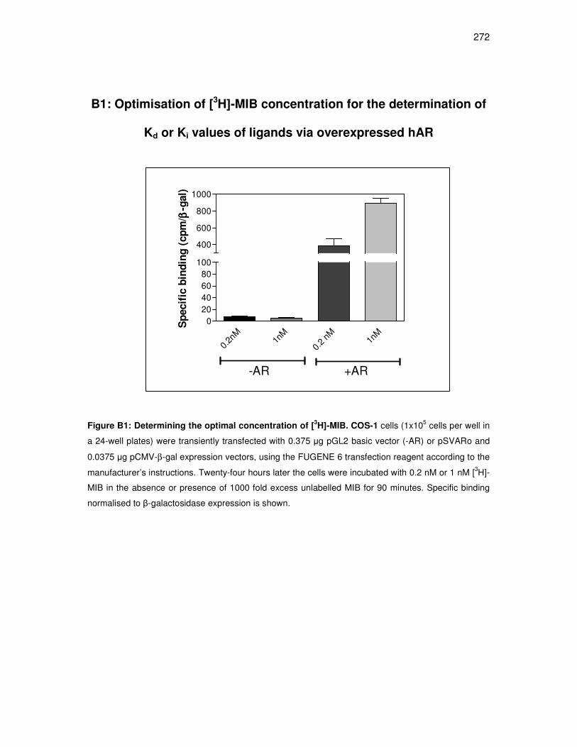
272
B1: Optimisation of [3H]-MIB concentration for the determination of
Kd or Ki values of ligands via overexpressed hAR
0.2n
M 1n
M
0.2
nM
1nM
0
20406080
100
400
600
800
1000
-AR +AR
Sp
ecif
ic b
ind
ing
(cp
m/ ββ ββ
-gal)
Figure B1: Determining the optimal concentration of [3H]-MIB. COS-1 cells (1x10⁵ cells per well in
a 24-well plates) were transiently transfected with 0.375 µg pGL2 basic vector (-AR) or pSVARo and
0.0375 µg pCMV-β-gal expression vectors, using the FUGENE 6 transfection reagent according to the
manufacturer’s instructions. Twenty-four hours later the cells were incubated with 0.2 nM or 1 nM [3H]-
MIB in the absence or presence of 1000 fold excess unlabelled MIB for 90 minutes. Specific binding
normalised to β-galactosidase expression is shown.

273
B2: Time course to establish equilibrium time for binding of 0.2 nM
[3H]-MIB to overexpressed hAR
0 4 8 12 16 20 240
1000
2000
3000
4000
5000
time (hours)
sp
ec
ific
bin
din
g (
cp
m)/
ββ ββg
al
Figure B2: Equilibrium time for 0.2 nM [3H]-MIB to bind to overexpressed AR is 16 hours. COS-1
cells (1x10⁵ cells per well in a 24-well plate) were transiently transfected with 0.375 µg pSVARo and
0.0375 µg pCMV-β-gal expression vectors, using the FUGENE6 transfection reagent according to the
manufacturer’s instructions. Twenty-four hours later the cells were incubated with 0.2 nM [3H]-MIB in
the absence and presence of 10 µM unlabelled MIB for varying times. Specific binding normalised to
β-galactosidase expression is shown.

274
B3: Transrepression assay in COS-1 cells in the absence and
presence of overexpressed hAR
Figure B3: Transrepression activity in the absence and presence of overexpressed hAR. COS-1
cells were transiently transfected with pCMV-β-galactosidase expression vectors and (A) pGL2basic
empty vector and NFĸB-containing promoter-luciferase reporter construct or (B) pSVARo and NFĸB-
containing promoter-luciferase reporter construct or (C) pGL2basic empty vector and AP-1-containing
promoter-luciferase reporter construct or (D) pSVARo and AP-1-containing promoter-luciferase
reporter construct. Subsequently, the cells were stimulated with PMA and incubated with 1 µM of
either MIB, DHT, Prog, MPA, NET-A or RU486 for 24 hours. The resulting plots show repression by
the individual ligands calculated as a percentage of EtOH (100%). Result shown is the average of
three independent experiments with each condition performed in triplicate (±SEM).
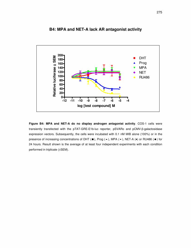
275
B4: MPA and NET-A lack AR antagonist activity
-12 -11 -10 -9 -8 -7 -6 -5 -40
20
40
60
80
100
120
140
160
180
200DHTProgMPANETRU486
log [test compound] M
Rela
tive l
ucif
era
se
±± ±± S
EM
Figure B4: MPA and NET-A do no display androgen antagonist activity. COS-1 cells were
transiently transfected with the pTAT-GRE-E1b-luc reporter, pSVARo and pCMV-β-galactosidase
expression vectors. Subsequently, the cells were incubated with 0.1 nM MIB alone (100%) or in the
presence of increasing concentrations of DHT (�), Prog (�), MPA (�), NET-A (♦) or RU486 (�) for
24 hours. Result shown is the average of at least four independent experiments with each condition
performed in triplicate (±SEM).

276
B5: MPA, but not NET-A, antagonizes the DHT-induced N/C-
interaction of the AR
0
50
100
150
--
-9 -
-8 -
-7 -
-7-9
-7-8
-7-7
-7-6
Log[DHT] (M)Log[NET-A] (M)
Lu
c/ββ ββ
-gal (r
lu)
Figure B5: In contrast to MPA, NET-A does not antagonize the DHT-induced N/C-interaction of
the hAR. COS-7 cells were transiently transfected with the pTAT-GRE-E1b-luc reporter, the pSG5-
hAR(DBD-LBD), the pSNATCH-II(hAR-NTD) and the pCMV-β-galactosidase expression vectors.
Subsequently, the cells were exposed to increasing concentrations of DHT, or 10-7 M DHT in the
presence of increasing concentrations of (A) MPA and (B) NET-A, for 24 hours. Result is shown as
luciferase (luc) activity normalized to β-galactosidase (β-gal) activity. Results are averages of three
independent experiments with each condition performed in triplicate (±SEM).
0
50
100
150
--
-9 -
-8 -
-7 -
-7-9
-7-8
-7-7
-7-6
Log[DHT] (M)Log[MPA] (M)
Lu
c/ββ ββ
-gal (r
lu)
A
B

277
APPENDIX C: DATA NOT INCLUDED IN CHAPTER 4

278
C1: Optimisation of [3H]-Ald concentration for the determination of
Kd or Ki values of ligands via overexpressed hMR
2nM
1nM
H
0.5n
M H
0.2n
M H
2nM
1nM
H
0.5n
M H
0.2n
M H
0
1
2
3
4
5
300060009000
120001500018000
-MR +MR
Sp
ecif
ic b
ind
ing
(cp
m/ ββ ββ
-gal)
Figure C1: Determining the optimal concentration of [3H]-MIB. COS-1 cells (1x10⁵ cells per well in
a 24-well plates) were transiently transfected with 0.375 µg pGL2 basic vector (-MR) or pRShMR and
0.0375 µg pCMV-β-gal expression vectors, using the FUGENE 6 transfection reagent according to the
manufacturer’s instructions. Twenty-four hours later the cells were incubated with 0.2 nM, 0.5 nM, 1
nM or 2 nM [3H]-Ald in the absence or presence of 1000 fold excess unlabelled Ald for 90 minutes.
Specific binding normalised to β-galactosidase expression is shown.
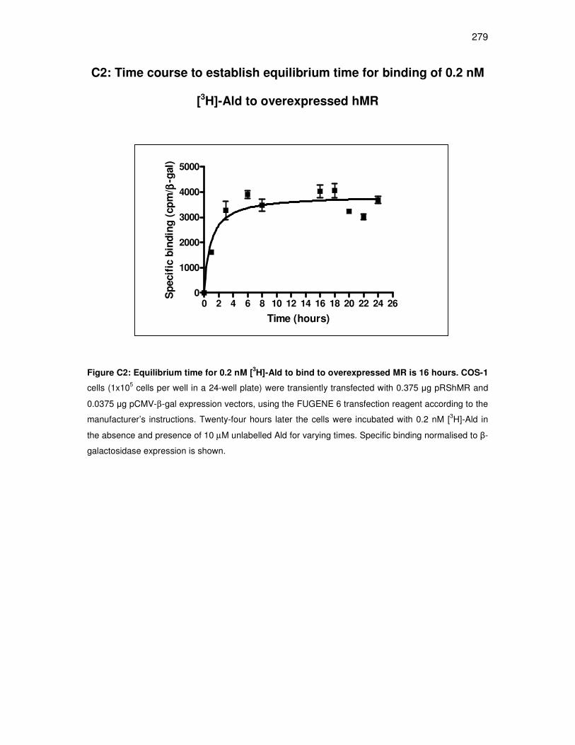
279
C2: Time course to establish equilibrium time for binding of 0.2 nM
[3H]-Ald to overexpressed hMR
0 2 4 6 8 10 12 14 16 18 20 22 24 260
1000
2000
3000
4000
5000
Time (hours)
Sp
ecif
ic b
ind
ing
(cp
m/ ββ ββ
-gal)
Figure C2: Equilibrium time for 0.2 nM [3H]-Ald to bind to overexpressed MR is 16 hours. COS-1
cells (1x10⁵ cells per well in a 24-well plate) were transiently transfected with 0.375 µg pRShMR and
0.0375 µg pCMV-β-gal expression vectors, using the FUGENE 6 transfection reagent according to the
manufacturer’s instructions. Twenty-four hours later the cells were incubated with 0.2 nM [3H]-Ald in
the absence and presence of 10 µM unlabelled Ald for varying times. Specific binding normalised to β-
galactosidase expression is shown.
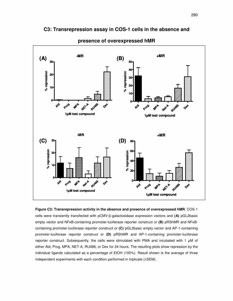
280
C3: Transrepression assay in COS-1 cells in the absence and
presence of overexpressed hMR
-MR
Ald
Pro
gM
PA
NET-A
RU48
6Dex
0
10
20
30
1µµµµM test compound
% r
ep
ressio
n
Ald
Prog
MPA
Net
-A
RU48
6Dex
0
10
20
30
40
50
+MR
1µµµµM test compound
% r
ep
ressio
n
-MR
Ald
Pro
gM
PA
Net
-A
RU48
6Dex
0
10
20
30
1µµµµM test compound
% r
ep
ressio
n
+MR
Ald
Prog
MPA
NET-A
RU48
6Dex
0
20
40
60
80
1µµµµM test compound
% r
ep
ressio
n
(A) (B)
(C) (D)
-MR
Ald
Pro
gM
PA
NET-A
RU48
6Dex
0
10
20
30
1µµµµM test compound
% r
ep
ressio
n
Ald
Prog
MPA
Net
-A
RU48
6Dex
0
10
20
30
40
50
+MR
1µµµµM test compound
% r
ep
ressio
n
-MR
Ald
Pro
gM
PA
Net
-A
RU48
6Dex
0
10
20
30
1µµµµM test compound
% r
ep
ressio
n
+MR
Ald
Prog
MPA
NET-A
RU48
6Dex
0
20
40
60
80
1µµµµM test compound
% r
ep
ressio
n
(A) (B)
(C) (D)
Figure C3: Transrepression activity in the absence and presence of overexpressed hMR. COS-1
cells were transiently transfected with pCMV-β-galactosidase expression vectors and (A) pGL2basic
empty vector and NFĸB-containing promoter-luciferase reporter construct or (B) pRShMR and NFĸB-
containing promoter-luciferase reporter construct or (C) pGL2basic empty vector and AP-1-containing
promoter-luciferase reporter construct or (D) pRShMR and AP-1-containing promoter-luciferase
reporter construct. Subsequently, the cells were stimulated with PMA and incubated with 1 µM of
either Ald, Prog, MPA, NET-A, RU486, or Dex for 24 hours. The resulting plots show repression by the
individual ligands calculated as a percentage of EtOH (100%). Result shown is the average of three
independent experiments with each condition performed in triplicate (±SEM).

281
C4: Prog displays weak partial agonist activity for transactivation
via overexpressed MR in COS-1 cells
EtOH
Ald
-6
Prog-6
MPA-6
NET-6
0
10
25
50
75
100
125
1µµµµM test compound
Fo
ld i
nd
ucti
on
(L
uc/B
-gal)
±± ±±S
EM
(n
=3)
Figure C4: Unlike Ald and Prog, MPA and NET-A do not display mineralocorticoid agonist
activity. COS-1 cells were transiently transfected with the pTAT-GRE-E1b-luc reporter plasmid plus,
pRShMR and pCMV-β-galactosidase expression vectors. Subsequently, the cells were incubated with
1 µM of either Ald, Prog, MPA or NET-A for 24 hours. Luciferase (luc) activity was normalized to β-
galactosidase (β-gal) activity, and is shown as fold induction relative to EtOH set as 1. Results shown
are the average of at least three independent experiments with each condition performed in triplicate
(±SEM).
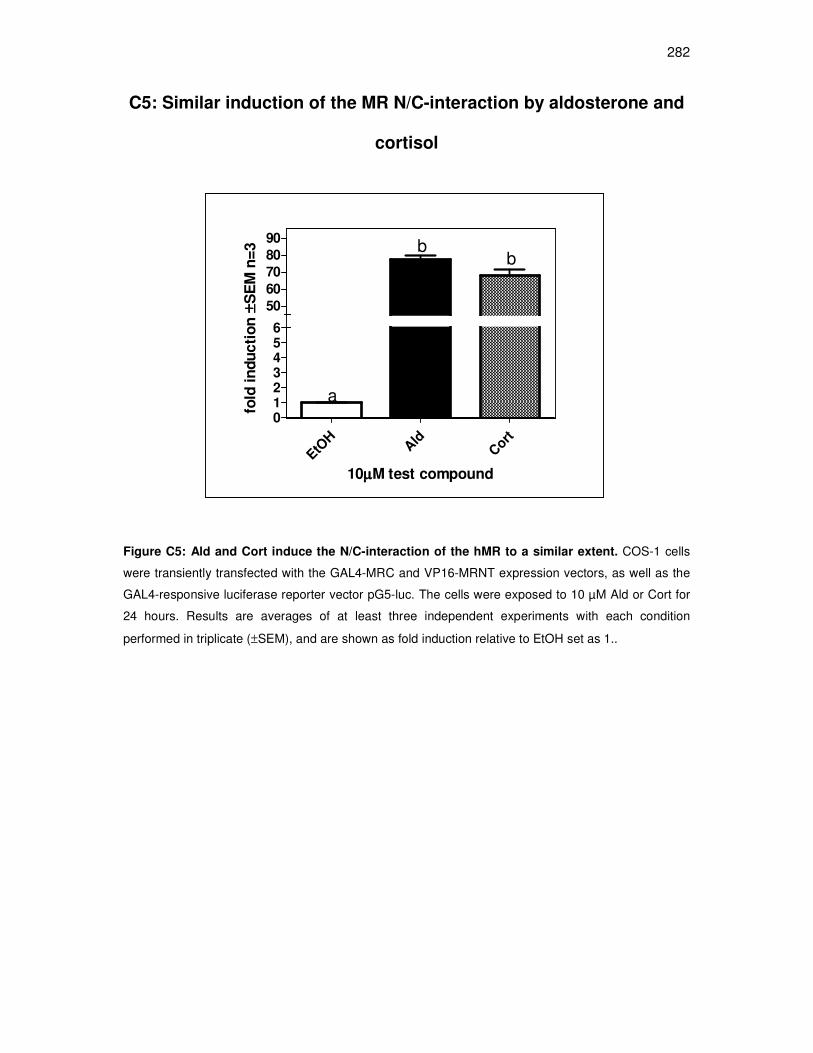
282
C5: Similar induction of the MR N/C-interaction by aldosterone and
cortisol
EtOH
Ald
Cort
0123456
50
60
70
80
90
a
bb
10µµµµM test compound
fold
in
du
cti
on
±± ±±S
EM
n=
3
Figure C5: Ald and Cort induce the N/C-interaction of the hMR to a similar extent. COS-1 cells
were transiently transfected with the GAL4-MRC and VP16-MRNT expression vectors, as well as the
GAL4-responsive luciferase reporter vector pG5-luc. The cells were exposed to 10 µM Ald or Cort for
24 hours. Results are averages of at least three independent experiments with each condition
performed in triplicate (±SEM), and are shown as fold induction relative to EtOH set as 1..

283
C5: Antagonist activity of NET-A via the hGR in COS-1 cells.
Figure C6: Relative antagonist activity of NET-A via the GR. COS-1 cells were transiently
transfected with HA-hGR-pCMV and pTAT-GRE-E1b-luc. The cells were incubated with 0.1 nM Dex in
the absence (100%) and presence of increasing concentrations of RU486 (A) or NET-A (B).
Thereafter the lysates were assayed for luciferase activity. One representative experiment for each
compound performed in triplicate (± S.E.M.) is shown.
contr
ol-1
0 -9 -8 -7 -6 -5
0
50
100
150
log [RU486] M
RL
U (
100 %
= 0
.1n
M D
ex)
A
contr
ol-1
0 -9 -8 -7 -6 -5
0
50
100
150
log [NET-A] M
RL
U (
100 %
= 0
.1n
M D
ex)
B

284
APPENDIX D: DEFINITIONS AND EXTRA DATA NOT INCLUDED IN
CHAPTERS
285
D1: Binding parameters and calculations
The relative binding affinity (RBA), also referred to as the EC50, is a measure of the
concentration of ligand that competes for half the specific binding. It is used to
estimate the binding affinity of a ligand for a receptor. The EC50 values obtained from
competitive binding curves however, are not equivalent to Kd or Ki (equilibrium
dissociation constant) values. The EC50 is a relative term, which is influenced by
different experimental conditions, such as receptor number or concentrations of
radiolabelled ligand, while the Kd or Ki is a constant value for a particular receptor,
which is not influenced by experimental variations (GraphPad Prism software).Thus,
like the EC50, the Kd or Ki, is a measure of the affinity of a ligand for a particular
receptor, that reflects the concentration of ligand needed to bind to half the available
receptors at equilibrium, but it represents a more accurate measure of binding affinity
of a ligand for its receptor than EC50 values.
Kd is typically determined in a saturation binding assay using varying concentrations
of a radiolabelled ligand. A low Kd or Ki value means that a low concentration of
ligand is needed to bind half the available receptors. Thus, a low Kd or Ki value
indicates that the receptor has a high affinity for the ligand. Ki refers to the equilibrium
constant of a ligand determined in inhibition studies, and is usually determined by the
alternative method for determining Kd or Ki, which is a homologous/heterologous
competitive binding assay. A homologous binding assay is one where the
radiolabelled and unlabelled are chemically identical, while in the heterologous
assay, they differ. In these experiments, the radioligand concentration remains
constant, and competes with varying concentrations of unlabelled competitor ligand
for the receptor.

286
Analysis of radioligand binding experiments is based on the law of mass action,
which assumes that binding is reversible. The number of binding events per unit of
time = [Ligand]. [Receptor].Kon, where Kon is defined as the association- or on-rate
constant (GraphPad Prism software). Following binding, the ligand remains bound to
the receptor for a random amount of time, and the possibility of dissociation is very
similar, at every instant of time. The rate of dissociation is the number of dissociation
events per unit time= [ligand.receptor]. Koff where Koff is defined as the dissociation-
or off-rate constant. Equilibrium is reached when the rate at which new ligand-
receptor complexes are formed equals the rate at which the ligand-receptor
complexes dissociate (GraphPad Prism software). Thus, at equilibrium,
[Ligand].[Receptor].Kon = [Ligand.Receptor].Koff. If this equation is rearranged, the Kd
is defined as Koff / Kon, which equals [Ligand].[Receptor] / [Ligand.Receptor]
(GraphPad Prism software).
To accurately analyse and interpret the results from competitive ligand binding
experiments, it is crucial to determine the time to reach binding equilibrium to the
receptor with the concentration of radiolabelled ligand used in the experiment. The
concentration of radiolabelled ligand used was in the range of two to ten times lower
than the EC50. In addition, the ligand depletion always needs to be considered.
Ligand depletion measures the fraction of the ligand bound to the receptor (specific
sites) vs. the fraction of the ligand bound to nonspecific sites. One would assume that
only a small fraction of the added ligand binds to the receptor, so that the free
concentration approximates to the concentration initially added. Less than 10%
ligand depletion is considered acceptable.
287
As [3H]-MPA and [3H]-NET-A are not commercially available, Kd and Ki values were
determined from pooled data of three independent experiments, by homologous and
heterologous binding experiments, respectively (Chapter 3 and 4). Once the optimal
concentration of radiolabelled ligand was established (0.2 nM for both [3H]-MIB
(Chapter 3) and [3H]-Ald (Chapter 4)), the incubation time for equilibrium to be
reached with 0.2 nM [3H]-MIB and [3H]-Ald was determined. The Kd was
subsequently calculated from homologous binding experiments by GraphPad Prism
software according to the following equation: Total binding = (Bmax.[Hot] / [Hot] +
[Cold] + Kd +NS, where NS is nonspecific binding and Bmax is the maximum binding
of ligand to receptors in cpm. Once the Kd for MIB was determined, heterologous
binding experiments were performed, and the Ki values for Prog, MPA, NET-A, DHT,
R1881, and RU486 were determined (Chapter 3) according to the Cheng- Prussoff
equation: Ki = EC50 / 1 + ([ligand] / Kd), where the EC50 value is that determined for
the competing ligand, the [ligand] is the radiollabelled igand concentration used, and
the Kd determined from the homologous binding experiments. Ki can only be
determined using the Cheng-Prussoff equation, provided that the EC50 is two to ten
times lower than the concentration of radiolabelled ligand, and that binding
equilibrium has been reached at that concentration. In the same manner, the Kd
value for Ald, and Ki values for Prog, MPA, NET-A, RU486 and Dex were
determined. All binding data were analysed using GraphPad Prism software, using
the following parameters: Nonlinear regression curve fit, homologous competitive
binding curve, one class of binding sites, weighting 1/y2 (this minimises relative
distances squared), consider the mean y value of each point and a stricter (slower)
criterion for convergence. In addition, run rest and residuals test were used to
determine whether the curve differed systematically from the data.
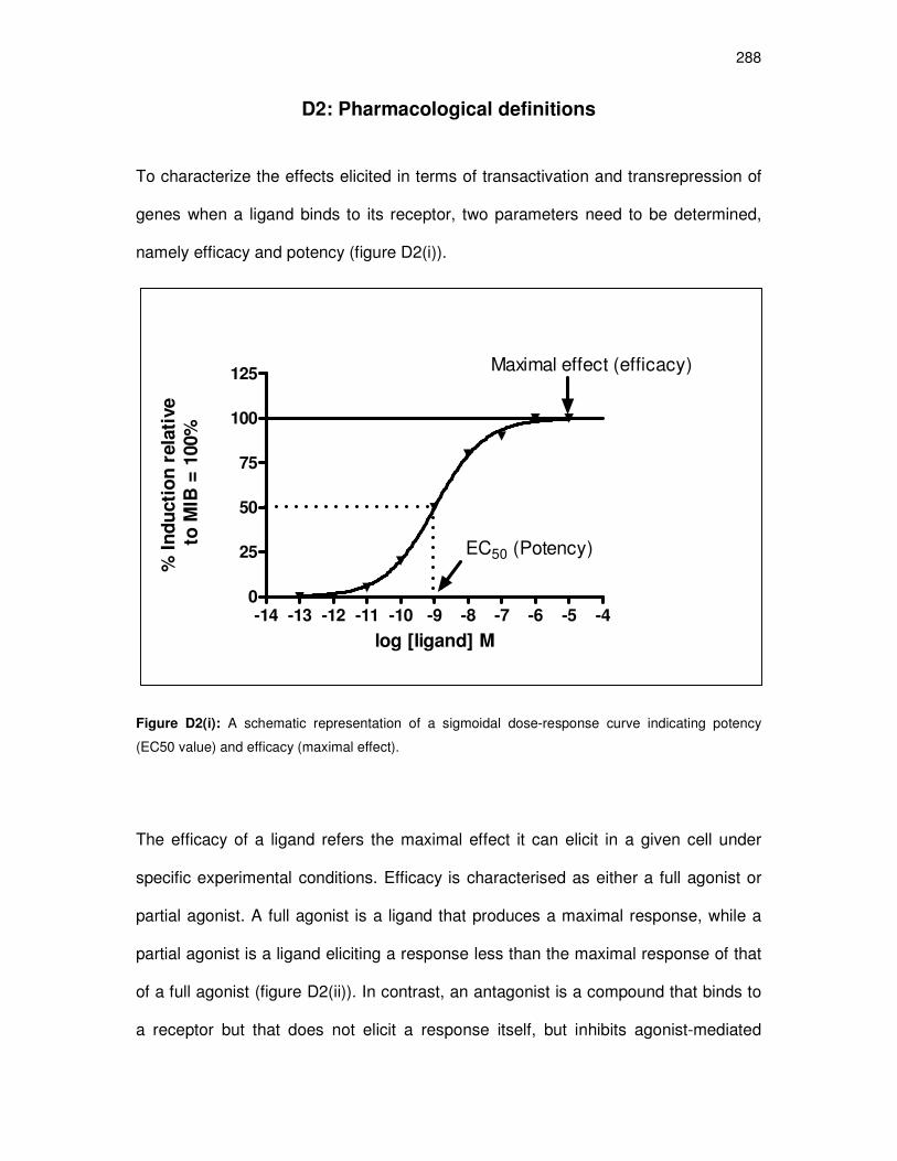
288
D2: Pharmacological definitions
To characterize the effects elicited in terms of transactivation and transrepression of
genes when a ligand binds to its receptor, two parameters need to be determined,
namely efficacy and potency (figure D2(i)).
Figure D2(i): A schematic representation of a sigmoidal dose-response curve indicating potency
(EC50 value) and efficacy (maximal effect).
The efficacy of a ligand refers the maximal effect it can elicit in a given cell under
specific experimental conditions. Efficacy is characterised as either a full agonist or
partial agonist. A full agonist is a ligand that produces a maximal response, while a
partial agonist is a ligand eliciting a response less than the maximal response of that
of a full agonist (figure D2(ii)). In contrast, an antagonist is a compound that binds to
a receptor but that does not elicit a response itself, but inhibits agonist-mediated
-14 -13 -12 -11 -10 -9 -8 -7 -6 -5 -40
25
50
75
100
125
EC50 (Potency)
Maximal effect (efficacy)
log [ligand] M
% I
nd
ucti
on
rela
tive
to
MIB
= 1
00%

289
responses (figure D2(ii)). The potency of a ligand is commonly quantified as the
EC50, which is the concentration that induces half the maximal response.
Figure D2(ii): A schematic representation of a sigmoidal dose-response curves indicating a full and
partial agonist, as well as an antagonist. (for ligands binding to a single site, with no co-operativity)
-14 -13 -12 -11 -10 -9 -8 -7 -6 -5 -40
25
50
75
100
125
full agonist
partial agonist
antagonist in thepresence of agonist
log [ligand] log M
% I
nd
ucti
on
rela
tive
to
MIB
= 1
00%

290
D3: Transactivation in COS-1 cells in the absence and presence of
overexpressed MR, AR or GR
(A) no transfected SR
(B) MR C) AR (D) GR
Figure D3: Agonist activity for transactivation via possible endogenous receptors, MR, AR or
GR in COS-1 cells. COS-1 cells were transiently transfected with 100 ng pTAT-GRE-E1b-luc reporter
plasmid plus, 10 ng pGL2basic (A), pRShMR (B), pSVARo (C) or HA-hGR-pCMV (D), and 10 ng
pCMV-β-galactosidase expression vectors in a 96-well plate. Subsequently, the cells were incubated
with 10 µM Prog, MPA, NET-A, RU486, MIB, DHT, Ald, Dex or Cort for 24 hours. Induction is shown
as fold induction relative to EtOH set as 1. Result shown is one representative figure with each
condition performed in triplicate (±SEM).
EtO
HAld
Pro
gM
PA
NET-A
RU48
6Dex
Cort
0
2
4
6
8
10
20
40
60
80
100
10 µµµµM test compound
Fo
ld i
nd
ucti
on
(lu
c)
EtOH
MIB
DHT
Prog
MPA
NET-A
RU48
60
2
4
6
8
10200
300
400
500
600
10 µµµµM test compound
Fo
ld i
nd
ucti
on
(lu
c)
EtOH
Dex
Cort
Ald
Prog
MPA
NET-A
RU48
6M
IBDHT
05
1015202530
1000
3000
5000
10 µµµµM test compound
Fo
ld i
nd
ucti
on
(lu
c)
EtO
HPro
gM
PA
NET-A
RU48
6M
IBDHT
Ald
Dex
Cort
0
2
4
6
8
10 µµµµM test compound
Fo
ld i
nd
ucti
on
(lu
c)
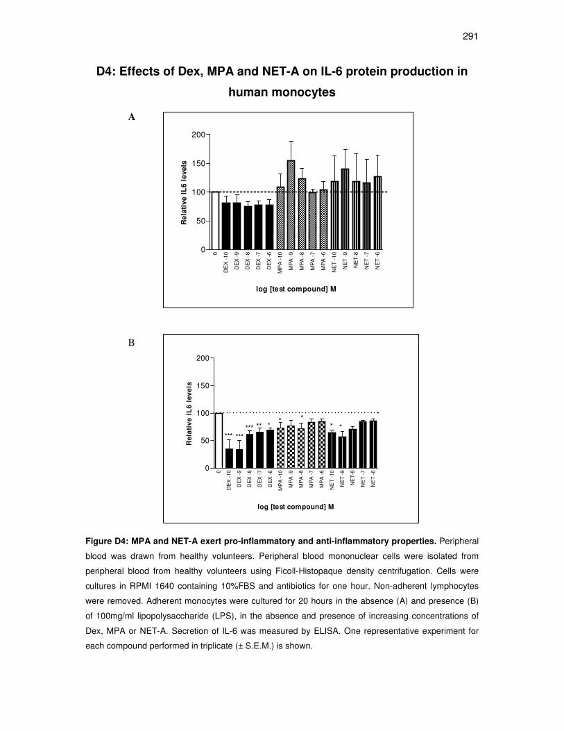
291
D4: Effects of Dex, MPA and NET-A on IL-6 protein production in
human monocytes
0
DE
X -
10
DE
X -
9
DE
X -
8
DE
X -
7
DE
X -
6
MP
A -
10
MP
A -
9
MP
A -
8
MP
A -
7
MP
A -
6
NE
T -
10
NE
T -
9
NE
T-8
NE
T -
7
NE
T -
60
50
100
150
200
log [test compound] M
Re
lati
ve I
L6 l
eve
ls
0
DE
X -
10
DE
X -
9
DE
X -
8
DE
X -
7
DE
X -
6
MP
A -
10
MP
A -
9
MP
A -
8
MP
A -
7
MP
A -
6
NE
T -
10
NE
T -
9
NE
T-8
NE
T -
7
NE
T -
60
50
100
150
200
****** ***
*****
* *
log [test compound] M
Re
lati
ve
IL
6 l
ev
els
Figure D4: MPA and NET-A exert pro-inflammatory and anti-inflammatory properties. Peripheral
blood was drawn from healthy volunteers. Peripheral blood mononuclear cells were isolated from
peripheral blood from healthy volunteers using Ficoll-Histopaque density centrifugation. Cells were
cultures in RPMI 1640 containing 10%FBS and antibiotics for one hour. Non-adherent lymphocytes
were removed. Adherent monocytes were cultured for 20 hours in the absence (A) and presence (B)
of 100mg/ml lipopolysaccharide (LPS), in the absence and presence of increasing concentrations of
Dex, MPA or NET-A. Secretion of IL-6 was measured by ELISA. One representative experiment for
each compound performed in triplicate (± S.E.M.) is shown.
A
B