comparative structural analysis of the caspase … structural analysis of the caspase family with...
TRANSCRIPT
Biochem. J. (2015) 466, 219–232 (Printed in Great Britain) doi:10.1042/BJ20141324 219
REVIEW ARTICLEComparative structural analysis of the caspase family with other clan CDcysteine peptidasesKaren McLuskey*1 and Jeremy C. Mottram*1
*Wellcome Trust Centre for Molecular Parasitology, Institute of Infection, Immunity and Inflammation, College of Medical, Veterinary and Life Sciences, University of Glasgow,Glasgow G12 8TA, UK
Clan CD forms a structural group of cysteine peptidases,containing seven individual families and two subfamilies ofstructurally related enzymes. Historically, it is most notable forcontaining the mammalian caspases, on which the structures of theclan were founded. Interestingly, the caspase family is split intotwo subfamilies: the caspases, and a second subfamily containingboth the paracaspases and the metacaspases. Structural data arenow available for both the paracaspases and the metacaspases,allowing a comprehensive structural analysis of the entire caspasefamily. In addition, a relative plethora of structural data hasrecently become available for many of the other families inthe clan, allowing both the structures and the structure–functionrelationships of clan CD to be fully explored. The present reviewcompares the enzymes in the caspase subfamilies with each other,
together with a comprehensive comparison of all the structuralfamilies in clan CD. This reveals a diverse group of structures withhighly conserved structural elements that provide the peptidaseswith a variety of substrate specificities and activation mechanisms.It also reveals conserved structural elements involved in substratebinding, and potential autoinhibitory functions, throughout theclan, and confirms that the metacaspases are structurally diversefrom the caspases (and paracaspases), suggesting that they shouldform a distinct family of clan CD peptidases.
Key words: caspase, clan CD, crystallography, metacaspase,peptidase, protein structure.
INTRODUCTION
Clan CD [1] cysteine peptidases use an active site cysteineresidue to catalyse the hydrolysis, and subsequent cleavage, ofpeptide bonds in proteins. These peptidases generally show a strictspecificity for the P1 residue of their substrates and depend only ontwo key catalytic residues: a highly conserved histidine/cysteinedyad. In clan CD, the order, type and surrounding tertiarystructure of the dyad are used to assign enzymes to theclan [2].
The founding member of clan CD was discovered in 1994 by thedetermination of the 3D crystal structure of human interleukin-1β-converting enzyme [3,4], also known as caspase-1. This structurerevealed a novel protein fold and was consequently placed ina new structural group of cysteine peptidases (clan CD [5]) asfamily C14, which is often referred to as the caspase family.Since its discovery, clan CD has been expanded to includeseven peptidase families: clostripains (C11); legumains (C13);caspases (C14); gingipains (C25); separases (C50); the cysteinepeptidase domain (CPD) of the multi-functional, autoprocessingrepeats in toxin (MARTX) toxins (C80); and most recently theenzymes related to the peptidase virulence factor PrtH fromTannerella forsythia [6] (C85). Notably, family C14 is furtherdivided to contain subfamilies C14A (the caspases) and C14B{both the metacaspases and the paracaspases [denoted C14B(M)and C14B(P), respectively]}. The phylogenetic distribution ofthe clan CD peptidases spans all the kingdoms of life (Table 1).
However, the caspase family (C14) is the only family that hasbeen identified in all kingdoms, although each subfamily is foundonly in certain branches [7] (Table 1).
Since the structural determination of caspase-1, approximately170 caspase structures have been deposited in the Protein DataBank (PDB [8], www.rcsb.org) with around 30 of them assumedto be unique (<90% sequence identity). In addition, X-raycrystal structures are also available for families C11 (unpublished,PDB ID 3UWS), C13 [9], C14B(P) [10], C14B(M) [11,12], C25[13,14] and C80 [15–17]. Apart from the original structure ofgingipain R [13] (RgpB, C25), all these structures have beendetermined within the last 6 years (Table 1), making it an excitingtime to analyse this diverse and structurally expanding clan. Thepresent review summarizes the collective structural informationof the families, evaluates and compares the structure–functionrelationships, and allows for greater understanding of the enzymesin clan CD.
FAMILY C14: CASPASES, METACASPASES AND PARACASPASES
Caspases
The name caspase is an abbreviation of cysteine-dependent,aspartate-specific peptidase, because caspases have a dominantspecificity for protein substrates that contain an aspartate inthe P1 position (Table 2). Functionally, the caspases are majorregulators of apoptotic cell death pathways, proliferation andinflammation, playing vital roles in the life and death of animal
Abbreviations: AP, activation peptide; CARD, caspase recruitment domain; CHF, caspase/haemoglobinase fold; CPD, cysteine peptidase domain;CSD, C-terminal subdomain; DD, death domain; DED, death effector domain; InsP6, myo-inositol hexakisphosphate; LSAM, legumain stabilization andactivity modulation; LSD1, lesion-simulating disease 1; MALT1, mucosa-associated lymphoid tissue translocation protein 1; MARTX, multi-functional,autoprocessing repeat in toxin; RMSD, root-mean-square deviation; SSE, secondary structural element; XIAP, X-linked inhibitor of apoptosis; Z-VRPR-FMK, benzoxycarbonyl-Val-Arg-Pro-Arg-fluoromethylketone.
1 To whom correspondence should be addressed (email [email protected] or [email protected]).
c© The Authors Journal compilation c© 2015 Biochemical Society
220 K. McLuskey and J.C. Mottram
Table 1 The structural availability and phylogenetic distribution of the clan CD families
The availability (�) and absence (×) of clan CD families in the phylogenetic kingdom. The year that the first structure became available is shown for each family (year).
FAMILY Representative member Structural data? (year) Bacteria Achaea Protozoa Fungi Plants Viruses Animals
C11 Clostripain Yes (2013) � � � × � × ×C13 Legumain Yes (2013) � � � � � × �C14A Caspase Yes (1994) × × × × × � �C14B(P) Paracaspase Yes (2011) � � × × × × �C14B(M) Metacaspase Yes (2012) � � � � � × ×C25 Gingipain R Yes (1999) � � × × × × ×C50 Separase No × × � � � × �C80 MARTX-CPD Yes (2008) � × × × × × �C84 PrtH peptidase No � × × × × × ×Clan CD � � � � � � �
Table 2 Enzymatic properties of the clan CD peptidases
Family Representative member Specificity in P1 Requirement for activation Self-inhibition observed? Region of self-inhibition
C11 Clostripain Arginine Ca2 + Unknown –C13 Legumain Asparagine and aspartate* Change in pH Yes C-terminal domainC14A Caspase Aspartate Dimerization or proteolysis† Unclear‡ N-terminal regionC14B(P) Paracaspase Arginine Dimerization Yes Substrate-binding loop (L5)C14B(M) Metacaspase Arginine and lysine§ Ca2 + and/or proteolysis‖ Yes N-terminal regionC25 Gingipain R Arginine or lysine¶ Proteolysis and/or Ca2 + ** Yes N-terminal prodomainC50 Separase Arginine Ca2 + Unknown –C80 MARTX-CPD Leucine Ligand binding Yes N-terminal regionC84 PrtH peptidase Arginine Unknown Unknown –
*Legumain will accept asparagine or aspartate residues depending on the pH.**Cleavage of the proform of gingipain is required for full activation of the enzyme and while Ca2+ is not reported as a prerequisite for activation, all active forms appear to have Ca2+ present.†Caspases are activated by dimerization or proteolysis depending on the type; typically initiator caspases are activated by dimerization whereas the effector caspases are activated by cleavage
(proteolysis).‡Self-inhibition using the N-terminal region has been suggested in the effector caspases but there are no structural data to date.§Metacaspases are known to accept arginine and lysine in P1.‖Type I metacaspases generally activated by Ca2 + ; this is also true for type II metacaspases but, in addition, proteolysis has also been shown to be important in some cases.¶Gingipain will accept arginine or lysine depending on the enzyme (gingipain R and K have a strict specificities for arginine and lysine, respectively).
cells. In humans, 11 caspases have been identified (caspase-1 to caspase-10 and caspase-14) and can be grouped togetheraccording to their sequence similarities [18], which are generallyassociated with their involvement in specific cellular processes[19]. They can (perhaps oversimply) be classified as eitherinflammatory (caspase-1, -4 and -5) or apoptotic caspases,with the latter being further organized into initiator (caspase-2, -8, -9 and -10) and effector (or executioner; caspase-3, -6 and -7) caspases [20]. Typically, caspases are described ashaving an N-terminal prodomain, which contains an aspartatesite for (auto)proteolysis and varies in length depending on thetype of caspase. The effector caspases have short prodomains(approximately 25 residues), whereas both the inflammatory andthe initiator caspases have long prodomains (approximately 100–200 residues), which contain either CARD (caspase recruitmentdomain – inflammatory and initiator caspases) or DED (deatheffector domain – initiator caspases) motifs [19].
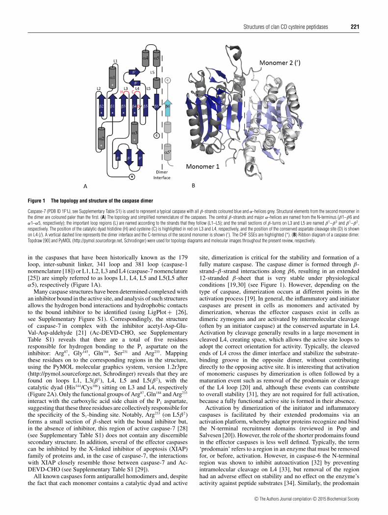
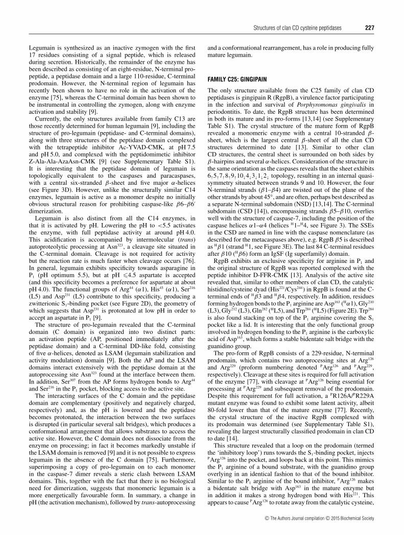
In contrast to their diverse N-terminal regions, the catalyticdomain of the caspases has a virtually identical fold in allthe crystal structures determined to date. However, in orderto describe the structure of the caspases in detail, the well-studied effector caspase, caspase-7 [21], has been chosen as ageneral representative of the caspases in the present review. Thestructure of the caspases is formed around a central six-strandedβ-sheet (β1–β6), consisting of five parallel and one antiparallel β-strand(s) with 2↑1↑3↑4↑5↑6↓ topology [22]. The central sheet issurrounded by five major α-helices (α1–α5), contains a small
three-stranded section of β-sheet situated between β3 and α3, andthe residues constituting the catalytic histidine/cysteine dyad arefound at the C-terminal ends of strands β3 and β4, respectively.This basic monomeric fold led to the identification of the otherclan CD members and the description of a minimal core structuralunit, the caspase/haemoglobinase fold (CHF) [23], which isdescribed as consisting of the first four strands of the β-sheet(2↑1↑3↑4↑) along with helices α1–α3 (Figure 1A).
A highly conserved proteolytic aspartate is found situatedbetween strands β4 and β5 of the caspases. As a result, theoriginal caspase structures were described as having a large (α orp20) and a small (β or p10) subunit, comprising strands 1–4 and5–6, respectively, linked together by an inter-subunit linker [4] (acleaved loop region). This description of two individual caspasesubunits predated any 3D structural information [24], althoughthe term ‘inter-subunit linker’ was most probably introduced later.In reality, caspase monomers do not contain individual subunitsbut are simply composed of a single polypeptide chain, whichfolds into a central six-stranded β-sheet with a highly conservedcleavage site. In addition, because of the abundance of caspasestructures available in the literature, other important loop regionshave been named in various ways. Therefore, to standardize thenomenclature used in the present review, and to allow structuralcomparisons with other families in the clan, all terms referring tocaspase subunits are omitted and the loop (L) regions are namedaccording to the strands that they follow (L1–L5, respectively)(Figure 1A). Consequently, the substrate-binding loop regions
c© The Authors Journal compilation c© 2015 Biochemical Society
Structures of clan CD cysteine peptidases 221
Figure 1 The topology and structure of the caspase dimer
Caspase-7 (PDB ID 1F1J, see Supplementary Table S1) is used to represent a typical caspase with all β-strands coloured blue and α-helices grey. Structural elements from the second monomer inthe dimer are coloured paler than the first. (A) The topology and simplified nomenclature of the caspases. The central β-strands and major α-helices are named from the N-terminus (β1–β6 andα1–α5, respectively); the important loop regions (L) are named according to the strands that they follow (L1–L5); and the small sections of β-turns on L3 and L5 are named β1–β3 and β1–β2,respectively. The position of the catalytic dyad histidine (H) and cysteine (C) is highlighted in red on L3 and L4, respectively, and the position of the conserved aspartate cleavage site (D) is shownon L4 (/). A vertical dashed line represents the dimer interface and the C-terminus of the second monomer is shown (′). The CHF SSEs are highlighted (*). (B) Ribbon diagram of a caspase dimer.Topdraw [90] and PyMOL (http://pymol.sourceforge.net, Schrodinger) were used for topology diagrams and molecular images throughout the present review, respectively.
in the caspases that have been historically known as the 179loop, inter-subunit linker, 341 loop and 381 loop (caspase-1nomenclature [18]) or L1, L2, L3 and L4 (caspase-7 nomenclature[25]) are simply referred to as loops L1, L4, L5 and L5(L5 afterα5), respectively (Figure 1A).
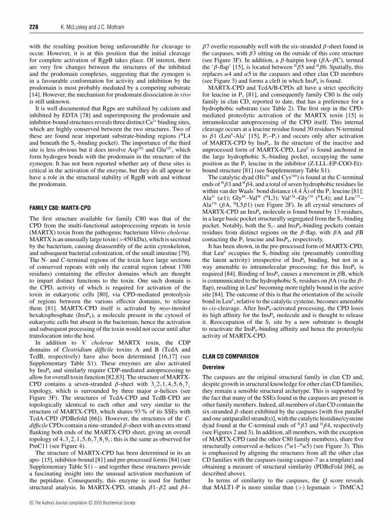
Many caspase structures have been determined complexed withan inhibitor bound in the active site, and analysis of such structuresallows the hydrogen bond interactions and hydrophobic contactsto the bound inhibitor to be identified (using LigPlot + [26],see Supplementary Figure S1). Correspondingly, the structureof caspase-7 in complex with the inhibitor acetyl-Asp-Glu-Val-Asp-aldehyde [21] (Ac-DEVD-CHO, see SupplementaryTable S1) reveals that there are a total of five residuesresponsible for hydrogen bonding to the P1 aspartate on theinhibitor: Arg87, Gly145, Gln184, Ser231 and Arg233. Mappingthese residues on to the corresponding regions in the structure,using the PyMOL molecular graphics system, version 1.2r3pre(http://pymol.sourceforge.net, Schrodinger) reveals that they arefound on loops L1, L3(β1), L4, L5 and L5(β2), with thecatalytic dyad (His144/Cys186) sitting on L3 and L4, respectively(Figure 2A). Only the functional groups of Arg87, Gln184 and Arg233
interact with the carboxylic acid side chain of the P1 aspartate,suggesting that these three residues are collectively responsible forthe specificity of the S1-binding site. Notably, Arg233 (on L5β2)forms a small section of β-sheet with the bound inhibitor but,in the absence of inhibitor, this region of active caspase-7 [28](see Supplementary Table S1) does not contain any discerniblesecondary structure. In addition, several of the effector caspasescan be inhibited by the X-linked inhibitor of apoptosis (XIAP)family of proteins and, in the case of caspase-7, the interactionswith XIAP closely resemble those between caspase-7 and Ac-DEVD-CHO (see Supplementary Table S1 [29]).
All known caspases form antiparallel homodimers and, despitethe fact that each monomer contains a catalytic dyad and active
site, dimerization is critical for the stability and formation of afully mature caspase. The caspase dimer is formed through β-strand–β-strand interactions along β6, resulting in an extended12-stranded β-sheet that is very stable under physiologicalconditions [19,30] (see Figure 1). However, depending on thetype of caspase, dimerization occurs at different points in theactivation process [19]. In general, the inflammatory and initiatorcaspases are present in cells as monomers and activated bydimerization, whereas the effector caspases exist in cells asdimeric zymogens and are activated by intermolecular cleavage(often by an initiator caspase) at the conserved aspartate in L4.Activation by cleavage generally results in a large movement incleaved L4, creating space, which allows the active site loops toadopt the correct orientation for activity. Typically, the cleavedends of L4 cross the dimer interface and stabilize the substrate-binding groove in the opposite dimer, without contributingdirectly to the opposing active site. It is interesting that activationof monomeric caspases by dimerization is often followed by amaturation event such as removal of the prodomain or cleavageof the L4 loop [20] and, although these events can contributeto overall stability [31], they are not required for full activation,because a fully functional active site is formed in their absence.
Activation by dimerization of the initiator and inflammatorycaspases is facilitated by their extended prodomains via anactivation platform, whereby adaptor proteins recognize and bindthe N-terminal recruitment domains (reviewed in Pop andSalvesen [20]). However, the role of the shorter prodomains foundin the effector caspases is less well defined. Typically, the term‘prodomain’ refers to a region in an enzyme that must be removedfor, or before, activation. However, in caspase-6 the N-terminalregion was shown to inhibit autoactivation [32] by preventingintramolecular cleavage on L4 [33], but removal of the regionhad an adverse effect on stability and no effect on the enzyme’sactivity against peptide substrates [34]. Similarly, the prodomain
c© The Authors Journal compilation c© 2015 Biochemical Society

222 K. McLuskey and J.C. Mottram
Figure 2 The S1-binding pockets of the clan CD family members
The catalytic dyad is shown in red and conserved aromatic residues are shown in green. With the exception of TbMCA2, residues that form hydrogen bonds to the P1 residue of a bound inhibitor areshown in blue (the darker shade of blue represents interactions through functional groups, whereas the lighter blue shows interactions from main chain atoms). Residues and SSEs involved in P1
binding are labelled and SSEs structurally homologous (but topologically diverse) to those found in the caspases are highlighted (H). (A) Caspase-7 PDB (ID 1F1J). (B) Inhibitor-free TbMCA2 (PDBID 4AFR) in which residues shown to be important in substrate binding are highlighted in blue, with those responsible for specificity in P1 [11] shown in navy blue. (C) MALT1 paracaspase domain(MALT1-P) (PDB IB 3UOA). (D) Legumain (PDB ID 4AW9). (E) Gingipain R (PDB ID 1CVR). (F) MARTX-CPD (PDB ID 3GCD). Inhibitors used in complex structures are shown in SupplementaryTable S1.
of caspase-3 was shown to keep the enzyme in a latent form untilits activation by a downstream caspase [35], but removal of theN-terminal region had no effect on the enzyme’s activity [36].This also holds true for caspase-7, for which the catalytic activityof the proform is indistinguishable from that of the wild type[28] and the cleaved form is reported to be more apoptoticallyactive [37]. Consequently, in contrast to some other peptidases(such as the clan CA peptidases, reviewed by Turk et al. [38]),removal of the effector caspase prodomain is not necessary forcatalytic activity, although it may play an inhibitory role untilenzymatic activity is required. In addition, the N-terminal regionof an effector caspase has so far escaped structural elucidation.This is despite the fact that both caspase-6 [33] and caspase-7 [28](see Supplementary Table S1) have been crystallized as inactiveproenzymes, suggesting that the N-terminal regions do not bind(tightly) to the surface of the enzymes and are assumed to bereasonably flexible in solution.
Metacaspases
Given the importance of the caspases in mammals, a searchfor orthologues in plants and other non-metazoan organisms
was undertaken by using the primary sequences of caspases, inand around the active site, in a PSI-BLAST (Position-SpecificIterative Basic Local Alignment Search Tool) search [39]. Thisresulted in the identification of two new groups of peptidasesthat were collectively assigned in MEROPS as a new caspasesubfamily (C14B). These peptidases, termed ‘paracaspases’ and‘metacaspases’, were both found to be present in the genomesof bacteria and Archaea [40,41]. In addition, the metacaspaseswere identified in protozoa, fungi and plants [39], whereasthe paracaspases were found distributed throughout the animalkingdom, from which the metacaspases were notably absent [42](see Table 1).
Early studies on the metacaspases attempted to draw parallelsbetween possible metacaspase function and the fundamentaland well-established processes carried out by the caspases [43–46]. Indeed, the yeast metacaspase Yca1 (from Saccharomycescerevisiae) has been implicated in cell death processes [43],suggesting a degree of functional homology with the caspases.This resulted in similar investigations being carried out onmetacaspases from other organisms, and revealed a role for severalfungi and plant metacaspases in cell death (reviewed in Tsiatsianiet al. [47]). However, a link with cell death mechanisms could notbe identified for all metacaspases and a number of other functions
c© The Authors Journal compilation c© 2015 Biochemical Society

Structures of clan CD cysteine peptidases 223
have since been established in various cellular processes includingcell-cycle progression [48], cell proliferation [49], endoplasmicreticulum (ER) stress [50], clearance of insoluble aggregates [51]and virulence [52].
Historically, two types of metacaspases have been described[39] (types I and II), with both types being found in plantswhereas yeast and the protozoa possess only type I. Inaddition, a further type of metacaspase (denoted type III) hasrecently been described in unicellular photosynthetic algae andbacteria [40]. It is of interest that the number and type ofmetacaspase genes identified in different organisms can varyconsiderably [47], although there is insufficient evidence toindicate whether this shows a degree of functional specializationor redundancy; both have, however, been reported [44,50,53–56]. In addition, multi-functional metacaspases have also beenidentified, particularly in organisms that have a single metacaspasegene, e.g. in S. cerevisiae Yca1 [43,48,51] and Leishmania majorLmMCA [57–59].
The original structural classification of all three types ofmetacaspases is based on a predicted domain structure originatingfrom the system adopted for the caspases. This describesmetacaspases as containing large (p20) and small (p10) subunits,with the addition of other variable structural features such asan N-terminal prodomain (type I), an extended inter-subunitlinker (type II) and a putative p20/p10 domain swap (type III)[40]. However, in contrast to the caspases, active metacaspasesshow a strict preference for substrates containing basic arginineand/or lysine residues [46,59–61] (see Table 2). Indeed, thispreference for basic substrates makes the name ‘metacaspase’technically incorrect. Metacaspases also differ significantly fromthe caspases in that they are active monomers [11], for whichactivation profiling has revealed a widespread, but not universal[62], requirement for calcium [45,60,63,64]. In addition, thereare no conserved cleavage sites reported in type I metacaspases,but this is different for type II metacaspases, which containhighly conserved cleavage sites that have been shown to playan important part in the activation mechanism of Arabidopsisthaliana, AtMC4 and AtMC9 [61,65].
Despite being described as having an N-terminal prodomain,there is no evidence in the type I metacaspases for removal ofthis region for activation. However, several distinct functions havebeen attributed to the N-terminal regions in type I metacaspases: inAtMC1, the N-terminal domain contains a conserved LSD1-like(lesion-simulating disease 1-like) zinc finger motif, which wasfound to interact with LSD1 and negatively regulate its function[44]; in Yca1, the N-terminal domain is essential for targetingthe enzyme to insoluble aggregates [51] and in Trypanosomabrucei MCA2 (TbMCA2) the N-terminal region is thought toact as a gatekeeper, controlling substrate access to the active site[11]. In addition, stronger autoprocessing has been observed inAtMC1 and AtMC2 when the N-terminal region is absent [44],suggesting that, similar to TbMCA2 (and the effector caspases),this region acts to inhibit/control enzymatic activity until it isrequired.
TbMCA2 structure
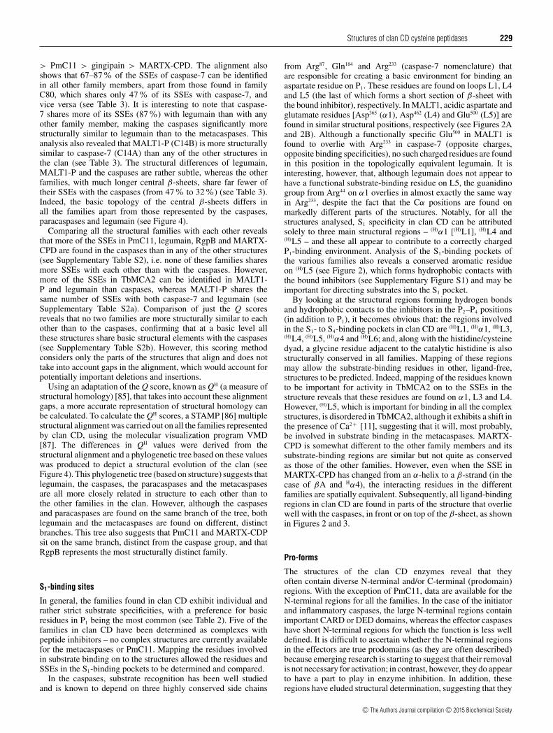
The structural basis for many of the functional differencesbetween the metacaspases and caspases was revealed by thefirst metacaspase structure, an inactive (C213G/A) mutant ofTbMCA2 [11]. The overall topology of this metacaspase wasrather unexpected, because the structure did not contain thesix-stranded β-sheet found in the caspases. Instead, TbMCA2contained two extra strands (β7 and β8) sandwiched between
β4 and β5, resulting in a monomeric structure with an eight-stranded β-sheet of 2↑1↑3↑4↑7↑8↓5↑6↓ topology [22]. Similar tothe caspases, five α-helices and a small section of β-sheet on L3were found surrounding the central sheet with various loop regionsconnecting the secondary structural elements (SSEs) (Figures 3Aand 3B). However, unlike the caspases, the N-terminal regionwas extremely well ordered and the 70-residue region precedingβ1 was found to encircle the enzyme and cross over the activesite.
For direct comparison of structures from different clan CDfamilies throughout the present review, caspase nomenclature ishighlighted for the SSEs and loops of all clan CD structures, whenthey are structurally conserved and similar to those found in thecaspases, e.g. β7 in TbMCA2 is structurally homologous to β5 inthe caspases – denoted as Hβ5 (H5; Figure 3B).
Structural determination of a metacaspase in the presence ofa bound substrate and/or inhibitor has so far escaped elucidationand as a consequence the metacaspase S1-binding pocket cannotbe mapped in the same way as the caspases. However, apotential S1-binding pocket was described for TbMCA2 andseveral residues were shown to be involved in substrate bindingand/or enzyme activity [11]: Cys92, Asp95, Ser156 and Asp211,which were found on α1, α1, L3 and L4, respectively (seeFigures 2B and 3B). In addition, the L7 (HL5) loop of TbMCA2was shown to undergo a structural rearrangement at Ala280
(Figure 3B), in the presence of Ca2 + , and is also thought to beimportant in substrate binding [11]. The structure of TbMCA2was determined in the presence of samarium, which facilitatedthe identification of an allosteric Ca2 + -binding site comprisingfour aspartate residues (Asp173, Asp189, Asp190 and Asp220), whichare highly conserved in a primary sequence alignment of bothtype I and type II metacaspases [11]. The PDB identitiesfor the individual structures are referenced in SupplementaryTable S1.
Yca1 structure
Elucidation of the structure of TbMCA2 was closely followed bythat of the crystal structure of another type I metacaspase fromyeast, Yca1 [12]. Comparing Yca1 with TbMCA2 revealed thatthe two structures are very similar, sharing 82 % of their SSEs(PDBeFold [66]), and that the predicted S1- and Ca2 + -bindingsites in TbMCA2 are completely conserved in Yca1, in terms ofboth structure and residue type (see Supplementary Figure S2).Unlike TbMCA2, the structure of Yca1 was determined fromfull-length active protein (residues 1–432), although the enzymewas treated with V8 peptidase before crystallization [12] and,consequently (or otherwise), the structure of Yca1 contains fourregions with missing residues: the N-terminus (89 residues), theturn of the β-hairpin on L3 (11 residues), L6 (51 residues) andL7 (HL5) (11 residues). It is of interest that these regions arerelatively diverse between TbMCA2 and Yca1, with L6 and theN-terminal region being the most notable (see SupplementaryFigure S2). In TbMCA2, L6 is 8 residues long (and well ordered),whereas in Yca1 it is 59 residues long and disordered, making L6a potentially interesting variation between the two enzymes. Inaddition, compared with TbMCA2, Yca1 has an extended, non-conserved, N-terminal region (136 as opposed to 70 residues,respectively). The first 68 residues of Yca1 consist of QXXQrepeats involved in targeting Yca1 to insoluble aggregates inyeast [51] and, although the first 89 residues are absent in thestructure, a further 48 N-terminal residues are found to be ordered.However, unlike TbMCA2 these residues do not wrap around theenzyme, but rather are found cradling the base of β5–β8 with
c© The Authors Journal compilation c© 2015 Biochemical Society

224 K. McLuskey and J.C. Mottram
Figure 3 The structural topologies of the clan CD enzymes
(A) Caspase-7; (B) TbMCA2; (C) MALT1-P; (D); legumain; (E) gingipain R; and (F) MARTX-CPD. The S1-binding pockets are highlighted as in Figure 2 and the topologies are based on the PDBcodes described in the same Figure. Strands in the central β-sheet are numbered from the N-terminus in black. Black numbering is also used for the five major α-helices and important S1-bindingloops (L) when they are located in the structure in the same order as they are in the caspases. SSEs that are structurally homologous to those found in the caspases, but appear in the structure in adifferent order, are highlighted with an (H), followed by the caspase numbering, and shown in purple (α and β have been omitted as a result of space constraints but are used in the text). The positionof the catalytic dyad (H/C) is shown in red on loops L3 and L4 (or HL3 and HL4), respectively.
a small β-hairpin section running parallel to the missing regionin L6.
Paracaspases
Paracaspases are the second group of enzymes classified inthe caspase subfamily C14B and, similar to the metacaspases,these enzymes recognize basic substrates, cleaving after arginineresidues (see Table 2). To date, the only available paracaspasestructures come from the human and murine mucosa-associatedlymphoid tissue translocation protein 1 (MALT1) [10,67].MALT1 is a large multi-domain protein, which exhibitsfunctionally important, arginine-specific, proteolytic activity asa result of its paracaspase domain. The full-length proteincomprises an N-terminal death domain (DD), followed by twoimmunoglobin (Ig)-like domains (Ig1 and Ig2), the paracaspasedomain, a further Ig-like domain (Ig3) and approximately 100C-terminal residues with no apparent secondary structure [10].The recombinant peptidase appears to be more stable (remainssoluble in solution) in vitro when it is expressed as a complex ofthe paracaspase/Ig3 domains [67,68], although the paracaspasedomain alone is active [68].
The original crystal structures of MALT1 were obtained for theapo-catalytic domain and the paracaspase/Ig3 domains with andwithout the peptide inhibitor benzoxycarbonyl-Val-Arg-Pro-Arg-fluoromethylketone (Z-VRPR-FMK) [10,67] (see SupplementaryTable S1).
The structure of the MALT1 paracaspase domain (MALT1-P)has a fold virtually identical to that of all known caspases [10](see Figure 3C). In addition, MALT1 requires dimerization togain activity [67] and the structures both revealed an antiparallelcaspase-like dimer along β6. However, unlike the caspases,cleavage in L4 is not required for activation and/or maturation ofthe enzyme and this is obvious from the inhibitor-bound formof the structure, which shows L4 to be intact and well ordered.Conversely, L4 is disordered in the apo-structure, suggesting thatthe inhibitor and/or a substrate is required to stabilize this loop.The structure of MALT1-P with Z-VRPR-FMK reveals that fourresidues are involved in hydrogen bonding to the P1 arginine ofthe inhibitor: Asp365 (α1), Gly416 (L3), Ala498 (L5) and Glu500 (L5).In addition, Asp462 (L4) is also found in the S1-binding pocket,suggesting that Asp365, Glu500 and Asp462 are responsible for thesubstrate specificity of MALT1 in P1 (see Figure 2C).
Apart from the ordering of L4, the most striking difference inthe apo- and inhibitor-bound forms of MALT1 is found within
c© The Authors Journal compilation c© 2015 Biochemical Society
Structures of clan CD cysteine peptidases 225
Table 3 Three-dimensional superposition of clan CD families with caspase 7
The Table is ordered in terms of the quality of the Cα alignment (Q score, QS), in which %SSEQ-C7 is the percentage of the SSEs in the query (Q) that can be identified in caspase-7 (where Q =MALT1-P, legumain, TbMCA2, PmC11, gingipain R and MARTX-CPD); %SSEC7-Q is the percentage of the SSEs in caspase-7 that can be identified in Q (see above); % Seq. ID is the percentage ofthe sequence identity found after structural alignment; Nalign is the number of matched residues; and RMSD is the root-mean-square deviation on the Cα positions of the matched residues.
Enzyme Family PDB ID Q SH %SSEQ-C7 %SSEC7-Q % Seq. ID Nalign RMSD (A)
Caspase-7 C14A 1F1J 1.00 100 100 100 230 0.00MALT1-P C14B(P) 3V4O 0.41 79 73 19 177 1.94Legumain C13 4AW9 0.34 65 87 13 173 2.05TbMCA2 C14B(M) 4AFR 0.22 59 67 13 175 2.69PmC11 C11 3UWS 0.14 38 73 11 151 3.03Gingipain R C25 1CVR 0.13 32 67 9 161 2.97MARTX-CPD C60 3GCD 0.10 47 47 6 109 3.60
L5, which undergoes a significant structural rearrangement,repositioning an important glutamine residue. In the ligand-free structure, this residue (Gln494) points directly into the S1-binding pocket, blocking access to the active site. However, inthe inhibitor-bound form L5 points away from the main body ofthe enzyme, towards the solvent, and forms an elbow with Gln494
sitting at the tip [69]. This is a substantial shift in Gln494 betweenthe two structures of approximately 13 Å (1 Å = 0.1 nm) andapproximately 180◦; when the inhibitor is bound L5 forms a smallβ-strand–β-strand interaction with the inhibitor, as observed incaspase-7. The conformation of Ig3 also changes on inhibitorbinding, leading to the suggestion that MALT1 activation is atwo-step process relying on both dimerization and, on substratebinding, release from Ig3-mediated autoinhibition [69].
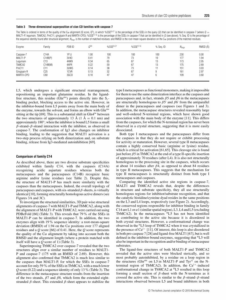
Comparison of family C14
As described above, there are two diverse substrate specificitiesexhibited within family C14, with the caspases (C14A)recognizing acidic aspartate residues whereas both themetacaspases and the paracaspases (C14B) recognize basicarginine and/or lysine residues (see Table 2). Despite this,the structure of MALT1-P has much more similarity with thecaspases than the metacaspases. Indeed, the overall topology ofparacaspases and caspases, with six-stranded β-sheets, is virtuallyidentical [10], forming structurally homologous active dimers (seeFigures 3A and 3C).
To investigate the structural similarities, 3D pair-wise structuralalignments of caspase-7 with MALT1-P and TbMCA2, along withthe alignment of MALT1-P with TbMCA2, were carried out usingPDBeFold [66] (Table 3). This reveals that 79% of the SSEs inMALT1-P can be identified in caspase-7. In addition, the twoenzymes align with 19% sequence identity: a root-mean-squaredeviation (RMSD) of 1.94 on the Cα positions over 177 alignedresidues and a Q score [66] of 0.41. Here, the Q score representsthe quality of the Cα alignment by taking into account both theRMSD and the alignment length (where a protein matched withitself will have a Q score of 1) (Table 3).
Superimposing TbMCA2 over caspase-7 revealed that the twostructures align over a similar number of residues to MALT1-P and caspase-7 (175 with an RMSD of 2.69). However, thisalignment also confirmed that TbMCA2 is much less similar tothe caspases than MALT1-P, for which the SSEs in caspase-7account for only 59% of the SSEs in TbMCA2, with a much lowerQ score (0.22) and a sequence identity of only 13% (Table 3). Thedifference in the metacaspase structure results from the insertionof the two strands, β7 and β8, which form the extended eight-stranded β-sheet. This extended β-sheet appears to stabilize the
type I metacaspases as functional monomers, making it impossiblefor them to use the same dimerization interface as the caspases andparacaspases and, in fact, strands β5 and β6 in the metacaspasesare structurally homologous to β5′ and β6′ from the antiparalleldimer in the paracaspases and caspases (see Figures 1 and 3).In addition, the metacaspase structures revealed reasonably largeand well-ordered N-terminal regions, which have shown goodassociation with the main body of the enzyme [11]. This differsfrom the caspases, for which the N-terminal region has never beenobserved in a crystal structure, suggesting that it is more easilydissociated.
Both type I metacaspases and the paracaspases differ fromthe caspases in that they do not require or exhibit processingfor activity or maturation. However, several type II metacaspasescontain a highly conserved basic (arginine or lysine) residue,which is critical for activation [61,65]. This cleavage site is foundjust before β5 in TbMCA2 at the end of a type II-specific insertionof approximately 70 residues (after L4). It is also not structurallyhomologous to the processing site in the caspases, which occursat about 14 residues after β4, as opposed to about 84 residuesin type II metacaspases. This suggests that the mechanism fortype II metacaspases is structurally distinct from both type Imetacaspases and caspases.
Comparing the identified active site regions in caspase-7,MALT1 and TbMCA2 reveals that, despite the differencesin structure and substrate specificity, they all use structurallyhomologous regions for inhibitor/substrate recognition, and thatthe catalytic histidine/cysteine dyad resides in the same positionson the L3 and L4 loops, respectively (see Figure 2). Accordingly,the conserved regions responsible for inhibitor binding in familyC14 are L1 or α1 (similar spatial region), L3, L4 and L5 (excludingTbMCA2). In the metacaspases HL5 has not been identifiedas contributing to the active site because it is disordered inboth crystal structures. However, a conformational change wasobserved in the HL5 loop of TbMCA2 when it was crystallized inthe presence of Ca2 + [11]. Of interest, this loop is also disorderedin both pro-caspase 7 [28] and ligand-free MALT1 [67], but is welldefined in the inhibitor-bound enzymes, suggesting that HL5 willalso be important in the recognition and/or binding of metacaspasesubstrates.
The ligand-free structures of both MALT1-P and TbMCA2revealed that the active sites were blocked sterically, and somost probably autoinhibited, by a residue on a loop region inthe structures (Gln494 on L5 in MALT1-P and Tyr31 on the N-terminal region of TbMCA2). In addition, the Ca2 + -inducedconformational change in TbMCA2 at HL5 resulted in this loopforming a small section of β-sheet with the N-terminus as itcrossed the active site. This is similar to the β-strand–β-strandinteractions observed between L5 and bound inhibitors in both
c© The Authors Journal compilation c© 2015 Biochemical Society

226 K. McLuskey and J.C. Mottram
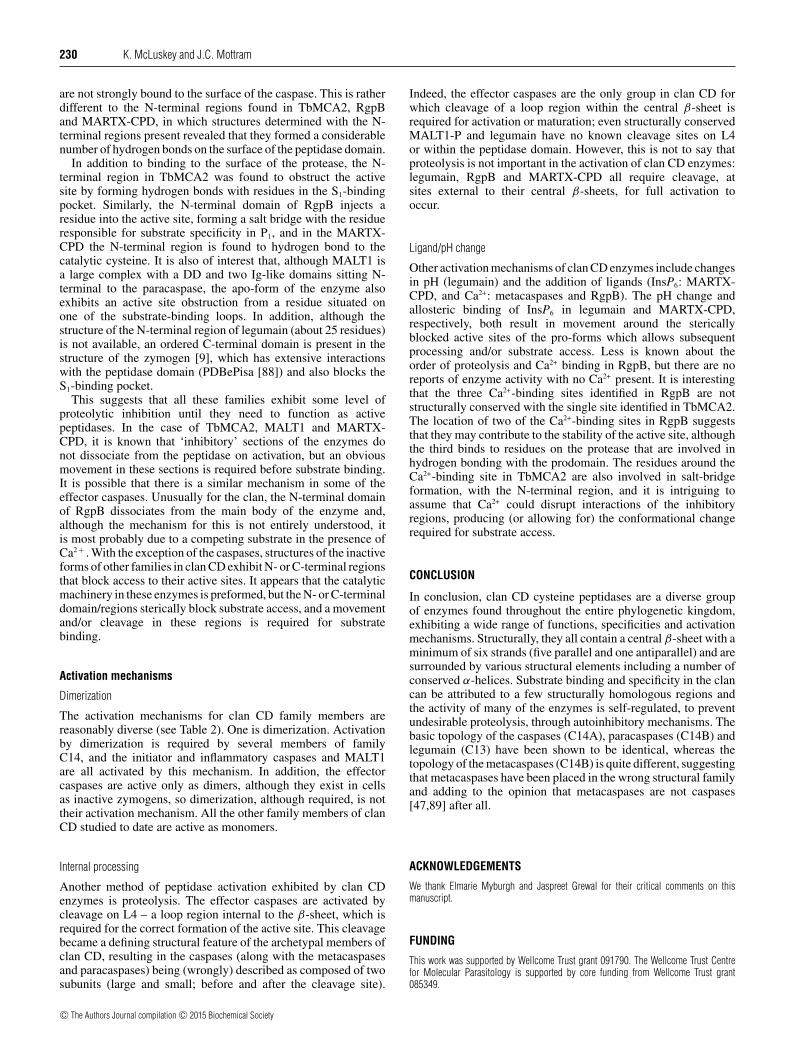
Figure 4 The structural diversity in the central β-sheet of the clan CD enzymes
(A) The β-sheet topologies exhibited by the families in the clan. The β-strands described by the CHF [23] are shown in grey whereas the other strands are shown in blue; the N- and C-terminal endsof the enzymes are labelled accordingly. (B) A phylogenetic tree based on structure, in which Q H [85] is a measure of structural homology. This Figure was produced using a STAMP [87] structuralalignment and VMD [87].
the caspases and MALT1, suggesting that this Ca2 + -inducedloop movement in TbMCA2 could mimic the conformationalchange required by HL5 to bind to a peptide/proteinsubstrate.
This structural family of enzymes, classed as C14, collectivelyexhibits a variety of substrate specificities, activation mechanisms,potential autoinhibitory machinery and N-terminal functionality.Structurally, the specificity-diverse caspases and paracaspasesare almost identical whereas the metacaspases have adifferent structural topology, and all the family members appearto use analogous structural elements to recognize and bind theirsubstrates. Regardless of the diversity exhibited by the family,it is fair to say that the monomeric forms of these structures(caspases, paracaspases and type I metacaspases) are all single-subunit, single-domain monomers, which, in the case of thecaspases and paracaspases, form homodimers. Correspondingly,the widespread nomenclature that describes this family ascontaining a homodimer of heterodimers [3,4], and/or consistingof small and large subunits [18], may need to be reconsidered.In addition, this analysis suggests that the metacaspases aresufficiently structurally and functionally diverse to be classedseparately from the caspases and paracaspases; to investigatethis fully, however, the structure–function relationships for otheravailable clan CD family members need to be considered.
FAMILY C11: CLOSTRIPAIN
The archetypal member of family C11 is clostripain: a cysteinepeptidase released by the anaerobic bacterium Clostridiumhistolyticum. This family of petidases is reportedly found in mostphylogenetic kingdoms but is missing from the Metazoa (see
Table 1). Clostripain is reportedly arginine specific, requiringCa2 + for activity and/or stabilization [70]; it needs the loss ofan N-terminal pro-peptide, along with cleavage and removalof an internal nine-residue peptide, for full activation [71]. Todate, there are no structures of clostripain available in the PDB,but there is a structure of an unassigned peptidase from familyC11. The Joint Centre for Structural Genomics [72] determinedthis structure from the bacterium Parabacteroides merdae, underthe gene name PARMER_00083 (PmC11, see SupplementaryTable S1). The primary sequence of PmC11 is almost 150 residuesshorter than that of clostripain but the two enzymes share a primarysequence identity of 23% (Clustal Omega [73]).
The structure of PmC11 has a nine-stranded β-sheet with4↑3↓2↑1↑5↑6↑7↓8↓9↑ topology, in which β1–β2 and β5–β8overlie well with the six-stranded β-sheet exhibited by thecaspases (Figure 4). Correspondingly, the His133 and Cys179
residues found at the ends of strands β5 and β6 (Hβ3 and Hβ4,respectively) are likely to be the catalytic dyad. PmC11 alsocontains five α-helices, which are structurally homologous toα1–α5 in the caspases. Apart from its extended β-sheet, PmC11differs most significantly from the caspases at its C-terminus,where a further seven α-helices and two β-turns are located afterβ8 (Hβ6).
FAMILY C13: LEGUMAIN
The archetypal member of family C13 is legumain, an asparagine-specific cysteine peptidase, which is found throughout mostphylogenetic kingdoms (see Table 1), although it has been mostextensively studied in the blood fluke parasite Schistosoma sp.,mammals and plants (in which it was originally identified [74]).
c© The Authors Journal compilation c© 2015 Biochemical Society
Structures of clan CD cysteine peptidases 227
Legumain is synthesized as an inactive zymogen with the first17 residues consisting of a signal peptide, which is releasedduring secretion. Historically, the remainder of the enzyme hasbeen described as consisting of an eight-residue, N-terminal pro-peptide, a peptidase domain and a large 110-residue, C-terminalprodomain. However, the N-terminal region of legumain hasrecently been shown to have no role in the activation of theenzyme [75], whereas the C-terminal domain has been shown tobe instrumental in controlling the zymogen, along with enzymeactivation and stability [9].
Currently, the only structures available from family C13 arethose recently determined for human legumain [9], including thestructure of pro-legumain (peptidase- and C-terminal domains),along with three structures of the peptidase domain complexedwith the tetrapeptide inhibitor Ac-YVAD-CMK, at pH 7.5and pH 5.0, and complexed with the peptidomimetic inhibitorZ-Ala-Ala-AzaAsn-CMK [9] (see Supplementary Table S1).It is interesting that the peptidase domain of legumain istopologically equivalent to the caspases and paracaspases,with a central six-stranded β-sheet and five major α-helices(see Figure 3D). However, unlike the structurally similar C14enzymes, legumain is active as a monomer despite no initiallyobvious structural reason for prohibiting caspase-like β6–β6′
dimerization.Legumain is also distinct from all the C14 enzymes, in
that it is activated by pH. Lowering the pH to <5.5 activatesthe enzyme, with full peptidase activity at around pH 4.0.This acidification is accompanied by intermolecular (trans)autoproteolytic processing at Asn323, a cleavage site situated inthe C-terminal domain. Cleavage is not required for activitybut the reaction rate is much faster when cleavage occurs [76].In general, legumain exhibits specificity towards asparagine inP1 (pH optimum 5.5), but at pH �4.5 aspartate is accepted(and this specificity becomes a preference for aspartate at aboutpH 4.0). The functional groups of Arg44 (α1), His45 (α1), Ser216
(L5) and Asp231 (L5) contribute to this specificity, producing azwitterionic S1-binding pocket (see Figure 2D), the geometry ofwhich suggests that Asp231 is protonated at low pH in order toaccept an aspartate in P1 [9].
The structure of pro-legumain revealed that the C-terminaldomain (C domain) is organized into two distinct parts:an activation peptide (AP, positioned immediately after thepeptidase domain) and a C-terminal DD-like fold, consistingof five α-helices, denoted as LSAM (legumain stabilization andactivity modulation) domain [9]. Both the AP and the LSAMdomains interact extensively with the peptidase domain at theautoprocessing site Asn323 found at the interface between them.In addition, Ser307 from the AP forms hydrogen bonds to Arg44
and Ser216 in the P1 pocket, blocking access to the active site.The interacting surfaces of the C domain and the peptidase
domain are complementary (positively and negatively charged,respectively) and, as the pH is lowered and the peptidasebecomes protonated, the interaction between the two surfacesis disrupted (in particular several salt bridges), which produces aconformational arrangement that allows substrates to access theactive site. However, the C domain does not dissociate from theenzyme on processing; in fact it becomes markedly unstable ifthe LSAM domain is removed [9] and it is not possible to expresslegumain in the absence of the C domain [75]. Furthermore,superimposing a copy of pro-legumain on to each monomerin the caspase-7 dimer reveals a steric clash between LSAMdomains. This, together with the fact that there is no biologicalneed for dimerization, suggests that monomeric legumain is amore energetically favourable form. In summary, a change inpH (the activation mechanism), followed by trans-autoprocessing
and a conformational rearrangement, has a role in producing fullymature legumain.
FAMILY C25: GINGIPAIN
The only structure available from the C25 family of clan CDpeptidases is gingipain R (RgpB), a virulence factor participatingin the infection and survival of Porphyromonas gingivalis inperiodontitis. To date, the RgpB structure has been determinedin both its mature and its pro-forms [13,14] (see SupplementaryTable S1). The crystal structure of the mature form of RgpBrevealed a monomeric enzyme with a central 10-stranded β-sheet, which is the largest central β-sheet of all the clan CDstructures determined to date [13]. Similar to other clanCD structures, the central sheet is surrounded on both sides byβ-hairpins and several α-helices. Consideration of the structure inthe same orientation as the caspases reveals that the sheet exhibits6↑5↑7↑8↑9↑10↓4↓3↓1↓2↓ topology, resulting in an internal quasi-symmetry situated between strands 9 and 10. However, the fourN-terminal strands (β1–β4) are twisted out of the plane of theother strands by about 45◦, and are often, perhaps best described asa separate N-terminal subdomain (NSD) [13,14]. The C-terminalsubdomain (CSD [14]), encompassing strands β5–β10, overlieswell with the structure of caspase-7, including the position of thecaspase helices α1–α4 (helices H1–H4, see Figure 3). The SSEsin the CSD are named in line with the caspase nomenclature (asdescribed for the metacaspases above), e.g. RgpB β5 is describedas Hβ1 (strand H1, see Figure 3E). The last 84 C-terminal residuesafter β10 (Hβ6) form an IgSF (Ig superfamily) domain.
RgpB exhibits an exclusive specificity for arginine in P1 andthe original structure of RgpB was reported complexed with thepeptide inhibitor D-FFR-CMK [13]. Analysis of the active siterevealed that, similar to other members of clan CD, the catalytichistidine/cysteine dyad (His221/Cys244) in RgpB is found at the C-terminal ends of Hβ3 and Hβ4, respectively. In addition, residuesforming hydrogen bonds to the P1 arginine are Asp163 (Hα1), Gly210
(L3), Gly212 (L3), Gln282 (HL5), and Trp284 (HL5) (Figure 2E). Trp284
is also found stacking on top of the P1 arginine covering the S1
pocket like a lid. It is interesting that the only functional groupinvolved in hydrogen bonding to the P1 arginine is the carboxylicacid of Asp163, which forms a stable bidentate salt bridge with theguanidino group.
The pro-form of RgpB consists of a 229-residue, N-terminalprodomain, which contains two autoprocessing sites at Arg126
and Arg229 (proform numbering denoted PArg126 and PArg229,respectively). Cleavage at these sites is required for full activationof the enzyme [77], with cleavage at PArg126 being essential forprocessing at PArg229 and subsequent removal of the prodomain.Despite this requirement for full activation, a PR126A/PR229Amutant enzyme was found to exhibit some latent activity, albeit80-fold lower than that of the mature enzyme [77]. Recently,the crystal structure of the inactive RgpB complexed withits prodomain was determined (see Supplementary Table S1),revealing the largest structurally classified prodomain in clan CDto date [14].
This structure revealed that a loop on the prodomain (termedthe ‘inhibitory loop’) runs towards the S1-binding pocket, injectsPArg126 into the pocket, and loops back at this point. This mimicsthe P1 arginine of a bound substrate, with the guanidino groupoverlying in an identical fashion to that of the bound inhibitor.Similar to the P1 arginine of the bound inhibitor, PArg126 makesa bidentate salt bridge with Asp163 in the mature enzyme butin addition it makes a strong hydrogen bond with His221. Thisappears to cause PArg126 to rotate away from the catalytic cysteine,
c© The Authors Journal compilation c© 2015 Biochemical Society
228 K. McLuskey and J.C. Mottram
with the resulting position being unfavourable for cleavage tooccur. However, it is at this position that the initial cleavagefor complete activation of RgpB takes place. Of interest, thereare very few changes between the structures of the inhibitedand the prodomain complexes, suggesting that the zymogen isin a favourable conformation for activity and inhibition by theprodomain is most probably mediated by a competing substrate[14]. However, the mechanism for prodomain dissociation in vivois still unknown.
It is well documented that Rgps are stabilized by calcium andinhibited by EDTA [78] and superimposing the prodomain andinhibitor-bound structures reveals three distinct Ca2+ binding sites,which are highly conserved between the two structures. Two ofthese are found near important substrate-binding regions (HL4and beneath the S1-binding pocket). The importance of the thirdsite is less obvious but it does involve Asp103 and Glu107, whichform hydrogen bonds with the prodomain in the structure of thezymogen. It has not been reported whether any of these sites iscritical in the activation of the enzyme, but they do all appear tohave a role in the structural stability of RgpB with and withoutthe prodomain.
FAMILY C80: MARTX-CPD
The first structure available for family C80 was that of theCPD from the multi-functional autoprocessing repeats in toxin(MARTX) toxin from the pathogenic bacterium Vibrio cholerae.MARTX is an unusually large toxin (>450 kDa), which is secretedby the bacterium, causing disassembly of the actin cytoskeleton,and subsequent bacterial colonization, of the small intestine [79].The N- and C-terminal regions of the toxin have large sectionsof conserved repeats with only the central region (about 1700residues) containing the effector domains which are thoughtto impart distinct functions to the toxin. One such domain isthe CPD, activity of which is required for activation of thetoxin in eukaryotic cells [80], via CPD-mediated proteolysisof regions between the various effector domains, to releasethem [81]. MARTX-CPD itself is activated by myo-inositolhexakisphosphate (InsP6), a molecule present in the cytosol ofeukaryotic cells but absent in the bacterium, hence the activationand subsequent processing of the toxin would not occur until aftertranslocation into the host.
In addition to V. cholerae MARTX toxin, the CDPdomains of Clostridium difficile toxins A and B (TcdA andTcdB, respectively) have also been determined [16,17] (seeSupplementary Table S1). These enzymes are also activatedby InsP6 and similarly require CDP-mediated autoprocessing toallow for overall toxin function [82,83]. The structure of MARTX-CPD contains a seven-stranded β-sheet with 3↓2↑1↑4↑5↑6↓7↓topology, which is surrounded by three major α-helices (seeFigure 3F). The structures of TcdA-CPD and TcdB-CPD aretopologically identical to each other and very similar to thestructure of MARTX-CPD, which shares 93% of its SSEs withTcdA-CPD (PDBefold [66]). However, the structures of the C.difficile CPDs contain a nine-stranded β-sheet with an extra strandflanking both ends of the MARTX-CPD sheet, giving an overalltopology of 4↑3↓2↑1↑5↑6↑7↓8↓9↑; this is the same as observed forPmC11 (see Figure 4).
The structure of MARTX-CPD has been determined in its anapo- [15], inhibitor-bound [81] and pre-processed forms [84] (seeSupplementary Table S1) – and together these structures providea fascinating insight into the unusual activation mechanism ofthe peptidase. Consequently, this enzyme is used for furtherstructural analysis. In MARTX-CPD, strands β1–β2 and β4–
β7 overlie reasonably well with the six-stranded β-sheet found inthe caspases, with β3 sitting on the outside of this core structure(see Figure 3F). In addition, a β-hairpin loop (βA–βC), termedthe ‘β-flap’ [15], is located between Hβ5 and Hβ6. Spatially, thisreplaces α4 and α5 in the caspases and other clan CD members(see Figure 3) and forms a cleft in which InsP6 is found.
MARTX-CPD and TcdA/B-CPDs all have a strict specificityfor leucine in P1 [81], and consequently family C80 is the onlyfamily in clan CD, reported to date, that has a preference for ahydrophobic substrate (see Table 2). The first step in the CPD-mediated proteolytic activation of the MARTX toxin [15] isintramolecular autoprocessing of the CPD itself. This internalcleavage occurs at a leucine residue found 30 residues N-terminalto β1 (Leu0-Ala1 [15], P1–P1) and occurs only after activationof MARTX-CPD by InsP6. In the structure of the inactive andunprocessed form of MARTX-CPD, Leu0 is found anchored inthe large hydrophobic S1-binding pocket, occupying the sameposition as the P1 leucine in the inhibitor (Z-LLL-EP-COO-Et)-bound structure [81] (see Supplementary Table S1).
The catalytic dyad (His91 and Cys140) is found at the C-terminalends of Hβ3 and Hβ4, and a total of seven hydrophobic residues liewithin van der Waals’ bond distance (4.4 Å) of the P1 leucine [81]:Ala47 (α1); Gly89–Val90 (HL3); Val138–Gly139 (HL4); and Leu175–Ala176 (βA, HL5β1) (see Figure 2F). In all crystal structures ofMARTX-CPD an InsP6 molecule is found bound by 13 residues,in a large basic pocket structurally segregated from the S1-bindingpocket. Notably, both the S1- and InsP6-binding pockets containresidues from distinct regions on the β-flap, with βA and βBcontacting the P1 leucine and InsP6, respectively.
It has been shown, in the pre-processed form of MARTX-CPD,that Leu0 occupies the S1-binding site (presumably controllingthe latent activity) irrespective of InsP6 binding, but not in away amenable to intramolecular processing; for this InsP6 isrequired [84]. Binding of InsP6 causes a movement in βB, whichis communicated to the hydrophobic S1 residues on βA (via the β-flap), resulting in Leu0 becoming more tightly bound in the activesite [84]. The outcome of this is that the orientation of the scissilebond in Leu0, relative to the catalytic cysteine, becomes amenableto cis-cleavage. After InsP6-activated processing, the CPD losesits high affinity for the InsP6 molecule and is thought to releaseit. Reoccupation of the S1 site by a new substrate is thoughtto reactivate the InsP6-binding affinity and hence the proteolyticactivity of MARTX-CPD.
CLAN CD COMPARISON
Overview
The caspases are the original structural family in clan CD and,despite growth in structural knowledge for other clan CD families,they remain a sensible structural archetype. This is supported bythe fact that many of the SSEs found in the caspases are present inother family members. Indeed, all members of clan CD contain thesix-stranded β-sheet exhibited by the caspases [with five paralleland one antiparallel strand(s)], with the catalytic histidine/cysteinedyad found at the C-terminal ends of Hβ3 and Hβ4, respectively(see Figures 2 and 3). In addition, all members, with the exceptionof MARTX-CPD (and the other C80 family members), share fivestructurally conserved α-helices (Hα1–Hα5) (see Figure 3). Thisis emphasized by aligning the structures from all the other clanCD families with the caspases (using caspase-7 as a template) andobtaining a measure of structural similarity (PDBeFold [66], asdescribed above).
In terms of similarity to the caspases, the Q score revealsthat MALT1-P is more similar than (>) legumain > TbMCA2
c© The Authors Journal compilation c© 2015 Biochemical Society
Structures of clan CD cysteine peptidases 229
> PmC11 > gingipain > MARTX-CPD. The alignment alsoshows that 67–87% of the SSEs of caspase-7 can be identifiedin all other family members, apart from those found in familyC80, which shares only 47% of its SSEs with caspase-7, andvice versa (see Table 3). It is interesting to note that caspase-7 shares more of its SSEs (87%) with legumain than with anyother family member, making the caspases significantly morestructurally similar to legumain than to the metacaspases. Thisanalysis also revealed that MALT1-P (C14B) is more structurallysimilar to caspase-7 (C14A) than any of the other structures inthe clan (see Table 3). The structural differences of legumain,MALT1-P and the caspases are rather subtle, whereas the otherfamilies, with much longer central β-sheets, share far fewer oftheir SSEs with the caspases (from 47% to 32%) (see Table 3).Indeed, the basic topology of the central β-sheets differs inall the families apart from those represented by the caspases,paracaspases and legumain (see Figure 4).
Comparing all the structural families with each other revealsthat more of the SSEs in PmC11, legumain, RgpB and MARTX-CPD are found in the caspases than in any of the other structures(see Supplementary Table S2), i.e. none of these families sharesmore SSEs with each other than with the caspases. However,more of the SSEs in TbMCA2 can be identified in MALT1-P and legumain than caspases, whereas MALT1-P shares thesame number of SSEs with both caspase-7 and legumain (seeSupplementary Table S2a). Comparison of just the Q scoresreveals that no two families are more structurally similar to eachother than to the caspases, confirming that at a basic level allthese structures share basic structural elements with the caspases(see Supplementary Table S2b). However, this scoring methodconsiders only the parts of the structures that align and does nottake into account gaps in the alignment, which would account forpotentially important deletions and insertions.
Using an adaptation of the Q score, known as QH (a measure ofstructural homology) [85], that takes into account these alignmentgaps, a more accurate representation of structural homology canbe calculated. To calculate the QH scores, a STAMP [86] multiplestructural alignment was carried out on all the families representedby clan CD, using the molecular visualization program VMD[87]. The differences in QH values were derived from thestructural alignment and a phylogenetic tree based on these valueswas produced to depict a structural evolution of the clan (seeFigure 4). This phylogenetic tree (based on structure) suggests thatlegumain, the caspases, the paracaspases and the metacaspasesare all more closely related in structure to each other than tothe other families in the clan. However, although the caspasesand paracaspases are found on the same branch of the tree, bothlegumain and the metacaspases are found on different, distinctbranches. This tree also suggests that PmC11 and MARTX-CDPsit on the same branch, distinct from the caspase group, and thatRgpB represents the most structurally distinct family.
S1-binding sites
In general, the families found in clan CD exhibit individual andrather strict substrate specificities, with a preference for basicresidues in P1 being the most common (see Table 2). Five of thefamilies in clan CD have been determined as complexes withpeptide inhibitors – no complex structures are currently availablefor the metacaspases or PmC11. Mapping the residues involvedin substrate binding on to the structures allowed the residues andSSEs in the S1-binding pockets to be determined and compared.
In the caspases, substrate recognition has been well studiedand is known to depend on three highly conserved side chains
from Arg87, Gln184 and Arg233 (caspase-7 nomenclature) thatare responsible for creating a basic environment for binding anaspartate residue on P1. These residues are found on loops L1, L4and L5 (the last of which forms a short section of β-sheet withthe bound inhibitor), respectively. In MALT1, acidic aspartate andglutamate residues [Asp365 (α1), Asp462 (L4) and Glu500 (L5)] arefound in similar structural positions, respectively (see Figures 2Aand 2B). Although a functionally specific Glu500 in MALT1 isfound to overlie with Arg233 in caspase-7 (opposite charges,opposite binding specificities), no such charged residues are foundin this position in the topologically equivalent legumain. It isinteresting, however, that, although legumain does not appear tohave a functional substrate-binding residue on L5, the guanidinogroup from Arg44 on α1 overlies in almost exactly the same wayin Arg233, despite the fact that the Cα positions are found onmarkedly different parts of the structures. Notably, for all thestructures analysed, S1 specificity in clan CD can be attributedsolely to three main structural regions – (H)α1 [(H)L1], (H)L4 and(H)L5 – and these all appear to contribute to a correctly chargedP1-binding environment. Analysis of the S1-binding pockets ofthe various families also reveals a conserved aromatic residueon (H)L5 (see Figure 2), which forms hydrophobic contacts withthe bound inhibitors (see Supplementary Figure S1) and may beimportant for directing substrates into the S1 pocket.
By looking at the structural regions forming hydrogen bondsand hydrophobic contacts to the inhibitors in the P2–P4 positions(in addition to P1), it becomes obvious that: the regions involvedin the S1- to S4-binding pockets in clan CD are (H)L1, (H)α1, (H)L3,(H)L4, (H)L5, (H)α4 and (H)L6; and, along with the histidine/cysteinedyad, a glycine residue adjacent to the catalytic histidine is alsostructurally conserved in all families. Mapping of these regionsmay allow the substrate-binding residues in other, ligand-free,structures to be predicted. Indeed, mapping of the residues knownto be important for activity in TbMCA2 on to the SSEs in thestructure reveals that these residues are found on α1, L3 and L4.However, (H)L5, which is important for binding in all the complexstructures, is disordered in TbMCA2, although it exhibits a shift inthe presence of Ca2 + [11], suggesting that it will, most probably,be involved in substrate binding in the metacaspases. MARTX-CPD is somewhat different to the other family members and itssubstrate-binding regions are similar but not quite as conservedas those of the other families. However, even when the SSE inMARTX-CPD has changed from an α-helix to a β-strand (in thecase of βA and Hα4), the interacting residues in the differentfamilies are spatially equivalent. Subsequently, all ligand-bindingregions in clan CD are found in parts of the structure that overliewell with the caspases, in front or on top of the β-sheet, as shownin Figures 2 and 3.
Pro-forms
The structures of the clan CD enzymes reveal that theyoften contain diverse N-terminal and/or C-terminal (prodomain)regions. With the exception of PmC11, data are available for theN-terminal regions for all the families. In the case of the initiatorand inflammatory caspases, the large N-terminal regions containimportant CARD or DED domains, whereas the effector caspaseshave short N-terminal regions for which the function is less welldefined. It is difficult to ascertain whether the N-terminal regionsin the effectors are true prodomains (as they are often described)because emerging research is starting to suggest that their removalis not necessary for activation; in contrast, however, they do appearto have a part to play in enzyme inhibition. In addition, theseregions have eluded structural determination, suggesting that they
c© The Authors Journal compilation c© 2015 Biochemical Society
230 K. McLuskey and J.C. Mottram
are not strongly bound to the surface of the caspase. This is ratherdifferent to the N-terminal regions found in TbMCA2, RgpBand MARTX-CPD, in which structures determined with the N-terminal regions present revealed that they formed a considerablenumber of hydrogen bonds on the surface of the peptidase domain.
In addition to binding to the surface of the protease, the N-terminal region in TbMCA2 was found to obstruct the activesite by forming hydrogen bonds with residues in the S1-bindingpocket. Similarly, the N-terminal domain of RgpB injects aresidue into the active site, forming a salt bridge with the residueresponsible for substrate specificity in P1, and in the MARTX-CPD the N-terminal region is found to hydrogen bond to thecatalytic cysteine. It is also of interest that, although MALT1 isa large complex with a DD and two Ig-like domains sitting N-terminal to the paracaspase, the apo-form of the enzyme alsoexhibits an active site obstruction from a residue situated onone of the substrate-binding loops. In addition, although thestructure of the N-terminal region of legumain (about 25 residues)is not available, an ordered C-terminal domain is present in thestructure of the zymogen [9], which has extensive interactionswith the peptidase domain (PDBePisa [88]) and also blocks theS1-binding pocket.
This suggests that all these families exhibit some level ofproteolytic inhibition until they need to function as activepeptidases. In the case of TbMCA2, MALT1 and MARTX-CPD, it is known that ‘inhibitory’ sections of the enzymes donot dissociate from the peptidase on activation, but an obviousmovement in these sections is required before substrate binding.It is possible that there is a similar mechanism in some of theeffector caspases. Unusually for the clan, the N-terminal domainof RgpB dissociates from the main body of the enzyme and,although the mechanism for this is not entirely understood, itis most probably due to a competing substrate in the presence ofCa2 + . With the exception of the caspases, structures of the inactiveforms of other families in clan CD exhibit N- or C-terminal regionsthat block access to their active sites. It appears that the catalyticmachinery in these enzymes is preformed, but the N- or C-terminaldomain/regions sterically block substrate access, and a movementand/or cleavage in these regions is required for substratebinding.
Activation mechanisms
Dimerization
The activation mechanisms for clan CD family members arereasonably diverse (see Table 2). One is dimerization. Activationby dimerization is required by several members of familyC14, and the initiator and inflammatory caspases and MALT1are all activated by this mechanism. In addition, the effectorcaspases are active only as dimers, although they exist in cellsas inactive zymogens, so dimerization, although required, is nottheir activation mechanism. All the other family members of clanCD studied to date are active as monomers.
Internal processing
Another method of peptidase activation exhibited by clan CDenzymes is proteolysis. The effector caspases are activated bycleavage on L4 – a loop region internal to the β-sheet, which isrequired for the correct formation of the active site. This cleavagebecame a defining structural feature of the archetypal members ofclan CD, resulting in the caspases (along with the metacaspasesand paracaspases) being (wrongly) described as composed of twosubunits (large and small; before and after the cleavage site).
Indeed, the effector caspases are the only group in clan CD forwhich cleavage of a loop region within the central β-sheet isrequired for activation or maturation; even structurally conservedMALT1-P and legumain have no known cleavage sites on L4or within the peptidase domain. However, this is not to say thatproteolysis is not important in the activation of clan CD enzymes:legumain, RgpB and MARTX-CPD all require cleavage, atsites external to their central β-sheets, for full activation tooccur.
Ligand/pH change
Other activation mechanisms of clan CD enzymes include changesin pH (legumain) and the addition of ligands (InsP6: MARTX-CPD, and Ca2+: metacaspases and RgpB). The pH change andallosteric binding of InsP6 in legumain and MARTX-CPD,respectively, both result in movement around the stericallyblocked active sites of the pro-forms which allows subsequentprocessing and/or substrate access. Less is known about theorder of proteolysis and Ca2+ binding in RgpB, but there are noreports of enzyme activity with no Ca2+ present. It is interestingthat the three Ca2+-binding sites identified in RgpB are notstructurally conserved with the single site identified in TbMCA2.The location of two of the Ca2+-binding sites in RgpB suggeststhat they may contribute to the stability of the active site, althoughthe third binds to residues on the protease that are involved inhydrogen bonding with the prodomain. The residues around theCa2+-binding site in TbMCA2 are also involved in salt-bridgeformation, with the N-terminal region, and it is intriguing toassume that Ca2+ could disrupt interactions of the inhibitoryregions, producing (or allowing for) the conformational changerequired for substrate access.
CONCLUSION
In conclusion, clan CD cysteine peptidases are a diverse groupof enzymes found throughout the entire phylogenetic kingdom,exhibiting a wide range of functions, specificities and activationmechanisms. Structurally, they all contain a central β-sheet with aminimum of six strands (five parallel and one antiparallel) and aresurrounded by various structural elements including a number ofconserved α-helices. Substrate binding and specificity in the clancan be attributed to a few structurally homologous regions andthe activity of many of the enzymes is self-regulated, to preventundesirable proteolysis, through autoinhibitory mechanisms. Thebasic topology of the caspases (C14A), paracaspases (C14B) andlegumain (C13) have been shown to be identical, whereas thetopology of the metacaspases (C14B) is quite different, suggestingthat metacaspases have been placed in the wrong structural familyand adding to the opinion that metacaspases are not caspases[47,89] after all.
ACKNOWLEDGEMENTS
We thank Elmarie Myburgh and Jaspreet Grewal for their critical comments on thismanuscript.
FUNDING
This work was supported by Wellcome Trust grant 091790. The Wellcome Trust Centrefor Molecular Parasitology is supported by core funding from Wellcome Trust grant085349.
c© The Authors Journal compilation c© 2015 Biochemical Society
Structures of clan CD cysteine peptidases 231
REFERENCES
1 Rawlings, N.D., Barrett, A.J. and Bateman, A. (2012) MEROPS: the database of proteolyticenzymes, their substrates and inhibitors. Nucleic Acids Res. 40, D343–D350CrossRef PubMed
2 Lillico, S. (2002) Essential Roles for GPI-anchored Proteins in African trypanosomesrevealed using mutants deficient in GPI8. Mol. Biol. Cell 14, 1182–1194 CrossRef
3 Walker, N.P., Talanian, R.V., Brady, K.D., Dang, L.C., Bump, N.J., Ferenz, C.R., Franklin,S., Ghayur, T., Hackett, M.C. and Hammill, L.D. (1994) Crystal structure of the cysteineprotease interleukin-1 beta-converting enzyme: a (p20/p10)2 homodimer. Cell 78,343–352 CrossRef PubMed
4 Wilson, K.P., Black, J.A., Thomson, J.A., Kim, E.E., Griffith, J.P., Navia, M.A., Murcko,M.A., Chambers, S.P., Aldape, R.A. and Raybuck, S.A. (1994) Structure and mechanism ofinterleukin-1 beta converting enzyme. Nature 370, 270–275 CrossRef PubMed
5 Barrett, A.J. and Rawlings, N.D. (1996) Families and clans of cysteine peptidases.Perspect. Drug Discov. Design 6, 1–11 CrossRef
6 Pei, J. and Grishin, N.V. (2009) Prediction of a caspase-like fold in Tannerella forsythiavirulence factor PrtH. Cell Cycle 8, 1453–1455 CrossRef PubMed
7 Rawlings, N.D. and Barrett, A.J. (1999) MEROPS: the peptidase database. Nucleic AcidsRes. 27, 325–331 CrossRef PubMed
8 Berman, H.M., Westbrook, J., Feng, Z., Gilliland, G., Bhat, T.N., Weissig, H., Shindyalov,I.N. and Bourne, P.E. (2000) The Protein Data Bank. Nucleic Acids Res. 28, 235–242CrossRef PubMed
9 Dall, E. and Brandstetter, H. (2013) Mechanistic and structural studies on legumainexplain its zymogenicity, distinct activation pathways, and regulation. Proc. Natl. Acad.Sci. U.S.A. 110, 10940–10945 CrossRef PubMed
10 Yu, J.W., Jeffrey, P.D., Ha, J.Y., Yang, X. and Shi, Y. (2011) Crystal structure of themucosa-associated lymphoid tissue lymphoma translocation 1 (MALT1) paracaspaseregion. Proc. Natl. Acad. Sci. U.S.A. 108, 21004–21009 CrossRef PubMed
11 McLuskey, K., Rudolf, J., Proto, W.R., Isaacs, N.W., Coombs, G.H., Moss, C.X. andMottram, J.C. (2012) Crystal structure of a Trypanosoma brucei metacaspase. Proc. Natl.Acad. Sci. U.S.A. 109, 7469–7474 CrossRef PubMed
12 Wong, A. H.-H., Yan, C. and Shi, Y. (2012) Crystal structure of the yeast metacaspaseyca1. J. Biol. Chem. 287, 29251–29259 CrossRef PubMed
13 Eichinger, A., Beisel, H.G., Jacob, U., Huber, R., Medrano, F.J., Banbula, A., Potempa, J.,Travis, J. and Bode, W. (1999) Crystal structure of gingipain R: an Arg-specific bacterialcysteine proteinase with a caspase-like fold. EMBO J. 18, 5453–5462 CrossRef PubMed
14 de Diego, I., Veillard, F.T., Guevara, T., Potempa, B., Sztukowska, M., Potempa, J. andGomis-Ruth, F.X. (2013) Porphyromonas gingivalis virulence factor gingipain RgpBshows a unique zymogenic mechanism for cysteine peptidases. J. Biol. Chem. 288,14287–14296 CrossRef PubMed
15 Lupardus, P.J., Shen, A., Bogyo, M. and Garcia, K.C. (2008) Small molecule-inducedallosteric activation of the Vibrio cholerae RTX cysteine protease domain. Science 322,265–268 CrossRef PubMed
16 Pruitt, R.N., Chagot, B., Cover, M., Chazin, W.J., Spiller, B. and Lacy, D.B. (2009)Structure–function analysis of inositol hexakisphosphate-induced autoprocessing inClostridium difficile toxin A. J. Biol. Chem. 284, 21934–21940 CrossRef PubMed
17 Shen, A., Lupardus, P.J., Gersch, M.M., Puri, A.W., Albrow, V.E., Garcia, K.C. and Bogyo,M. (2011) Defining an allosteric circuit in the cysteine protease domain of Clostridiumdifficile toxins. Nat. Struct. Mol. Biol. 18, 364–371 CrossRef PubMed
18 Fuentes-Prior, P. and Salvesen, G.S. (2004) The protein structures that shape caspaseactivity, specificity, activation and inhibition. Biochem. J. 384, 201–232CrossRef PubMed
19 MacKenzie, S.H. and Clark, A.C. (2012) Death by caspase dimerization. Adv. Exp. Med.Biol. 747, 55–73 CrossRef PubMed
20 Pop, C. and Salvesen, G.S. (2009) Human caspases: activation, specificity, andregulation. J. Biol. Chem. 284, 21777–21781 CrossRef PubMed
21 Wei, Y., Fox, T., Chambers, S.P., Sintchak, J., Coll, J.T., Golec, J.M., Swenson, L., Wilson,K.P. and Charifson, P.S. (2000) The structures of caspases-1, -3, -7 and -8 reveal thebasis for substrate and inhibitor selectivity. Chem. Biol. 7, 423–432 CrossRef PubMed
22 Zhang, C. and Kim, S.H. (2000) The anatomy of protein beta-sheet topology. J. Mol. Biol.299, 1075–1089 CrossRef PubMed
23 Aravind, L. and Koonin, E.V. (2002) Classification of the caspase-hemoglobinase fold:detection of new families and implications for the origin of the eukaryotic separins.Proteins 46, 355–367 CrossRef PubMed
24 Thornberry, N.A., Bull, H.G., Calaycay, J.R., Chapman, K.T., Howard, A.D., Kostura, M.J.,Miller, D.K., Molineaux, S.M., Weidner, J.R. and Aunins, J. (1992) A novel heterodimericcysteine protease is required for interleukin-1 beta processing in monocytes. Nature 356,768–774 CrossRef PubMed
25 Chai, J., Shiozaki, E., Srinivasula, S.M., Wu, Q., Datta, P., Alnemri, E.S., Shi, Y. and Dataa,P. (2001) Structural basis of caspase-7 inhibition by XIAP. Cell 104, 769–780CrossRef PubMed
26 Laskowski, R.A. and Swindells, M.B. (2011) LigPlot + : multiple ligand-proteininteraction diagrams for drug discovery. J. Chem. Inf. Model 51, 2778–2786CrossRef PubMed
27 Reference deleted PubMed28 Chai, J., Wu, Q., Shiozaki, E., Srinivasula, S., Alnemri, E. and Shi, Y. (2001) Crystal
structure of a procaspase-7 zymogen: Mechanisms of activation and substrate binding.Cell 107, 399–407 CrossRef PubMed
29 Chai, J., Shiozaki, E., Srinivasula, S.M., Wu, Q., Datta, P., Alnemri, E.S., Shi, Y. and Dataa,P. (2001) Structural basis of caspase-7 inhibition by XIAP. Cell 104, 769–780CrossRef PubMed
30 Bose, K. and Clark, A.C. (2001) Dimeric procaspase-3 unfolds via a four-state equilibriumprocess. Biochemistry 40, 14236–14242 CrossRef PubMed
31 Renatus, M., Stennicke, H.R., Scott, F.L., Liddington, R.C. and Salvesen, G.S. (2001)Dimer formation drives the activation of the cell death protease caspase 9. Proc. Natl.Acad. Sci. U.S.A. 98, 14250–14255 CrossRef PubMed
32 Klaiman, G., Champagne, N. and LeBlanc, A.C. (2009) Self-activation ofcaspase-6 in vitro and in vivo: caspase-6 activation does not induce cell death inHEK293T cells. Biochim. Biophys. Acta 1793, 592–601 CrossRef PubMed
33 Cao, Q., Wang, X.J., Li, L.F. and Su, X.D. (2014) The regulatory mechanism of thecaspase 6 pro-domain revealed by crystal structure and biochemical assays. ActaCrystallogr. D Biol. Crystallogr. 70, 58–67 CrossRef PubMed
34 Vaidya, S., Velazquez-Delgado, E.M., Abbruzzese, G. and Hardy, J.A. (2011)Substrate-induced conformational changes occur in all cleaved forms of caspase-6. J.Mol. Biol. 406, 75–91 CrossRef PubMed
35 Meergans, T., Hildebrandt, A.K., Horak, D., Haenisch, C. and Wendel, A. (2000) The shortprodomain influences caspase-3 activation in HeLa cells. Biochem. J. 349, 135–140CrossRef PubMed
36 Stennicke, H.R., Jurgensmeier, J.M., Shin, H., Deveraux, Q., Wolf, B.B., Yang, X., Zhou,Q., Ellerby, H.M., Ellerby, L.M., Bredesen, D. et al. (1998) Pro-caspase-3 is a majorphysiologic target of caspase-8. J. Biol. Chem. 273, 27084–27090 CrossRef PubMed
37 Denault, J.-B. and Salvesen, G.S. (2003) Human caspase-7 activity and regulation by itsN-terminal peptide. J. Biol. Chem. 278, 34042–34050 CrossRef PubMed
38 Turk, B., Turk, D. and Turk, V. (2000) Lysosomal cysteine proteases: more thanscavengers. Biochim. Biophys. Acta 1477, 98–111 CrossRef PubMed
39 Uren, A.G., O’Rourke, K., Aravind, L.A., Pisabarro, M.T., Seshagiri, S., Koonin, E.V. andDixit, V.M. (2000) Identification of paracaspases and metacaspases: two ancient familiesof caspase-like proteins, one of which plays a key role in MALT lymphoma. Mol. Cell 6,961–967 PubMed
40 Choi, C.J. and Berges, J.A. (2013) New types of metacaspases in phytoplankton revealdiverse origins of cell death proteases. Cell Death Dis. 4, e490 CrossRef PubMed
41 Asplund-Samuelsson, J., Bergman, B. and Larsson, J. (2012) Prokaryotic caspasehomologs: phylogenetic patterns and functional characteristics reveal considerablediversity. PLoS ONE 7, e49888 CrossRef PubMed
42 Vercammen, D., Declercq, W., Vandenabeele, P. and Van Breusegem, F. (2007) Aremetacaspases caspases? J Cell Biol 179, 375–380 CrossRef PubMed
43 Madeo, F., Herker, E., Maldener, C., Wissing, S., Lachelt, S., Herlan, M., Fehr, M., Lauber,K., Sigrist, S.J., Wesselborg, S. et al. (2002) A caspase-related protease regulatesapoptosis in yeast. Cell 9, 911–917
44 Coll, N.S., Vercammen, D., Smidler, A., Clover, C., Van Breusegem, F., Dangl, J.L. andEpple, P. (2010) Arabidopsis type I metacaspases control cell death. Science 330,1393–1397 CrossRef PubMed
45 Bozhkov, P.V., Suarez, M.F., Filonova, L.H., Daniel, G., Zamyatnin, A.A., Rodriguez-Nieto,S., Zhivotovsky, B. and Smertenko, A. (2005) Cysteine protease mcII-Pa executesprogrammed cell death during plant embryogenesis. Proc. Natl. Acad. Sci. U.S.A. 102,14463–14468 CrossRef PubMed
46 Watanabe, N. and Lam, E. (2005) Two Arabidopsis metacaspases AtMCP1b and AtMCP2bare arginine/lysine-specific cysteine proteases and activate apoptosis-like cell death inyeast. J. Biol. Chem. 280, 14691–14699 CrossRef PubMed
47 Tsiatsiani, L., Van Breusegem, F., Gallois, P., Zavialov, A., Lam, E. and Bozhkov, P.V.(2011) Metacaspases. Cell Death Differ. 18, 1279–1288 CrossRef PubMed
48 Lee, R.E.C., Puente, L.G., Kaern, M. and Megeney, L.A. (2008) A non-death role of theyeast metacaspase: Yca1p alters cell cycle dynamics. PLoS ONE 3, e2956CrossRef PubMed
49 Castanys-Munoz, E., Brown, E., Coombs, G.H. and Mottram, J.C. (2012) Leishmaniamexicana metacaspase is a negative regulator of amastigote proliferation in mammaliancells. Cell Death Dis. 3, e385 CrossRef PubMed
50 Richie, D.L., Miley, M.D., Bhabhra, R., Robson, G.D., Rhodes, J.C. and Askew, D.S.(2007) The Aspergillus fumigatus metacaspases CasA and CasB facilitate growth underconditions of endoplasmic reticulum stress. Mol. Microbiol. 63, 591–604CrossRef PubMed
51 Lee, R.E.C., Brunette, S., Puente, L.G. and Megeney, L.A. (2010) Metacaspase Yca1 isrequired for clearance of insoluble protein aggregates. Proc. Natl. Acad. Sci. U.S.A. 107,13348–13353 CrossRef PubMed
c© The Authors Journal compilation c© 2015 Biochemical Society
232 K. McLuskey and J.C. Mottram
52 Proto, W.R., Castanys-Munoz, E., Black, A., Tetley, L., Moss, C.X., Juliano, L., Coombs,G.H. and Mottram, J.C. (2011) Trypanosoma brucei metacaspase 4 is a pseudopeptidaseand a virulence factor. J. Biol. Chem. 286, 39914–39925 CrossRef PubMed
53 Helms, M.J., Ambit, A., Appleton, P., Tetley, L., Coombs, G.H. and Mottram, J.C. (2006)Bloodstream form Trypanosoma brucei depend upon multiple metacaspases associatedwith RAB11-positive endosomes. J. Cell Sci. 119, 1105–1117 CrossRef PubMed
54 Hamann, A., Brust, D. and Osiewacz, H.D. (2007) Deletion of putative apoptosis factorsleads to lifespan extension in the fungal ageing model Podospora anserina. Mol.Microbiol. 65, 948–958 CrossRef PubMed
55 Colabardini, A.C., De Castro, P.A., De Gouvea, P.F., Savoldi, M., Malavazi, I., Goldman,M.H.S. and Goldman, G.H. (2010) Involvement of the Aspergillus nidulans protein kinaseC with farnesol tolerance is related to the unfolded protein response. Mol. Microbiol. 78,1259–1279 CrossRef PubMed
56 Laverriere, M., Cazzulo, J.J. and Alvarez, V.E. (2012) Antagonic activities of Trypanosomacruzi metacaspases affect the balance between cell proliferation, death and differentiation.Cell Death Differ. 19, 1358–1369 CrossRef PubMed
57 Ambit, A., Fasel, N., Coombs, G.H. and Mottram, J.C. (2008) An essential role for theLeishmania major metacaspase in cell cycle progression. Cell Death Differ. 15, 113–122CrossRef PubMed
58 Zalila, H., Gonzalez, I.J., El-Fadili, A.K., Delgado, M.B., Desponds, C., Schaff, C. andFasel, N. (2011) Processing of metacaspase into a cytoplasmic catalytic domain mediatingcell death in Leishmania major. Mol. Microbiol. 79, 222–239 CrossRef PubMed
59 Gonzalez, I.J., Desponds, C., Schaff, C., Mottram, J.C. and Fasel, N. (2007) Leishmaniamajor metacaspase can replace yeast metacaspase in programmed cell death and hasarginine-specific cysteine peptidase activity. Int. J. Parasitol. 37, 161–172CrossRef PubMed
60 Moss, C.X., Westrop, G.D., Juliano, L., Coombs, G.H. and Mottram, J.C. (2007)Metacaspase 2 of Trypanosoma brucei is a calcium-dependent cysteine peptidase activewithout processing. FEBS Lett. 581, 5635–5639 CrossRef PubMed
61 Vercammen, D., van de Cotte, B., De Jaeger, G., Eeckhout, D., Casteels, P., Vandepoele,K., Vandenberghe, I., Van Beeumen, J., Inze, D. and Van Breusegem, F. (2004) Type IImetacaspases Atmc4 and Atmc9 of Arabidopsis thaliana cleave substrates after arginineand lysine. J. Biol. Chem. 279, 45329–45336 CrossRef PubMed
62 Zhang, Y. and Lam, E. (2011) Sheathing the swords of death: post-translationalmodulation of plant metacaspases. Plant Signal. Behav. 6, 2051–2056CrossRef PubMed
63 Meslin, B., Beavogui, A.H., Fasel, N. and Picot, S. (2011) Plasmodium falciparummetacaspase PfMCA-1 triggers a z-VAD-fmk inhibitable protease to promote cell death.PLoS ONE 6, e23867 CrossRef PubMed
64 Watanabe, N. and Lam, E. (2011) Calcium-dependent activation and autolysis ofarabidopsis metacaspase 2d. J. Biol. Chem. 286, 10027–10040CrossRef PubMed
65 Watanabe, N. and Lam, E. (2011) Arabidopsis metacaspase 2d Is a positive mediator ofcell death induced during biotic and abiotic stresses. Plant J Cell Mol. Biol. 66, 969–982CrossRef
66 Krissinel, E.E. and Henrick, K.K. (2004) Secondary-structure matching (SSM), a new toolfor fast protein structure alignment in three dimensions. Acta Crystallogr. D Biol.Crystallogr. 60, 2256–2268 CrossRef PubMed
67 Wiesmann, C., Leder, L., Blank, J., Bernardi, A., Melkko, S., Decock, A., D’Arcy, A.,Villard, F., Erbel, P., Hughes, N. et al. (2012) Structural determinants of MALT1 proteaseactivity. J. Mol. Biol. 419, 4–21 CrossRef PubMed
68 Hachmann, J., Snipas, S.J., van Raam, B.J., Cancino, E.M., Houlihan, E.J., Poreba, M.,Kasperkiewicz, P., Drag, M. and Salvesen, G.S. (2012) Mechanism and specificity of thehuman paracaspase MALT1. Biochem. J. 443, 287–295 CrossRef PubMed
69 Staal, J. and Beyaert, R. (2012) A two-step activation mechanism of MALT1 paracaspase.J. Mol. Biol. 419, 1–3 CrossRef PubMed
70 Kembhavi, A.A., Buttle, D.J., Rauber, P. and Barrett, A.J. (1991) Clostripain:characterization of the active site. FEBS Lett. 283, 277–280 CrossRef PubMed
71 Witte, V., Wolf, N. and Dargatz, H. (1996) Clostripain linker deletion variants yield activeenzyme in Escherichia coli: a possible function of the linker peptide as intramolecularinhibitor of clostripain automaturation. Curr. Microbiol. 33, 281–286 CrossRef PubMed
72 Elsliger, M.A., Deacon, A.M., Godzik, A., Lesley, S.A., Wooley, J., Wuthrich, K. andWilson, I.A. (2010) The JCSG high-throughput structural biology pipeline. ActaCrystallogr. F Struct. Biol. Cryst. Commun. 66, 1137–1142 CrossRef
73 Sievers, F., Wilm, A., Dineen, D., Gibson, T.J., Karplus, K., Li, W., Lopez, R., McWilliam,H., Remmert, M., Soding, J. et al. (2011) Fast, scalable generation of high-quality proteinmultiple sequence alignments using Clustal Omega. Mol. Syst. Biol. 7, 539CrossRef PubMed
74 Kembhavi, A.A., Buttle, D.J., Knight, C.G. and Barrett, A.J. (1993) The two cysteineendopeptidases of legume seeds: purification and characterization by use of specificfluorometric assays. Arch. Biochem. Biophys. 303, 208–213 CrossRef PubMed
75 Dall, E. and Brandstetter, H. (2012) Activation of legumain involves proteolytic andconformational events, resulting in a context- and substrate-dependent activity profile.Acta Crystallogr. F Struct. Biol. Cryst. Commun. 68, 24–31 CrossRef
76 Li, D.N., Matthews, S.P., Antoniou, A.N., Mazzeo, D. and Watts, C. (2003) Multistepautoactivation of asparaginyl endopeptidase in vitro and in vivo. J. Biol. Chem. 278,38980–38990 CrossRef PubMed
77 Mikolajczyk, J., Boatright, K.M., Stennicke, H.R., Nazif, T., Potempa, J., Bogyo, M. andSalvesen, G.S. (2003) Sequential autolytic processing activates the zymogen ofArg-gingipain. J. Biol. Chem. 278, 10458–10464 CrossRef PubMed
78 Chen, Z., Potempa, J., Polanowski, A., Wikstrom, M. and Travis, J. (1992) Purificationand characterization of a 50-kDa cysteine proteinase (gingipain) from Porphyromonasgingivalis. J. Biol. Chem. 267, 18896–18901 PubMed
79 Olivier, V., Haines, G.K., Tan, Y. and Satchell, K.J.F. (2007) Hemolysin and themultifunctional autoprocessing RTX toxin are virulence factors during intestinal infectionof mice with Vibrio cholerae El Tor O1 strains. Infect. Immun. 75, 5035–5042CrossRef PubMed
80 Sheahan, K.-L., Cordero, C.L. and Satchell, K.J.F. (2007) Autoprocessing of the Vibriocholerae RTX toxin by the cysteine protease domain. EMBO J. 26, 2552–2561CrossRef PubMed
81 Shen, A., Lupardus, P.J., Albrow, V.E., Guzzetta, A., Powers, J.C., Garcia, K.C. and Bogyo,M. (2009) Mechanistic and structural insights into the proteolytic activation of Vibriocholerae MARTX toxin. Nat. Chem. Biol. 5, 469–478 CrossRef PubMed
82 Egerer, M., Giesemann, T., Jank, T., Satchell, K.J.F. and Aktories, K. (2007) Auto-catalyticcleavage of Clostridium difficile toxins A and B depends on cysteine protease activity. J.Biol. Chem. 282, 25314–25321 CrossRef PubMed
83 Reineke, J., Tenzer, S., Rupnik, M., Koschinski, A., Hasselmayer, O., Schrattenholz, A.,Schild, H. and Eichel-Streiber, von, C. (2007) Autocatalytic cleavage of Clostridiumdifficile toxin B. Nature 446, 415–419 CrossRef PubMed
84 Prochazkova, K., Shuvalova, L.A., Minasov, G., Voburka, Z., Anderson, W.F. and Satchell,K.J.F. (2009) Structural and molecular mechanism for autoprocessing of MARTX toxin ofVibrio cholerae at multiple sites. J. Biol. Chem. 284, 26557–26568 CrossRef PubMed
85 O’Donoghue, P. and Luthey-Schulten, Z. (2005) Evolutionary profiles derived from the QRfactorization of multiple structural alignments gives an economy of information. J. Mol.Biol. 346, 875–894 CrossRef PubMed
86 Russell, R.B. and Barton, G.J. (1992) Multiple protein sequence alignment from tertiarystructure comparison: assignment of global and residue confidence levels. Proteins 14,309–323 CrossRef PubMed
87 Eargle, J., Wright, D. and Luthey-Schulten, Z. (2006) Multiple alignment of proteinstructures and sequences for VMD. Bioinformatics 22, 504–506 CrossRef PubMed
88 Krissinel, E. and Henrick, K. (2007) Inference of macromolecular assemblies fromcrystalline state. J. Mol. Biol. 372, 774–797 CrossRef PubMed
89 Enoksson, M. and Salvesen, G.S. (2010) Metacaspases are not caspases – always doubt.Cell Death Differ. 17, 1221 CrossRef PubMed
90 Bond, C.S. (2003) TopDraw: a sketchpad for protein structure topology cartoons.Bioinformatics 19, 311–312 CrossRef PubMed
Received 22 October 2014/19 November 2014; accepted 8 December 2014Published on the Internet 20 February 2015, doi:10.1042/BJ20141324
c© The Authors Journal compilation c© 2015 Biochemical Society