comparative karyotype analysis and chromosome evolution in the
TRANSCRIPT

Gruber et al. BMC Genetics 2012, 13:28http://www.biomedcentral.com/1471-2156/13/28
RESEARCH ARTICLE Open Access
Comparative karyotype analysis and chromosomeevolution in the genus Aplastodiscus(Cophomantini, Hylinae, Hylidae)Simone Lilian Gruber1*, Juliana Zina2, Hideki Narimatsu1, Célio Fernando Baptista Haddad3 and Sanae Kasahara1
Abstract
Background: The frogs of the Tribe Cophomantini present, in general, 2n = 24 karyotype, but data on Aplastodiscusshowed variation in diploid number from 2n= 24 to 2n = 18. Five species were karyotyped, one of them for the firsttime, using conventional and molecular cytogenetic techniques, with the aim to perform a comprehensivecomparative analysis towards the understanding of chromosome evolution in light of the phylogeny.
Results: Aplastodiscus perviridis showed 2n = 24, A. arildae and A. eugenioi, 2n = 22, A. callipygius, 2n = 20, and A.leucopygius, 2n = 18. In the metaphase I cells of two species only bivalents occurred, whereas in A. arildae, A.callipygius, and A. leucopygius one tetravalent was also observed besides the bivalents. BrdU incorporation producedreplication bands especially in the largest chromosomes, and a relatively good banding correspondence wasnoticed among some of them. Silver impregnation and FISH with an rDNA probe identified a single NOR pair: the11 in A. perviridis and A. arildae; the 6 in A. eugenioi; and the 9 in A. callipygius and A. leucopygius. C-bandingshowed a predominantly centromeric distribution of the heterochromatin, and in one of the species distinctmolecular composition was revealed by CMA3. The telomeric probe hybridised all chromosome ends andadditionally disclosed the presence of telomere-like sequences in centromeric regions of three species.
Conclusions: Based on the hypothesis of 2n = 24 ancestral karyotype for Aplastodiscus, and considering thekaryotype differences and similarities, two evolutionary pathways through fusion events were suggested. One ofthem corresponded to the reduction of 2n = 24 to 22, and the other, the reduction of 2n = 24 to 20, andsubsequently to 18. Regarding the NOR, two conditions were recognised: plesiomorphy, represented by thehomeologous small-sized NOR-bearing pairs, and derivation, represented by the NOR in a medium-sized pair. Inspite of the apparent uniformity of C-banding patterns, heterogeneity in the molecular composition of somerepetitive regions was revealed by CMA3 staining and by interstitial telomeric labelling. The meiotic tetravalentmight be due to minute reciprocal translocations or to non-chiasmatic ectopic pairing between terminal repetitivesequences. The comparative cytogenetic analysis allowed to outline the chromosome evolution and contributed toenlighten the relationships within the genus Aplastodiscus.
Keywords: Amphibian, BrdU, FISH, Ag-NOR, C-band, CMA3, Phylogeny
* Correspondence: [email protected], Universidade Estadual Paulista, Instituto de Biociências,Departamento de Biologia, Av. 24A, 1515, 13506-900, Rio Claro, SP, BrazilFull list of author information is available at the end of the article
© 2012 Gruber et al.; licensee BioMed Central Ltd. This is an Open Access article distributed under the terms of the CreativeCommons Attribution License (http://creativecommons.org/licenses/by/2.0), which permits unrestricted use, distribution, andreproduction in any medium, provided the original work is properly cited.
Gruber et al. BMC Genetics 2012, 13:28 Page 2 of 9http://www.biomedcentral.com/1471-2156/13/28
BackgroundThe original description of Aplastodiscus Lutz, 1950, inthe family Hylidae, was based on the species A. perviri-dis. However, many questions regarding the taxonomy ofthis genus remained, because the traits used for its char-acterisation were shared with representatives of thegenus Hyla [1]. This fact led to the assignment of thename Hyla perviridis [2], and this species was includedin the H. albomarginata group, along with H. albomar-ginata, H. albosignata, and H. albofrenata, due to,among other characters, the green colour typical of thespecies [3].Based on morphological and bioacoustics data, as well
as breeding behaviour of Hyla cochranae and H. perviri-dis [1], the genus Aplastodiscus was re-characterised, butthe authors emphasised that further taxonomic studieswere still necessary. Later, based on reproductive mode,it was suggested that the Hyla albosignata and H. albo-frenata species complexes should be included in thegenus Aplastodiscus [4]. Subsequently, comprehensivereviews of the taxonomy and phylogeny of the familyHylidae were performed [5,6], confirming the previoussuggestion [4]. The 15 known species of Aplastodiscusare currently distributed into three groups: the A. albo-frenatus group (A. albofrenatus, A. arildae, A. ehrhardti,A. eugenioi, A. weygoldti, and A. musicus), the A. albo-signatus group (A. albosignatus, A. callipygius, A. cavi-cola, A. flumineus, A. ibirapitanga, A. leucopygius, andA. sibilatus), and the A. perviridis group (A. cochranaeand A. perviridis) [7].About half of the known species of Aplastodiscus have
been karyotyped and the former analysis, based only onstandard staining, showed 2n = 24 and 2n = 22 in A.albofrenatus, and 2n = 20 and 2n = 18 in A. albosignatus,collected in distinct Brazilian localities [8]. The authoradmitted that the different karyotypes might correspond,in fact, to distinct species.Recently, four species of Aplastodiscus with 2n = 22,
i.e., A. albofrenatus, A. arildae, A. ehrhardti, and A. euge-nioi were karyotyped and some species-specific chromo-some markers were found [9]. Analysing specimens of A.
Table 1 Species, number of individuals, sex, voucher number
Species Number Sex Vouc
Aplastodiscus perviridis 4 males 2239
Aplastodiscus arildae 1 male 2238
4 males 2858
Aplastodiscus eugenioi 2 male, female 2237
Aplastodiscus callipygius 7 males 7514
Aplastodiscus leucopygius 3 males 2238
1 female 2238
6 males 2858
CFBH: Célio Fernando Baptista Haddad Collection, UNESP, Rio Claro, SP, Brazil.
perviridis and A. cochranae with 2n=24, A. albosignatuswith 2n=20, and A. leucopygius with 2n=18, the sameauthors suggested that the karyotype differentiation ofthese species might have resulted from a reduction in thenumber of the small-sized chromosomes [10]. These dataconfirmed the karyotype variability in Aplastodiscus, anunusual finding in anurans, which are characterized, ingeneral, by conserved chromosome constitution [11,12].In the present paper, a comprehensive comparative
analysis was carried out for the first time based on fivespecies of Aplastodiscus with distinct diploid numbers,one of them (A. callipygius) never karyotyped before. Be-sides Ag-NOR impregnation, C-banding, and FISH withprobes of rDNA and of telomeric repeats, which hadbeen previously used for some species [9,10], the chro-mosomes of our sampled species were also analysed withfluorochrome staining and replication-banding afterBrdU incorporation. The aim was to search for add-itional markers, towards a better understanding ofchromosome evolution in light of the phylogeny [5,6],contributing to make clear the relationships within thegenus Aplastodiscus.
MethodsAnalysed speciesCytogenetic analyses were performed on 28 individualsrepresenting five species of Aplastodiscus (Table 1), col-lected in the states of São Paulo (SP) and Minas Gerais(MG). The animals were identified and deposited in theamphibian collection Célio Fernando Baptista Haddad(CFBH) housed in the Department of Zoology, UNESP,Rio Claro, SP, Brazil.
Chromosome preparation and cytogenetic techniqueDirect cytological preparations were obtained from bonemarrow, liver, and testes [13] and from intestinal epithe-lium [14]. In vivo treatment with 5-bromodeoxiuridine(BrdU) was carried out for some specimens [15]. Theslides were standard stained with Giemsa, and submittedto Ag-NOR technique [16], C-banding [17], fluorochromestaining with AT-specific DAPI and GC-specific CMA3
, and collecting localities in Brazil
her number (CFBH) Collecting localities
4, 22395, 22401, 22402 Camanducaia, MG
7 Serra do Japí, Jundiaí, SP
2, 30409, 30410, 30411 Mogi das Cruzes, SP
3, A505 Ubatuba, SP
, 7515, 7516, 22396, 22397, 22403, 22404 Camanducaia, MG
9, A732, A733 Serra do Japí, Jundiaí, SP
8
3, 28584, 30412, 30413, 30414, 30415 Mogi das Cruzes, SP

Gruber et al. BMC Genetics 2012, 13:28 Page 3 of 9http://www.biomedcentral.com/1471-2156/13/28
[18], and replication band differentiation using Fluoro-chrome Plus Giemsa (FPG) or Acridine Orange [19,20].The ribosomal probe HM123 [21] was hybridised usingthe fluorescence in situ hybridisation (FISH) technique[22] and a telomeric probe, following the manufacturer'smanual (Dako Cytomation Denmark A/S Kit). The bi-armed chromosomes were classified as metacentric, sub-metacentric, or subtelocentric by visual inspection, follow-ing the nomenclature of Green and Sessions [23,24].
ResultsKaryotype constitution and meiosisThe specimens of A. perviridis showed 2n = 24, FN= 48(Figure 1A), and a karyotype formed by five large pairswith slight variation in size from pairs 2 to 5, onemedium pair 6, and six small pairs 7 to 12, with subtlevariation in size. Pair 1 was metacentric, pairs 2, 3, 4,and 5 were submetacentric, pair 6 was subtelocentric,and the remaining pairs were classified as metacentric orsubmetacentric. Aplastodiscus arildae and A. eugenioi,with 2n = 22, FN= 44 (Figure 1B, 1C), had very similarkaryotype constitution compared with A. perviridis, ex-cept that the small-sized group included five pairs 7 to11 and that pair 2 was clearly metacentric. Aplastodiscuscallipygius and A. leucopygius, with 2n = 20, FN= 40, and2n = 18, FN= 36, respectively (Figure 1D, 1E), exhibitedlarge-sized pairs 1 to 7, with slight variation from 2 to 7,one medium pair 8, and two small-sized pairs 9 and 10in A. callipygius, and only one small-sized pair 9 in A.leucopygius. Pair 1 was metacentric, pairs 2 to 7 weresubmetacentric, pair 8 was subtelocentric, and theremaining pairs were metacentric or submetacentric inboth species.
Figure 1 Giemsa-stained karyotypes of Aplastodiscus. A. A. perviridis, mA. callipygius, male, 2n = 20. E. A. leucopygius, male, 2n = 18. Insets show maBar = 10 μm.
Secondary constriction was noticed in one or bothhomologues of chromosome pair 11 in A. perviridis and A.arildae, as well as in one or both homologues of chromo-some pair 9 in A. callipygius and A. leucopygius. No sex-related chromosome heteromorphism was observed inmale (XY) or female (ZW) of A. eugenioi and A. leucopy-gius; neither in males (XY) of the remaining species.In metaphase I cells of A. perviridis (Figure 2A) and A.
eugenioi, 12 and 11 bivalents, respectively, were observed,while during metaphase II, 12 chromosomes were observedin the former species; for A. eugenioi this meiotic stage wasnot available. In A. arildae and A. callipygius, diplotene andmetaphase I cells invariably showed one tetravalent, plus 9and 8 bivalents, respectively (Figure 2B, 2C). Aplastodiscuscallipygius exhibited 10 chromosomes in metaphase II cells,but this meiotic stage was not available for A. arildae. Inmetaphase I cells of A. leucopygius two of the nine bivalentsappeared to be connected (Figure 2D), and during themetaphase II, 9 chromosomes were observed.
Differential staining and FISHThe technique of nucleolar organiser region by silverimpregnation was performed in almost all individuals ofthe sampled species, excepting in two individuals of A.callipygius, showing a single pair of Ag-NOR: at the ter-minal region of the long arms of chromosome 11 in A.perviridis and A. arildae, at the terminal region of thelong arms of chromosome 6 and 9 in A. eugenoi and A.callipygius, respectively, and at the terminal region ofthe short arms of chromosome 9 in A. leucopygius(Figure 1). One single Ag-NOR, as shown in Figure 1Cfor A. eugenioi, was observed eventually in metaphasesof some of the individuals in all analysed species. The
ale, 2n = 24; B. A. arildae, male, 2n = 22; C. A. eugenioi, male, 2n = 22; D.rker pairs, visualised by Ag-NOR and FISH with the rDNA probe.

Figure 2 Giemsa-stained meiotic cells of Aplastodiscus. A. metaphase I of A. perviridis, with 12 bivalents; B. metaphase I of A. arildae, withnine bivalents and one quadrivalent (arrow); C. metaphase I of A. callipygius, with eight bivalents and one quadrivalent (arrow and inset); D.metaphase I of A. leucopygius, with nine bivalents (arrow and inset, connected bivalents). Bar = 10 μm.
Gruber et al. BMC Genetics 2012, 13:28 Page 4 of 9http://www.biomedcentral.com/1471-2156/13/28
sites of Ag-impregnation were coincident with the sec-ondary constrictions in most cases. The FISH techniquecarried out in one single individual of each species con-firmed that ribosomal sequences were in the sites previ-ously identified by silver impregnation, always in the twohomologues of the corresponding NOR-bearing pair(Figure 1).
Figure 3 C-banded karyotypes of Aplastodiscus. A. A. perviridis; B. A. ari
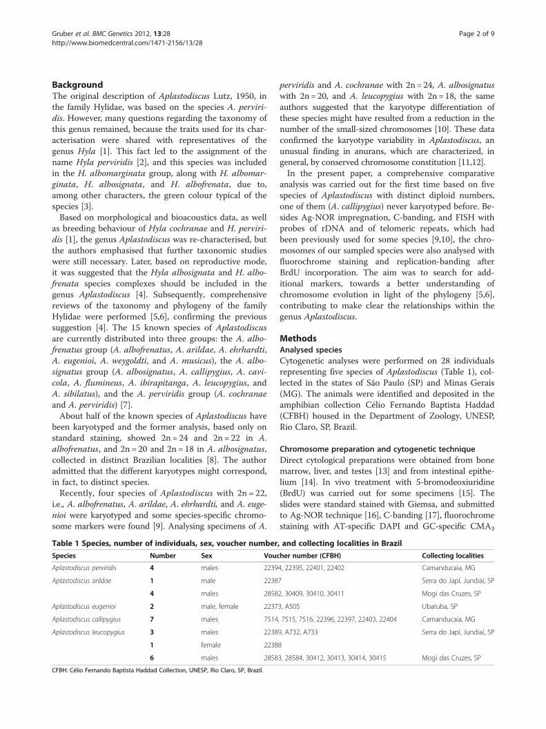
The heterochromatin in all species had a predomin-antly centromeric distribution, with additional labellingat the NOR site (Figure 3), but in some metaphases thisC-band was very slight or not well visualised as in theFigure 3C for A. eugenioi. Fluorochrome staining wascarried out in four species, with exception of A. eugenioi.In A. perviridis, bright fluorescence was observed withCMA3 in the NOR and in the centromere of all
ldae; C. A. eugenioi; D. A. callipygius; E. A. leucopygius. Bar = 10 μm.
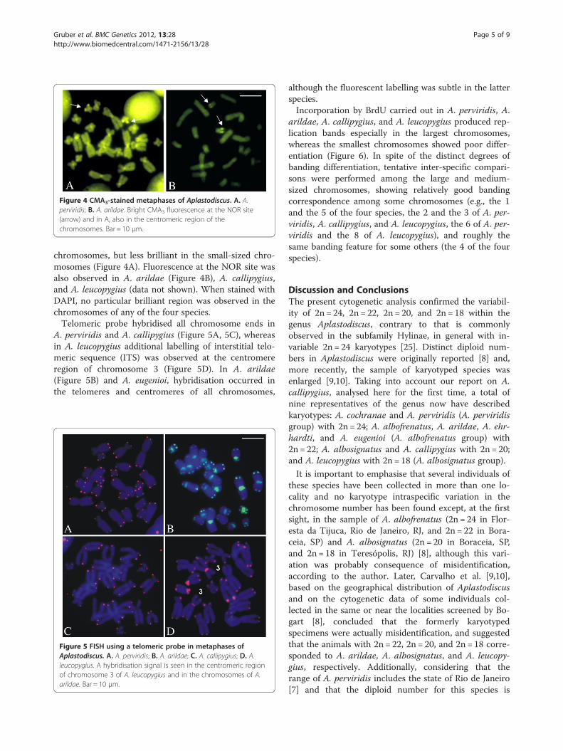
Figure 4 CMA3-stained metaphases of Aplastodiscus. A. A.perviridis; B. A. arildae. Bright CMA3 fluorescence at the NOR site(arrow) and in A, also in the centromeric region of thechromosomes. Bar = 10 μm.
Gruber et al. BMC Genetics 2012, 13:28 Page 5 of 9http://www.biomedcentral.com/1471-2156/13/28
chromosomes, but less brilliant in the small-sized chro-mosomes (Figure 4A). Fluorescence at the NOR site wasalso observed in A. arildae (Figure 4B), A. callipygius,and A. leucopygius (data not shown). When stained withDAPI, no particular brilliant region was observed in thechromosomes of any of the four species.Telomeric probe hybridised all chromosome ends in
A. perviridis and A. callipygius (Figure 5A, 5C), whereasin A. leucopygius additional labelling of interstitial telo-meric sequence (ITS) was observed at the centromereregion of chromosome 3 (Figure 5D). In A. arildae(Figure 5B) and A. eugenioi, hybridisation occurred inthe telomeres and centromeres of all chromosomes,
Figure 5 FISH using a telomeric probe in metaphases ofAplastodiscus. A. A. perviridis; B. A. arildae; C. A. callipygius; D. A.leucopygius. A hybridisation signal is seen in the centromeric regionof chromosome 3 of A. leucopygius and in the chromosomes of A.arildae. Bar = 10 μm.
although the fluorescent labelling was subtle in the latterspecies.Incorporation by BrdU carried out in A. perviridis, A.
arildae, A. callipygius, and A. leucopygius produced rep-lication bands especially in the largest chromosomes,whereas the smallest chromosomes showed poor differ-entiation (Figure 6). In spite of the distinct degrees ofbanding differentiation, tentative inter-specific compari-sons were performed among the large and medium-sized chromosomes, showing relatively good bandingcorrespondence among some chromosomes (e.g., the 1and the 5 of the four species, the 2 and the 3 of A. per-viridis, A. callipygius, and A. leucopygius, the 6 of A. per-viridis and the 8 of A. leucopygius), and roughly thesame banding feature for some others (the 4 of the fourspecies).
Discussion and ConclusionsThe present cytogenetic analysis confirmed the variabil-ity of 2n = 24, 2n = 22, 2n = 20, and 2n = 18 within thegenus Aplastodiscus, contrary to that is commonlyobserved in the subfamily Hylinae, in general with in-variable 2n = 24 karyotypes [25]. Distinct diploid num-bers in Aplastodiscus were originally reported [8] and,more recently, the sample of karyotyped species wasenlarged [9,10]. Taking into account our report on A.callipygius, analysed here for the first time, a total ofnine representatives of the genus now have describedkaryotypes: A. cochranae and A. perviridis (A. perviridisgroup) with 2n = 24; A. albofrenatus, A. arildae, A. ehr-hardti, and A. eugenioi (A. albofrenatus group) with2n = 22; A. albosignatus and A. callipygius with 2n = 20;and A. leucopygius with 2n = 18 (A. albosignatus group).
It is important to emphasise that several individuals ofthese species have been collected in more than one lo-cality and no karyotype intraspecific variation in thechromosome number has been found except, at the firstsight, in the sample of A. albofrenatus (2n = 24 in Flor-esta da Tijuca, Rio de Janeiro, RJ, and 2n = 22 in Bora-ceia, SP) and A. albosignatus (2n = 20 in Boraceia, SP,and 2n = 18 in Teresópolis, RJ) [8], although this vari-ation was probably consequence of misidentification,according to the author. Later, Carvalho et al. [9,10],based on the geographical distribution of Aplastodiscusand on the cytogenetic data of some individuals col-lected in the same or near the localities screened by Bo-gart [8], concluded that the formerly karyotypedspecimens were actually misidentification, and suggestedthat the animals with 2n = 22, 2n = 20, and 2n = 18 corre-sponded to A. arildae, A. albosignatus, and A. leucopy-gius, respectively. Additionally, considering that therange of A. perviridis includes the state of Rio de Janeiro[7] and that the diploid number for this species is
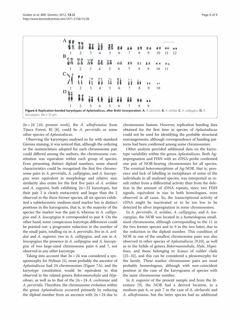
Figure 6 Replication-banded karyotypes of Aplastodiscus, after BrdU incorporation. A. A. perviridis; B. A. arildae; C. A. callipygius; D. A.leucopygius. Bar = 10 μm.
Gruber et al. BMC Genetics 2012, 13:28 Page 6 of 9http://www.biomedcentral.com/1471-2156/13/28
2n = 24 [10, present work], the A. albofrenatus fromTijuca Forest, RJ [8], could be A. perviridis or someother species of Aplastodiscus.Observing the karyotypes analysed so far with standard
Giemsa staining, it was noticed that, although the orderingor the nomenclature adopted for each chromosome paircould differed among the authors, the chromosome con-stitution was equivalent within each group of species.Even presenting distinct diploid numbers, some sharedcharacteristics could be recognized: the first five chromo-some pairs in A. perviridis, A. callipygius, and A. leucopy-gius were equivalent in morphology and relative size;similarity also exists for the first five pairs of A. arildaeand A. eugenioi, both exhibiting 2n= 22 karyotypes, buttheir pair 2 is clearly metacentric and larger than the 2observed in the three former species; all six species exhib-ited a subtelocentric medium-sized marker but in distinctpositions in the karyograms, that is, in the majority of thespecies the marker was the pair 6, whereas in A. callipy-gius and A. leucopygius it corresponded to pair 8. On theother hand, some conspicuous karyotype differences couldbe pointed out: a progressive reduction in the number ofthe small pairs, totalling six in A. perviridis, five in A. aril-dae and A. eugenioi, two in A. callipygius, and one in A.leucopygius; the presence in A. callipygius and A. leucopy-gius of two large-sized chromosome pairs 6 and 7, notobserved in any other karyotype.Taking into account that 2n= 24 was considered a syn-
apomorphy for Hylinae [5], most probably the ancestor ofAplastodiscus had 24 chromosomes (Figure 7A), and thekaryotype constitution would be equivalent to thatobserved in the related genera Bokermannohyla and Hyp-siboas, as well as to that of the 2n= 24 A. cochranae andA. perviridis. Therefore, the chromosome evolution withinthe genus Aplastodiscus occurred primarily by reducingthe diploid number from an ancestor with 2n= 24 due to
chromosome fusions. However, replication banding dataobtained for the first time in species of Aplastodiscuscould not be used for identifying the probable structuralrearrangements, although correspondence of banding pat-terns had been confirmed among some chromosomes.Other analysis provided additional data on the karyo-
type variability within the genus Aplastodiscus. Both Ag-impregnation and FISH with an rDNA probe confirmedone pair of NOR-bearing chromosomes for all species.The eventual heteromorphism of Ag-NOR, that is, pres-ence and lack of labelling in metaphases of some of theindividuals in all analysed species, was interpreted as re-sult rather from a differential activity than from the dele-tion in the amount of rDNA repeats, since two FISHsignals, equivalent in size in both homologues, wereobserved in all cases. So, the transcriptional activity ofrDNA might be inactivated or to be too low to bedetected by silver impregnation in some chromosomes.In A. perviridis, A. arildae, A. callipygius, and A. leu-
copygius, the NOR was located in a homeologous small-sized chromosome, although corresponding to the 11 inthe two former species and to 9 in the two latter, due tothe reduction in the diploid number. This condition ofNOR in one of the smallest chromosome pairs was alsoobserved in other species of Aplastodiscus [9,10], as wellas in the hylids of genera Bokermannohyla, Hyla, Hypsi-boas, and those belonging to Scinax of rubber clade[25–32], and this can be considered a plesiomorphy forthe family. These marker chromosome pairs are mostprobably homeologous, although with non-coincidentposition in the case of the karyograms of species withthe same chromosome number.In A. eugenioi of the present sample and from the lit-
erature [9], the NOR had a derived location, in amedium-pair 6, or pair 7 in the case of A. ehrhardti andA. albofrenatus, but the latter species had an additional
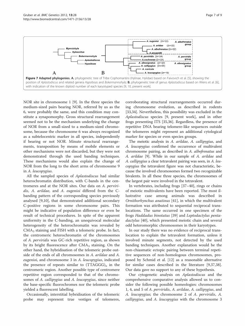
Figure 7 Adapted phylogenies. A. phylogenetic tree of Tribe Cophomantini (Hylinae, Hylidae) based on Faivovich et al. [5], showing theposition of Aplastodiscus and related genera Hypsiboas and Bokermannohyla; B. phylogenetic tree of genus Aplastodiscus based on Wiens et al. [6],with indication of the known diploid number of each karyotyped species [9, 10, present work].
Gruber et al. BMC Genetics 2012, 13:28 Page 7 of 9http://www.biomedcentral.com/1471-2156/13/28
NOR site in chromosome 1 [9]. In the three species themedium-sized pairs bearing NOR, referred by us as the6, were probably the same, and this condition may con-stitute a synapomorphy. Gross structural rearrangementseemed not to be the mechanism underlying the changeof NOR from a small-sized to a medium-sized chromo-some, because the chromosome 6 was always recognizedas a subtelocentric marker in all species, independentlyif bearing or not NOR. Minute structural rearrange-ments, transposition by means of mobile elements orother mechanisms were not discarded, but they were notdemonstrated through the used banding techniques.These mechanisms would also explain the change ofNOR from the long to the short arms of chromosome 9in A. leucopygius.All the sampled species of Aplastodiscus had similar
heterochromatin distribution, with C-bands in the cen-tromeres and at the NOR sites. Our data on A. perviri-dis, A. arildae, and A. eugenioi differed from the C-banding pattern of the corresponding species previouslyanalysed [9,10], that demonstrated additional secondaryC-positive regions in some chromosome pairs. Thismight be indicative of population difference or even beresult of technical procedures. In spite of the apparentuniformity in the C-banding, an unequivocal molecularheterogeneity of the heterochromatin was revealed byCMA3 staining and FISH with a telomeric probe. In fact,the centromeric heterochromatin of the chromosomesof A. perviridis was GC-rich repetitive region, as shownby its bright fluorescence after CMA3 staining. On theother hand, the hybridisation of the telomeric probe out-side of the ends of all chromosomes in A. arildae and A.eugenioi, and chromosome 3 in A. leucopygius, indicatedthe presence of repeats similar to (TTAGGG)n in thecentromeric region. Another possible type of centromererepetitive region corresponded to that of the chromo-somes of A. callipygius and A. leucopygius, since neitherthe base-specific fluorochromes nor the telomeric probeyielded a fluorescent labelling.Occasionally, interstitial hybridisation of the telomeric
probe may represent true vestiges of telomeres,
corroborating structural rearrangements occurred dur-ing chromosome evolution, as described in rodents[33,34]. Nevertheless, this possibility was excluded in theAplastodiscus species [9, present work], and in otherfrogs presenting ITS [35,36]. Regardless, the presence ofrepetitive DNA bearing telomere-like sequences outsidethe telomeres might represent an additional cytologicalmarker for species or even species groups.The meiotic analysis in A. arildae, A. callipygius, and
A. leucopygius confirmed the occurrence of multivalentchromosome pairing, as described in A. albofrenatus andA. arildae [9]. While in our sample of A. arildae andA. callipygius a clear tetravalent pairing was seen, in A. leu-copygius the tetravalent figure was not characteristic, be-cause the involved chromosomes formed two recognizablebivalents. In all these three species, the chromosomes ofthe largest pair were involved in the tetravalent.In vertebrates, including frogs [37–40], rings or chains
of meiotic multivalents have been reported. The most il-lustrative case among animals was described inOrnithorhynchus anatinus [41], in which the multivalentformation was attributed to sequential reciprocal trans-locations. The same occurred in one specimen of thefrogs Haddadus binotatus [39] and Leptodactylus penta-dactylus [40], which presented meiotic chain and severalodd heteromorphic chromosomes in their karyotypes.In our study there was no evidence of reciprocal trans-
location to explain the tetravalent formation, unless itinvolved minute segments, not detected by the usedbanding techniques. Another explanation would be thenon-chiasmatic ectopic pairing between terminal repeti-tive sequences of non-homologous chromosomes, pro-posed by Schmid et al. [12] as a reasonable alternativefor similar cases described in the literature [9,37,38].Our data gave no support to any of these hypothesis.Our cytogenetic analysis on Aplastodiscus and the
comprehensive comparative analysis allowed us to con-sider the following possible homeologies: chromosomes1, 4, and 5 of A. perviridis, A. arildae, A. callipygius, andA. leucopygius; the chromosome 2 of A. perviridis, A.callipygius, and A. leucopygius with the chromosome 3

Gruber et al. BMC Genetics 2012, 13:28 Page 8 of 9http://www.biomedcentral.com/1471-2156/13/28
of A. arildae and A. eugenioi; the chromosomes 3 of A.perviridis, A. callipygius, and A. leucopygius; the chro-mosomes 6 and 11 of A. perviridis, A. arildae, and A.eugenioi with the chromosomes 8 and 9, respectively, ofA. callipygius and A. leucopygius; the chromosomes 7, 8,9, and 10 of A. perviridis, A. arildae, and A. eugenioi;and the chromosome 12 of A. perviridis with thechromosome 10 of A. callipygius. The correspondingchromosome 2 of A. arildae and A. eugenioi, and thechromosomes 6 and 7 of A. callipygius and A. leucopy-gius were interpreted as resulted of rearrangement.Based on these presumed data, the chromosome evolu-tion in the genus Aplastodiscus from an ancestor with2n = 24 was outlined. Nevertheless, two evolutionarypathways were proposed: one involving two fusionsevents, in which participate the small elements 7, 8, 9,and 10, giving rise to two new large-sized pairs 6 and 7,as in the karyotype with 2n = 20 of A. callipygius andwith 2n = 18 of A. leucopygius; and the other, fusion in-volving the small chromosome 12 and the large chromo-some 3, giving rise to the metacentric pair 2, as in thekaryotypes with 2n = 22 of A. arildae and A. eugenioi.This hypothesis is supported by our present cytogeneticdata, but undoubtedly, other resolute approaches (e.g.,chromosome painting, gene linkage, among others) arestill necessary in order to confirm the chromosome evo-lution within the genus Aplastodiscus.Another achievement of the present study was the
confirmation, by means of chromosome analysis, of therelationships among species or species groups of Aplas-todiscus, as shown in the adapted phylogenetic treebased in Wiens et al. [6], and shown in Figure 7B. In-cluding the known diploid numbers of all karyotypedspecies, the two pathways in the chromosome evolutionwere well visualised, and the cytogenetic data gave sup-port to the molecular phylogeny and distribution of thespecies in the known groupings. Certainly, further spe-cies sampling, especially of those that have never beenkaryotyped, will be of great interest to confirm or notthe relationships within the genus Aplastodiscus.
Abbreviations2n: diploid number; Ag-NOR: nucleolar organiser region marked by silverimpregnation; BrdU: 5-bromodeoxiuridine; CMA3: Chromomycin A3; DAPI: 40-6-diamidino-2-phenylindole; FISH: Fluorescent in situ hybridization;FPG: Fluorochrome Plus Giemsa; FN: Fundamental number; ITS: Interstitialtelomeric sequence; NOR: Nucleolar organiser region; rDNA: Ribosomal DNA.
Competing interestsNon-financial competing interests.
Authors' contributionsSLG performed the cytogenetic studies and drafted the manuscript. JZcollected some animals and helped in the review of the manuscript. HNcollected some animals and helped with identification. CFBH providedsupport on zoological information, carried out the species identification, andrevised the manuscript. SK supervised the cytogenetic studies, participated in
the draft, and in the revision of the final text. All authors read and approvedthe final manuscript.
AcknowledgmentsThe authors are grateful to Fundação de Amparo à Pesquisa do Estado deSão Paulo (FAPESP) and Conselho Nacional de Desenvolvimento Científico eTecnológico (CNPq) for financial support. They also thank to InstitutoBrasileiro do Meio Ambiente e dos Recursos Naturais Renováveis (IBAMA) forproviding the collection license to JZ, HN, and CFBH.
Author details1UNESP, Universidade Estadual Paulista, Instituto de Biociências,Departamento de Biologia, Av. 24A, 1515, 13506-900, Rio Claro, SP, Brazil.2Universidade Estadual do Sudoeste da Bahia, Departamento de CiênciasBiológicas, Rua José Moreira Sobrinho, s/n, 45206-000, Jequié, BA, Brazil.3UNESP, Universidade Estadual Paulista, Instituto de Biociências,Departamento de Zoologia, Av. 24A, 1515, 13506-900, Rio Claro, SP, Brazil.
Received: 10 January 2012 Accepted: 15 April 2012Published: 20 April 2012
References1. Garcia PCA, Caramaschi U, Kwet A: O status taxonômico de Hyla
cochranae Mertens e a recaracterização de Aplastodiscus A. Lutz (Anura,Hylidae). Revista Brasileira de Zoologia 2001, 18:1197–1218.
2. Caramaschi U: Aplastodiscus A. Lutz, 1950, um sinônimo júnior de HylaLaurenti, 1768 (Amphibia, Anura, Hylidae) [abstract]. Congresso Brasileirode Zoologia 1983, 10:307.
3. Cruz CAG, Peixoto AL: Espécies verdes de Hyla: o complexo “albofrenata”(Amphibia, Anura, Hylidae). Arquivos da Universidade Federal Rural do Riode Janeiro 1985, 8:59–70.
4. Haddad CFB, Faivovich J, Garcia PCA: The specialized reproductive modeof the treefrog Aplastodiscus perviridis (Anura: Hylidae). Amphib-reptil2005, 26:87–92.
5. Faivovich J, Haddad CFB, Garcia PCA, Frost DR, Campbell JA, Wheeler WC:Systematic review of the frog family Hylidae, with special reference toHylinae: phylogenetic analysis and taxonomic revision. Bull Am Mus NatHis 2005, 294:1–240.
6. Wiens JJ, Kuczynski CA, Hua X, Moen D: An expanded phylogeny oftreefrogs (Hylidae) based on nuclear and mitochondrial sequence data.Mol Phylogenet Evol 2010, 55:871–882.
7. Frost DR: Amphibians of the world: an on-line reference. V5.5. [http://research.amnh.org/herpetology/amphibia/index.html]
8. Bogart JP: Evolution of anuran karyotypes. In Evolutionary Biology ofAnurans. Edited by Vial JL. Columbia: University of Missouri Press;1973:337–349.
9. Carvalho KA, Garcia PCA, Recco-Pimentel SM: NOR dispersion, telomericsequence detection in centromeric regions and meiotic multivalentconfigurations in species of the Aplastodiscus albofrenatus group (Anura,Hylidae). Cytogenet Genome Res 2009, 126:359–367.
10. Carvalho KA, Garcia PCA, Recco-Pimentel SM: Cytogenetic comparison oftree frogs of the genus Aplastodiscus and the Hypsiboas faber group(Anura, Hylidae). Genet Mol Res 2009, 8:1498–1508.
11. Schmid M, Steinlein C, Nanda I, Epplen JT: Chromosome banding inAmphibia. In Cytogenetics of Amphibians and Reptiles. Edited by Olmo E.Basel: Birkhauser Verlag; 1990:21–45.
12. Schmid M, Steinlein C, Bogart JP, Feichtinger W, León P, La Marca E, DiazLM, Sans A, Chen S-H, Hedges SB: The chromosomes of Terraranan frogs:insights into vertebrate cytogenetics. Cytogenet Genome Res 2010, 130–131:1–568.
13. Baldissera FA Jr, Oliveira PSL, Kasahara S: Cytogenetics of four BrazilianHyla species (Amphibia-Anura) and description of a case with asupernumerary chromosome. Rev Bras Genet 1993, 16:335–345.
14. Schmid M: Chromosome banding in Amphibia I. Constitutiveheterochromatin and nucleolus organizers regions in Bufo and Hyla.Chromosoma 1978, 66:361–388.
15. Silva APZ, Haddad CFB, Kasahara S: Chromosomal studies on five speciesof the genus Leptodactylus Fitzinger, 1826 (Amphibia, Anura) usingdifferential staining. Cytobios 2000, 103:25–38.
Gruber et al. BMC Genetics 2012, 13:28 Page 9 of 9http://www.biomedcentral.com/1471-2156/13/28
16. Howell WM, Black DA: Controlled silver-staining of nucleolus organizerregions with a protective colloidal developer: 1-step method. Experientia1980, 36:1014–1015.
17. Sumner AT: A simple technique for demonstrating centromericheterochromatin. Exp Cell Res 1972, 75:304–306.
18. Christian A, McNiel E, Robinson J, Drabek J, LaRue C, Wadren C, Bedford JA:A versatile image analysis approach for simultaneous chromosomeidentification and localization of FISH probes. Cytogenet Cell Genet 1998,82:172–179.
19. Dutrillaux B, Couturier J: La Pratique de l´Analyse Chromosomique. Paris:Masson; 1981.
20. Matsuda Y, Chapman VM: Application of fluorescence in situ hybridizationin genome analysis of the mouse. Electrophoresis 1995, 16:261–272.
21. Meunier-Rotival M, Cortadas J, Macaya G: Isolation and organization of calfribosomal DNA. Nucleic Acids Res 1979, 6:2109–2123.
22. Pinkel D, Straume T, Gray JW: Cytogenetic analysis using quantitative,high-sensitivity, fluorescence hybridization. Proc Natl Acad Sci U S A 1986,83:2934–2938.
23. Green DM, Sessions SK: Nomenclature for chromosomes. In AmphibianCytogenetics and Evolution. Edited by Green DM, Sessions SK. San Diego:Academic Press; 1991:431–432.
24. Green DM, Sessions SK: Karyology and Cytogenetics. In Amphibian Biology.Volume 7. Edited by Heatwole H, Tyler M. Chipping Norton: Surrey Beattyand Sons; 2007:2756–2841.
25. Catroli GF, Kasahara S: Cytogenetic data on species of the family Hylidae(Amphibia, Anura): results and perspectives. Publicatio: Ciências Biológicase da Saúde 2009, 15:67–86.
26. Wiley JE: Chromosome banding patterns of treefrogs (Hylidae) of theEastern United States. Herpetologica 1982, 38:507–520.
27. King M, Contreras N, Honeycutt RL: Variation within and betweennucleolar organizer regions in Australian hylid frogs (Anura) shown by18 S+ 28 S in-situ hybridization. Genetica 1990, 80:17–29.
28. Anderson K: Chromosome evolution in Holarctic Hyla treefrogs. InAmphibian Cytogenetics and Evolution. Edited by Green DM, Sessions SK. SanDiego: Academic Press; 1991:299–331.
29. Kasahara S, Silva APZ, Gruber SL, Haddad CFB: Comparative cytogeneticanalysis on four tree frog species (Anura, Hylidae, Hylinae) from Brazil.Cytogenet Genome Res 2003, 103:155–162.
30. Gruber SL, Haddad CFB, Kasahara S: Chromosome banding in threespecies of Hypsiboas (Hylidae, Hylinae), with special reference to a newcase of B-chromosome in anuran frogs and to the reduction of thediploid number of 2n= 24 to 2n = 22 in the genus. Genetica 2007,130:281–291.
31. Cardozo DE, Leme DM, Bortoleto JF, Catroli GF, Baldo D, Faivovich J, KolencF, Silva APZ, Borteiro C, Haddad CFB, Kasahara S: Karyotypic data on 28species of Scinax (Amphibia: Anura: Hylidae): diversity and informativevariation. Copeia 2011, 2:251–263.
32. Catroli GF, Faivovich J, Haddad CFB, Kasahara S: Conserved karyotypes inCophomantini: cytogenetic analysis of 12 species from 3 species groupsof Bokermannohyla (Amphibia: Anura: Hylidae). J Herpetol 2011,45:120–128.
33. Fagundes V, Yonenaga-Yassuda Y: Evolutionary conservation of wholehomeologous chromosome arms in the Akodont rodents Bolomys andAkodon (Muridae, Sigmodontinae): maintenance of interstitial telomericsegments (ITBs) in recent event of centric fusion. Chrom Res 1998, 6:643–648.
34. Ventura K, O’Brien PCM, Yonenaga-Yassuda Y, Ferguson-Smith MA:Chromosome homologies of the highly rearranged karyotypes of fourAkodon species (Rodentia, Cricetidae) resolved by reciprocalchromosome painting: the evolution of the lowest diploid number inrodents. Chrom Res 2009, 17:1063–1078.
35. Meyne J, Baker AJ, Hobart HH, Hsu TC, Ryder OA, Ward OG, Wiley JE,Wurster-Hill DH, Yates TL, Moyziz RK: Distribution of non-telomeric sites ofthe (TTAGGG)n telomeric sequence in vertebrate chromosomes.Chromosoma 1990, 99:3–10.
36. Wiley JE, Meyne J, Little ML, Stout JC: Intersticial hybridization sites of the(TTAGGG)n telomeric sequence on the chromosomes of some NorthAmerican hylid frogs. Cytogenet Cell Genet 1992, 61:55–57.
37. Lourenço LB, Recco-Pimentel SM, Cardoso AJ: Polymorphism of thenucleolus organizer regions (NORs) in Physalaemus petersi (Amphibia,Anura, Leptodactylidae) detected by silver staining and fluorescence insitu hybridization. Chrom Res 1998, 6:621–628.
38. Siqueira S Jr, Ananias F, Recco-Pimentel SM: Cytogenetics of three Brazilianspecies of Eleutherodactylus (Anura, Leptodactylidae) with 22chromosomes and re-analysis of multiple translocations in E. binotatus.Genet Mol Biol 2004, 27:363–372.
39. Campos JRC, Ananias F, Haddad CFB, Kasahara S: Karyotypic similarityamong Barycholos ternetzi and five species of the genusEleutherodactylus from southeastern Brazil (Anura, Brachycephalidae).Micron 2006, 39:151–159.
40. Gazoni T, Gruber SL, Silva APZ, Araújo OGS, Strüssmann C, Haddad CFB,Kasahara S: Comparative cytogenetic analyses of Leptodactylus(Amphibia, Anura, Leptodactylidae), with description of a new karyotypeand a case of multiple sequential translocations [abstract]. ReuniãoBrasileira de Citogenética 2011, 2:22.
41. Grützner F, Rens W, Tsend-Ayush E, El-Mogharbell N, O’Brien PCM, Jones RC,Ferguson-Smith MA, Graves JAM: In the platypus a meiotic chain of tensex chromosomes shares genes with the bird Z and mammal Xchromosomes. Nature 2004, 3021:1–5.
doi:10.1186/1471-2156-13-28Cite this article as: Gruber et al.: Comparative karyotype analysis andchromosome evolution in the genus Aplastodiscus (Cophomantini,Hylinae, Hylidae). BMC Genetics 2012 13:28.
Submit your next manuscript to BioMed Centraland take full advantage of:
• Convenient online submission
• Thorough peer review
• No space constraints or color figure charges
• Immediate publication on acceptance
• Inclusion in PubMed, CAS, Scopus and Google Scholar
• Research which is freely available for redistribution
Submit your manuscript at www.biomedcentral.com/submit