comparative genomics between human and animal associated
TRANSCRIPT

RESEARCH ARTICLE Open Access
Comparative genomics between humanand animal associated subspecies of theMycobacterium avium complex: a basis forpathogenicityVerlaine J. Timms1,3, Karl A. Hassan2, Hazel M. Mitchell1 and Brett A. Neilan1*
Abstract
Background: A human isolate of Mycobacterium avium subsp. paratuberculosis (M. paratuberculosis 43525) wassequenced and compared genomically to other mycobacterial pathogens. M. paratuberculosis 43525 was recentlyisolated from a patient with ulcerative colitis and belongs to the M. avium complex, a group known to infect bothhumans and animals. While M. paratuberculosis is a known pathogen of livestock, there are only 20 human isolatesfrom the last 20 years, therefore we took the opportunity to perform a whole genome comparison between humanand animal mycobacterial pathogens. We also compared virulence determinants such as the mycobactin cluster, PE/PPE genes and mammalian cell entry (mce) operons between MAC subspecies that infect animals and those that infecthumans. M. tuberculosis was also included in these analyses given its predominant role as a human pathogen.
Results: This genome comparison showed the PE/PPE profile of M. paratuberculosis 43525 to be largely the same asother M. paratuberculosis isolates, except that it had one PPE and one PE_PGRS protein that are only present in humanMAC strains and M. tuberculosis. PE/PPE proteins that were unique to M. paratuberculosis 43525, M. avium subsp.hominissuis and a caprine M. paratuberculosis isolate, were also identified. In addition, the mycobactin cluster differedbetween human and animal isolates and a unique mce operon flanked by two mycobactin genes, mbtA and mbtJ, wasidentified in all available M. paratuberculosis genomes.
Conclusions: Despite the whole genome comparison placing M. paratuberculosis 43525 as closely related to bovine M.paratuberculosis, key virulence factors were similar to human mycobacterial pathogens. This study highlights key factorsof mycobacterial pathogenesis in humans and forms the basis for future functional studies.
Keywords: Mycobacterium avium, Mycobacterium paratuberculosis, Genome analysis, Pathogenicity, PE/PPE family,Mycobactin, Mammalian cell entry, Inflammatory bowel disease, Johne’s disease, Host-pathogen interactions
BackgroundM. avium subsp. paratuberculosis (M. paratuberculosis),of the M. avium complex (MAC), is one of the slowestgrowing mycobacteria and like other pathogenic myco-bacteria, is difficult to detect and treat. It is widelyrecognised as the cause of Johne’s disease, a gastrointes-tinal disease of livestock, and is also implicated in hu-man Crohn’s disease [1–3]. The MAC contain
subspecies that infect animals and subspecies pathogenicto humans [4]. The closely related MAC display slightgenomic differences depending on their host and com-parison of these differences has the potential to identifyhost specific pathogenicity factors, leading to improveddiagnosis and treatment.In the current study we compared the genome of a
newly isolated strain of M. paratuberculosis (M. paratu-berculosis 43525) from a female patient with ulcerativecolitis [5], to other pathogenic mycobacteria using Sin-gle Nucleotide Polymorphism (SNP) analysis, BLASTp(homology based) and phmmer (non-homology based)
* Correspondence: [email protected] of Biotechnology and Biomolecular Sciences, University of NewSouth Wales, Sydney 2052, AustraliaFull list of author information is available at the end of the article
© 2015 Timms et al. Open Access This article is distributed under the terms of the Creative Commons Attribution 4.0International License (http://creativecommons.org/licenses/by/4.0/), which permits unrestricted use, distribution, andreproduction in any medium, provided you give appropriate credit to the original author(s) and the source, provide a link tothe Creative Commons license, and indicate if changes were made. The Creative Commons Public Domain Dedication waiver(http://creativecommons.org/publicdomain/zero/1.0/) applies to the data made available in this article, unless otherwise stated.
Timms et al. BMC Genomics (2015) 16:695 DOI 10.1186/s12864-015-1889-2

algorithms. While M. paratuberculosis normally infectsanimals, its isolation from humans is rare, with lessthan 20 isolates reported in the last 20 years [6–9]. Likeother M. paratuberculosis from humans, M. paratuber-culosis 43525 is cattle type (C-Type) [5]. Therefore, fur-ther analysis of this strain provides a uniqueopportunity to explore other possible variations in hostpathogenicity factors.While genomic studies have compared a number of
these isolates to other M. paratuberculosis strains andM. hominissuis [10–15], all sequences of M. paratuber-culosis to date have been obtained from laboratorystrains of unknown subculture number, and often haveundergone many years of laboratory passage. Currentevidence would suggest that multiple subculture of M.tuberculosis may affect virulence properties with, forexample, marked changes in cell wall lipids observedafter extensive laboratory passage [16]. Important tonote is that M. bovis BCG, widely used in vaccines dueto its attenuation in immunocompetent hosts, was pro-duced by multiple subculture in vitro [17]. In contrast,the genome of M. paratuberculosis 43525 was se-quenced after only four subcultures and therefore pro-vides a more accurate representation of the wild-type invivo mycobacterial genome.The virulence factors explored in this study, the PE/
PPE (proline-glutamate/proline-proline-glutamate motif )genes, mammalian cell entry (mce) operons and themycobactin cluster, were chosen based on studies intoM. tuberculosis and M. avium pathogenicity. The ana-lysis of these genomic loci afford the representation ofpathogenicity elements present in M. tuberculosis iso-lated from human infections and M. paratuberculosisisolated from livestock infections [4, 18–22].The PE/PPE families are unique to mycobacteria and
were first identified for their ability to stimulate IFN-γ
[19]. They are GC rich and thought to be the main sourceof strain variability within the MAC [4]. PE and PPE referto the residues, Proline-Glutamate and Proline-Proline-Glutamate, respectively, located at the N termini of theirencoded proteins. The M. tuberculosis genome devotes10 % of its protein coding potential to this protein familywith various functions attributed to them [23]. Similar toM. tuberculosis, some PPE of M. paratuberculosis areexpressed on the cell surface, while others are cell wall as-sociated and interact with the immune system via TLR-2[19], however, this gene family only represents 2.5 % ofthe M. paratuberculosis genome [24].The mammalian cell entry (mce) operons of M. tuber-
culosis were first discovered in studies to elucidate howM. tuberculosis enters non-phagocytic cells [25]. Thegenes exist in many bacterial species, however, only inthe mycobacteria do they exist as operons [26]. Thefunction of these operons is now thought to be diverseand not confined to cell entry, given that they have beenfound in non-pathogenic, environmental mycobacteria[26]. There are four mce operons in M. tuberculosis,each has two yrbE genes and six mce genes, oftencoupled to a mce regulator gene. The genes in each op-eron are contiguous but differentially expressed, depend-ing on growth conditions and/or nutrient supply [21]. Inthe MAC, additional mce operons have been reportedthat do not appear to have orthologues in M. tubercu-losis [26, 18].Mycobactin dependency in vitro is a major phenotypic
difference between M. paratuberculosis and other sub-species of the MAC complex. Mycobactins are sidero-phores that transport or scavenge iron, particularly inenvironments where free iron is limited, such as inside ahost cell [27]. Like other siderophores, mycobactin is asecondary metabolite, a product of non-ribosomal pep-tide synthases (NRPS) and polyketide synthases (PKS)
Fig. 1 Graphical representation of the 6310 orthologous clusters of annotated protein sequences encoded in 27 mycobacterial strains. Comparativeblastp 2.2.22+ searches [61] were conducted using Proteinortho [30] and orthologous clusters were visualised using FriPan (http://www.vicbioinformatics.com/software.fripan.shtml)
Timms et al. BMC Genomics (2015) 16:695 Page 2 of 11

(an integrated NRPS-PKS) [28]. The mycobactin genecluster contains 10 genes (A-J) and the mycobactin op-eron promoter is active in M. paratuberculosis, with allmycobactin genes able to be transcribed inside bovinemacrophages [29]. M. paratuberculosis 43525 has a pe-culiar mycobactin phenotype as it grows on some typesof media, such as Middlebrook 7H10, without theaddition of mycobactin [5]. Given this, comparison ofM. paratuberculosis 43525 with other MAC strains hasthe potential to provide unique genomic informationand the basis for their pathogenicity.
ResultsThe general features of the assembled draft genome ofM. paratuberculosis 43525 are presented in Table 1. Outof a total of 4433 protein coding sequences (CDS), 1517(35 %) belonged to recognised subsystems. Of the 2781non-hypothetical CDS, 1450 belonged to recognisedsubsystems while 1715 CDS were hypothetical and ofthese, 67 belonged to subsystems according to RAST.Comparative blastp searches and clustering analyses exe-
cuted through Proteinortho [30], suggested that 165 puta-tive protein sequences annotated in theM. paratuberculosis43525 genome were unique to this strain (Fig. 1). These pu-tatively unique sequences included a large number of hypo-thetical proteins, as well as PE-PGRS and mce genes thatwill be described below. In addition, differences were ob-served between the genes encoding the mycobactin clusterand this cluster was analysed in more detail.
Single nucleotide polymorphism (SNP) analysisTo better characterise M. paratuberculosis 43525, vari-ation between this bacterium and 27 other mycobacterialstrains (including two M. avium subsp. avium strains)
were compared at the nucleotide level. Of these strains,nine were M. paratuberculosis isolates from humans,one was an ovine isolate and one was a caprine isolate.The SNPs of all strains were concatenated and used forphylogenetic analysis on a genome-wide level with M.paratuberculosis K10 as the reference strain. The rootedtree (Fig. 2) shows M. paratuberculosis 43525 to beclosely related to M. paratuberculosis CLIJ644, a bovineisolate from Victoria, Australia [12].
PE/PPE genesThe nomenclature of the MAC complex PE/PPE geneswas used as previously described [4], and a summary ofthe M. paratuberculosis 43525 genes shared between theMAC and M. tuberculosis is presented in Fig. 3 andAdditional file 1. Thirty seven PPE genes were found inM. paratuberculosis 43525, none of which were uniqueto this strain, while 17 were conserved in all strains ex-amined. In the bovine strain M. paratuberculosis K10,MACPPE15 is a fragmented pseudogene, whereas the fullgene is present in the human isolate M. paratuberculosis43525, and this gene is homologous to Mav2514 from M.avium 104. Although MACPPE41 and MACPPE42 aresaid to be unique to the M. paratuberculosis subspecies,here only MACPPE42 was found in M. paratuberculosis43525 [4].Ten PE genes and one PE fragment were present in M.
paratuberculosis 43525 (Fig. 3, Additional file 1). PE13was the only PE gene that was not conserved in allstrains studied, being found only in M. paratuberculosis43525 and M. avium 104. The genome of M. paratuber-culosis 43525 also had gene PE_PGRS11 which was alsofound in M. paratuberculosis K10, M. tuberculosis andM. avium 104, but absent in M. avium ATCC25291.
MycobactinA total of 17 NRPS/ PKS clusters were identified by anti-SMASH in the M. paratuberculosis 43525 genome. Thecluster identified as the mycobactin cluster was analysedand found to have a different primary structure as com-pared with that of other MAC strains, with respect tothe spacing of genes and gene size (Fig. 4). The myco-bactin cluster of M. paratuberculosis has previously beenshown to have both NRPS and PKS modules [24]. Whilethe mbtA, mbtC, mbtD, mbtG and mbtI genes of the M.paratuberculosis 43525 mycobactin cluster were foundto be identical to the equivalent genes in M. paratuber-culosis K10, the remaining 5 genes (3 of which are NRPSmodules) were found to encode larger proteins.Furthermore, the mbtB gene, shown by others to be
the first gene involved in mycobactin synthesis [31], en-codes a polypeptide of 1420 amino acids in M. paratu-berculosis 43525, which was larger than the mbtB geneproduct of strains such as M. paratuberculosis K10 and
Table 1 Chromosome features of M. paratuberculosis 43525
Parameters M. paratuberculosis 43525
Reference organism M. paratuberculosis K10
Chromosome size(base pairs)
4,812,039
G + C (%) 69.7
Number of contigs 466
Protein-codingsequences (CDS)
4433
CDS belonging to subsystems 1517
Non-hypothetical CDS 2781
Non-hypothetical CDSbelonging to subsytems
1450
Hypothetical CDS 1715
Hypothetical CDS belonging to subsystems 67
Number of Subsystems 372
No. of RNAs 49
Timms et al. BMC Genomics (2015) 16:695 Page 3 of 11

M. avium 104 but similar in size to the equivalent MbtBin M. tuberculosis. The polypeptide size difference be-tween these strains was due to the thioesterase domainof MbtB being encoded on a separate gene in M. paratu-berculosis K10 and other MAC, but not in M. paratuber-culosis 43525 or M. tuberculosis (Fig. 4).The size and organisation of genes encoding MbtE
vary greatly across strains (Fig. 4). The AMP bindingand peptidyl carrier protein (PCP) domains were encodedon the one mbtE gene in M. paratuberculosis 43525, how-ever, in M. paratuberculosis K10 the AMP binding wassplit across MAP2172c and MAP2173c, while the PCPdomain is encoded on MAP2172c. Confirmation byamplicon sequencing demonstrated a 187 nucleotide indel
in M. paratuberculosis 43525 compared to M. paratuber-culosis K10 (bases 2411868 to 2412055). The AMP bind-ing domain is a 215 aa protein in M. paratuberculosis K10as compared with a 394 residue protein in M. paratuber-culosis 43525 and a 408 aa protein in M. avium 104. Anti-smash uses a number of databases to predict the substratefor each NRPS domain. The predicted substrate for MbtEof M. paratuberculosis 43525 is tyrosine. In contrast, therewas no consensus on the predicted substrate for MbtE ofM. paratuberculosis K10 and M. avium 104.The mbtF gene in M. paratuberculosis 43525 encoded
a longer protein than in the M. paratuberculosis K10equivalent. The main difference was in the epimerisationdomain that encodes a shorter polypeptide in M.
Fig. 2 Phylogenetic analysis of M. paratuberculosis 43525 relative to other M. paratuberculosis isolates and two M. avium isolates. Neighbour-joiningphylogenetic tree based on SNPs of M. paratuberculosis isolates from bovine, ovine and human hosts and M. avium isolates derived from human andavian hosts with M. paratuberculosis K10 as the reference. The colour box indicates the clade in which M. paratuberculosis 43525 belongs andalso contains human isolates M. paratuberculosis 4 and 4B. In addition, this clade contains recent (compared to the type strains) bovineisolates, including the bovine isolate CLIJ644. The reference strain M. paratuberculosis K10 is in a different clade to M. paratuberculosis43525 and the type strain ATCC19698
Timms et al. BMC Genomics (2015) 16:695 Page 4 of 11

paratuberculosis K10 (179 aa) as compared with M.paratuberculosis 43525 (299 aa), M. avium 104 (299 aa),M. avium ATCC25291 (299 aa) and M. tuberculosis (288aa).Five copies of mbtH were found in M. paratuberculosis
43525, four of which (mbtH_1, mbtH_2, mbtH_3 andphoP) had 100 % sequence similarity with the equivalentgenes in M. paratuberculosis K10. The mbtH_3 gene wassituated adjacent to the mycobactin cluster. However, thefifth mbtH gene of M. paratuberculosis 43525, adjacent topstA was found to have an 85 % match to mbtH_2 in K10but 100 % sequence similarity with 18 other mbtH likegenes including the D522_08303 gene in another M. para-tuberculosis strain (S5) originally isolated from a goat,MAP4_2610 from M. paratuberculosis MAP4 (a humanisolate), MAH_2060 of M. avium TH135 and geneOCQ_31530 in M. intracellulare (strain MOTT-64).In addition to differences in the size of genes within
the mycobactin gene cluster, there were also differencesin the spacing between the genes and gene clusters. Thegap between mbtA and mbtJ (lipK) was comparable be-tween M. paratuberculosis K10 and M. paratuberculosis43525 and contained a mce operon 8.7 kbp downstreamfrom mbtA. However, the gap between mbtJ (lipK) andmbtI (trpE2) in M. paratuberculosis 43525 was 2 kbshorter compared with the 6.6 kb spacer region in M.paratuberculosis K10 (Fig. 4).
mce genesThe M. paratuberculosis 43525 genome contained eightmce operons that encode 74 mce proteins, although notall are complete. The four best known mce operons identi-fied in M. tuberculosis are labelled 1–4 in Table 2. Otheridentified mce operons include mce 5, 6, and 7. Based onthese findings it is suggested to include the gene designa-tion mce 8, an operon that was originally described as aduplicate of mce 7. However, mce 8 has low nucleotideand amino acid sequence similarity (72 and 63 %, respect-ively), when compared to the existing mce7 in M. paratu-berculosis [26]. Table 2 shows the amino acid sequencesimilarity of the mce genes in M. paratuberculosis 43525compared to equivalent genes in related bacteria of theMAC and M. tuberculosis. Of particular note is that in allM. paratuberculosis isolates included in this study, mce6was found 8.7 kb downstream from mbtA of the mycobac-tin cluster.Four genes of the mce1 operon were longer than the
corresponding genes in M. paratuberculosis K10 andthe same size as the corresponding genes of M. paratu-berculosis MAP4 and other MAC. While the mce2 op-eron was conserved among M. paratuberculosis, M.avium strains 104 and TH135, the mce3R gene ap-peared to be missing from M. paratuberculosis 43525and other M. paratuberculosis strains. As reported by
Fig. 3 A summary of the presence and absence of the MACPE/PPE.The genes were sorted according to their distribution profiles(Additional file 1). Orthologues in M. tuberculosis were also added forcomparison. Blue indicates the gene (listed on the right hand side) ispresent while yellow indicates the gene is missing. The strain orderacross the top is determined by the relative presence/absence ofPE/PPE genes
Timms et al. BMC Genomics (2015) 16:695 Page 5 of 11

others, the mce4a gene was highly conserved across allmycobacteria [21].Of particular note was the finding that the conserved
hypothetical integral membrane protein yrbe3B waspresent in M. paratuberculosis 43525 but missing in M.paratuberculosis K10. Interestingly, yrbe3B has beenfound in a M. paratuberculosis strain (S397) isolatedfrom sheep, M. avium 104, M. avium TH135, M. aviumATCC25291, M. intracellulare and M. tuberculosis.
DiscussionUsing comparative genomics a rare human isolate of M.paratuberculosis was compared to both animal and hu-man pathogens of the MAC and M. tuberculosis. Afterbroad analysis by Blast and SNP typing, this study fo-cused on comparisons of PE/PPE genes, the mycobactincluster and the mce operons, all of which are key viru-lence factors across the species examined.
When compared at the nucleotide level, M. paratuber-culosis 43525 displayed a close relationship to a bovineisolate M. paratuberculosis CLIJ644 (Fig. 2). This re-quires further investigation, particularly as M. paratu-berculosis is shed in the milk of infected cows even atthe early subclinical stage and that M. paratuberculosiscan survive pasteurisation [32].As in prior work, it was found that the complement of
PPE genes was variable across strains while the PEgenes showed a high degree of conservation (Fig. 3 andAdditional file 1) [4]. A possible human associated PPE,MACPPE43 was present in M. paratuberculosis 43525and M. intracellulare, which was orthologous to Rv3621c(PPE65 of M. tuberculosis). In contrast, MACPPE43 wasnot present in any strains of animal origin, including otherM. paratuberculosis isolates. In M. tuberculosis, this genewas not essential for in vitro growth but could be detectedin M. tuberculosis H37Rv infected guinea pig lungs at 30and 90 days post infection suggesting a critical function
Fig. 4 Comparison of the mycobactin cluster between members of the MAC complex and M. tuberculosis. Amino acid length is indicated aboveeach gene, gene names are indicated and colour matched to equivalent genes in other strains (M. paratuberculosis 43525, M. paratuberculosis K10,M. paratuberculosis S397 M. avium 104, M. avium ATCC25291 and M. tuberculosis). M. paratuberculosis K10, M. paratuberculosis S397 and M. aviumATCC25291 are of animal origin, the remaining three are from human hosts. The NRPS modules MbtB and MbtE are one gene in M. paratuberculosis43525 and M. tuberculosis but two genes in other MAC. In addition, all M. paratuberculosis strains had an mce operon (mce6) present in the gapbetween MbtA and MbtJ (indicated for M. paratuberculosis 43525 and K10)
Timms et al. BMC Genomics (2015) 16:695 Page 6 of 11

for this gene product in vivo [33, 34]. PE_PGRS11 wasalso found to be present in strains isolated from humans,as well as M. paratuberculosis K10 although its functiontoo is unknown. Two PE/PPE genes, MACPPE51 and aMav2927 orthologue, were only found in the new humanisolate M. paratuberculosis 43525, M. hominissuis and acaprine isolate of M. paratuberculosis, S5. Given the sig-nificance of the PE/PPE family to virulence, through gen-eration of antigenic variations, the functions of the PE/PPE identified here should be investigated further.MACPPE42 is unique to M. paratuberculosis and is
located on a Large Sequence Polymorphism (LSP)-14[4, 35]. There is some evidence that LSPs are associ-ated with the cellular immune response [36] and it isco-transcribed with the iron-regulated transporters(irt) A and B equivalents MAP3734-3735 in macro-phages [35]. IrtA and B are thought to be involved inthe trafficking of carboxymycobactin, which is se-creted in contrast to cell wall associated mycobactin[37]. As proposed in a recent study, MACPPE42 mayact as a signal transduction protein for the IrtA and Bequivalents which in turn form a single ABC transporterfor Fe-carboxymycobactin and iron assimilation via ferriciron reduction [35]. Structural studies demonstrating thesimilarity of M. tuberculosis PPE proteins to signal trans-duction molecules and the observation that some PE/PPEproteins are up-regulated during iron limitation and re-pressed by the regulator ideR, form the basis of theabove proposal [38, 39]. In addition, the finding that M.
tuberculosis mbtB mutants that are unable to synthesisemycobactin or carboxymycobactin, but have irtAB in-tact, can grow in the presence of exogenous Fe-carboxymycobactin [40], may explain how mycobactindependent strains of M. paratuberculosis survive thehostile environment of the macrophage as well as themycobactin independence of other strains in vitro (aslong as 1 % ferric ammonium citrate is added) [41, 42].An attenuated strain of M. paratuberculosis (strain316FNOR1960) has lost two of the irtA and B ortholo-gues (MAP3734c and MAP3735c) as part of the LargeVariable Genomic Island-19 deletion [43]. This strainwas used in early vaccine preparations and was exten-sively subcultured before attenuation on Dubos mediawith pyruvate [44]. M. paratuberculosis is usually main-tained on media containing ferric ammonium citraterather than pyruvate, as the two are antagonistic oncemycobactin is added [45]. MACPPE42 was not part ofthe vGI-19 deletion in the attenuated strain.M. paratuberculosis 43525 did not require additional
mycobactin on Middlebrook agar, a phenotype that hasbeen described before in M. paratuberculosis isolatesfrom sheep [46]. Therefore the mycobactin cluster requiredcloser scrutiny and was found to differ in its primary struc-ture when compared to M. paratuberculosis K10.The three NRPS domains of the mycobactin cluster
were larger in M. paratuberculosis 43525 as comparedto M. paratuberculosis K10 and encode larger pro-teins. The size of the mbtE gene varies greatly across
Table 2 Percentage amino acid sequence similarity of mce operons in 43525 compared to M. paratuberculosis K10 (MAP K10),M. avium 104, M. avium TH135, M. intracellulare and M. tuberculosis
MAPK10
Mav104
MavTH135
MavATCC25291
M.intracellulare
M.tuberculosis
Remarks
Operon
mce1 99–100 77–100 99–100 81–100 92–99 76–93 3 genes in 43525 are longer, fadD5 (453aalonger than MAP3601), yrbE1B (40aa longer thanMAP3603), mce1E (233aa longer than MAP3608 andMavATCC25291_4409
mce2 99–100 97–99 99–100 97–100a 62–78 72–91 Mce2E missing in M. avium ATCC25291
mce3 99–100a 60–100 99 98–99 84–96 50–62 yrbE3B missing in MAPK10
mce4 100 99–100a 99–100 99–100 93–99 81–95 Frameshift in M. avium 104 Mce4F
mce5 99–100a 99–100a 65–99a 90–99 85–99 – Deletion at position 7862892 of MAPK10 resultsin truncation of MAP0762 and M. avium ATCC25291_0785 proteins by 241aa and 70aa respectively. Frameshiftin Mav0951.
mce6 99–100 – – – 88–92 – Another human MAP strain, MAP4 had 100 % a.a.homology to all genes of 43525 in this operon.
mce7 99–100 98–100 98–99 98–100 88–97 – This operon also found in M. marinum (>69 %homology) and M. abscessus.
mce8 97–100 96–100 96–100 95–99 80–94 – Frameshift in MAP K10 (MAP0116). Truncated proteinin MavATCC25291_0099. 43525 is 100 % identical tosheep MAP strains S397 and S5 for all genes in thisoperon.
Each value is the range across each operon. No value indicates that the operon is missing in that species/strain. aindicates one or two genes are missing in thatoperon relative to isolate 43525 (see remarks)
Timms et al. BMC Genomics (2015) 16:695 Page 7 of 11
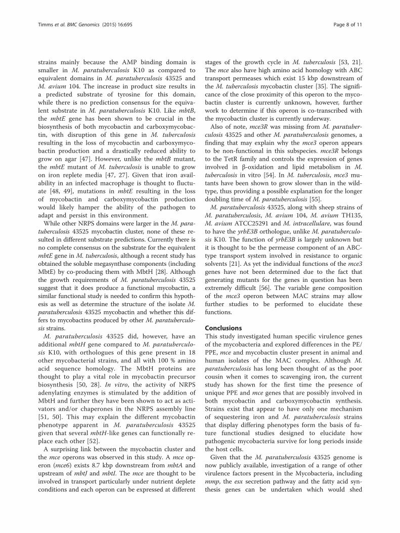
strains mainly because the AMP binding domain issmaller in M. paratuberculosis K10 as compared toequivalent domains in M. paratuberculosis 43525 andM. avium 104. The increase in product size results ina predicted substrate of tyrosine for this domain,while there is no prediction consensus for the equiva-lent substrate in M. paratuberculosis K10. Like mbtB,the mbtE gene has been shown to be crucial in thebiosynthesis of both mycobactin and carboxymycobac-tin, with disruption of this gene in M. tuberculosisresulting in the loss of mycobactin and carboxymyco-bactin production and a drastically reduced ability togrow on agar [47]. However, unlike the mbtB mutant,the mbtE mutant of M. tuberculosis is unable to growon iron replete media [47, 27]. Given that iron avail-ability in an infected macrophage is thought to fluctu-ate [48, 49], mutations in mbtE resulting in the lossof mycobactin and carboxymycobactin productionwould likely hamper the ability of the pathogen toadapt and persist in this environment.While other NRPS domains were larger in the M. para-
tuberculosis 43525 mycobactin cluster, none of these re-sulted in different substrate predictions. Currently there isno complete consensus on the substrate for the equivalentmbtE gene in M. tuberculosis, although a recent study hasobtained the soluble megasynthase components (includingMbtE) by co-producing them with MbtH [28]. Althoughthe growth requirements of M. paratuberculosis 43525suggest that it does produce a functional mycobactin, asimilar functional study is needed to confirm this hypoth-esis as well as determine the structure of the isolate M.paratuberculosis 43525 mycobactin and whether this dif-fers to mycobactins produced by other M. paratuberculo-sis strains.M. paratuberculosis 43525 did, however, have an
additional mbtH gene compared to M. paratuberculo-sis K10, with orthologues of this gene present in 18other mycobacterial strains, and all with 100 % aminoacid sequence homology. The MbtH proteins arethought to play a vital role in mycobactin precursorbiosynthesis [50, 28]. In vitro, the activity of NRPSadenylating enzymes is stimulated by the addition ofMbtH and further they have been shown to act as acti-vators and/or chaperones in the NRPS assembly line[51, 50]. This may explain the different mycobactinphenotype apparent in M. paratuberculosis 43525given that several mbtH-like genes can functionally re-place each other [52].A surprising link between the mycobactin cluster and
the mce operons was observed in this study. A mce op-eron (mce6) exists 8.7 kbp downstream from mbtA andupstream of mbtJ and mbtI. The mce are thought to beinvolved in transport particularly under nutrient depleteconditions and each operon can be expressed at different
stages of the growth cycle in M. tuberculosis [53, 21].The mce also have high amino acid homology with ABCtransport permeases which exist 15 kbp downstream ofthe M. tuberculosis mycobactin cluster [35]. The signifi-cance of the close proximity of this operon to the myco-bactin cluster is currently unknown, however, furtherwork to determine if this operon is co-transcribed withthe mycobactin cluster is currently underway.Also of note, mce3R was missing from M. paratuber-
culosis 43525 and other M. paratuberculosis genomes, afinding that may explain why the mce3 operon appearsto be non-functional in this subspecies. mce3R belongsto the TetR family and controls the expression of genesinvolved in β-oxidation and lipid metabolism in M.tuberculosis in vitro [54]. In M. tuberculosis, mce3 mu-tants have been shown to grow slower than in the wild-type, thus providing a possible explanation for the longerdoubling time of M. paratuberculosis [55].M. paratuberculosis 43525, along with sheep strains of
M. paratuberculosis, M. avium 104, M. avium TH135,M. avium ATCC25291 and M. intracellulare, was foundto have the yrbE3B orthologue, unlike M. paratuberculo-sis K10. The function of yrbE3B is largely unknown butit is thought to be the permease component of an ABC-type transport system involved in resistance to organicsolvents [21]. As yet the individual functions of the mce3genes have not been determined due to the fact thatgenerating mutants for the genes in question has beenextremely difficult [56]. The variable gene compositionof the mce3 operon between MAC strains may allowfurther studies to be performed to elucidate thesefunctions.
ConclusionsThis study investigated human specific virulence genesof the mycobacteria and explored differences in the PE/PPE, mce and mycobactin cluster present in animal andhuman isolates of the MAC complex. Although M.paratuberculosis has long been thought of as the poorcousin when it comes to scavenging iron, the currentstudy has shown for the first time the presence ofunique PPE and mce genes that are possibly involved inboth mycobactin and carboxymycobactin synthesis.Strains exist that appear to have only one mechanismof sequestering iron and M. paratuberculosis strainsthat display differing phenotypes form the basis of fu-ture functional studies designed to elucidate howpathogenic mycobacteria survive for long periods insidethe host cells.Given that the M. paratuberculosis 43525 genome is
now publicly available, investigation of a range of othervirulence factors present in the Mycobacteria, includingmmp, the esx secretion pathway and the fatty acid syn-thesis genes can be undertaken which would shed
Timms et al. BMC Genomics (2015) 16:695 Page 8 of 11
further light on the ability of specific mycobacterialstrains to colonise and cause disease in different tissuesof different hosts.
MethodsBacterial growth and genome sequencingM. paratuberculosis 43525, isolated from a female withulcerative colitis in 2009 [5], was grown on a slope ofMiddlebrook 7H10 agar supplemented with 10 % oleicacid-albumin-dextrose-catalase (OADC) (Difco) and2 μg/mL mycobactin J (Allied Monitor) for 3 months.DNA was extracted as previously reported and the con-centration and quality of DNA was measured using aNanodrop ND-1000 spectrophotometer (Nanodrop Tech-nologies) [57]. The genome of M. paratuberculosis 43525was sequenced, using an Illumina HiSeq sequencer withthe TruSeq SBS v4 GA kit. Paired-end indexed librarieswere prepared from purified DNA fragments of approxi-mately 320 bp in length generating raw reads of 100 bp inlength. Sequencing was performed at the RamaciottiCentre for Gene Function Analysis, University of NewSouth Wales (Sydney, Australia). The sequence reads weresubmitted to the Sequence Read Database (http://www.ncbi.nlm.nih.gov/sra) and the SRA study accessionfor the M. paratuberculosis 43525 genome sequence isSRP033522.
Genome assemblyRead quality was controlled by FASTQC (BabrahamBioinformatics) (http://www.bioinformatics.bbsrc.ac.uk/projects/fastqc) using default values. Raw reads werefiltered for quality (mean phred > 20) and trimmed10 bp on each end using custom Perl scripts, redu-cing each read to 80 bp. Paired-reads were then usedto estimate the genome size using the programkhmerfreq (kmer = 17). The trimmed reads were thenassembled using Velvet 1.0.09 [58] and SoapDenovo[59] with a range of kmer lengths (57–64) the finalassembly being based on assembly size, number ofcontigs and contig size compared to M. paratubercu-losis K10 (Accession number AE016958).
Genome analysisAnnotation of the M. paratuberculosis 43525 genome wasperformed using the Rapid Annotation and SubsystemTechnology (RAST) web application server [60].Proteinortho was used to conduct comparative blastp
searches and clustering analyses [30, 61], which were visua-lised using Fripan (http://www.vicbioinformatics.com/software.fripan.shtml). Single Nucleotide Polymorphisms(SNPs) were called and used to infer phylogeny using theprogram CSI Phylogeny 1.0a (https://cge.cbs.dtu.dk/services/CSIPhylogeny/). The parameters for SNP calling were:Minimum depth 10x, minimum relative depth > 10 %,
minimum distance between SNPs > 10 bp, minimum SNPquality = 30, read mapping quality score > 25 and mini-mum Z score 1.96. The phylogenetic tree was importedto FigTree (http://tree.bio.ed.ac.uk/software/figtree/) forvisualisation.Probable orthologues in M. paratuberculosis 43525 for
PE, PPE and mce genes were defined using both theBLASTp algorthrim and Hmmer3 (http://hmmer.jane-lia.org/) [62]. Orthologues with > 70 % amino acid identityand over 50 % of the sequence length compared to publicsequences of the MAC complex and M. tuberculosis wereconsidered. Protein databases such as the PFAM databasewere also used for comparative purposes [63].In order to investigate the mycobactin cluster of M.
paratuberculosis 43525 the annotated genome wasuploaded to Version 2.0 of the antiSMASH (Antibioticsand Secondary Metabolite Analysis SHell) program [64].The antiSMASH algorithm identifies backbone enzymes,usually polyketide synthase (PKS), nonribosomal synthe-tase (NRPS), hybrid PKS-NRPS, or NRPS-like enzymes.Adjacent genes are scanned for the presence of commonsecondary metabolite gene domains and boundaries arepredicted for each cluster. The clusters were then manu-ally analysed and synteny of the mycobactin cluster wasvisually evaluated by examining whether a gene had ortho-logues in other mycobacterial species.Given that the mbtE gene of M. paratuberculosis
43525 was found to be different to other mycobacter-ial species. PCR primers mbtE fwd (5′ gttacttccccgtcgatccc) and mbtE rev (5′ gtagtagagctcccccacca) weredesigned to amplify the region of mbtE that differedfrom the equivalent gene in M. paratuberculosis K10.Automated sequencing to identify PCR products wascarried out using the PRISM BigDye™ cycle sequen-cing system v3.1 and ABI 3730 capillary sequencer(Applied Biosystems).The mce and mycobactin cluster genes were com-
pared across MAC and M. tuberculosis with emphasison members of the MAC complex that infect animals;M. paratuberculosis K10 (bovine), M. avium ATCC25291 (avian), M. paratuberculosis S397 (sheep) andthose that infect humans; M. paratuberculosis 43525,M. avium 104, M. avium TH135. The PE/PPE geneswere compared to defined PE/PPE genes from com-pleted genomes only.
Ethics statementEthics approval was not required for this study. All ex-periments were conducted according to the regulationsof the University of New South Wales.
Availability of supporting dataAll supporting data for this article are included asadditional files.
Timms et al. BMC Genomics (2015) 16:695 Page 9 of 11
Additional file
Additional file 1: The amino acid percent homology between the PEand PPE genes of M. paratuberculosis 43525 compared to other MACspecies. (XLSX 15 kb)
Competing interestsThe authors declare that they have no competing interests.
Authors’ contributionsVJT: conceived and planning of experiments, data collection, analysis andmanuscript preparation, KH: data analysis and manuscript preparation, HMM:supervision and planning of experiments, manuscript preparation, BAN:conceived, supervision and planning of experiments, manuscript preparation.All authors read and approved the final manuscript.
AcknowledgmentsThe authors thank Rati Sinha for assistance with genome assembly. This workwas funded by the Australian Research Council.
Author details1School of Biotechnology and Biomolecular Sciences, University of NewSouth Wales, Sydney 2052, Australia. 2Department of Chemistry andBiomolecular Sciences, Macquarie University, Sydney, Australia. 3Centre forInfectious Diseases and Microbiology, Institute of Clinical Microbiology andMedical Research, Westmead Hospital, Sydney, NSW, Australia.
Received: 4 March 2015 Accepted: 1 September 2015
References1. Chiodini RJ, Rossiter CA. Paratuberculosis: a potential zoonosis? Vet Clin
North Am Food Anim Pract. 1996;12(2):457–67.2. Autschbach F, Eisold S, Hinz U, Zinser S, Linnebacher M, Giese T, et al. High
prevalence of Mycobacterium avium subspecies paratuberculosis IS900 DNAin gut tissues from individuals with Crohn’s disease. Gut. 2005;54(7):944–9.
3. Behr MA, Kapur V. The evidence for Mycobacterium paratuberculosis inCrohn’s disease. Curr Opin Gastroenterol. 2008;24(1):17–21.
4. Mackenzie N, Alexander DC, Turenne CY, Behr MA, De Buck JM. Genomiccomparison of PE and PPE genes in the Mycobacterium avium complex. JClin Microbiol. 2009;47(4):1002–11. doi:10.1128/JCM.01313-08.
5. Timms VJ, Gehringer MM, Mitchell HM, Daskalopoulos G, Neilan BA, editors.Isolation of Mycobacterium sp. from patients with Inflammatory BowelDisease. 11th International Colloquium on Paratuberculosis. Sydney,Australia: Proceedings of the 11th Colloquim on Paratuberculosis; 2012.
6. Kirkwood CD, Wagner J, Boniface K, Vaughan J, Michalski WP, Catto-SmithAG, et al. Mycobacterium avium subspecies paratuberculosis in children withearly-onset Crohn’s disease. Inflamm Bowel Dis. 2009;15(11):1643–55.doi:10.1002/ibd.20967.
7. Naser SA, Schwartz D, Shafran I. Isolation of Mycobacterium avium subspparatuberculosis from breast milk of Crohn’s disease patients. Am JGastroenterol. 2000;95(4):1094–5.
8. Naser SA, Ghobrial G, Romero C, Valentine JF. Culture of Mycobacterium aviumsubspecies paratuberculosis from the blood of patients with Crohn’s disease.Lancet. 2004;364(9439):1039–44. doi:10.1016/S0140-6736(04)17058-XS014067360417058X.
9. Chiodini RJ, Van Kruiningen HJ, Thayer WR, Merkal RS, Coutu JA. Possiblerole of mycobacteria in inflammatory bowel disease. I. An unclassifiedMycobacterium species isolated from patients with Crohn’s disease. Dig DisSci. 1984;29(12):1073–9.
10. Bannantine JP, Baechler E, Zhang Q, Li L, Kapur V. Genome scalecomparison of Mycobacterium avium subsp. paratuberculosis withMycobacterium avium subsp. avium reveals potential diagnostic sequences.J Clin Microbiol. 2002;40(4):1303–10.
11. Semret M, Zhai G, Mostowy S, Cleto C, Alexander D, Cangelosi G, et al.Extensive genomic polymorphism within Mycobacterium avium. J Bacteriol.2004;186(18):6332–4.doi:10.1128/JB.186.18.6332-6334.2004186/18/6332.
12. Wynne JW, Bull TJ, Seemann T, Bulach DM, Wagner J, Kirkwood CD, et al.Exploring the zoonotic potential of Mycobacterium avium subspecies
paratuberculosis through comparative genomics. PLoS One. 2011;6(7),e22171. doi:10.1371/journal.pone.0022171PONE-D-11-06874.
13. Ghosh P, Hsu C, Alyamani EJ, Shehata MM, Al-Dubaib MA, Al-Naeem A, etal. Genome-wide analysis of the emerging infection with Mycobacteriumavium subspecies paratuberculosis in the Arabian camels (Camelusdromedarius). PLoS One. 2012;7(2), e31947. doi:10.1371/journal.pone.0031947PONE-D-11-17590.
14. Hsu CY, Wu CW, Talaat AM. Genome-Wide Sequence Variation amongMycobacterium avium Subspecies paratuberculosis Isolates: a betterunderstanding of Johne’s Disease Transmission Dynamics. Front Microbiol.2011;2:236. doi:10.3389/fmicb.2011.00236.
15. Paustian ML, Amonsin A, Kapur V, Bannantine JP. Characterization of novelcoding sequences specific to Mycobacterium avium subsp. paratuberculosis:implications for diagnosis of Johne’s Disease. J Clin Microbiol.2004;42(6):2675–81.
16. Domenech P, Reed MB. Rapid and spontaneous loss of phthioceroldimycocerosate (PDIM) from Mycobacterium tuberculosis grown in vitro:implications for virulence studies. Microbiology. 2009;155(Pt 11):3532–43.doi:10.1099/mic.0.029199-0.
17. Calmette A. La Vaccination Preventive Contre la Tuberculose. Masson Paris.1927.
18. Uchiya K, Takahashi H, Yagi T, Moriyama M, Inagaki T, Ichikawa K, et al.Comparative genome analysis of Mycobacterium avium revealed geneticdiversity in strains that cause pulmonary and disseminated disease. PLoSOne. 2013;8(8), e71831. doi:10.1371/journal.pone.0071831PONE-D-13-12459.
19. Sampson SL. Mycobacterial PE/PPE proteins at the host-pathogen interface.Clin Dev Immunol. 2011;2011:497203. doi:10.1155/2011/497203.
20. McGuire AM, Weiner B, Park ST, Wapinski I, Raman S, Dolganov G, et al.Comparative analysis of Mycobacterium and related Actinomycetes yieldsinsight into the evolution of Mycobacterium tuberculosis pathogenesis. BMCGenomics. 2012;13:120. doi:10.1186/1471-2164-13-120.
21. Zhang F, Xie JP. Mammalian cell entry gene family of Mycobacteriumtuberculosis. Mol Cell Biochem. 2011;352(1-2):1–10.doi:10.1007/s11010-011-0733-5.
22. Whittington RJ, Begg DJ, de Silva K, Plain KM, Purdie AC. Comparativeimmunological and microbiological aspects of paratuberculosis as a modelmycobacterial infection. Vet Immunol Immunopathol. 2012;148(1-2):29–47.doi:10.1016/j.vetimm.2011.03.003.
23. Cole ST, Brosch R, Parkhill J, Garnier T, Churcher C, Harris D, et al.Deciphering the biology of Mycobacterium tuberculosis from the completegenome sequence. Nature. 1998;393(6685):537–44. doi:10.1038/31159.
24. Li L, Bannantine JP, Zhang Q, Amonsin A, May BJ, Alt D, et al. The completegenome sequence of Mycobacterium avium subspecies paratuberculosis.Proc Natl Acad Sci U S A. 2005;102(35):12344–9.
25. Arruda S, Bomfim G, Knights R, Huima-Byron T, Riley LW. Cloning of an M.tuberculosis DNA fragment associated with entry and survival inside cells.Science. 1993;261(5127):1454–7.
26. Casali N, Riley LW. A phylogenomic analysis of the Actinomycetales mceoperons. BMC Genomics. 2007;8:60. doi:10.1186/1471-2164-8-60.
27. De Voss JJ, Rutter K, Schroeder BG, Su H, Zhu Y, Barry 3rd CE. Thesalicylate-derived mycobactin siderophores of Mycobacterium tuberculosisare essential for growth in macrophages. Proc Natl Acad Sci U S A.2000;97(3):1252–7.
28. McMahon MD, Rush JS, Thomas MG. Analyses of MbtB, MbtE, and MbtFsuggest revisions to the mycobactin biosynthesis pathway inMycobacterium tuberculosis. J Bacteriol. 2012;194(11):2809–18.doi:10.1128/JB.00088-12.
29. Janagama HK, Senthilkumar TM, Bannantine JP, Rodriguez GM, Smith I,Paustian ML, et al. Identification and functional characterization of theiron-dependent regulator (IdeR) of Mycobacterium avium subsp.paratuberculosis. Microbiology. 2009;155(Pt 11):3683–90.doi:10.1099/mic.0.031948-0.
30. Lechner M, Findeiss S, Steiner L, Marz M, Stadler PF, Prohaska SJ. Proteinortho:detection of (co-)orthologs in large-scale analysis. BMC Bioinformatics.2011;12:124. doi:10.1186/1471-2105-12-124.
31. Quadri LE, Sello J, Keating TA, Weinreb PH, Walsh CT. Identification of aMycobacterium tuberculosis gene cluster encoding the biosynthetic enzymesfor assembly of the virulence-conferring siderophore mycobactin. ChemBiol. 1998;5(11):631–45.
32. Grant IR. Mycobacterium paratuberculosis and milk. Acta Vet Scand.2003;44(3-4):261–6.
Timms et al. BMC Genomics (2015) 16:695 Page 10 of 11

33. Griffin JE, Gawronski JD, Dejesus MA, Ioerger TR, Akerley BJ, Sassetti CM.High-resolution phenotypic profiling defines genes essential formycobacterial growth and cholesterol catabolism. PLoS Pathog. 2011;7(9),e1002251. doi:10.1371/journal.ppat.1002251PPATHOGENS-D-11-00689.
34. Kruh NA, Troudt J, Izzo A, Prenni J, Dobos KM. Portrait of a pathogen: theMycobacterium tuberculosis proteome in vivo. PLoS One. 2010;5(11), e13938.doi:10.1371/journal.pone.0013938.
35. Lamont EA, Xu WW, Sreevatsan S. Host-Mycobacterium avium subsp.paratuberculosis interactome reveals a novel iron assimilation mechanismlinked to nitric oxide stress during early infection. BMC Genomics.2013;14(1):694. doi:10.1186/1471-2164-14-694.
36. Deb R, Goswami PP. Expression of a gene encoding 34.9 kDa PPE antigenof Mycobacterium avium subsp. paratuberculosis in E. coli. Mol Biol Int.2010;2010:628153. doi:10.4061/2010/628153.
37. Ratledge C, Dover LG. Iron metabolism in pathogenic bacteria. Annu RevMicrobiol. 2000;54:881–941. doi:10.1146/annurev.micro.54.1.88154/1/881.
38. Strong M, Sawaya MR, Wang S, Phillips M, Cascio D, Eisenberg D. Towardthe structural genomics of complexes: crystal structure of a PE/PPE proteincomplex from Mycobacterium tuberculosis. Proc Natl Acad Sci U S A.2006;103(21):8060–5. doi:10.1073/pnas.0602606103.
39. Rodriguez GM, Voskuil MI, Gold B, Schoolnik GK, Smith I. ideR, An essentialgene in Mycobacterium tuberculosis: role of IdeR in iron-dependent geneexpression, iron metabolism, and oxidative stress response. Infect Immun.2002;70(7):3371–81.
40. Rodriguez GM, Smith I. Identification of an ABC transporter required for ironacquisition and virulence in Mycobacterium tuberculosis. J Bacteriol.2006;188(2):424–30. doi:10.1128/JB.188.2.424-430.2006.
41. Farhana A, Kumar S, Rathore SS, Ghosh PC, Ehtesham NZ, Tyagi AK, et al.Mechanistic insights into a novel exporter-importer system ofMycobacterium tuberculosis unravel its role in trafficking of iron. PLoS One.2008;3(5), e2087. doi:10.1371/journal.pone.0002087.
42. Ryndak MB, Wang S, Smith I, Rodriguez GM. The Mycobacterium tuberculosishigh-affinity iron importer, IrtA, contains an FAD-binding domain. JBacteriol. 2010;192(3):861–9. doi:10.1128/JB.00223-09.
43. Bull TJ, Schock A, Sharp JM, Greene M, McKendrick IJ, Sales J, et al. Genomicvariations associated with attenuation in Mycobacterium avium subsp.paratuberculosis vaccine strains. BMC Microbiol. 2013;13:11.doi:10.1186/1471-2180-13-11.
44. Saxegaard F, Fodstad FH. Control of paratuberculosis (Johne’s disease) ingoats by vaccination. Vet Rec. 1985;116(16):439–41.
45. Merkal RS, Curran BJ. Growth and metabolic characteristics ofMycobacterium paratuberculosis. Appl Microbiol. 1974;28(2):276–9.
46. Aduriz JJ, Juste RA, Cortabarria N. Lack of mycobactin dependence ofmycobacteria isolated on Middlebrook 7H11 from clinical cases of ovineparatuberculosis. Vet Microbiol. 1995;45(2-3):211–7.
47. Reddy PV, Puri RV, Chauhan P, Kar R, Rohilla A, Khera A, et al. Disruption ofmycobactin biosynthesis leads to attenuation of Mycobacterium tuberculosisfor growth and virulence. J Infect Dis. 2013;208(8):1255–65.doi:10.1093/infdis/jit250.
48. Pandey R, Rodriguez GM. IdeR is required for iron homeostasis andvirulence in Mycobacterium tuberculosis. Mol Microbiol. 2014;91(1):98–109.doi:10.1111/mmi.12441.
49. Wagner D, Maser J, Lai B, Cai Z, Barry 3rd CE, Honer Zu Bentrup K, etal. Elemental analysis of Mycobacterium avium-, Mycobacteriumtuberculosis-, and Mycobacterium smegmatis-containing phagosomesindicates pathogen-induced microenvironments within the host cell’sendosomal system. J Immunol. 2005;174(3):1491–500.
50. Felnagle EA, Barkei JJ, Park H, Podevels AM, McMahon MD, Drott DW, et al.MbtH-like proteins as integral components of bacterial nonribosomalpeptide synthetases. Biochemistry. 2010;49(41):8815–7.doi:10.1021/bi1012854.
51. Imker HJ, Krahn D, Clerc J, Kaiser M, Walsh CT. N-acylation duringglidobactin biosynthesis by the tridomain nonribosomal peptide synthetasemodule GlbF. Chem Biol. 2010;17(10):1077–83.doi:10.1016/j.chembiol.2010.08.007.
52. Lautru S, Oves-Costales D, Pernodet JL, Challis GL. MbtH-like protein-mediated cross-talk between non-ribosomal peptide antibiotic andsiderophore biosynthetic pathways in Streptomyces coelicolor M145.Microbiology. 2007;153(Pt 5):1405–12. doi:10.1099/mic.0.2006/003145-0.
53. Kumar A, Chandolia A, Chaudhry U, Brahmachari V, Bose M. Comparison ofmammalian cell entry operons of mycobacteria: in silico analysis and
expression profiling. FEMS Immunol Med Microbiol. 2005;43(2):185–95.doi:10.1016/j.femsim.2004.08.013.
54. Santangelo MP, Blanco FC, Bianco MV, Klepp LI, Zabal O, Cataldi AA, et al.Study of the role of Mce3R on the transcription of mce genes ofMycobacterium tuberculosis. BMC Microbiol. 2008;8:38.doi:10.1186/1471-2180-8-38.
55. Gioffre A, Infante E, Aguilar D, Santangelo MP, Klepp L, Amadio A, et al.Mutation in mce operons attenuates Mycobacterium tuberculosis virulence.Microbes Infect. 2005;7(3):325–34. doi:10.1016/j.micinf.2004.11.007.
56. Klepp LI, Forrellad MA, Osella AV, Blanco FC, Stella EJ, Bianco MV, et al.Impact of the deletion of the six mce operons in Mycobacterium smegmatis.Microbes Infect. 2012;14(7-8):590–9. doi:10.1016/j.micinf.2012.01.007.
57. Bull TJ, McMinn EJ, Sidi-Boumedine K, Skull A, Durkin D, Neild P, et al.Detection and verification of Mycobacterium avium subsp. paratuberculosisin fresh ileocolonic mucosal biopsy specimens from individuals with andwithout Crohn’s disease. J Clin Microbiol. 2003;41(7):2915–23.
58. Zerbino DR, Birney E. Velvet: algorithms for de novo short read assemblyusing de Bruijn graphs. Genome Res. 2008;18(5):821–9.doi:10.1101/gr.074492.107.
59. Tang Z, Wu H, Cort JR, Buchko GW, Zhang Y, Shao Y, et al. Constraint ofDNA on functionalized graphene improves its biostability and specificity.Small. 2010;6(11):1205–9. doi:10.1002/smll.201000024.
60. Aziz RK, Bartels D, Best AA, DeJongh M, Disz T, Edwards RA, et al. The RASTServer: rapid annotations using subsystems technology. BMC Genomics.2008;9:75. doi:10.1186/1471-2164-9-75.
61. Altschul SF, Gish W, Miller W, Myers EW, Lipman DJ. Basic local alignmentsearch tool. J Mol Biol. 1990;215(3):403–10.doi:10.1016/S0022-2836(05)80360-2.
62. Finn RD, Clements J, Eddy SR. HMMER web server: interactive sequencesimilarity searching. Nucleic Acids Res. 2011;39(Web Server issue):W29–37.doi:10.1093/nar/gkr367.
63. Punta M, Coggill PC, Eberhardt RY, Mistry J, Tate J, Boursnell C, et al. ThePfam protein families database. Nucleic Acids Res. 2012;40(Databaseissue):D290–301. doi:10.1093/nar/gkr1065.
64. Medema MH, Blin K, Cimermancic P, de Jager V, Zakrzewski P, FischbachMA, et al. antiSMASH: rapid identification, annotation and analysis ofsecondary metabolite biosynthesis gene clusters in bacterial and fungalgenome sequences. Nucleic Acids Res. 2011;39(Web Server issue):W339–46.doi:10.1093/nar/gkr466.
Submit your next manuscript to BioMed Centraland take full advantage of:
• Convenient online submission
• Thorough peer review
• No space constraints or color figure charges
• Immediate publication on acceptance
• Inclusion in PubMed, CAS, Scopus and Google Scholar
• Research which is freely available for redistribution
Submit your manuscript at www.biomedcentral.com/submit
Timms et al. BMC Genomics (2015) 16:695 Page 11 of 11