comparative genomics and evolution of the amylase-binding
TRANSCRIPT

RESEARCH ARTICLE Open Access
Comparative genomics and evolution ofthe amylase-binding proteins of oralstreptococciElaine M. Haase1*, Yurong Kou1,4, Amarpreet Sabharwal1, Yu-Chieh Liao2, Tianying Lan3, Charlotte Lindqvist3
and Frank A. Scannapieco1
Abstract
Background: Successful commensal bacteria have evolved to maintain colonization in challenging environments.The oral viridans streptococci are pioneer colonizers of dental plaque biofilm. Some of these bacteria have adaptedto life in the oral cavity by binding salivary α-amylase, which hydrolyzes dietary starch, thus providing a source ofnutrition. Oral streptococcal species bind α-amylase by expressing a variety of amylase-binding proteins (ABPs).Here we determine the genotypic basis of amylase binding where proteins of diverse size and function share acommon phenotype.
Results: ABPs were detected in culture supernatants of 27 of 59 strains representing 13 oral Streptococcus speciesscreened using the amylase-ligand binding assay. N-terminal sequences from ABPs of diverse size were obtainedfrom 18 strains representing six oral streptococcal species. Genome sequencing and BLAST searches usingN-terminal sequences, protein size, and key words identified the gene associated with each ABP. Among thesequenced ABPs, 14 matched amylase-binding protein A (AbpA), 6 matched amylase-binding protein B (AbpB),and 11 unique ABPs were identified as peptidoglycan-binding, glutamine ABC-type transporter, hypothetical, orcholine-binding proteins. Alignment and phylogenetic analyses performed to ascertain evolutionary relationshipsrevealed that ABPs cluster into at least six distinct, unrelated families (AbpA, AbpB, and four novel ABPs) with nophylogenetic evidence that one group evolved from another, and no single ancestral gene found within eachgroup. AbpA-like sequences can be divided into five subgroups based on the N-terminal sequences. Comparativegenomics focusing on the abpA gene locus provides evidence of horizontal gene transfer.
Conclusion: The acquisition of an ABP by oral streptococci provides an interesting example of adaptive evolution.
Keywords: Commensal, Adaptation, Amylase, Phylogenetics, Horizontal gene transfer
BackgroundThe importance of commensal bacteria to the healthand wellbeing of the human host is now becoming morerecognized. Successful commensals find ways to main-tain colonization in challenging environments. In theoral cavity, bacteria are subjected to extremes in pH, thefluctuating availability of nutrients, the stress of salivaryflow, and the physical shear forces mediated by chewingand swallowing. Oral bacteria have evolved numerous
mechanisms that influence colonization of this envir-onment. One such mechanism is the production ofsurface proteins that facilitate adhesive interactionswith host and bacterial ligands. These interactionsoften result in enzymatic activity and/or intra- andinter-species signaling functions that help to maintaina relatively stable collection of bacterial species com-prising the oral microbiome.Viridans streptococci are the predominant commensal
bacteria colonizing the oral cavity and dental plaquebiofilm. These bacteria cluster into five groups (mitis,mutans, anginosus, salivarius and bovis) based on 16SrRNA gene sequencing [1, 2]. Some species within each
* Correspondence: [email protected] of Oral Biology, School of Dental Medicine, University atBuffalo, State University of New York, Buffalo, NY, USAFull list of author information is available at the end of the article
© The Author(s). 2017 Open Access This article is distributed under the terms of the Creative Commons Attribution 4.0International License (http://creativecommons.org/licenses/by/4.0/), which permits unrestricted use, distribution, andreproduction in any medium, provided you give appropriate credit to the original author(s) and the source, provide a link tothe Creative Commons license, and indicate if changes were made. The Creative Commons Public Domain Dedication waiver(http://creativecommons.org/publicdomain/zero/1.0/) applies to the data made available in this article, unless otherwise stated.
Haase et al. BMC Microbiology (2017) 17:94 DOI 10.1186/s12866-017-1005-7

group, with the exception of bovis, have surface proteinsthat bind α-amylase [3–6], the predominant enzyme andone of the most abundant components in the saliva ofmany mammalian species [7–9].Amylase, together with other salivary components,
forms a pellicle that covers all surfaces of the oral cavity[10]. Bacteria have evolved mechanisms to adhere to thesalivary pellicle by binding to various components withinit, thus providing the substrate for dental plaque forma-tion. Several species of oral streptococci produce cellwall-associated amylase-binding proteins [3, 6, 11]. Inaddition to mediating the adhesion of bacteria to the sal-ivary pellicle, the binding of host amylase to the bacterialcell surface can facilitate starch metabolism and bacterialgrowth [12, 13].Three types of amylase-binding proteins (ABPs)
have been formally named, amylase-binding protein A(AbpA) [14], amylase-binding protein B (AbpB) [15],and amylase-binding protein C (AbpC) [16]. AbpA,essential for amylase binding to the streptococcalsurface, is a unique protein with no known conserveddomains. It is co-transcribed with the downstreamsortase B (srtB) gene, shown to be essential for co-valent attachment of AbpA to the bacterial cell wall[17–19]. AbpB, which is not essential for amylasebinding to the bacterial cell surface [15], is homolo-gous to and functions as a dipeptidyl-peptidase [20].Currently, in the NCBI database, AbpA and AbpB aredesignated as ‘amylase-binding protein’ and ‘peptidaseC69’, respectively. AbpC differs considerably from AbpAand AbpB, and its functional significance remains to bedetermined [16].In addition to these three ABPs, oral streptococcal
genomes encode other ABPs with molecular weightsranging from 20 to 87 kDa [3]. To date, there has beenno formal analysis of the relationship of each of theseproteins to each other. We postulate that some or all ofthese proteins are related and have been acquired byinterspecies horizontal gene transfer followed by muta-tion or recombination. The goal of this study was totherefore investigate how ABPs of oral streptococci areevolutionarily related. Here, using a combination ofapproaches, we provide evidence of the amylase-bindingphenotype among a diverse group of oral streptococci,and the possible acquisition of these genes throughhorizontal gene transfer. These results provide an inter-esting example of adaptive evolution.
ResultsDetection of amylase-binding proteinsAt the time these experiments were initiated in 2013,few ORFs were annotated as ABPs in the NCBI data-base. The aim of this study was to search for additionalABPs both in oral streptococcal isolates and in silico.
We began by screening 59 oral streptococcal isolatesfrom our collection (Table 1) for the presence of ABPsusing the in vitro amylase-ligand binding assay. AlthoughAbpA is initially associated with the cell wall in mid-logphase, by stationary phase AbpA is secreted into thesupernatant providing easy access to the native protein.Therefore, AbpA-like proteins and other potential ABPswere obtained from concentrated culture supernatants,resolved by SDS-PAGE, followed by staining withCoomassie blue (Fig. 1a). Proteins from a duplicate gelrun at the same time were electrotransferred to polyvinyli-dene fluoride (PVDF) membrane and assayed for bindingto salivary α-amylase in the amylase ligand-binding assay(Fig. 1b). To further characterize these ABPs, selectedbands, as listed in Table 2 and outlined by a box in Fig. 1a,were cut from a corresponding Coomassie blue-stainedblot (not shown) and sent for N-terminal sequencing. TheN-terminal sequence of ABPs from Streptococcus para-sanguinis MGH413 and Streptococcus salivarius KB005were determined previously (unpublished).Several streptococcal isolates expressed more than
one ABP. Two ABPs were identified previously inStreptococcus gordonii Challis CH1, namely AbpA(20 kDa) and AbpB (82 kDa) [14, 15]. As shown inFig. 1b, additional ABPs from different strains wereidentified ranging from 20 to 87 kDa. N-terminalsequencing of all ABPs was cost prohibitive, there-fore, proteins were selected to represent a range ofmolecular weights. Of the 36 protein bands sent forsequencing, 33 bands yielded N-terminal sequencesfor further in silico analysis. Results from this and aprevious study [3] revealed that 41 of 79 (52%) oralstreptococcal strains showed the ability to bindamylase by one or more methods (Table 1). Basedon these data, none (0/6) of the anginosus group (S.anginosus, S. intermedius); 64% (30/47) of the mitisgroup (S. australis, S. cristatus, S. gordonii, S. infan-tis, S. mitis, S. oralis, S. parasanguinis, S. sanguinis);none (0/15) of the mutans. group (S. mutans, S.sobrinus), and all (11/11) of the salivarius group (S.salivarius) were able to bind salivary α-amylase. Aspreviously noted, all S. gordonii and no S. mutansstrains bound amylase. Draft genomes of S. infantisstrains ATCC 700779 and SK970 do not carry aabpA or srtB sequence, unlike S. infantis strainsSK1076, SK1302, UC921A, and UC6950A. Thus, it isnot surprising that S. infantis ATCC 700779 wasnegative in the amylase-ligand binding assay. ManyStreptococcus species were heterogeneous with re-spect to amylase binding [6]. Of the 59 strainsscreened, 18 were selected for genome sequencing.The ability of bacterial cells from each of thesestrains to bind amylase correlated with the amylasebinding of their supernatant proteins (Table 1).
Haase et al. BMC Microbiology (2017) 17:94 Page 2 of 16

Table 1 Amylase binding by streptococcus strains in our collectionStreptococcus species Strain designation Amylase ligand-binding
overlaya (ca. mw kDa)Amylase-binding activityb GenBank accession number
S. anginosus ATCC 33397 (NCTC 10713) - -
UC2953 - ND
UC9218 - ND
NCTC 10708c - -
NCTC 10709c - -
S. australis ATCC 700641 22, 35, 86 + NZ_AFUD01000015
S. cristatus CC5A 26, 26, 84 + NZ_JYGJ00000000d.j
CR3 28, 28, 84 + NZ_JYGK00000000d,j
CR311 (ATCC 51100) 30, 82e + NZ_AFUE01000002
S. gordonii ATCC 10558 (NCTC 7865) 20, 82e ND
Blackburn (NCTC 10231) 20, 82e ND
Challis CH1 20, 82e + NC_009785
I141i 20, 84 + NZ_JYOZ00000000d
CN2814 20, 82e ND
FAS4 20, 82e ND
GEO2 20, 82e ND
G9B 20, 82e + NZ_JYGL00000000d,j
UB10712f,i 20, 37, 82e + NZ_JYGN00000000d
JF2 20, 82e
LGR2 20, 82e ND
MJ2 20, 82e ND
M5 20, 82e ND
SPED3 20, 82e ND
S. infantis ATCC 700779 - - PRJNA158721
UC921Ai 26, 30 + NZ_JYGT00000000d
UC6950Ai 30 + NZ_JYOV00000000d
S. intermedius ATCC 27335 (SK54) - - PRJNA197004
S. mitis NS51/SD142 36 g + EF989012.1
(ATCC 49456)
(NCTC 12261)
OT25 36 + NZ_JYGP00000000d, j
SK137 36, 50, 63 + NZ_JYGQ00000000d,j
SK145 37, 48, 65 + NZ_JYGS00000000d,j
UC2948 - -
UC3161 - -
S. mutans NCTC 10449 (ATCC 25175) - -
BM71 - ND
GS5 - ND
Ingbritt - -
LT11 - ND
OMZ175 - ND
NG8 - ND
VT321 - ND
V202 - ND
S. oralis
Haase et al. BMC Microbiology (2017) 17:94 Page 3 of 16
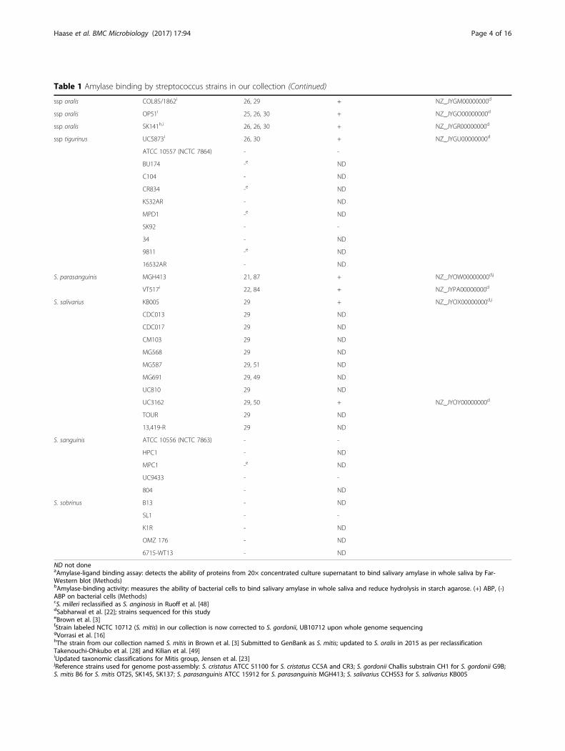
Table 1 Amylase binding by streptococcus strains in our collection (Continued)
ssp oralis COL85/1862i 26, 29 + NZ_JYGM00000000d
ssp oralis OP51i 25, 26, 30 + NZ_JYGO00000000d
ssp oralis SK141h,i 26, 26, 30 + NZ_JYGR00000000d
ssp tigurinus UC5873i 26, 30 + NZ_JYGU00000000d
ATCC 10557 (NCTC 7864) - -
BU174 -e ND
C104 - ND
CR834 -e ND
KS32AR - ND
MPD1 -e ND
SK92 - -
34 - ND
9811 -e ND
16532AR - ND
S. parasanguinis MGH413 21, 87 + NZ_JYOW00000000d,j
VT517i 22, 84 + NZ_JYPA00000000d
S. salivarius KB005 29 + NZ_JYOX00000000d,j
CDC013 29 ND
CDC017 29 ND
CM103 29 ND
MG568 29 ND
MG587 29, 51 ND
MG691 29, 49 ND
UC810 29 ND
UC3162 29, 50 + NZ_JYOY00000000d
TOUR 29 ND
13,419-R 29 ND
S. sanguinis ATCC 10556 (NCTC 7863) - -
HPC1 - ND
MPC1 -e ND
UC9433 - -
804 - ND
S. sobrinus B13 - ND
SL1 - -
K1R - ND
OMZ 176 - ND
6715-WT13 - ND
ND not doneaAmylase-ligand binding assay: detects the ability of proteins from 20× concentrated culture supernatant to bind salivary amylase in whole saliva by Far-Western blot (Methods)bAmylase-binding activity: measures the ability of bacterial cells to bind salivary amylase in whole saliva and reduce hydrolysis in starch agarose. (+) ABP, (-)ABP on bacterial cells (Methods)cS. milleri reclassified as S. anginosis in Ruoff et al. [48]dSabharwal et al. [22]; strains sequenced for this studyeBrown et al. [3]fStrain labeled NCTC 10712 (S. mitis) in our collection is now corrected to S. gordonii, UB10712 upon whole genome sequencinggVorrasi et al. [16]hThe strain from our collection named S. mitis in Brown et al. [3] Submitted to GenBank as S. mitis; updated to S. oralis in 2015 as per reclassificationTakenouchi-Ohkubo et al. [28] and Kilian et al. [49]iUpdated taxonomic classifications for Mitis group, Jensen et al. [23]jReference strains used for genome post-assembly: S. cristatus ATCC 51100 for S. cristatus CC5A and CR3; S. gordonii Challis substrain CH1 for S. gordonii G9B;S. mitis B6 for S. mitis OT25, SK145, SK137; S. parasanguinis ATCC 15912 for S. parasanguinis MGH413; S. salivarius CCHSS3 for S. salivarius KB005
Haase et al. BMC Microbiology (2017) 17:94 Page 4 of 16

Assembly of whole genome sequencing dataSequenced genomes were assembled de novoThe recently developed algorithm, MyPro [21], facili-tated assembly and annotation of the draft genomes.When closely related streptococcal genomes were avail-able for reference, a post-assembly algorithm was usedto reduce gaps in the de novo assemblies [21, 22]. Post-assembled genomes consisted of fewer gaps in theassembly, with a median of 6 contigs. De novo assem-bled genomes ranged in size from 1.79 to 2.29 Mb, withan average G + C content of 40.8% and 1916 codingsequences [22].
Taxonomic designationThere have been changes in the taxonomic assign-ment of several mitis group Streptococcus species sub-sequent to the availability of comparative phylogeneticanalyses of core genomes, multilocus sequence ana-lysis (MLSA), and 16S rRNA gene sequence data [23].In the current study, revised streptococcal taxonomyshowed changes in species nomenclature of 25 strainsused in phylogenetic analyses. Briefly, several mem-bers of mitis, oligofermentans, pseudopneumoniae, andsanguinis species that represent ABS were revised toother Streptococcus species based on MLSA andwhole genome core phylogeny [23]. Taxonomic reas-signments are summarized in Additional file 1: TableS1, and updates have also been made to our draftgenome submissions [22].
Identification of AbpA-like sequencesThe AbpA protein sequence (20–26 kDa) from ournewly sequenced strains and homologs obtained in silicowere used to further categorize AbpA-like producingstreptococcal strains into five subgroups based on theN-terminal sequence. In-depth phylogenetic analysis ofAbpA for this study was confined to predominanthuman oral streptococcal strains and to strains where asortase B (srtB) gene was immediately downstream ofthe putative abpA gene.Comparison of complete abpA gene sequences revealed
more heterogeneity among amylase-binding streptococcithan previously observed. The Streptococcus species andstrains within each AbpA-like subgroup along with theassociated N-terminal sequences are listed in Additionalfile 2: Table S2. Subgroup 1 consists primarily of S. gordo-nii strains, but also includes a strain of S. cristatus. Thewell-characterized AbpA protein of S. gordonii ChallisCH1 is a member of this subgroup [12, 14]. Subgroup 2 ispredominantly S. cristatus strains, but also contains theGram-positive opportunistic pathogen Gemella haemoly-sans, a member of the human flora of the oral cavity andupper respiratory tract [24, 25]. Since G. haemolysansabpA lacks the typical downstream srtB gene, it wasexcluded from phylogenetic analysis. S. salivarius domi-nates Subgroup 3, but also contains some S. oralis and Sparasanguinis strains. Subgroup 4 is a mix of S. oralis, S.infantis, and S. parasanguinis. Subgroup 5 is the mostdiverse group; although S. parasanguinis predominates, S.australis, S. infantis, and Streptococcus vestibularis strainsare represented among others. The N-terminal sequencesnot only serve to subgroup AbpA-like proteins, but maycontribute to the ability of AbpA to bind amylase, as in S.gordonii [19, 26].The AbpA-like protein sequences from each of the sub-
groups were aligned using CLUSTAL Omega [27]. AbpAsequences of individual subgroup members are well con-served with the exception of subgroup 5, which is themost diverse. When the consensus sequence from eachsubgroup was aligned, the signal sequence, sortase B-binding motif, and C-terminal end are the most concord-ant (Fig. 2). These protein sequences contain 195 to 228amino acid residues and each subgroup revealed a similararrangement schematically represented in Fig. 3, i.e. awell-conserved signal sequence of 23 residues, followed byone of five subgroup-specific N-terminal consensussequences (11–13 residues), followed by a variable regionof low to moderate homology (152–186 residues), andending with a putative sortase B-binding motif [LP(K/N)T(S/A/H)] and a termination sequence [A(V/A)K].
Identification of AbpB-like sequencesSeveral strains of streptococci produce an 80–87 kDaABP that is consistent with AbpB (Fig. 1b, Additional
Fig. 1 Composite of (a) Coomassie-stained gels and (b) blots from theamylase-ligand overlay assay. a Boxes represent protein bands cut outfrom the Coomassie-stained blot for N-terminal sequencing. b AbpA-like, red boxes; AbpB-like, blue boxes; Novel ABPs, green boxes;Indeterminant, black boxes. Summary of ABPs are listed in Table 2
Haase et al. BMC Microbiology (2017) 17:94 Page 5 of 16
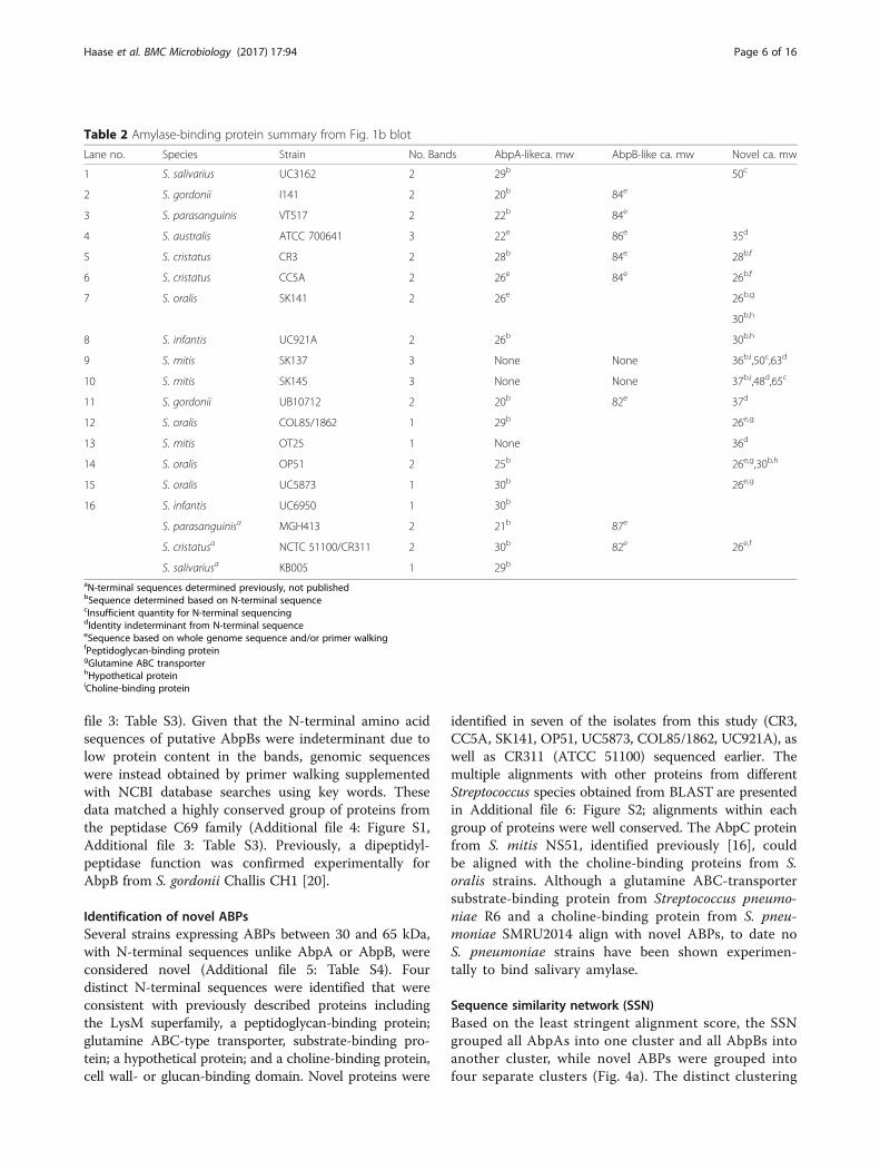
file 3: Table S3). Given that the N-terminal amino acidsequences of putative AbpBs were indeterminant due tolow protein content in the bands, genomic sequenceswere instead obtained by primer walking supplementedwith NCBI database searches using key words. Thesedata matched a highly conserved group of proteins fromthe peptidase C69 family (Additional file 4: Figure S1,Additional file 3: Table S3). Previously, a dipeptidyl-peptidase function was confirmed experimentally forAbpB from S. gordonii Challis CH1 [20].
Identification of novel ABPsSeveral strains expressing ABPs between 30 and 65 kDa,with N-terminal sequences unlike AbpA or AbpB, wereconsidered novel (Additional file 5: Table S4). Fourdistinct N-terminal sequences were identified that wereconsistent with previously described proteins includingthe LysM superfamily, a peptidoglycan-binding protein;glutamine ABC-type transporter, substrate-binding pro-tein; a hypothetical protein; and a choline-binding protein,cell wall- or glucan-binding domain. Novel proteins were
identified in seven of the isolates from this study (CR3,CC5A, SK141, OP51, UC5873, COL85/1862, UC921A), aswell as CR311 (ATCC 51100) sequenced earlier. Themultiple alignments with other proteins from differentStreptococcus species obtained from BLAST are presentedin Additional file 6: Figure S2; alignments within eachgroup of proteins were well conserved. The AbpC proteinfrom S. mitis NS51, identified previously [16], couldbe aligned with the choline-binding proteins from S.oralis strains. Although a glutamine ABC-transportersubstrate-binding protein from Streptococcus pneumo-niae R6 and a choline-binding protein from S. pneu-moniae SMRU2014 align with novel ABPs, to date noS. pneumoniae strains have been shown experimen-tally to bind salivary amylase.
Sequence similarity network (SSN)Based on the least stringent alignment score, the SSNgrouped all AbpAs into one cluster and all AbpBs intoanother cluster, while novel ABPs were grouped intofour separate clusters (Fig. 4a). The distinct clustering
Table 2 Amylase-binding protein summary from Fig. 1b blot
Lane no. Species Strain No. Bands AbpA-likeca. mw AbpB-like ca. mw Novel ca. mw
1 S. salivarius UC3162 2 29b 50c
2 S. gordonii I141 2 20b 84e
3 S. parasanguinis VT517 2 22b 84e
4 S. australis ATCC 700641 3 22e 86e 35d
5 S. cristatus CR3 2 28b 84e 28b,f
6 S. cristatus CC5A 2 26e 84e 26b,f
7 S. oralis SK141 2 26e 26b,g
30b,h
8 S. infantis UC921A 2 26b 30b,h
9 S. mitis SK137 3 None None 36b,i,50c,63d
10 S. mitis SK145 3 None None 37b,i,48d,65c
11 S. gordonii UB10712 2 20b 82e 37d
12 S. oralis COL85/1862 1 29b 26e,g
13 S. mitis OT25 1 None 36d
14 S. oralis OP51 2 25b 26e,g,30b,h
15 S. oralis UC5873 1 30b 26e,g
16 S. infantis UC6950 1 30b
S. parasanguinisa MGH413 2 21b 87e
S. cristatusa NCTC 51100/CR311 2 30b 82e 26e,f
S. salivariusa KB005 1 29b
aN-terminal sequences determined previously, not publishedbSequence determined based on N-terminal sequencecInsufficient quantity for N-terminal sequencingdIdentity indeterminant from N-terminal sequenceeSequence based on whole genome sequence and/or primer walkingfPeptidoglycan-binding proteingGlutamine ABC transporterhHypothetical proteiniCholine-binding protein
Haase et al. BMC Microbiology (2017) 17:94 Page 6 of 16
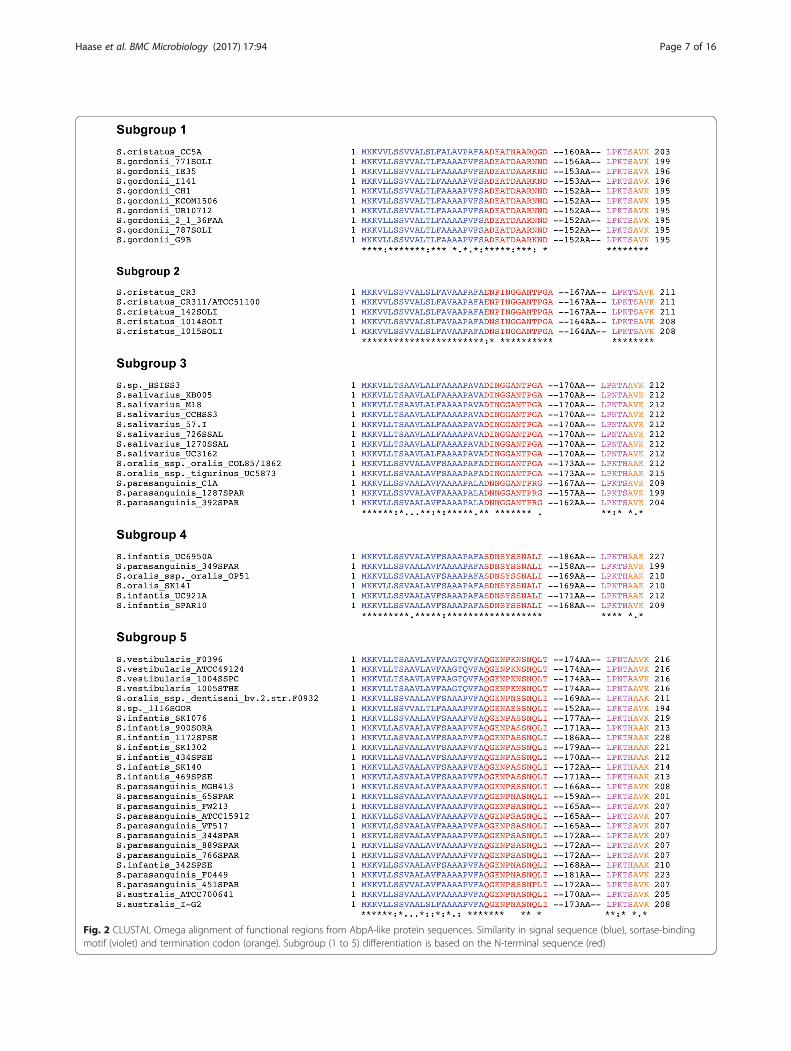
Fig. 2 CLUSTAL Omega alignment of functional regions from AbpA-like protein sequences. Similarity in signal sequence (blue), sortase-bindingmotif (violet) and termination codon (orange). Subgroup (1 to 5) differentiation is based on the N-terminal sequence (red)
Haase et al. BMC Microbiology (2017) 17:94 Page 7 of 16
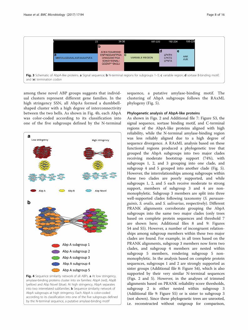
among these novel ABP groups suggests that individ-ual clusters represent different gene families. In thehigh stringency SSN, all AbpAs formed a dumbbell-shaped cluster with a high degree of interconnectivitybetween the two bells. As shown in Fig. 4b, each AbpAwas color-coded according to its classification intoone of the five subgroups defined by the N-terminal
sequence, a putative amylase-binding motif. Theclustering of AbpA subgroups follows the RAxMLphylogeny (Fig. 5).
Phylogenetic analysis of AbpA-like proteinsAs shown in Figs. 2 and Additional file 7: Figure S3, thesignal sequence, sortase binding motif, and C-terminalregions of the AbpA-like proteins aligned with highreliability, while the N-terminal amylase-binding regionwas less reliably aligned due to a high degree ofsequence divergence. A RAxML analysis based on thesefunctional regions produced a phylogenetic tree thatgrouped the AbpA subgroups into two major cladesreceiving moderate bootstrap support (74%), withsubgroups 1, 2, and 3 grouping into one clade, andsubgroup 4 and 5 grouped into another clade (Fig. 5).However, the interrelationships among subgroups withinthese two clades are poorly supported, and whilesubgroups 1, 2, and 5 each receive moderate to strongsupport, members of subgroup 3 and 4 are non-monophyletic. Subgroup 3 members are split into threewell-supported clades following taxonomy (S. parasan-guinis, S. oralis, and S. salivarius, respectively). DifferentPRANK alignments corroborate grouping the AbpAsubgroups into the same two major clades (only treesbased on complete protein sequences and threshold 7are shown here; Additional files 8 and 9: FiguresS4 and S5). However, a number of incongruent relation-ships among subgroup members within these two majorclades are found. For example, in all trees based on thePRANK alignments, subgroup 3 members now form twoclades, and subgroup 4 members are nested withinsubgroup 5 members, rendering subgroup 5 non-monophyletic. In the analysis based on complete proteinsequences, subgroups 1 and 2 are strongly supported assister groups (Additional file 8: Figure S4), which is alsosupported by their very similar N-terminal sequences(Figs. 2 and 5). However, in the analyses of trimmedalignments based on PRANK reliability score thresholds,subgroup 2 is either nested within subgroup 3(Additional file 9: Figure S5) or is sister to subgroup 3(not shown). Since these phylogenetic trees are unrooted,i.e. reconstructed without outgroup for comparison,
Fig. 3 Schematic of AbpA-like proteins. a Signal sequence; b N-terminal regions for subgroups 1–5; c variable region; d sortase B-binding motif;and (e) termination codon
Fig. 4 Sequence similarity network of all ABPs. a At low stringency,amylase-binding proteins cluster into six families: AbpA (red), AbpB(yellow) and Abp Novel (blue). At high stringency, AbpA separatesinto two interrelated subfamilies. b Sequence similarity network ofAbpA subgroups at high stringency. Each AbpA is color-codedaccording to its classification into one of the five subgroups definedby the N-terminal sequence, a putative amylase-binding motif
Haase et al. BMC Microbiology (2017) 17:94 Page 8 of 16
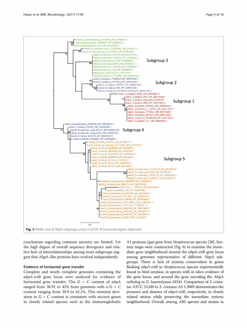
conclusions regarding common ancestry are limited. Yetthe high degree of overall sequence divergence and rela-tive lack of interrelationships among most subgroups sug-gest that AbpA-like proteins have evolved independently.
Evidence of horizontal gene transferComplete and nearly complete genomes containing theabpA-srtB gene locus were analyzed for evidence ofhorizontal gene transfer. The G + C content of abpAranged from 38.91 to 42% from genomes with a G + Ccontent ranging from 39.9 to 42.1%. This minimal devi-ation in G + C content is consistent with ancient genesin closely related species such as the immunoglobulin
A1 protease (iga) gene from Streptococcus species [28]. Syn-teny maps were constructed (Fig. 6) to examine the imme-diate gene neighborhood around the abpA-srtB gene locusamong genomes representative of different AbpA sub-groups. There is lack of synteny conservation in genesflanking abpA-srtB in Streptococcus species experimentallyfound to bind amylase, in species with in silico evidence ofthe gene locus, and around the gene encoding the AbpAortholog in G. haemolysans M341. Comparison of S. crista-tus ATCC 51100 to S. cristatus AS 1.3089 demonstrates thepresence and absence of abpA-srtB, respectively, in closelyrelated strains while preserving the immediate syntenyneighborhood. Overall, among ABS species and strains in
Fig. 5 RAxML tree of AbpA subgroups using CLUSTAL W functional regions alignment
Haase et al. BMC Microbiology (2017) 17:94 Page 9 of 16

this study, the genes flanking abpA-srtB show conservationwithin species, but not between species. In addition, trans-poson-mediated mutagenesis with Tn916 used to in-activate abpA demonstrated a transposon integrationhotspot immediately upstream of abpA [14]. Together,these observations suggest the co-transcribed abpA-srtB locus is a genetic island that may have beenacquired by horizontal transfer.
Molecular modeling of AbpA subgroupsIn silico modeling with I-TASSER revealed similar tertiarystructures for representative AbpA proteins from eachsubgroup. The molecular modeling performed here islimited by the availability of NMR data on AbpA struc-ture, which until recently was not reported [29]. Over-all, helical arrangement predominated the N-terminalhalf of the molecule and C-terminal half showed acoiled structure (Fig. 7).
DiscussionIn this study, we expand upon previous studies to betterunderstand how oral streptococci use different proteins tobind amylase. This interaction allows the bacteria to adaptto the oral environment by adhering to the salivary pellicleand initiating colonization of the tooth surface, as well as toefficiently metabolize dietary starch as an energy source.An in vitro assay to assess amylase binding to the cell sur-face of oral Streptococcus species has also been used previ-ously in an attempt to classify viridans streptococci [6, 11,30]. Here, this method was used to confirm the results ofamylase binding to denatured proteins in the culture super-natant. Most Streptococcus strains examined produced anAbpA-like protein, the best-studied and most predominant
protein that mediates the binding of amylase to the bacter-ial surface. But bacterial cells from four strains, S. mitisOT25, SK137, SK145, and NS51 bound amylase withoutAbpA. These strains produced novel proteins, in particularthe AbpC of S. mitis NS51. The actual function, cellular lo-cation, exposure to the extracellular environment, and rele-vance of these novel ABPs to the fitness of these strains,remains to be determined.At least six separate families of ABPs comprising
AbpA-like, AbpB-like, and novel proteins were identifiedin Streptococcus strains sequenced in this study incombination with homologs obtained from NCBI RefSeqhttps://www.ncib.nlm.nih.gov/refseq/. There was nosignificant similarity between the families based on
Fig. 6 Synteny mapping of abpA-srtB. S. cristatus AS1.3089 did not carry the abpA-srtB locus. ORFs flanking the abpA-srtB locus were deducedfrom genome sequences available at NCBI. All strains except S. cristatus AS1.3089, S. vestibularis F0396, and G. haemolysans M341 have beentested in vitro for the ability to bind amylase. Flanking genes include: (1) ribose-phosphate pyrophosphokinase; (2) aminotransferase; (3) haloaciddehalogenase; (4) MFS transporter; (5) peptidase M42; (6) peptidase; (7) DNA-binding transcriptional regulator, XRE-family; (8) metallophosphatase;(9) CoA-binding protein; and (10) IS200/IS605 family transposase. Ain silico evidence only
Fig. 7 Tertiary protein structure of representative AbpA sequences.N-terminal half of AbpA is predominated by helical spatial arrangementand the C-terminal half is largely coiled. NMR structure of S. gordonii CH1is in red and the predicted structures for representative sequences arecoded in various colors (Green-Subgroup1-WP_008810020.1; Yellow-Subgroup2-WP_005591359.1; Purple-Subgroup3-WP_002886387.1; Blue-Subgroup4-WP_033629133.1; Orange-Subgroup5-WP_003092326.1)
Haase et al. BMC Microbiology (2017) 17:94 Page 10 of 16

protein sequence alignment. No readily identifiableamylase-binding or sortase-binding motifs were presentexcept within the AbpA-like family. Thus, binding ofamylase by each of these protein families is likely due tosecondary structural interaction rather than by primaryamino acid sequences. This was especially evident forAbpA-like proteins having moderate amino acid similar-ity and identity, but with similar predicted secondarystructures as presented in this study and by Liang et al.[19].General functions of ABPs may be in promoting oral
colonization and fitness. When ABPs bind salivary amyl-ase, they enable close contact of bacteria with oral sur-faces coated with saliva and promote subsequentcolonization. When first described, AbpA was a novelprotein [14]. Homologs are now found in many oralstreptococcal species of humans and animals whosesaliva contains amylase activity [32, 33]. In addition toplaying a role in adhesion, biofilm formation, providinga convenient source of saccharolytic nutrition throughthe hydrolysis of dietary starch, and possibly providingrelief from oxidative stress, there may be as yet un-described functions for AbpA [12, 17]. AbpB proteinsequences are conserved (63–100% identical), suggestingthey have a primary function as dipeptidases, which mayhave a role in nutrient acquisition pathways [20, 34].The novel ABPs have been annotated with a variety ofcell wall-associated functions. Association of AbpA withthe bacterial cell wall, especially at the site of nascentcell wall synthesis, was previously demonstrated [32], sothe finding that other unrelated ABPs may also associatewith the cell wall is supportive of the hypothesis thatamylase binding is beneficial to the growth of the organ-ism [12, 34]. Alternatively, the ability to bind amylasemay just be a coincidental function for AbpB and thenovel ABPs. How the acquisition of the abpA-srtB locusor any of the other genes encoding potentially novelABPs enhances the survival and role of the commensalstreptococcal strains as primary colonizers of the oralcavity requires further study.The ability of a bacterial species to bind salivary amyl-
ase allowing it to adapt to the oral environment is notconfined to cell wall components of Streptococcus spe-cies. Fimbria-associated proteins from S. sanguinis SK36[33] and S. mutans CS2 [35], a Streptococcus species notnormally known to bind amylase have been shown tobind amylase. Amylase-binding components have beendescribed in a few other non-streptococcal oral speciesincluding a 110-kDa protein of Fusobacterium nuclea-tum subsp. polymorphum [36], and the outer membranelipopolysaccharide of Aggregatibacter actinomycetemco-mitans [37] and Porphyromonas gingivalis [38]. In A.actinomycetemcomitans, amylase interferes with bac-terial adherence and biofilm formation. Amylases from
several sources are able to inhibit the growth of P. gingi-valis [39]. How the binding of amylase in vivo in amixed species biofilm enables or hinders interspeciesinteractions is not known.Comparative pan-genomic studies provide evidence of
genetic exchange with related species sharing the sameecological niche [40, 41]. Our analyses of complete andnearly complete draft genomes of AbpA-encoding oralStreptococcus strains revealed that all contained theabpA-srtB gene locus. Sortase B is essential for initialcovalent attachment of AbpA to the bacterial cell wall[18, 19]. In fact this gene locus was found in 15/18strains sequenced in this study, supporting the role ofAbpA as the predominate ABP. The abpA gene may bean ancient gene, not unlike another gene in the mitisgroup of streptococci, iga, which encodes IgA protease[28] and is also found in several closely related Strepto-coccus species. AbpA is located in a region of conservedgene order indicating relatively recent subgroup diver-gence. AbpA subgroups generally correlate with species,although there is evidence of interspecies divergence.For example, within the mitis group of Streptococcusspecies, the S. oralis clade is more distantly related tothe S. parasanguinis clade [23], and both contain strainsthat carry AbpA-like proteins from a variety of sub-groups. This apparent random distribution of the co-transcribed abpA-srtB genes in more distantly relatedStreptococcus species, the lack of synteny conservationin closely related species, and the presence of an AbpAortholog in a different genus (Gemella) suggests thatthese genes are part of a genomic island acquired byhorizontal gene transfer.Many oral streptococci are known to be naturally
competent [42], a benefit to organisms living in a biofilmsuch as dental plaque. While screening our collection ofStreptococcus strains, it became apparent that not allstrains of a given species possess an ABP. The transferof DNA encoding a beneficial trait from one member ofthe community to another enhances the survival of thecommunity. It is therefore possible that the acquisitionof the amylase-binding function by some oral strepto-coccal strains, mediated by different proteins enablesthese bacteria to take advantage of their ecological nichewithin the oral cavity.This study demonstrates that ABPs are a diverse group
of proteins that sort into at least six distinct groups with avariety of putative functions. Sequence divergence of theABPs, especially within the AbpA group, suggests that theamylase-binding function evolved independently in separ-ate protein families. The ability to bind amylase may bene-fit the host by serving to remove bacteria in saliva. Thehigh prevalence of amylase binding among primary colon-izing streptococci in dental plaque may demonstrate howadaptive evolution can enhance bacterial colonization and
Haase et al. BMC Microbiology (2017) 17:94 Page 11 of 16
survival in the oral cavity. Amylase binding may not onlyfacilitate the nutrition of bacteria in the immediate envir-onment, but it may also serve another unrecognized func-tion to benefit the whole microbial community.
ConclusionsIn this study, we explored the genomic and evolutionaryaspects of ABPs. We demonstrated that ABPs (AbpA,AbpB and novel ABPs) cluster into six distinct but unre-lated families. Further, the AbpA protein family could besubgrouped based on N-terminal sequences. Whileevidence for evolution of these protein families was notconclusive, comparative genomics of abpA gene pro-vided evidence of horizontal gene transfer. Importantly,the acquisition of ABP by oral streptococci provided aninteresting example of how bacteria may evolve to adaptto the human host.
MethodsBacterial strains and culture conditionsA total of 79 oral streptococcal strains representing 13species were screened for ABPs, which included 20strains studied previously and 59 strains examined here(Table 1). All strains were obtained from our culturecollection, with the exception of S. australis ATCC700641 and S. infantis ATCC 700779, which were ob-tained from the American Type Culture Collection(ATCC, Manassas, VA). Bacteria were cultured from fro-zen stocks to agar plates containing tryptic soy brothsupplemented with 0.5% (w/v) yeast extract (TSBY) and1.5% Bacto agar (Becton Dickinson and Co., Sparks,MD) in a candle jar at 37 °C for 2 days. Isolated colonieswere subcultured into TSBY broth and incubated in acandle jar at 37 °C for 14 to 16 h.
Human saliva collectionThe University at Buffalo Human Subjects InstitutionalReview Board approved the saliva collection protocol.Briefly, unstimulated whole saliva, as the source ofhuman salivary amylase, was collected from severaldonors by expectoration into ice-chilled tubes, clarifiedby centrifugation at 12,800 x g for 10 min at 4 °C, ali-quoted, and stored at −20 °C until use.
Detection of amylase-binding proteins (ABPs) by theamylase ligand-binding assayStreptococcal culture supernatants from overnight cul-tures were collected by centrifugation at 5000×g for10 min followed by 20-fold concentration using AmiconUltra-4 centrifugal filter devices (Merck Millipore Ltd.,Tullagreen, Carrigtwohill, Co. Cork, IRL). The concen-trated supernatant proteins were separated on a 12.5%gel by SDS-PAGE. Proteins were either stained withCoomassie blue or transblotted to PVDF membrane.
The amylase-ligand binding assay was performed aspreviously described [14, 20]. Whole human saliva wasused as the source of amylase.
N-terminal amino acid sequencingConcentrated supernatants containing ABPs identifiedby the aforementioned amylase ligand-binding assaywere subjected to 12.5% SDS-PAGE and separatedproteins were electrotransferred from gels to a smallpore PVDF ProBlott membrane (Applied BiosystemsInc., Foster City, CA) and stained with 0.1% (w/v)Coomassie brilliant blue R-250. The protein bandscorresponding to the identified ABPs were cut from theblot and N-terminal sequenced by standard methods(ProSeq Inc., Oxford, MA). The N-terminal amino acidsequences of ABPs were used to search for homologousproteins in the National Center for Biotechnology Infor-mation database (NCBI, http://www.ncbi.nlm.nih.gov).
Purification and sequencing of streptococcal DNAGenomic DNA isolated from streptococcal strains usinga previously described method [20] was used as thetemplate in PCR reactions. For high-throughput se-quencing, the genomic DNA was first treated withRNase A/T1 Mix (Thermo Fisher Scientific Inc., Pitts-burgh, PA) according to the manufacturers’ instructions.The QIAamp DNA Mini Kit (Qiagen, Hilden, Germany)was then used to further purify the genomic DNA. Thequality was assessed by ethidium bromide stained agar-ose gels. DNA was quantified at A260 and A280 using theNanodrop spectrophotometer and the Quant-iT dsDNAkit (Invitrogen, Carlsbad, CA). Samples were stored at−80 °C prior to library construction for whole genomesequencing (Center for Excellence in Bioinformatics atthe University at Buffalo, Buffalo, NY).
Sequencing, quality control, de novo assembly, andannotation of streptococcal genomesIllumina HiSeq 2500 Next-Generation Sequencing wasused in rapid 150-cycle paired-end mode to performgenome sequencing of the appropriately preparedstreptococcal strains. The paired-end sequencing readsof 150-bp read length and over 100X coverage wereprocessed using MyPro (http://sourceforge.net/projects/sb2nhri/files/MyPro) [21], a customized software pipe-line designed for prokaryotic genome assembly andannotation. Quality control tests, de novo assembly, andannotation were applied to genomes of 18 streptococcalstrains. When closely related reference genomes wereavailable, post-assembly function of MyPro was used toalign and order contigs and reduce gaps in the de novogenome assemblies [22]. All draft genome sequenceswere deposited in GenBank and corresponding accessionnumbers were published prior to data mining [22].
Haase et al. BMC Microbiology (2017) 17:94 Page 12 of 16
Identification of amylase-binding protein a sequencesComplete abpA were manually obtained from 18 strepto-coccal genomes by using corresponding N-terminal se-quence as a query. Thereafter, complete abpA sequenceswere used as multifasta query to BLAST against the non-redundant protein sequence (nr) database restricted toStreptococcus (taxid:1301) using an evalue of 1e-5. Resultswere curated for presence of N-terminal - like sequencesas a measure of confirmation.
Identification of amylase-binding protein B sequencesTo obtain genomic sequences previously annotated asAbpB, the NCBI RefSeq database was searched forcomplete streptococcal AbpB sequences by using thekeywords ‘amylase-binding protein’ or ‘amylase-bindingprotein B’, and filters ‘bacteria’ and ‘sequence length morethan 300’. The results were un-collapsed and manuallysorted to include all non-redundant streptococcal strains.To identify any additional AbpB-like sequences that maybe annotated differently (e. g. peptidase C69), the databasewas also searched with sequences obtained from primerwalking of AbpB.
Primer walking of abpBTo determine the abpB sequence from each of thestrains expressing AbpB in the amylase-ligand bindingassay, five sets of degenerate primers were designedaccording to the alignment of nucleotide sequences ofknown AbpBs. Primers were designed to conserved re-gions where possible. The primer sequences (Invitrogen)listed in Additional file 10: Table S5 cover approximately84% of abpB. Conventional PCR was initiated with 90 sat 95 °C, followed by 34 cycles of 30 s at 95 °C, 30 s at50–52 °C according to the annealing temperatures, and2 min at 72 °C, then 10 min at 72 °C using a T100thermo cycler (Bio-Rad, CA). Amplified products wereseparated in a 1.5% agarose gel, stained with ethidiumbromide, and visualized under UV light. Genomic DNAof S. gordonii Challis CH1 and a no template controlwere used as the positive and negative controls, res-pectively. PCR amplicons purified by QIAquick PCRPurification kit or QIAquick Gel Extraction kit (Qiagen,Hilden, Germany) were sent for nucleic acid sequencing(Roswell Park Cancer Institute, Buffalo, NY). Sequencesobtained from each strain were assembled using SeqMansoftware (www.dnastar.com) into one sequence contain-ing the nearly complete abpB gene.
Identification of novel ABP sequencesProteins that bound amylase in the amylase ligand-binding assay with molecular weights ranging from 30 to65 kDa and whose N-terminal sequences were not simi-lar to one of the five AbpA, or AbpB N-terminal se-quences were considered novel ABPs. The NCBI RefSeq
database was searched with the N-terminal sequences ofthese strains to identify novel ABP sequences.
Sequence similarity networkA sequence similarity network (SSN) of all the ABP proteinsequences was generated using EFI-EST [43]. The related-ness is described by sequence similarity based on pairwisealignment scores calculated from an all-by-all BLAST witha default E-value 1e-05. Networks were generated based onthe minimum alignment score 6 (low stringency) and themaximum alignment score 16 (high stringency), respect-ively. Each node, which represents one protein sequence,was color-coded according to its classification in the threeABP groups: AbpA, AbpB, and Abp Novel.
Phylogenetic analysis of ABPsMultiple sequence alignment was performed using CLUS-TAL W to align the functional regions of AbpA, includingthe signal sequence, the N-terminal amylase-binding re-gion, the sortase B-binding motif, and the C-terminus.Maximum Likelihood (ML) analysis was performed onthe resulting alignment using the RAxML (RandomizedAccelerated Maximum Likelihood) algorithm [31]. Sincethe AbpA-like protein sequences are highly divergent,we also applied PRANK (Probabilistic Alignment Kit)[44] to align the complete protein sequences of AbpAs,and the reliability score of each aligned site was thenannotated. The scale of the reliability score is definedas 0 to 9, indicating less reliably aligned site to morereliably aligned site. The alignment was then trimmedbased on different reliability score thresholds: 1, 4, 5, 6,7 and 9. ML analysis using RAxML was performed foreach of these trimmed alignments, as well as thecomplete protein sequences.
Bacterial amylase-binding assayThe starch agarose assay was performed as previously[6, 11] to access the ability of bacterial cells to bindamylase in clarified whole human saliva. Control sam-ples included saliva alone, and saliva pre-incubatedwith streptococcal strains known to bind amylase orunable to bind amylase.
Signatures of horizontal gene transfer surrounding abpAGenBank files for ABS were compared for synteny (onegene upstream and downstream) flanking the abpA-srtBlocus. Representative genomes for each AbpA subgroupwere selected based on experimental evidence of AbpAto illustrate the variation in synteny neighborhood ofabpA-srtB locus. The closely related genome S. cristatusATCC 51100 was selected based on the lack of theabpA-srtB locus, and the G. haemolysans M341 genomewas selected to illustrate an abpA ortholog without thetypical srtB.
Haase et al. BMC Microbiology (2017) 17:94 Page 13 of 16
Molecular modeling of AbpAMultispecies sequences (representing 20 sequences) fromall AbpA subgroups were submitted for molecular model-ing using the I-TASSER (Iterative Threading ASSEmblyRefinement) web service, which uses a hierarchical ap-proach for protein structure and function prediction [45].We included the AbpA sequence from S. gordonii ChallisCH1 as a positive control. The NMR structure of AbpAfrom S. gordonii G9B has been determined [29], and theprotein sequence of S. gordonii Challis CH1 differs fromG9B only at residue 33 containing Asn and Lys, res-pectively. I-TASSER predicted secondary structures andtertiary structure models (.pdb format) were obtained forall sequences along with their confidence scores. Modelwith highest confidence score for each subgroup wereused as input for Protean3D (http://www.dnastar.com/t-protean-3D.aspx) for structural comparison and out-put in graphical format.
Streptococcal taxonomyConcatemers of seven housekeeping genes for viridansgroup Streptococci in multifasta format were obtainedfrom the authors of a previous publication [46]. Thesesequences were curated to represent the amylase-bindingstreptococcus (ABS) species used in this study. Curatedsequences were used as query to BLAST against a localdatabase comprised of whole genome sequences of ABSwith an Expected (E) value of 1e-5 [47]. The results wereconcatenated in-frame, in the order (map-pfl-ppaC-pyk-rpoB-sodA-tuf ) and used for phylogenetic analysis. ABSconcatemers were aligned using CLUSTAL W of theMEGA 7.0.18 package and the aligned sequences wereused to construct a minimum evolution tree.
Additional files
Additional file 1: Table S1. MLSA based revised streptococcal taxonomy.Includes the genome accession number, strain identifier, old taxonomicname, and new taxonomic name for each strain. (DOCX 91 kb)
Additional file 2: Table S2. Amylase-binding protein A-like subgroupcomparison. Includes NCBI protein identifiers, molecular weights, andN-terminal sequences. (DOCX 107 kb)
Additional file 3: Table S3. Amylase-binding protein B-like comparison.Includes NCBI protein identifiers, molecular weights, and N-terminalsequences. (DOCX 89 kb)
Additional file 4: Figure S1. CLUSTAL alignment of AbpB-like proteinsequences. (DOCX 167 kb)
Additional file 5: Table S4. Novel amylase-binding protein comparison.Includes NCBI protein identifiers, molecular weights, and N-terminalsequences. (DOCX 82 kb)
Additional file 6: Figure S2. CLUSTAL alignment of novel amylase-binding proteins. (DOCX 145 kb)
Additional file 7: Figure S3. PRANK reliability annotation of thealignment of functional regions (signal sequence and N-terminalsequence) from AbpA-like proteins. (TIFF 11836 kb)
Additional file 8: Figure S4. PRANK tree of AbpA subgroups using theentire gene sequence. (TIFF 14602 kb)
Additional file 9: Figure S5. PRANK tree of AbpA subgroups using theentire gene sequence, reliability score 7. (TIFF 14758 kb)
Additional file 10: Table S5. Degenerate PCR primers for genesencoding AbpB-like proteins. (DOCX 73 kb)
AbbreviationsAbpA: Amylase-binding protein A; AbpB: Amylase-binding protein B;AbpC: Amylase-binding protein C; ABPs: Amylase-Binding Proteins;ATCC: American Type Culture Collection; BLAST: Basic Local AlignmentSearch Tool; CLUSTAL W: CLUSTer ALignment W; G + C: Guanine andCytosine; IgA: Immunoglobulin A1 protease; iga: Immunoglobulin A1protease gene;I-TASSER: Iterative Threading ASSEmbly Refinement; kDa: kilo Dalton;MLSA: MultiLocus Sequence Analysis; NCBI: National Center forBiotechnology Information; NMR: Nuclear Magnetic Resonance;PRANK: PRobabilistic AligNment Kit; PVDF: PolyVinyliDene Fluoride;RAxML: Randomized Accelerated Maximum Likelihood; SDS-PAGE: SodiumDodecyl Sulfate- PolyAcrylamide Gel Electrophoresis; srtB: Sortase B gene;SrtB: Sortase B protein; SSN: Sequence Similarity Network; TSBY: Tryptic SoyBroth supplemented with 0.5% (w/v) Yeast extract
AcknowledgementsThe National Institute of Dental and Craniofacial Research GrantR01DE022673 supported this work. We thank Dr. Victor Albert and Dr.Gerald Kuldelka in the Department of Biology, and Dr. M. MargaretVickerman in the Department of Oral Biology at the University at Buffalofor their helpful discussions. We also thank Steven Hintermeier fortechnical laboratory assistance.
FundingThis work was supported by the National Institute of Dental and CraniofacialResearch Grant R01DE022673.
Availability of data and materialsThe datasets generated and/or analyzed during the current study areavailable at the NCBI in GenBank at https://www.ncbi.nlm.nih.gov/bioproject/PRJNA274768. Draft genomes are described in [22].
Authors’ contributionsEMH contributed to the design of all experiments, analyzed Western blotdata, performed amylase activity assays, collated strain data and results,participated in bioinformatic data analysis, co-drafted and co-wrote themanuscript. YK performed sample preparation for Western blot andN-terminal sequencing, amylase ligand binding assay and analysis, DNAisolation for genomic sequencing, primer design and primer walking ofAbpB, and co-wrote a draft of the manuscript. AS collaborated with YL ingenome analysis, performed CLUSTAL alignments, horizontal gene transferanalysis, and structural modeling of AbpA, and co-wrote part of themanuscript. TL performed all phylogenetic analyses. CL consulted on types ofphylogenetic analyses, and together with TL co-wrote part of the manuscript.FAS conceived of the project and advised on all aspects of experimental designand analyses, and co-wrote this manuscript. All authors have read andapproved the final manuscript.
Competing interestsThe authors declare that they have no competing interests.
Consent for publicationNot applicable.
Ethics approval and consent to participateThe University at Buffalo Human Subjects Institutional Review Boardapproved the human saliva collection protocol. The protocol includes astatement that the saliva will be used for research purposes, and the consentwas signed by all donors.
Haase et al. BMC Microbiology (2017) 17:94 Page 14 of 16

Publisher’s NoteSpringer Nature remains neutral with regard to jurisdictional claims inpublished maps and institutional affiliations.
Author details1Department of Oral Biology, School of Dental Medicine, University atBuffalo, State University of New York, Buffalo, NY, USA. 2Division ofBiostatistics and Bioinformatics, Institute of Population Health Sciences,National Health Research Institutes, Miaoli, Taiwan. 3Department of BiologicalSciences, University at Buffalo, State University of New York, Buffalo, NY, USA.4Department of Oral Biology, School of Stomatology, China MedicalUniversity, Shenyang, People’s Republic of China.
Received: 23 December 2016 Accepted: 8 April 2017
References1. Bentley RW, Leigh JA, Collins MD. Intrageneric structure of Streptococcus
based on comparative analysis of small-subunit rRNA sequences. Int J SystBacteriol. 1991;41(4):487–94. doi:10.1099/00207713-41-4-487.
2. Kawamura Y, Hou XG, Sultana F, Miura H, Ezaki T. Determination of 16SrRNA sequences of Streptococcus mitis and Streptococcus gordonii andphylogenetic relationships among members of the genus Streptococcus. IntJ Syst Bacteriol. 1995;45(2):406–8. doi:10.1099/00207713-45-2-406.
3. Brown AE, Rogers JD, Haase EM, Zelasko PM, Scannapieco FA. Prevalence ofthe amylase-binding protein A gene (abpA) in oral streptococci. J ClinMicrobiol. 1999;37(12):4081–5.
4. Douglas CW, Heath J, Hampton KK, Preston FE. Identity of viridansstreptococci isolated from cases of infective endocarditis. J Med Microbiol.1993;39(3):179–82. doi:10.1099/00222615-39-3-179.
5. Gwynn JP, Douglas CW. Comparison of amylase-binding proteins in oralstreptococci. FEMS Microbiol Lett. 1994;124(3):373–9.
6. Kilian M, Nyvad B. Ability to bind salivary alpha-amylase discriminates certainviridans group streptococcal species. J Clin Microbiol. 1990;28(11):2576–7.
7. Karn RC, Shulkin JD, Merritt AD, Newell RC. Evidence for post-transcriptionalmodification of human salivary amylase (amyl) isozymes. Biochem Genet.1973;10(4):341–50.
8. Boehlke C, Zierau O, Hannig C. Salivary amylase - The enzyme ofunspecialized euryphagous animals. Arch Oral Biol. 2015;60(8):1162–76. doi:10.1016/j.archoralbio.2015.05.008.
9. Aguirre A, Levine MJ, Cohen RE, Tabak LA. Immunochemical quantitation ofalpha-amylase and secretory IgA in parotid saliva from people of variousages. Arch Oral Biol. 1987;32(4):297–301.
10. Scannapieco FA, Torres G, Levine MJ. Salivary alpha-amylase: role in dentalplaque and caries formation. Crit Rev Oral Biol Med. 1993;4(3–4):301–7.
11. Douglas CW. The binding of human salivary alpha-amylase by oral strains ofstreptococcal bacteria. Arch Oral Biol. 1983;28(7):567–73.
12. Rogers JD, Palmer Jr RJ, Kolenbrander PE, Scannapieco FA. Role of Streptococcusgordonii amylase-binding protein A in adhesion to hydroxyapatite, starchmetabolism, and biofilm formation. Infect Immun. 2001;69(11):7046–56.
13. Nikitkova AE, Haase EM, Vickerman MM, Gill SR, Scannapieco FA. Responseof fatty acid synthesis genes to the binding of human salivary amylase byStreptococcus gordonii. Appl Environ Microbiol. 2012;78(6):1865–75. doi:10.1128/AEM.07071-11.
14. Rogers JD, Haase EM, Brown AE, Douglas CW, Gwynn JP, Scannapieco FA.Identification and analysis of a gene (abpA) encoding a major amylase-binding protein in Streptococcus gordonii. Microbiology. 1998;144(Pt 5):1223–33.
15. Li L, Tanzer JM, Scannapieco FA. Identification and analysis of the amylase-binding protein B (AbpB) and gene (abpB) from Streptococcus gordonii.FEMS Microbiol Lett. 2002;212(2):151–7.
16. Vorrasi J, Chaudhuri B, Haase EM, Scannapieco FA. Identification andcharacterization of amylase-binding protein C from Streptococcus mitis NS51.Mol Oral Microbiol. 2010;25(2):150–6. doi:10.1111/j.2041-1014.2009.00554.x.
17. Haase EM, Feng X, Pan J, Miecznikowski JC, Scannapieco FA. Dynamics ofthe Streptococcus gordonii transcriptome in response to media composition,salivary alpha-amylase and starch. Appl Environ Microbiol. 2015. doi:10.1128/AEM.01221-15.
18. Liang X, Chen Y, Wu H. Sortase B assemble amylase-binding protein A tostreptococcal cell surface. 89th Gen. Sess. of Internat. Assoc. Dent. Res. SanDiego: IADR; 2011. p. 72.
19. Liang X, Liu B, Zhu F, Scannapieco FA, Haase EM, Matthews S, et al. A distinctsortase SrtB anchors and processes a streptococcal adhesin AbpA with a novelstructural property. Sci Rep. 2016;6:30966. doi:10.1038/srep30966.
20. Chaudhuri B, Paju S, Haase EM, Vickerman MM, Tanzer JM, Scannapieco FA.Amylase-binding protein B of Streptococcus gordonii is an extracellular dipeptidyl-peptidase. Infect Immun. 2008;76(10):4530–7. doi:10.1128/IAI.00186-08.
21. Ruoff KL. Streptococcus anginosus (“Streptococcus milleri”): theunrecognized pathogen. Clin Microbiol Rev. 1988;1(1):102–8.
22. Sabharwal A, Liao YC, Lin HH, Haase EM, Scannapieco FA. Draft genomesequences of 18 oral streptococcus strains that encode amylase-binding proteins.Genome Announc. 2015;3(3):e00510–5. doi:10.1128/genomeA.00510-15.
23. Takenouchi-Ohkubo N, Mortensen LM, Drasbek KR, Kilian M, Poulsen K.Horizontal transfer of the immunoglobulin A1 protease gene (iga) fromStreptococcus to Gemella haemolysans. Microbiology. 2006;152(Pt 7):2171–80.doi:10.1099/mic.0.28801-0.
24. Kilian M, Poulsen K, Blomqvist T, Havarstein LS, Bek-Thomsen M, Tettelin H,et al. Evolution of Streptococcus pneumoniae and its close commensalrelatives. PLoS One. 2008;3(7):e2683. doi:10.1371/journal.pone.0002683.
25. Jensen A, Scholz CF, Kilian M. Re-evaluation of the taxonomy of the Mitisgroup of the genus Streptococcus based on whole genome phylogeneticanalyses, and proposed reclassification of Streptococcus dentisani asStreptococcus oralis subsp. dentisani comb. nov., Streptococcus tigurinus asStreptococcus oralis subsp. tigurinus comb. nov., and Streptococcusoligofermentans as a later synonym of Streptococcus cristatus. Int J Syst EvolMicrobiol. 2016;66(11):4803–20. doi:10.1099/ijsem.0.001433.
26. Liao YC, Lin HH, Sabharwal A, Haase EM, Scannapieco FA. MyPro: A seamlesspipeline for automated prokaryotic genome assembly and annotation.J Microbiol Methods. 2015;113:72–4. doi:10.1016/j.mimet.2015.04.006.
27. Facklam R, Elliott JA. Identification, classification, and clinical relevance ofcatalase-negative, gram-positive cocci, excluding the streptococci andenterococci. Clin Microbiol Rev. 1995;8(4):479–95.
28. Collins M. The Genus Gemella. In: Dworkin M, Falkow S, Rosenberg E, SchleiferH, Stackebrandt E, editors. The Prokaryotes. New York: Springer; 2006. p. 511–8.
29. Gopal P, Ragunath C, Vyas V, Shanmugam M, Ramasubbu N. Probing theinteraction of human salivary alpha-amylase and amylase binding protein A(AbpA) of Streptococcus gordonii. Mol Biol. 2013;2:111. doi:10.4172/2168-9547.100011.
30. Sievers F, Wilm A, Dineen D, Gibson TJ, Karplus K, Li W, et al. Fast, scalablegeneration of high-quality protein multiple sequence alignments usingCLUSTAL Omega. Mol Syst Biol. 2011;7:539. doi:10.1038/msb.2011.75.
31. Sethi A, Mohanty B, Ramasubbu N, Gooley PR. Structure of amylase-bindingprotein A of Streptococcus gordonii: a potential receptor for human salivaryalpha-amylase enzyme. Protein Sci. 2015;24(6):1013–8. doi:10.1002/pro.2671.
32. Douglas CW, Pease AA, Whiley RA. Amylase-binding as a discriminatoramong oral streptococci. FEMS Microbiol Lett. 1990;54(1–3):193–7.
33. Nikitkova AE, Haase EM, Scannapieco FA. Taking the starch out of oralbiofilm formation: molecular basis and functional significance of salivaryalpha-amylase binding to oral streptococci. Appl Environ Microbiol. 2013;79(2):416–23. doi:10.1128/AEM.02581-12.
34. Scannapieco FA, Haraszthy GG, Cho MI, Levine MJ. Characterization of anamylase-binding component of Streptococcus gordonii G9B. Infect Immun.1992;60(11):4726–33.
35. Okahashi N, Nakata M, Terao Y, Isoda R, Sakurai A, Sumitomo T, et al. Pili oforal Streptococcus sanguinis bind to salivary amylase and promote thebiofilm formation. Microb Pathog. 2011;50(3–4):148–54. doi:10.1016/j.micpath.2011.01.005.
36. Perrone M, Gfell LE, Fontana M, Gregory RL. Antigenic characterization offimbria preparations from Streptococcus mutans isolates from caries-free andcaries-susceptible subjects. Clin Diagn Lab Immunol. 1997;4(3):291–6.
37. Zulfiqar M, Yamaguchi T, Sato S, Oho T. Oral Fusobacterium nucleatumsubsp. polymorphum binds to human salivary alpha-amylase. Mol OralMicrobiol. 2013;28(6):425–34. doi:10.1111/omi.12036.
38. Baik JE, Hong SW, Choi S, Jeon JH, Park OJ, Cho K, et al. Alpha-amylase is ahuman salivary protein with affinity to lipopolysaccharide of Aggregatibacteractinomycetemcomitans. Mol Oral Microbiol. 2013;28(2):142–53. doi:10.1111/omi.12011.
39. Choi S, Baik JE, Jeon JH, Cho K, Seo DG, Kum KY, et al. Identification ofPorphyromonas gingivalis lipopolysaccharide-binding proteins in human saliva.Mol Immunol. 2011;48(15–16):2207–13. doi:10.1016/j.molimm.2011.06.434.
40. Ochiai A, Harada K, Hashimoto K, Shibata K, Ishiyama Y, Mitsui T, et al. alpha-Amylase is a potential growth inhibitor of Porphyromonas gingivalis, a
Haase et al. BMC Microbiology (2017) 17:94 Page 15 of 16
periodontal pathogenic bacterium. J Periodontal Res. 2014;49(1):62–8. doi:10.1111/jre.12079.
41. Donati C, Hiller NL, Tettelin H, Muzzi A, Croucher NJ, Angiuoli SV, et al. Structureand dynamics of the pan-genome of Streptococcus pneumoniae and closelyrelated species. Genome Biol. 2010;11(10):R107. doi:10.1186/gb-2010-11-10-r107.
42. Richards VP, Palmer SR, Pavinski Bitar PD, Qin X, Weinstock GM, HighlanderSK, et al. Phylogenomics and the dynamic genome evolution of the genusStreptococcus. Genome Biol Evol. 2014;6(4):741–53. doi:10.1093/gbe/evu048.
43. Cvitkovitch DG. Genetic competence and transformation in oralstreptococci. Crit Rev Oral Biol Med. 2001;12(3):217–43.
44. Atkinson HJ, Morris JH, Ferrin TE, Babbitt PC. Using sequence similaritynetworks for visualization of relationships across diverse proteinsuperfamilies. PLoS One. 2009;4(2):e4345. doi:10.1371/journal.pone.0004345.
45. Stamatakis A. RAxML version 8: a tool for phylogenetic analysis and post-analysis of large phylogenies. Bioinformatics. 2014;30(9):1312–3. doi:10.1093/bioinformatics/btu033.
46. Loytynoja A, Goldman N. webPRANK: a phylogeny-aware multiple sequencealigner with interactive alignment browser. BMC Bioinformatics. 2010;11:579.doi:10.1186/1471-2105-11-579.
47. Zhang Y. I-TASSER server for protein 3D structure prediction. BMCBioinformatics. 2008;9:40. doi:10.1186/1471-2105-9-40.
48. Bishop CJ, Aanensen DM, Jordan GE, Kilian M, Hanage WP, Spratt BG.Assigning strains to bacterial species via the internet. BMC Biol. 2009;7:3.doi:10.1186/1741-7007-7-3.
49. Altschul SF, Gish W, Miller W, Myers EW, Lipman DJ. Basic local alignmentsearch tool. J Mol Biol. 1990;215(3):403–10. doi:10.1016/S0022-2836(05)80360-2.
• We accept pre-submission inquiries
• Our selector tool helps you to find the most relevant journal
• We provide round the clock customer support
• Convenient online submission
• Thorough peer review
• Inclusion in PubMed and all major indexing services
• Maximum visibility for your research
Submit your manuscript atwww.biomedcentral.com/submit
Submit your next manuscript to BioMed Central and we will help you at every step:
Haase et al. BMC Microbiology (2017) 17:94 Page 16 of 16