comparative biochemistry and physiology, part a et al compbioc… · 376 b.l. jones et al. /...
TRANSCRIPT
Stress granules form in Brachionus manjavacas (Rotifera) in response toa variety of stressors
Brande L. Jones a,⁎, Josephine VanLoozen b, Min H. Kim a, Stacey J. Miles c, Christine M. Dunham c,Loren Dean Williams a, Terry W. Snell ba School of Chemistry and Biochemistry, Georgia Institute of Technology, Atlanta, GA 30332, USAb School of Biology, Georgia Institute of Technology, Atlanta, GA 30332, USAc Department of Biochemistry, Emory University School of Medicine, Atlanta, GA 30322, USA
a b s t r a c ta r t i c l e i n f o
Article history:Received 13 April 2013Received in revised form 3 July 2013Accepted 4 July 2013Available online 12 July 2013
Keywords:SGsTIA-1eIF3BeIF4ERNA granules
Many eukaryotes share a common response to environmental stresses. The responses include reorganizationof cellular organelles and proteins. Similar stress responses between divergent species suggest that these pro-tective mechanisms may have evolved early and been retained from the earliest eukaryotic ancestors. Manyeukaryotic cells have the capacity to sequester proteins and mRNAs into transient stress granules (SGs) thatprotect most cellular mRNAs (Anderson and Kedersha, 2008). Our observations extend the phylogeneticrange of SGs from trypanosomatids, insects, yeast and mammalian cells, where they were first described,to a species of the lophotrochozoan animal phylum Rotifera. We focus on the distribution of three proteinsknown to be associated with both ribosomes and SG formation: eukaryotic initiation factors eIF3B, eIF4Eand T-cell-restricted intracellular antigen 1. We found that these three proteins co-localize to SGs in rotifersin response to temperature stress, osmotic stress and nutrient deprivation as has been described in other eu-karyotes. We have also found that the large ribosomal subunit fails to localize to the SGs in rotifers. Further-more, the SGs in rotifers disperse once the environmental stress is removed as demonstrated in yeast andmammalian cells. These results are consistent with SG formation in trypanosomatids, insects, yeast andmammalian cells, further supporting the presence of this protective mechanism early in the evolution ofeukaryotes.
© 2013 Elsevier Inc. All rights reserved.
1. Introduction
Posttranscriptional control of geneexpression is crucial for cell devel-opment, differentiation, immune signaling and neuronal plasticity(Richter, 2007; Anderson and Kedersha, 2008; Thomas et al., 2011).Translational repression is associated with RNA aggregation into gran-ules, which have been identified in various eukaryotic cells (AndersonandKedersha, 2006). Diverse types of RNAgranules have been observed,including neuronal RNA granules, germ cell granules (GCGs), processingbodies (PBs) and stress granules (SGs) (Kedersha and Anderson, 2002;Navarro and Blackwell, 2005; Anderson and Kedersha, 2006; Kedershaand Anderson, 2007; Anderson and Kedersha, 2008, 2009b). TheseRNA granules are all functional by-products of mRNA metabolism thatperform distinct functions.
Germ cell granules (GCGs) are polarized cytoplasmic aggregates.During embryogenesis, many maternal mRNAs are translationallyrepressed and transported to specific locations within the oocytes
(Anderson and Kedersha, 2006). GCGs define sites of germ cell differen-tiation within organisms. Analogous GCGs have been found in multipleinsect species as well as, Xenopus laevis, Drosophilia melanogaster andCaenorhabditis elegans (Schisa et al., 2001). The GCGs contain maternalmRNA required for germ cell specification and direct the timing of ma-ternal mRNA translation. GCGs also contain proteins involved in trans-lation initiation, translation control and mRNA decay (Anderson andKedersha, 2006). Other RNA granules have also been found withinGCGs (Navarro and Blackwell, 2005; Anderson and Kedersha, 2006).
PBs are cytoplasmic aggregates that contain RNA silencing or RNAdecay machinery (Bashkirov et al., 1997; Ingelfinger et al., 2002; vanDijk et al., 2002; Sheth and Parker, 2003; Brengues et al., 2005;Teixeira et al., 2005; Eulalio et al., 2007; Franks and Lykke-Andersen,2007, 2008; Buchan and Parker, 2009). PBs are constitutively expressedin cells and can be further induced upon exposure to a variety ofstressors (Brengues et al., 2005; Kedersha et al., 2005; Loschi et al.,2009). PBs have been observed in yeast, plants, insects, nematodes,vertebrates and trypanosomatids (Franks and Lykke-Andersen, 2008;Buchan and Parker, 2009). PBs and SGs share some components eventhough they are very distinct structures.
Eukaryotic cells commonly respond to environmental stresses byaltering their translational machinery and forming stress granules
Comparative Biochemistry and Physiology, Part A 166 (2013) 375–384
⁎ Corresponding author at: School of Biology, Georgia Institute of Technology, 310Ferst Drive, Atlanta, GA 30332-0230, USA. Tel.: +1 404 894 3700; fax: +1 404 8940519.
E-mail address: [email protected] (B.L. Jones).
1095-6433/$ – see front matter © 2013 Elsevier Inc. All rights reserved.http://dx.doi.org/10.1016/j.cbpa.2013.07.009
Contents lists available at SciVerse ScienceDirect
Comparative Biochemistry and Physiology, Part A
j ourna l homepage: www.e lsev ie r .com/ locate /cbpa

(SGs) (Kedersha and Anderson, 2002). Environmental stress preventstranslational initiation, leading to the formation of unproductive andtranslationally silenced pre-initiation small subunits complexed withmRNAs (Anderson and Kedersha, 2002b, 2006, 2008). The pre-initiation small subunits aggregate to form microscopically visible cy-toplasmic structures called SGs. Stalled small ribosomal subunits arefound in the SGs along with mRNAs bound to early initiation factors(eIF4E, eIF3, eIF4A, eIFG), but not large ribosomal subunits (Table 1)(Kedersha and Anderson, 2007). SGs may also contain RNA bindingproteins that regulate mRNA translation and decay such as T-cell-restricted intercellular antigen 1 (TIA-1), and other proteins involvedin RNA metabolism (Kedersha and Anderson, 2007).
SGs appear only under stress conditions, and their presence corre-lates with translational silencing (Kedersha et al., 1999, 2000, 2002;Kimball et al., 2003; Anderson and Kedersha, 2008; Cassola, 2011). SGsare transient andmay only be present for a limited period of time follow-ing exposure to stress (Anderson and Kedersha, 2008). In heat-shockedplants, SGs are believed to sequester mRNAs, transiently preventingtheir translation until homeostasis is restored (Nover et al., 1989).
SGs have been found in yeast, insects, trypanosomatids and mam-malian cell lines (Farny et al., 2009; Thomas et al., 2011). Althoughthey were first discovered within the cytoplasm of heat stressed cul-tured tomato cells (Nover et al., 1989), SGs have also been observedwithin mammalian cells upon environmental stress such heat,hyperosmolarity, oxidative conditions and UV irradiation (Andersonand Kedersha, 2002b; Kedersha and Anderson, 2002). It is notknown how widely distributed SGs are among animals, especially in-vertebrates, and whether this cell protective mechanism is a generalstress response or a specialized process of a few groups.
Invertebrate animals from the phylum Rotifera can sustain growthand reproduction over awide range of environmental conditions. How-ever, exposure to high temperature, low pH and high solute concentra-tions induce a generalized stress response. Brachinoid rotifers possess afull suite of heat shock genes and mobilize them in conventional waysupon exposure to high temperatures (Denekamp et al., 2009; Smith etal., 2012). Brachinoid rotifers also possess a suite of late embryo abun-dant (LEA) genes that assist in the osmotic adjustments during theirlife cycle (Denekamp et al., 2010). It is not known whether formationof SGs is part of the rotifer response to stress andwhether SG formationconfers resistance to future stressors.
Here, we aimed to elucidate mechanisms employed by the rotiferBrachionus manjavacas in response to specific, environmental chal-lenges usingfluorescence confocalmicroscopy and pharmacological ap-proaches. We focus on the distribution of three proteins known to beassociated with both ribosomes and SG formation: translation factorseIF3B, eIF4E and TIA-1. Our results indicate that SGs are part of the stressresponse repertoire of B. manjavacas. These observations extend thephylogenetic range of SGs to lophotrochozoan invertebrates and
suggest that SG formation is a common element of the stress responseof animals.
2. Materials and methods
2.1. Rotifer species and culturing
Weused B.manjavacas (Fontaneto et al., 2007) as test animals in thisstudy. B. manjavacas was originally collected from Azov Sea and previ-ously known as Brachionus plicatilis (Rico-Martínez and Snell, 1997;Snell et al., 2006). B. manjavacas has been cultured continuously in theSnell laboratory since 1983, with periodic collection and storage of rest-ing eggs (Stout et al., 2010). Rotifer neonates are hatched from restingeggs in 15 ppt artificial seawater (ASW, prepared from Instant Oceansalts) under constant fluorescent illumination at 25 °C in all of the fol-lowing experiments.
2.2. Assessing survival of neonates during periods of environmental stress
Neonates were subjected to one of three environmental stressorswithin three hours of hatching to determine the effect on survivorshipof the rotifers. The stressors are (1) osmotic stress, (2) heat stress or(3) nutrient deprivation. Rotifers were judged dead by immobility andlack of any ciliary or internal movements for 3 s of observation as con-sistent with the other studies (Snell et al., 1991; Jones et al., 2012).Twenty technical replicates were used for each of the stressors, and allsurvival experiments were conducted with six biological replicates.
2.3. Temperature stress
In heat stress experiments, 50 neonates in 150-µl ASW wereplaced in 500-µl thin-walled PCR tubes. The incubation temperatureof the neonates was shifted from 22 °C to 40 °C in a PCR thermocyclerfor 0, 5, 10, 20, 40, 80 or 160 min. After heating, the temperature wasshifted back to 22 °C for 24 h of recovery, after which the neonateswere assessed for survival. In a second experiment, neonates were in-cubated with translation-inhibiting antibiotics to assess their abilityto inhibit the formation of SGs. The rotifers were incubated in30 µM of either cycloheximide or puromycin for 30 min prior toheat stressing the animals.
2.4. Osmotic stress
To assess survival after osmotic stress, neonates were hatched in15 ppt and shifted to 15-ppt (control), 25-ppt, 35-ppt or 45-pptASW for 1 h. After the osmotic stress, the neonates were transferredback to 15-ppt ASW and were allowed to recover for 24 h and thenassessed for survival.
Table 1Selected eukaryotic initiation factors.
Initiation of translation is promoted by a number of proteins called eukaryotic initiation factors. The form a complex with the ribosome and promote scanning of mRNA. The func-tions of the initiation factors essential for SG formation are provided.
Multi-complexedeukaryoticinitiation factors
Subunit Function Reference
eIF4 eIF4A A DEAD box RNA helicase important for resolving mRNA secondary structures. Cheng and Gallie (2006)eIF4B Interacts with the 18S portion of the small ribosomal subunit and interacts non-specifically with mRNA.
Acts as an anchor and is a critical cofactor for eIF4A.Cheng and Gallie (2006)
eIF4E Recognizes and binds to the 5′ cap structure on mRNA. Anderson and Kedersha (2009a)eIF4G Interacts with both eIF4A and eIF4E and enhances cap binding to eIF4E. Craig et al. (1998)
eIF3 Serves as an adaptor between eIF2, eIF4G and the small ribosomal subunit. Facilitates initiation andstabilization of the closed loop of polysomal mRNA.
Anderson and Kedersha (2009a)
eIF2 eIF2a A GTP binding protein responsible for bringing the initiator tRNA to the P-site of the pre-initiation complex. Rajesh et al. (2008)eIF2b Catalyzes the exchange of GDP for GTP, which recycles the eIF2 complex for another round of initiation. Rajesh et al. (2008)eIF2g Required to turn off protein synthesis globally. Its phosphorylation sequesters eIF2B. Hasenohrl et al. (2008)
376 B.L. Jones et al. / Comparative Biochemistry and Physiology, Part A 166 (2013) 375–384

2.5. Nutrient deprivation
The response of neonates to nutrient deprivation was determinedby incubating the neonates without food for 24, 48, 72 and 96 h, afterwhich survival was assessed.
2.6. Analysis of protein accumulation
2.6.1. Protein identificationWe have examined three proteins normally associated with SGs in
mammals: eIF3B, eIF4E and TIA-1 (Higgins et al., 1992; Thompson etal., 1994). Commercial antibodies available for these mammalian pro-teins are expected to cross-react with rotifer proteins, depending on se-quence similarity. Rotifer protein sequences were compared to theirmammalian counterparts with ClustalW (Supplementary Figs. 1–and3) (Higgins et al., 1992; Thompson et al., 1994). Commercial antibodiesraised against regions of high rotifer/mammalian sequence similaritywere purchased from Santa Cruz Biotechnology (Santa Cruz, California).An eIF3B mouse monoclonal antibody contains a specific epitope map-ping between amino acids 215 and 242 near the C-terminus of the eIF3Bof human origin (catalog number sc-271539). A rabbit polyclonal TIA-1/TIAR antibody contains an epitope corresponding to the amino acids21–140, mapping near the N-terminus of the TIA-1 of human origin(catalog number sc-28237). The eIF4E goat polyclonal antibody con-tains an epitope analogous to the C-terminus of eIF4E of human origin(catalog number sc-6968). Fluorescently tagged secondary antibodiesspecific for each of the primary antibodies were obtained from JacksonImmunoResearch (West Grove, PA). A donkey anti-mouse antibodyconjugated to Alexa Fluor 647 (code number 715-605-150) binds spe-cifically to the eIF3B primary antibody. The donkey anti-rabbit antibodyconjugated to Cy3 (code number 711-166-152) binds specifically to theTIA-1 primary antibody. The donkey anti-goat antibody conjugated toAlexa Fluor 488 (code number 705-545-147) binds specifically to theeIF4E primary antibody.
2.6.2. Immunohistochemistry and confocal microscopyFifty newly hatched female rotifers were transferred into 2-ml
ASW to follow protein localization during periods of stress. The neo-nates were anesthetized with club soda and immediately fixed afterstress exposure by adding 20% formalin directly to the rotifers withfinal concentration of 4%. They were incubated at room temperaturefor 10 min. The fixed neonates were separated from the formalinand ASWmixture by centrifugation and aspiration of the supernatant.The rotifers were then rinsed with PBS (130 mM NaCl, 10 mMNaH2PO4, pH 7.2) and blocked for 1 h with 5% donkey serum to re-duce non-specific antibody binding. The donkey serum was removedfollowing incubation, and the rotifers were incubated in phosphatebuffer saline with Tween 20 [PBT: 130 mM NaCl, 10 mM NaH2PO4,pH 7.2, 0.5% (v/v) Tween 20] for 10 min. The rotifers were then incu-bated with the appropriate primary antibody (against eIF3B, eIF4E orTIA-1) (1:50 in PBS). After incubation for 4 h at room temperature,the primary antibody binding solution was removed and the rotiferswere washed three times with the PBS solution. The rotifers werethen exposed to the appropriate secondary antibody.
The rotifers were then transferred to a Nunc Lab-Tek chamberedcoverglass, 8-well microscope slide from Thermo Scientific (catalognumber 12-565-470) for imaging. Twenty replicates were assessedfor each experimental group. The rotifers were imaged using a ZeissLSM 700-405 confocal microscope with an oil immersion objectivelens magnification of 63!. An argon laser at 488 nm was used to ex-cite Alexa Fluor 488. Emissions were collected using a 505- to550-nm band-pass filter. A helium-neon laser was used to excite Cy3at 543 nm, with emission collected using a band-pass 560–615 nm.Alexa Fluor 647 was excited at 633 nm and detected from 650 to710 nm. Excitation wavelengths were 350 nm for Hoechst; emissionwavelengths were 400–460 nm for Hoechst. The emission spectra of
several of the fluorophores overlap. We kept the signals of the dyesseparate by collecting the images from the fluorophores withoverlapping spectra sequentially instead of simultaneously. Imageswere taken under multi-track mode. Images from fluorophores inthe same track were taken simultaneously. The first track consistedof the Hoechst and Cy3 readings. The second track was collectedafter the first track. The second track consisted of Alexa Fluors 488and 647. To prevent autofluorescence when imaging, the laserpower and gains were set to a level to ensure that there was no
Fig. 1. Brachionusmanjavacas neonate survival after environmental stress. (a) B. manjavacasneonateswere starved for 24, 48, 72 or 96 h and the proportion of animals surviving at eachtime point was determined. (b) Neonates were exposed to 15 ppt (control), 25 ppt, 35 pptand 45-ppt ASW for 1 h, returned to 15 ppt for 24 h, and the proportion surviving was de-termined. (c) Neonates were heat stressed at 40 °C for 0, 5, 10, 20, 40, 80 or 160 min,returned to 22 °C for 24 h and the proportion survivingwas determined. These experimentsidentify three possible environmental stresses that induce stress granule (SG) formation.
377B.L. Jones et al. / Comparative Biochemistry and Physiology, Part A 166 (2013) 375–384
fluorescence detected in our negative control samples (rotifers in-cubated with secondary antibodies only and not with primaryantibodies).
For co-localization analysis, the voxel dimensions were set accordingto the Nyquist criteria (Scriven et al., 2008). Calculations were madeusing an online Nyquist calculator (www.svi.nl/NyquistCalculator). Thex, y and z dimensions of the pixel sizes were calculated for 488-nm exci-tation wavelength as x = 43 nm, y = 43 nm and z = 130 nm; zoom(confocal) 3.1. The calculated x, y and z dimensions for the 547-nm exci-tation wavelength were x = 490 nm, y = 490 nm and z = 146 nm.The calculated x, y and z dimension s for the 647-nm excitation wave-length were x = 58 nm, y = 58 nm and z = 174 nm; zoom (confo-cal) = 2.4. The pinhole size was 1 Airy unit.
Fiji software was used to analyze the confocal pictures (http://fiji.sc/Fiji). The quantitative co-localization analyses were also performedwith Fiji software using the Coloc_2 program. Our co-localization ex-periments were performed using the red and green signals. TheColoc_2 plug-in calculates multiple coefficients of co-localization, in-cluding Pearson's and Manders' coefficients. We present Manders' co-efficients (Rr) in this work. Rr describes the extent of overlap betweenimage pairs. The Rr value is between 0 and 1, with 0 being no overlapand 1 being perfect overlap of the 2 images (Manders' et al., 1992,1993).
3. Results
3.1. Stress response of rotifer neonates when subjected to heat stress,osmotic stress and nutrient deprivation
Of the neonates, 100%were able to survive 24 h of nutrient depriva-tion, 70% survived 48 h without feeding, only 15% survived 72 h andnone survived 96 h of nutrient deprivation (Fig. 1a). Neonates incubat-ed in 15 and 25 ppt exhibited 100% survival after 24 h. Only about 60%of the neonates survived exposure to 35 ppt, and none of the neonatessurvived incubation in 45-ppt ASW (Fig. 1b). Heat-shocked neonatesexhibited a drastic decline in survival after 20 min at 40 °C. Onlyabout 10% of the neonates were able to survive 80 min at 40 °C, andno neonates were able to survive 40 °C heat stress for 160 min (Fig. 1c).
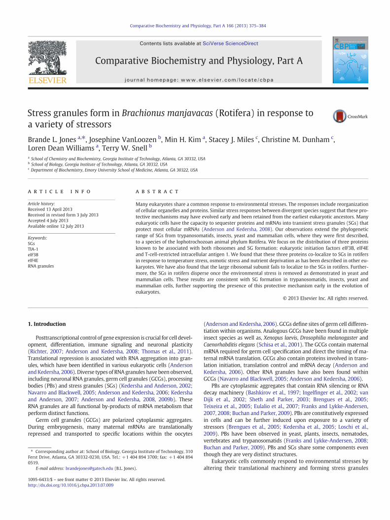
Having demonstrated the impact of our stress treatments on roti-fer survival, we followed cellular responses to these treatments. Weexamined the intracellular distribution of three proteins associatedwith stress granules, eIF3B, eIF4E and TIA-1, in response to each envi-ronmental stressor. We used immunohistochemistry and confocalmicroscopy to follow the localization of the three proteins withinthe cells of stressed rotifers (Fig. 2).
Neonates were fixed immediately after nutrient deprivation at 0, 24,48 or 72 h and probed with antibodies for localization of eIF4E (green),eIF3B (magenta) and TIA-1 (red). Nutrient deprivation led to obviousaggregation and co-localization of TIA-1, eIF4E and eIF3B into particlesresembling previous descriptions of stress granules. Manders' coeffi-cient for co-localization of the proteins was used for statistical analysis.It yielded a value of 0.349 in the control group and peaked at 0.728 at24 h of nutrient deprivation. This was followed by a decrease inco-localization at 48 h. The Manders' coefficient dropped to 0.338 at48 h (Fig. 3). A Manders' coefficient of 1 would indicate completeco-localization of the proteins, while a 0 indicates no localization.
For osmotic stress, neonates were transferred from 15-ppt ASW to15-ppt (control), 25-ppt, 35-ppt or 45-ppt ASW for 1 h and changes
Fig. 2. Microscopy images from Brachionus manjavacas immunohistochemistry. This is asingle slice of a confocal image illustrating the visualization of proteins and DNA usingfluorescent tags. The neonates were probed with antibodies specific for TIA-1 (red),eIF4E (green) and eIF3B (magenta). Nuclei were stained using Hoechst (blue). The bottompanel is a bright-fieldmicroscopic image showing B.manjavacasneonates. This image illus-trates the localization of theprobed proteins andDNAwithin neonate tissues. The scale baris 10 µm.
378 B.L. Jones et al. / Comparative Biochemistry and Physiology, Part A 166 (2013) 375–384

Fig. 3. eIF4E, eIF3B and TIA-1 are recruited to SGs in response to nutrient deprivation. Brachionus manjavacas neonates were deprived of nutrients for 0 h (control), 24 h and 48 h.They were fixed and probed with antibodies specific for eIF4E (green), eIF3B (magenta) and TIA-1 (red). The probed neonates were visualized using immunohistochemistry. Thebright-field microscopy image (BF) is provided to illustrate the localization of the proteins within the organs of the rotifers. Manders' correlation coefficient (Rr) shows that theproteins increasingly co-localize with increased time period of starvation, suggesting the formation of SGs. There is also a decrease in co-localization seen with extended durationsof nutrient deprivation. The scale bar is 20 µm.
Fig. 4. eIF4E, eIF3B and TIA-1 are recruited to SGs in response to osmotic stress. Brachionus manjavacas neonates were incubated in 15 ppt (control), 25 ppt or 35-ppt ASW for 1 hand fixed. The neonates were probed with antibodies specific for eIF4E (green), eIF3B (magenta) and TIA-1 (red). The fluorescently tagged macromolecules were visualized usingimmunohistochemistry. The bright field (BF) is provided to illustrate the localization of the proteins within the organs of the rotifers. The proteins are more uniformly distributedthroughout the cells in the control group (15-ppt ASW) and become co-localized with increasing osmotic shock (35-ppt ASW), indicating the formation of SGs. Rr value confirms theproteins co-localize with increased osmotic shock. The co-localization of these proteins provides further evidence for formation of SGs. The scale bar is 20 µm.
379B.L. Jones et al. / Comparative Biochemistry and Physiology, Part A 166 (2013) 375–384

in localization of the three proteins was assessed. After treatment,the neonates were immediately fixed and probed with antibodies. Os-motic stress led to co-localization of eIF4E, TIA-1 and eIF3B within thecorona, mastax, stomach and vitellarium of the rotifers. Manders' co-efficient of co-localization yielded a value of 0.354 in the control(15-ppt ASW), which increased to 0.994 in the 35-ppt sample (Fig. 4).
In the third experiment, neonates were heat stressed at 40 °C for0, 5, 10 or 20 min and immediately fixed and immunostained withantibodies. Heat stress resulted in the co-localization of TIA-1, eIF3Band eIF4E in the major organ systems of the neonates (Fig. 5). TheManders' coefficient was 0.02 in the control group (0 min heat stress)and increased to 1.00 in the rotifers subjected to 20 min of heatstress. The protein aggregation dissipated after recovery from heatstress for 3 h at 22 °C. The Manders' coefficient was 0.289 in the con-trol group; it increased to 0.734 after 30 min of heat stress anddropped back down to 0.375 with 3 h of removal from the heat stress(Fig. 6).
3.2. The large ribosomal subunit does not co-localize to SGs
We probed the heat stressed and fixed neonates with C-puromycinconjugated to fluorescein isothiocyanate (FITC) to discern the locationof the large ribosomal subunit. C-puromycin binds to the A site of thelarge ribosomal subunit (Luhrmann et al., 1981; Gilly and Pellegrini,1985; Starck et al., 2004). Incubation with the fluorescently labeled
C-puromycin enabled us to visualize the location of the large ribo-somal subunit. The neonates were heat stressed at 40 °C for 0, 10or 30 min. They were then anesthetized, fixed and probed withC-puromycin-FITC, eIF3B and TIA-1 antibodies. Antibodies bindingto eIF4E were not used for this experiment because Alexa Fluor488 has a very similar excitation and emission spectra as FITC.The large ribosomal subunits aggregated when subjected to heatstress; however, they did not co-localize with eIF3B and TIA-1proteins of the small ribosomal subunit. The calculated Manders'coefficient for the co-localization of the large ribosomal subunitwith eIF3B was 0.781 in the control group and decreased to 0.348in the group subjected to heat stress (Fig. 7).
3.3. SGs respond to the presence of both puromycin and cycloheximide
Next, we tested the ability of the TIA-1, eIF3B and eIF4E proteins toaggregate in the presence of two antibiotics, cycloheximide and puro-mycin. Cycloheximide has been demonstrated to prevent stress gran-ule formation in both yeast and mammalian cells, while puromycinpromotes stress granule formation in both yeast and mammaliancells (Kedersha et al., 2000; Anderson and Kedersha, 2002a, 2008;Grousl et al., 2009; Cassola, 2011). These proteins aggregated in thepresence of puromycin but failed to aggregate in the presence of cy-cloheximide. The Manders' coefficient for co-localization began at
Fig. 5. eIF4E, eIF3B and TIA-1 are recruited to SGs in response to heat stress. Brachionus manjavacas neonates were incubated at 40 °C for 0, 5, 10 and 20 min. The organisms wereimmediately fixed and probed with antibodies specific for eIF4E (green), eIF3B (magenta) and TIA-1 (red). The bright field (BF) is provided to illustrate the localization of the pro-teins within the organs of the rotifers. The localization of the probed proteins shifts from being more uniformly distributed throughout the neonates to a significant amount ofco-localization within major organs. The Rr values calculated using Manders' coefficient confirms the co-localization of the proteins. The data indicate the formation of SGs withinthe organs in response to heat stress. The scale bar is 20 µm.
380 B.L. Jones et al. / Comparative Biochemistry and Physiology, Part A 166 (2013) 375–384

0.358 in the control group. The Manders' coefficient increased to0.992 in animals incubated in the presence of puromycin (Fig. 8).
4. Discussion
We have analyzed the impact of environmental stresses onB. manjavacas neonates. We used fluorescent confocal microscopy
to demonstrate for the first time in the phylum Rotifera thatmammalian-like SGs form in response to heat stress, osmotic stressand nutrient deprivation. This extension of stress granule formationto lophotrochozoan invertebrates suggests that it may be part of ageneralized stress response in most animals.
Brachinoid rotifers are adapted to environmentswhere their growthis episodically restricted by harsh environments (Denekamp et al.,
Fig. 6. The SGs disperse after removal of the organism from the environmental stress. Neonates were heated at 40 °C for 30 min then were allowed to recover for 3 h at room tem-perature (22 °C). Each experimental group was immediately fixed and probed with antibodies specific for eIF4E (green), eIF3B (magenta) and TIA-1 (red). The cells were visualizedusing confocal microscopy. The proteins co-localize at the beginning of the environmental stress and disperse after its removal. The Manders' coefficient (Rr) increases with theinitial environmental stress; however, it decreases when the stress is removed. These data suggest that SG formation within neonates is a dynamic response to environmentalstress. The scale bar is 20 µm.
Fig. 7. The large ribosomal subunit does not localize to SGs. Neonates were heated at 40 °C for 0 and 30 min. The neonates were immediately removed from the heat stress, fixedand probed with antibodies specific for eIF3B (magenta) and TIA-1 (red). Puromycin conjugated to fluorescein isothiocyanate (FITC, green) was used to probe for the large ribo-somal subunit. The cells were visualized using confocal microscopy. eIF3Β and TIA-1 co-localize upon exposure to environmental stress, suggesting the formation of SGs. In contrast,the large ribosomal subunit aggregates in a separate location (green). The Manders' correlation coefficient (Rr) provides further evidence that the large ribosomal subunit doesnot co-localize to SGs. These data indicate that the large ribosomal subunit does not localize to stress granules. SGs found in both mammalian and yeast cells also lack the largeribosomal subunit. The constituents of SGs in Brachionus manjavacas neonates appear to be consistent with what has been seen in other eukaryotes. The scale bar is 20 µm.
381B.L. Jones et al. / Comparative Biochemistry and Physiology, Part A 166 (2013) 375–384

2009). Several adaptations have been suggested to explain the capabil-ity of organisms to survive environmental stresses, particularly treha-lose, sucrose, yolk proteins and LEA proteins (Crowe et al., 1998;Hoekstra et al., 2001; Boschetti et al., 2011; Hand et al., 2011; Jones etal., 2012). Although the precise function of SGs in the physiology of ro-tifers is yet to be understood, their formation ismost probably related tosurvival during periods of stress. SG formation might allow for tempo-rary suspension of energy consuming protein synthesis until the envi-ronmental stress has passed or until the rotifer moves to a morefavorable environment. Interestingly, the survival of animals decreasedin groups that displayed a decrease in SG formation during periods ofstress. This was especially evident in rotifers with extended periods ofnutrient deprivation.
Ourwork identifies three types of environmental factors that inducestress granule formation and inhibit translation in rotifers: nutrientdeprivation, heat stress and osmotic stress. Rotifers often occupy habi-tats that are only temporarily suitable for growth (García-Roger et al.,2006). We found that nutrient deprivation for more than 48 h, osmoticchanges where animals were transferred from 15 to more than 35 pptand temperature stress at 40 °C formore than 1 h stressed the neonatesto their limits for survival. These stressful conditions are similar to thosethe neonates could encounter in natural environments, and have beendocumented as conditions that negatively affect neonate hatching(Minkoff et al., 1983; Pérez-Legaspi and Rico-martínez, 1998; García-Roger et al., 2006; Dahms et al., 2011).
We identified TIA1-like, eIF4E-like and eIF3B-like proteins inB. manjavacas that bind antibodies raised against similar mammalianproteins. These proteins may play essential roles in the stress responseduring periods of nutrient deprivation, thermal and osmotic stress.TIA-1 is an RNA binding protein known to promote arrest of translationin environmentally stressed cells (Kedersha and Anderson, 2002; Gilkset al., 2004). The overexpression of TIA-1 induces stress granule
formation even in the absence of stress (Gilks et al., 2004). Likewise,cells that lack TIA-1 exhibit impaired stress granule assembly(Kedersha et al., 1999; Gilks et al., 2004). A key conserved componentof eIF3 is required for SG formation (Anderson and Kedersha, 2009a).Ohn et al. have shown that knockdown of eIF3 subunits inhibits stressgranule assembly (Ohn et al., 2008).
Using immunohistochemistry, we followed the localization of TIA-1,eIF3B and eIF4E in rotifers during environmental stress. Our finding ofthe co-localization of these three proteins during periods of nutrientdeprivation, osmotic and thermal stress are consistent with previousstudies with trypanosomatids, animal and yeast models of stress gran-ule formation (Kedersha and Anderson, 2002, 2007; Anderson andKedersha, 2008; Kramer et al., 2008; Farny et al., 2009; Grousl et al.,2009; Cassola, 2011; Thomas et al., 2011).
Thermal stress induced the greatest extent of protein co-localizationin B.manjavacas. Thermal stress has also been found to be a powerful in-ducer of stress granule formation in Saccharomyces cerevisiae (Grousl etal., 2009), D. melanogaster (Farny et al., 2009), and Trypanosoma brucei(Kramer et al., 2008). This pattern of SG formation is found in all ani-mals thus far investigated, with the exception being yeast (Buchanand Parker, 2009; Grousl et al., 2009).
SGs in B. manjavacas neonates failed to form in the presence ofcycloheximide; however, incubation in the presence of puromycindid promote stress granule formation. This result is consistent withdata from trypanosomatids, yeast and mammalian cells treated withthese drugs. Studies have shown that cycloheximide prevents SG for-mation and treatment with puromycin does not inhibit the formationof SGs in the presence during heat stress experiments (Kramer et al.,2008; Cassola, 2011). The rotifer stress granules disassembled oncethe heat stress was removed. This dynamic behavior is also seen inboth mammalian and yeast cells suggesting a conserved evolutionaryrole for SGs (Anderson and Kedersha, 2002a).
Fig. 8. eIF3B, eIF4E and TIA-1 are recruited to SGs respond to the presence of the antibiotic puromycin, not cycloheximide. Brachionus manjavacas neonates were incubated in 30 µMpuromycin or 30 µM cycloheximide for 30 min. The neonates were probed with antibodies specific for eIF3B (magenta), eIF4E (green) and TIA-1 (red). The proteins are uniformlydistributed throughout the cells in the control group (unstressed rotifers) and co-localize upon treatment with puromycin but not cycloheximide. This indicates the formation ofstress granules. These results are consistent with SG formation in trypanosomatids, yeast and mammalian cells. SGs also fail to form within trypanosomatids, yeast and mammaliancells in the presence of cycloheximide. The scale bar is 20 μm.
382 B.L. Jones et al. / Comparative Biochemistry and Physiology, Part A 166 (2013) 375–384

While the presence of SGs in rotifers is certain, we certainly ob-served that the appearance, disappearance and protein make up of theobserved SGs vary with the type of environmental stress as well asthe duration of the environmental stress. These observations are consis-tentwithwhat has been observed in the SGs of other eukaryotes, specif-ically both yeast and mammalian cells. Heterogeneous populations ofall RNA granules have been observed (Thomas et al., 2011). Both PBsand SGs are highly dynamic. Proteins and RNA have been observedbeing exchange among the two. SGs and PBs can also release both tran-scripts and proteins in order to promote translation of messages(Anderson and Kedersha, 2006; Franks and Lykke-Andersen, 2008;Buchan and Parker, 2009). Furthermore, the composition of SGs variesduring any one-stress response. Also, SGs can be subtly differentdepending on the nature of the stress condition (Buchan and Parker,2009; Buchan et al., 2011).
SGs have been implicated in signaling cascades that determinesurvival in a variety of eukaryotes. Therefore SGs appear to be anear universal and versatile means of managing cellular stress(Nilsson and Sunnerhagen, 2011). Cells prevented from assemblingSGs show a reduced level of survival during periods of environmentalstress (McEwen et al., 2005; Kwon et al., 2007; Kim et al., 2012), andthe assembly of SGs enhances the survival of stressed cells (Andersonand Kedersha, 2008). The identification of SGs in the Phylum Rotiferafurther suggests that SGs have an ancient origin, early in the evolu-tion of animals.
Acknowledgments
We thank Jessica Bowman for her critical review of this manu-script. This work was supported by the NASA Astrobiology Instituteand the Center for Ribosomal Origins and Evolution. C.M.D is a PewScholar in the Biomedical Sciences.
Appendix A. Supplementary data
Supplementary data to this article can be found online at http://dx.doi.org/10.1016/j.cbpa.2013.07.009.
References
Anderson, P., Kedersha, N., 2002a. Stressful initiations. J. Cell Sci. 115, 3227–3234.Anderson, P., Kedersha, N., 2002b. Visibly stressed: the role of eIF2, TIA-1, and stress
granules in protein translation. Cell Stress Chaperones 7, 213–221.Anderson, P., Kedersha, N., 2006. RNA granules. J. Cell Biol. 172, 803–808.Anderson, P., Kedersha, N., 2008. Stress granules: the Tao of RNA triage. Trends
Biochem. Sci. 33, 141–150.Anderson, P., Kedersha, N., 2009a. RNA granules: post-transcriptional and epigenetic
modulators of gene expression. Nat. Rev. Mol. Cell Biol. 10, 430–436.Anderson, P., Kedersha, N., 2009b. Stress granules. Curr. Biol. 19, R397–R398.Bashkirov, V.I., Scherthan, H., Solinger, J.A., Buerstedde, J.M., Heyer, W.D., 1997. A
mouse cytoplasmic exoribonuclease (mXRN1p) with preference for G4 tetraplexsubstrates. J. Cell Biol. 136, 761–773.
Boschetti, C., Pouchkina-Stantcheva, N., Hoffmann, P., Tunnacliffe, A., 2011. Foreigngenes and novel hydrophilic protein genes participate in the desiccation responseof the bdelloid rotifer Adineta ricciae. J. Exp. Biol. 214, 59–68.
Brengues, M., Teixeira, D., Parker, R., 2005. Movement of eukaryotic mRNAs betweenpolysomes and cytoplasmic processing bodies. Science 310, 486–489.
Buchan, J.R., Parker, R., 2009. Eukaryotic stress granules: the ins and outs of translation.Mol. Cell 36, 932–941.
Buchan, J.R., Yoon, J.H., Parker, R., 2011. Stress-specific composition, assembly andkinetics of stress granules in Saccharomyces cerevisiae. J. Cell Sci. 124, 228–239.
Cassola, A., 2011. RNA granules living a post-transcriptional life: the trypanosomes'case. Curr. Chem. Biol. 5, 108–117.
Cheng, S., Gallie, D.R., 2006. Wheat eukaryotic initiation factor 4B organizes assemblyof RNA and eIFiso4G, eIF4A, and poly(A)-binding protein. J. Biol. Chem. 281,24351–24364.
Craig, A.W., Haghighat, A., Yu, A.T., Sonenberg, N., 1998. Interaction of polyadenylate-binding protein with the eIF4G homologue PAIP enhances translation. Nature392, 520–523.
Crowe, J.H., Carpenter, J.F., Crowe, L.M., 1998. The role of vitrification in anhydrobiosis.Annu. Rev. Physiol. 60, 73–103.
Dahms, H.U., Hagiwara, A., Lee, J.S., 2011. Ecotoxicology, ecophysiology, and mechanis-tic studies with rotifers. Aquat. Toxicol. 101, 1–12.
Denekamp, N.Y., Thorne, M.A., Clark, M.S., Kube, M., Reinhardt, R., Lubzens, E., 2009.Discovering genes associated with dormancy in the monogonont rotifer Brachionusplicatilis. BMC Genomics 10, 108.
Denekamp, N.Y., Reinhardt, R., Kube, M., Lubzens, E., 2010. Late embryogenesis abun-dant (LEA) proteins in nondesiccated, encysted, and diapausing embryos of roti-fers. Biol. Reprod. 82, 714–724.
Eulalio, A., Behm-Ansmant, I., Izaurralde, E., 2007. P bodies: at the crossroads of post-transcriptional pathways. Nat. Rev. Mol. Cell Biol. 8, 9–22.
Farny, N.G., Kedersha, N.L., Silver, P.A., 2009. Metazoan stress granule assembly is medi-ated by P-eIF2alpha-dependent and -independent mechanisms. RNA 15, 1814–1821.
Fontaneto, D., Giordani, I., Melone, G., Serra, M., 2007. Disentangling the morphologicalstasis in two rotifer species of the Brachionus plicatilis species complex. Hydrobiologia583, 297–307.
Franks, T.M., Lykke-Andersen, J., 2007. TTP and BRF proteins nucleate processing bodyformation to silence mRNAs with AU-rich elements. Genes Dev. 21, 719–735.
Franks, T.M., Lykke-Andersen, J., 2008. The control of mRNA decapping and P-bodyformation. Mol. Cell 32, 605–615.
García-Roger, E.M., Martínez, A., Serra, M., 2006. Starvation tolerance of rotifers pro-duced from parthenogenetic eggs and from diapausing eggs: a life table approach.J. Plankton Res. 28, 257–265.
Gilks, N., Kedersha, N., Ayodele, M., Shen, L., Stoecklin, G., Dember, L.M., Anderson, P.,2004. Stress granule assembly is mediated by prion-like aggregation of TIA-1.Mol. Biol. Cell 15, 5383–5398.
Gilly, M., Pellegrini, M., 1985. Puromycin photoaffinity labels small- and large-subunitproteins at the A site of the Drosophila ribosome. Biochemistry 24, 5781–5786.
Grousl, T., Ivanov, P., Frydlova, I., Vasicova, P., Janda, F., Vojtova, J., Malinska, K.,Malcova, I., Novakova, L., Janoskova, D., Valasek, L., Hasek, J., 2009. Robust heatshock induces eIF2alpha-phosphorylation-independent assembly of stress gran-ules containing eIF3 and 40S ribosomal subunits in budding yeast, Saccharomycescerevisiae. J. Cell Sci. 122, 2078–2088.
Hand, S.C., Menze, M.A., Toner, M., Boswell, L., Moore, D., 2011. LEA proteins duringwater stress: not just for plants anymore. Annu. Rev. Physiol. 73, 115–134.
Hasenohrl, D., Lombo, T., Kaberdin, V., Londei, P., Blasi, U., 2008. Translation initiation fac-tor a/eIF2(!gamma) counteracts 5′ to 3′ mRNA decay in the archaeon Sulfolobussolfataricus. Proc. Natl. Acad. Sci. U. S. A. 105, 2146–2150.
Higgins, D.G., BLeasby, A.J., Fuchs, R., 1992. Clustal V: improved software for multiplesequence alignment. Comput. Appl. Biosci. 8, 189–191.
Hoekstra, F.A., Golovina, E.A., Buitink, J., 2001. Mechanisms of plant desiccation toler-ance. Trends Plant Sci. 6, 431–438.
Ingelfinger, D., Arndt-Jovin, D.J., Luhrmann, R., Achsel, T., 2002. The human LSm1-7proteins colocalize with the mRNA-degrading enzymes Dcp1/2 and Xrnl in distinctcytoplasmic foci. RNA 8, 1489–1501.
Jones, B.L., Schneider, D.M., Snell, T.W., 2012. Thermostable proteins in the diapausingeggs of Brachionus manjavacas (Rotifera). Comp. Biochem. Physiol. A Mol. Integr.Physiol. 162, 193–199.
Kedersha, N., Anderson, P., 2002. Stress granules: sites of mRNA triage that regulatemRNA stability and translatability. Biochem. Soc. Trans. 30, 963–969.
Kedersha, N., Anderson, P., 2007. Mammalian stress granules and processing bodies.Methods Enzymol. 431, 61–81.
Kedersha, N.L., Gupta, M., Li, W., Miller, I., Anderson, P., 1999. RNA-binding proteinsTIA-1 and TIAR link the phosphorylation of eIF-2 alpha to the assembly of mamma-lian stress granules. J. Cell Biol. 147, 1431–1442.
Kedersha, N., Cho, M.R., Li, W., Yacono, P.W., Chen, S., Gilks, N., Golan, D.E., Anderson, P.,2000. Dynamic shuttling of TIA-1 accompanies the recruitment of mRNA to mam-malian stress granules. J. Cell Biol. 151, 1257–1268.
Kedersha, N., Chen, S., Gilks, N., Li, W., Miller, I.J., Stahl, J., Anderson, P., 2002. Evidencethat ternary complex (eIF2-GTP-tRNA(i)(Met))-deficient preinitiation complexesare core constituents of mammalian stress granules. Mol. Biol. Cell 13, 195–210.
Kedersha, N., Stoecklin, G., Ayodele, M., Yacono, P., Lykke-Andersen, J., Fritzler, M.J.,Scheuner, D., Kaufman, R.J., Golan, D.E., Anderson, P., 2005. Stress granules andprocessing bodies are dynamically linked sites of mRNP remodeling. J. Cell Biol.169, 871–884.
Kim, B., Cooke, H.J., Rhee, K., 2012. DAZL is essential for stress granule formation impli-cated in germ cell survival upon heat stress. Development 139, 568–578.
Kimball, S.R., Horetsky, R.L., Ron, D., Jefferson, L.S., Harding, H.P., 2003. Mammalianstress granules represent sites of accumulation of stalled translation initiationcomplexes. Am. J. Physiol. Cell Physiol. 284, C273–C284.
Kramer, S., Queiroz, R., Ellis, L., Webb, H., Hoheisel, J.D., Clayton, C., Carrington, M.,2008. Heat shock causes a decrease in polysomes and the appearance of stressgranules in trypanosomes independently of eIF2(alpha) phosphorylation atThr169. J. Cell Sci. 121, 3002–3014.
Kwon, S., Zhang, Y., Matthias, P., 2007. The deacetylase HDAC6 is a novel critical compo-nent of stress granules involved in the stress response. Genes Dev. 21, 3381–3394.
Loschi, M., Leishman, C.C., Berardone, N., Boccaccio, G.L., 2009. Dynein and kinesin reg-ulate stress-granule and P-body dynamics. J. Cell Sci. 122, 3973–3982.
Luhrmann, R., Bald, R., Stoffler-Meilicke, M., Stoffler, G., 1981. Localization of the puromy-cin binding site on the large ribosomal subunit of Escherichia coli by immunoelectronmicroscopy. Proc. Natl. Acad. Sci. U. S. A. 78, 7276–7280.
Manders', E.M., Stap, J., Brakenhoff, G.J., van Driel, R., Aten, J.A., 1992. Dynamics of three-dimensional replication patterns during the S-phase, analysed by double labelling ofDNA and confocal microscopy. J. Cell Sci. 103 (Pt 3), 857–862.
Manders', E.M., Verbeek, F.J., Aten, J.A., 1993. Measurement of co-localization of objectsin dual-colour confocal images. J. Microsc. 169, 375–382.
McEwen, E., Kedersha, N., Song, B., Scheuner, D., Gilks, N., Han, A., Chen, J.J., Anderson,P., Kaufman, R.J., 2005. Heme-regulated inhibitor kinase-mediated phosphoryla-tion of eukaryotic translation initiation factor 2 inhibits translation, induces stress
383B.L. Jones et al. / Comparative Biochemistry and Physiology, Part A 166 (2013) 375–384

granule formation, and mediates survival upon arsenite exposure. J. Biol. Chem.280, 16925–16933.
Minkoff, G., Lubzens, E., Kahan, D., 1983. Environmental factors affecting hatching ofrotifer (Brachionus plicatilis) resting eggs. Hydrobiologia 104, 61–69.
Navarro, R.E., Blackwell, T.K., 2005. Requirement for P granules and meiosis for accu-mulation of the germline RNA helicase CGH-1. Genesis 42, 172–180.
Nilsson, D., Sunnerhagen, P., 2011. Cellular stress induces cytoplasmic RNA granules infission yeast. RNA 17, 120–133.
Nover, L., Scharf, K.D., Neumann, D., 1989. Cytoplasmic heat shock granules are formedfrom precursor particles and are associated with a specific set of mRNAs. Mol. Cell.Biol. 9, 1298–1308.
Ohn, T., Kedersha, N., Hickman, T., Tisdale, S., Anderson, P., 2008. A functional RNAiscreen links O-GlcNAc modification of ribosomal proteins to stress granule andprocessing body assembly. Nat. Cell Biol. 10, 1224–1231.
Pérez-Legaspi, I.A., Rico-martínez, R., 1998. Effect of temperature and food concentra-tion in two species of littoral rotifers. Hydrobiologia 387, 341–348.
Rajesh, K., Iyer, A., Suragani, R.N., Ramaiah, K.V., 2008. Intersubunit and interprotein in-teractions of alpha- and beta-subunits of human eIF2: effect of phosphorylation.Biochem. Biophys. Res. Commun. 374, 336–340.
Richter, J.D., 2007. CPEB: a life in translation. Trends Biochem. Sci. 32, 279–285.Rico-Martínez, R., Snell, T.W., 1997. Comparative binding of antibody to a mate recog-
nition pheromone on female Brachionus plicatilis and Brachionus rotundiformis(Rotifera). Hydrobiologia 358, 71–76.
Schisa, J.A., Pitt, J.N., Priess, J.R., 2001. Analysis of RNA associated with P granules ingerm cells of C. elegans adults. Development 128, 1287–1298.
Scriven, D.R., Lynch, R.M., Moore, E.D., 2008. Image acquisition for colocalization usingoptical microscopy. Am. J. Physiol. Cell Physiol. 294, C1119–C1122.
Sheth, U., Parker, R., 2003. Decapping and decay of messenger RNA occur in cytoplas-mic processing bodies. Science 300, 805–808.
Smith, H.A., Burns, A.R., Shearer, T.L., Snell, T.W., 2012. Three heat shock proteins areessential for rotifer thermotolerance. J. Exp. Mar. Biol. Ecol. 413, 1–6.
Snell, T.W., Moffat, B.D., Janssen, C., Persoone, G., 1991. Acute toxicity tests using roti-fers: IV. Effects of cyst age, temperature, and salinity on the sensitivity ofBrachionus calyciflorus. Ecotoxicol. Environ. Saf. 21, 308–317.
Snell, T.W., Kubanek, J., Carter, W., Payne, A.B., Kim, J., Hicks, M.K., Stelzer, C., 2006. Aprotein signal triggers sexual reproduction in Brachionus plicatilis (Rotifera). Mar.Biol. 149, 763–773.
Starck, S.R., Green, H.M., Alberola-Ila, J., Roberts, R.W., 2004. A general approach to de-tect protein expression in vivo using fluorescent puromycin conjugates. Chem.Biol. 11, 999–1008.
Stout, E.P., La Clair, J.J., Snell, T.W., Shearer, T.L., Kubanek, J., 2010. Conservation of proges-terone hormone function in invertebrate reproduction. Proc. Natl. Acad. Sci. U. S. A.107, 11859–11864.
Teixeira, D., Sheth, U., Valencia-Sanchez, M.A., Brengues, M., Parker, R., 2005. Process-ing bodies require RNA for assembly and contain nontranslating mRNAs. RNA 11,371–382.
Thomas, M.G., Loschi, M., Desbats, M.A., Boccaccio, G.L., 2011. RNA granules: the good,the bad and the ugly. Cell. Signal. 23, 324–334.
Thompson, J.D., Higgins, D.G., Gibson, T.J., 1994. Clustal W: improving sensitivity ofprogressive multiple sequence alignment through sequence weighting, position-specific gap penalties and weight matrix choice. Nucleic Acids Res. 22, 4673–4680.
van Dijk, E., Cougot, N., Meyer, S., Babajko, S., Wahle, E., Seraphin, B., 2002. HumanDcp2: a catalytically active mRNA decapping enzyme located in specific cytoplas-mic structures. EMBO J. 21, 6915–6924.
384 B.L. Jones et al. / Comparative Biochemistry and Physiology, Part A 166 (2013) 375–384