community eye health journal · in this issue of the journal, we address the eye health needs of...
TRANSCRIPT

In this issue of the journal, we address the eye health needs of young children, focusing on those aged less than six years old. All of you who have tried to examine or measure the visual acuity of young children will know that this can be very challenging and difficult; it can be very tempting to give up and send the child home, particularly if the clinic is busy. We hope that, after reading this edition of the journal and putting into practice some of the practical suggestions, you will feel more confident in managing young children. Also, if referral is needed, you will have a better idea of the degree of urgency required and how this should be communicated to parents.
The impact of eye disease in childrenIt is said that almost three quarters of a child’s early learning comes through vision and that over one third of the adult visual cortex responds to visual stimuli. This tells us that vision is not only very important for early development in infancy, but also that visual information is used and processed by many different parts of the brain. It is, therefore, not surprising that visual loss early in a child’s life can have a major impact on that child’s development, leading to delays in crawling and walking, for example. Preventing visual loss, or ensuring that a child has the correct treatment at the correct time to restore visual function, will have a
major impact on the child’s development.The impact of visual loss also extends
beyond the child to the family: studies have shown that having a disabled child can increase stress and depression among parents and can lead to increases in divorce or separation.1 Conversely, some families find that it brings them closer together, as they jointly share the challenges of living with a disabled child.
Listening to the motherAll mothers, regardless of educational attainment, want what is best for their child and also know their child very well. They observe their children closely over long periods of time and under different lighting conditions and circumstances. Mothers will
notice if there is something wrong with the eye or eyes, or if their child is behaving differently. One of the key messages of this issue is that health workers need to listen to, and believe, mothers: they know more about their child than anyone.
With this in mind, the article on page 4 is based around what parents might say when they bring their child to see you. We hope that you will find this approach useful.
The challengesAssessing visual acuity in young children can be very difficult indeed, but children aged five years or above can usually be tested using a Snellen E or Landolt C chart. (Tip: If children can reach over the top of
When your eye patient is a child
Community Eye Health
Journal
Continues overleaf ➤
1 When your eye patient is a child Arvind Chanda and Clare Gilbert
4 Managing eye health in young children Aderonke Baiyeroju, Richard Bowman, Clare Gilbert, and David Taylor
12 Understanding, detecting and managing strabismus Eugene M Helveston
Arvind ChandnaConsultant Paediatric Ophthalmologist and Chair: Vision for Children, Alder Hey Children’s Hospital. Liverpool, UK.Clare Gilbert Co-director, International Centre for Eye Health, London School of Hygiene and Tropical Medicine; Clinical Advisor, Sightsavers, UK.
In ThIs IssUe
CC
BR
T/D
iete
r Tel
eman
s
1CommuniTy EyE HEalTH Journal | Vol 23 iSSuE 72 | marCH 2010
hOW TO17 Instilling eye drops and ointment in a baby or young child Ingrid Mason and Sue Stevens
12 UseFUL ResOURCes
19 CPD: TesT YOURseLF
20 neWs AnD nOTICes
16 Looking after young eye patients in hospital Caroline Ayilo, Dianne Pickering, Fay Gallant, and Ingrid Mason
18 how to make an eye unit child friendly Joan McLeod-Omawale and Alamgir Hossain
VolumE 23 | iSSuE 72 | marCH 2010
a young child with bilateral cataract. TanZania
Nowwith CPDSee page 19

Volume 23 | Issue 72 | March 2010
editorElmien Wolvaardt Ellison [email protected]
editorial committeeNick AstburyAllen FosterClare GilbertIan MurdochGVS MurthyDaksha PatelRichard WormaldDavid Yorston
special advisor for Issue 72Clare Gilbert
Regional consultantsSergey Branchevski (Russia)Miriam Cano (Paraguay)Professor Gordon Johnson (UK)Susan Lewallen (Tanzania)Wanjiku Mathenge (Kenya)Joseph Enyegue Oye (Francophone Africa)Babar Qureshi (Pakistan)BR Shamanna (India)Professor Hugh Taylor (Australia)Min Wu (China)Andrea Zin (Brazil)
AdvisorsLiz Barnett (Teaching and Learning) Catherine Cross (Infrastructure and Technology) Pak Sang Lee (Ophthalmic Equipment)Dianne Pickering (Ophthalmic Nursing)
editorial assistant Anita ShahDesign Lance BellersPrinting Newman Thomson
Online edition Sally Parsleyemail [email protected]
exchange articlesAnita Shah [email protected]
Website Back issues are available at:
www.cehjournal.orgsubscriptions and back issuesCommunity Eye Health Journal, International Centre for Eye Health, London School of Hygiene and Tropical Medicine, Keppel Street, London WC1E 7HT, UK.Tel +44 207 612 7964/72Fax +44 207 958 8317 email [email protected]
The Community Eye Health Journal is sent freeto applicants from low- and middle-income countries. French, Spanish, Portuguese, and Chinese translations are available and a special supplement is produced for India (in English). Please send details of your name, occupation, and postal address to the Community Eye Health Journal, at the address above. Subscription rates for applicants elsewhere: one year UK £50; three years UK £100. Send credit card details or an international cheque/banker’s order made payable to London School of Hygiene and Tropical Medicine to the address above.
© International Centre for Eye Health, LondonArticles may be photocopied, reproduced or translated provided these are not used for commercial or personal profit. Acknowledgementsshould be made to the author(s) and to Community Eye Health Journal. Woodcut-style graphics by Victoria Francis.
ISSN 0953-6833
The journal is produced in collaboration with the World Health Organization. Signed articles are the responsibility of the named authors alone and do not necessarily reflect the policies of the World Health Organization. The World Health Organization does not warrant that the information contained in this publication is complete and correct and shall not be liable for any damages incurred as a result of its use. The mention of specific companies or of certain manufacturers’ products does not imply that they are endorsed or recommended by the World Health Organization in preference to others of a similar nature that are not mentioned.
Supporting VISION 2020: The Right to Sight
When YOUR eYe PATIenT Is A ChILD Continued Community Eye Health
Journal their head with one arm, and touch the opposite ear, then they are at least five years old.) Below the age of five years, other methods need to be used to assess vision, such as matching tests, or recognising or finding small objects against a plain background. However, even in tertiary referral hospitals where all the latest equipment and acuity testing charts are available, formal visual acuity measurement is not always possible. Once again, we have to rely on what the mother has noticed or on what we can detect about the child’s visual behaviour. The chart on page 5 tells you what a normally sighted child should be able to do at different ages, so allowing you to identify children whose vision may be a cause for concern.
Examining young children can also be challenging as they do not understand what you are doing and are likely to be frightened of the whole experience. This issue contains some tips on how to examine babies and young children (page 6). There are also practical suggestions on how to look after a young child in hospital and how to support parents (page 16) as well as how to make a facility child friendly (page 18). There is also an article on understanding, diagnosing, and managing strabismus/squint (page 12).
You can make a differenceIt is important to realise that the correct action taken by an eye care worker can play a major role in preserving the sight of a child; correct action can even save a child’s life! For example, identification and correct referral of a child with a white reflex, which may indicate retinoblastoma, may lead to life-saving treatment – retinoblastoma is often fatal if not treated early. Correctly diagnosing the signs of vitamin A deficiency and giving a child high-dose vitamin A will also reduce the risk that the child will die. Studies undertaken in Indonesia indicate that children with Bitot’s spots and night blindness are fifteen times more likely to die than children without these signs.
Congenital or developmental cataract, one of the commonest treatable causes of blindness in children, requires early detection and treatment to prevent permanent visual impairment from amblyopia (‘lazy eye’). This
is also likely to be noticed by the mother first. Health workers can check for cataract using the red reflex test (page 11), which should ideally form part of the routine examination of all newborn babies.
Even when you suspect a child with an eye abnormality will not benefit from medical treatment, you should still refer that child to an ophthalmologist or tertiary eye care centre. Such children may be helped by refraction and low vision services, and it is important to refer them as soon as possible to minimise the impact of any visual impairment on their development.
Beyond the clinicWe encourage you to think about what you can do beyond the clinic to reduce eye disease and visual loss in children. For example, you could talk to staff who work in maternal and child health clinics, or immuni-sation staff, and suggest that they refer any child to you if they have concerns. Indeed, a programme in Uganda very successfully trained immunisation staff to look at the eyes of the infants and children they were immunising and ask the mothers whether they had any concerns. This intervention led to many children being referred earlier than they would have been for examination, assessment, and treatment.
You can encourage mothers of young children to ensure they receive all their immunisations and take their vitamin A supplement. Discussions with traditional birth attendants could include the impor-tance of cleaning the eyelids immediately after the head is delivered to prevent conjunctivitis of the newborn (ophthalmia neonatorum).
If you see one child with trachoma, vitamin A deficiency, or measles infection, it is likely that there are other children in the same area who have the same problem. You should find out which community or area the child is from and inform the relevant authority.
The ten key activities listed in on this page are those suggested by the World Health Organization and are intended for primary level staff.2 If these activities were to be
2 CommuniTy EyE HEalTH Journal | Vol 23 iSSuE 72 | marCH 2010
1 Clean the eyes immediately after birth and instil topical antibiotic eye ointment or topical antiseptic eye drops.
2 Give the mother 200,000 international units (IU) of vitamin A immediately after delivery.3 Promote breastfeeding and good nutrition.4 Immunise children against measles at nine months and give vitamin A 100,000 IU.
Encourage second immunisation for extra protection.5 Give any child with measles or suspected undernutrition vitamin A 100,000 IU (if less
than twelve months) or 200,000 IU (if twelve months of age or older).6 Keep children’s faces clean.7 Refer any child who cannot see well to an eye care worker as soon as possible.8 Urgently refer any child with a white pupil or other obvious abnormality to an eye care worker.9 Refer any child with a serious eye injury or a red eye to an eye care worker immediately.10 Do not put traditional medicines in the eyes.
Ten key activities for primary health care workers

widely and consistently implemented, then the effects will spread far beyond eye health and promote the overall health of children.
Health promotion and education, particularly of mothers and women of child-bearing age, is also important. Tell them what they can do to prevent visual loss and eye diseases in their child. The ten key activities are a good place to start.
It is also important to know that most communities hold beliefs about the causes and treatment of illness, including eye conditions. Many commu-nities believe that visual loss of early onset, particularly if it is congenital (i.e., since birth), cannot be treated. As a result, parents do not seek help. Therefore, if a parent attends a health facility very late, do not blame them, as it is likely that they hold the same beliefs as many others in their community.
In summaryRemember that children are not smaller versions of adults, nor are their eyes smaller versions of adult eyes – children’s eye care needs are different, and often more urgent than those of adults. It is true that managing young children in the clinic can be a challenge. However, there is a lot that you and your colleagues, as well as parents and the community, can do to prevent eye disease and visual loss in children. We hope that this issue will inspire you to provide the best possible eye care to children in a way that supports children and parents and makes the experience of the clinic less traumatic for them.
references1 Reichman NE, Corman H, Noonan K. Impact of
child disability on the family. Matern Child Health J. 2008 Nov;12(6): 679–83.
2 World Health Organization. A five year project for the prevention of childhood blindness. Report of WHO Consultation. Geneva 18–20 June 2002. Geneva; 2002.
3 Jones G, Steketee RW, Black RE, Bhutta ZA, Morris SS. How many child deaths can we prevent this year? Lancet. 2003 Jul 5;362(9377): 65–71.
4 Gilbert C, Foster A. Childhood blindness in the context of VISION 2020 – the right to sight. Bull World Health Organ. 2001;79(3): 227–32.
5 Ahmad OB, Lopez AD, Inoue M. The decline in child mortality: a reappraisal. Bull World Health Organ. 2000;78(10): 1175–91.
3
Under-5 mortality rates (U5MRs) can be used to estimate the prevalence of blindness in children (see Table 1). The justification for this is that many of the conditions that cause blindness in children are also causes of child mortality, such as measles, vitamin A deficiency disorders, meningitis, and congenital rubella.3 At the launch of VISION 2020 in 1999, there were estimated to be 1.4 million blind children in the world, almost three quarters of whom lived in low- and middle-income countries.4
Worldwide, over the last ten years, the total number of children aged 0–15 years has increased slightly, to almost 1.9 billion. However, U5MRs, and hence blindness prevalence estimates, have declined in most countries since 1980.5
By multiplying the number of children by the relevant blindness prevalence estimate, it has been possible to calculate that that the number of children who are blind globally has declined by around 10 per cent over the last ten years, to 1.26 million in 2010 (Table 2). Better measles immunisation and vitamin A supplementation are two important public health interventions which have undoubtedly contributed.
In Table 2, countries are grouped by World Bank region rather than geographical region, because socio-economic factors play such an important role in determining the prevalence and causes of visual loss in children.
The revised 2010 estimate suggests that the greatest changes have occurred in China and the Other Asia and Islands region (which includes Indonesia, the Philippines, and Bangladesh), where U5MRs, and hence blindness prevalence estimates, have dropped considerably and the child population has stayed relatively stable.
In Sub-Saharan Africa, the number of blind children has increased by 31%, in part because this is the only region with a signif-icant increase in the child population. However, in many countries in this region, U5MRs have increased, mainly as a consequence of the HIV epidemic and the large number of children who are orphaned, leading to poorer child health.
how to estimate the number of children who are blindFind the relevant U5MR for your country or region. This can be found in several publica-tions and documents, including UNICEF’s State of the World’s Children, which is published every year.
To estimate the number of children who are currently blind, use the U5MR of five years ago, i.e. for 2005, as this will reflect the most relevant time period.
Find the U5MR in Table 1 and read off the blindness prevalence estimate.
Multiply the prevalence by the number of children aged 0-15 years in your country to estimate the number of children who are blind.
Example: Country P has a total population of 12 million in 2010, and 40% are children aged 0–15 years. The number of children is therefore 12,000,000 x 40/100 = 4,800,000. The U5MR in 2005 was 112 per 1,000 live births. So the prevalence estimate is 0.8 per 1,000 children (from Table 1). In country P there will be 4,800,000 x 0.8/1000 = 3,840 children who are blind.
note: These estimates do not include blindness from refractive error as there is very little data on which to base estimates.
a boy plays after eye surgery. BanGlaDESH
Zul M
ukhi
da/S
ight
save
rs
under-5 mortality rate per 1,000 live births
Estimated prevalence of blindness per 1,000 children
0–19 0.3
20–39 0.4
40–59 0.5
60–79 0.6
80–99 0.7
100–119 0.8
120–139 0.9
140–159 1.0
160–179 1.1
180–199 1.2
200–219 1.3
220–239 1.4
240+ 1.5
Table 1. Under-5 mortality rates and blindness prevalence estimates in children
Table 2. Estimates of the number of blind children worldwide in 2010
How many children are blind?
2010 estimate % change between1999 and 2010
Child pop (millions)
Blind children
in child pop
in estimates of blind children
lower in 2010 than in 1999
China 340 116,000 0% - 44.8%
Other Asia and Islands
266 136,000 - 2.3% - 38.2%
EME + FSE 244 70,000 - 1.6% - 22.2%
Latin America and Caribbean
170 71,000 0% -29.0%
not much change
Middle East Crescent
241 168,000 +0.4% - 11.6%
India 345 280,000 - 1.4% + 3.7%
Higher in 2010 than in 1999
Sub-Saharan Africa
274 419,000 + 5.4% + 30.9%
ToTal: 1,880 1,260,000 +0.6% -10%EME = established market economies; FSE = former socialist economies. These regions have been combined as some countries were re-designated between 1999 and 2010.
Copyright © 2010 Arvind Chandna and Clare Gilbert. This is an open access article distributed under the Creative Commons Attribution License, which permits unrestricted use, distribution, and reproduction in any medium, provided the original work is properly cited.

4 CommuniTy EyE HEalTH Journal | Vol 23 iSSuE 72 | marCH 2010
In The CLInIC
Managing eye health in young children
Children are brought to us with a range of conditions, usually when their parents or carers notice something is wrong. This article focuses on the more challenging complaints in babies and young children, who are the most difficult to assess. This is not an exhaustive list of presenting complaints or examination techniques, but it will give a starting point.
General principlesWhen your patient is a young child:
• Do the best you can, and start treatment or refer the child as quickly as possible. The earlier the treatment starts, the better a child’s vision is likely to be after treatment. Even if there is no treatment, a blind baby or child still needs help to develop as normally as possible and should also be referred.
• Believe the parents. Most things parents notice and mention to you are real and relevant. Parents are usually right! They spend a great deal of time with their children, and will observe how children behave and what their eyes look like.
• listen more than you speak. Usually the parent will help you towards the diagnosis.
• Don’t take any chances – play it safe. If in doubt, ask a colleague or refer the child to a specialist.
• Be patient. It takes time to let the parents tell their story and to examine a child properly, especially one who cannot or will not cooperate.
• Plan ahead. If you have a busy clinic, see any young children first. If you don’t, they may get tired and irritable, which will cause stress for their parents or carers; it also makes children difficult to examine.
Communication with parentsGood communication with parents is essential:
• Speak in ways that parents can understand. Speak in simple, everyday terms and use diagrams or drawings to support your explanations.
• Be as honest as you can. This could include saying that you are uncertain of what exactly is wrong.
• Be kind. Parents want what is best for their children, but because of lack of education or resources they may not always make the best choices. Do not blame parents for what they have done, or what they have not done. This may make them less likely to seek further help. With careful explanation, you can help them to make the best decision for their child’s eyes and vision.
ReferralWhen you refer a child, it is very useful to write a referral letter. Give the letter to the parents to take with them and keep a copy for your records. In the letter, state:• what the mother complained of or noticed• what you found when you examined the
child• what you have done, if anything (e.g.,
started antibiotics).
It is important to encourage parents to take up a referral.
• Explain why you might refer the child.If a child needs to be referred, it is very important to convince the parents that
specialist testing and treatment will help their child.
• Help parents to understand the urgency of seeking further help.Tell them how important it is to get the advice. However, don’t alarm parents unnecessarily. Explain that the quicker a child gets treatment, the better the outcome will be.
• Be supportive. Advise parents about what support is available to help them take up the referral, such as transport, subsidies, and so on. If you can, tell them what to expect at the hospital and what they should bring with them (such as the referral letter, clothes, or food).
Always refer children with the following eye problems urgently:
• One or both eyes are abnormally small or large (Figure 1)
• One or both eyes stick out (Figure 2)• There is a red mark on the eyelid (Figure 3)• One or both eyes are obviously abnormal;
for example, white all over (Figure 4).
Assessing vision in a baby (0–1 year) Don’t be anxious about examining a baby. If the baby is awake and attentive, there is a lot you can find out by asking the parents and simply observing the baby’s reactions.
• First ask the parents what they think about their baby’s vision.
• Notice how the baby looks at things in the room, such as the window or any lights.
Aderonke BaiyerojuProfessor of Ophthalmology, College of Medicine, University of Ibadan, Nigeria.
Richard BowmanOphthalmologist and Director of Training, CCBRT Hospital, Dar es Salaam, Tanzania; Honorary Senior Lecturer, London School of Hygiene and Tropical Medicine.
Clare GilbertCo-director, International Centre for Eye Health, London School of Hygiene and Tropical Medicine; Clinical Advisor, Sightsavers, UK.
David TaylorChairman, International Council of Ophthalmology Examinations, International Council of Ophthalmology, 11-43 Bath Street, London EC1V 9EL. [email protected]
always believe the mother. TanZania
CC
BR
T/D
iete
r Tel
eman
s
5CommuniTy EyE HEalTH Journal | Vol 23 iSSuE 72 | marCH 2010
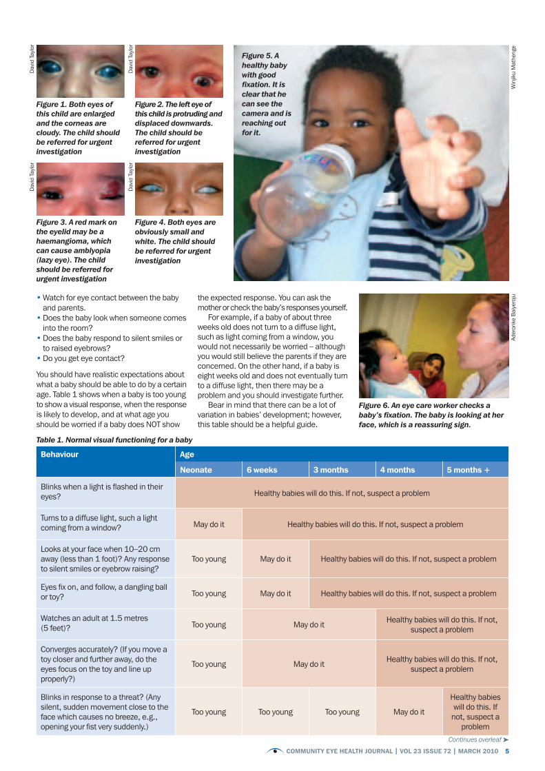
• Watch for eye contact between the baby and parents.
• Does the baby look when someone comes into the room?
• Does the baby respond to silent smiles or to raised eyebrows?
• Do you get eye contact?
You should have realistic expectations about what a baby should be able to do by a certain age. Table 1 shows when a baby is too young to show a visual response, when the response is likely to develop, and at what age you should be worried if a baby does NOT show
the expected response. You can ask the mother or check the baby’s responses yourself.
For example, if a baby of about three weeks old does not turn to a diffuse light, such as light coming from a window, you would not necessarily be worried – although you would still believe the parents if they are concerned. On the other hand, if a baby is eight weeks old and does not eventually turn to a diffuse light, then there may be a problem and you should investigate further.
Bear in mind that there can be a lot of variation in babies’ development; however, this table should be a helpful guide.
Figure 6. An eye care worker checks a baby’s fixation. The baby is looking at her face, which is a reassuring sign.
Figure 5. A healthy baby with good fixation. It is clear that he can see the camera and is reaching out for it.
Continues overleaf ➤
Behaviour age
neonate 6 weeks 3 months 4 months 5 months +
Blinks when a light is flashed in their eyes? Healthy babies will do this. If not, suspect a problem
Turns to a diffuse light, such a light coming from a window? May do it Healthy babies will do this. If not, suspect a problem
Looks at your face when 10–20 cm away (less than 1 foot)? Any response to silent smiles or eyebrow raising?
Too young May do it Healthy babies will do this. If not, suspect a problem
Eyes fix on, and follow, a dangling ball or toy? Too young May do it Healthy babies will do this. If not, suspect a problem
Watches an adult at 1.5 metres (5 feet)? Too young May do it Healthy babies will do this. If not,
suspect a problem
Converges accurately? (If you move a toy closer and further away, do the eyes focus on the toy and line up properly?)
Too young May do it Healthy babies will do this. If not, suspect a problem
Blinks in response to a threat? (Any silent, sudden movement close to the face which causes no breeze, e.g., opening your fist very suddenly.)
Too young Too young Too young May do it
Healthy babies will do this. If not, suspect a
problem
Table 1. Normal visual functioning for a baby
Figure 1. Both eyes of this child are enlarged and the corneas are cloudy. The child should be referred for urgent investigation
Figure 2. The left eye of this child is protruding and displaced downwards. The child should be referred for urgent investigation
Figure 3. A red mark on the eyelid may be a haemangioma, which can cause amblyopia (lazy eye). The child should be referred for urgent investigation
Figure 4. Both eyes are obviously small and white. The child should be referred for urgent investigation
Dav
id T
aylo
rD
avid
Tay
lor
Dav
id T
aylo
r
Win
jiku
Mat
heng
eAd
eron
ke B
aiye
roju
Dav
id T
aylo
r

In The CLInIC Continued
6 CommuniTy EyE HEalTH Journal | Vol 23 iSSuE 72 | marCH 2010
Tips for examining a baby• Try to carry out as much of the examination
as possible without touching the baby. Children often resist having their eyes held open, for example.
• Have many toys available (Figure 7). For each new toy, the baby will momentarily hold their eyes steady, allowing a quick examination. If available, use toys which are bright and can flash on and off. A good rule to remember is one toy, one look.
• Don’t be embarrassed about making funny noises! These help to attract the baby’s attention and to keep them interested and calm.
• In order to be able to do a more detailed examination in an infant, examine the child while he or she is being bottle fed or breast fed.
• If you are struggling, ask the parent’s permission to wrap the baby. Place the baby on a blanket or sheet, hold the arms to the side and the legs straight, and wrap the blanket around the body and arms (Figure 8). Ask the parent to hold the baby. Either the parent or a helper can then carefully open one eye at a time for the examination (without putting pressure on the eye – see Figure 2 on page 17). Remember that this may be very stressful for both the baby and the parent.
Cla
re G
ilber
t
Cla
re G
ilber
t
ICEH
Figure 9. Be playful and make a game of the examination
Figure 7. A lot of toys are needed to attract children’s attention and to make examination fun!
Figure 8. Wrapping a baby for an eye examination
Assessing vision in a young child (1–5 years)Children in this age group should have steady eyes, no squint, no history of sight difficulties and, if in a good mood, show interest in colourful or interesting objects in the room. They should respond to silent smiles, eyebrow raising, and winking.
Children in this age group should also be able to see objects presented in their peripheral visual field by a colleague while you draw their attention to your face, perhaps by making a funny noise. Cover one eye at a time if the child will allow it and ask them to identify different sized objects or, with older children, letters – make it a game.
Many children can accurately name colours by the age of three years but many cannot until they are older; it is reassuring if they can.
After the age of three, most children can participate in accurate visual acuity, visual field, and colour vision testing by someone trained and with age-appropriate equipment.
If you do not have that equipment or have not been trained to use it, you can still test a child’s functional vision using everyday objects as described above.
Tips for examining a young childThe tips for examining a baby (above) apply equally well to young children. In addition:
• Be playful and make a game of the examination (Figure 9). For example,
shine a light into the mother’s eye first, or pretend you are playing ‘hide and seek’ or ‘peekaboo’ when covering one eye.
• Observe children when they don’t know they are being observed, for example while you are talking to the mother or taking a history.
• The tip about wrapping up a baby will work for a younger child, but may be more difficult in an older child. Ask the parents what they think would be appropriate or would work best. For example, parents may prefer to hold their child’s arms gently.
‘Observe children when they don’t know they are being observed, for example while you are talking to the mother or taking a history’
The rest of this article is divided into four sections, each of which is based on what the mother or parents will say when they bring their child to see you:
1 “My child cannot see”2 “There is something white in my
child’s eye(s)”3 “My child’s eyes are wobbly” or
“My child has a squint”4 “My child’s eyes are red and/or sticky”
For each problem, the article describes the likely causes, what you should ask the parents, what you should look for, what action you should take, and how you can talk to the parents. Where appro-priate, these are described separately for babies and young children. We hope you find this useful!
How to use this article

7CommuniTy EyE HEalTH Journal | Vol 23 iSSuE 72 | marCH 2010
Possible causes Further possible causes: babies
Further possible causes: young children
• Corneal scar/opacity• Cataract• Glaucoma• Developmental problems (retina, optic nerve, brain)
• Retinal conditions such as meningitis and retinopathy of prematurity (ROP), which is rare in Africa
• Central nervous system conditions, e.g. following prolonged or difficult birth
• Retinal conditions, such as retinal dystrophies, CMV retinitis (a complication of HIV), late presentation of ROP
• Central nervous system conditions, e.g. following meningitis, malaria, or head injuries
What to ask the parents additional questions:babies
additional questions: young children
• When did you first suspect there was something wrong with your child’s vision?
• Does your child dislike bright light? If yes, suspect glaucoma or some form of retinal dystrophy.
• Does the eye water? If yes, this may simply be a blocked nasolacrimal duct, in which case the eye will probably also be sticky. However, if the watering happens when the child is in bright light, or if the child also cannot see or is in pain, you should suspect congenital glaucoma (Figure 10).
• Does the baby seem to be in pain? If yes, it may be glaucoma or there may be a problem with the cornea.
• Was the baby premature and cared for in a neonatal unit? If yes, it may be ROP.
• Was the birth of the baby difficult or long? If yes, it may be cerebral visual impairment.
• Is there a history of fever? If yes, suspect neonatal meningitis.
• Is there a history of head injury or fever immediately before the difficulty with vision was noticed? If yes, suspect a central nervous system condition.
• Can the child walk around and hear normally? If no, suspect a central nervous system condition.
• Do the parents or brothers and sisters have (similar) vision problems? If yes, suspect an inherited retinal problem or an environmental problem such as maternal ingestion of drugs or alcohol.
What to look for What else to look for: babies
What else to look for: young children
• Use a torch to examine the cornea. Is there a corneal ulcer or scar/opacity? How big is it? Is the pupil completely covered?
• Check the lens in each eye. Use a torch to look just behind the pupil. A cataract will appear white.
• Do the red reflex test (see box on page 11). A cataract blocks the red reflex, so it will appear black or partially black (Figure 11).
• Assess the visual milestones given in Table 1, page 5.
• Assess the child’s vision using the tips on page 6.
Hint: In this age group, doing a red reflex test is often easier because you can turn it into a game. For instance, tell a child of two or three years old to ‘blow the light out’ (you switch it off!). You don’t need to get very near: 30 cm (about 1 foot) will do, as long as the light is bright. Practise on an older brother or sister, or the mother, first; this reassures the child that the test is not frightening.
1 “My child cannot see”P
Khaw
Dav
id T
aylo
r
What to do• Always refer babies or young children who
have something obviously wrong with their eyes and/or vision
• Always refer when you and/or the parents are concerned about the baby’s vision and when you think their vision may be outside the normal for their age.
• Remember to err on the side of caution – always believe the mother. If you are unsure, it is better to refer than to miss something serious.
• When a baby needs a referral, refer him or her to an ophthalmologist, preferably one trained in paediatric ophthalmology, whatever the suspected cause.
remember: Cataracts in children are not the same as cataracts in adults. Children with visual loss from cataracts need urgent surgical treatment to prevent them from developing amblyopia (lazy eyes) which may not be reversible if surgery is delayed. They should not be told to “wait for the cataract to mature”; nor to “come back when your child is older”. These messages can cause delays in treatment which can have a lasting impact on the child.
What to tell parents when you refer their child• It is important to persuade parents to take
up their baby’s referral urgently – just as
soon as they can. The sooner the exact nature of the condition is known, the sooner they can be treated and the better the outcome is likely to be.
• Say something like this: “It’s difficult for me to find out exactly what is wrong and/or how much your child can see – your child may need more tests. Knowing exactly what is wrong will help us find out whether your child’s condition can be treated.”
• Try to dissuade parents from seeking the advice of traditional healers or using traditional remedies. These may be harmful and may delay much-needed investigation and treatment.
Figure 10. Congenital glaucoma
Figure 11. The pupil was dilated using one drop of cyclopentolate 0.5%. The cataract is visible as a black shadow obstructing the red reflex.
Continues overleaf ➤

8 CommuniTy EyE HEalTH Journal | Vol 23 iSSuE 72 | marCH 2010
In The CLInIC Continued
Possible causes Further possible causes:babies
Further possible causes:young children
1 on the surface of the eye: corneal ulcer (Figure 12) or corneal scar/opacity (Figure 13) which may cover the pupil.
2 Just inside the eye: cataracts (Figure 14), which can cause a white pupil.
3 at the back of the eye: retinoblastoma (Figure 16), coloboma (Figure 17), ROP (unlikely in Africa). These can also cause a white pupil, but the whiteness comes from deeper inside the eye.
• A white spot on the surface of the eye can be due to congenital abnormalities and is often bilateral.
• Corneal scars, ulcers, or opacities can be due to ophthalmia neonatorum (usually bilateral), trauma, or the use of harmful traditional remedies.
Figure 14. Bilateral cataract
• Corneal ulcer or scar/opacity is usually due to harmful traditional remedies or measles and vitamin A deficiency.
• At the back of the eye, additional causes can include CMV retinitis (a complication of HIV), late presentation of ROP, or other developmental abnormalities; all are serious.
2 “There’s something white in my child’s eye(s)”P
Vija
yala
kshm
i
ICEH
ICEH
Pak
San
g L
ee
ICEHC
lare
Gilb
ert
Figure 12. Corneal ulcer with circumcorneal congestion
Figure 15. Retinoblastoma presenting as a white reflex. It can also present as a squint, or with loss of vision (if bilateral).
Figure 13. Corneal scar/opacity
What to ask the parents additional questions: young children
• When did you first notice it?• Is it both eyes or just one eye? Most of these causes can affect one or both eyes.• When do you see it? All the time, or just when the light is coming from any particular
direction – such as over your shoulder when you are feeding your baby or cuddling your child? If all the time, it’s likely to be due to corneal opacity or cataract, if only some of the time, then it could be cataract, retinoblastoma or coloboma.
• Was your baby premature and cared for in a neonatal unit? If yes, it could be ROP, or late presentation of ROP.
• Have you used any treatment or traditional remedies?
• Did the child have a fever, rash, or diarrhoea before the white spot developed? If yes, this could indicate corneal ulcer or scarring as a result of measles or vitamin A deficiency.
What to look foron the surface of the eye• Use a torch to examine the cornea. Is there a
corneal ulcer or scar/opacity? How big is it? Is the pupil completely covered?
• Are there Bitot’s spots (Figure 18)? If yes, this is an indication of vitamin A deficiency.
Just inside the eye• Check the lens in both eyes using
a torch. A cataract will appear white.
• Do the red reflex test. A cataract blocks the red reflex, so it will appear black or partially black (Figure 5).
at the back of the eye• Do the red reflex test. A white
reflex is abnormal and could be retinoblastoma/coloboma or another problem. If you have dilating drops, dilate the pupils and examine with a direct ophthalmoscope.
Figure 16. Retinoblastoma as seen with a direct ophthalmoscope. Can present as a white reflex or a squint, or with loss of vision (if bilateral)
Figure 17. Chorioretinal coloboma as seen with a direct ophthalmoscope. Can give a white reflex in young children

9CommuniTy EyE HEalTH Journal | Vol 23 iSSuE 72 | marCH 2010
ICEH
What to do• if there is an ulcer, start a topical
antibiotic immediately, show the parents how to instil the antibiotic (every 30 minutes), and refer very urgently. For babies, you may need two people to instil the antibiotic – one to hold the baby and the other to instil the drops (see article on page 17).
• in older children with corneal ulcers, this may be due to vitamin A deficiency, in particular if Bitot’s spots are also present. Give a dose of 200,000 international units (IU) immediately if the child is over 12 months of age and also start a topical antibiotic. Refer.
• Refer all children with suspected corneal scarring/opacity so vision can be assessed and they can be examined to see whether treatment is possible.
• Refer all children when there is an obvious white reflex just inside the eye(s) or deeper in the eye(s).
• Refer all children whose parents say they
have seen something white in the eye – even if you can’t see it. It’s really important not to miss a retinoblastoma – if diagnosed and treated early, this can save a child’s sight and their life. Err on the side of caution: refer the child with a letter explaining what you have seen or what the parents have reported and urge parents to take them to the hospital within two days.
• Refer all cases to an ophthalmologist, preferably one trained in paediatric ophthalmology.
What to tell parents when you refer their child• Try and dissuade parents from seeking the
advice of traditional healers or using traditional remedies. These may be harmful, but just as important, they may cause a delay in the proper investigation and treatment of children.
• If you suspect an ulcer, explain that parents must put the eye drops in every 30 minutes until they reach the hospital. They must be urged to go immediately – no delay. Explain that it is important to find out the exact cause of the ulcer so that it can be treated properly; the antibiotic eye drops are just an emergency treatment.
• If you can see something white just inside or at the back of the eye, say something like: “I agree with you that there does seem to be something white inside the eye. To find out exactly what the condition is and what the right course of treatment would be, your child needs to be seen by a trained ophthalmologist who has more equipment than I do. It is important to go within two days.”
• If you cannot see something white in the eye, say something like: “Even though I cannot see anything today, I believe you and you did the right thing to bring your child for examination. To find out exactly what the condition is and what the right course of treatment would be, your child needs to be seen by a trained ophthalmo-logist who has more equipment than I do. It is important to go within two days.”
Implications beyond the clinic• If measles is the underlying cause of a
corneal problem, you need to be aware that more children may be affected. You should alert the agency responsible for immunisation.
• If you suspect vitamin A deficiency, be aware that there is likely to be more children affected in the community.
• If traditional remedies have been used, health education is important.
Possible causesThere are two main causes of wobbly eyes (nystagmus) and squint (where the eyes are misaligned):
1 Any condition which causes loss of vision may result in wobbly eyesor squint. If the loss of vision is in both eyes, the eyes can become wobbly; if the loss is in one eye, it can lead to squint.
2 An abnormality in the brain mecha-nisms or muscles which control the movement and position of the eyes can also lead to wobbly eyes or squint, even if the eyes themselves are entirely normal.
What to ask the parents• When did the parents first notice the
condition?• Do the parents think their child can see
normally?• Does the squint point inwards or
outwards?• Have the parents noticed any other
abnormality in one or both eyes, such as a white pupil?
What to look for• are the eyes straight and steady most
of the time? Before six weeks, many children’s eyes wander from time to time. This is entirely normal. After six weeks the eyes should be basically steady and point in the same direction most of the time. There should some eye contact when your face is near theirs.
• Check the vision. If you cover each eye in turn with your or the mother’s hand, does the baby object to you covering one eye in particular? The child might move their head or try to remove your hand. If this happens, the eye not being covered may have poor vision.
• Check for any obvious abnormality in one or both eyes, including something white in the eye (see above).
• Do a red reflex test (see page 11). • Check pupil reactions.• Which eye is turning?
What to do• Refer any children with wobbly eyes or a
definite squint, especially babies with recent squint or eyes that point outwards. A squint may be the first sign of a more serious condition, such as retinoblastoma.
• Refer to an ophthalmologist, preferably one trained in paediatric ophthalmology.
• In all cases, refer to the hospital with a letter saying what you have seen. Make sure that the parents know that they need to be seen within a month.
• In some communities, a squint is seen as attractive, particularly in girls. However, it is important that parents realise that a squint may be a sign of something more serious.
What to tell parents when you refer their child• Tell parents that there may be something
wrong with their child’s eye and/or vision. Their child needs further examination and may be helped by treatment.
• Urge parents to take up the referral within one month.
For more information on squint, see article on page 12.
3 “My child’s eyes are wobbly” or “My child has a squint”
Figure 19. A child with squint. The right eye is turned inwards
Dav
id T
aylo
r
Figure 18. Bitot’s spots are an indication of Vitamin A deficiency. Note the typical white, foamy appearance on the surface of the conjunctiva, next to the iris.
Hint: Babies and young children can be difficult to examine and you may not be able to see a white reflex, particularly if the reflex is coming from the back of the eye. Parents often see the white reflex more easily than you do because they are with the child more and they are likely to see the eyes in different lighting conditions – such as when they are looking at the child with the light coming over their shoulder. That is just one reason why it is important to always believe the parents!

10 CommuniTy EyE HEalTH Journal | Vol 23 iSSuE 72 | marCH 2010
In The CLInIC Continued
Possible causes Further possible causes:babies
Further possible causes:young children
• Viral, bacterial or fungal conjunctivitis • Corneal ulcers• Traditional eye remedies• Foreign bodies• Trauma
• ophthalmia neonatorum. This is infective keratoconjunctivitis starting within 28 days of birth.
• allergic conjunctivitis. This can occur at any age but is unusual in infancy.
• Vernal keratoconjunctivitis (spring catarrh). This is unusual below three years of age but can occur in older children. It is usually bilateral.
• Trachoma. This can occur at any age but is more common in young children.
4 “My child’s eyes are red and/or sticky”
What to look for What else to look for: babies What else to look for: young children
• Is the discharge watery or thick and yellow? Thick and yellow discharge is likely due to bacteria, including Gonococcus. If it is watery, this may be due to viral conjunctivitis or a corneal ulcer.
• Can you see a corneal ulcer? This might be due to an injury that became infected, traditional eye remedies, or infection with Gonococcus or another organism.
• Examine the eyes carefully for signs of injury. Evert the eyelids to look for foreign bodies.
• Are both eyes affected, or just one eye?
• If the child is under 28 days old, the eyelids are swollen, and the discharge is thick and yellow, it is most likely due to ophthalmia neonatorum.
• Evert the upper eyelids. ‘Cobblestones’ (Figure 22) are a sign of vernal conjunctivitis. The eyes are usually irritable with a watery, stringy discharge.Figure 22. ‘Cobblestones’ typical of vernal keratoconjunctivitis
• Follicles and/or intensive inflammation (Figure 23) on the inner surface of the upper eyelids are likely due to trachoma. Active trachoma will often be irritating and have a watery discharge. Figure 23. Follicles and inflammation typical of trachoma
• If the eyes are itchy, watery, and red it could be allergic conjunctivitis. The conjunctiva may also be swollen.
• If the child is over 28 days old, there is no ulcer, and the eyes are watery and red, this could be viral or bacterial conjunctivitis – especially if the eyes are sticky.
What to ask the parents additional questions: babies additional questions: young children
• How old is the child? If under 28 days, suspect ophthalmia neonatorum.
• When did the redness and stickiness start? • Is there a history of trauma or eye injury?
Ask the parents exactly what happened.• Have traditional eye remedies been used?
• Does the mother or father have a urogenital infection? If yes, suspect ophthalmia neonatorum.
• Does anyone else in the family or community have the same problem? If yes, suspect vernal conjunctivitis or trachoma.
• Does the child have any other problems – itchy skin rash or wheezing? If yes, suspect allergic conjunctivitis.
ICEH
Emm
a H
ardi
ng-E
sch
P Vi
jaya
laks
hmi Figure 20. Bacterial
conjunctivitis. The eyes are inflamed and there is a purulent discharge. It is usually bilateral. If unilateral, it may follow mild trauma or be due to a foreign body in the eye.
Figure 21. Ophthalmia neonatorum due to Gonococcus infection. The lids are swollen and there is copious discharge. The eye is in serious, immediate danger
Mur
ray
McG
avin
What to do• If you suspect ophthalmia neonatorum,
start treatment immediately – clean the eyelids and instil topical antibiotics. Show parents how to clean the eyelids and instil antibiotic eye drops. Then refer urgently – tell parents to continue eye drops until the child is seen. Systemic antibiotics are also needed.
• if there is an ulcer, start a topical antibiotic immediately, show the parents how to instil the antibiotic (every 30 minutes), and refer urgently.
• Suspected viral/bacterial conjunctivitis: Start a topical antibiotic (repeated every two hours) and follow up in two to three days. Show the parent or carer how to instil the eye drops (see page 17).
• allergic and vernal conjunctivitis can be
treated with sodium chromoglycate drops or topical antihistamine drops, if available. Children with severe vernal conjunctivitis will need more aggressive treatment and should be referred to an ophthalmologist.
• Trachoma: The child should be treated with one dose of systemic azithromycin. If unavailable, use topical tetracycline eye ointment which will have to be applied twice a day for six weeks.
• Burns: If a chemical or other fluid entered the eye, wash the eyes as shown in ‘How to irrigate the eye’ (Vol 18 No 55, see Useful Resources on page 11) and refer immediately.
• Foreign body: Carefully remove it with the edge of a clean, folded cloth or a matchstick covered in cotton wool. Refer if embedded.
• Blunt injury: Advise rest. Refer children with
hyphaema (blood in the anterior chamber) if it looks severe or has not improved after three days of rest. Aspirin should be avoided.
• Penetrating injury: Refer urgently.• if the child is in pain, analgesics (paracetamol
or ibuprofen) may be given. Avoid the use of aspirin.
• For any injury, the most important thing is to give frequent antibiotic drops and make sure that the child is taken to an ophthalmologist as soon as possible.
What to tell parents when you refer their child• If you think the baby might have ophthalmia
neonatorum, the baby and both parents need to be investigated and treated. Delay in treatment can permanently

11CommuniTy EyE HEalTH Journal | Vol 23 iSSuE 72 | marCH 2010
damage the child’s sight. • If you suspect an ulcer, explain that
parents must put the eye drops in every 30 minutes until they reach the hospital. They must be urged to go immediately – no delay. Explain that it is important to find out the exact cause of the ulcer so that it can be treated properly; the antibiotic eye drops are just an emergency treatment.
• Suspected viral, bacterial, allergic, and vernal conjunctivitis: Tell parents that the infection should get better, but that you want to see the child again in a few
days to make sure there is improvement. Even if the eye or eyes get better quickly, parents should still bring the child back so you can see them again, because there may be incomplete healing or there may be some damage that still needs treatment.
• Whatever the cause of the redness or discharge, tell parents to avoid using traditional remedies or seeking the advice of a traditional healer.
• Explain that it is really important to instil eye drops as often as instructed, and in the correct way.
• In case of a suspected penetrating injury, explain that an ophthalmologist needs to see the child urgently and parents should continue with the antibiotics until the child is seen.
• In case of a blunt injury, parents should come back if the eye does not settle within a few days of the injury. The child may need a referral.
Implications beyond the clinicIf a child presents with trachoma, it is almost certain that other children and adults living in the same village or community will also have trachoma. Unless the whole community is treated, the child will be re-infected. Record the community the child is from and alert the people responsible for trachoma control in your district.
ConclusionYou can have a significant impact on reducing visual loss and blindness in children by examining and referring them. Remember, one of your most powerful tools is your good communication with the parents. By helping parents to understand the importance of a referral and supporting them to take up that referral quickly, you can improve the chances of a good visual outcome for their child.
Even if you suspect that there’s nothing that can be done to help the eye or vision, there is still a lot you can do to help the child and parents. In addition to referring the child to an ophthalmologist, make sure the family receives all the other services it may need, including support for the parents and low vision care, rehabilitation, and visual stimu-lation for the child.
The red reflex test can reveal problems in the cornea, lens, and sometimes the vitreous. It can alert you to large lesions in the retina but it cannot be used to identify causes of poor vision related to retinal or optic nerve damage, such as retinal dystrophy or optic nerve hypoplasia.
• The red reflex is much easier to see in a darkened room, so switch off the lights, draw the curtains or ask the parents to accompany you into a room which doesn’t have a window.
• Use a direct ophthalmoscope or a red reflex scope (both of these devices allow you to look directly down the light beam) and make sure the batteries are well charged!
• Stand between one and two feet away (around one third to two thirds of a metre) and direct the light to one eye at a time: you should see a bright red reflex from the pupil.
• Sometimes the reflex is more pink than red. This is when the light beam is directed towards the optic disc which is normally pink, not red like the retina (see Figure 24). It is useful to practice looking for the pink reflex. With a co-operative patient, ask them to look slightly away from the light, for example at one of your ears (your left ear if you’re examining their left eye, and your right ear if you’re examining their right eye). Move nearer and further away until you can spot the pink reflex.
How to see the red reflex
Figure 24. A normal red reflex (right eye) and a normal pink reflex (left eye)
strabismusVisit www.cybersight.org to learn more about strabismus or get advice on managing the condition.
Child-friendly careThe Child-Friendly Health Care Initiative (www.cfhiuk.org) has produced a practical manual on child-friendly health care. Download it free at www.cfhiuk.org/publi-cations/cfhi_manual.htm
equipment for childrenstandard list for a VIsIOn 2020 eye care service unitDownload free from www.v2020.org. For a print version, please send your name, occupation, and address to TALC. Cost is UK £3 plus post and packing (free to low- and middle-income countries).ICee Global Resource CentreFor reasonably priced spectacle frames and small-diameter, high-power intraocular lenses. Write to ICEE Global Resource Centre, 272 Umbilo Road, Durban, South Africa, call +27 31 202-3811, or visit www.iceegrc.org
BooksHelping children who are blind (Hesperian Foundation). Available in English and Spanish. Available from TALC. UK £9.50 plus post and packing or download (free) from www.hesperian.org
Sonksen P and Stiff B. Show me what my friends can see. A developmental guide for parents of babies with severely impaired sight and their professional advisors. Available from the Institute for Child Health, London. Non-UK applications should be sent in writing to: ‘Developmental Guide’, The Wolfson Centre, Mecklenburgh Square, London WC1N 2AP, UK,or by fax to: + 44 207 833 9469.
Suresh K. Pandey, m. Edward Wilson, rupal Trivedi. Pediatric cataract surgery: techniques, complications, and management. Lippincott Williams and Wilkins, 2005. UK £99. Available from Waterstones: 71–74 North Street, Brighton, East Sussex BN1 1ZA, UK. Email: [email protected]
CehJ back issuesAll back issues are available for free download on www.cehjournal.org/journal.asp. Paper copies can be ordered from ICEH.
The visually impaired child. Volume 20, Issue 62, 2007.assessing and managing eye injuries. Volume 18, Issue 55, 2005.Childhood cataract: magnitude, management, economics and impact. Volume 17, Issue 50, 2004.red eye: the role of primary care. Volume 18, Issue 53, 2005;18(53).
suppliersTeaching aids at low Cost (TalC): PO Box 49, St Albans, Hertfordshire, AL1 5TX, UK. Email: [email protected] Website: www.talcuk.org/featured-publishers.htm
international Centre for Eye Health (iCEH): London School of Hygiene and Tropical Medicine, Keppel Street, London WC1E 7HT, UK. Email [email protected] Website: www.iceh.org.uk
UseFUL ResOURCes
Useful resources: when your eye patient is a young child
Dav
id T
aylo
r
Copyright © 2010 Aderonke Baiyeroju, Richard Bowman, Clare Gilbert, and David Taylor. This is an open access article distributed under the Creative Commons Attribution License, which permits unrestricted use, distribution, and reproduction in any medium, provided the original work is properly cited.

12 CommuniTy EyE HEalTH Journal | Vol 23 iSSuE 72 | marCH 2010
sTRABIsMUs
Understanding, detecting, and managing strabismus
Whereas many animals have eyes located on either side of their head (such as horses, for example), the eyes of humans look forwards – in the same direction. When normal, the eyes move in a coordinated manner, so that the object being looked at is centred in each eye. Because the eyes are set a small distance apart, the image in each eye is slightly different. The brain fuses the images coming from both eyes to produce a three-dimensional image that has depth. This three-dimensional vision, also known as stereoscopic or binocular vision, gives us depth perception. This allows us to judge distances more accurately, especially with objects close to us. Try to thread a needle with only one eye open and you will see the advantage of binocular vision!
In order to achieve normal binocular vision, the eyes must see well, be aligned (looking in the same direction), and be focused properly on the same object. To maintain alignment, the eyes must also move in a coordinated manner, a process involving twelve different muscles (six in each eye). The four rectus muscles move the eyes up, down, to the right, and to the left, and the two oblique muscles have more complex actions, helping the eyes to look down and in (towards the tip of the nose) or up and in (towards the bridge of the nose). Three different cranial nerves are involved in the contraction and relax-ation of these muscles and the main coordinating centre is in the brain.
Misalignment of the eyes is called strabismus (or squint). Misalignment means that the eyes are not lined up to look at the same thing. In every case of strabismus or misalignment, one eye is fixed on what the person intends to look at (the fixing eye) and the other eye is looking at something else (the deviated eye).
Types of strabismus1 One eye can be deviated inwards
(sometimes referred to as being ‘cross-eyed’). This is called esotropia.
2 One eye can be deviated outward (sometimes referred to as a ‘wall eye’). This is called exotropia.
3 One eye can be deviated vertically, either upwards (hypertropia) or downwards (hypotropia).
how does strabismus affect vision?In a person with strabismus, the eyes are looking at different things; therefore, each eye is sending a different image to the brain. Depending on the individual, the brain will manage these two images in different ways:
1 Very rarely, the person with squint will see two different objects in the same place.
The images appear to merge into one, creating what is called visual confusion.
2 More commonly, the person with acute squint will see two images, or ‘double’. The image from the fixing eye will appear normal, and the image from the deviated eye will usually appear blurred.
3 In longer-standing squint, the second or blurred image, produced by the deviated eye, is ignored or suppressed by the brain and only a single object is seen.
In all of the above, the person will have reduced depth perception.
If a person with squint covers their fixing eye, the deviated eye will usually move into the ‘normal’ position, and look at what the person intends to look at. The image produced by that eye will also now be normal (not blurred), even if it was suppressed before.
Sometimes, the vision in the deviated eye will be permanently reduced, even when the fixing eye is covered. This is called amblyopia, or ‘lazy eye’.
Amblyopia develops when the strabismus (and suppression) started at a very early age and the brain has been continually suppressing the image from the deviated eye. As a result of this suppression, the part of the visual cortex responsible for inter-preting images coming from that eye doesn’t receive the stimulation it needs to develop normally. The loss of vision is therefore due to changes in the brain and will persist even if the deviated eye is normal in every way, except for the misalignment.
eugene M helvestonDirector, ORBIS Telemedicine, Cyber-Sight, ORBIS International, 520 8th Ave, New York, NY, 10018, USA.
Figure 2. This boy’s right eye is deviated outwards, an example of exotropia
Figure 1. This boy’s right eye is deviated inwards, an example of esotropia
OR
BIS
Figure 3. The left eye is higher in this girl, an example of hypertropia, a vertical deviation
OR
BIS
OR
BIS

Detecting strabismusStrabismus can be barely detectable in some people, but most of the time the misalignment of the eyes is obvious and can be seen easily.
It may help to shine a small light, such as a penlight, in the patient’s eyes. A patient who has straight, aligned eyes (no strabismus) will have a reflection in the centre of each pupil or nearly so (Figure 4).
Figure 4. Straight eyes. The light reflex is seen in the centre of the pupils in both eyes. Although this boy’s eyes tend to look crossed (because he is looking slightly to the left and the bridge of his nose is broad), the light reflexes in the centre of his pupils confirm that his eyes are straight and aligned.
The person who has strabismus will have the reflection in the centre of the pupil of only one eye (the fixing eye) and the other reflection will be seen over the iris or definitely away from the centre of the pupil (the deviated eye). See Figures 5 and 6.
Figure 5. Esotropia in the right eye. The light reflex is central in the left eye (the non-deviated eye), but over the iris in the right eye (the deviated eye).
Figure 6. Exotropia in the left eye. The light reflex is central in the right eye (the non-deviated eye), but over the iris in the left eye (the deviated eye).
Another way to check for strabismus is to cover the fixing eye – the eye that appears to be looking at the target. This will cause the deviating eye (which is not covered) to move in order to look at, or take up fixation, on the target.
For example, if the right (fixing) eye of the person in Figure 6 is covered, the left (deviating) eye will move inwards, or toward the nose, confirming the presence of strabismus.
Some individuals with strabismus will have straight eyes part of the time, but will have restriction of movement of one or both eyes, causing strabismus when they look in particular directions. This is called intermittent strabismus.
When checking a person for strabismus, it is therefore necessary to confirm that the eyes can move freely in all directions. There are nine possible positions of gaze, as shown in Figure 7. Check eye movement by holding the patient’s head still and asking
him or her to follow your finger or a light as you move it to each position.
The strabismus can be present all of the time or only some of the time. Constant strabismus is more serious. In some instances, the person with constant strabismus assumes an abnormal position of the head to try to keep the eyes aligned.
For example, the child or adult will turn their head or raise or lower the chin to help the eyes to become aligned with what they are looking at (Figure 8). This abnormal head posture can be uncom-fortable. If it occurs in a very young child and is persistent, it can cause abnormal growth of the bones of the head.
13CommuniTy EyE HEalTH Journal | Vol 23 iSSuE 72 | marCH 2010
Continues overleaf ➤
Figure 8. This child assumes an abnormal head posture, facing right while the eyes look to the left, to enable the eyes to be aligned.
Figure 7. This patient demonstrates the full range of movement of the eyes while maintaining alignment (straight eyes)
OR
BIS
OR
BIS
OR
BIS
OR
BIS
OR
BIS

When do patients develop strabismus? • Some children are born with a tendency
for the eyes to cross in (esotropia). This condition tends to run in families and is usually noticed in the first year of life.
• Some strabismus develops later due to defective nerves or muscles, or as a result of trauma.
• Some children develop esotropia when they are aged three to six years old because they are hypermetropic (farsighted) and need spectacles, both to help them focus and to keep their eyes aligned.
• Sometimes strabismus develops due to a serious disease affecting the nervous system or eye. For example, squint (both exotropia and esotropia) can be the first sign of retinoblastoma, a cancer of the eye that is fatal unless treated promptly. Outward deviation of the eye with drooping of the lid occurs due to nerve damage which can be a sign of a brain tumour (Figure 9). Although relatively rare, these causes must be ruled out before treatment for the misalignment can begin.
Managing the patient with strabismusRegardless of the type of strabismus detected, it is necessary for the affected person, whether child or adult, to have a thorough examination by an ophthalmologist or the best-trained person available.
Once underlying causes such as retin-oblastoma have been ruled out, is it important to check the patient for amblyopia urgently, as this has to be treated first. The patient’s strabismus can then be treated.
It is important to talk to the patient, and the parents, about the treatment plan and what to expect.
Treating amblyopiaAmblyopia may be reversible if treated as early in life as possible, when the brain and
nervous system are still capable of change. If left too late, the amblyopia will be permanent.
Amblyopia is treated by forcing the brain to use the eye with reduced vision (the deviating eye). This is done by covering or handicapping the ‘good’ eye (the fixing eye) with a patch or using medicine to blur vision in that eye. This is usually done from several hours a day to all, or nearly all, of the waking hours and can continue for weeks or months.
By forcing the brain to use the deviated eye, the visual cortex responsible for that eye receives additional visual stimulation which allows it to re-establish, or develop for the first time, a normal level of vision.
Amblyopia treatment should be monitored by an ophthalmologist or orthoptist and the schedule adjusted according to the change in vision.
Urgent treatment of amblyopia in young children is very important. If good vision is restored by patching, and if this good vision is maintained beyond the ages of six to eight years, the child will have a chance to retain good vision in that eye for life.
It is important to note that some children with amblyopia may not have strabismus. Their amblyopia is usually a result of significant differences in refractive error between the two eyes. Because the eyes appear straight, these children can only be identified after careful screening. For them, amblyopia treatment begins by providing glasses that equalise the focus in the two eyes.
It is important to be diligent in finding all children at risk of amblyopia and to convince both the parents and the child of the impor-tance of treatment by patching.
Treating strabismusWhen strabismus is encountered, the first step is to find out the cause. As was stated earlier, this is best done by a doctor who is familiar with the diagnosis and treatment of strabismus in all of its forms.
After taking a careful history, the misalignment is measured and the range of eye movement is checked. After this, refraction under cycloplegia (paralysis of the ciliary muscle of the eye) should be done. Cycloplegia is required so that the full extent of any hypermetropia (farsightedness) can be assessed.
In some children, simply prescribing hypermetropic, or plus, spectacle correction will straighten the eyes. A few children may need bifocals to make sure their eyes stay straight when they are looking at near objects and, in rare cases, to make up for a congenital absence of accommodation
(near focusing power). Other children with strabismus require
surgery on the eye muscles to straighten the eyes. This procedure is done in hospital, either as an outpatient or with a short stay. Children require a general anaesthetic.
The surgeon may operate on muscles in one or both eyes: either strengthening the action of the muscles (usually by short-ening them), or weakening the action of the muscles (usually by altering where they attach onto the sclera).
After surgery, the patient is expected to have minimal discomfort. Both antibiotic and steroid eye drops or ointment are used for a few days. A patch may also be used, but it is not required in
all cases. Patients can return to full activities after a few days, but should not submerge their heads in water for a week or two.
It is important to explain to patients (if old enough) and their parents that a second operation is sometimes needed soon after the first. It is also common for additional surgery to be required if
strabismus recurs years after initially successful surgery. For children, alignment attained and maintained to the mid-teens tends to remain stable.
how would a patient benefit from treatment, optical or surgical, to straighten the eyes? The most apparent benefits of strabismus treatment are:
1 development or restoration of binocular vision (binocular depth perception)
2 elimination of double vision3 restoration of normal head posture 4 increased visual field in patients with
esotropia 5 creation of a normal appearance.
Even if it is not possible to improve vision, successful strabismus treatment helps the eyes look ‘normal’. This has significant psychological and social benefits, as both children and adults prefer to look like their peers.
Further studyEye health care workers, especially those who deal with children, should have a good understanding of the importance of early detection and prompt effective treatment for children with strabismus. More infor-mation about strabismus is available on line at www.cybersight org – see Useful Resources on page 11.
sTRABIsMUs Continued
‘It is important to check patients with strabismus for amblyopia, as this has to be treated first’
Figure 9. This child has outward deviation of the eye with ptosis, or a drooping lid. This can be the sign of serious neurologic disease, including a brain tumour.
OR
BIS
Copyright © 2010 Eugene M Helveston. This is an open access article distributed under the Creative Commons Attribution License, which permits unrestricted use, distribution, and reproduction in any medium, provided the original work is properly cited.

Extra care is necessary when instilling eye drops and eye ointment in babies and children. It is also important that parents and carers are taught how to continue the treatment when the child leaves the eye clinic or hospital setting. Allow parents to practice, and supervise and support them until they feel confident to do it on their own.
Before performing this procedure• Wash your hands (and afterwards too).• Ask the parent and any other helper to
wash their hands.• Minimise distractions.• Ensure good lighting.• Explain to the parent (and child, if old
enough to understand) that the medicine needs to be put into the eye and that it will make the eye better.
• Explain that, once the medication has been put in, the vision may be blurred for some time.
• Some eye drops cause a stinging sensation – tell the parent and the child so they can expect it: doing so is important as it builds trust.
You will need• Eye drops • Ointment• Treatment card or prescription note• Cotton wool, swab, or paper tissue• Cooled boiled water, if the eye needs
cleaning• A toy or colourful picture
Preparation• Show the child what the container of eye
drops/ointment looks like. Put some on the back of the child’s hand so he or she knows what it feels like.
• Use your finger to point at your own eye and show where the drop/ointment is going to be instilled. You can also pretend to instil some in the parent’s eye to show the child what to expect.
• Ask the parent to hold the child in a gentle, comforting manner.
• Encourage the parent to speak to the child in a comforting way throughout the procedure and to cuddle the child immediately afterwards.
• A baby or child who is too young to cooperate may be wrapped in a sheet or blanket to restrain their arms (see Figure 8 on page 6).
• Work as quickly and calmly as you can – this minimises the child’s distress.
Method• Check the medication label against the
treatment chart (if in hospital) or against the prescription note (if at home).
• Ensure the eye(s) are clean. To clean the eye, moisten cotton wool, a swab, or a paper tissue with cooled, boiled water and gently wipe the closed eye from the inner to outer canthus. Use each swab/cotton wool/tissue once only.
• Ask the child to look upwards. You can ask a helper to hold up a toy. Alternatively, attach a toy or colourful picture to the ceiling.
• Gently pull down the lower eyelid to create a ‘sac’ (Figure 1).
NOTE: In the case of a child who is unable to cooperate, you will need to ease the eye open gently (do not pull), by holding the upper and lower eyelids apart simultane-ously while you instil the drop or ointment (Figure 2). It is very important to avoid any pressure on the eye.
• Hold the tip of the bottle, dropper, or tube close to the eye, but do not allow it to touch the eye. Instil one drop (Figure 3) or apply a line of ointment (Figure 4) in the ‘sac’. Release the eyelid(s) so the eye can close.
Wait three to five minutes before instilling any other prescribed drop or ointment.
After the procedure• Replace the top of the bottle, dropper, or
tube immediately after use.• Reassure the parent and the child that
you have finished.• Wash your hands.• Praise the child and emphasise how well
the child has behaved – whatever the reaction has been!
• With agreement from the parent, give the child some sort of reward, such as a sweet or a special food or toy. For babies, encourage the mother to put them to her breast.
• If the child has an infection in either eye, ensure that the bottle or tube is only used for this child and thrown away when the child is discharged, or given only to this child to take home.
Advising parents who have to continue treatment at homeAdvise the parent to ask someone to help them hold the child when instilling the eye drops or ointment. If this is not possible, they can wrap the child in a blanket so their arms are restrained. A helper may still be needed to gently hold the eye open as described above. Ensure that any helpers have washed their hands.
hOW TO
Instilling eye drops and ointment in a baby or young child
Ingrid MasonCBM Capacity Development Officer and Medical Advisor, PO Box 58004, 00200 City Square, Ring Road Parklands, Nairobi, Kenya.
sue stevensFormer Nurse Advisor, Community Eye Health Journal, International Centre for Eye Health, London School of Hygiene and Tropical Medicine, Keppel Street, London WC1E 7HT, UK.
Bro
nwen
Wal
ters
, Gre
at O
rmon
d S
tree
t Chi
ldre
n’s
Hos
pita
lB
ronw
en W
alte
rs, G
reat
Orm
ond
Str
eet C
hild
ren’
s H
ospi
tal
Pak
San
g Le
eIC
EH
Figure 1
Figure 3
Figure 2
Figure 4
Copyright © 2010 Ingrid Mason and Sue Stevens. This is an open access article distributed under the Creative Commons Attribution License, which permits unrestricted use, distribution, and reproduction in any medium, provided the original work is properly cited.

16 CommuniTy EyE HEalTH Journal | Vol 23 iSSuE 72 | marCH 2010
hOsPITAL CARe
Looking after young eye patients in hospital
Admission into hospital can be an anxious and unsettling time for a child, whether it is for a planned eye procedure or as a result of an eye injury. There is a lot you can do to make the hospital experience as positive as possible to reduce stress and anxiety for both child and parent.
The physical environment It is advisable to have a dedicated ward for children. It should be bright, clean, and colourful. You can use simple drawings to decorate the walls and ceilings. Colourful curtains and bedspreads will also brighten up the ward.
If a child is visually impaired, it is necessary to make the area around them as free from obstacles as possible. Checking that the child is properly acquainted with their surroundings will help to ensure that they are safe and feel more confident. Painting doors, windows, door handles, and sign boards in contrasting colours will help a child with low vision navigate more easily and feel more at ease in a new environment.
Friendly conductIt is important to be kind and to show your concern for the children in your care. Spend time with children and talk to them in a language they can understand. By listening to children and treating them with respect, you build their confidence and make it possible for them to voice their fears or concerns.
When talking to children, it is important to try and sit next to them or to crouch down so that your head is at approximately the same height as theirs. This will help you to avoid ‘talking down’ at them, which children can find intimidating.
Children like being called by their name or nickname – you can ask the parents what name the child prefers.
Keeping parents and children togetherAllow parents or carers to stay with their children as much as possible. In most hospitals, the only place a parent is not allowed to enter is the operating theatre.
If a child needs to stay overnight, strongly encourage parents to stay with them. This will support the child’s recovery and reduce anxiety and pain.
Where possible, conduct all treatments (apart from operations) with the parents present. The nurse or doctor can describe the treatment in simple terms to the parent before it begins. The parent can then explain this to the child in a manner that the child will understand.
The parent or guardian should hold the child during treatments or interventions as this reduces the stress of the child and makes it easier for the doctor or nurse to work quickly and efficiently.
The importance of playPlay is very important to children. Do what you can to support and encourage children to play. A play area should be available for children and this can usually be provided, even if the area is small. Allow children to play with their siblings (under supervision), provided that it will not affect their recovery.
Play can also be used to alleviate children’s fear and prepare them for their stay in
hospital – some hospitals even have dedicated ‘play specialists’ to work with children.
Encourage parents to role play with their child to win their confidence. For example, parents can let the nurse pretend to instil eye drops into their own eye; this will show
the child that it is safe and will help them know what to expect.
Distraction is also a helpful technique. For example, getting children to focus their attention on a toy or a game before a blood test can help to reduce their anxiety and pain.
Admission to hospital• As already mentioned, children should
ideally have their own ward. If that is not possible, place the child in a bed close to the nurses’ station.
• Welcome the parent and child, show them where their bed is, and explain where everything is on the ward.
• Give the parent a name band with the child’s name (and nickname, if necessary) to put on the child’s arm.
• The child does not need to wear hospital clothes unless they are going to the operating theatre, but clean clothes are advisable.
• Favourite toys or games can be brought in by the parents.
• Identify any food preferences. Depending on the hospital policy, parents can bring in food for their child.
Caroline AyiloPaediatric nurse, Gertrude’s Garden Children’s Hospital, Kenya. [email protected]
Dianne Pickering Registered General Nurse, Norfolk and Norwich University Hospital, [email protected]
Fay GallantRegistered Paediatric Nurse. James Paget Hospital, Norfolk, UK.
Ingrid MasonCBM Capacity Development Officer and Medical Advisor, PO Box 58004, 00200 City Square, Ring Road Parklands, Nairobi, Kenya.
Lum
bini
Eye
Inst
itute
, Nep
al
a child-friendly ward. nEPal
‘Be kind to children and their parents, and show your concern for the children in your care.’

The operation• The ophthalmologist should explain the
procedure, the process, and the prognosis to the parent away from the child. The parents will then be able explain this in a way their child can understand. It is important that the child knows what is going to happen to them as this helps to build trust.
• If the child is to have a general anaesthetic, it is important to make sure the parents understand that their child must not have any solid food for at least six hours before the operation. Babies can be breast fed up to four hours before the operation, but babies who have formula milk must not be fed for at least six hours before the operation. Babies and children can and should drink water during this time, but must stop at least two hours before the operation.
• Encourage the parent or carer to refer to the anaesthetic as a ‘special sleep’. Explain that, upon waking, the child’s eye may be sore for a short while and that their eye may be covered by a special patch. Covering a favourite toy’s eye with an eye patch may also be helpful.
• Ideally, the parent should accompany their child from the ward as far as the operating theatre. If the child is to have a general anaesthetic, given in the anaesthetic room, then the parent can stay with the child until he or she is ‘asleep’. Parents are not allowed inside the operating theatre.
• Explain to the parents that a child who has had a general anaesthetic may feel nauseous for the next 24 hours. Parents should encourage, but not force, their child to drink. As long as the child is drinking, it does not matter if they do not want to eat for a few days.
Postoperative care• Encourage the parents to put their child’s
favourite toy or comforter on the bed so it is there when the child returns from the operating theatre. This will help to reduce the child‘s anxiety.
• Make sure that the parents are with their child when he or she wakes from the eye operation. Parents can accompany – or even carry – their child back to the ward.
• The doctor should talk to the parents away from the child to explain how the operation went, what was found or done, and the likely prognosis. Understanding the prognosis will help parents to comply with instructions for care and use of medication after they return home.
• The parents should explain what is going to happen with the support of a nurse, if required. The nurse can again make use of the toys in the play area to demonstrate to the child how their eye pad will be removed and how the parent will instil their eye medication.
Going homeWhen appropriate, nursing staff should show parents the treatment or ongoing care the child is going to need after discharge from the hospital, such as putting on an eye patch, instilling eye drops, or putting in eye ointment (see page 16). Parents should be encouraged to assist with these and to take over doing the procedure when they feel ready. This will make parents more confident and able to look after their child once they leave the hospital.
In most instances, going home after a successful eye operation is a happy occasion. However, if a child has lost their vision, or has a poor visual outcome, they and their parents need additional support. Ensure that the parents understand what additional services are available and encourage them to make use of them, including low vision care and rehabilitation. Parents may need further support, particu-larly if they are distressed (see box). It is important that parents get help, as the child should not be discharged into a stressed family setting.
In addition, the child (and parent) may have started to feel secure in a hospital environment and may be fearful of going
home. Counselling may be required, which will include guidance on the roles of family members once the child is at home.
If a child has become blind, the parents will need guidance on how to communicate with and behave towards their child. This is a very large and important topic which goes beyond the scope of this article; however, the following are some ideas to discuss with parents:
• Always say who you are when you enter the room the child is in.
• Explain what you are doing or are going to do.
• Do not whisper or make noises without explaining what you are doing.
• Encourage the child to explore their environment using their hands, feet, and other senses.
Two helpful books are Show me what my friends can see and Helping children who are blind – see Useful Resources on page 11.
If there is a counsellor or occupational therapist, refer the child and parent for their advice and support.
ConclusionThis article gives some suggestions for making the hospital environment less intimi-dating and more friendly for children and their parents. It should be possible to adapt these ideas to suit your particular work environment, at very little cost. Taking time to consider the needs of both the child and parent in hospital will assist in the child’s recovery and raise the profile of the hospital in the eyes of the community.
Cla
re G
ilber
t
it is important to keep children and parents together. BanGlaDESH
If parents are told bad news (for example, if their child will lose an eye or the sight in one eye) they may react in a number of ways. Some become withdrawn as they take in the information, while others may become aggressive and demanding. Both these reactions are quite normal, and represent a response to stress or grief: the grief of losing the completely healthy child they thought they had.
Be patient and kind, but honest. If parents become distressed, offer to talk to them again later in the day or the following day. Always try to keep these appointments as doing so instils confidence and trust.
It may be advisable to enlist the help of professional counsellors, where available, as they have skills in counselling.
Encourage parents to form self-help groups. Put parents of children with the same problems in contact with each other so that they have someone to talk to who shares their experiences and concerns.These counsellors should help the parents to discover ways to tell the child. A counsellor can be present with the parents when the child is being told. This takes time and should not be rushed.
Support for parents
Copyright © 2010 Caroline Ayilo, Dianne Pickering, Fay Gallant, and Ingrid Mason. This is an open access article distributed under the Creative Commons Attribution License, which permits unrestricted use, distribution, and reproduction in any medium, provided the original work is properly cited.

The eYe UnIT
How to make an eye unit child friendly
Children are not simply small adults – they have very different needs! Children are more easily frightened than adults, they get restless and irritable more quickly than adults, and they have a need to play and explore their environment.
Parents and carers also need support in the eye care environment. Mothers of young babies need to have space and privacy for breastfeeding, for example. Parents also need to know what is happening to their child and what is required of them.
Meeting the needs of children and their parents in an eye unit, whether at primary, district, or tertiary level, has a significant impact on the eye team’s ability to provide good quality eye care.
At the most basic level, a friendly atmos-phere and thoughtful treatment of children and parents will:
• Reduce fear of hospitals and doctors among both children and parents, which encourages parents to bring their children back for necessary and important referral or follow-up visits.
• Reduce children’s distress, which will allow doctors to examine them better.
The authors have been involved in setting up sophisticated child eye units in India and Bangladesh. These eye units are designed to meet the specific needs of children but are very expensive, costing between US $15,000 and US $20,000 each. In this article, the authors hope to share the key principles of making an eye unit child friendly. Even on a very small budget, there is much that can be done to improve the eye care experiences of children.
space to playChildren should be able to spend waiting time doing enjoyable things such as playing with toys, looking at pictures, or reading stories (some of which may include a health education component). Children who enjoy their visit will be more willing to come back for follow-up!
You can produce your own simple toys, such as dolls made from cloth (beware of buttons as they can be a choking hazard), or wooden blocks painted with non-toxic paints. Ask for donations of books and toys from local well-wishers, the community, or places of worship. Provide a play area that is safe from sharp corners and has soft, comfortable flooring where children can sit down and play (Figure 1). This needn’t be expensive – even a blanket will be better than a hard, cold floor!
Encourage local artists to draw colourful pictures, cartoons, or slogans on the walls. All images and text should be appropriate for children and sensitive to the local culture, perhaps using popular fictional or real-world figures. Figure 2 shows an example from Africa and Figure 3 one from India.
shorter waiting timesA child-friendly eye clinic should be sensitive to the need for timely care. Long waiting times may contribute to children’s boredom and/or distress.
Ideally, services for children should all be provided in one place. For example, regis-tration of children can be in the children’s unit rather than in the main registration area. Medical records can also be kept in the children’s unit and children’s spectacles could be available on site.
In a general eye unit, make time to see children first! Provide a separate queuing area for mothers and children and ensure that people know that children will be seen first – this may encourage parents to bring their children.
It is helpful to test children’s visual acuity (VA) in a separate area, away from adults, as it is less distracting for them. Testing VA in children is also more time-consuming. If possible, one member of the team should be trained in testing VA in children and assigned to do this.
Child-friendly facilities Ideally, the eye unit should be safe, clean, spacious, colourful, attractive and enjoyable, with child-sized furniture and bathrooms.
To achieve this on a small budget:
• Paint different-sized crates or wooden boxes in bright, contrasting colours (using lead-free paint) or cover them in strong fabric to create child-sized tables and seats. Ensure that all sharp edges and nails are removed first!
Joan McLeod-OmawaleSenior Technical Advisor/LAC Director, ORBIS International, 520 8th Ave, 11th Floor, New York, NY 10018, USA.
Alamgir hossain Programme Coordinator, BCCC, Sightsavers International, Apt 5B and 5C, House 7, Road 33, Gulshan, Dhaka 1212, Bangladesh.
Figure 1. Provide a play area that is safe and comfortable
Figure 2. Drawings of animals and objects, Tanzania.
Figure 3. Popular cartoon figures in a child eye unit in Assam, India
Cla
re G
ilber
t
Cla
re G
ilber
tS
ri S
anka
rade
va N
ethr
alay
a, A
ssam
Copyright © 2010 Joan McLeod-Omawale and Alamgir Hossain. This is an open access article distributed under the Creative Commons Attribution License, which permits unrestricted use, distribution, and reproduction in any medium, provided the original work is properly cited.

•Provide a small step or sturdy box (approximately 15–20 cm high) to help children use an adult toilet and wash basin. The box can also help children ‘reach’ the slit lamp. Remind parents that these should be used under the supervision of an adult.
Parent-friendlyfacilitiesDepending on the cultural context, there could be a separate, quiet room with comfortable seating for breastfeeding mothers. A low-cost alternative is to hang a curtain across a corner or section of the waiting room to create a private space.
Parents will also appreciate a sturdy table or enough floor space for changing nappies – this can be in the bathroom nearest the waiting area. Provide a basin for hand washing.
EquipmentandtechnologyThe outpatient department and the operating theatre should be fully equipped so that children can be adequately examined and assessed, and undergo high-quality surgery.
The IAPB Standard List, 2009 edition, has separate sections for the equipment and consumables needed in a child eye care centre (see Useful Resources on page 11).
The examination room(s) should have a table or patient chair that can be raised and lowered as required and can also be used for supine examination of infants.
Consumables appropriate for children should be available, such as paediatric spectacle frames and small-diameter, high-power intraocular lenses. Many of these can be purchased through the ICEE Global Resource Centre in Durban, South Africa (see Useful Resources on page 11).
Child-andparent-friendlystaffIdentify, support, and reward staff who are good at dealing with children. Train all staff to be welcoming, caring, and supportive of children and their parents.
Encourage all staff to wear casual clothes instead of uniforms – preferably no hats or caps!
Encourage all staff to communicate with children and their parents. Children will respond if you are friendly, even if they can’t understand what you say. If you are friendly with the parents, this will help to win the child’s trust.
Most parents will need your help to under-stand what they have to do, whether it is to instil eye drops regularly, to take their child to a referral centre, or to bring the child back for follow-up.
It may be helpful to have written infor-mation available which explains the more common eye conditions of childhood. However, some parents may struggle to read for various reasons – it is never a good idea to rely on such materials alone. You still need to talk to the parents or carers yourself; the materials are merely there to support you and reinforce your message.
19Community EyE HEaltH Journal | Vol 23 iSSuE 72 | marCH 2010
1. a mother brings her child, aged nine months, to you, an eye health worker, because she is concerned about her child’s ability to see properly. Which of the following statements are true and which are false?
true False
a The red reflex test can detect even small problems with the retina.
b Parents often tell you things about their child’s vision that are helpful for diagnosis.
c An infant with strabismus (‘squint’ or ‘cross-eyes’) does not need referral.
d If there is no treatment for a blinding condition, nothing can be done to help an infant.
2. think about making an eye department child friendly. Which of the following statements are true and which are false? true False
a Electronic toys are better for children than simple ones.
b The more formal you are, the more a child will respect you.
c Children prefer casually dressed staff to those in uniform.
d It is always expensive to make an eye department child-friendly.
3. think about the management of a young child in hospital. Which of the following statements are true and which are false? true False
a Young children do not mind about cleanliness so it should not be a priority on a ward.
b Young children need to be involved in the consent process before a procedure.
c If a child refuses to wear a hospital gown for surgery, the operation should be cancelled.
d The parents need to know all the risks of surgery, including the possibility of death.
4. think about the use of eye drops in children and how to instil them. Which of the following statements are true and which are false? true False
a It is good practice for children to share bottles of antibiotic eye drops.
b The bottle label should be checked after putting the drop in the eye.
c Eye ointment has a longer lasting effect than eye drops.
d If an eye drop stings, tell the child before putting the drop in.
1. a. False. Refer a child if you are unsure. b. True. c. False. d. False.2. a. False. b. False. You may frighten the child – it is better to be friendly. c. True. d. False.3. a. False. A clean ward is a safe ward! b. True. c. False. You can cover the child with a sterile sheet to keep
the operating area sterile. d. True.4. a. False. This can spread infection to other children. b. False. Check the label before putting in eye drops.
c. True. d. True
Continuing professional development (CPD) describes courses and activities which help professionals such as health care workers to broaden their knowledge and improve their skills so that they can provide a better service to their patients.
From this issue onwards, the Community Eye Health Journal will support your continued professional development by providing questions about the topics covered in each issue. We hope that you will use these questions to test your knowledge and under-standing, and that you will also discuss them with your colleagues and other members of the eye care team. Sharing what we know with others can be a useful and enjoyable way to support each other’s learning!
These questions have been developed by the International Council of Ophthalmology and are based on the style of the ICO Advanced Examination. For more information, visit www.icoexams.org/exams/advanced
anSWErS
CONTINUINGPROFESSIONALDEVELOPMENT
CPD:Test yourself

Lutfu
l Hus
ain
Next issue
The next issue of the CommunityEye Health Journal will be onVIsIOn 2020: ten years to go
Community Eye Health
JournalSupported by:
neWs AnD nOTICes
20 CommuniTy EyE HEalTH Journal | Vol 23 iSSuE 72 | marCH 2010
Book reviewDiabetic Retinopathy for the comprehensive ophthalmologist. Walker J.Reviewed by Nick Astbury
This 280-page paperback provides a readable and practical guide for ophthal-mologists who have to manage
and treat patients with diabetic retinopathy.Unlike other, similar texts, Jonathan Walker
adds a humorous bent with a colloquial style. Although some may not like his style,I found it engaging. His ‘tips’ and well-described illustrations were particularly helpful.
Walker’s ‘Most Useful Chapter in the Book’ deals with systemic factors such as blood glucose control and hypertension, smoking, and pregnancy.
This book passes on a wealth of experience with a refreshing style and would be ideal for a general ophthalmologist wishing to become profi cient at looking after patients with diabetic retinopathy.
Available for free download or topurchase (free delivery) from drcobook.com We have six copies to give away. Sendus your name, address, occupation and 200 words describing how you have benefi ted from the Community Eye Health Journal. Email [email protected] or write to The Editor, Community Eye Health Journal, International Centre for Eye Health, London School of Hygiene and Tropical Medicine, Keppel Street, London WC1E 7HT, UK.
Online ophthalmology training in strabismus ORBIS International offers free residency/fellowship training through the E-Learning programme on its Cyber-Sight website. Comprehensive courses on strabismus are currently available and further ophthalmic specialties will follow. Registration is free and the course material is also available on a free CD. To get a copy, please send your postal address to [email protected]. View the online version at:www.cybersight.org
Free international development publicationinsights is a free publication that gives an overview of recent research fi ndings thatare relevant to international development, including health. Read it online atwww.eldis.org/go/topics/insightsTo subscribe, send your email or postal address to [email protected] or write to Institute of Development Studies, University of Sussex, Brighton, BN1 9RE, UK.
CoursesInternational Centre for eye health, UK Application forms available from The Registry, 50 Bedford Square, London WC1B 3DP,UK. Tel: +44 207 299 4646. Email [email protected] or visit www.iceh.org.uk mSc Community Eye Health, 27 September 2010 to 16 September 2011. UK £15,150 (overseas students) or UK £4,815 (home and EU students)Public health planning for hearing impairment, 12–16 July 2010. UK £750.
Community eye health Institute, south AfricaShort courses: All courses ZA R2,500 (approximately US $350). Contact Aayesha Patel, Community Eye Health Institute, University of Cape Town, Private Bag 3, RONDEBOSCH, 7700, South Africa, Tel +27 21 406 6215.Email: [email protected] or visit www.cehi.uct.ac.za
Project management for ViSion 2020, 26–30 July 2010monitoring and evaluation for ViSion 2020, 23–27 August 2010leadership and management forViSion 2020, 20–23 September 2010.
Postgraduate diploma in communityeye health For national VISION 2020 coordinators and district VISION 2020 programme managers. Duration: one year. Cost:US $4,500 for students from SADEC countries and US $8,250 for non-SADEC students. Recommended living expenses for 12 weeks in Cape Town: approximately US $3,000. Closing date: 30 October 2010.
Kilimanjaro Centre for Community Ophthalmology, Tanzania All courses paid for by various sponsors. Contact Genes Mng’anya, Good Samaritan Foundation, PO Box 2254, Moshi, Tanzania, Tel +255 27 275 3547.Email: genes@ kcco.netor visit www.kcco.net addressing the challenges of childhood cataract in africa, 20–23 September 2010 (Nigeria)Certifi cate course in Community Eye Health, 27 October to 26 November 2010.
MeetingsWorld Congress on refractive Error, Durban, 20–22 September and Sixth World Conference on optometric Education, Durban, 22–24 September 2010. Visit www.icee2010.co.za
Video and photo competitionOur digital video competition has now been extended to include photographs as well. Send a photograph or short video showing us how you use the journal in your daily work – whether to teach others, to refresh your knowledge, or to improve the lives of patients. There is a prize in each category and the overall winner will receive Kanski’s Clinical Ophthalmology. Deadline: 1 September 2010. Patient permission: If your photograph or video shows any patients, you must get their written permission and include this in your entry. Send to: The Editor, Community Eye Health Journal, International Centre for Eye Health, London School of Hygiene and Tropical Medicine, Keppel Street, London WC1E 7HT, UK, or email [email protected] More information: www.cehjournal.org/competition