communications - british journal of ophthalmology · peri-orbital tumours the eyelids surgical...
TRANSCRIPT

Brit. J. Ophthal. (1952) 36, 57.
COMMUNICATIONS
PERI-ORBITAL TUMOURS*BY
SIR CECIL WAKELEY, P.R.C.S.London
I FULLY realize the honour the Institute of Ophthalmology has paid me inasking me to give this address to-day. I wondered how a general surgeon
could interest listeners who have devoted so much of their time to thestudy of ophthalmology. Many years ago I was ophthalmic house surgeon
to Mr. Cargill and Mr. Lyle, and it was during that time that I first becameinterested in peri-orbital tumours, a subject which still is full of curiosityas far as I am concerned, and is one which embraces a wide field ofpathology, coming within the spheres of the ophthalmologist, generalsurgeon, neurosurgeon, and dermatologist.The commonest types of tumour around the eye are those which occur in
the skin or subcutaneous tissues. The basal and squamous-cell carcinomataneed little introduction, being well known by reason of their frequency, anddiffering pathologically in no respect from those occurring elsewhere.
RODENT ULCERS.-These grow by predilection in the skin of the lids, thesupra-orbital region, both canthi, and the skin of the cheek, when comparedwith their incidence in other parts of the body (Figs 1 and 2). They are
estimated roughly to be ten times more frequent than squamous-cellcarcinomas. They arise from the deeper layers of either the epidermis or
the sweat glands and present the characteristic microscopical appearance of
FIG. 1 Rodent ulcer at the inner canthus in a man aged 68. Thisulcer was excised.
* The Inaugural Lecture of the Academic Year 1951-52, delivered at the Institute of Ophthalmology, London.on October 3, 1951.
57
on July 12, 2020 by guest. Protected by copyright.
http://bjo.bmj.com
/B
r J Ophthalm
ol: first published as 10.1136/bjo.36.2.57 on 1 February 1952. D
ownloaded from

CECIL WAKELEY
FIG. 2.-Rodent ulcer in a woman aged 88 with ulcer present for 12 years.No treatment prior to excision.
ramifying and often pointed processes which invade the dermis and sub-cutaneous tissue. Sometimes in microscopical sections the connection withthe basal layer of the epidermis can be made out, while in others continuitywith the pilo-sebaceous organs is demonstrable. The peripheral layer ofcells resembles columnar epithelium, while the more central cells are irregularand contain a considerable number of mitoses. The amount of stroma isvariable, being relatively large and fibrous in the more chronic type andembryonic in the more malignant ones.
Rodent ulcers tend to occur after the age of 40 years and like epitheliomataare often preceded by hypertrophic changes in the affected area of epithelium,which results in the formation of a prerodent keratosis, firmer and flatter thanthat of a squamous epithelioma. The reason for this predilection for theface is no doubt due to its exposure to a number of factors believed to beconnected with carcinogenesis, such as trauma, and exposure to the weatherand to the sun's rays, and in some cases to products of coal tar. All formsof solar or senile keratosis should be regarded with suspicion and treated asprecancerous.The natural history of a rodent ulcer varies from case to case. The first-
noticed lesion may be a pimple or a small red nodule. Over a period offrom 1 to 30 years the lesion grows slowly. For some years there is noulceration, but eventually the epithelium over the lesion breaks down and,after abortive attempts at healing, a chronic ulcer remains with a heaped-up,rolled-over edge which is indurated and even hard to the touch. Theclinical appearances are not always uniform. A useful classification of thedifferent types is that by Sequeira into superficial cicatrizing, non-cicatrizingor the typical rodent ulcer, and the terebrant type with rapid spread.TREATMENT.-This has been a subject of controversy, the choice lying between
surgical excision and irradiation. Both may play a part but certain points demandconsideration. .
Site.-Almost all rodent ulcers can be excised, many quite simply with a margin ofhealthy tissue, others requiring a variety of plastic repairs including free skin grafts orflaps, but all within the scope of the general surgeon (Fig. 3). In some regions around
58
on July 12, 2020 by guest. Protected by copyright.
http://bjo.bmj.com
/B
r J Ophthalm
ol: first published as 10.1136/bjo.36.2.57 on 1 February 1952. D
ownloaded from
PERI-ORBITAL TUMOURS
the eyelids surgical treatment may be difficult, buttreatment by irradiation demands care to neighbour-ing vital structures such as the eye.
Surface Area ofLesion.-Small lesions are naturallythe more readily excised, but these are also moresuitable to treatment by irradiation. Skin defects leftby surgical excision of large tumours are readilyclosed by graft or plastic repair. On occasion thecosmetic effect may be better from irradiation oflarge areas, but greater exposure with its attendant ldisadvantages is necessary.
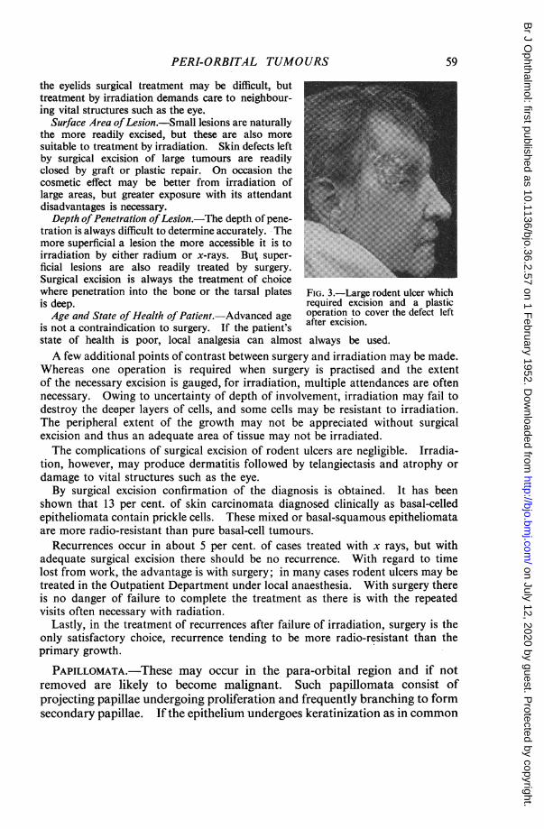
Depth of Penetration ofLesion.-The depth of pene-tration is always difficult to determine accurately. Themore superficial a lesion the more accessible it is toirradiation by either radium or x-rays. But super-ficial lesions are also readily treated by surgery.Surgical excision is always the treatment of choicewhere penetration into the bone or the tarsal plates FIG. 3.-Large rodent ulcer whichis deep. required excision and a plasticAge and State of Health of Patient.-Advanced age operation to cover the defect left
is not a contraindication to surgery. If the patient's after excision.state of health is poor, local analgesia can almost always be used.A few additional points of contrast between surgery and irradiation may be made.
Whereas one operation is required when surgery is practised and the extentof the necessary excision is gauged, for irradiation, multiple attendances are oftennecessary. Owing to uncertainty of depth of involvement, irradiation may fail todestroy the deeper layers of cells, and some cells may be resistant to irradiation.The peripheral extent of the growth may not be appreciated without surgicalexcision and thus an adequate area of tissue may not be irradiated.The complications of surgical excision of rodent ulcers are negligible. Irradia-
tion, however, may produce dermatitis followed by telangiectasis and atrophy ordamage to vital structures such as the eye.By surgical excision confirmation of the diagnosis is obtained. It has been
shown that 13 per cent. of skin carcinomata diagnosed clinically as basal-celledepitheliomata contain prickle cells. These mixed or basal-squamous epitheliomataare more radio-resistant than pure basal-cell tumours.
Recurrences occur in about 5 per cent. of cases treated with x rays, but withadequate surgical excision there should be no recurrence. With regard to timelost from work, the advantage is with surgery; in many cases rodent ulcers may betreated in the Outpatient Department under local anaesthesia. With surgery thereis no danger of failure to complete the treatment as there is with the repeatedvisits often necessary with radiation.
Lastly, in the treatment of recurrences after failure of irradiation, surgery is theonly satisfactory choice, recurrence tending to be more radio-resistant than theprimary growth.
PAPILLOMATA.-These may occur in the para-orbital region and if notremoved are likely to become malignant. Such papillomata consist ofprojecting papillae undergoing proliferation and frequently branching to formsecondary papillae. If the epithelium undergoes keratinization as in common
59
on July 12, 2020 by guest. Protected by copyright.
http://bjo.bmj.com
/B
r J Ophthalm
ol: first published as 10.1136/bjo.36.2.57 on 1 February 1952. D
ownloaded from

CECIL WAKELEY
warts, these become hard and may constitute horn-like outgrowths.Haemorrhage from such a papilloma is a sign of malignant invasion andalways demands radical excision. It is pathetic to see these papillomataallowed to progress towards malignant disease before radical treatment isentertained. It is surprising that members of the public will tolerate largepapillomatous masses in the para-orbital region (Fig. 4).
FIG. 4.-Papilloma of the inner canthus. FIG. 5.-Melanoma of the outer canthus.
SQUAMOUS-CELL CARcINOMATA.-Epithelioma is a new growth ofsquamouscells often occurring close to the lid margin in contradistinction to rodentulcer. It infiltrates and destroys local tissue and so forms a malignant ulcer.The constituent cells are large and of irregular shape, forming a variablenumber of cell nests according to the grade of differentiation and thereforeof malignancy. Fortunately, epitheliomata in this situation are not often ofa high grade of malignancy and glandular metastases are therefore infrequentexcept in neglected cases. The treatment is in many respects similar to thatfor rodent ulcers with a choice of excision or radiotherapy. If radiotherapyis used, it is important to perform a biopsy in order that the diagnosis maybe confirmed and the grading known. Surgical treatment is in many casesmore satisfactory. The treatment of the lymphatic glands depends upontheir clinical state when first seen and upon the age of the patient. Shouldthey be thought to be involved, then a block dissection is considered bymany at the present time to be of greater curative value, but irradiation isless disturbing to the patient, particularly in the elderly.MALIGNANT MELANOMATA.-The third type of malignant tumour of the
skin is the malignant melanoma. This may occur around the eye just as inother parts of the body. Bleeding, increase in size, or pain occurring in aprevious mole are ominous signs (Fig. 5). It can hardly be stressed too oftenthat any local interference with a pigmented naevus is dangerous for fear ofstimulating actual malignant degeneration. When this has occurred nobiopsy should be performed, but instead a wide excision should be made toinclude the tumour, a good margin of healthy tissue, and the deep fascia.These tumours are radio-resistant; they tend to spread by the lymphatics
60
on July 12, 2020 by guest. Protected by copyright.
http://bjo.bmj.com
/B
r J Ophthalm
ol: first published as 10.1136/bjo.36.2.57 on 1 February 1952. D
ownloaded from
PERI-ORBITAL TUMOURS
first but also widely by the blood stream at a variable period of time afteronset. Although the prognosis is generally regarded as bad a few cases livefor many years, especially when adequately treated. There seems to be noway of distinguishing these and the histology is of no help.
BENIGN LESIONS.-Of benign lesions in the peri-orbital region there areonly a few which call for brief attention. Simple papillomata occur frequentlyin the skin of the eyelids, and are formed by several layers of squamousepithelium covering a vascular stroma. Excessive keratinization forms onevariety of cutaneous horn and malignant degeneration is rare. Xanthomapalpebrarum, a deposit of yellow lipoid in large polyhedral cells beneath theskin, is sometimes large enough to draw the patient's attention. Both theseconditions are easily excised.
HAEMANGIOMATA.-Haemangiomata are the commonest tumours to occurin the peri-orbital region in childhood, being congenital in origin but tendingto increase in size during the first few years of life. They are not, therefore,strictly tumours, with the exception of the less common hypertrophic orangio-endotheliomatous type, but congenital malformations of embryonicvaso-formative tissue. They are presented for medical attention because ofthe disfigurement they cause, being either of the superficial capillary type orof the deeper and more spongy cavernous type.
Superficial.-It is common knowledge that in a proportion of superficial angio-mata, and particularly, in the "strawberry birthmark", spontaneous thrombosisoccurs if left long enough. In order to initiate or promote this curative phenomenona variety of procedures such as cauterization with CO2 snow, electrocoagulation,and irradiation with x-rays, radium, or radon seeds is in current use. In somecases surgical excision is always more satisfactory being quickly performed withoutfear of complication and leaving a thin linear scar. These methods are as a ruleineffective in dealing with the deeper subcutaneous and cavernous angiomata andparticularly with the advanced type which has acquired an extensive blood supplyand now amounts to a cirsoid aneurysm. These methods are not only ineffectivebut also better avoided, because of the importance of maintaining an unbrokenepithelial covering, since loss of viability may lead to sloughing, infection, andhaemorrhage with serious risk of grave consequences.
Cavernous.-The useful methods of treating cavernous angiomata are surgicalexcision, ligation of feeding vessels, injection of sclerosive chemicals, and co-agulation by injection of boiling water. In many cases simple surgical excisionis effective provided that the angioma is suitably situated, certain types requiringsubsequent plastic repair. The size of large angiomata, especially those with deepramifications, may be reduced by preliminary injections of irritating solutions suchas 40 per cent. saline or glucose, ethamolin, or boiling water. Reduction in sizefrom thrombosis follows and surgical excision may then be facilitated. The samereduction in size or even complete thrombosis can sometimes be effected by theimplantation of radon seeds.
Ligation of the vessels feeding the angioma is never necessary in the absence ofpulsation but is sometimes useful as an adjunct to sclerosis and where surgical
61
on July 12, 2020 by guest. Protected by copyright.
http://bjo.bmj.com
/B
r J Ophthalm
ol: first published as 10.1136/bjo.36.2.57 on 1 February 1952. D
ownloaded from

62 CECIL WAKELEY
excision is impossible as when both eyelids are involved. It is necessary toremember that the vessels feeding a haemangioma arise from the blood vesselssupplying the part concerned, but that the blood passing through the angiomais a circulation by itself and independent of adjacent tissue. When the angiomais arteriovenous in type the shunt of blood may on occasion be so great that thequantity available for nourishing neighbouring tissues is reduced even to theextent of producing ischaemic lesions.INVOLVEMENT OF NEIGHBOURING TISSUE.-The subject of haemangiomata
is a suitable one by which to pass from the more common superficial tumoursto the less common deep ones involving the orbital walls or neighbouringtissues. Angiomata of the bony walls of the orbit are uncommon but wellknown, and occur in capillary and cavernous forms. The typical radio-graphic appearances of radiating trabeculations give considerable help inmaking a firm diagnosis.Although uncommon in themselves, angiomata may be reckoned among the
more common causes of exophthalmos by the creation of a space-occupyinglesion posterior to the globe. The following types are recognized:
(a) Capillary.(b) Cavernous.(c) Angioblastic or hypertrophic.(d) Racemose or cirsoid.
The angioblastic type is firm, being composed of a solid mass of endothelialcells with a minimum of small patent vessels, the lumina becoming obliterated bythe proliferation of embryonic epithelium.The racemose type is composed of a pulsating mass of dilated, thickened, and
tortuous vessels. They occur in adults and have a relatively sudden onset. Theymay represent an ordinary cavernous angioma which has established a com-munication with a neighbouring artery or arteriole. After this communicationhas been established the mass becomes locally destructive by reason of constantarterial pulsation. These tumours may erode the walls of the orbit and surroundingstructures and even lead to fatal haemorrhage.A characteristic of haemangiomata, except those of the cirsoid type, is that they
do not show unlimited growth but tend to reach a certain size and then remainstationary. This is an important point when considering treatment in a difficult case.Two noteworthy characteristics of this type of ' tumour' in the orbit are:(1) The mobility of the eye is unaffected.(2) The variations in size may result in exophthalmos. These variations may be
spontaneous and may be due to sinple venous stasis from coughing, crying, bending thehead down, or compression of the jugular vein.
Sometimes the haemangiomatous nature of the lesion is revealed by an extensionforwards rendering it visible from the exterior in a subconjunctival position, or else bythe association of a port-wine stain around the eye. When considering treatment, theposition, size, and type of angioma must be taken into account and many of the principlesdiscussed for superficial angiomata applied.DERMOID and EPIDERMoID TuMouRs.-Dermoid and epidermoid tumours
of the orbit are common if the para-orbital type is included, but althoughwell-known, intra-orbital dermoids are not frequent. They are found inmany situations-external, intra-orbital, intracranial, and in the diploe.
62
on July 12, 2020 by guest. Protected by copyright.
http://bjo.bmj.com
/B
r J Ophthalm
ol: first published as 10.1136/bjo.36.2.57 on 1 February 1952. D
ownloaded from

PERI-ORBITAL TUMOURS
FiG. 6 (a).-External angular dermoid FIG. 6 (b).-External angular dermoid(front view). (lateral view).
External.-The commonest site is the lateral end of the eyebrow where theyare known as external angular dermoids (Fig. 6 a and b). In this situationthey give rise to cysts either tense or soft, which are not attached to the skin butwhich characteristically form a depressikn in the underlying skull which may befelt clinically. The depression sometimes amounts to a defect with firm attach-ment of the epidermoid to the dura (Fig. 7).
Intra-Orbital.-These dermoids occur most commonly in the superior temporalregion, causing proptosis and requiring to be distinguished from the less commontumours of the lacrimal gland. In this situation a defect may occur in the bonein the form of either a depression or aforamen which is easily demonstrated inthe x-ray.
Other Sites.-Intracranial extensionsometimes occurs, and some cysts appearto arise in the diploe of the frontal boneextending into both orbit and cranium.The treatment for both dermoids
and epidermoids is the same andwhere possible consists of excision.In the deeper and larger varieties ofintra-orbital cyst this may not be an FIG. 7 Mid-line dermoid at base of nose.easy matter because of the involvementof neighbouring structures and cavities demanding transfrontal approach. Inothers the cyst may first be incised with evacuation of its contents and thenits wall may be removed piece-meal.A few words are required regarding the terms dermoid cyst and epidermoid
cyst. Although they are closely related in origin and behaviour and requiresimilar treatment, much has been written of their differentiation. The termepidermoid has replaced the older cholesteatoma or tumeur perlee; thedifferentiation between the two is perhaps academic, since both are derivedfrom embryonic nests originating from the primitive ectodermal layer, theactual differentiation depending upon either the time at which the cell nest
63
on July 12, 2020 by guest. Protected by copyright.
http://bjo.bmj.com
/B
r J Ophthalm
ol: first published as 10.1136/bjo.36.2.57 on 1 February 1952. D
ownloaded from

CECIL WAKELEY
developed or the degree or depth of inclusion of the original dermal layer.It was Renak in 1854 who first suggested that the 'pearly tumour' ofCurveillier originated in ectodermal nests, and this hypothesis has been verystrongly supported by subsequent observers. The evidence, however, restsentirely on the histological features of the tumour itself and particularly onthe details of the capsule.The ectodermal layer of the head end of the foetus develops into both skin
and neural tissue. The cause for differentiation of this one layer is speculativebut it has been suggested that what is known as " organizer influence" isexcreted by the mesodermal tissue or cells which later become neural tissue.Jefferson believes that the epidermoids found in the brain substance or withinthe membrane may possibly be due to failure of this influence to compelgroups of ectodermal cells towards neural differentiation.Dermoids vary in size from that of a seed to that of an apple. They are
encapsulated and oval, round, or hourglass in shape. They are soft andusually cystic, the actual consistency of the contents being determined bythe presence of sebaceous material, fat, and cholesterol crystals in varyingproportions. The presence of hair has been regarded as pathognomonic ofa dermoid but intermediate forms have been found. Microscopically thewall of a dermoid resembles skin and one can distinguish from withoutinwards a fibrous capsule, a connective tissue layer containing blood vessels(especially at the site of the dermoid tuft which represents the original siteof the dermal implantation), and an inner epithelial layer. This inner layervaries in thickness but resembles skin, there being a number of layers ofcells, the innermost being flattened and without nuclei. The wall nearlyalways shows some evidence of inflammatory irritation.
Epidermoids are simple in nature, and form a more solid mass which maybe described as being made up of two layers: the outer layer, or stratumdurum as it is called, is formed of layers of collagen fibres with little or nocellular formation; it is this outer layer which gives the epidermoid itscharacteristic appearance. The inner layer forms the interior with a depthoffrom two to ten cells; fine granules ofkeratohyalin are scattered throughoutthe cytoplasm of these cells and the interior of the cyst is composed ofaccumulated products of the desquamated epithelial layer. These tumoursare of course quite different from the cholesteatomata that occur in themiddle ear; the latter are associated with inflammation and consist mainlyof epithelial debris, granulation tissue, leucocytes, and cholesterol crystalswhich are the accumulated products of excess desquamation and reaction toinflammation.OSmOMATA.-Tumours arising in the walls of the orbit may be either
benign or malignant, primary or secondary, or due to one of a group ofmiscellaneous diseases of doubtful aetiology.
Osteomata are described as either cancellous or ivory but the latter termis the better. Ivory osteomata occur in almost any part of the skull including
64
on July 12, 2020 by guest. Protected by copyright.
http://bjo.bmj.com
/B
r J Ophthalm
ol: first published as 10.1136/bjo.36.2.57 on 1 February 1952. D
ownloaded from
PERI-ORBITAL TUMOURS
the orbit. They are often dome-shaped and are formed of very hard,mature bone. They are found in both sexes in later life, and are slow-growing and painless. They sometimes follow trauma and after some yearsof growth tend to come to a standstill. On the vault of the skull they areusually symptomless but when they occur in the orbit proptosis may followor in advanced cases blindness may result from pressure on the optic nerve.Their usual form is sessile like a limpet and covered with periosteum, thesurface being hard and frequently polished. Occasionally, small compactpedunculated tumours are found, which have a short stout pedicle and amushroom-like head. They are composed throughout of mature, compactbone continuous with the outer table of the adjacent bone and formed ofconcentrically-arranged laminae containing small haversian systems.A special type of these osteomata is that which occurs in either the frontal
or the ethmoid sinus or in the antrum of Highmore; many remain small andundetected, but others grow steadily larger and sooner or later produce sub-jective or objective symptoms. They enlarge in a centrifugal mannerdestroying the bony walls of the sinus by pressure atrophy. In this positionsymptoms arise of inflammation or obstruction of the sinus. Later theyspread into the cranial cavity, nose, and orbit, causing pressure symptoms,proptosis, and sometimes the formation of mucoceles or aeroceles.The precise aetiology of these tumours is not known. Associated infection
is more likely to follow than to cause them. A history of trauma is commonbut its association is difficult to prove. The ethmoid type is formed incartilage but those on the frontal bone in membrane. In this latter regionhowever the frontal sinus continues to grow until after puberty and it maybe that islands of cartilage are misplaced and stimulated to grow, possibly bytrauma. The treatment of these osteomata often involves a complicatedneurological operation; in the old days it terminated only too frequentlyin meningitis and cerebrospinalrhinorrhoea.The so-called cancellous type of
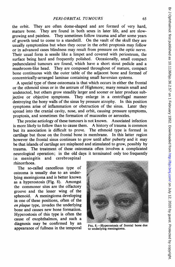
osteoma is usually due to an under-lying meningioma and is better knownas a hyperostosis (Fig. 8). Amongstthe commoner sites are the olfactorygroove and the lesser wing of thesphenoid. A meningioma developingin one of these positions, often of theen plaque type, invades the underlyingbone and causes new bone formation.Hyperostosis of this type is often thecause of exophthalmos, and such adiagnosis may be confirmed by an 7FIG. 8.-Hyperostosis of frontal bone dueappearance of fullness in the temporal to underlying meningioma.
65
on July 12, 2020 by guest. Protected by copyright.
http://bjo.bmj.com
/B
r J Ophthalm
ol: first published as 10.1136/bjo.36.2.57 on 1 February 1952. D
ownloaded from

CECIL WAKELEY
fossa, or of thickening of the sphenoidal wings, or it may be seen by x-rayor may cause an increase of cerebrospinal fluid protein.Of malignant tumours of the bone around the orbit, osteogenic sarcoma
and fibrosarcoma are rare but secondary tumours are more common. Aparticular example of this is neuroblastoma-the so-called Hutchison'ssyndrome. Secondary deposits are also rarely found as the result of somehaemopoietic disease, such as lymphatic leukaemia in children and
myelogenous leukaemiain adults.
In Schiiller-Christiandisease, alternativelyknown as xanthomatosis,~~~~~~~~~~~~~~~~~~~~~~.... ... e ., .. < ::-deposits of hpoid material
*... 7occur in the membrane,bones, lymphatic glands,
M: ~~~~~~~~~~andviscera. X rays dis-^ ; ^ i i ;; iplay the typical punched-
...'. :2 l''outappearance of thebone.
Paget's disease attackselderly people and occa-sionally picks out one
... :F bone such as the upperjaw. A similar deformitymay arise from leontiasisossea (Fig. 9), a conditionof bone overgrowth ofuncertain origin, whichproduces asymmetricaldeformity with proptosisof one or both eyesand in late cases opticatrophy.
This survey of peri-FIG. 9. Skull of case of leontiasis ossea (Royal College of orbital tumours, althoughSurgeons). comprehensive, may haveomitted a few rare tumours which should have been included, but I haveconfined my remarks to clinical material that has come under my care duringt4e past thirty years.
66
on July 12, 2020 by guest. Protected by copyright.
http://bjo.bmj.com
/B
r J Ophthalm
ol: first published as 10.1136/bjo.36.2.57 on 1 February 1952. D
ownloaded from