common genetic variants and risk of brain injury after...
TRANSCRIPT
Common Genetic Variants and Risk of Brain InjuryAfter Preterm Birth
WHAT’S KNOWN ON THIS SUBJECT: Preterm birth is stronglyassociated with alterations in brain development and long-termneurocognitive impairment that are not fully explained byenvironmental factors.
WHAT THIS STUDY ADDS: Common genetic variation in genesassociated with schizophrenia and lipid metabolism modulatesthe risk for preterm brain injury; known susceptibilities toneurologic disease in later life may be exposed by the stressof preterm birth.
abstractBACKGROUND: The role of heritable factors in determining the com-mon neurologic deficits seen after preterm birth is unknown, but thecharacteristic phenotype of neurocognitive, neuroanatomical, andgrowth abnormalities allows principled selection of candidate genesto test the hypothesis that common genetic variation modulates therisk for brain injury.
METHODS: We collected an MRI-linked genomic DNA library from 83preterm infants and genotyped tag single nucleotide polymorphismsin 13 relevant candidate genes. We used tract-based spatial statisticsand deformation-based morphometry to examine the risks conferredby carriage of particular alleles at tag single nucleotide polymorphismsin a restricted number of genes and related these to the preterm cerebralendophenotype.
RESULTS: Carriage of the minor allele at rs2518824 in the armadillorepeat gene deleted in velocardiofacial syndrome (ARVCF) gene, whichhas been linked to neuronal migration and schizophrenia, andrs174576 in the fatty acid desaturase 2 gene, which encodes a rate-limiting enzyme for endogenous long chain polyunsaturated fatty acidsynthesis and has been linked to intelligence, was associated withwhite matter abnormality measured in vivo using diffusion tensorimaging (P = .0009 and P = .0019, respectively).
CONCLUSIONS: These results suggest that genetic variants modulatewhite matter injury after preterm birth, and known susceptibilities toneurologic status in later life may be exposed by the stress of prematureexposure to the extrauterine environment. Pediatrics 2014;133:e1655–e1663
AUTHORS: James P. Boardman, FRCPCH, PhD,a AndrewWalley, PhD,b,c Gareth Ball, PhD,d Petros Takousis, PhD,b
Michelle L. Krishnan, MRCPCH,d Laurelle Hughes-Carre,RN,e Paul Aljabar, PhD,d Ahmed Serag, PhD,f Caroline King,BSc,e Nazakat Merchant, MD,d Latha Srinivasan, PhD,e
Philippe Froguel, MD, PhD, F Med Sci,b,g Jo Hajnal, PhD,d
Daniel Rueckert, PhD,h Serena Counsell, PhD,d and A. DavidEdwards, F Med Scid
aMedical Research Council/University of Edinburgh Centre forReproductive Health, Edinburgh, United Kingdom; bDepartmentsof Genomics of Common Disease, School of Public Health,cMolecular Genetics and Genomics, National Heart and LungInstitute, ePaediatrics, and hComputing, Imperial College London,London, England, United Kingdom; dCentre for the DevelopingBrain, King’s College London, London, England, United Kingdom;fThe Advanced Pediatric Brain Imaging Research Laboratory,Children’s National Medical Center, Washington, District ofColumbia; and gCentre National de la Recherche Scientifique-CNRS 8199, Lille 2 University, Paris, France
KEY WORDSmagnetic resonance image, neonate, preterm, genetics
ABBREVIATIONSARVCF—armadillo repeat gene deleted in velocardiofacial syn-dromeCOMT—catechol-O-methyl transferase geneDTI—diffusion tensor imagingFA—fractional anisotropyFADS2—fatty acid desaturase 2 geneFDR—false discovery rateLC-PUFA—long-chain polyunsaturated fatty acidLD—linkage disequilibriumMAF—minor allele frequencySNP—single nucleotide polymorphismTBSS—tract-based spatial statistics
(Continued on last page)
PEDIATRICS Volume 133, Number 6, June 2014 e1655
ARTICLE
by guest on June 26, 2018www.aappublications.org/newsDownloaded from
Preterm delivery affects 10% of birthsworldwide and is associated with neu-rodevelopmental impairment.1,2 Adverseoutcome is strongly associated witha phenotype that commonly combines:cognitive dysfunction and special educa-tional needs; reduced early somatic andbrain growth; and cerebral white matterinjury.3–6 This is only partly explained byknown environmental stresses such asischemia, inflammation, and altered nu-trition.7 However, neuroanatomic andneurocognitive functions are stronglyheritable8–11 and the characteristic ab-normalities seen in preterm infants allowprincipled choices of candidate genesthat might modulate the risk for adverseneurodevelopmental outcome after pre-term birth.
The role of prematurity in cerebral de-velopment and damage has been muchinvestigated by using MRI and diffusiontensor imaging (DTI), which providesensitive measures of abnormal mor-phology and microstructure thatcorrelate with neurodevelopmentaloutcome.5,12–16 Computational techni-ques are available that define a cere-bral endophenotype that is closelyrelated to later neurocognitive outcome.These include tract-based spatial statis-tics (TBSS), a method for group-wiseanalysis of multiple fractional anisot-ropy (FA) images derived from DTI, anddeformation-based morphometry, whichquantitates local brain growth.17,18 Thesemethods allow powerful unbiasedgroup-wise studies of genetic risk.19–21
Based on studies of the imaging andclinical phenotypewehypothesized thatthe development of preterm cerebralinjurycouldberelated togenes involvedin white matter development or humancognition and behavior. We used TBSSand deformation-based morphometryto examine the risks conferred bycarriage of particular alleles at singlenucleotide polymorphisms (SNPs)22
located within a small number of can-didate genes (Supplemental Table 1).
These genes were selected because ofprevious functional evidence related tothe preterm cerebral endophenotypeincluding cognitive/behavioral dysfunc-tion, neuronal/glial development, andendogenous long-chain polyunsaturatedfatty acid production, which is essentialfor neural development. We now report 2genetic variants that modulate whitematter injury after preterm birth.
METHODS
The study was conducted according tothe principles of the Declaration ofHelsinki, and ethical approval wasobtained from theUKNational ResearchEthics Service. Written parental in-formed consent was obtained.
The cohort consisted of preterm neo-nates who received care at QueenCharlotte’s and Chelsea Hospital be-tween January 2005 and October 2008,underwent DTI and MR imaging in theneonatal period, and whose parentsconsented to the collection of DNA.Infants were not eligible if they hada chromosomal abnormality, congenitalmalformation, or congenital infection.
Genotyping
TheDNAOG-575 kitwasused for samplingof saliva at term equivalent age (DNAGe-notek Inc, Kanata, Canada). Genotypingwas performed by using the Sequenomi-PLEX genotyping assay and MALDI-TOFmass spectrometry (Sequenom Inc, SanDiego, CA). To maximize the genetic in-formation obtained from studying thecandidate genes, an efficient set of SNPsfor genotyping were identified by usingthe Taggeralgorithm22 as implemented inthe Haploview software.23 Tag SNP se-quences were extracted from the Hap-Map genome browser release♯28 andincluded if the minor allele frequency(MAF) exceeded 5% using populationdescriptor CEU (Utah residents withNorthern and Western European ances-try from the CEPH collection). Exclusioncriteria: ,85% genotyping success per
SNP, and/or statistically significant de-viation from Hardy-Weinberg equilibrium(P, .001).
Imaging
MR imaging was performed on a Philips3-Tesla system (Philips Medical Systems,Netherlands) using an 8-channel phasedarray head coil. Single-shot echo-planarimaging DTI was acquired in the trans-verse plane in 15 non-collinear direc-tions using the following parameters:repetition time (TR): 8000 ms; echotime (TE): 49 ms; slice thickness: 2 mm;field of view: 224 mm; matrix: 1283 128(voxel size: 1.7531.7532mm3); b value:750 s/mm2; SENSE factor: 2. T1-weighted3D MPRAGE were acquired with pa-rameters: TR = 17 ms, TE = 4.6 ms, in-version delay = 1500ms, flip angle = 13°,acquisition plane = sagittal, voxel size =0.82 3 1.03 3 1.6 mm, FOV = 210 3167 mm and acquired matrix = 2563163. T2-weighted fast spin-echo: TR =8700 ms, TE = 160 ms, flip angle = 90°,acquisition plane = axial, voxel size =1.15 3 1.18 3 2 mm, FOV = 220 mm,and acquired matrix = 192 3 186.
All examinations were supervised bya pediatrician experienced in MR pro-cedures. Infants were sedated with oralchloral hydrate (25–50 mg/kg) beforescanning, and pulse oximetry, tempera-ture, and electrocardiography dataweremonitored throughout. Ear protectionwas used for each infant, comprisingearplugs molded from a silicone-basedputty (ColteneWhaledent, Mahwah, NJ)placed in the external ear and neonatalearmuffs (MiniMuffs, Natus Medical Inc,San Carlos, CA).
Data Analysis
DTI
DTI analysis was performed by usingFMRIB’s Diffusion Toolbox (v2.0) asimplemented in FMRIB’s Software Li-brary (FSL v4.1.5; www.fmrib.ox.ac.uk/fsl).24 Each infant’s diffusion-weightedimage DWI was registered to their
e1656 BOARDMAN et al by guest on June 26, 2018www.aappublications.org/newsDownloaded from

respective non–diffusion-weighted (b=0)image and corrected for differences inspatial distortion owing to eddy cur-rents. Images were brain extractedusing Brain Extraction Tool (BET v2.1),diffusion tensors calculated voxelwise,and FA maps generated. TBSS18 wasperformed by using amodified pipelinespecifically optimized for neonatal DTIanalysis.19 The aligned data were usedto create amean FAmap and amean FAskeleton that represents the center ofall white matter tracts common to thegroup. The FA skeleton was thresh-olded at FA $0.15 before each infant’saligned FA data were projected onto it.
TBSSwasusedtoassesstherelationshipbetween FA and genotype. For each SNP,infants were grouped according topresence of the minor allele, which en-abled a 2-group comparison of minorhomozygote or heterozygote versusmajor allele homozygote. SNP data wereexcluded if MAF in the study populationwas,5%. The FA values of the 2 groupsof infants were compared by usingvoxel-wise cross-subject linear regres-sion, with gestational age at birth andage at scan entered in the model, be-cause these are known to influence FAduring early development. All imageswere subject to family-wise error cor-rection for multiple comparisons afterthreshold-free cluster enhancement.25
Control of type 1 error for multiple SNPtesting was carried out using the q-valuealgorithm, selecting the bootstrapmethodto calculate the Benjamini and HochbergFDR of 5%.26,27
Morphometric Analysis
Volumetric analyseswere performed in2 stages by using publishedmethods.28,29
First, the MR image of each subject wasco-aligned with a spatiotemporal atlasof the anatomy of the neonatal brainclosest to the age of the subject.29,30
The correspondence obtained by thisregistration allowed the average esti-mates of structural segmentations to
be used as priors in a segmentationmethod, which estimated the samestructures in the native space of thesubject.28 The resulting segmentationof each structure is represented asa “soft” density map with the value ateach voxel ranging from 0 to 1 in-dicating the proportion of the voxeloccupied by each structure. By sum-ming these values over all voxels, anestimate of the volume for each struc-ture in each image was calculated.
Second, a spatiotemporal atlas was con-structed from 142 T2-weighted imagesacquired between 29 and 44 weeks’postmenstrual age (PMA)31,32 and is pub-licly available at www.brain-development.org. At each specific age in the range,the corresponding atlas volume is anestimate of the average of the anato-mies of subjects close in age. By iter-atively applying kernel regression tothe affine transformations betweeneach subject’s image and the average,an unbiased estimate of the averageshape is obtained. The transforma-tions between each acquired image andthe final atlas were used to propagatestructural segmentations from eachsubject’s native space. In this way, aswell as obtaining a spatiotemporal at-las of the MR images, further spatio-temporal atlases of white matter, greymatter, cerebrospinal fluid, subcorticalgrey matter, brainstem, and cerebel-lum were generated. Automatic tha-lamic segmentations were acquiredusing published methods.33
Clinical, demographic, and nutritionaldata were tested for normality by usingShapiro-Wilk test and are presented asmedians and ranges if the data did notconform to a normal distribution. Datawere analyzed by using independentsample t test for continuous variablesif there was equality of variance be-tween groups (Levene’s test) and x2
test for proportions. Statistical analy-sis was performed by using SPSS v19.0(SPSS Inc, Chicago, IL).
RESULTS
WecollectedanMRI-linkedgenomicDNAlibrary from 83 preterm infants whohad a median PMA at birth of 28+5
weeks (range, 23+2 to 32+6 weeks) andmedian birth weight of 1052 g (range,550–2140 g). MR images were acquiredat median PMA 40+6 weeks (range, 29+2
to 47+6 weeks). Forty-five participantswere male (54%), and 38 (45%) werewhite British (see complete ethnic dis-tribution in Supplemental Table 2).Fourteen (17%) had bronchopulmonarydysplasia defined as a need for re-spiratory support and/or supplementaloxygen at 36 weeks’ PMA.
Common Genetic Variants andWhite Matter Microstructure
Thirty-five tag SNPs across 13 genesweretested (Supplemental Table 1). After cor-rection for multiple testing we found that2 tag SNPs were associated with signifi-cant alterations in FA in white matter:rs2518824 in the ARVCF gene (P = .0009;b coefficient20.029, 95% confidenceinterval [CI], 20.039 to 20.019) andrs174576 in the fatty acid desaturase2 (FADS2) gene (P = .0019; b coefficient20.034, 95% CI,20.047 to20.021). Car-riage of the minor allele in no other tagSNP was significantly associated with FAin white matter after correction for mul-tiple tests (Fig 1 and Supplemental Ta-ble 1). Therewas no significant differencein the prevalence of bronchopulmonarydysplasia between infants who had andthose who did not have the minor allelefor either of the significant SNPs.
Aftercorrectingfordegreeofprematurityat birth and PMA at scan, carriage of theminorallele (G) at rs2518824 in theARVCFgene was associated with reduced FA inwhite matter for the 80 individuals whohad genotype data for this SNP and DTI(P= .0009). Theminorallele frequencywas66% (minor homozygote/heterozygote).Additional SNPs rs1110477 in ARVCF andrs9332377 in the catechol-O-methyltransferase (COMT) gene, both at locus
ARTICLE
PEDIATRICS Volume 133, Number 6, June 2014 e1657 by guest on June 26, 2018www.aappublications.org/newsDownloaded from

22q11.2, were nominally associatedwithchange in FA, but these relationshipswere not significant after the omnibuscorrection threshold was applied (P =.0282 and P = .0413, respectively; forlinkage disequilibrium plots see Sup-plemental Fig 1). Carriage of the minorallele (A) at rs174576 in FADS2 was as-sociated with reduced FA in specificwhite matter tracts for the 73 individu-als who had genotype data for this SNPand DTI (P = .0019), and none of theremaining tag SNPs in the FADS2 genewere associated with FA changes aftercorrection for multiple tests. The MAFwas 57%.
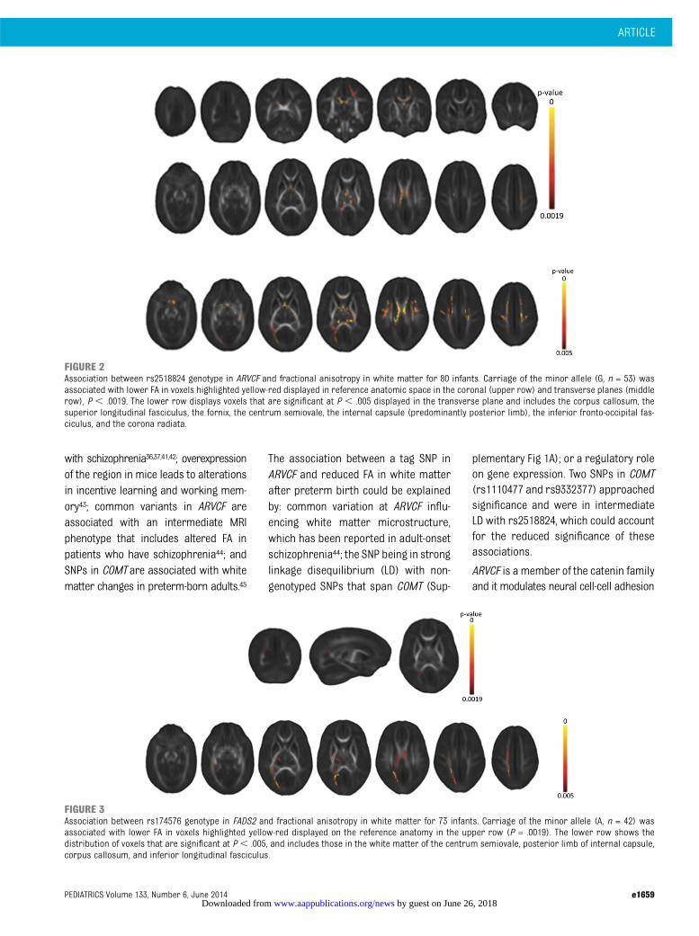
Infants who carried theminor allele (G)at rs2518824 in the ARVCF gene hadreduced FA in the corpus callosum, thesuperior corona radiata, the fornix,and the centrum semiovale (Fig 2).Infants who carried the minor allele (A)at rs174576 in FADS2 had reduced FA inwhite matter within the posterior co-rona radiata (Fig 3). The mean PMA atbirth of the group that carried the mi-
nor allele was 29.28 weeks, which wasolder than that of the group without theminor allele (28 weeks) (P = .032).
Because of a possible interaction be-tween polymorphisms in FADS2 andbreast milk exposure on IQ in child-hood,34,35 we investigated whether the2 groups differed in early nutritionalexposures. There was no difference inthe proportion of infants who wereexclusively breastfed at term equiva-lent age between the 2 groups (60% vs55%, P = .812), and there was no sig-nificant difference in parenteral nutri-tion use between those who had theminor allele and those who did not(4 vs 5.5 days, P = .132).
TBSS is designed to identify supra-threshold voxels, so when a regional ef-fect is detected without due cause, thepossibility that it underlies amore globaleffectshouldbeconsidered.Thereforeweassessed the spatial distribution of dif-ference inFAat thesignificance thresholdP , .005 for rs2518824 and rs174576(Figs 2 and 3). No other tag SNP was
associated with voxel-wise differencesthat were significant at this threshold(Fig 1 and Supplemental Table 1).
The Effect of SNP-Associated WhiteMatter Microstructural Change onCerebral Volume
Geneticfactorsinfluencebrainstructure8,9
and the integrity of white matter tractscan influence cerebral volume in thedeveloping brain,31 so we investigatedwhether the SNP-associated alterations intract microstructure identified in TBSSanalyses (Figs 2 and 3) were accompa-nied by alterations in brain morphologyassessed by automatic segmentation oftissue compartments.
There was a linear increase in wholebrainvolumewith increasingageat timeof scan,whichwasexpectedandreflectsthe rapid rate of cerebral growth thattakes place at this stage in human de-velopment.16 However, there was nosignificant relationship between geno-type andwhole brain volume (Fig 4), andthere were no significant differencesbetween SNP genotype and the volumesof cortical gray matter, white matter,deep gray matter including a separateanalysis of the thalami, or the cerebel-lum (Supplemental Table 3).
DISCUSSION
Based on studies of the characteris-tic preterm phenotype, we predicteda number of genes that could modulatethe risks for cerebral abnormalities,and found that SNPs linked to geneticregions associated with schizophre-nia,36,37 and linking lipid metabolism tointelligence,34,35 predicted changes inthe cerebral endophenotype.
Common genetic variation at chromo-some22q11.2 is consistently associatedwith neuropsychiatric disorders.37–40
Within this cytogenetic band we focusedon ARVCF and COMT for a number ofreasons: haplotypic associations span-ning these genes have been associated
FIGURE 1Scatterplot of –Log10(P value) calculated from voxel-wise analyses of the effect of minor allele carriageon FA for 35 SNPs. Carriage of the minor allele at rs2518824 (P = .0009) and rs174576 (P = .0019) wasassociated with decreased FA in the white matter (Benjamini and Hochberg FDR threshold 5%, P #.0019). Dotted line equates to P = .05, and hashed line P = .005.
e1658 BOARDMAN et al by guest on June 26, 2018www.aappublications.org/newsDownloaded from
with schizophrenia36,37,41,42; overexpressionof the region in mice leads to alterationsin incentive learning and working mem-ory43; common variants in ARVCF areassociated with an intermediate MRIphenotype that includes altered FA inpatients who have schizophrenia44; andSNPs in COMT are associated with whitematter changes in preterm-born adults.45
The association between a tag SNP inARVCF and reduced FA in white matterafter preterm birth could be explainedby: common variation at ARVCF influ-encing white matter microstructure,which has been reported in adult-onsetschizophrenia44; the SNP being in stronglinkage disequilibrium (LD) with non-genotyped SNPs that span COMT (Sup-
plementary Fig 1A); or a regulatory roleon gene expression. Two SNPs in COMT(rs1110477 and rs9332377) approachedsignificance and were in intermediateLD with rs2518824, which could accountfor the reduced significance of theseassociations.
ARVCF is a member of the catenin familyand it modulates neural cell-cell adhesion
FIGURE 2Association between rs2518824 genotype in ARVCF and fractional anisotropy in white matter for 80 infants. Carriage of the minor allele (G, n = 53) wasassociated with lower FA in voxels highlighted yellow-red displayed in reference anatomic space in the coronal (upper row) and transverse planes (middlerow), P , .0019. The lower row displays voxels that are significant at P , .005 displayed in the transverse plane and includes the corpus callosum, thesuperior longitudinal fasciculus, the fornix, the centrum semiovale, the internal capsule (predominantly posterior limb), the inferior fronto-occipital fas-ciculus, and the corona radiata.
FIGURE 3Association between rs174576 genotype in FADS2 and fractional anisotropy in white matter for 73 infants. Carriage of the minor allele (A, n = 42) wasassociated with lower FA in voxels highlighted yellow-red displayed on the reference anatomy in the upper row (P = .0019). The lower row shows thedistribution of voxels that are significant at P, .005, and includes those in the white matter of the centrum semiovale, posterior limb of internal capsule,corpus callosum, and inferior longitudinal fasciculus.
ARTICLE
PEDIATRICS Volume 133, Number 6, June 2014 e1659 by guest on June 26, 2018www.aappublications.org/newsDownloaded from

and migration. It is richly expressed inthe human ganglionic eminence and inneurons that migrate from the gangli-onic eminence to the intermediatezone, the amygdaloid complex, and thethalamus during fetal life.46,47 We andothers have reported that volume14
and neuronal48 loss in the dorsomedialnucleus of the thalamus is associatedwith preterm birth, and these data fo-cus attention on perturbed neuronalmigration as a possible mechanismfor abnormal thalamic development,which contributes to the encephalop-athy of prematurity.49,50
The FADS2 gene, located on chromo-some 11q12.2, encodes d-6 desaturase,a rate-limiting enzyme on the pathway ofendogenous docosohexaeneoic acidand arachidonic acid production. Theselong-chain polyunsaturated fatty acids(LC-PUFAs) accumulate in the brain inabundance from the third trimester to18 months postpartum and are essen-tial for neurogenesis, neurotransmis-sion, and protection from oxidativestress.51–53 The candidacy of FADS2 asa risk modulator for preterm brain in-jury is raised because FADS2 gene var-iants have functional effects on LC-PUFA
availability, with minor allele carriage atcommon SNPs including rs174576 beingassociated with altered levels of arachi-donic acid and docosohexaeneoic acidin phospholipid, serum, and breastmilk,54–58 and FADS2 variants may in-teract with early dietary exposures toinfluence childhood IQ.34,35
We found that minor allele carriage atrs174576 is associated with lower FA inwhite matter, after controlling for non-genetic confounders. rs174576 is as-sociated with LC-PUFA phenotypes andis in strong LD with other functionalSNPs at the FADS2 locus (SupplementalFig 1B). The effect size of SNPs in theFADS2 locus is large, with approxi-mately one-third of the variability ofPUFA and LC-PUFA levels in human tis-sues attributable to genotype.55,59
These effects were detected through theuse of TBSS, which provides a noveland powerful method for detectinggroup-wise differences in white mattermicrostructure predictive of later neuro-developmental outcome,20,21 overcomingthe diagnostic imprecision of neuro-developmental assessment in earlychildhood and allowing significantlysmaller sample sizes. Modeling and
neonatal clinical studies have shownthat clinically significant changes in FAcan be detected in groups as small as 10patients per group.60,61 The quantitativeabnormalities described here showsimilarity to alterations seen in olderpreterm children and adolescents,62–64
suggesting that the influence of pre-maturity65 and genetic factors on neuralsystems that underpin neurodevelop-mental function is operative before thetime of normal birth.
We used the International HapMap Pro-ject population descriptor CEU (Utahresidents with Northern and WesternEuropean ancestry) to define haplotypefrequency, because this descriptorrepresents the largest proportion of thestudy population. Our study group wasdiverse, and although it is possible thatethnicity is a confounding factor in theanalysis, we did not adjust for it for thefollowing reasons: firstly, self-declaredmaternal ethnicity is an imprecise sur-rogate for neonatal ethnicity; secondly,both parents did not always share thesame ethnicity; and thirdly, we adjustedthe TBSSmodel only for factors that areknown to influenceFA. In future studies itmay be possible to investigate the effect
FIGURE 4Brain tissue volume in relation to SNP genotype. There was no difference in whole brain volume between individuals who had theminor allele (blue) and thosewhodid not have theminorallele (green) for rs2518824 (ARVCF) or rs174576 (FADS2). Therewas a linear increase in brain volume across the age range from28to 47 weeks’ PMA (r2 = 0.682 rs2518824 and r2 = 0.706 rs174576).
e1660 BOARDMAN et al by guest on June 26, 2018www.aappublications.org/newsDownloaded from
of ethnicity on FA by studying very largepopulations, which would allow for ad-justment for genetic ethnicity, or byrestricting analyses to ethnically ho-mogenous groups.
This study provides proof of concept ofgenetic effects, but there is no reason tosuspect that these are the only heritablemodulators of preterm brain injury;there are other genetic pathways thatwarrant investigation in large-scale as-sociation studies, including inflamma-tion, hypoxia signaling, and myelinationpathways. The detected effects werespecific to white matter, despite brainvolume being highly heritable and thecommon association between whitematter damage and reduced brain vol-umes.66,67 This raises the possibility that
although volumetric and white matterchanges are associated in the pretermphenotype, genetic vulnerability may beseparable. It is also possible that tech-niques that provide a more highly re-solved quantification of structure, butwhich are not yet validated for use in thedeveloping brain, may be sensitive togenetic difference.
CONCLUSIONS
These results suggest that genetic vul-nerabilities to cognitive and neuropsy-chiatric abnormalities are revealed bythe environmental stress of pretermdelivery. This is consistent with recentdata showing that preterm infants havehigher rates of neuropsychiatric diseasein adult life,68 as well as the observation
that schizophrenia- and autism-associatedgenes are overexpressed in developingmouse cortical subplate,69 a region ofspecific vulnerability during pretermlife.70 Future research could focus onperturbations in neuronal migrationand lipid metabolism in the causalpathway to preterm brain injury.
ACKNOWLEDGMENTSWe are grateful to the families who con-sented to take part in the study and tothe nursing and medical staff who par-ticipated in scanning the infants. Wethank Dr Salvatore Aversa and Dr SaraLunardi for collating nutritional infor-mation. Philips Healthcare providedtechnical support to facilitate researchusing Philips MRI systems.
REFERENCES
1. Moore T, Hennessy EM, Myles J, et al.Neurological and developmental outcomein extremely preterm children born inEngland in 1995 and 2006: the EPICurestudies. BMJ. 2012;345:e7961
2. Beck S, Wojdyla D, Say L, et al. The worldwideincidence of preterm birth: a systematicreview of maternal mortality and morbidity.Bull World Health Organ. 2010;88(1):31–38
3. Delobel-Ayoub M, Arnaud C, White-Koning M,et al; EPIPAGE Study Group. Behavioral prob-lems and cognitive performance at 5 yearsof age after very preterm birth: the EPIPAGEStudy. Pediatrics. 2009;123(6):1485–1492
4. MacKay DF, Smith GC, Dobbie R, Pell JP.Gestational age at delivery and specialeducational need: retrospective cohortstudy of 407,503 schoolchildren. PLoS Med.2010;7(6):e1000289
5. Woodward LJ, Anderson PJ, Austin NC,Howard K, Inder TE. Neonatal MRI to predictneurodevelopmental outcomes in preterminfants. N Engl J Med. 2006;355(7):685–694
6. Dyet LE, Kennea N, Counsell SJ, et al. Nat-ural history of brain lesions in extremelypreterm infants studied with serial mag-netic resonance imaging from birth andneurodevelopmental assessment. Pediat-rics. 2006;118(2):536–548
7. Volpe JJ. Systemic inflammation, oligo-dendroglial maturation, and the encepha-
lopathy of prematurity. Ann Neurol. 2011;70(4):525–529
8. Toga AW, Thompson PM. Genetics of brainstructure and intelligence. Annu Rev Neu-rosci. 2005;28:1–23
9. Thompson PM, Cannon TD, Narr KL, et al.Genetic influences on brain structure. NatNeurosci. 2001;4(12):1253–1258
10. Geng X, Prom-Wormley EC, Perez J, et al.White matter heritability using diffusiontensor imaging in neonatal brains. TwinRes Hum Genet. 2012;15(3):336–350
11. Plomin R, Owen MJ, McGuffin P. The geneticbasis of complex human behaviors. Sci-ence. 1994;264(5166):1733–1739
12. Miller SP, Vigneron DB, Henry RG, et al.Serial quantitative diffusion tensor MRI ofthe premature brain: development in new-borns with and without injury. J MagnReson Imaging. 2002;16(6):621–632
13. Hüppi PS, Maier SE, Peled S, et al. Micro-structural development of human newborncerebral white matter assessed in vivo bydiffusion tensor magnetic resonance im-aging. Pediatr Res. 1998;44(4):584–590
14. Boardman JP, Craven C, Valappil S, et al. Acommon neonatal image phenotype predictsadverse neurodevelopmental outcome inchildren born preterm. Neuroimage. 2010;52(2):409–414
15. Neil JJ, Shiran SI, McKinstry RC, et al. Nor-mal brain in human newborns: apparentdiffusion coefficient and diffusion anisotropymeasured by using diffusion tensor MRimaging. Radiology. 1998;209(1):57–66
16. Kapellou O, Counsell SJ, Kennea N, et al. Ab-normal cortical development after prematurebirth shown by altered allometric scaling ofbrain growth. PLoS Med. 2006;3(8):e265
17. Rueckert D, Frangi AF, Schnabel JA. Auto-matic construction of 3-D statistical de-formation models of the brain usingnonrigid registration. IEEE Trans Med Im-aging. 2003;22(8):1014–1025
18. Smith SM, Jenkinson M, Johansen-Berg H,et al. Tract-based spatial statistics: voxel-wise analysis of multi-subject diffusiondata. Neuroimage. 2006;31(4):1487–1505
19. Ball G, Counsell SJ, Anjari M, et al. Anoptimised tract-based spatial statisticsprotocol for neonates: applications to pre-maturity and chronic lung disease. Neuro-image. 2010;53(1):94–102
20. van Kooij BJ, de Vries LS, Ball G, et al.Neonatal tract-based spatial statisticsfindings and outcome in preterm infants.AJNR Am J Neuroradiol. 2012;33(1):188–194
21. Counsell SJ, Edwards AD, Chew AT,et al. Specific relations between neuro-developmental abilities and white matter
ARTICLE
PEDIATRICS Volume 133, Number 6, June 2014 e1661 by guest on June 26, 2018www.aappublications.org/newsDownloaded from

microstructure in children born preterm.Brain. 2008;131(Pt 12):3201–3208
22. de Bakker PI, Yelensky R, Pe’er I, Gabriel SB,Daly MJ, Altshuler D. Efficiency and powerin genetic association studies. Nat Genet.2005;37(11):1217–1223
23. Barrett JC, Fry B, Maller J, Daly MJ.Haploview: analysis and visualization of LDand haplotype maps. Bioinformatics. 2005;21(2):263–265
24. Smith SM, Jenkinson M, Woolrich MW, et al.Advances in functional and structural MRimage analysis and implementation as FSL.Neuroimage. 2004;23(suppl 1):S208–S219
25. Smith SM, Nichols TE. Threshold-free clus-ter enhancement: addressing problems ofsmoothing, threshold dependence andlocalisation in cluster inference. Neuro-image. 2009;44(1):83–98
26. Benjamini Y, Hochberg Y. Controlling thefalse discovery rate: a practical and pow-erful approach to multiple testing. J R StatSoc. 1995;57(1):289–300
27. Storey JD, Tibshirani R. Statistical signifi-cance for genomewide studies. Proc NatlAcad Sci USA. 2003;100(16):9440–9445
28. Ledig C, Wolz R, Aljabar P, et al. Multi-classbrain segmentation using atlas propaga-tion and EM-based refinement. Biomed Eng.2012;5:896–899
29. Kuklisova-Murgasova M, Aljabar P, Srinivasan L,et al. A dynamic 4D probabilistic atlas ofthe developing brain. Neuroimage. 2011;54(4):2750–2763
30. Rueckert D, Sonoda LI, Hayes C, Hill DL, LeachMO, Hawkes DJ. Nonrigid registration usingfree-form deformations: application to breastMR images. IEEE Trans Med Imaging. 1999;18(8):712–721
31. Ball G, Boardman JP, Rueckert D, et al. Theeffect of preterm birth on thalamic andcortical development. Cereb Cortex. 2012;22(5):1016–1024
32. Serag A, Aljabar P, Ball G, et al. Construc-tion of a consistent high-definition spatio-temporal atlas of the developing brainusing adaptive kernel regression. Neu-roimage. 2012;59(3):2255–2265
33. Gousias IS, Hammers A, Counsell SJ, et al.Magnetic resonance imaging of the new-born brain: automatic segmentation ofbrain images into 50 anatomical regions.PLoS ONE. 2013;8(4):e59990
34. Caspi A, Williams B, Kim-Cohen J, et al.Moderation of breastfeeding effects on theIQ by genetic variation in fatty acid me-tabolism. Proc Natl Acad Sci USA. 2007;104(47):18860–18865
35. Steer CD, Davey Smith G, Emmett PM, HibbelnJR, Golding J. FADS2 polymorphisms modify
the effect of breastfeeding on child IQ. PLoSONE. 2010;5(7):e11570
36. Sanders AR, Rusu I, Duan J, et al. Haplotypicassociation spanning the 22q11.21 genesCOMT and ARVCF with schizophrenia. MolPsychiatry. 2005;10(4):353–365
37. Li T, Ball D, Zhao J, et al. Family-basedlinkage disequilibrium mapping using SNPmarker haplotypes: application to a poten-tial locus for schizophrenia at chromosome22q11. Mol Psychiatry. 2000;5(4):452
38. Szatmari P, Paterson AD, Zwaigenbaum L,et al; Autism Genome Project Consortium.Mapping autism risk loci using geneticlinkage and chromosomal rearrangements.Nat Genet. 2007;39(3):319–328
39. Wentzel C, Fernström M, Ohrner Y, AnnerénG, Thuresson AC. Clinical variability of the22q11.2 duplication syndrome. Eur J MedGenet. 2008;51(6):501–510
40. Murphy KC, Jones LA, Owen MJ. Highrates of schizophrenia in adults with velo-cardio-facial syndrome. Arch Gen Psychia-try. 1999;56(10):940–945
41. Xie L, Ju GZ, Liu SZ, Shi JP, Yu YQ, Wei J.Searching for a schizophrenia susceptibil-ity gene in the 22q11 region. Biomed Envi-ron Sci. 2005;18(1):31–35
42. Mas S, Bernardo M, Parellada E, et al. ARVCFsingle marker and haplotypic association withschizophrenia. Prog NeuropsychopharmacolBiol Psychiatry. 2009;33(6):1064–1069
43. Suzuki G, Harper KM, Hiramoto T, et al. Over-expression of a human chromosome22q11.2 segment including TXNRD2, COMTand ARVCF developmentally affects incentivelearning and working memory in mice. HumMol Genet. 2009;18(20):3914–3925
44. Sim K, Chan WY, Woon PS, et al. ARVCF ge-netic influences on neurocognitive andneuroanatomical intermediate phenotypesin Chinese patients with schizophrenia. JClin Psychiatry. 2012;73(3):320–326
45. Dutt A, Shaikh M, Ganguly T, et al. COMTgene polymorphism and corpus callosummorphometry in preterm born adults.Neuroimage. 2011;54(1):148–153
46. Ulfig N, Chan WY. Expression of ARVCF in thehuman ganglionic eminence during fetaldevelopment. Dev Neurosci. 2004;26(1):38–44
47. Bystron I, Blakemore C, Rakic P. De-velopment of the human cerebral cortex:Boulder Committee revisited. Nat Rev Neu-rosci. 2008;9(2):110–122
48. Ligam P, Haynes RL, Folkerth RD, et al. Thalamicdamage in periventricular leukomalacia:novel pathologic observations relevant tocognitive deficits in survivors of pre-maturity. Pediatr Res. 2009;65(5):524–529
49. Ball G, Boardman JP, Aljabar P, et al. Theinfluence of preterm birth on the de-
veloping thalamocortical connectome.Cortex. 2012
50. Volpe JJ. Brain injury in premature infants:a complex amalgam of destructive anddevelopmental disturbances. Lancet Neu-rol. 2009;8(1):110–124
51. Clandinin MT, Chappell JE, Leong S, Heim T,Swyer PR, Chance GW. Extrauterine fattyacid accretion in infant brain: implicationsfor fatty acid requirements. Early Hum Dev.1980;4(2):131–138
52. Larque E, Demmelmair H, Koletzko B. Peri-natal supply and metabolism of long-chainpolyunsaturated fatty acids: importance forthe early development of the nervous sys-tem. Ann N Y Acad Sci. 2002;967:299–310
53. Farquharson J, Cockburn F, Patrick WA,Jamieson EC, Logan RW. Infant cerebralcortex phospholipid fatty-acid compositionand diet. Lancet. 1992;340(8823):810–813
54. Schaeffer L, Gohlke H, Müller M, et al.Common genetic variants of the FADS1FADS2 gene cluster and their reconstructedhaplotypes are associated with the fattyacid composition in phospholipids. HumMol Genet. 2006;15(11):1745–1756
55. Koletzko B, Lattka E, Zeilinger S, Illig T, SteerC. Genetic variants of the fatty acid desa-turase gene cluster predict amounts of redblood cell docosahexaenoic and otherpolyunsaturated fatty acids in pregnantwomen: findings from the Avon Longitudi-nal Study of Parents and Children. Am JClin Nutr. 2011;93(1):211–219
56. Xie L, Innis SM. Genetic variants of theFADS1 FADS2 gene cluster are associatedwith altered (n-6) and (n-3) essential fattyacids in plasma and erythrocyte phospho-lipids in women during pregnancy and inbreast milk during lactation. J Nutr. 2008;138(11):2222–2228
57. Lattka E, Rzehak P, Szabó E, et al. Geneticvariants in the FADS gene cluster are as-sociated with arachidonic acid concen-trations of human breast milk at 1.5 and 6mo postpartum and influence the course ofmilk dodecanoic, tetracosenoic, and trans-9-octadecenoic acid concentrations overthe duration of lactation. Am J Clin Nutr.2011;93(2):382–391
58. Moltó-Puigmartí C, Plat J, Mensink RP, et al.FADS1 FADS2 gene variants modify the as-sociation between fish intake and the doco-sahexaenoic acid proportions in humanmilk. Am J Clin Nutr. 2010;91(5):1368–1376
59. Gieger C, Geistlinger L, Altmaier E, et al. Ge-netics meets metabolomics: a genome-wideassociation study of metabolite profiles in hu-man serum. PLoS Genet. 2008;4(11):e1000282
60. Tusor N, Wusthoff C, Smee N, et al. Predic-tion of neurodevelopmental outcome after
e1662 BOARDMAN et al by guest on June 26, 2018www.aappublications.org/newsDownloaded from

hypoxic-ischemic encephalopathy treatedwith hypothermia by diffusion tensor im-aging analyzed using tract-based spatialstatistics. Pediatr Res. 2012;72(1):63–69
61. Ball G, Boardman JP, Arichi T, et al. Testingthe sensitivity of Tract-Based Spatial Statis-tics to simulated treatment effects in pre-term neonates. PLoS ONE. 2013;8(7):e67706
62. Aljabar P, Bhatia KK, Murgasova M, et al.Assessment of brain growth in earlychildhood using deformation-based mor-phometry. Neuroimage. 2008;39(1):348–358
63. Nagy Z, Westerberg H, Skare S, et al. Pre-term children have disturbances of whitematter at 11 years of age as shown bydiffusion tensor imaging. Pediatr Res. 2003;54(5):672–679
64. Giménez M, Junqué C, Narberhaus A,Bargalló N, Botet F, Mercader JM. Whitematter volume and concentration reduc-tions in adolescents with history of verypreterm birth: a voxel-based morphometrystudy. Neuroimage. 2006;32(4):1485–1498
65. Rathbone R, Counsell SJ, Kapellou O, et al.Perinatal cortical growth and childhoodneurocognitive abilities. Neurology. 2011;77(16):1510–1517
66. Boardman JP, Counsell SJ, Rueckert D,et al. Abnormal deep grey matter de-velopment following preterm birth detec-ted using deformation-based morphometry.Neuroimage. 2006;32(1):70–78
67. Inder TE, Huppi PS, Warfield S, et al. Peri-ventricular white matter injury in the pre-
mature infant is followed by reducedcerebral cortical gray matter volume atterm. Ann Neurol. 1999;46(5):755–760
68. Nosarti C, Reichenberg A, Murray RM, et al.Preterm birth and psychiatric disorders inyoung adult life. Arch Gen Psychiatry. 2012;69(6):E1–E8
69. Hoerder-Suabedissen A, Oeschger FM,Krishnan ML, et al. Expression profiling ofmouse subplate reveals a dynamic genenetwork and disease association with au-tism and schizophrenia. Proc Natl Acad SciUSA. 2013;110(9):3555–3560
70. McQuillen PS, Sheldon RA, Shatz CJ,Ferriero DM. Selective vulnerability of sub-plate neurons after early neonatal hypoxia-ischemia. J Neurosci. 2003;23(8):3308–3315
(Continued from first page)
Dr Boardman conceptualized and designed the study, made substantial contributions to data acquisition (participant recruitment and image acquisition) andanalysis and interpretation of data (identification of candidate genes and tag-SNP information and statistical analysis of image and genotype data), and drafted themanuscript; Dr Walley conceptualized and designed the study, made substantial contributions to analysis and interpretation of genetic data (identification of tag-SNP information, design of Sequenom assays, and performance of SNPSpD and FDR analyses), and revised the manuscript for important intellectual content; Dr Ballmade substantial contributions to analysis and interpretation of image data (tract-based spatial statistics) and revised the manuscript for important intellectualcontent; Dr Takousis made substantial contributions to analysis and interpretation of genetic data (design of Sequenom assays, genotyping experiments, QA tests,collation of data output, and generation of summary statistics) and revised the manuscript for important intellectual content; Dr Krishnan made substantialcontributions to analysis and interpretation of image data (tract-based spatial statistics) and revised the manuscript for important intellectual content; MrsHughes-Carre made substantial contributions to the acquisition of data (genetic material) and revised the manuscript for important intellectual content; Dr Aljabarmade substantial contributions to data analysis and interpretation (structural magnetic resonance image data) and revised the manuscript for importantintellectual content; Dr Serag made substantial contributions to image data analysis and interpretation (structural magnetic resonance image data) and revisedthe manuscript for important intellectual content; Mrs King made substantial contributions to the acquisition and interpretation of nutritional data and revised themanuscript for important intellectual content; Drs Merchant and Srinivasan made substantial contributions to acquisition of image data and revised themanuscript for important intellectual content; Professor Froguel, Professor Hajnal, and Professor Rueckert made substantial contributions to conception anddesign and revised the manuscript for important intellectual content; Professor Counsell made substantial contributions to conception and design of the study andacquisition, analysis, and interpretation of image data and revised the manuscript for important intellectual content; Professor Edwards conceptualized anddesigned the study, made substantial contributions to analysis and interpretation of data, and drafted the manuscript; and all authors approved the finalmanuscript as submitted.
www.pediatrics.org/cgi/doi/10.1542/peds.2013-3011
doi:10.1542/peds.2013-3011
Accepted for publication Feb 27, 2014
Address correspondence to Dr James Boardman, FRCPCH, PhD, Room C1.26 Queen’s Medical Research Institute, 47 Little France Crescent, Edinburgh EH16 4TJ, UnitedKingdom. E-mail: [email protected]
PEDIATRICS (ISSN Numbers: Print, 0031-4005; Online, 1098-4275).
Copyright © 2014 by the American Academy of Pediatrics
FINANCIAL DISCLOSURE: The authors have indicated they have no financial relationships relevant to this article to disclose.
FUNDING: We thank the NIHR Imperial College Biomedical Research Centre, the Medical Research Council, the Engineering and Physical Sciences Research Council,Imperial College London School of Public Health, the Garfield Weston Foundation, and Theirworld for financial support.
POTENTIAL CONFLICT OF INTEREST: The authors have indicated they have no potential conflicts of interest to disclose.
ARTICLE
PEDIATRICS Volume 133, Number 6, June 2014 e1663 by guest on June 26, 2018www.aappublications.org/newsDownloaded from

DOI: 10.1542/peds.2013-3011 originally published online May 12, 2014; 2014;133;e1655Pediatrics
Serena Counsell and A. David EdwardsNazakat Merchant, Latha Srinivasan, Philippe Froguel, Jo Hajnal, Daniel Rueckert,
Krishnan, Laurelle Hughes-Carre, Paul Aljabar, Ahmed Serag, Caroline King, James P. Boardman, Andrew Walley, Gareth Ball, Petros Takousis, Michelle L.Common Genetic Variants and Risk of Brain Injury After Preterm Birth
ServicesUpdated Information &
http://pediatrics.aappublications.org/content/133/6/e1655including high resolution figures, can be found at:
Referenceshttp://pediatrics.aappublications.org/content/133/6/e1655#BIBLThis article cites 69 articles, 13 of which you can access for free at:
Subspecialty Collections
y_subhttp://www.aappublications.org/cgi/collection/traumatic_brain_injurTraumatic Brain Injuryhttp://www.aappublications.org/cgi/collection/neurology_subNeurologyhttp://www.aappublications.org/cgi/collection/genetics_subGeneticsfollowing collection(s): This article, along with others on similar topics, appears in the
Permissions & Licensing
http://www.aappublications.org/site/misc/Permissions.xhtmlin its entirety can be found online at: Information about reproducing this article in parts (figures, tables) or
Reprintshttp://www.aappublications.org/site/misc/reprints.xhtmlInformation about ordering reprints can be found online:
by guest on June 26, 2018www.aappublications.org/newsDownloaded from
DOI: 10.1542/peds.2013-3011 originally published online May 12, 2014; 2014;133;e1655Pediatrics
Serena Counsell and A. David EdwardsNazakat Merchant, Latha Srinivasan, Philippe Froguel, Jo Hajnal, Daniel Rueckert,
Krishnan, Laurelle Hughes-Carre, Paul Aljabar, Ahmed Serag, Caroline King, James P. Boardman, Andrew Walley, Gareth Ball, Petros Takousis, Michelle L.Common Genetic Variants and Risk of Brain Injury After Preterm Birth
http://pediatrics.aappublications.org/content/133/6/e1655located on the World Wide Web at:
The online version of this article, along with updated information and services, is
http://pediatrics.aappublications.org/content/suppl/2014/05/08/peds.2013-3011.DCSupplementalData Supplement at:
ISSN: 1073-0397. 60007. Copyright © 2014 by the American Academy of Pediatrics. All rights reserved. Print the American Academy of Pediatrics, 141 Northwest Point Boulevard, Elk Grove Village, Illinois,has been published continuously since 1948. Pediatrics is owned, published, and trademarked by Pediatrics is the official journal of the American Academy of Pediatrics. A monthly publication, it
by guest on June 26, 2018www.aappublications.org/newsDownloaded from