common errors in xray interpretation dr sally candy department of radiology gsh
TRANSCRIPT
COMMON ERRORS IN XRAY INTERPRETATION
DR SALLY CANDY
DEPARTMENT OF RADIOLOGY
GSH


Misinterpretation
• Forgivable
• Regrettable
• Leave town
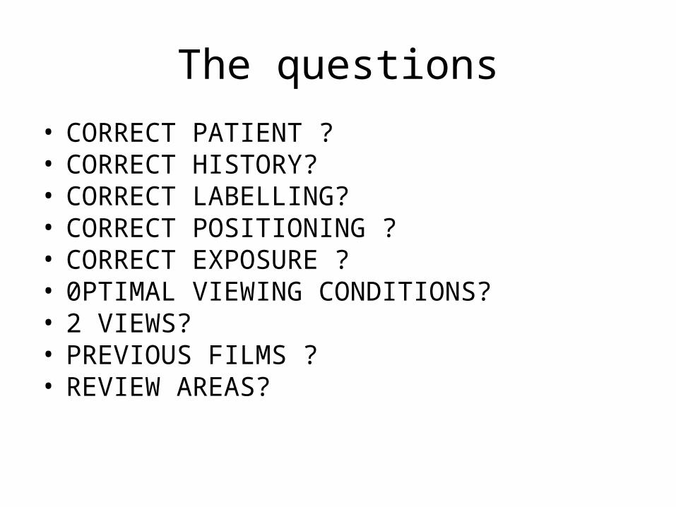
The questions
• CORRECT PATIENT ?• CORRECT HISTORY?• CORRECT LABELLING?• CORRECT POSITIONING ?• CORRECT EXPOSURE ? • 0PTIMAL VIEWING CONDITIONS?• 2 VIEWS?• PREVIOUS FILMS ? • REVIEW AREAS?
The Billion Dollar questions
• Is it real ? Technical / artefact
• Is it incidental ? Normal structure Variant
• Is it significant ?
“ …you can’t see what you don’t know ….”

CXR - REVIEW AREAS
• APICES• HILA• BEHIND THE HEART• CP ANGLES• BREASTS• BONES• PARASPINAL
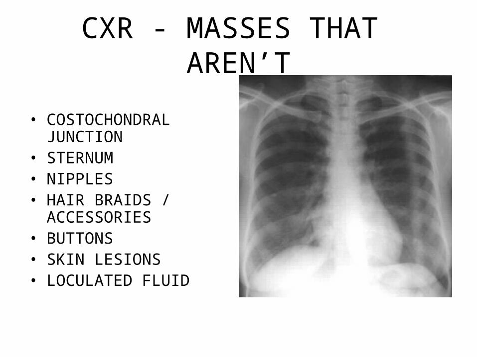
CXR - MASSES THAT AREN’T
• COSTOCHONDRAL JUNCTION
• STERNUM• NIPPLES• HAIR BRAIDS /
ACCESSORIES• BUTTONS• SKIN LESIONS• LOCULATED FLUID

LEFT UPPER LOBE COLLAPSE

LEFT LOWER LOBE COLLAPSE
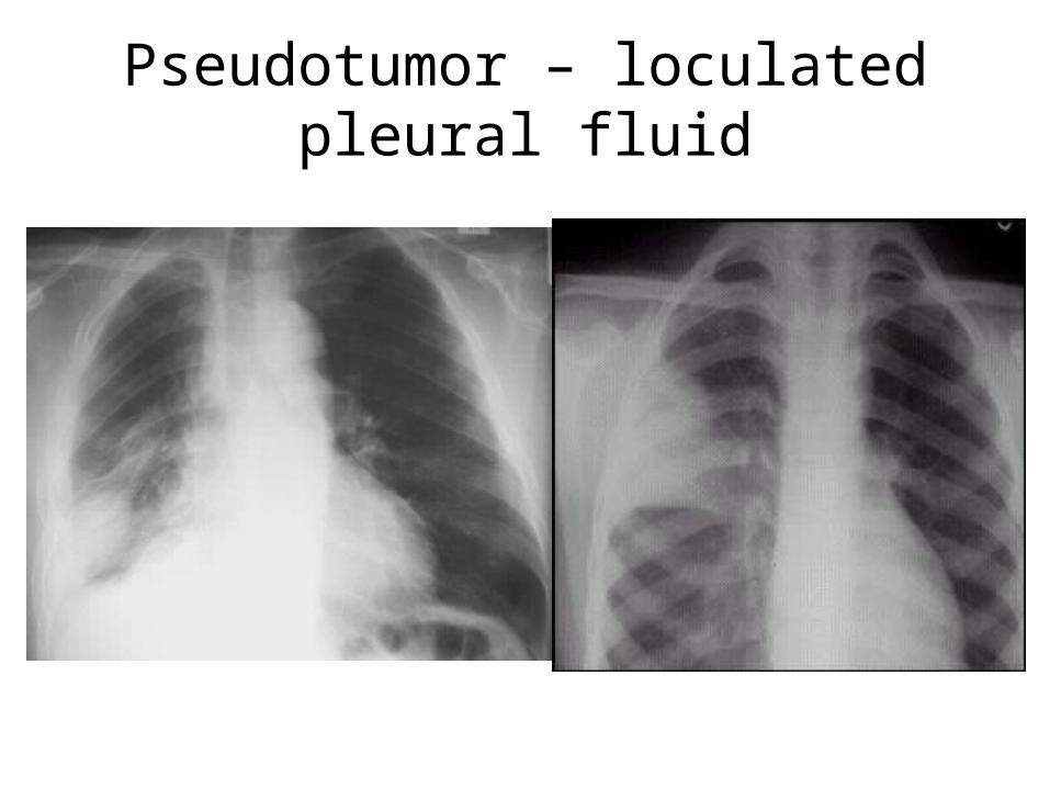
Pseudotumor – loculated pleural fluid
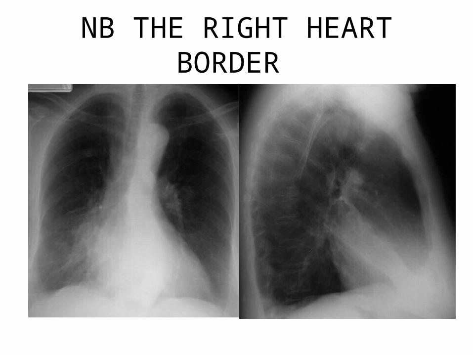
NB THE RIGHT HEART BORDER
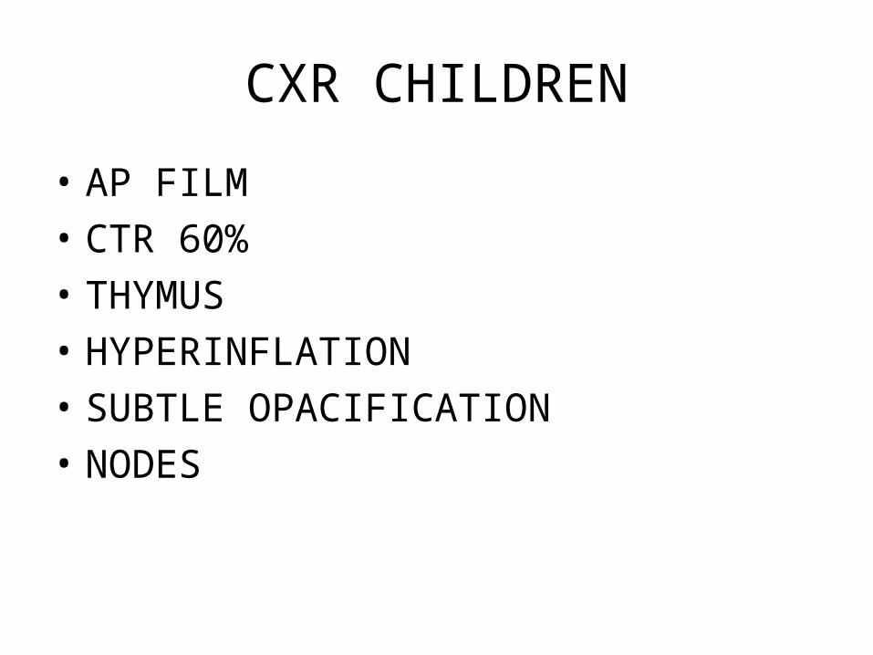
CXR CHILDREN
• AP FILM
• CTR 60%
• THYMUS
• HYPERINFLATION
• SUBTLE OPACIFICATION
• NODES

The Thymus

ASPIRATION OF FB

PNEUMOMEDIASTINUM
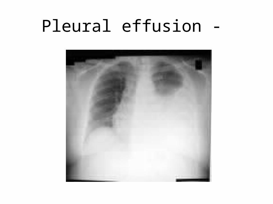
Pleural effusion -
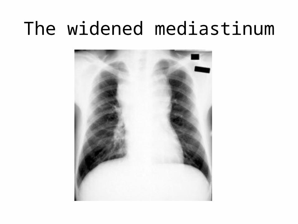
The widened mediastinum

Abdominal XRay
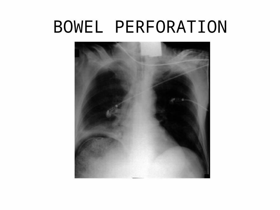
BOWEL PERFORATION
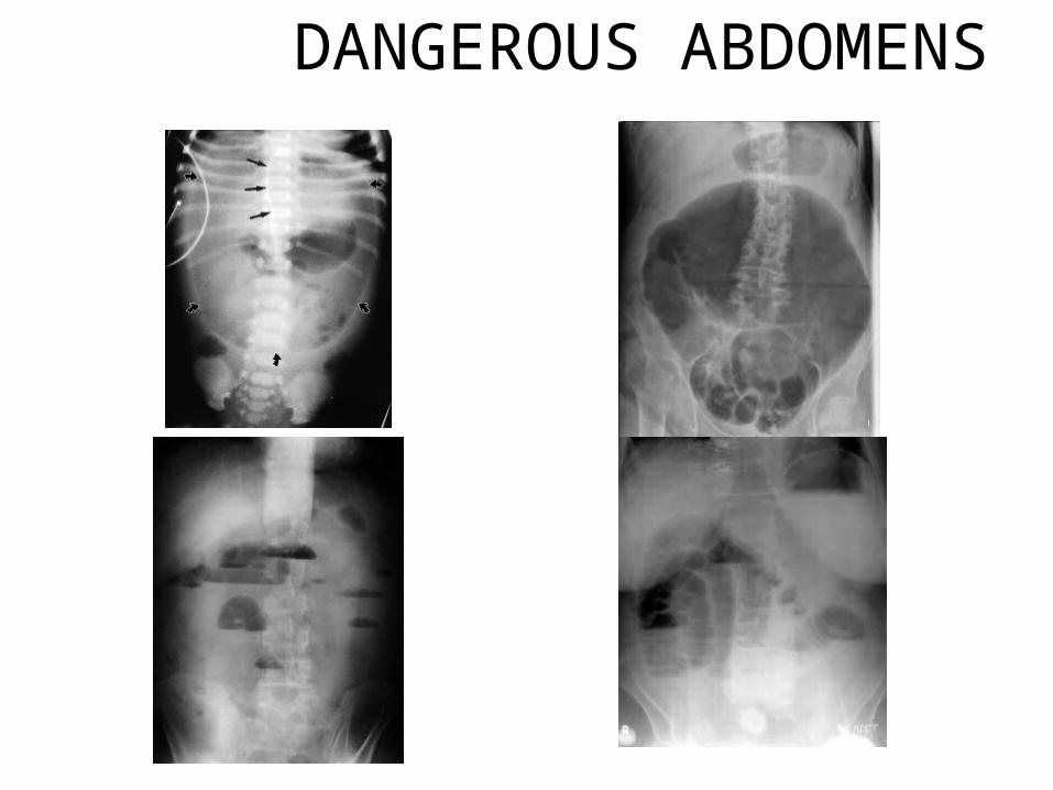
DANGEROUS ABDOMENS
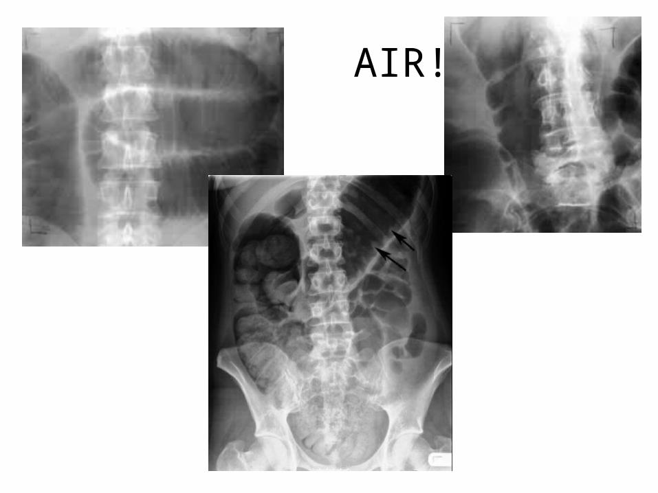
AIR!
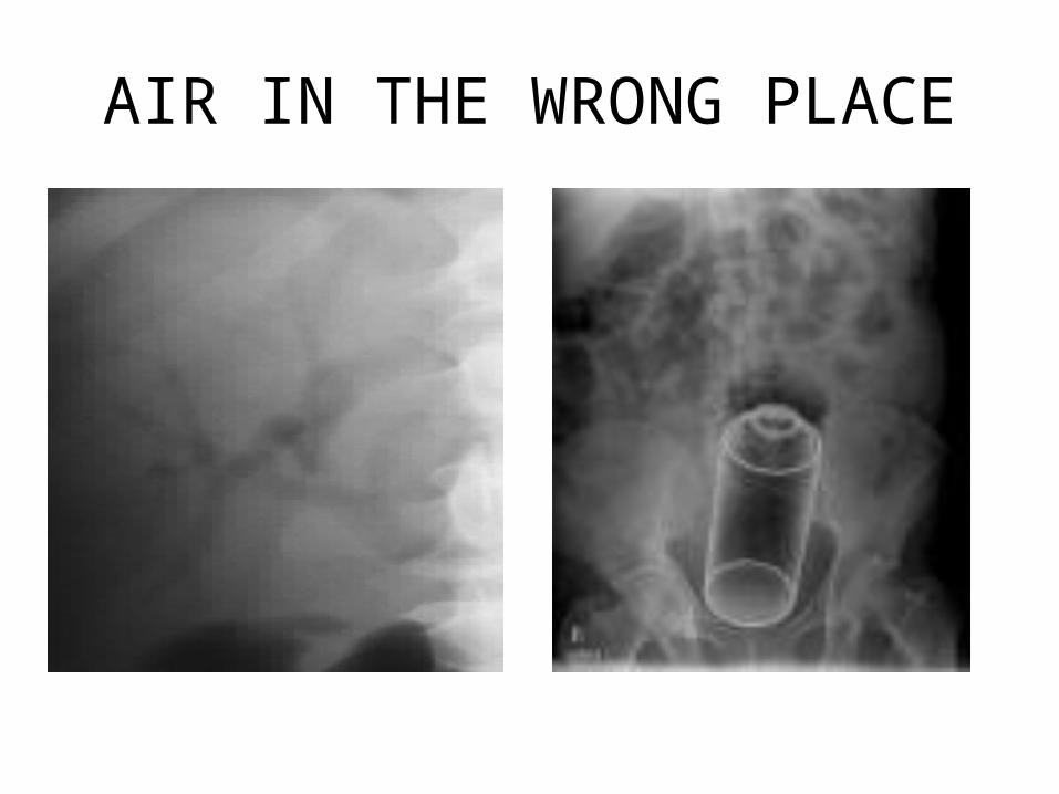
AIR IN THE WRONG PLACE
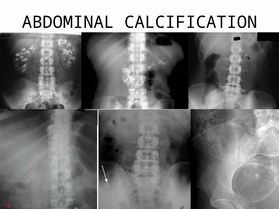
ABDOMINAL CALCIFICATION

BONES
• NB 2 VIEWS - ALWAYS
• COMPARE WITH OPPOSITE SIDE
• REPEAT XRAY IN 2 WEEKS ( PANNUS )
• CONSULT FRIENDLY TEXT ( KEATS )
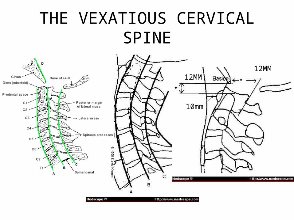
THE VEXATIOUS CERVICAL SPINE
12MM12MM
10mm
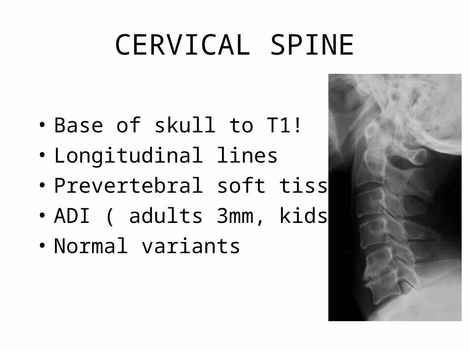
CERVICAL SPINE
• Base of skull to T1!
• Longitudinal lines
• Prevertebral soft tissue
• ADI ( adults 3mm, kids 5mm )
• Normal variants
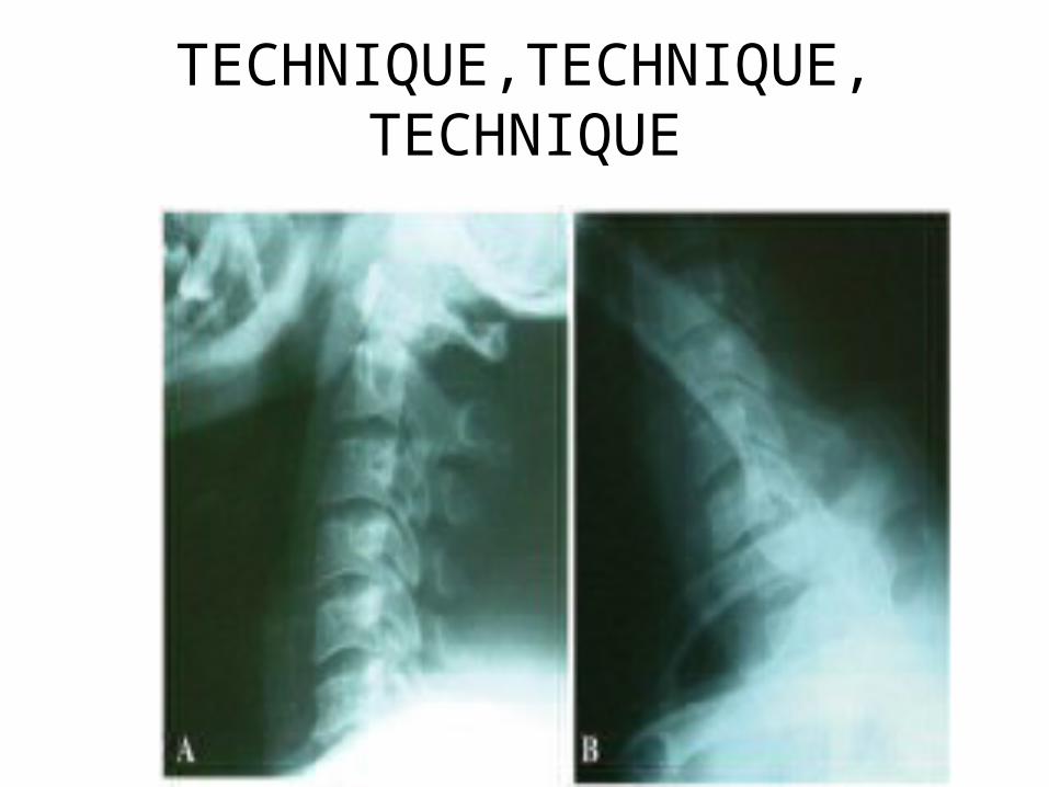
TECHNIQUE,TECHNIQUE,TECHNIQUE

THE OPEN MOUTH VIEW
MISCHIEVOUS FRACTURES
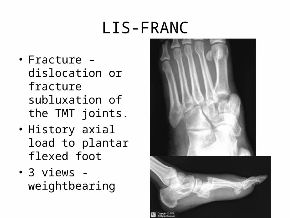
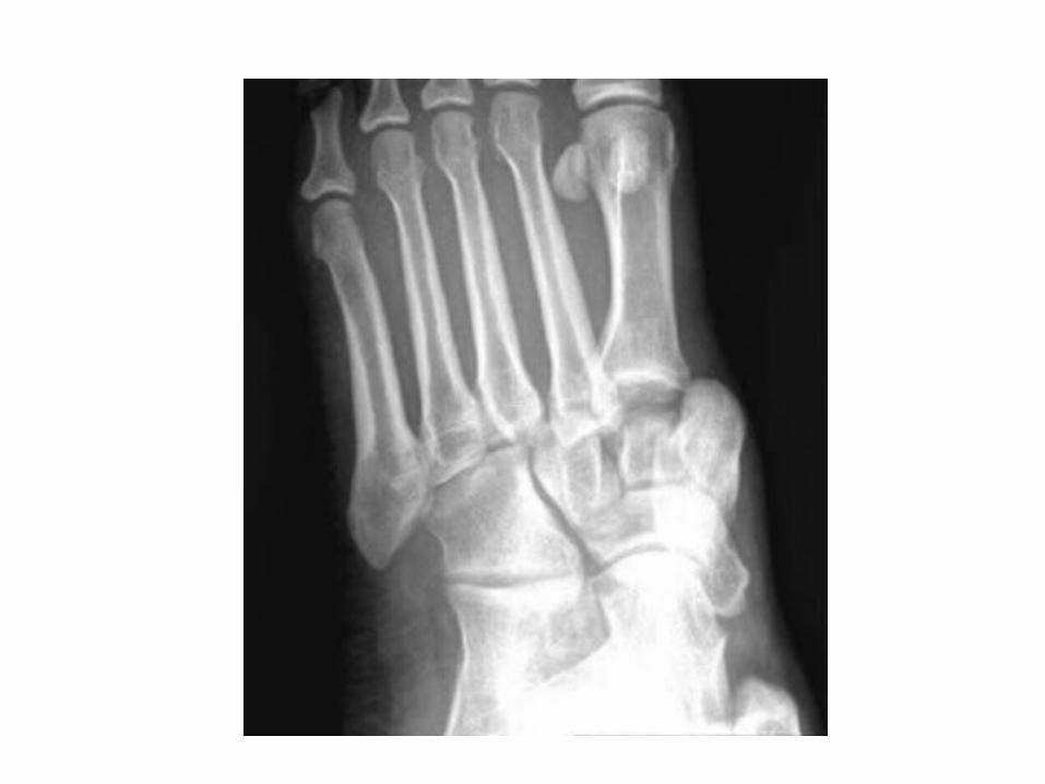
LIS-FRANC
• Fracture –dislocation or fracture subluxation of the TMT joints.
• History axial load to plantar flexed foot
• 3 views - weightbearing

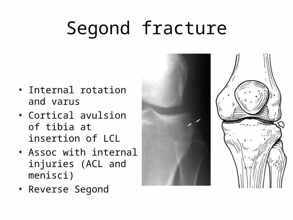
Segond fracture
• Internal rotation and varus
• Cortical avulsion of tibia at insertion of LCL
• Assoc with internal injuries (ACL and menisci)
• Reverse Segond

Maisonneuve fracture
• Pronation external rotation
• # upper third fibula• rupture distal tibiofibular
syndesmosis and interosseous membrane
• UNSTABLE• OUT OF ANKLE VIEW
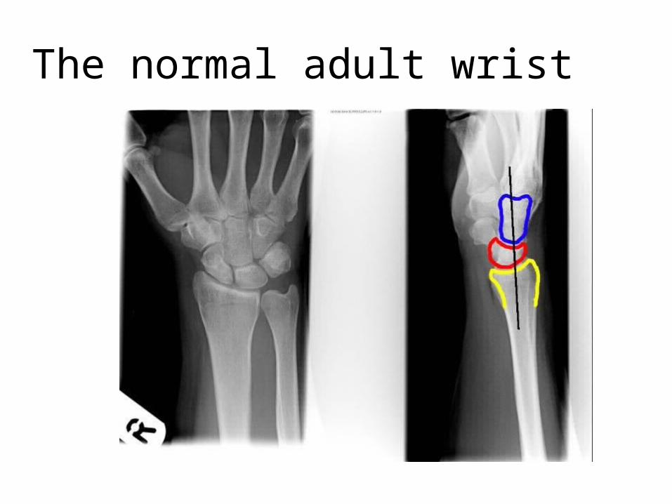
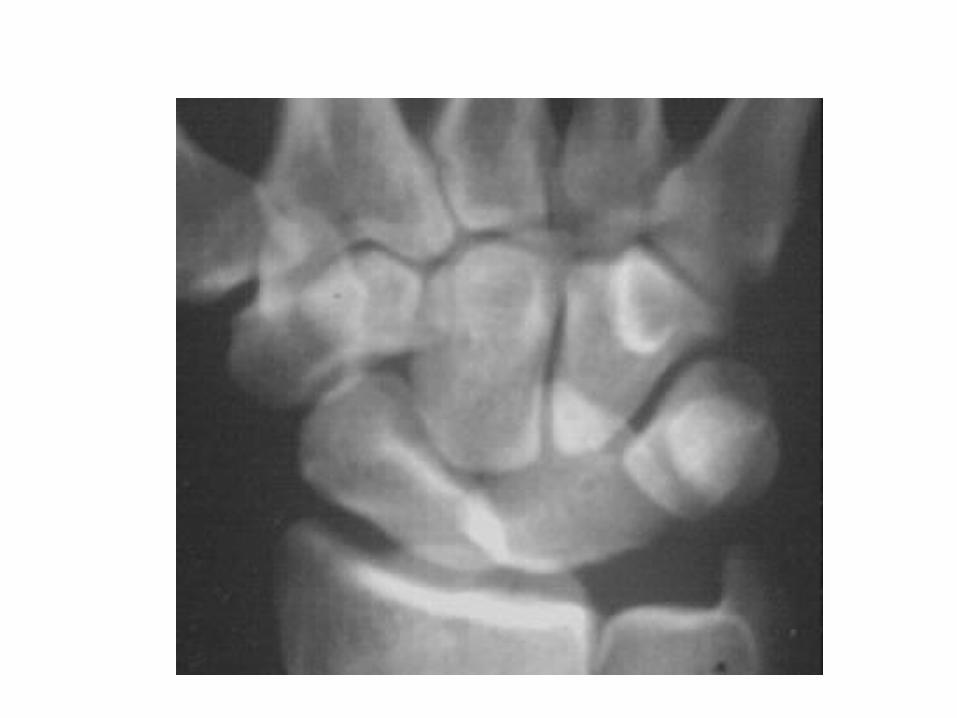
The normal adult wrist

Lunate dislocation
• Lunate loses its articulation with both the capitate and the radius and is displaced volarly with up to 90 degrees rotation. The capitate remains aligned with the radius but sinks proximally

Perilunate dislocation
• The lunate maintains its normal articulation with the radius.
• The capitate articular surface is dislocated from the lunate, normally dorsally

Salter Harris Physeal Injuries
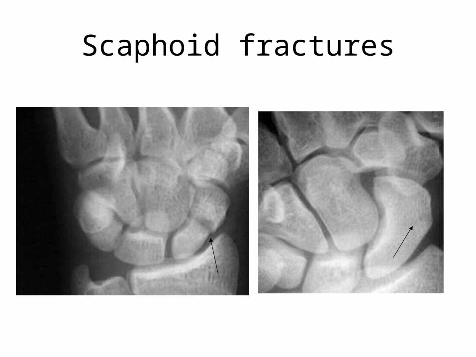
Scaphoid fractures
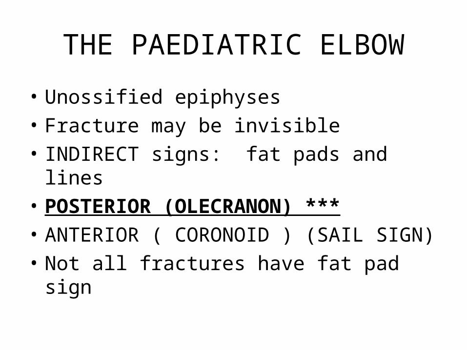
THE PAEDIATRIC ELBOW
• Unossified epiphyses
• Fracture may be invisible
• INDIRECT signs: fat pads and lines
• POSTERIOR (OLECRANON) ***
• ANTERIOR ( CORONOID ) (SAIL SIGN)
• Not all fractures have fat pad sign

THE ELEVATED FAT PAD
ANT CORONOID
POST OLECRANON

Normal alignment elbowAnterior humeral line
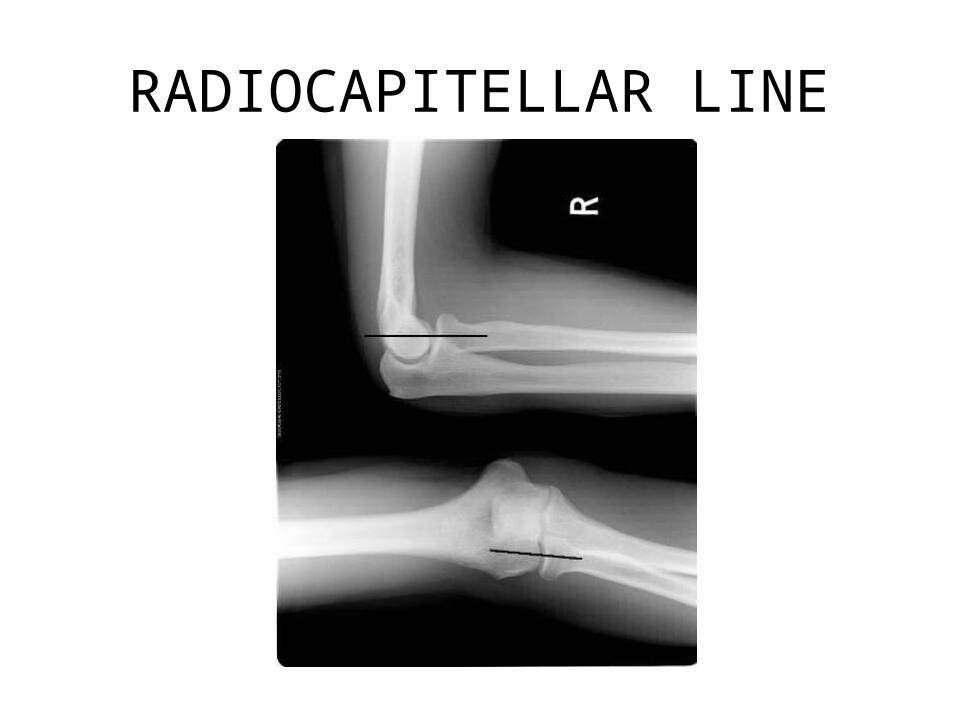
RADIOCAPITELLAR LINE
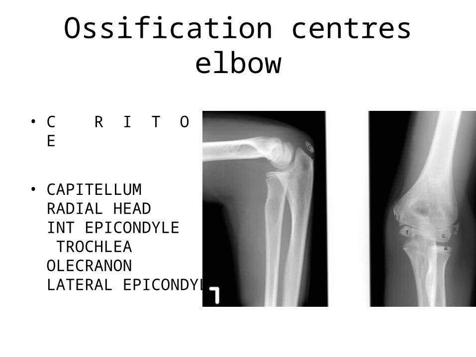
Ossification centres elbow
• C R I T O L E
• CAPITELLUMRADIAL HEADINT EPICONDYLE TROCHLEA OLECRANONLATERAL EPICONDYLE


THANK YOU!