committee report: publication guidelines and ... - eeg-emgeeg-emg.hu/textbox/media/keil.pdf ·...
TRANSCRIPT

Committee report: Publication guidelines and recommendations forstudies using electroencephalography and magnetoencephalography
ANDREAS KEIL,a STEFAN DEBENER,b GABRIELE GRATTON,c MARKUS JUNGHÖFER,d
EMILY S. KAPPENMAN,e STEVEN J. LUCK,e PHAN LUU,f GREGORY A. MILLER,g and CINDY M. YEEh
aDepartment of Psychology and Center for the Study of Emotion and Attention, University of Florida, Gainesville, Florida, USAbDepartment of Psychology, University of Oldenburg, Oldenburg, GermanycDepartment of Psychology and Beckman Institute, University of Illinois at Urbana-Champaign, Urbana-Champaign, Illinois, USAdInstitute for Biomagnetism and Biosignalanalysis, University of Münster, Münster, GermanyeCenter for Mind & Brain and Department of Psychology, University of California, Davis, California, USAfElectrical Geodesics, Inc. and Department of Psychology, University of Oregon, Eugene, Oregon, USAgDepartment of Psychology, University of California, Los Angeles, California, USA andDepartment of Psychology and Beckman Institute, University of Illinois at Urbana-Champaign, Urbana-Champaign, Illinois, USAhDepartment of Psychiatry and Biobehavioral Sciences and Department of Psychology, University of California, Los Angeles, California, USA
Abstract
Electromagnetic data collected using electroencephalography (EEG) and magnetoencephalography (MEG) are of centralimportance for psychophysiological research. The scope of concepts, methods, and instruments used by EEG/MEGresearchers has dramatically increased and is expected to further increase in the future. Building on existing guidelinepublications, the goal of the present paper is to contribute to the effective documentation and communication of suchadvances by providing updated guidelines for conducting and reporting EEG/MEG studies. The guidelines also includea checklist of key information recommended for inclusion in research reports on EEG/MEG measures.
Descriptors: Electrophysiology, Methods, Recording techniques, Data analysis, Good practices
Electrophysiological measures derived from the scalp-recordedelectroencephalogram (EEG) have provided a window into thefunction of the living human brain for more than 80 years. Morerecently, technical advancements allow magnetic fields associatedwith brain function to be measured as well, resulting in a growingresearch community using magnetoencephalography (MEG).Although hemodynamic imaging and transcranial magnetic stimu-lation have expanded the understanding of psychophysiologicalprocesses considerably, electromagnetic measures have not losttheir importance, largely due to their unparalleled temporal reso-lution. In fact, recent technological developments have fundamen-tally widened the scope of methodologies available to researchersinterested in electromagnetic brain signals. At the time of writing,the increased use of MEG as well as the development of powerfulnew hardware and software tools have led to a dramatic growth inthe range of research questions addressed. With the growing reali-zation that diverse electrical, magnetic, optical, and hemodynamicpsychophysiological methods are complementary rather than com-petitive, attempts at multimodal neuroimaging integration are
growing. In addition, a wide spectrum of data recording, artifactcontrol, and signal processing approaches are currently used, someof which are intimately known only to a small number of research-ers. With these richer opportunities comes a growth in demands fortechnical expertise. For example, there is much greater use of andneed for advanced statistical and other signal-processing methods,both adapted from other fields and developed anew. At the sametime, the availability of “turn-key” recording systems used by awide variety of scholars has been accompanied by a trend of report-ing less detail about recording and data analysis parameters. Bothdevelopments are at odds with the need to communicate experi-mental procedures, materials, and analytic tools in a way thatallows readers to evaluate and replicate the research described in apublished manuscript.
The goal of the present paper is to update and expand existingpublication guidelines for reporting on studies using measuresderived from EEG. If not otherwise specified, these guidelines alsoapply to MEG measures. This report is the result of the collabora-tive effort of a committee appointed by the Society forPsychophysiological Research, whose members are listed as theauthors of this manuscript. Previous guideline publications andcommittee reports have laid an excellent foundation for researchand publication standards in our field (Donchin et al., 1977; Pictonet al., 2000; Pivik et al., 1993). A recent paper by Gross et al.(2013) provided specific recommendations and reporting guide-lines for the recording and analysis of MEG data. The reader isreferred to these publications in addition to the present report. To
The authors would like to thank the many members of the Society forPsychophysiological Research who have contributed to the discussion ofthese guidelines.
Address correspondence to: Andreas Keil, PhD, Department of Psy-chology and Center for the Study of Emotion & Attention, University ofFlorida, PO Box 112766, Gainesville, FL 32611, USA. E-mail: [email protected]
bs_b
s_ba
nner
Psychophysiology, •• (2013), ••–••. Wiley Periodicals, Inc. Printed in the USA.Copyright © 2013 Society for Psychophysiological ResearchDOI: 10.1111/psyp.12147
1

facilitate reading and to assist in educating researchers early intheir careers, certain key points will be discussed in the presentdocument, despite having been extensively covered in previouspublication guidelines.
How To Use This Document
Publication guidelines and recommendations are not intended tolimit the ability of individual researchers to explore novel aspectsof electromagnetic data, innovative analyses, or new ways of illus-trating results. Rather, the goal of this document is to facilitatecommunication between authors and readers as well as editors andreviewers, by providing guidelines for how such communicationcan successfully be implemented. There will be instances in whichauthors will want to deviate from these guidelines. That will oftenbe acceptable, provided that such deviations are explicitly docu-mented and explained. To aid editors, reviewers, and authors, wehave compiled a checklist of key information required when sub-mitting a research report on EEG/MEG measures. The checklist,provided in the Appendix, summarizes central aspects of theguidelines.
Guidelines
Hypotheses and Predictions
In most cases, specific hypotheses and predictions about the elec-tromagnetic activity of interest should be provided in the introduc-tion. Picton et al. (2000) offered helpful examples regarding thepresentation of such predictions and their relation to the scientificrationale and behavioral constructs under study. This involvesmaking predictions about exactly how the electromagnetic indexwill differ by condition, group, or measurement site, reflecting themain hypotheses of the study. It is rarely sufficient to make ageneral prediction that the measures will differ, without describingthe specific ways in which they are expected to differ. For example,a manuscript might describe predicted differences in the amplitudeor latency of specific components of the event-related potential(ERP) or event-related field (ERF), or differences between fre-quency bands contained in EEG/MEG recordings. The predictionsshould directly relate to the theories and previous findingsdescribed in the introduction and describe, to the extent possible,the specific components, time ranges, topography, electrode/sensorsites, frequency bands, connectivity indices, etc., where effects arepredicted.
Participant Characteristics
The standards for obtaining informed consent and reporting onhuman participants, consistent with the Helsinki Accords, theBelmont Report, and the publication manual of the AmericanPsychological Association (APA, 2010), apply fully to electromag-netic studies. It is well established that interindividual differencesin the physiological or psychological status of research participantswill affect electromagnetic recordings. In fact, such differences areoften the focus of a given study. Even in studies not targetinginterindividual differences, suitable indicators of participant statusshould be reported, including age, gender, educational level, andother relevant characteristics. The specifics of what to report mayvary somewhat with the nature of the sample, the experimentalquestion, or even the sociocultural context of the research.
In their ERP guidelines report, Picton et al. (2000) discussedareas of importance regarding selection of participants for research
and reporting procedures. These include reporting the number ofparticipants, their sensory and cognitive status, and appropriateinformation on health status. In addition, recent epidemiologicalwork and EEG/MEG research in clinical populations suggest thatfor certain research questions it is useful to report additional infor-mation on the sample. When relying on measures that are sensitiveto factors such as psychopathology or alcohol and substance use,for example, additional screening procedures may be appropriateeven in studies involving nonclinical samples. Results of epidemio-logical studies in the United States suggest that past-year andlifetime prevalence rates for a mental disorder are approximately25% and 46%, respectively (Kessler, Berglund et al., 2005;Kessler, Chiu, Demler, Merikangas, & Walters, 2005). The rate ofcurrent illicit drug use in the United States among 18- to 25-year-olds is over 20%, and rates of binge and heavy alcohol use in 21-to 25-year-olds approach 32% and 14%, respectively (SAMHSA,2012). Among college students, who comprise a significant pro-portion of research participants in the United States, most estimatesplace the binge-drinking rate at 40%–50% (e.g., Wechsler &Nelson, 2008). If screening is undertaken, authors should report themethod and measures used as well as all inclusion and exclusioncriteria.
The issue of matching of groups in clinical studies involvingelectromagnetic data also warrants careful attention. It has longbeen noted that matching on one or more characteristics maysystematically mismatch groups on other characteristics (e.g.,Resnick, 1992). Thus, researchers should carefully consider notonly the variables on which to match their groups but the possibleunintended consequences of doing so. It may be that a singlecomparison group cannot handle all relevant issues and that multi-ple control groups are needed.
Recording Characteristics and Instruments
Evaluation, comparison, and replication of a given psycho-physiological study depend heavily on the description of theinstrumentation and recording settings used. Many of the relevantparameters are listed in Picton et al. (2000) as well as Gross et al.(2013) for MEG, and the reader is referred to these publicationsfor a discussion of how to report on instrumentation. Here, werecapitulate key information that needs to be provided and offer anupdate on new requirements and recommendations pertaining torecent developments in hardware and software.
Sensor types. The type of MEG sensor or EEG electrode shouldbe indicated, ideally accompanied by make and model. In additionto traditional sensor types, recent developments in EEG sensortechnology include active electrodes. Active electrodes have cir-cuitry at the electrode site that is designed to maintain good signal-to-noise ratio. Electrode-scalp impedances are often of less concernwhen using active electrodes, and authors may want to emphasizethis aspect when using a system that allows reporting impedancevalues. For passive and active electrodes, the electrode materialshould be specified (e.g., Ag/AgCl).
Two types of dry sensor technology have become morewidely used in recent years (Grozea, Voinescu, & Fazli, 2011).Microspikes penetrate the stratum corneum (Ng et al., 2009), thehighly resistive layer of dead cells. Capacitive sensors (Taheri,Knight, & Smith, 1994) rely on conductive materials, such asrubber (Gargiulo et al., 2010), foam, or fabric (Lin et al., 2011).Common to these dry sensor technologies is the challenge of rec-ording the EEG over scalp regions covered by hair. Most previous
2 A. Keil et al.

applications with dry sensors have involved recordings from theforehead and often have been in the context of research on brain-computer interfaces. When using dry electrodes, the type and tech-nology should be indicated clearly in the manuscript.
Sensor locations. Although MEG sensor locations are fixed rela-tive to each other within a recording system, the position of theparticipant’s head relative to the sensor should be reported alongwith an index of error/variability of position measurement. Whenreporting EEG research, electrode positions should be clearlydefined. Standard electrode positions include the 10-20 system andthe revision to a 10-10 system proposed by the AmericanElectroencephalographic Society (1994). This standard is similar tothe 10-10 system of the International Federation of ClinicalNeurophysiology (Nuwer et al., 1999). Oostenveld and Praamstra(2001) proposed an extension of the 10-10 system, referred to asthe 10-5 system, in order to accommodate electrode arrays withmore than 75 electrode sites. An alternative electrode placementsystem employs a description of the scalp surface based on geo-desic (equidistant) partitioning of the head surface with up to 256positions (e.g., Tucker, 1993), rather than the percentage approachof the 10-20 and related systems. The electrode positions in thissystem vary according to channel count, as the geodesic partitionsdiffer for different spatial frequencies and ensure regular spacingbetween electrodes.
Regardless of the system used, a standard nomenclature shouldbe employed and one or more appropriate citations reported. If anequidistant/geodesic placement system is used, the average dis-tance between electrodes should be reported, and the coverage ofthe head sphere should be described in relation to the 10-20 or10-10 system. This information can be conveyed in the text or in afigure showing the sensor layout. Ground and reference electrodelocations should be specified. For active electrode systems andMEG recordings, the type and location of additional sensors usedto reduce ambient and/or subject noise should be indicated. Forexample, the location of the so-called common mode sense orsimilar reference electrode should be mentioned if an amplifiersystem uses such an arrangement. In general, individual referenceelectrodes are recommended over physically linked reference elec-trodes (see Miller, Lutzenberger, & Elbert, 1991). Other referencemontages can be computed offline.
Spatial sampling. The relationship between the rate ofdiscretization (i.e., digital sampling) and accurate description of thehighest frequency of an analog, time-series signal (e.g., the EEG orMEG waveform) is well known as the Nyquist theorem. Thistheorem posits that signal frequencies equal to or greater than halfof the sampling frequency (i.e., the Nyquist frequency) will bemisrepresented. This principle of discretization also holds for sam-pling in the spatial domain for EEG and MEG data, where thesignal is the voltage or field data across or above the scalp surface(Srinivasan, Nunez, & Silberstein, 1998). Undersampling in thespatial domain results in high-spatial-frequency features being mis-taken for low-spatial frequency information.
In addition to the importance of spatial sampling density, spatialcoverage is critical. Traditionally, the head surface is not coveredinferior to the axial (horizontal) plane, particularly when using10-20 EEG positions, whereas equidistant layouts are commonlydesigned to provide more coverage of the head sphere. In manycontexts, especially for source estimation involving much or all ofthe brain, it is important that the sensor montage extend inferior atleast to the equivalent of the axial plane containing F9/F10 in the
10-10 system. At issue is how well local activity from the ventralaspects of the brain is represented. Inadequate spatial sampling canresult in a biased estimate of averaged-reference data (Junghöfer,Elbert, Tucker, & Braun, 1999) and misinterpretation regarding theunderlying sources (Lantz, Grave de Peralta, Spinelli, Seeck, &Michel, 2003). Authors should address these limitations, particu-larly when reporting on topographical distributions of electromag-netic data.
Measuring sensor locations. With the increased use of densesensor arrays and source estimation procedures, the exact locationof sensors for a given participant is of increasing importance.Current methods for the determination of 3D sensor positionsinclude manual methods, such as measuring all intersensor dis-tances with digital calipers (De Munck, Vijn, & Spekreijse, 1991),or measuring a subset of sensors arrayed in a known configurationand then interpolating the positions of the remaining sensors (Le,Lu, Pellouchoud, & Gevins, 1998). Another group of methodsrequires specialized equipment such as electromagnetic digitizers,near-infrared cameras, ultrasound sensors, photographic imagesacquired from multiple views (Baysal & Sengul, 2010; Russell,Jeffrey Eriksen, Poolman, Luu, & Tucker, 2005), and electrodescontaining a magnetic resonance marker together with MR images(e.g., Koessler et al., 2008). When 3D coordinates are reported, themethod for obtaining these parameters should be detailed and anindex of spatial variability or measurement error provided.
The resolution with which sensor positions can be measuredvaries considerably, with a potentially strong impact on the validityand reliability of source localization solutions. Under some circum-stances, such measurement error contributes most of the sourcelocalization error. At best, spatial localization methods are limitedlargely by the accuracy of sensor position measurement, butcompare favorably to the spatial resolution provided by routinefunctional magnetic resonance imaging (fMRI) procedures (Aineet al., 2012; Miller, Elbert, Sutton, & Heller, 2007). Thus, includingthe recommended information on measurement of sensor positionsis quite important in studies seeking discrete source estimation.
Amplifier type. Accurate characterization of signal amplitude isdependent on many factors, one of which is an amplifier’s inputimpedance. Amplifier systems differing in input impedance varydrastically in their sensitivity to variations in electrode impedance(Ferree, Luu, Russell, & Tucker, 2001; Kappenman & Luck, 2010).Thus, authors are encouraged to report the input impedance of theiramplifiers. At minimum, the make and model of the recordingsystem should be indicated in the Methods section.
Impedance levels. If the impedance of the connection between theelectrode and the skin is high, this may increase noise in the data.High impedances may also increase the incidence and amplitude ofelectrodermal (skin) potentials related to sweat gland activity. Tra-ditionally, researchers have addressed these issues by reducingelectrode-scalp impedance at each site to be equal to or less than agiven threshold (e.g., 10 kΩ). The impedance at a given electrodeand time point may be less important than the relationship betweenelectrode impedance and amplifier input impedance in rejectingelectrical noise, although sensitivity to skin potentials depends oninput impedance even when the amplifier is held constant(Kappenman & Luck, 2010). In addition, the variability and rangeof impedances across time points and electrodes may add noise tothe signal, impacting topographical and temporal information andinferences about sources. EEG systems with active sensors have
Guidelines for EEG and MEG 3

different requirements in terms of impedance and may have differ-ent ways of reporting indices of data quality. Thus, no singlethreshold for maximum acceptable electrode impedance can beoffered. Reporting should follow the recommendations of themanufacturer. The use of amplifiers with a very high input imped-ance also reduces the importance of matching impedance for spe-cific sets of electrodes that will be compared, such as athemispherically homologous sites (Miller et al., 1991).
Obtaining impedances at individual electrodes below a targetthreshold typically requires preparation (e.g., abrasion) of eachsite, because the stratum corneum is highly resistive. When suchprocedures are used, the method should be described. Electrodeimpedances should be reported when appropriate, or an equivalentsignal quality index given when impedances cannot be obtained.When using high-input impedance systems with passive electrodes,information should be given about the range of electrode-scalpimpedances, the amplifier’s input impedance, and informationabout how ambient recording conditions were controlled.
Recording settings. The settings of recording devices should bereported in sufficient detail to allow replication. At minimum,parameters should include resolution of the analog-to-digital con-verter and the sampling rate. In addition, any online filters usedduring data recording must be specified, including the type of filterand filter roll-off and cut-off values (stated in dB or specifyingwhether cut-off is the half-power or half-amplitude value; Cook &Miller, 1992).
Stimulus and Timing Parameters
Timing. As noted in Picton et al. (2000), reporting the exacttiming of all stimuli and responses occurring during electromag-netic studies is critical. Such information should be providedin a fashion that allows replication of the sequence of events.Required parameters include stimulus durations, stimulus-onsetasynchronies, and intertrial intervals, where applicable. Manyexperimental control platforms synchronize stimulus presentationrelative to the vertical retrace of one of the monitors comprising thesystem, unless otherwise specified. If used, such a linkage shouldbe reported and presentation times accurately indicated in multi-ples of the video signal retrace (monitor refresh) rate.
When an aspect of the timing is intentionally variable (e.g.,variable interstimulus intervals), then a uniform (rectangular) dis-tribution of the time intervals is assumed unless otherwisespecified. The relative timing of trials belonging to different experi-mental conditions should be specified, including any rules orrestrictions during randomization, permutation, or balancing. Thenumber of trials in each condition should be explicitly specified,along with the number of trials remaining in each condition whenelimination of trials is used to remove artifacts, poor performance,etc. (In many contexts, the number of trials per condition is aprimary factor affecting signal-to-noise ratio.) Information aboutpractice trials should also be provided. The total recording durationshould be specified along with the duration of rest breaks betweenrecording blocks, where applicable.
Stimulus properties. Replication of a given study is possible onlyif the relevant parameters defining the stimuli are described. Thismay include a description of the experimental setting in which thestimuli are presented, including relevant physical and psychosocialaspects of the participants’ environment. For instance, it may beappropriate for some studies to report the experimenter’s gender,
the participants’ body posture, or the presence or absence of aux-iliary instructions such as to avoid eye blinking during particulartime periods of the experiment. Other studies may require preciseinformation regarding ambient lighting conditions, or the size ofthe recording chamber.
Ideally, examples of the stimuli should be provided in theMethods section, especially when nonstandard or complex stimuliare used. For visual stimuli, parameters may include stimulus sizeand viewing distance, often together with the visual angles spannedby the stimuli. Because of the strong impact of contrast and inten-sity of a stimulus on the amplitude and latency of electromagneticresponses, these parameters should be reported where applicable.Different measures of contrast and luminance exist, but often lumi-nance density in units of cd/m2 can be reported along with either ameasure of luminance variability across the experimental displayor an explicit measure of contrast, such as the Michelson contrast.Reporting on stimulus color should follow good practices in therespective area of research, which may include specifying CIEcoordinates. Additional requirements regarding the display devicemay exist in studies focusing on color processing. In all instances,the type of display should be reported along with the frame rate orany similar parameter. For example, authors may report that a LEDscreen of a given make and model with a vertical frame rate of120 Hz was used. In studies that focus on higher-order processing,it may suffice to specify approximate characteristics of stimulusand display (e.g., “a gray square of moderate luminance,” “a redfixation cross”).
Similarly, investigators examining responses to auditory stimulishould report the intensity of the stimuli in decibels (dB). Becausethe dB scale reflects a ratio between two values (not absoluteintensity), the report of dB values should include the specificationof whether this relates to sound pressure level (SPL), sensationlevel (SL), hearing level (HL), or another operational definition.When appropriate, the frequency content of the stimulus should bereported along with measures of onset and offset envelope timingand a suitable measure of the energy over time, such as the rootmean square. Paralleling the visual domain, the make and model ofthe delivery device (e.g., headphones, speakers) should be speci-fied. The procedure for intensity calibration should also bereported. If this was adjusted for individual subjects, that procedureshould also be reported.
Electromagnetic studies with stimuli in other modalities (e.g.,olfactory, tactile) should follow similar principles by defining thenature, timing, and intensity of stimuli and task parameters, pergood practice in the respective field of research.
Response parameters. The nature of any response devices shouldbe clearly specified (e.g., serial computer mouse, USB mouse,keyboard, button box, game pad, etc.). Suitable indices ofbehavioral performance should be indicated, likely includingresponse times, accuracy, and measures of their variability.Because computer operating systems differ in their ability tosupport accurate response recording, reports may include thisinformation when appropriate.
Data Preprocessing
Data preprocessing commonly refers to a diverse set of pro-cedures that are applied to the data prior to averaging or othermajor analysis procedures. These procedures may transform thedata into a form that is appropriate for more generic computationsand eliminate some types of artifact that cannot be dealt with
4 A. Keil et al.

satisfactorily through averaging methods, spectral analysis, orother procedures. The end product is a set of “clean” continuousor single-trial records ready for subsequent analysis. Reportsshould include clear descriptions of the methods used for each ofthe preprocessing steps along with the temporal order in whichthey were carried out. The following section considers preproc-essing steps.
Transformation from A/D units into physical units. This step isnecessary for comparing data across experiments. Either the dataneed to be presented in physical units (e.g., microvolts, femtotesla),or waveforms presented in the paper should report a suitable com-parison scale with respect to a physical unit.
Rereferencing. The reference issue is an important problem inEEG studies, and aspects of it were also discussed above (seeRecording Characteristics and Instruments). In EEG research, adifferent reference may be used for online recording and offlineanalysis. For example, data can be recorded referenced to a spe-cific electrode (e.g., left mastoid), and a different reference (suchas average mastoids, or average reference) can be computedoffline. Typically, this involves a simple linear transformation cor-responding to adding or subtracting a particular waveform fromall the channels (unless a Hjorth or Laplacian reference system isused, see Application of Current Source Density or LaplacianTransformations). As data change substantially depending on thereference method used, the type of reference used for online rec-ording and offline analysis should be indicated clearly. When theaverage of multiple sites is used as the reference, all the sitesshould be clearly specified, even if they are not used as “active”sites in the analyses.
Interpolation of missing data. Some EEG/MEG channels maycontain excessive artifacts and thus may not be usable. Forexample, an electrode may detach during recording, or a connec-tion may become faulty. The probability of such artifacts increaseswith dense sensor arrays. It may nevertheless be preferable toinclude data from missing channels in the analysis; for example, incases where the analysis software requires identical sensor layoutsfor all participants. For this reason, problematic or missing data areoften replaced with interpolated data. Interpolation is a mathemati-cal technique for estimating unobserved data, according to somedefined function (e.g., linear, sphere, spline functions, average ofneighbors), from those measured. Interpolation can be used toestimate data between sensor locations (such as those used forcolor-coded topographic maps) or to replace missing data at a givensensor. For spatial interpolation, it is important to note that theestimated data do not provide higher-density spatial content thanwhat is contained in the original data (Perrin, Pernier, Bertrand,Giard, & Echallier, 1987). Moreover, when interpolation is used toreplace missing data, the limit to accuracy is a function of thespatial frequency of the missing data and the number and distribu-tion of sensors. That is, if missing data points are spatially contigu-ous, and the data to be replaced are predominantly of high spatialcontent, then it is likely that the estimated data are spatially aliasedand do not provide an accurate replacement. Publications shouldreport the interpolation algorithm used for estimating missingchannels or for spatial interpolation resulting in topographicalmaps. Information should be provided as to how many missingchannels were interpolated for each participant. Often, informationabout the spatial distribution of interpolated channels will berequired.
Segmentation. Most analysis methods, including traditionalcross-trial averages and spectral and time-frequency analyses, arebased on EEG/MEG segments of a specific length and a givenlatency range with respect to an event. Although some data are stillrecorded in short epochs, data are now more typically recorded ina continuous fashion over extended periods of time and then seg-mented offline into appropriate epochs. The time range used tosegment the data should be reported.
Baseline removal. ERP data are changes in voltage betweenlocations on the recording volume occurring over time. Similarly,ERFs are changes in magnetic field strength. To quantify thischange, researchers often define a time period during which themean activity is to be used as an arbitrary zero value. This periodis called the “baseline” period. The mean value recorded duringthis baseline period is then subtracted or divided from the rest ofthe segment to result in a measure of change with respect to thiszero level. In most ERP/ERF studies, a temporally local baselinevalue is computed for each recording channel. The choice ofbaseline period is up to the investigators and should be appropri-ate to the experimental design. The baseline period should bespecified in the manuscript and should ideally be chosen suchthat it contains no condition-related differences. In addition, asdiscussed in the section Results Figures, the baseline periodshould be displayed in waveform plots. Alternative proceduresmay be used to establish change with respect to a baseline,including regression or filter-based methods. Authors should indi-cate the method and the data segments used for any baseline pro-cedures. Additional recommendations exist for baseline removalwith spectral or time-frequency analyses, as discussed in theSpectral Analysis section.
Artifact rejection. There are many types of artifacts that cancontaminate EEG and MEG recordings, including artifacts gener-ated by the subject (e.g., eye blinks, eye movements, muscle activ-ity, and skin potentials) and artifacts induced by the recordingequipment or testing environment (e.g., amplifier saturation andline noise). These artifacts are often very large compared to thesignal of interest and may differ systematically across conditions orgroups of subjects, making it necessary in many experiments toremove the data segments with artifacts from the data to obtain aclean signal for analysis. It should be specified whether artifactswere rejected by visual inspection, automatically based on an algo-rithm, or a combination of visual inspection and automatic detec-tion. The algorithms used for automatic detection of artifactsshould be described in the paper (e.g., a moving window peak-to-peak algorithm). It should also be stated whether thresholds forautomatic detection procedures were set separately for individualsubjects or channels of data. If any aspect of these procedures wascontrolled by the experimenter (e.g., manual rejection or subject-specific parameter settings), it should be indicated whether this wasdone in a manner that was blind to experimental condition orparticipant group. Unless otherwise specified, it is assumed that allchannels are rejected for a segment of data if an artifact is identifiedin a single channel.
Importantly, because the number of artifacts may differ substan-tially between experimental conditions or between groups of sub-jects (e.g., some patient populations may exhibit more artifacts thanhealthy comparison subjects), the number or percentage of trialsrejected for each group of subjects must be specified (especially ifpeak amplitude measurement is used; see section below on meas-urement procedures). It should be made clear to the reader whether
Guidelines for EEG and MEG 5

the number of trials contributing to the averages after artifact rejec-tion differs substantially across conditions or groups of subjects.
Artifact correction. It is often preferable and feasible to estimatethe influence of an artifact on the EEG or MEG signal and tosubtract the estimated contribution of the artifact, rather than reject-ing the portions of data that contain artifacts. A number of correc-tion methods have been proposed, including regression methods(such as Gratton, Coles, & Donchin, 1983; Miller, Gratton, & Yee,1988), independent component analysis (ICA; Jung et al., 2000),frequency-domain methods (Gasser, Schuller, & Gasser, 2005),and source-analysis methods (e.g., Berg & Scherg, 1994b). Anumber of studies have investigated the relative merits of thevarious procedures (e.g., Croft, Chandler, Barry, Cooper, & Clarke,2005; Hoffmann & Falkenstein, 2008). By and large, most of thesestudies have shown that correction methods are generally effective.The major issues are the extent to which they might undercorrect(leaving some of the artifact present in the data) or overcorrect(eliminating some of the nonartifact activity from the data) andwhether such correction errors are inconsistent (e.g., larger forsensors near the eyes).
Regardless of which artifact correction procedure is chosen, thepaper should provide details of the procedure and all steps used toidentify and correct artifacts so that another laboratory can repli-cate the methods. For example, it is not sufficient to state “artifactswere corrected with ICA.” Paralleling artifact rejection (above),other necessary information includes whether the procedure wasapplied in whole or in part automatically. If nonautomatic correc-tion procedures were used, it should be specified whether the indi-vidual performing the procedure was blind to condition, group, orchannel. In addition, a detailed list of the preprocessing stepsperformed before correction (including filtering, rejection of largeartifacts, segmentation, etc.) should be described. If a statisticalapproach such as ICA is used, it is necessary to describe the criteriafor determining which components were removed.
If a participant blinks or moves his/her eyes during the presen-tation of a visual stimulus, the sensory input that reaches the brainis changed, and ocular correction procedures are not able to com-pensate for the associated change in brain-related processing. Inexperimental designs where compliance with fixation instructionsis crucial (e.g., visual hemifield studies), it is recommended thatsegments of data that include ocular artifacts during the presenta-tion of the stimuli be rejected prior to ocular correction.
Offline filtering. Filters can be used to improve the signal-to-noiseratio of EEG and MEG data. As filters lead to loss of information,it is often advisable to do minimal online analog filtering andinstead use appropriately designed offline digital filters. In general,both online and offline filters work better with temporally extendedepochs and therefore may be best applied to continuous than tosegmented data. This is particularly the case for high-pass filtersused to eliminate very low frequencies (drift) and for low-passfilters with a very sharp roll-off. The type of filters used in theanalysis should be reported along with whether they were appliedto continuous or segmented data. It is not sufficient to simplyindicate the cut-off frequency or frequencies; the filter familyand/or algorithm should be indicated together with the filter orderand descriptive indices of the frequency response function. Forexample, a manuscript may report that “a 5th order infinite impulseresponse (IIR) Butterworth filter was used for low-pass filteringon the continuous (nonsegmented data), with a cut-off frequency(3 dB point) of 40 Hz and 12 dB/octave roll-off.” Other ways of
reporting filter characteristics are possible, but they should includethe filter family (e.g., Boxcar, Butterworth, Elliptic, Chebychev,etc.) as well as information on the roll-off or steepness of thetransition in the filter response function (e.g., by indicating that theroll-off was 12 dB/octave). The most common single index of afilter’s frequency response function is the half-amplitude or half-power cutoff (the frequency at which the amplitude or power isreduced by 50%). The half-power and half-amplitude frequenciesare not the same, so as noted above it is important to indicatewhether the cutoff frequency specifies the half-amplitude (−6 dB)point or the half-power (−3 dB) point (Cook & Miller, 1992; Edgar,Stewart, & Miller, 2005).
Measurement Procedures
After preprocessing is completed, the data are typically reduced toa much smaller number of dependent variables to be subjected tostatistical analyses. This often consists of measuring the amplitudesor latencies of specific ERP/ERF components or quantifying thepower or amplitude within a given time-frequency range. Increas-ingly, it involves multichannel analysis via principal componentanalysis (PCA) or ICA, dipole or distributed source analysis,and/or quantification of relationships between channels or sourcesto evaluate connectivity. Choosing the appropriate measurementtechnique for quantifying these features is important, and there area variety of measurement techniques available (some describedbelow; see Fabiani, Gratton, & Federmeier, 2007, Handy, 2005,Kappenman & Luck, 2012b, Kiebel, Tallon-Baudry, & Friston,2005, or Luck, 2005, for more information). The following guide-lines are described primarily in the context of conventional, time-domain ERP/ERF analyses, but many points apply to otherapproaches as well.
Isolating components. Successful measurement requires care thatthe ERP/ERF component of interest is isolated from other activity.Even a simple experimental design will elicit multiple componentsthat overlap in time and space, and therefore it is usually importantto take steps to avoid multiple components contributing to a singlemeasurement. As a first step, researchers typically choose a timewindow and a set of channels for measurement that emphasize thecomponent of interest. However, source-analysis procedures havedemonstrated that multiple components are active at a given sensorsite at almost every time point in the waveform (Di Russo,Martinez, Sereno, Pitzalis, & Hillyard, 2002; Picton et al., 1999).Many investigators have acknowledged this problem and have pro-posed various methods for addressing it in specific cases (e.g.,Luck, 2005). Methods for isolating a single component includecreating difference waves or applying a component decompositiontechnique, such as ICA or spatiotemporal PCA, discussed inthe Principal Component Analysis and Independent ComponentAnalysis section (Spencer, Dien, & Donchin, 1999; for generaldiscussion of techniques for isolating components of interest, seeKappenman & Luck, 2012a; Luck, 2005). The method for compo-nent isolation used in a particular study should be reported. If nosuch method is used, authors are encouraged to discuss componentoverlap as a potential limitation. Most effective in separatingthe sources of variance, when feasible, is an experimental designthat manipulates the overlapping components orthogonally (seeKappenman & Luck, 2012b, for a description).
Description of measurement procedures. The measurementprocess should be described in detail in the Method section. This
6 A. Keil et al.

includes specifying the measurement technique (e.g., mean ampli-tude) as well as the time window and baseline period used formeasurement (e.g., “Mean amplitude of the N2pc wave was meas-ured 175 to 225 ms poststimulus, relative to a −200 to 0 msprestimulus baseline period.”). A justification for the time windowand baseline period should be included (as described in more detailbelow).
For measurement techniques that use peak values—such aspeak amplitude, peak latency, and adaptive mean amplitude, whichrelies in part on the peak value—additional information about themeasurement procedures is helpful. First, the method used tofind the peak value should be specified, including whether thepeak values were determined automatically by an algorithm ordetermined (in whole or in part) by visual inspection of the wave-forms. If visual inspection was used, the manuscript should specifywhether the individual performing the inspection was blind tocondition, group, or channel information. It should also be statedwhether the peak was defined as the absolute peak or the local peak(e.g., a point that was greater than the adjacent points even if theadjacent points fell outside the measurement window—see Luck,2005). It should also be stated whether the peak value was deter-mined separately for each channel or whether the peak latency inone channel was used to determine the measurement latency at theother channels. Generally, it would be misleading to plot the scalpor field distribution of a peak unless it was measured at the sametime at all sites.
Peak measures become biased as the signal-to-noise ratiodecreases (Clayson, Baldwin, & Larson, 2013), as scores will bemore subject to exaggeration due to overlapping noise. In principle,the signal-to-noise ratio improves as a function of the square root ofthe number of trials in an average. When analyzing cross-trialaverages, it is therefore problematic to compare peak values fromconditions or groups with significantly different numbers of trials.Studies that rely on peak measures scored from averages (or othermeasures that may be biased in a similar manner) should report thenumber of trials in each condition and in each group of subjects.This should include the mean number of trials in each cell, as wellas the range of trials included.
Acommon alternative to peak scoring is area scoring. In general,area measures are less susceptible to signal-to-noise-ratio problemsresulting from few (or differing numbers of) trials. An area measureis typically the sum or average of the amplitudes of the measuredpoints in a particular scoring window (after baseline removal).However, the term area can be ambiguous, because the area of ageometric shape can never be negative. For measurement techniquesthat use area measures, it is recommended that the field adopt thefollowing terminology: positive area (area of the regions on thepositive side of the baseline); negative area (area of the regions onthe negative side of the baseline); integrated area (positive areaminus negative area); geometric area (positive area plus negativearea; this is the same as positive area preceded by rectification).
Inferences about magnitude and timing. Care must be taken indrawing conclusions about the magnitude and timing of the under-lying neural activity on the basis of changes in the amplitude andlatency of ERPs/ERFs. For example, a change in the relative ampli-tudes or latencies of two underlying components that overlap intime can cause a shift in the latency of scalp peaks (see Donchin &Heffley, 1978; Kappenman & Luck, 2012a). Thus, a change in peakamplitude does not necessarily imply a change in component mag-nitude, and a change in peak latency does not necessarily imply achange in component timing. As noted above, suitable methods for
isolating specific spatiotemporal processes assist in the interpreta-tion of overlapping neural events and should be reported in suffi-cient detail to allow evaluation and replication.
Measurement windows and electrode sites must be welljustified and avoid inflation of Type I error rate. EEG/MEG datasets are extremely rich, and it is almost always possible to finddifferences that are “statistically significant” by choosing measuresthat take advantage of noise in the data, even if the null hypothesis istrue. Opportunities for inflation of Type I error rate (i.e., an increasein false positives) almost always arise when the observed waveformsare used to determine how the data are quantified and analyzed (seeInferential Statistical Analyses). For example, if the time range andsensor sites for measuring a component are chosen on the basis of thetiming and topographical distribution of an observed differencebetween conditions in the same data set, this will increase thelikelihood that the difference will be statistically significant even ifthe difference is a result of noise rather than a reliable effect.
Choosing time windows and sensor sites that maximize sensi-tivity to real effects while avoiding this kind of bias is a majorpractical challenge faced by researchers. This problem has becomemore severe as the typical number of recording sites increased fromone to three in the 1970s to dozens at present. Fortunately, concep-tual and data-driven approaches have been developed that make itpossible to select electrode sites and time windows in a way that isboth unbiased and reasonably powerful. An important guideline isto provide an explicit justification for the choice of measurementwindows and sensor sites, ensuring that this does not bias results.
A variety of ways of selecting scoring windows and sites arevalid. In many cases, the best approach is to use time windows andsensor sites selected on the basis of prior research. When this is notpossible, a common alternative approach is to create an averageacross all participants and conditions and to use this information toidentify the time range and topographical distribution of a givencomponent. This is ordinarily an unbiased approach with respect todifferences between groups or conditions. However, care must betaken if a given component is larger in one group or condition thanin another group or condition or if group or condition numbersdiffer substantially. For example, Group A may have a larger P3than Group B, and the average across Group A and Group B is usedto select the electrode sites for measuring P3 latency. If Groups Aand B differ in P3 scalp distribution, then a suboptimal set of sensorsites would be used to measure P3 latency in Group B, and thiscould bias the results. The issue of making scoring decisions basedon the data being analyzed is considered further in the InferentialStatistical Analyses section.
Another approach is to obtain measures from a broad set of timeranges or sensor locations and include this as a factor in the statis-tical analysis. For example, one might measure the mean amplitudeover every consecutive 100-ms time bin between 200 and 800 msand then include time as a factor in the statistical analysis. If aneffect is observed, then each time window or site can be testedindividually, using an appropriate adjustment for multiple compari-sons. A recent approach is to use cluster-based analyses that takeadvantage of the fact that component and time-frequency effectstypically extend over multiple consecutive sample points andmultiple adjacent sensor sites (see, e.g., Groppe, Urbach, & Kutas,2011; Maris, 2012).
Both descriptive and inferential statistics should be provided.Reporting of results should include both descriptive and inferentialstatistics (see Inferential Statistical Analyses). For descriptive
Guidelines for EEG and MEG 7

statistics, the group-mean values for each combination of conditionand group should be provided, along with a measure of variability(e.g., standard deviation, standard error, 95% confidence interval,etc.). It is generally not sufficient to present inferential statisticsthat indicate the significance of differences between means withoutalso providing the means themselves. These descriptive statisticscan be included in the text, a table, a figure, or a figure caption asappropriate. Descriptive statistics should ordinarily be presentedprior to the inferential statistics. This is further discussed in theInferential Statistical Analyses section.
Results Figures
Electromagnetic data are multidimensional in nature, oftenincluding dimensions of sensor channel, topography, or voxel,time and/or frequency band, experimental condition, and group,among others. As a result, particular efforts are necessary toensure their proper graphical representation on a journal page.Documentation of data quality in most cases will require presen-tation of a waveform or frequency spectrum in the manuscript. Asdescribed in Picton et al. (2000) for ERP data, it is virtually man-datory that figures be provided for the relevant comparisons. Theyshould include captions and labels with all information needed tounderstand what is being plotted. Ideally, the set of figures in agiven paper will graphically convey both the nature of thedependent variable (voltage, field, dipole source strength, fre-quency, time-frequency, coherence, etc.) and the temporal andspatial properties of the data, as they differ across experimentalconditions or groups. As a result, papers will typically havefigures highlighting time (e.g., line plots) or spatial distribution(e.g., topographies).
Line plots (see Figure 1) often show change in a measure as afunction of time (e.g., amplitude or power of voltages or mag-netic fields). Inclusion of line plots is strongly recommended forERP/ERF studies. It is recommended that waveforms be overlaid,facilitating comparison across conditions, groups, sensors, etc.This may sometimes involve presenting the same waveformtwice: for example, patients overlaid with controls for each con-dition and each condition overlaid separately for patients andcontrols. Figures should be clearly labeled with the spatial loca-tion from which the data were obtained, such as a sensor, source
location, or a topographically defined group of sensors. A base-line segment should be included in any data figure that depicts atime course. This baseline segment should be of sufficient lengthto contain a valid estimate of pre-event or postevent activity. Thetime segment used as the baseline should be included in thefigure (see Data Preprocessing, above). In the case of time-varying power or amplitude in a given frequency band (e.g.,event-related (de)synchronization), the baseline segment shouldcontain at least the duration of two cycles of the frequency underconsideration (e.g., at least 200 ms of baseline are needed whendisplaying time-varying 10 Hz activity) or the duration that cor-responds to the time resolution of the specific analysis method(e.g., the temporal full width at half maximum of the impulseresponse).
Each waveform plot should include a labeled x axis with appro-priate time units and a labeled y axis with appropriate amplitudeunits. In most cases, waveforms should include a y-axis line at timezero (on the x axis) and an x-axis line at the zero of the physical unit(e.g., voltage). Generally, unit ticks should be shown, at sufficientlydense intervals, for every set of waveforms, so that the time andamplitude of a given deflection are immediately visible. Line plotsdisplaying EEG segments or ERPs also should clearly indicatepolarity, and information regarding both the electrode montage (e.g.,32-channel) and reference (e.g., average mastoid) should be pro-vided in the caption. Including this information in the caption isimportant to facilitate readers comparing waveforms across studies,which may use different references that contribute to reporteddifferences in the waveforms. The waveforms should be sufficientlylarge that readers can easily see important differences among them.Where difference waveforms are a standard in a given literature, theymay suffice to illustrate a given effect, but in most cases the corre-sponding condition waveforms should be provided. Because densearrays of EEG and MEG sensors are increasingly used, it is often notpractical to include each sensor’s time-varying data in a given figure.For most situations, a smaller number of representative sensors (orsensor clusters) or another suitable figure with temporal informationwill suffice (e.g., the root mean square). Spatial information relatedto specific effects may be communicated with additional scalptopographies, as described in the next section.
Scalp potential or magnetic field topographies and source plotsare typically used to highlight the spatial distribution of voltage,magnetic fields, current/source densities, and spectral power,among other measures. If the figure includes mapping by means ofan interpolation algorithm, such as that suggested by Perrin et al.(1987), the method should be indicated. Interpolation algorithmsimplemented in some commercial or open-source software suitesare optimized for smooth voltage topographies, leading to severelimitations when mapping nonvoltage data or topographies con-taining higher spatial frequencies (see Data Preprocessing for rec-ommendations on interpolation). Paralleling the recommendationsfor line plots, captions and labels should accompany topographicalfigures, indicating the type of data mapped along with a keyshowing the physical unit. In addition, the perspective should beclearly indicated (e.g., front view, back view; left/right) when using3D head models. For flat maps, clearly labeling the front/nose andleft/right and indicating the projection method in the caption areimportant. The location of the sensors on a head volume or surfaceshould be visible, to communicate any differences between inter-polation across the sensor array, versus extrapolation of databeyond the area covered with sensors. Authors have increasinglyused structural brain images overlaid with functional variables
PO7/PO8200 400 600 800 ms−200
N2pc
−5
7
1
−1
5
3
−3
ContraIpsi
µVN1
P1
Figure 1. Example of an ERP time series plot suitable for publication.Note that the electrode location is indicated, and both axes are labeled withphysical units at appropriate intervals. The onset of the event (in thisexample, a visual stimulus) is clearly indicated at time zero. Overlaying twoexperimental conditions fosters comparison of relevant features. (In thisexample, positive voltage is plotted “up.” Both positive up and negative upare common in the EEG/ERP literature, and the present paper makes norecommendation for one over the other.)
8 A. Keil et al.

derived from EEG and MEG. In many such cases, a key to relevantanatomical regions (slice location or a reference area) shownshould be provided.
Connectivity figures and time-frequency plots are increasinglyemployed to illustrate different types of time-frequency and con-nectivity analyses. Although these figures will necessarily varygreatly, inclusion of figures showing original data is recommended,along with the higher-order analyses. Where possible, authorsshould provide illustrations that maintain a close relationship to thedata and analyses performed. For example, results of connectivityanalyses carried out for scalp voltages should normally bedisplayed on a scalp model and not on a standard brain. For time-frequency figures (see Figure 2), a temporal baseline segmentshould be included of sufficient length to properly display thelowest frequency in the figure, as noted above. In general, many ofthe recommendations discussed above for line plots and topo-graphical figures apply to other figure types, and authors areencouraged to apply them as appropriate.
Inferential Statistical Analyses
Data analysis is a particularly rich and rapidly advancing area in theEEG/MEG literature. Because electromagnetic data are often mul-tidimensional in nature, statistical analysis may pose considerablecomputational demands. As noted in Picton et al. (2000), research-ers must ensure that statistical analyses are appropriate both to thenature of the data and to the goal of the study. Generally, statisticalapproaches applied to EEG/MEG data fall into two categories: onewhere data reduction of dependent variables to relatively smallnumbers is accomplished on the basis of a priori assumptions, asdescribed above, and one where the number of dependent variablesto which inferential statistical testing is applied remains too large toallow meaningful application of traditional statistical methods.Various aspects of these two approaches are discussed below.Approaches in which multivariate methods are used to reduce thedimensionality of the data prior to statistical analysis are discussedin the Principal Component Analysis and Independent ComponentAnalysis section.
Studies with preselected dependent variables. Even when sta-tistical analyses are conducted on a limited number of dependentvariables that are defined in advance, authors must ensure theappropriateness of the procedure for the data type that is to beanalyzed. Where assumptions of common statistical methods areviolated (e.g., normality, sphericity), it should be noted and anadequate correction applied when available. For example, it isexpected that the homogeneity/heterogeneity of covariance inwithin-subjects designs is examined and addressed by Greenhouse-Geisser, Huynh-Feldt, or equivalent correction if the assumption ofsphericity is violated (Jennings, 1987) or that multivariate analysisof variance (MANOVA) is undertaken (Vasey & Thayer, 1987).Often, nonparametric statistical approaches are appropriate for agiven data type, and authors are encouraged to consider suchalternatives. For example, permutation tests and bootstrappingmethods have considerable and growing appeal (e.g., Maris, 2012;Wasserman & Bockenholt, 1989). Concerns specific to studies ofgroup effects, as noted in Picton et al. (2000), include the impor-tance of demonstrating that the dependent variable (e.g., an ERPcomponent, a magnetic dipole estimate) is not qualitatively differ-ent as a function of group or condition. This is an obvious problemwhen latencies or waveforms of components or spectral eventsdiffer by group or condition. In these situations, authors should becareful to ensure that their dependent variables are indeed measur-ing the same phenomena or constructs across groups, conditions, orbrain regions.
Jackknife approaches have been specifically designed toexamine latency differences (Miller, Patterson, & Ulrich, 1998),enhancing the signal-to-noise ratio of the measurement by averag-ing across observations within a group (in n − 1 observations) andassessing the variability using all possible n − 1 averages. Theseand similar methods should be considered when latency differencesare of interest. Because jackknife methods are based on grandmeans, they depend greatly on assumptions regarding variabilitywithin conditions and groups and on the criterion used to identifythe latency of an event (e.g., 50% vs. 90% of an ERP peak value).Thus, authors should report measures of variability as well asindicate the criterion threshold and a rationale for its selection.
In studies designed to evaluate group differences, often as afunction of psychopathology or aging, participants may also differin their ability to perform a task. Task performance by healthyyoung-adult comparison subjects may be more accurate and fasterthan that by patients, children, or older participants. Assuming thatonly electromagnetic data obtained under comparable conditionsare to be included in each average (e.g., correct trials only), it is notunusual for psychometric issues concerning group differences inperformance to arise. Patient participants, for example, may requiremore trials (and consequently a longer session) in order to obtainas many correct trials as healthy comparison subjects. Becauseanalyzing sessions of varying durations in a single experiment risksa variety of confounds, identifying and selecting a subset (andequal number) of trials that occur at approximately the same timepoint for each patient and their demographically matched compari-son participant can help to address the issue. Statistical methodsthat explicitly address group differences in mixed designs, or withnested data in general, include multilevel models (Kristjansson,Kircher, & Webb, 2007). These approaches allow researchers toexplicitly test hypotheses on the level of individuals versus groupsand separately assess different sources of variability, for instance,regarding their respective predictive value. Researchers reportingon multilevel models should indicate the explicit model used inequation form.
Figure 2. Example of a time-frequency figure suitable for publication. Thefigure shows a wide range of frequencies (vertical axis) to allow readers acomparison of temporal dynamics in different frequency bands. Thevertical axis and the horizontal (time) axis are labeled with appropriatephysical units (a), and so is the color bar (b), which here indicates the powerchange from baseline levels in percent. Hence, it is important that asufficiently long baseline segment (c) be shown on the plot.
Guidelines for EEG and MEG 9

Explicit interactions supporting inferences. Hypotheses orinterpretations that involve regional differences in brain activityshould be supported by appropriate tests. For example, hemi-sphere should be included as a factor in statistical analyses, ifinferences are made about lateralized effects. In general, suchinferences are not justified when the analysis has essentiallyinvolved only a simple-effects test for each sensor, voxel, orregion of interest. Simple-effects tests may be appropriate forexploring an interaction involving hemisphere but are not them-selves a sufficient basis for inferences about lateralization. Thisexample of an effect of hemisphere generalizes to inferencesregarding region-specific findings. If two groups or conditionsdiffer in region X but not in region Y, a test for a Group × Regionor Condition × Region interaction is usually needed in order toinfer that the effect differs in regions X and Y. It may seem logi-cally sufficient to show that the effect in X differs from zero, andthe effect in Y does not. In such an analysis, however, it is essen-tial to demonstrate that the confidence intervals do not overlap.Even if the means of X and Y fall on opposite sides of zero, it ispossible that their confidence intervals overlap.
Scaling of topographic effects. Because topography is a tradi-tional criterion for defining ERP and ERF components, topo-graphic differences can suggest differences in neural generators.However, even varying amplitudes of a single, invariant neuralsource can produce Location × Condition interactions in statisticalanalyses, despite their fundamentally unchanged topography.McCarthy and Wood (1985) brought this issue to the attention ofthe literature and recommended scaling of the data to avoid theproblem, using a normalization of the amplitude of the scalp dis-tribution. This recommendation was widely endorsed for a time.Urbach and Kutas (2002, 2006), however, showed that scaling mayeliminate overall amplitude differences between distributionswithout altering the topography. They argued convincingly that therecommended scaling should not be performed routinely, becauseit does not resolve the interpretive problem of the source of elec-tromagnetic data. They also demonstrated that standard baselinesubtraction further compromises the scaled data.
Although such scaling is no longer recommended, the interpre-tative problem remains. This issue does not affect inferences thatare confined to whether two scalp topographies differ, which maysuffice in many experimental contexts. The issue arises, however,when the goal is to make an inference about whether underlyingsources differ. Generally, formal source analysis may be needed toaddress such a question, rather than relying solely on analyses insensor space.
Handling of baseline levels. Even the routine removal of baselinedifferences between groups, conditions, and recording sites canpotentially be problematic. First, whether to remove baseline vari-ance by subtracting baseline levels or by partialing them out isoften not obvious, and the choice is rarely justified explicitly.Second, baseline activity may be related to other activity of inter-est. If groups, conditions, or brain regions differ at baseline,removal of baseline levels may inadvertently distort or eliminateaspects of the phenomenon of interest (discussed below). It istherefore recommended that any baseline differences betweengroups or conditions be examined, where appropriate. Third, base-line removal can be particularly problematic for some types ofbrain source localization analyses. If a source estimation algorithmattempts to identify a source based on the spatial pattern of scalp-recorded signal intensities, but those values have been adjusted by
removal of site-specific baselines, the topography of the scalpsignals will in some cases no longer represent the original topog-raphy of the source. Urbach and Kutas (2002) discussed this issuein detail as it applies to normalization of topographies, and theircritique of baseline removal applies to source analysis as well.Authors are encouraged to assess the robustness of any observedeffects against baseline variations and scaling, where appropriate,and to document these steps in the manuscript.
Analytic circularity. A recent controversy in the hemodynamicneuroimaging literature (e.g., Kriegeskorte, Simmons, Bellgowan,& Baker, 2009; Lieberman, Berkman, & Wager, 2009; Viviani,2010; Vul, Harris, Winkielman, & Pashler, 2009) applies as well toelectromagnetic data. When two groups or conditions are com-pared, and one group or condition is used to define key aspects ofthe analysis such as the latency window, frequency band, or set ofvoxels or sensors (topography) for scoring, this approach can biasthe results. Resampling methods, replication, and basing scoringdecisions on independent data sets can help to avoid this problem.Patterns of results that are contrary to the bias may be accepted aswell, providing a particularly conservative test (e.g., Engels et al.,2007).
Psychometric challenges. Electromagnetic measurement issubject to the same psychometric considerations as are othertypes of measurement, yet issues of task matching, measurereliability, item discriminability, and general versus differentialdeficit (see Chapman & Chapman, 1973) are often not acknowl-edged in this literature. These issues go well beyond basic con-cerns about reliability and validity. For example, given twomeasurements that differ in reliability yet reflect identical effectsizes, it is generally easier to find a difference with the less noisymeasure, suggesting a differential deficit when none may bepresent. It also is the case that noise in measuring a covariatewill tend to lead to underadjustment for the latent variablemeasured by the covariate (Zinbarg, Suzuki, Uliaszek, & Lewis,2010). Evaluating the relationship between an independentvariable and a dependent variable can be further complicatedif a third variable shares variance with either the independent ordependent variable. For example, two groups of subjects maynot be ideally matched on age. Even if the discrepancy isnot large, the difference may be statistically significant. Age-related changes in electromagnetic activity can occur. Thus,partialing out shared variance provided by the third variable (e.g.,age) may not be an option, as the resulting partialed variablesmay no longer represent the intended construct or phenomenon(Meehl, 1971; Miller & Chapman, 2001). These issues can beparticularly difficult to address in clinical or developmentalresearch where random assignment to group is not an option, andgroups may differ on variables other than those of interest. Often,explicit modeling of the different effects at different levels mayrepresent a quantitative approach to addressing these issues(Tierney, Gabard-Durnam, Vogel-Farley, Tager-Flusberg, &Nelson, 2012).
Studies with massive statistical testing. Electromagneticdata sets increasingly involve many sensor locations or voxels,time points, or frequency bands and, essentially, massively par-allel significance testing. Recent developments in statistical meth-odology have led to specific procedures to treat such data setsappropriately. The methods may involve calculation of permuta-tion or bootstrapped distributions, without assuming that the data
10 A. Keil et al.

are uncorrelated or normally distributed (Groppe et al., 2011;Maris, 2012; Mensen & Khatami, 2013). When using suchmethods, it is recommended that authors report the number ofrandom permutations or a suitable quantitative measure of thedistribution used for thresholding, along with the significancethreshold employed. Because many variants of massivelyunivariate testing exist, the algorithm generating the referencedistribution should be indicated in sufficient detail to allowreplication.
Effect sizes. Increasing attention is being paid to effect size andstatistical power, beyond the traditional emphasis on significancetesting. Effect sizes may be large yet not very useful, or small buttheoretically or pragmatically important (Hedges, 2008). Thegrowing attention to effect size, while overdue, can be overdone.First, there is no consensus about what constitutes a “large” or a“small” effect across diverse contexts, although Cohen (1992) pro-vided a commonly cited set of suggestions. Second, effect size isnot always important to the research question at hand. In manyinferential contexts, the size of the effect is not as important aswhether groups or conditions differ reliably, as a basis for inferringwhether they are from the same population. Third, there is oftenlittle information on which to base a prediction about how large aneffect size will be. On the other hand, underpowered studies appearquite commonly, including in the psychophysiology literature.Small-N studies are generally limited to finding large effects.Whether that is acceptable in a given case is rarely discussed.
In summary, effect size is often but not always an importantissue. When it is important, it should be addressed explicitly. Effectsizes obtained in other contexts rarely suffice to justify an assump-tion of an effect size anticipated in a given study. Unless a model isemployed to make a point prediction of effect size, in most caseswhat should be discussed is how small an effect a study should bepowered to find, not merely what size effect may be likely.
Spectral Analyses
Changes in the spectral properties of raw waveforms during taskperformance are prima facie properties of EEG and MEG—theyare obvious when visually inspecting the ongoing electromagnetictime series or spatial topography. Given its salience even with basicrecording setups, the oscillatory character of EEG/MEG and therelationship between different types of oscillations and mentalprocesses have been the focus of pioneering work in EEG (Berger,1929; in English in 1969) and MEG research (Cohen, 1972). Thepresent discussion will focus on time-domain spectral analyses, butthese comments generally apply to spatial spectra as well.
Subsequent to the publication of guidelines for recording andquantitative analysis of EEG (Pivik et al., 1993), the availability ofpowerful research tools and the increasing awareness that spectralanalysis can provide rich information about brain function haveled to a sharp increase in the number of scholars interested inspectral properties of electromagnetic phenomena (e.g., Voytek,D’Esposito, Crone, & Knight, 2013). Many recent approachescapitalize on phase information, often used to compute metrics ofphase coherence and phase synchrony across trials, time points, orchannels. Variables that are reflective of some concept of causalityor dependence across spatial location in source or sensor spaceoften are tied to frequency-domain or time-frequency-domainanalyses and are therefore discussed in this section as well. Presentcomments supplement the guidelines presented in Pivik et al.(1993). More recent discussions (Herrmann, Grigutsch, & Busch,
2005; Keil, 2013; Roach & Mathalon, 2008) provide additionalresources regarding frequency and time-frequency analyses.
Frequency-domain analyses: Power and amplitude. The fre-quency spectrum of a sufficiently long data segment can beobtained with a host of different methods, with traditional Fourier-based methods being the most prevalent. These methods areapplied to time-domain data (where data points represent a tempo-ral sequence) and transform them into a spectral representation(where data points represent different frequencies), called the fre-quency domain. Both the power (or amplitude) and phase spectrumof ongoing sine waves that model the original data may be identi-fied in the frequency domain. Most authors use Fourier-basedalgorithms for sampled, noncontinuous data (discrete Fouriertransform, DFT; fast Fourier transform, FFT), the principles ofwhich are explained in Pivik et al (1993; see also Cook & Miller,1992). Many different implementations of Fourier algorithms exist,defined, for example, by the use of tapering (in which the signal ismultiplied with a symmetrical, tapered window function to addressdistortions of the spectrum due to edge effects), padding (in whichzeros, data, or random values are appended to the signal, e.g., toachieve a desired signal duration), and/or windowing (in which theoriginal time series is segmented in overlapping pieces, the spec-trum of which is averaged according to specific rules). These stepsshould be clearly described and the relevant quantities numericallydefined. For example, the shape of the tapering window is typicallyidentified by the name of the specific window type. Popular taperwindows are Hann(ing), Hamming, Cosine (square), and Tukey.Nonstandard windows should be mathematically described, indi-cating the duration of the taper window at the beginning and end ofthe function (i.e., the time it takes until the taper goes from zero tounit level and vice versa). The actual frequency resolution of thefinal spectrum should be specified in Hz. This is particularly criticalwhen using averaging techniques with overlapping windows, suchas the popular Welch-Periodogram method, frequently imple-mented in commercial software for spectral density estimation.These procedures apply multiple overlapping windows to a time-domain signal and estimate the spectrum for each of thesewindows, followed by averaging across windows. As a result, thefrequency resolution is reduced, because shorter time windows areused, but the signal-to-noise ratio of the spectral estimates isenhanced. Researchers using such a procedure should indicate thetype, size, and overlap of the window functions used.
Another important source of variability across studies relates tothe quantities extracted from Fourier algorithms and shown infigures and means. Some authors and commercial programs stand-ardize the power or amplitude spectrum by the signal duration orsome related variable, to enable power/amplitude comparisonsacross studies or across different trial types. Other transformationscorrect for the symmetry of the Fourier spectrum, for example, bymultiplying the spectrum by two. All such transformations thataffect the scaling of phase or amplitude/power should be reportedand a reference to the numerical recipes used should be given.Importantly, the physical unit of the final power or amplitudemeasure should be given. When using commercial software, it isnot sufficient to indicate that the spectrum was calculated using aparticular software package; the information described above needsto be provided.
Fourier methods model the data as a sum of sine waves. Thesesine waves are known as the “basis functions” for that type ofanalysis. Other basis functions are used in some non-Fourier-basedmethods, such as nontrigonometric basis functions implemented in
Guidelines for EEG and MEG 11

half-wave analysis, for example, where the data serve as the basisfunction, or in wavelet analysis. In a similar vein, autoregressivemodeling of the time series is often used to determine the spectrum.Any such transform should be accompanied by mathematicalspecification of the basis function(s) or an appropriate referencecitation. It is important to note that any waveform, no matter howit was originally generated, can be deconstructed by frequency-domain techniques. The presence of spectral energy at a particularfrequency does not indicate that the brain is oscillating at thatfrequency. It merely reflects the fact that this frequency would berequired to reconstruct the time-domain waveform, for example, bycombining sine waves or wavelets. Consequently, conclusionsabout oscillations in a given frequency band should not be drawnsimply by transforming the data into the frequency domain andmeasuring the amplitude in that band of frequencies. Additionalevidence is necessary, as discussed in the following paragraphs.
Frequency-domain analyses: Phase and coherence. At anygiven time, an oscillatory signal is at some “phase” in its cycle,such as crossing zero heading positive. The phase of an oscillatorysignal is commonly represented relative to a reference function,typically a sine wave of the same frequency. It is common toassume a reference function that crosses zero heading positive atsome reference time, such as the start of an epoch, or stimulusonset, and to describe the difference in phase between the data timeseries and the reference time series as a “phase lag” of between 0and 360 degrees or between 0 and 2π radians. Phase and phase lagare also used when two signals from a given data set are compared,whether or not they have the same frequency.
As with spectral power, the phase at a given frequency for agiven data segment can be extracted from any time series. Whenpresenting a phase spectrum, authors should detail how the phaseinformation was extracted, following the same steps as indicatedabove for power/amplitude. Because the reference function is typi-cally periodic, phase cannot be estimated unambiguously. Forexample, an oscillation may be regarded as lagging behind a halfcycle or being advanced a half cycle relative to the referencefunction, because these two lags would lead to the same time seriesif the data were periodic. This is typically addressed by so-calledunwrapping of the phase. If used, the method for phase unwrappingshould be given.
Spectral coherence coefficients can be regarded as correlationindices defined in the frequency domain. They are easily calculatedfrom the Fourier spectra of multiple time series, recorded, forexample, at multiple sensors or at different points in time. Correctlyreporting the extraction of coherence values may follow the stepsfor spectral power as described above. Authors using coherence orsynchrony measures to address spatial relationships should indicatehow they have addressed nonspecific effects such as effects of thereference sensor, volume conduction, or deep dipolar generators,which may lead to spurious coherence between sensors because thegenerators affect multiple sites (Nolte et al., 2004). Several algo-rithms have been proposed to address this problem (e.g., Stam,Nolte, & Daffertshofer, 2007) and should be considered by authors.Coherence and intersite synchrony may be better addressed at thesource level than at the sensor level, but then information should beincluded regarding how the source-estimating algorithm addressesthe shared-variance problem.
Time-frequency analyses. Spectral analyses as discussed abovecannot fully address the issue of overlapping and rapidly changingneural oscillations during behavior and experience. Generally, the
methods above are appropriate only to the extent that the spectralproperties of the signal are stable throughout the analyzed interval.Time-frequency (TF) analyses have been developed to handle moredynamic contexts (Tallon-Baudry & Bertrand, 1999). They allowresearchers to study changes in the signal spectrum over time. Inaddition to providing a dynamic view of electrocortical activityacross frequency ranges, the TF approach is sensitive to so-calledinduced oscillations that are reliably initiated by an event but arenot consistently phase-locked to it. To achieve this sensitivity,single trials are first transformed into the time-frequency plane andthen averaged. TF transformation of the time-domain averaged data(the ERP or ERF) will result in an evolutionary spectrum of thetime- and phase-locked activity but will not contain inducedoscillations, which were averaged out. As a consequence, theorder of averaging steps, if any, should be specified. If effortsare undertaken to eliminate or reduce the effect of the ERP or ERFon the evolutionary spectrum, for example, by subtracting theaverage from single trials, this should be indicated, and any effectsof this procedure should be documented by displaying thenoncorrected spectrum. The sensitivity of most TF methods totransient, nonoscillatory processes including ocular artifacts(Yuval-Greenberg, Tomer, Keren, Nelken, & Deouell, 2008) hasled to recommendations to analyze a sufficiently wide range offrequencies and display them in illustrations. This recommendationshould be followed particularly when making claims about thefrequency specificity of a given process.
The Fourier uncertainty principle dictates that time and fre-quency cannot both be measured at arbitrary accuracy. Rather,there is a trade-off between the two domains. To obtain betterresolution in the frequency domain, longer time segments areneeded, reducing temporal resolution. Conversely, better time reso-lution comes at the cost of frequency resolution. Methods devel-oped to address this challenge include spectrograms, complexdemodulation, wavelet transforms, the Hilbert transform, and manyothers.
Providing or citing a mathematical description of the procedureand its temporal and spectral properties is recommended. In mostcases, this will be supplied by a mathematical formulation of thebasis function (e.g., the sine and cosine waves used for complexdemodulation, the parameters of an autoregressive model, or theequation for a Morlet wavelet). Typically, the general proceduresare adapted to meet the requirements of a given study by adjustingparameters (e.g., the filter width for complex demodulation; thewindow length for a spectrogram). These parameters should begiven together with their rationale. The time and frequency sensi-tivity of the resulting function should be specified exactly. Forexample, the frequency resolution of a wavelet at a given frequencycan be indicated by reporting the full width at half maximum(FWHM) of this wavelet in the frequency domain. If multiplefrequencies are examined, resulting in an evolutionary spectrum,the relationship between analytic frequencies and their varyingFWHM should be reported.
Paralleling frequency-domain analyses, windowing proceduresshould be specified along with relevant parameters such as thelength of the window used. This is particularly relevant for pro-cedures that aim to minimize artifacts at the beginning and end ofthe EEG/MEG segment to be analyzed. Methods that use digitalfilters (complex demodulation, event-related desynchronization/event-related synchronization [ERD/ERS], Hilbert transform, etc.)or convolution of a particular basis function (e.g., wavelet analyses)lead to distortions at the edges of the signal, where the empiricaltime series is not continuous. Even when successfully attenuating
12 A. Keil et al.

these effects (e.g., by tapering, see above), the validity of thesetemporal regions for hypothesis testing is compromised, so theyshould generally not be considered as a dependent variable. Seg-ments that are subject to onset/offset artifacts or tapering shouldalso not be used in a baseline. Therefore, the temporal position ofthe baseline segment with respect to the onset/offset of the timesegment should be indicated. In the same vein, the baselinesegment typically cannot be selected to lie in close temporal prox-imity to the onset of an event of interest, because of the temporaluncertainty of the TF representation, which may change as afunction of frequency. Reporting the temporal smearing at thelowest frequency included in the analysis helps readers to assessthe appropriateness of a given baseline segment. To facilitate cross-study comparisons, the type of baseline adjustment (subtraction,division, z-transform, dB-transform, etc.) should be specified andthe physical unit displayed in figures.
Analyses of phase over time or across recording segmentshave led to a very productive area of research in which authorsare interested in coupling/dependence among phase and ampli-tude values at the same or different frequencies, within orbetween brain regions. Many indices exist that quantify suchdependencies or interactions in the time domain as well. Morerecently, inferred causality and dependence approaches have beenincreasingly used to identify or quantify patterns of connectivity(Greenblatt, Pflieger, & Ossadtchi, 2012). For instance, Grangercausality algorithms have been used to index directed dependen-cies between electromagnetic signals (Keil et al., 2009). Pathanalysis, structural equation modeling, graph theory, and methodsdeveloped for hemodynamic data (e.g., dynamic causal modeling)can readily be applied to electromagnetic time series. Parallelingcoherence and synchrony analyses (above), such measures shouldbe described in sufficient detail to allow replication. New algo-rithms and implementations should be accompanied by extensive,accessible documentation addressing aspects of reliability andvalidity.
Source-Estimation Procedures
Source-estimation techniques are increasingly used to show that aset of sensor-space (voltage or magnetic field) data is consistentwith a given set of intracranial generator locations, strengths, andin some methods orientations. As with other neuroimagingmethods, these techniques cannot provide direct and unambigu-ous evidence about underlying neural activity, such as that therecorded data were actually generated in a given brain area. Awealth of different approaches exists to estimate the intracranialsources underlying the extracranial EEG and MEG recordings.These are known as estimated solutions to the “inverse prob-lem”—inferring the brain sources from remote sensors. (The“forward solution” refers to the calculation of sensor-space databased on given source-space configurations and volume conduc-tor models). The EEG and MEG inverse problem isunderdetermined; that is, the generators of measured potentialsand fields cannot uniquely be reconstructed without further con-straints (Hämäläinen & Ilmoniemi, 1984; von Helmholtz, 1853).Thus, source localization methods provide a model of the internaldistribution of electrical activity, which should be described. Thespecific model assumptions, including the constraints chosen bythe researcher, should be reported. A mathematical description ora reference to a publication providing such a description shouldbe provided for the steps involved in the source estimation pro-cedure, as outlined below.
Choice and implementation of the head model. All source-estimation approaches rely on a source and a conductivity model ofthe head. The source model describes the location, orientation, anddistribution of possible intracranial neural generators (e.g., graymatter, cerebellum). The conductivity model describes the conduc-tivity configuration within the head that defines the flow ofextracellular currents generated by an active brain region, which incombination with intracellular currents eventually leads to meas-urable scalp potentials or magnetic fields. For any given source andconductivity model, a lead field (i.e., the transfer matrix) can becomputed specifying the sensitivity of a given sensor to a givensource (e.g., Nolte & Dassios, 2005). The simplest common con-ductivity model employed in EEG research is a spherical modelthat describes three to four concentric shells of tissues (brain and/orcerebrospinal fluid [CSF], skull, and scalp), with each tissuevolume having homogeneous conductivities in all directions.Because conductivities and thickness of CSF, skull, and scalp donot affect magnetic fields in concentric spherical models, a simplesphere with homogenous conductivity is the easiest model forMEG.
Although simple or concentric spherical models with uniformconductivity assumptions are only rough approximations of realis-tic conditions, they tend to be feasible and computationally effi-cient (Berg & Scherg, 1994a). At some cost in computation timeand in some cases some cost in anatomic estimates, more realisticmodels can be generated, with varying resolutions. As more real-istic models are employed (e.g., restricting the source model to thegray matter), more constraints are considered in the lead field, thusrestricting the number of possible source solutions. An atlas headmodel, based on geometries of different tissues from populationaverages, or individual source and conductivity models, based onindividual magnetic resonance imaging (MRI)/ computed tomog-raphy (CT) data, can be derived. In these cases, a detailed descrip-tion of the structural images (including acquisition, resolution,segmentation, and normalization parameters) and their transforma-tion into head models is strongly recommended. The potentiallyhigh spatial accuracy attainable for individual source/conductivitymodels should be considered in the context of the limited spatialaccuracy affecting other important variables such as sensor posi-tion measurements. With less precision, but at far lower cost, theindividual head surface can be mapped, for example, with the sameequipment that maps sensor locations. To describe the geometriesand properties (such as conductivity) of each tissue, boundaryelement (BEM), finite element (FEM), or finite difference (FDM)methods can be employed, with each model type having specificstrengths and weaknesses, which should be discussed in the manu-script (Mosher, Leahy, & Lewis, 1999). Source-estimation soft-ware (especially commercially available programs) often providesmultiple source and volume conductor models. The conductivitymodel should be clearly specified and should include (a) thenumber of tissue types, including their thickness where appropri-ate; (b) the conductivity (directional or nondirectional) values ofeach tissue; (c) the method (BEM, FEM, or FDM) if it is a realisticmodel; and (d) how the sensor positions (such as the averageposition or positions from individual data) are registered to thehead (volume conductor) model. The source model should includethe location, orientation, and distribution of the sources.
Description of the source-estimation algorithm. Once sourceand conductivity models are established, one of two major groupsof estimation algorithms is used: focal or distributed sourcemodels.
Guidelines for EEG and MEG 13

Focal source models (sometimes referred to as dipole models)typically assume that a single or small number of individual dipolarsources—with overall fewer degrees of freedom than the number ofEEG/MEG sensors—can account for the observed data (Scherg &Von Cramon, 1986). Estimates from this approach depend on manya priori assumptions (or models) that the user adopts, such asnumber, rough configuration, and orientation of all active sources,and the segment of data (time course) to be modeled. Once a userestablishes a model, a search for the residual source freedoms suchas location, orientation, and time course of the source generator(s)is performed, and a solution is established when a cost function isminimized (e.g., the residual variance). A solution can thereforesuffer from the existence of local minima. Thus, it is generallyadvisable that any solution derived from a particular model betested repeatedly for stability, through repeated runs using the samemodel parameters but with different starting points. It is recom-mended that the manuscript describe how stability was assessed. Itis important that the model parameters, including the starting con-ditions and the search method, be specified along with qualityindices such as residual variance or goodness-of-fit.
Distributed source methods (in contrast to focal source models)assume that the source locations are numerous and distributedacross a given source volume such as the cortical surface or graymatter (Hauk, 2004). The number, spatial density, and orientationof the sources should be specified in the manuscript. The goal is todetermine the active sources, with their magnitudes and potentiallytheir orientations. Again, additional assumptions are necessary toselect a unique solution. A common assumption is the L1 or L2minimum norm (least amount of overall source activity or energy,respectively) assumption, but many other assumptions can beemployed (such as smoothness in 3D space). Once an assumptionis adopted, the user should specify how this assumption is imple-mented in the solution. Often this step is linked to regularization ofthe data or the lead field, in which the presence of noise or prag-matic mathematical needs are addressed. The type and strength ofregularization should not differ across experimental conditions orgroups and should be reported together with an appropriate metricdescribing its influence on the data.
Spatial filtering is most often implemented as algorithms thatinvolve a variant of so-called beamforming, in which spatial filtersare constructed to capture the specific contribution of a givensource location to the measured electrical or magnetic field andsuppress contributions from other source locations. This approachdiffers from the algorithms outlined above, as cost functions orfitting the data are not normally involved in beamforming.However, spatial filtering approaches also depend on norms or apriori assumptions. Inherent assumptions of beamforming includea suppression of synchronous activity occurring at different loca-tions. Authors should mention such assumptions and discuss theirimplications for a given data set. Beamforming depends greatly onthe accuracy of the source and conductivity models (Steinstrater,Sillekens, Junghoefer, Burger, & Wolters, 2010), which should bespecified in detail, as discussed above. Many types of beamformingexist (Huang et al., 2004), and the exact type used should be math-ematically described or a reference to a full description given.
Several points mentioned by Picton et al. (2000) should bebriefly highlighted here given their growing importance, as sourceestimation becomes more common and methods for doing it morevaried. Source estimation can be performed on grand-average data,but use on individual participant data is often advisable. Options
include using grand-average source estimates as starting points forsource estimation done on individual subjects and using differentsignals for estimating different sources. An explicit rationaleshould be provided for any such strategy. In many cases, evidenceof the reliability (including operator-independence) of the locali-zation should be given, with appropriate figures provided illustrat-ing the variability of the source estimate. The accuracy of atechnique may vary across cortical locations (e.g., sulci versus gyri;deep versus superficial sources). Such limitations should be noted,particularly when reporting deep or subcortical sources. Somemethods may have desirable properties under ideal conditions,many of which may however not be met in a given study usingmethodology presently available. To document the validity of agiven source estimation approach, it is therefore not sufficient torefer to previous studies of the accuracy of a given technique unlessthe conditions of the present study are comparable (e.g., signal-to-noise ratio, number and position of sensors, availability of single-subject structural MRI scans).
Principal Component Analysis and IndependentComponent Analysis
PCA. Principal component analysis has a long history in EEG/MEG research. Its recommended use is described in detail in Dien(2010), Donchin & Heffley (1978), and Picton et al. (2000).Spatial, temporal, and combined variants are often used (Tenke &Kayser, 2005). The structure and preprocessing of the EEG/MEGdata submitted to PCA should be described. Comments in the nextsection about preprocessing for ICA apply for the most part to PCAas well. The specific PCA algorithm used should be described,including the type of association matrix as well as whether and howrotation was applied. Initial PCA and subsequent rotation are sepa-rate steps, and both should be described (e.g., “PCA followed byvarimax rotation were employed”). In addition, the decision rulefor retaining versus discarding PCA components should beprovided.
ICA. Independent component analysis refers not to a specificanalysis method but a family of linear decomposition algorithms.It has been brought to the EEG/MEG literature much morerecently than PCA and is increasingly used for biosignal analysis.Assuming a linear superposition of (an unknown number of)signals originating from brain and nonbrain sources, the aim ofICA in multichannel EEG/MEG data decomposition is typicallyto disentangle these source contributions into maximally inde-pendent statistical components (Makeig, Jung, Bell, Ghahremani,& Sejnowski, 1997; Onton, Westerfield, Townsend, & Makeig,2006). ICA is now frequently used to attenuate artifacts in EEG/MEG recordings (see artifact correction section above) and, morerecently, to identify and distinguish signals from different brainsources. Compared to PCA, ICA solutions do not require a rota-tional postprocessing step to facilitate interpretation, as independ-ence imposes a more severe restriction than orthogonality (thelatter requires second-order statistical moments, the formerincludes higher-order moments). For the same reason, however,ICA decompositions can be computationally demanding. Intro-ductions to ICA can be found in Hyvärinen, Karhunen, and Oja(2001) and Onton et al. (2006).
Various ICA algorithms and implementations are available.They have been reported to produce generally similar results, butthey estimate independence in different ways. The use of differentalgorithms may also entail different practical considerations.
14 A. Keil et al.

Among the most popular ICA algorithms for EEG and MEG analy-sis are infomax ICA (Bell & Sejnowski, 1997) and fastICA(Hyvärinen, 1999). Several implementations exist in different com-mercial and open source software packages. Since default param-eters may vary between implementations and algorithms, thealgorithm and software implementation should be reported in themanuscript. Moreover, all parameter settings should be reported.Some iterative ICA algorithms may not converge to the same solu-tion after repeated decomposition of the same data. It is thereforenecessary to confirm the reliability of the ICA componentsobtained. Dedicated procedures have been developed to do so.They should be applied and described (Groppe et al., 2009).
EEG/MEG data are typically of high dimensionality and can bearranged in various different ways before being submitted to ICA.As with PCA, the structure and preprocessing of the EEG/MEGdata submitted to ICA should be detailed in the manuscript. Themost common data arrangement is a 2D Channels × Time Pointsmatrix structure. Group, subject, condition, etc., may be higher-order dimensions within which the channel dimension is nested.Using ICA this way, the aim is to achieve maximally temporallyindependent time series. Regarding the first dimension (channels)in such an approach, it is usually good practice to use all recordedEEG channels (returning good-quality voltage fluctuations) for thedecomposition. If some channels are excluded from ICA training,this should be reported.
Although dense-array EEG/MEG recordings provide higher-dimensional data and thus in principle enable a more fine-grained decomposition, higher-dimensionality decomposition iscomputationally more demanding. If the dimensionality of dense-array EEG\MEG data is reduced before ICA decomposition, thisshould be specified, and the model selection justified. Good resultsfor the separation of some basic EEG features have been achievedfor low-density EEG recordings as well. The second dimensionsubmitted to the decomposition, that is, the time points, may be forexample the raw voltage time series as originally recorded. Itcould also represent the preprocessed (e.g., high-pass filtered,rereferenced) raw data, a subset of the raw data after the removal ofparticular artifacts (e.g., severe, nonrepetitive artifacts), or a subsetof the raw data where a particular brain activity pattern is expectedto dominate (e.g., the intervals following some events of interest).For most algorithms it is necessary that the second dimension besignificantly larger than the first to achieve a satisfactory decompo-sition result and a reliable solution (Onton et al., 2006). Accord-ingly, which and how many data points are selected for ICA shouldbe described.
ICA decomposition quality depends strongly on how well thedata comply with the statistical assumptions of the ICA approach.How ICA decomposition quality can be evaluated is a matter ofongoing discussion (Delorme, Palmer, Onton, Oostenveld, &Makeig, 2012; Groppe et al., 2009; Onton et al., 2006). One statis-tical assumption of the ICA approach is the covariance stationarityof the data, which is for example violated by very low-frequencyartifacts such as drift. Accordingly, the application of a high-passfilter, de-trending, or de-meaning of the input data is recommendedto improve ICA reliability and decomposition quality. These stepsshould be described. A key feature for the evaluation of decompo-sition quality is the dipolarity of the inverse weights characterizingthe independent components. Infomax ICA appears to outperformother algorithms in this respect (Delorme et al., 2012), and dipolarICA components have been found to be more reliable thannondipolar ones (Debener, Thorne, Schneider, & Viola, 2010).Given that dipolar projections are biophysically plausible assump-
tions for spatially circumscribed brain generators (Onton et al.,2006), these two criteria, component dipolarity and componentreliability, should be carefully considered in ICA outcomeevaluation.
The main ICA outcome, the unmixing weights, may beregarded as a set of spatial filters, which can be used to identifyone or several statistical sources of interest. It is important torecognize that sign and magnitude of the raw data are arbitrarilydistributed between the resulting temporally maximally independ-ent component activations and the corresponding spatial projec-tions (inverse weights). Accordingly, the grouping of inverseweights across subjects and the grouping of component activa-tions (or component activation ERPs or ERFs) across subjectsinto a group average representation require some form of nor-malization and definition of polarity. Alternatively, a back-projection of components of interest to the sensor level solves thesign and magnitude ambiguity problem and produces signals inoriginal physical units and polarity (e.g., microvolts orfemtotesla). If taken, these procedural steps should be docu-mented in the manuscript.
The above-mentioned approach requires that the single-subjector single-session EEG/MEG data be submitted to ICA. Accord-ingly, the decomposition results will differ by subject to someextent, requiring some form of clustering to identify which com-ponents reflect the same process. How the component selectionprocess was guided should be specified. For some biological arti-facts such as eye blinks, eye movements, or electrocardiac activity,this grouping or clustering process can be achieved efficiently andin an objective procedure by the use of templates (Viola et al.,2009). Here, the inverse unmixing weights of independent compo-nents reflect the projection strength of a source to each EEG/MEGchannel and thus can be plotted as a map. The spatial properties ofthese maps can guide component interpretation, as they shouldshow some similarity to the EEG/MEG features a user might beinterested in. The identification of ICA components reflecting brainsignals should be based not only on spatial information (inverseweights) but also on temporal information (ICA activations). Howmuch (spatial, temporal, or a combination of both) variance orpower in the raw signal or the time- or frequency-domain averagedsignal a component explains can be determined and can guide theICA component selection step. All thresholds used for selectionshould be indicated. Given that the assumption of an equal numberof sensors and sources is likely violated in ICA decompositions ofreal EEG/MEG data, authors should specify how many compo-nents representing a given brain activity pattern were consideredper subject.
Multimodal Imaging/Joint Recording Technologies
Diverse measures of brain function, including noninvasiveneuroimaging such as EEG, MEG, and fMRI, have differentstrengths and weaknesses and are often best used as complements.EEG and MEG can readily be recorded simultaneously. Increas-ingly, EEG and MEG analysis is being augmented with per-subjectstructural MRI recorded in separate sessions. The concurrentrecording of electromagnetic and hemodynamic measures ofbrain activity is a rapidly evolving field. Electromagnetic andhemodynamic measures are also sometimes recorded from thesame subjects performing the same tasks in separate sessions.
Different imaging modalities reflect different aspects of brainactivity (e.g., Nunez & Silberstein, 2000), which makes their inte-gration difficult, although the fact that they are substantially
Guidelines for EEG and MEG 15

nonredundant also makes their integration appealing. As describedabove for single-modality data, hypotheses must be clearly statedand all preprocessing steps fully described for each measure used.A number of specific issues should also be considered inmultimodal imaging studies. Importantly, practical issues ofsubject safety apply (Lemieux, Allen, Franconi, Symms, & Fish,1997; Mullinger & Bowtell, 2011). Therefore, only certified hard-ware should be used for EEG recordings in an MRI system, and thehardware should be specified in the manuscript.
Artifact handling should be detailed. EEG data recorded duringMRI are severely contaminated by specific types of artifacts, notcovered above (Data Preprocessing), such as the cardioballisticartifact. How these artifacts are handled determines signal quality.The processing of MRI-specific and MRI-nonspecific artifactsshould be described. This includes a description of the softwareused, the order of the signal processing steps taken, and the param-eter settings applied.
Single-modality results should be reported. Multimodal inte-gration is a quickly evolving field, and new analysis proceduresare being developed that aim to optimize the integration or fusionof information from different modalities (Huster, Debener,Eichele, & Herrmann, 2012). The underlying rationale is that tosome extent different modalities capture different aspects of thesame or related neural processes. Given that the amount ofoverlap between modalities is to a large extent unknown, andgiven that signal quality from multimodal recordings may becompromised, it is generally necessary to report single-modalityfindings in addition to results reporting multimodality integration.For EEG-fMRI integration, for example, EEG or ERP-alone find-ings should typically be described, illustrated, and statisticallyanalyzed in addition to the multimodal integration analysis,which might be based on the same (e.g., ERP) or related (single-trial EEG) signals.
Application of Current Source Density or LaplacianTransformations
As discussed in Source-Estimation Procedures, for any given dis-tribution of EEG/MEG data recorded at the surface of the headthere exists an infinite number of possible source configurationsinside the head. Another theorem, however, maintains that thepotential of magnetic field distribution on any closed surface,which encloses all the generators, can be determined uniquely fromthe extracranial potential or magnetic field map. In MEG, thisprocedure can, for example, be applied to project the magnetic fielddistribution of a subject-specific sensor configuration onto a stand-ard configuration. Because there are no neural sources between thescalp and the cortical surface, mapping procedures can be appliedto EEG data to estimate the potential distribution on the corticalsurface (Junghöfer, Elbert, Leiderer, Berg, & Rockstroh, 1997).This mathematical transformation called “cortical mapping” (CM)can compensate for the strong blurring of the electrical potentialdistribution, which is primarily a consequence of the low conduc-tivity of the skull. Magnetic fields, in contrast, are almost unaf-fected by conductivity properties of intervening tissues. MEGtopographies thus reveal higher spatial frequencies, convergent tothe CM topography, and sensor, scalp, and cortex MEG topogra-phies do not differ much. The uniqueness of CM does not dependon adequate modeling of conductivities or the head shape. Inad-equate modeling will, however, give rise to an inaccurate estima-
tion of the cortical surface potential. Authors should thereforedescribe the model parameters (e.g., assumed conductivities) in themanuscript.
An alternative to cortical mapping that also compensates for thespatial low-pass filter effect reflecting the signal transition betweencortex and scalp in EEG is the current source density (CSD) cal-culation that is based on the spatial Laplacian or the second spatialderivative of the scalp potential (Tenke & Kayser, 2005). Becausethe reference potential, against which all other potentials are meas-ured, is extracted by the calculation of a spatial gradient—that is,constant addends are removed—both CM and CSD measures areindependent of the reference choice. They constitute “reference-free” methods—convergent with reference-free MEG. As a result,CM and CSD may serve as a bridge between reference-dependentscalp potentials and the estimation of the underlying neural gen-erators. Many implementations exist, and authors should indicatethe software and parameter settings used to calculate CM and CSDmaps.
Importantly, CM and CSD transformations of EEG data andalso projections of MEG topographies are unique and accurate ifand only if the electrical potential or magnetic field is known forthe entire surface surrounding all neural generators. The signal,however, can only be measured at discrete locations covering muchless than the entire head. Data at other locations are then estimatedthrough interpolation, in order to compute the CM or the CSD. Inthe context of CSD mapping, the most common interpolation func-tions are three- or two-dimensional spline functions, which meetthe physical constraint of minimal energy. Moreover, in contrast tothe method of nearest neighbors, which has often been used in thepast, these functions take the complete distribution at all sensorsinto consideration. There exist a large set of such functions that allmeet this criterion and interpolate the topography in different ways,resulting in different projection topographies. Thus, authors shouldindicate the interpolation functions that were used. In the samevein, the CM or CSD algorithms applied in a given study should bereported, for example, by providing a brief mathematical descrip-tion together with a reference to the full algorithm or by describingthe full mathematical formulation.
Interpolation-dependent effects are smaller with less interpola-tion, that is, with denser sensor configurations (Junghöfer et al.,1997). Since CM and CSD act as spatial high-pass filters for EEGtopographies, these projections are quite vulnerable to high-spatial-frequency noise (e.g., noise with differential effects on neighboringelectrodes). Even if dense-array electrode configurations (e.g., 256-sensor systems) may spatially oversample the brain-generatedaspect of the scalp topography, such configurations may reducehigh-spatial-frequency noise as a consequence of spatial averaging.Thus, although CM and CSD procedures can be applied with sparseelectrode coverage, estimates of CM and CSD measures gain sta-bility with increasing electrode density. Accordingly, authors areencouraged to address the specific vulnerability of CM and CSD tohigh-spatial-frequency noise with respect to the specific electrodeconfiguration used in a given study.
Single-Trial Analyses
Given recent advances in recording hardware and signal processingas described in the previous sections, researchers have increasinglycapitalized on information contained in single trials of electromag-netic recordings. Many such applications exist now, ranging frommapping or plotting routines that graphically illustrate properties ofsingle trials (voltage or field, spatial distribution, spectral phase or
16 A. Keil et al.

power) to elaborate algorithms for extracting specific dependentvariables (e.g., component latency and amplitude) from single-trialdata. Recent advances in statistics such as multilevel modelinghave also enabled analyses of electromagnetic data on the singletrial level (Zayas, Greenwald, & Osterhout, 2010).
Because single-trial EEG and MEG typically have low signal-to-noise ratios, authors analyzing such data are encouraged toaddress the validity and reliability of the specific proceduresemployed. The type of temporal and/or spatial filters used is criticalfor the reliability and validity of latency and amplitude estimates ofsingle-trial activity (e.g., De Vos, Thorne, Yovel, & Debener, 2012;Gratton, Kramer, Coles, & Donchin, 1989). Thus, preprocessingsteps should be documented in particular detail when single-trialanalyses are attempted. Where multivariate or regression-basedmethods are used to extract tendencies inherent in single-trial data,measures of variability should be given. Where feasible, readers
should be enabled to relate the extracted data to an index withgreater signal-to-noise ratio such as a time-domain average (ERP orERF) or a frequency spectrum. To foster replication, a researchreport should include a mathematical description of the algorithmemployed to extract single-trial parameters or a reference to a paperwhere such a description is provided.
Conclusion
As discussed in the introductory section, the present guidelinesare not intended to discourage the application of diverse andinnovative methodologies. The authors anticipate that the spec-trum of methods available to researchers will increase rapidly inthe future. It is hoped that this document contributes to the effec-tive documentation and communication of such methodologicaladvances.
References
Aine, C. J., Sanfratello, L., Ranken, D., Best, E., MacArthur, J. A., &Wallace, T. (2012). MEG-SIM: A web portal for testing MEG analysismethods using realistic simulated and empirical data. Neuroinformatics,10, 141–158. doi: 10.1007/s12021-011-9132-z
American Electroencephalographic Society (1994). AmericanElectroencephalographic Society. Guideline thirteen: Guidelines forstandard electrode position nomenclature. Journal of ClinicalNeurophysiology, 11, 111–113.
American Psychological Association (APA) (2010). The publicationmanual of the American Psychological Association (6th ed.). Washing-ton, DC: American Psychological Association.
Baysal, U., & Sengul, G. (2010). Single camera photogrammetry system forEEG electrode identification and localization. Annals of BiomedicalEngineering, 38, 1539–1547. doi: 10.1007/s10439-010-9950-4
Bell, A. J., & Sejnowski, T. J. (1997). The “independent components” ofnatural scenes are edge filters. Vision Research, 37, 3327–3338. doi:10.1016/S0042-6989(97)00121-1
Berg, P., & Scherg, M. (1994a). A fast method for forward computation ofmultiple-shell spherical head models. Electroencephalography andClinical Neurophysiology, 90, 58–64.
Berg, P., & Scherg, M. (1994b). A multiple source approach to thecorrection of eye artifacts. Electroencephalography and ClinicalNeurophysiology, 90, 229–241.
Berger, H. (1929). Über das Elektrenkephalogramm des Menschen [Onthe electroencephalogram of man]. Archiv für Psychiatrie undNervenkrankheiten, 87, 527–570.
Berger, H. (1969). On the electroencephalogram of man. [Supplement].Electroencephalography and Clinical Neurophysiology, 28, 37–73.
Chapman, L. J., & Chapman, J. P. (1973). Problems in the measurement ofcognitive deficit. Psychological Bulletin, 79, 380–385.
Clayson, P. E., Baldwin, S. A., & Larson, M. J. (2013). How doesnoise affect amplitude and latency measurement of event-related poten-tials (ERPs)? A methodological critique and simulation study.Psychophysiology, 50, 174–186. doi: 10.1111/psyp.12001
Cohen, D. (1972). Magnetoencephalography: Detection of the brain’s elec-trical activity with a superconducting magnetometer. Science, 175, 664–666.
Cohen, J. (1992). A power primer. Psychological Bulletin, 112, 155–159.Cook, E. W., III, & Miller, G. A. (1992). Digital filtering: Background and
tutorial for psychophysiologists. Psychophysiology, 29, 350–367.Croft, R. J., Chandler, J. S., Barry, R. J., Cooper, N. R., & Clarke, A. R.
(2005). EOG correction: A comparison of four methods.Psychophysiology, 42, 16–24. doi: 10.1111/j.1468-8986.2005.00264.x
De Munck, J. C., Vijn, P. C., & Spekreijse, H. (1991). A practicalmethod for determining electrode positions on the head.Electroencephalography and Clinical Neurophysiology, 78, 85–87.
De Vos, M., Thorne, J. D., Yovel, G., & Debener, S. (2012). Let’s face it,from trial to trial: Comparing procedures for N170 single-trialestimation. Neuroimage, 63, 1196–1202. doi: 10.1016/j.neuroimage.2012.07.055
Debener, S., Thorne, J., Schneider, T. R., & Viola, F. C. (2010). Using ICAfor the analysis of EEG data. In M. Ullsperger & S. Debener (Eds.),Integration of EEG and fMRI. New York, NY: Oxford UniversityPress.
Delorme, A., Palmer, J., Onton, J., Oostenveld, R., & Makeig, S. (2012).Independent EEG sources are dipolar. PLoS One, 7, e30135. doi:10.1371/journal.pone.0030135PONE-D-11-14790
Di Russo, F., Martinez, A., Sereno, M. I., Pitzalis, S., & Hillyard, S. A.(2002). Cortical sources of the early components of the visual evokedpotential. Human Brain Mapping, 15, 95–111. doi: 10.1002/hbm.10010
Dien, J. (2010). Evaluating two-step PCA of ERP data with Geomin,Infomax, Oblimin, Promax, and Varimax rotations. Psychophysiology,47, 170–183. doi: 10.1111/j.1469-8986.2009.00885.x
Donchin, E., Callaway, E., Cooper, R., Desmedt, J. E., Goff, W. R., &Hillyard, S. A. (1977). Publication criteria for studies of evoked poten-tials in man. In J. E. Desmedt (Ed.), Attention, voluntary contractionand event-related cerebral potentials (Vol. 1, pp. 1–11). Brussels,Belgium: Karger.
Donchin, E., & Heffley, E. F. (1978). Multivariate analysis of event-relatedpotential data: A tutorial review. In D. Otto (Ed.), Multidisciplinaryperspectives in event-related brain potentials research (pp. 555–572).Washington, DC: U.S. Environmental Protection Agency.
Edgar, J. C., Stewart, J. L., & Miller, G. A. (2005). Digital filtering inEEG/ERP research. In T. C. Handy (Ed.), Event-related potentials: Ahandbook. Cambridge, MA: MIT Press.
Engels, A. S., Heller, W., Mohanty, A., Herrington, J. D., Banich, M. T., &Webb, A. G. (2007). Specificity of regional brain activity in anxietytypes during emotion processing. Psychophysiology, 44, 352–363. doi:10.1111/j.1469-8986.2007.00518.x
Fabiani, M., Gratton, G., & Federmeier, K. D. (2007). Event-related brainpotentials: Methods, theory, and applications. In J. T. Cacioppo & L. G.Tassinary (Eds.), Handbook of psychophysiology (3rd ed., pp. 85–119).New York, NY: Cambridge University Press.
Ferree, T. C., Luu, P., Russell, G. S., & Tucker, D. M. (2001).Scalp electrode impedance, infection risk, and EEG data quality.Clinical Neurophysiology, 112, 536–544. doi: 10.1016/S1388-2457(00)00533-2
Gargiulo, G., Calvo, R. A., Bifulco, P., Cesarelli, M., Jin, C., & Mohamed,A. (2010). A new EEG recording system for passive dry electrodes.Clinical Neurophysiology, 121, 686–693. doi: 10.1016/j.clinph.2009.12.025
Gasser, T., Schuller, J. C., & Gasser, U. S. (2005). Correction of muscleartefacts in the EEG power spectrum. Clinical Neurophysiology, 116,2044–2050. doi: 10.1016/j.clinph.2005.06.002
Gratton, G., Coles, M. G., & Donchin, E. (1983). A new method for off-lineremoval of ocular artifact. Electroencephalography and ClinicalNeurophysiology, 55, 468–484.
Gratton, G., Kramer, A. F., Coles, M. G., & Donchin, E. (1989). Simulationstudies of latency measures of components of the event-related brainpotential. Psychophysiology, 26, 233–248.
Guidelines for EEG and MEG 17
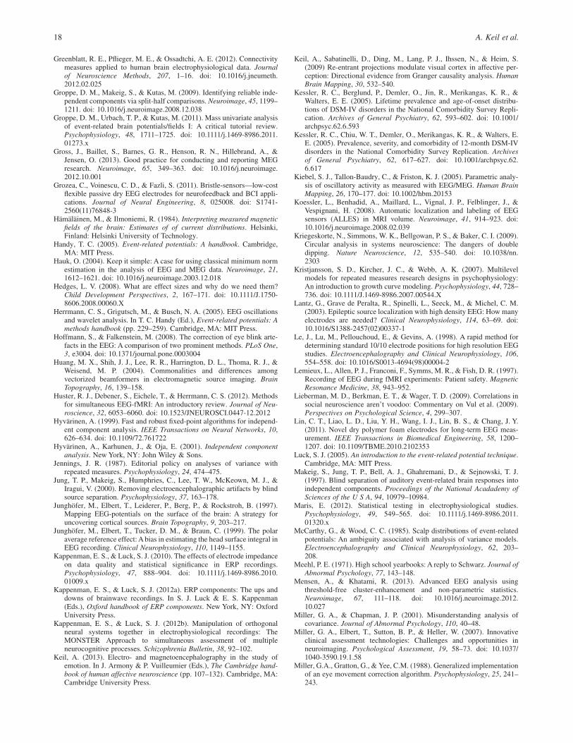
Greenblatt, R. E., Pflieger, M. E., & Ossadtchi, A. E. (2012). Connectivitymeasures applied to human brain electrophysiological data. Journalof Neuroscience Methods, 207, 1–16. doi: 10.1016/j.jneumeth.2012.02.025
Groppe, D. M., Makeig, S., & Kutas, M. (2009). Identifying reliable inde-pendent components via split-half comparisons. Neuroimage, 45, 1199–1211. doi: 10.1016/j.neuroimage.2008.12.038
Groppe, D. M., Urbach, T. P., & Kutas, M. (2011). Mass univariate analysisof event-related brain potentials/fields I: A critical tutorial review.Psychophysiology, 48, 1711–1725. doi: 10.1111/j.1469-8986.2011.01273.x
Gross, J., Baillet, S., Barnes, G. R., Henson, R. N., Hillebrand, A., &Jensen, O. (2013). Good practice for conducting and reporting MEGresearch. Neuroimage, 65, 349–363. doi: 10.1016/j.neuroimage.2012.10.001
Grozea, C., Voinescu, C. D., & Fazli, S. (2011). Bristle-sensors—low-costflexible passive dry EEG electrodes for neurofeedback and BCI appli-cations. Journal of Neural Engineering, 8, 025008. doi: S1741-2560(11)76848-3
Hämäläinen, M., & Ilmoniemi, R. (1984). Interpreting measured magneticfields of the brain: Estimates of of current distributions. Helsinki,Finland: Helsinki University of Technology.
Handy, T. C. (2005). Event-related potentials: A handbook. Cambridge,MA: MIT Press.
Hauk, O. (2004). Keep it simple: A case for using classical minimum normestimation in the analysis of EEG and MEG data. Neuroimage, 21,1612–1621. doi: 10.1016/j.neuroimage.2003.12.018
Hedges, L. V. (2008). What are effect sizes and why do we need them?Child Development Perspectives, 2, 167–171. doi: 10.1111/J.1750-8606.2008.00060.X
Herrmann, C. S., Grigutsch, M., & Busch, N. A. (2005). EEG oscillationsand wavelet analysis. In T. C. Handy (Ed.), Event-related potentials: Amethods handbook (pp. 229–259). Cambridge, MA: MIT Press.
Hoffmann, S., & Falkenstein, M. (2008). The correction of eye blink arte-facts in the EEG: A comparison of two prominent methods. PLoS One,3, e3004. doi: 10.1371/journal.pone.0003004
Huang, M. X., Shih, J. J., Lee, R. R., Harrington, D. L., Thoma, R. J., &Weisend, M. P. (2004). Commonalities and differences amongvectorized beamformers in electromagnetic source imaging. BrainTopography, 16, 139–158.
Huster, R. J., Debener, S., Eichele, T., & Herrmann, C. S. (2012). Methodsfor simultaneous EEG-fMRI: An introductory review. Journal of Neu-roscience, 32, 6053–6060. doi: 10.1523/JNEUROSCI.0447-12.2012
Hyvärinen, A. (1999). Fast and robust fixed-point algorithms for independ-ent component analysis. IEEE Transactions on Neural Networks, 10,626–634. doi: 10.1109/72.761722
Hyvärinen, A., Karhunen, J., & Oja, E. (2001). Independent componentanalysis. New York, NY: John Wiley & Sons.
Jennings, J. R. (1987). Editorial policy on analyses of variance withrepeated measures. Psychophysiology, 24, 474–475.
Jung, T. P., Makeig, S., Humphries, C., Lee, T. W., McKeown, M. J., &Iragui, V. (2000). Removing electroencephalographic artifacts by blindsource separation. Psychophysiology, 37, 163–178.
Junghöfer, M., Elbert, T., Leiderer, P., Berg, P., & Rockstroh, B. (1997).Mapping EEG-potentials on the surface of the brain: A strategy foruncovering cortical sources. Brain Topography, 9, 203–217.
Junghöfer, M., Elbert, T., Tucker, D. M., & Braun, C. (1999). The polaraverage reference effect: A bias in estimating the head surface integral inEEG recording. Clinical Neurophysiology, 110, 1149–1155.
Kappenman, E. S., & Luck, S. J. (2010). The effects of electrode impedanceon data quality and statistical significance in ERP recordings.Psychophysiology, 47, 888–904. doi: 10.1111/j.1469-8986.2010.01009.x
Kappenman, E. S., & Luck, S. J. (2012a). ERP components: The ups anddowns of brainwave recordings. In S. J. Luck & E. S. Kappenman(Eds.), Oxford handbook of ERP components. New York, NY: OxfordUniversity Press.
Kappenman, E. S., & Luck, S. J. (2012b). Manipulation of orthogonalneural systems together in electrophysiological recordings: TheMONSTER Approach to simultaneous assessment of multipleneurocognitive processes. Schizophrenia Bulletin, 38, 92–102.
Keil, A. (2013). Electro- and magnetoencephalography in the study ofemotion. In J. Armony & P. Vuilleumier (Eds.), The Cambridge hand-book of human affective neuroscience (pp. 107–132). Cambridge, MA:Cambridge University Press.
Keil, A., Sabatinelli, D., Ding, M., Lang, P. J., Ihssen, N., & Heim, S.(2009) Re-entrant projections modulate visual cortex in affective per-ception: Directional evidence from Granger causality analysis. HumanBrain Mapping, 30, 532–540.
Kessler, R. C., Berglund, P., Demler, O., Jin, R., Merikangas, K. R., &Walters, E. E. (2005). Lifetime prevalence and age-of-onset distribu-tions of DSM-IV disorders in the National Comorbidity Survey Repli-cation. Archives of General Psychiatry, 62, 593–602. doi: 10.1001/archpsyc.62.6.593
Kessler, R. C., Chiu, W. T., Demler, O., Merikangas, K. R., & Walters, E.E. (2005). Prevalence, severity, and comorbidity of 12-month DSM-IVdisorders in the National Comorbidity Survey Replication. Archivesof General Psychiatry, 62, 617–627. doi: 10.1001/archpsyc.62.6.617
Kiebel, S. J., Tallon-Baudry, C., & Friston, K. J. (2005). Parametric analy-sis of oscillatory activity as measured with EEG/MEG. Human BrainMapping, 26, 170–177. doi: 10.1002/hbm.20153
Koessler, L., Benhadid, A., Maillard, L., Vignal, J. P., Felblinger, J., &Vespignani, H. (2008). Automatic localization and labeling of EEGsensors (ALLES) in MRI volume. Neuroimage, 41, 914–923. doi:10.1016/j.neuroimage.2008.02.039
Kriegeskorte, N., Simmons, W. K., Bellgowan, P. S., & Baker, C. I. (2009).Circular analysis in systems neuroscience: The dangers of doubledipping. Nature Neuroscience, 12, 535–540. doi: 10.1038/nn.2303
Kristjansson, S. D., Kircher, J. C., & Webb, A. K. (2007). Multilevelmodels for repeated measures research designs in psychophysiology:An introduction to growth curve modeling. Psychophysiology, 44, 728–736. doi: 10.1111/J.1469-8986.2007.00544.X
Lantz, G., Grave de Peralta, R., Spinelli, L., Seeck, M., & Michel, C. M.(2003). Epileptic source localization with high density EEG: How manyelectrodes are needed? Clinical Neurophysiology, 114, 63–69. doi:10.1016/S1388-2457(02)00337-1
Le, J., Lu, M., Pellouchoud, E., & Gevins, A. (1998). A rapid method fordetermining standard 10/10 electrode positions for high resolution EEGstudies. Electroencephalography and Clinical Neurophysiology, 106,554–558. doi: 10.1016/S0013-4694(98)00004-2
Lemieux, L., Allen, P. J., Franconi, F., Symms, M. R., & Fish, D. R. (1997).Recording of EEG during fMRI experiments: Patient safety. MagneticResonance Medicine, 38, 943–952.
Lieberman, M. D., Berkman, E. T., & Wager, T. D. (2009). Correlations insocial neuroscience aren’t voodoo: Commentary on Vul et al. (2009).Perspectives on Psychological Science, 4, 299–307.
Lin, C. T., Liao, L. D., Liu, Y. H., Wang, I. J., Lin, B. S., & Chang, J. Y.(2011). Novel dry polymer foam electrodes for long-term EEG meas-urement. IEEE Transactions in Biomedical Engineering, 58, 1200–1207. doi: 10.1109/TBME.2010.2102353
Luck, S. J. (2005). An introduction to the event-related potential technique.Cambridge, MA: MIT Press.
Makeig, S., Jung, T. P., Bell, A. J., Ghahremani, D., & Sejnowski, T. J.(1997). Blind separation of auditory event-related brain responses intoindependent components. Proceedings of the National Acadademy ofSciences of the U S A, 94, 10979–10984.
Maris, E. (2012). Statistical testing in electrophysiological studies.Psychophysiology, 49, 549–565. doi: 10.1111/j.1469-8986.2011.01320.x
McCarthy, G., & Wood, C. C. (1985). Scalp distributions of event-relatedpotentials: An ambiguity associated with analysis of variance models.Electroencephalography and Clinical Neurophysiology, 62, 203–208.
Meehl, P. E. (1971). High school yearbooks: A reply to Schwarz. Journal ofAbnormal Psychology, 77, 143–148.
Mensen, A., & Khatami, R. (2013). Advanced EEG analysis usingthreshold-free cluster-enhancement and non-parametric statistics.Neuroimage, 67, 111–118. doi: 10.1016/j.neuroimage.2012.10.027
Miller, G. A., & Chapman, J. P. (2001). Misunderstanding analysis ofcovariance. Journal of Abnormal Psychology, 110, 40–48.
Miller, G. A., Elbert, T., Sutton, B. P., & Heller, W. (2007). Innovativeclinical assessment technologies: Challenges and opportunities inneuroimaging. Psychological Assessment, 19, 58–73. doi: 10.1037/1040-3590.19.1.58
Miller, G.A., Gratton, G., & Yee, C.M. (1988). Generalized implementationof an eye movement correction algorithm. Psychophysiology, 25, 241–243.
18 A. Keil et al.

Miller, G. A., Lutzenberger, W., & Elbert, T. (1991). The linked-referenceissue in EEG and ERP recording. Journal of Psychophysiology, 5,273–276.
Miller, J., Patterson, T., & Ulrich, R. (1998). Jackknife-based method formeasuring LRP onset latency differences. Psychophysiology, 35,99–115.
Mosher, J. C., Leahy, R. M., & Lewis, P. S. (1999). EEG and MEG:Forward solutions for inverse methods. IEEE Transactions in Biomedi-cal Engineering, 46, 245–259.
Mullinger, K., & Bowtell, R. (2011). Combining EEG and fMRI. Methods inMolecular Biology, 711, 303–326. doi: 10.1007/978-1-61737-992-5_15
Ng, W. C., Seet, H. L., Lee, K. S., Ning, N., Tai, W. X., & Sutedja, M.(2009). Micro-spike EEG electrode and the vacuum-casting technologyfor mass production. Journal of Materials Processing Technology, 209,4434–4438.
Nolte, G., Bai, O., Wheaton, L., Mari, Z., Vorbach, S., & Hallett, M. (2004).Identifying true brain interaction from EEG data using the imaginarypart of coherency. Clinical Neurophysiology, 115, 2292–2307. doi:10.1016/j.clinph.2004.04.029
Nolte, G., & Dassios, G. (2005). Analytic expansion of the EEG lead fieldfor realistic volume conductors. Physics in Medicine and Biology, 50,3807–3823.
Nunez, P. L., & Silberstein, R. B. (2000). On the relationship of synapticactivity to macroscopic measurements: Does co-registration of EEGwith fMRI make sense? Brain Topography, 13, 79–96.
Nuwer, M. R., Comi, G., Emerson, R., Fuglsang-Frederiksen, A., Guerit, J.M., & Hinrichs, H. (1999). IFCN standards for digital recordingof clinical EEG. The International Federation of ClinicalNeurophysiology. [Supplement]. Electroencephalography and ClinicalNeurophysiology, 52, 11–14.
Onton, J., Westerfield, M., Townsend, J., & Makeig, S. (2006). Imaginghuman EEG dynamics using independent component analysis. Neuro-science and Biobehavioral Reviews, 30, 808–822. doi: 10.1016/j.neubiorev.2006.06.007
Oostenveld, R., & Praamstra, P. (2001). The five percent electrode systemfor high-resolution EEG and ERP measurements. ClinicalNeurophysiology, 112, 713–719. doi: 10.1016/S1388-2457(00)00527-7
Perrin, F., Pernier, J., Bertrand, O., Giard, M. H., & Echallier, J. F.(1987). Mapping of scalp potentials by surface spline interpolation.Electroencephalography and Clinical Neurophysiology, 66, 75–81.
Picton, T. W., Alain, C., Woods, D. L., John, M. S., Scherg, M., &Valdes-Sosa, P. (1999). Intracerebral sources of human auditory-evokedpotentials. Audiology and Neurootology, 4, 64–79.
Picton, T. W., Bentin, S., Berg, P., Donchin, E., Hillyard, S. A., & Johnson,R., Jr. (2000). Guidelines for using human event-related potentialsto study cognition: Recording standards and publication criteria.Psychophysiology, 37, 127–152.
Pivik, R. T., Broughton, R. J., Coppola, R., Davidson, R. J., Fox, N., &Nuwer, M. R. (1993). Guidelines for the recording and quantitativeanalysis of electroencephalographic activity in research contexts.Psychophysiology, 30, 547–558.
Resnick, S. M. (1992). Matching for education in studies of schizophrenia.Archives of General Psychiatry, 49, 246.
Roach, B. J., & Mathalon, D. H. (2008). Event-related EEG time-frequencyanalysis: An overview of measures and an analysis of early gamma bandphase locking in schizophrenia. Schizophrenia Bulletin, 34, 907–926.doi: 10.1093/schbul/sbn093
Russell, G. S., Jeffrey Eriksen, J. K., Poolman, P., Luu, P., & Tucker, D. M.(2005). Geodesic photogrammetry for localizing sensor positions indense-array EEG. Clinical Neurophysiology, 116, 1130–1140. doi:10.1016/j.clinph.2004.12.022
SAMHSA. (2012). Results from the 2011 National Survey on Drug Use andHealth: Summary of national findings (NSDUH Series H-44, HHSPublication No. (SMA) 12-4713). Rockville, MD: Substance Abuse andMental Health Services Administration.
Scherg, M., & Von Cramon, D. (1986). Evoked dipole source potentials ofthe human auditory cortex. Electroencephalography and ClinicalNeurophysiology, 65, 344–360.
Spencer, K. M., Dien, J., & Donchin, E. (1999). A componential analysis ofthe ERP elicited by novel events using a dense electrode array.Psychophysiology, 36, 409–414.
Srinivasan, R., Nunez, P. L., & Silberstein, R. B. (1998). Spatial filteringand neocortical dynamics: Estimates of EEG coherence. IEEE Trans-actions in Biomedical Engineering, 45, 814–826. doi: 10.1109/10.686789
Stam, C. J., Nolte, G., & Daffertshofer, A. (2007). Phase lag index: Assess-ment of functional connectivity from multi channel EEG and MEG withdiminished bias from common sources. Human Brain Mapping, 28,1178–1193. doi: 10.1002/hbm.20346
Steinstrater, O., Sillekens, S., Junghoefer, M., Burger, M., & Wolters, C. H.(2010). Sensitivity of beamformer source analysis to deficiencies inforward modeling. Human Brain Mapping, 31, 1907–1927. doi:10.1002/hbm.20986
Taheri, B. A., Knight, R. T., & Smith, R. L. (1994). A dry electrode for EEGrecording. Electroencephalography and Clinical Neurophysiology, 90,376–383.
Tallon-Baudry, C., & Bertrand, O. (1999). Oscillatory gamma activity inhumans and its role in object representation. Trends in Cognitive Sci-ences, 3, 151–162.
Tenke, C. E., & Kayser, J. (2005). Reference-free quantification of EEGspectra: Combining current source density (CSD) and frequency prin-cipal components analysis (fPCA). Clinical Neurophysiology, 116,2826–2846. doi: 10.1016/j.clinph.2005.08.007
Tierney, A. L., Gabard-Durnam, L., Vogel-Farley, V., Tager-Flusberg, H., &Nelson, C. A. (2012). Developmental trajectories of resting EEG power:An endophenotype of autism spectrum disorder. PLoS One, 7, e39127.doi: 10.1371/journal.pone.0039127 PONE-D-12-01143
Tucker, D. M. (1993). Spatial sampling of head electrical fields:The geodesic sensor net. Electroencephalography and ClinicalNeurophysiology, 87, 154–163.
Urbach, T. P., & Kutas, M. (2002). The intractability of scaling scalpdistributions to infer neuroelectric sources. Psychophysiology, 39, 791–808. doi: 10.1017/S0048577202010648
Urbach, T. P., & Kutas, M. (2006). Interpreting event-related brain potential(ERP) distributions: Implications of baseline potentials and variabilitywith application to amplitude normalization by vector scaling. Biologi-cal Psychology, 72, 333–343. doi: 10.1016/j.biopsycho.2005.11.012
Vasey, M. W., & Thayer, J. F. (1987). The continuing problem of falsepositives in repeated measures ANOVA in psychophysiology: A multi-variate solution. Psychophysiology, 24, 479–486.
Viola, F. C., Thorne, J., Edmonds, B., Schneider, T., Eichele, T., & Debener,S. (2009). Semi-automatic identification of independent componentsrepresenting EEG artifact. Clinical Neurophysiology, 120, 868–877.doi: 10.1016/j.clinph.2009.01.015
Viviani, R. (2010). Unbiased ROI selection in neuroimaging studiesof individual differences. Neuroimage, 50, 184–189. doi: 10.1016/j.neuroimage.2009.10.085
von Helmholtz, H. (1853). Ueber einige Gesetze der Vertheilungelektrischer Ströme in körperlichen Leitern mit Anwendung auf diethierisch elektrischen Versuche [On the laws of electrical current distri-bution in volume conductors, with an application to animal-electricexperiments]. Annalen der Physik, 165, 211–233.
Voytek, B., D’Esposito, M., Crone, N., & Knight, R. T. (2013). A methodfor event-related phase/amplitude coupling. Neuroimage, 64, 416–424.doi: 10.1016/j.neuroimage.2012.09.023
Vul, E., Harris, C., Winkielman, P., & Pashler, H. (2009). Puzzlingly highcorrelations in fMRI studies of emotion, personality, and social cogni-tion. Perspectives on Psychological Science, 4, 274–290.
Wasserman, S., & Bockenholt, U. (1989). Bootstrapping: Applications topsychophysiology. Psychophysiology, 26, 208–221.
Wechsler, H., & Nelson, T. F. (2008). What we have learned from theHarvard School of Public Health College Alcohol Study: Focusingattention on college student alcohol consumption and the environmentalconditions that promote it. Journal of Studies on Alcohol and Drugs, 69,481–490.
Yuval-Greenberg, S., Tomer, O., Keren, A. S., Nelken, I., & Deouell, L. Y.(2008). Transient induced gamma-band response in EEG as a manifes-tation of miniature saccades. Neuron, 58, 429–441.
Zayas, V., Greenwald, A. G., & Osterhout, L. (2010). Unintentional covertmotor activations predict behavioral effects: Multilevel modeling oftrial-level electrophysiological motor activations. Psychophysiology,48, 208–217. doi: 10.1111/j.1469-8986.2010.01055.x
Zinbarg, R. E., Suzuki, S., Uliaszek, A. A., & Lewis, A. R. (2010). Biasedparameter estimates and inflated Type I error rates in analysis of covari-ance (and analysis of partial variance) arising from unreliability: Alter-natives and remedial strategies. Journal of Abnormal Psychology, 119,307–319. doi: 10.1037/a0017552
(Received July 30, 2013; Accepted July 31, 2013)
Guidelines for EEG and MEG 19

Appendix
Authors’ Checklist
The checklist is intended to facilitate a brief overview of important guidelines, sorted by topic. Authors may wish to use it prior tosubmission, to ensure that the manuscript provides key information.
Hypotheses YES NO
Specific hypotheses and predictions for the electromagnetic measures are described in the introduction
Participants YES NO
Characteristics of the participants are described, including age, gender, education level, and other relevant characteristics
Recording characteristics and instruments YES NO
The type of EEG/MEG sensor is described, including make and model
All sensor locations are specified, including reference electrode(s) for EEG
Sampling rate is indicated
Online filters are described, specifying the type of filter and including roll-off and cut-off parameters (in dB, or by indicatingwhether the cut-off represents half-power/half amplitude)
Amplifier characteristics are described
Electrode impedance or similar information is provided
Stimulus and timing parameters YES NO
Timing of all stimuli, responses, intertrial intervals, etc., are fully specified; ensure clarity that intervals are from onset or offset
Characteristics of the stimuli are described such that replication is possible
Description of data preprocessing steps YES NO
The order of all data preprocessing steps is included
Rereferencing procedures (if any) are specified, including the location of all sensors contributing to the new reference
Method of interpolation (if any) is described
Segmentation procedures are described, including epoch length and baseline removal time period
Artifact rejection procedures are described, including the type and proportion of artifacts rejected
Artifact correction procedures are described, including the procedure used to identify artifacts, the number of components removed,and whether they were performed on all subjects
Offline filters are described, specifying the type of filter and including roll-off and cut-off parameters (in dB, or by indicatingwhether the cut-off represents half-power/half amplitude)
The number of trials used for averaging (if any) is described, reporting the number of trials in each condition and each group ofsubjects. This should include both the mean number of trials in each cell and the range of trials included
Measurement procedures YES NO
Measurement procedures are described, including the measurement technique (e.g., mean amplitude), the time window and baseline period,sensor sites, etc.
For peak amplitude measures, the following is included: whether the peak was an absolute or local peak, whether visual inspection orautomatic detection was used, and the number of trials contributing to the averages used for measurement
An a priori rationale is given for the selection of time windows, electrode sites, etc.
Both descriptive and inferential statistics are included
Statistical analyses YES NO
Appropriate correction for any violation of model assumptions is implemented and described (e.g., Greenhouse-Geisser, Huynh-Feldt, orsimilar adjustment)
The statistical model and procedures are described and results are reported with test statistics, in addition to p values
An appropriate adjustment is performed for multiple comparisons
If permutation or similar techniques are applied, the number of permutations is indicated together with the method used to identify athreshold for significance
20 A. Keil et al.

Figures YES NO
Data figures for all relevant comparisons are included
Line plots (e.g., ERP/ERF waveforms) include the following: sensor location, baseline period, axes at zero points (zero physical unitsand zero ms), appropriate x- and y-axis tick marks at sufficiently dense intervals, polarity, and reference information, where appropriate
Scalp topographies and source plots include the following: captions and labels including a key showing physical units, perspective ofthe figure (e.g., front view; left/right orientation), type of interpolation used, location of electrodes/sensors, and reference
Coherence/connectivity and time-frequency plots include the following: a key showing physical units, clearly labeled axes,a baseline period, the locations from which the data were derived, a frequency range of sufficient breadth to demonstratefrequency-specificity of effects shown
Spectral analyses YES NO
The temporal length and type of data segments (single trials or averages) entering frequency analysis are defined
The decomposition method is described, and an algorithm or reference given
The frequency resolution (and the time resolution in time-frequency analyses) is specified
The use of any windowing function is described, and its parameters (e.g., window length and type) are given
The method for baseline adjustment or normalization is specified, including the temporal segments used for baseline estimation,and the resulting unit
Source-estimation procedures YES NO
The volume conductor model and the source model are fully described, including the number of tissues, the conductivity values of eachtissue, the (starting) locations of sources, and how the sensor positions are registered to the head geometry
The source estimation algorithm is described, including all user-defined parameters (e.g., starting conditions, regularization)
Principle component analysis (PCA) YES NO
The structure of the EEG/MEG data submitted to PCA is fully described
The type of association matrix is specified
The PCA algorithm is described
Any rotation applied to the data is described
The decision rule for retaining/discarding PCA components is described
Independent component analysis (ICA) YES NO
The structure of the EEG/MEG data submitted to ICA is described
The ICA algorithm is described
Preprocessing procedures, including filtering, detrending, artifact rejection, etc., are described
The information used for component interpretation and clustering is described
The number of components removed (or retained) per subject is described
Multimodal imaging YES NO
Single-modality results are reported
Current source density and Laplacian transformations YES NO
The algorithm used and the interpolation functions are described
Single-trial analyses YES NO
All preprocessing steps are described
A mathematical description of the algorithm is included or a reference to a complete description is provided
Guidelines for EEG and MEG 21