committee 1 task group report low-dose extrapolation of
TRANSCRIPT

1 2 3 4 5 6 7 8 9
10 11 12 13 14 15 16 17 18 19 20 21 22 23 24 25 26 27 28 29 30 31 32 33 34 35 36 37 38 39 40
12/421/04 Committee 1 Task Group Report Low-dose Extrapolation of Radiation-Related Cancer Risk
Task Group Members CE Land, PA Jeggo, AM Kellerer, JB Little, DA Pierce and RL Ullrich Corresponding Members V Beral, ES Gilbert, K Mabuchi, WK Sinclair, Z Tao Draft of December 10, 2004

CONTENTS PAGE 1 2 3 4 5 6 7 8 9
10 11 12 13 14 15 16 17 18 19 20 21 22 23 24 25 26 27 28 29 30 31 32 33 34 35 36 37 38 39 40 41 42 43 44 45 46 47 48 49 50
PREFACE 8 EXECUTIVE SUMMARY 9 1. INTRODUCTION 13 1.1 References 18 2. EPIDEMIOLOGICAL CONSIDERATIONS 20
2.1 Introduction 20 2.1.1 Evidence regarding radiation-related transgenerational risk 21
2.2 Dependence of cancer risk on radiation dose 22 2.2.1 Existence of a dose response 24 2.2.2 Estimating the dose response 24
2.3 Inferences based on acute doses in the moderate-to-high dose range 27 2.3.1 Modification of dose response by age and sex 27 2.3.2 Modification by lifestyle factors 28 2.3.3 Variation by population 28 2.3.4 Radiation quality 30
2.4 Estimation of risk at low doses and low dose rates 31 2.4.1 Difficulties of direct estimation of low-dose risk 32 2.4.2 Illustrative example 32 2.4.3 Studies of low-dose exposure 34 i) Medical studies 34 ii) Occupational studies 35 iii) A-bomb survivor studies 35 2.4.4 Extrapolation of risk to low doses and dose rates 37 i) Dose and dose rate effectiveness factor (DDREF) 38
2

1 2 3 4 5 6 7 8 9
10 11 12 13 14 15 16 17 18 19 20 21 22 23 24 25 26 27 28 29 30 31 32 33 34 35 36 37 38 39 40 41 42 43 44 45 46 47 48 49 50
ii) Site-specific differences 39 2.5 Thresholds vs. the linear, no-threshold hypothesis 40 2.6 Conclusion: Implications for low-dose cancer risk 42
2.7 References 43
3. LOW-DOSE RISK – BIOLOGY 58 3.1 Introduction 58 3.2 Damage caused by radiation 58 3.3 Damage Response Pathways 60 3.3.1 DNA DSB repair 61 i) Non-homologous End Joining 61 ii) Homologous Recombination 64 iii) Single strand annealing 65
iv) Contributions of HR and NHEJ to DNA DSB repair in mammalian cells 65
3.3.2 Cell cycle checkpoint control. 66 i) DNA integrity checkpoints in yeast. 66 ii) Checkpoint responses in mammalian cells. 67 3.3.3 Early sensors of DNA damage. 68 i) Role of ATM 68 ii) Role of Nbs1, hMre11 and hRad50 69 iii) BRCA1 and BRCA2 69 iv) Role of H2AX 70 Iv) MDC1 and 53BP1 71 3.3.4 Signal transduction after irradiation. 71 i) Role of p53 71
3

1 2 3 4 5 6 7 8 9
10 11 12 13 14 15 16 17 18 19 20 21 22 23 24 25 26 27 28 29 30 31 32 33 34 35 36 37 38 39 40 41 42 43 44 45 46 47 48 49 50
ii) G1/S arrest 72 iii) S phase arrest 73 iv) G2/M arrest 73 v) Apoptosis 74 3.4 Fidelity of DSB repair 75 3.4.1 The fidelity achievable by HR and NHEJ 75 3.4.2 The fate of non-rejoined and misrejoined breaks 77
3.4.3 The impact of the nature of DNA damage on repair 77 3.5 Impact of defects in DNA repair, checkpoint control and apoptosis 79 3.6. Conclusions 81 3.7 References 82 4. CELLULAR CONSEQUENCES OF RADIATION-INDUCED DAMAGE 98 4.1 Radiation- induced Chromosome Aberrations 98 4.2 Radiation-Induced Somatic Cell Mutations 101 4.2.1 Summary 103 4.3 Bystander effect, adaptive response, and genomic instability 103 4.3.1 Adaptive Response 103 4.3.2 Radiation Induced Genomic Instability 107 4.3.3 The Bystander Effect in Irradiated Cell Populations 110 4.4 Conclusions: Implications for Risk Assessment 115 4.5 References 118 5. CARCINOGENIC EFFECTS OF IONIZING RADIATION 135 5.1 Mechanisms of radiation-induced cancer 135 5.2 Tissue Modifying Factors 142 5.2.1 Target cells 143
4

1 2 3 4 5 6 7 8 9
10 11 12 13 14 15 16 17 18 19 20 21 22 23 24 25 26 27 28 29 30 31 32 33 34 35 36 37 38 39 40 41 42 43 44 45 46 47 48 49 50
5.3 Radiation-induced Cancer in Animals 146 5.3.1 Leukemia 146 5.3.2 Solid Tumors 147 5.4 Life Shortening 150 5.5 Summary 151 5.6 Conclusions: Implications for radiation-related cancer at low doses 153 5.7 References 155 6. QUANTITATIVE UNCERTAINTY ANALYSIS 169 6.1 Overview 169 6.2 Sources of uncertainty 172 6.2.1 Statistical estimate of excess risk per Gy 172 6.2.2 Diagnostic misclassification 173 6.2.3 Dose reconstruction errors 174 6.2.4 Transfer between populations 174 6.2.5 DDREF 176 6.2.6 Variation by sex 177 6.2.7 Expression of risk in absolute terms 177
6.2.8 Gradualism in DDREF and threshold effects 178 6.3 Allowing for the uncertain possibility of a threshold 179
6.4 Conclusions 182 6.6 References 185
7. CONCLUSIONS 189 TABLES 193 Table 2.1 Some sources and amounts of ionizing radiation exposure 193 Table 2.2. Parameter estimates corresponding to the general
dose-response model 194
5

1 2 3 4 5 6 7 8 9
10 11 12 13 14 15 16 17 18 19 20 21 22 23 24 25 26 27 28 29 30 31 32 33 34 35 36 37 38 39 40 41 42 43 44 45 46 47 48 49 50
Table 2.3 Modification of radiation-related risk by individual and
lifestyle factors 195 Table 2.4. Statistical power calculations for a hypothetical study 196 Table 2.5. Distribution of subjects, solid cancers, and excess solid 196 cancers in the Life Span Study cohort
FIGURES 197 Figure 2.1. Dose-specific excess relative risk of solid cancer among
atomic bomb survivors, 1958-87 197 Figure 2.2. General dose-response modelδD2), fit to the dose-response data of Figure 2.1 198 Figure 2.3. Estimated low-dose relative risks 199 Figure 2.4. Linear regression estimates of the ERR per Gy 200 Figure 2.5. Linear regression estimates of the ERR per Gy 201 Figure 2.6. All-age linear regression estimates of ERR per Gy for female breast cancer 202 Figure 3.1. Model for DNA NHEJ 203 Figure 3.2. Model for DNA homologous recombination 204 Figure 6.1. Lognormal distribution with 90% confidence limits 18%-43%, representing statistical uncertainty about cancer excess relative risk per Gy. 205
Figure 6.2. Normal uncertainty distribution for dosimetry bias correction factor, with corrected mean 0.84 and 90% uncertainty limits 0.69-1.0. 205
Figure 6.3. Approximate lognormal, uncertainty distribution for ERR per Gy, with mean 0.26 and 90% uncertainty limits 0.15-0.46. 205
Figure 6.4. Comparison of age-specific baseline rates for female breast cancer incidence in Japan and the United States 206 Figure 6.5. Monte Carlo simulation of the uncertainty distribution for cancer ERR at 1 Gy, after transfer to a U.S. population 207
Figure 6.6. Continuous subjective uncertainty distributions for DDREF used in recent analyses. 207
6

1 2 3 4 5 6 7 8 9
10 11 12 13 14 15
Figure 6.7. Discrete uncertainty distribution for DDREF used in NCI/CDC (2003) analysis. 207
Figure 6.8. Influence of uncertain DDREF assumptions on uncertainty for ERR/Gy 208 Figure 6.9. Mean and upper 95% probability limit for ERR/Gy as functions of threshold probability p, given (in the absence of a threshold) a lognormal uncertainty distribution with mean 0.17 and upper 95% limit 0.36. 209 Figure 6.10. Effect of uncertain threshold probability on the uncertain distribution for low-dose ERR/Gy. 210
7

PREFACE 1 2 3 4 5 6 7 8 9
10 11 12 13 14 15 16 17 18 19 20 21 22 23 24 25 26 27 28 29 30 31 32 33 34 35 36 37 38 39 40 41 42 43
Following its meeting in Oxford, UK, in 1997, Committee 1 of ICRP (the International Commission on Radiological Protection) proposed a Task Group to prepare a report on low-dose extrapolation of radiation-related cancer risk estimates based largely on higher-dose epidemiological data, and the possible implications for radiological protection. The Commission accepted this recommendation and established a Task Group, which began its work in April, 1998. The membership of the Task Group was as follows: C.E. Land (Chair) P.A. Jeggo A.M. Kellerer J.B. Little D.A. Pierce R.L. Ullrich
Corresponding members were: V. Beral E.S. Gilbert K. Mabuchi W.K. Sinclair Z. Tao R. Cox, J.H. Hendry, C.R. Muirhead, and R. J. Preston of Committee 1 contributed additional text to the report. The membership of Committee 1 during the period of preparation of this report was: (1997 – 2001) R. Cox (Chair) A.V. Akleyev R.J.M. Fry (Vice-Chair) J.H. Hendry A.M. Kellerer C.E. Land J.B. Little K. Mabuchi R. Masse C.R. Muirhead (Secretary) R.J. Preston K.Sankaranarayanan R.E. Shore C. Streffer K. Wei H.R. Withers (2001 – 2004) R. Cox (Chair) A.V. Akleyev M. Blettner J.H. Hendry A.M. Kellerer C.E. Land J.B. Little C.R. Muirhead (Secretary) O. Niwa D.L. Preston R.J. Preston E. Ron K.Sankaranarayanan R.E. Shore F.A. Stewart M. Tirmarche R.L. Ullrich (Vice-Chair) P.-K. Zhou
8

1 2 3 4
5
6
7
8
9
10
11
12
13
14
15
16
17
18
19
20
21
22
23
24
25
26
27
28
29
30
31
32
33
34
EXECUTIVE SUMMARY
The present report, of a task group of ICRP Committee 1, considers the evidence
relating to cancer risk associated with exposure to low doses of low-LET radiation, and
particularly doses below current recommended limits for protection of radiation workers
and the general public. The focus is on evidence regarding linearity of dose response for
all cancers considered as a group, but not necessarily individually, at low doses (the so-
called linear, no-threshold (LNT) hypothesis), and the possibility of a universal threshold
dose below which there is no risk of radiation-related cancer. According to the LNT
hypothesis, the same number of radiation-related cancers would be predicted in a
population of a given size exposed to a certain small average radiation dose and in an
otherwise similar population many times times larger and exposed to a proportionally
smaller average dose. According to the threshold hypothesis, the radiation-related risk in
the larger population would be zero if its average dose were sufficiently small.
The present document has been preceded by other, recent reports, notably those of
the United Nations Scientific Committee on the Effects of Atomic Radiation and the U.S.
National Council of Radiation Protection and Measurements. These reports recommended
that radiation protection continue to be guided by the LNT hypothesis. The task group
concurs with those recommendations.
The report is organized by scientific discipline, beginning with epidemiological
studies of exposed human populations (Chapter 2). Epidemiological studies offer the most
directly relevant information for risk-based radiation protection. The major scientific
issues, as illustrated by the example of cancer incidence from all solid tumors combined
in the Life Span Study (LSS) population of atomic bomb survivors, are (1) establishment
of the existence of a dose-related risk in this population, (2) modeling radiation-related
risk as a statistically uncertain parametric function of dose, modified by other factors such
as sex, exposure age, attained age, and time following exposure, (3) extrapolation of
estimated risk to other potentially exposed populations, with possibly different baseline
cancer rates, (4) projection of the risk in the population to the end of its natural life, and
(5) extrapolation of risk estimates from moderate-to-high dose levels of acute exposure,
characteristic of the most informative atomic bomb survivor data, to the far more
common low-dose and/or protracted exposures that occur in occupational and general
settings. Consideration of each of these issues leads to more refined risk estimates but,
9
1
2
3
4
5
6
7
8
9
10
11
12
13
14
15
16
17
18
19
20
21
22
23
24
25
26
27
28
29
30
31
32
33
because information about each is uncertain, the overall uncertainty of the improved
estimates is increased. There is some evidence of increased cancer risk associated with
exposures on the order of 10 mGy which will be discussed in the report, and other
evidence placing an upper limit on the value of any universal threshold that might exist.
Also, the risk of mortality and morbidity from all solid cancers combined is proportional
to radiation dose down to about 100 mGy, below which statistical variation in baseline
risk, as well as small and uncontrollable biases, tend to obscure evidence concerning
radiation-related risk. Extrapolation of risk estimates based on observations at moderate
to high doses continues to be the primary basis for estimation of radiation-related risk at
low doses and dose rates, for example at the present recommended limit for members of
the public of 1mGy per year from non-medical man-made sources.
The fundamental role of radiation-induced DNA damage in the induction of
mutations and chromosome aberrations and the apparent critical involvement of
aberrations and mutations in the pathogenesis of cancer provides a framework for the
analysis of risks at low radiation doses and low dose rate exposures (Chapter 3). A
characteristic type of damage produced by ionizing radiation (IR) involves multiple
lesions within close spatial proximity. Such clustered damage can be induced even by a
single radiation track through a cell. Although cells have a vast array of damage response
mechanisms that facilitate the repair of DNA damage and the removal of damaged cells,
these mechanisms are not fool-proof, and emerging evidence suggests that closely spaced
lesions can compromise the repair machinery. Also, while many of the cells containing
such radiation-induced damage may be eliminated by damage response pathways
involving cell cycle checkpoint control and apoptotic pathways, it is clear from analysis
of cytogenetics and mutagenesis that damaged or altered cells are capable of escaping
these pathways and propagating.
Cellular consequences of radiation-induced damage (Chapter 4) include
chromosome aberrations and somatic cell mutations. The processing and misrepair of
radiation-induced DSBs, particularly complex forms, are responsible for
chromosome/gene alterations that manifest as chromosome aberrations and mutations.
Current understanding of mechanisms and quantitative data on dose and time-dose
relationships support a linear dose response at low doses (i.e., LNT) for total cancer risk.
Considered as a whole, the emerging results with regard to radiation-related adaptive
response, genomic instability, and bystander effects suggest that the risk of low level
10

1
2
3
4
5
6
7
8
9
10
11
12
13
14
15
16
17
18
19
20
21
22
23
24
25
26
27
28
29
30
31
32
33
exposure to ionizing radiation is uncertain, and a simple extrapolation from high dose
effects may not be wholly justified in all instances. However, a better understanding of
the mechanisms for these phenomena, the extent to which they are active in vivo, and how
they are interrelated is needed before they can be evaluated as factors to be included in
the estimation of potential risk to the human population of exposure to low levels of
ionizing radiation.
Experimental approaches using animal models (Chapter 5) are well suited to
precise control of radiation dose and dose rate, as well as genetic background and other
possible modifiers of dose response, and can facilitate precise determination of biological
outcomes. Recent studies using newly developed animal models, cellular, cytogenetic and
molecular data for acute myelogenous leukemia (AML), intestinal tumors, and mammary
tumors, and cytogenetic and molecular studies on the induction of AML and mammary
cancer support the view that the essential radiation-associated events in the tumorigenic
process are predominantly early events involving DNA losses targeting specific genomic
regions harboring critical genes. As such, the response for early initiating events is likely
to correspond to that for the induction of cytogenetic damage. On this basis, mechanistic
arguments support a linear response in the low dose region, i.e., the process should be
independent of dose rate because interactions between different electron tracks should be
rare. Quantitative analyses of dose responses for tumorigenesis and for life shortening in
laboratory animals also support this prediction. These studies also support a dose and
dose rate effectiveness factor (DDREF), for reduction of estimated risk per unit dose
based on acute, high-dose data, in the range of about 2 when data are extrapolated to low
doses from effects induced by doses in the range of 2-3 Gy. Extrapolation of results from
less than 1 Gy would result in lower DDREF values.
Chapter 6 presents a formal exercise in quantitative uncertainty analysis, in which
the different uncertain components (as identified in Chapter 2) of estimated cancer risk
associated with low-dose, low-LET radiation exposure to a non-Japanese population, in
this case that represented by the U.S. National Cancer Institute’s SEER registry, are
combined. Attention is paid to the resulting uncertainty distribution for excess relative
risk per Gy (ERR/Gy), with and without allowing for the uncertain possibility of a
universal low-dose threshold, below which there would be no radiation-related risk. In the
example, which involves risk from all cancers combined including leukemia, except for
non-melanoma skin cancer, the major sources of uncertainty are statistical variation in the
11

1
2
3
4
5
6
7
8
9
10
11
12
13
14
15
estimated ERR at 1 Gy for the atomic bomb survivors population, subjective uncertainty
(informed by experimental and epidemiological data) about the DDREF to be applied at
low doses and dose rates, and the postulated uncertainty concerning the existence of a
universal threshold at some dose above that for which the calculation was being made.
Unless the existence of a threshold was assumed to be virtually certain, the effect of
introducing the uncertain possibility of a threshold was equivalent to that of an uncertain
increase in the value of DDREF, i.e., merely a variation on the result obtained by ignoring
the possibility of a threshold.
The conclusions of this report are given in Chapter 7. While existence of a low-
dose threshold does not seem unlikely for radiation-related cancers of certain tissues, and
cannot be ruled out for all cancers as a group, the evidence as a whole does not favor the
existence of a universal threshold, and there seems to be no particular reason to factor the
possibility of a threshold into risk calculations for purposes of radiation protection. The
LNT hypothesis, combined with an uncertain DDREF for extrapolation from high doses,
remains a prudent basis for radiation protection at low doses and low dose rates.
12

1. INTRODUCTION 1
2
3
4
5
6
7
8
9
10
11
12
13
14
15
16
17
18
19
20
21
22
23
24
25
26
27
28
29
30
31
32
33
34
The purpose of the present report is to summarize scientific evidence relevant to
the quantification of cancer risk associated with radiation exposure at (effective) doses of
interest for radiation protection, particularly doses below current recommended limits for
protection of radiation workers (e.g., 20 mSv per year) and the general public (e.g., 1 mSv
per year). (As a rough rule of thumb, effective doses on the order of 1 Sv, 100 mSv, 10
mSv, 1 mSv, and 0.1 mSv will be called “moderately high”, “moderate”, “low”, “very
low”, and “extremely low”, respectively, in this report.)
Ionizing radiation exposure is an established cancer risk factor. Compared to other
common environmental carcinogens, it is relatively easy to determine organ-specific
radiation dose and, as a result, radiation dose-response relationships tend to be highly
quantified. Nevertheless, there can be considerable uncertainty about questions of
radiation-related cancer risk as they apply to risk protection and public policy, and the
interpretations of interested parties can differ radically. A major reason for disagreement
is that public and regulatory concern often is focused on exposures at radiation doses far
lower than those at which useful information about cancer risk can be obtained directly,
that is, than can be obtained by studying populations with such exposures. Thus, risk
estimates promulgated by expert committees, for example, are usually based upon
epidemiological dose-response data obtained at doses ranging up to 0.2 Gy, 0.5 Gy, 1 Gy,
or higher, and the resulting estimates are then extrapolated, with appropriate caveats, to
lower doses. The extrapolation rules are based in part upon epidemiological observations,
such as the degree of curvature of fitted linear-quadratic dose response models for
leukemia and solid cancer morbidity among atomic bomb survivors, and on models
derived from experimental systems.
The discussion in the present report is concerned ultimately with biological effects
of ionizing radiations of low linear energy transfer (low LET), such as photons (gamma
rays and X rays) and electrons (beta particles) of various energies, as contrasted with
high-LET radiations such as neutrons and alpha particles. However, some biological
effects that have been observed mainly in connection with high-LET exposure are clearly
relevant to questions of cancer risk at low levels of low-LET radiation.
Currently, the ICRP radiation protection philosophy is based on the so-called
linear, non-threshold (LNT) hypothesis, according to which, at low doses (on the order of
100 mGy or less) and dose rates (less than 6 mGy/hour averaged over the first few hours)
13

1
2
3
4
5
6
7
8
9
10
11
12
13
14
15
16
17
18
19
20
21
22
23
24
25
26
27
28
29
30
31
32
33
34
(UNSCEAR 1993, EPA 1999) total radiation-related cancer risk is proportional to dose.
The hypothesis is not universally accepted as biological truth, but rather, because we do
not actually know what level of risk is associated with very low-dose exposure, is
considered by many as a prudent rule of thumb for public policy aimed at avoiding
unnecessary risk from exposure.
A logical consequence of the hypothesis is that, at a sufficiently low dose D,
exposure of N people to average dose D would result in the same number of radiation-
related cancers as exposure of k H N people to average dose D / k, for arbitrary k > 1. This
logical consequence can be used to justify the concept of “collective dose”, that the
product of average dose and the number of people exposed is proportional to the number
of radiation-related cancers. The concept of collected dose is sometimes used to support a
moral argument against widespread use of technologies or practices that would, according
to the LNT hypothesis, involve individual exposures at doses so low that any associated
risk, from the standpoint of the individual, would be far smaller than other risks that are
casually taken in everyday life. A so-called threshold hypothesis, according to which
there is no radiation-related risk associated with exposures at doses below some universal
threshold dose, would obviate concern about exposures at doses below the threshold and,
specifically, arguments based on the concept of collective dose. Aside from collective
dose, however, it is worth emphasizing that the practical importance of the LNT vs.
threshold question is associated with doses at which the associated risks, if they exist, are
high enough to be of “legitimate” concern, as determined by the usual social and political
processes.
Historically, the LNT vs. threshold controversy has been associated with public
policy issues related to exposures that are widespread but (typically) low for individuals,
such as local and worldwide exposure to radioactive fallout from above-ground nuclear
test explosions carried out by different governments, mainly during the 1950s (Lewis,
1957, 1963; Caron, 2004). The threshold hypothesis, as applied to ionizing radiation and
to fallout exposure in particular, drew some of its legitimacy from the field of chemical
toxicology, where thresholds are the rule (Brues, 1958, 1960), whereas the LNT
hypothesis is more consistent with findings from experimental radiation mutagenesis. As
described by Caron (2004), the intellectual positions taken by proponents of the opposing
sides during the fallout controversy of the 1950s (no compelling evidence of increased
cancer risk at low radiation doses, vs. no compelling evidence against a radiation-related
increased risk) are very similar to the situation at the present time. Some differences
14

1
2
3
4
5
6
7
8
9
10
11
12
13
14
15
16
17
18
19
20
21
22
23
24
25
26
27
28
29
30
31
32
33
34
discussed in the present report include the present general acceptance of a mutational
basis for carcinogenesis, and evidence that radiation-related mutations tend to be more
complex than more common mutations associated with endogenous and other causes.
The present report has been preceded by other surveys of the biological and
epidemiological information that underlies our understanding of low-dose risk and its
estimation by extrapolation from data obtained at higher doses, notably and recently the
comprehensive reports of the United Nations Scientific Committee on the Effects of
Atomic Radiation (UNSCEAR 2000, Annexes G and I) and of a committee of the U.S.
National Council of Radiation Protection and Measurements, entitled “Evaluation of the
Linear-Nonthreshold Dose-Response Model for Ionizing Radiation” (NCRP 2001). The
existence of these reports has allowed the present ICRP Task Group to be somewhat less
comprehensive in its coverage of the field than might otherwise have been necessary, and
to concentrate on updated coverage of developments in areas of epidemiology,
fundamental biology, experimental radiation mutagenesis and carcinogenesis, and
uncertainty analysis.
Studies of cancer risk following exposure of human populations are the most
obvious sources of information applicable to radiation protection policy. However, as
discussed in Chapter 2, generalization of risk information, obtained from one exposed
population, to other populations with different characteristics and potentially exposed to
radiation from different sources, at different doses and dose rates, requires the use of
dose-response models to describe the behavior of risk as a function of radiation dose, as
well as possible modification of dose response by individual and environmental factors. It
also requires making assumptions that are often based on uncertain information.
Chapter 3 deals with events believed to be fundamental to radiation
carcinogenesis: radiation-induced DNA damage and its repair. In particular, the chapter
discusses the nature of radiation-induced damage and damage response pathways
including repair of DNA double-strand breaks (DSB), cell cycle checkpoint control, early
sensors of DNA damage, and signal transduction after irradiation. Questions of particular
relevance for the current investigation are comparability of molecular damage from
radiation exposure and endogenous causes, and comparability between radiation-related
damage from ionizing radiation at high cf. low doses and dose rates with respect to
mechanisms, pathways, and fidelity of repair.
Cellular consequences of radiation-induced damage are discussed in Chapter 4.
Rates of radiation-induced chromosome aberrations and somatic cell mutations were
15

1
2
3
4
5
6
7
8
9
10
11
12
13
14
15
16
17
18
19
20
21
22
23
24
25
26
27
28
29
30
31
32
33
34
among the earliest quantitative measures of the cellular effects of ionizing radiation, and
studies of these outcomes have been highly informative about dose response over a wide
range of doses, and about effects of dose rate and fractionation. Induction of bystander
effects in cells not directly irradiated, genomic instability in the progeny of irradiated
cells, and adaptive response are radiation-related phenomena that evoke questions about
the generality of inferences based on cellular studies.
Considerations of statistical power, and possible bias due to unobservable and
uncontrollable confounders, govern the extent to which useful epidemiological
information can be obtained at exposure levels of regulatory interest, and some degree of
extrapolation is unavoidable. Experimental approaches using animal models, discussed in
Chapter 5, offer considerably more control of radiation exposure and dose, genetic
background, and modifying factors including other exposures, and can facilitate very
precise determination of biological outcomes. On the other hand, analogies between
radiation-related risks in human beings on the one hand and inbred strains of
experimental animals on the other are necessarily limited. Low statistical power for low-
dose studies is problematic for experimental and epidemiological studies alike, but
indirect approaches, based on protraction and fractionation of exposure resulting in
moderate to high cumulative doses, offer insights into low-dose effects. Experimental
studies can of course be replicated, to provide a firmer basis for insights into mechanisms,
tissue modifying factors, and quantitative dose response.
The material of Chapters 2-5 highlights statistical variation inherent in estimates
obtained by fitting parametric models to epidemiological and experimental data, but also
more fundamental uncertainties about important factors that cannot be ignored, but about
which there may be only limited information. The implications of these uncertainties for
conventional estimates of radiation-related cancer risk, especially at low doses and/or low
dose rates characteristic of exposures most commonly encountered by radiation workers
and the general public, are investigated in Chapter 6. The approach taken is an exercise in
quantitative uncertainty analysis similar to approaches used in a number of recent
exercises by expert committees concerned with such risks. Central to the approach is
recognition of the fact that radiation protection is a political process, responsive to the
interests and perceptions of stakeholders with differing points of view, and relying upon a
knowledge base that is extensive but also uncertain. Acceptance of this fact implies that it
is important, for the benefit and information of participants and stakeholders in the
radiation protection process, to identify sources of uncertainty and to quantify the
16

implications of such uncertainty for estimated risk. Among the questions addressed is the
impact on radiation protection policy of treating the existence of a universal low-dose
threshold for radiation-related cancer risk as an uncertain possibility.
1
2
3
17

1.1 References 1
2
3
4
5
6
7
8
9
10
11
12
13
14
15
16
17
18
19
20
21
22
23
24
25
26
27
28
29
30
31
32
Brues AM. Critique of the linear theory of carcinogenesis. Science 1958 Sep
26;128(3326):693-9.
Brues AM. Critique of mutational theories of carcinogenesis. Acta Unio Int Contra
Cancrum. 1960;16:415-7.
Caron J. Biology and “the bomb”. Engineering and Science 2004;47(2):16-27. California
Institute of Technology, Pasadena, California.
EPA (1999) Environmental Protection Agency. Estimating Radiogenic Cancer Risks.
EPA Report 402-R-00-003, Washington, D.C., May 1999.
Lewis EB. Leukemia and ionizing radiation. Science 1957;125:965-972.
Lewis EB. Leukemia, multiple myeloma, and aplastic anemia in American radiologists.
Science 1963;142:1492-1494.
NCRP (2001) National Council on Radiation Protection and Measurements. Evaluation of
the linear-nonthreshold dose-response model for ionizing radiation. NCRP Report
No. 136. National Council on Radiation Protection and Measurements, Bethesda,
MD, 2001.
UNSCEAR 1993. United Nations Scientific Committee on the Effects of Atomic
Radiation. Sources, Effects and Risks of Ionizing Radiation, No. E.94.IX.2,
United Nations, New York, 1993.
UNSCEAR 2000 Report to the General Assembly, with Scientific Annexes. Volume II:
Effects. Annex G: Biological effects at low radiation doses. No. E.00.IX.4. United
Nations, New York, 2000
18

UNSCEAR 2000 Report to the General Assembly, with Scientific Annexes. Volume II:
Effects. Annex I: Epidemiological Evaluation of Radiation-Induced Cancer. No.
E.00.IX.4. United Nations, New York, 2000
1
2
3
19

2. EPIDEMIOLOGICAL CONSIDERATIONS 1
2
3
4
5
6
7
8
9
10
11
12
13
14
15
16
17
18
19
20
21
22
23
24
25
26
27
28
29
30
31
32
33
34
2.1 Introduction
Like other areas of epidemiological research, the study of radiation-related cancer
risk began with clinical observations, the earliest of which may have been the 16th century
identification by the physician Georg Bauer (more often known by his Latinized name,
Agricola) of a specific condition, which he called “Joachimsthal Mountain Disease”,
among miners in the Joachimsthal region of the present-day Czech Republic. The disease,
the description of which now appears consistent with radon-related lung cancer but could
also include other lung diseases such as silicosis (NAS/NRC, 1999; Toohey, 1987), was
thought by Agricola to be caused by “metallic vapors” in mine atmospheres. Roentgen’s
discovery of x rays in 1895 and Becquerel’s discovery of natural radioactivity the
following year, and the subsequent use of both in science, medicine and industry, led to
the recognition, documented by case reports early in its history, that radiation exposure
might be harmful (Doll, 1995). The Court Brown and Doll study of mortality among
British radiologists (1958; Smith and Doll, 1981; Berrington et al, 2001), which
demonstrated a significantly increased risk of cancer mortality among radiologists who
had registered with a radiological society before 1921 and who were therefore likely to
have received higher doses than radiologists who began their practice later, is an example
of an influential study in which the fact of exposure was related to risk but individual
dose estimates were not available. However, experimental studies of radiation effects
such as cell inactivation, mutation, and carcinogenesis have taken advantage of the
experimenters’ ability to regulate, with precision, radiation dose to target cells or tissues.
Similarly, epidemiological investigations of exposed populations have benefited
enormously from information enabling scientists to reconstruct individual, and even
organ-specific, radiation doses. Benefits include the estimation of dose-response
relationships and of the modification of such relationships by individual properties such
as sex, age, lifestyle, and genetic inheritance. Thus, dose reconstruction is a fundamental
component of the epidemiology of radiation carcinogenesis, and tends to be well worth
the often considerable effort and expense required.
“Risk” is a concept in common use that is often applied to the past and future
experiences of individuals, but a numerical risk value can be estimated and verified only
on the basis of population rates, e.g., by comparing cancer rates, in a population exposed
20

1
2
3
4
5
6
7
8
9
10
11
12
13
14
15
16
17
18
19
20
21
22
23
24
25
26
27
28
29
30
31
32
33
34
to a given radiation dose, with rates in an otherwise comparable population that is either
not exposed or exposed to a much lower radiation dose. Thus, when we speak of an
individual’s risk we are really referring to a property of a population similar to that to
which the individual is assumed to belong.
The implications of risk for public policy, and for radiation protection in
particular, are controversial in large part because risk estimates are uncertain and because
there are legitimate interests both in avoiding radiation-related risks on the one hand and
in maintaining radiation-related benefits and/or avoiding costs associated with
unnecessary exposure reduction on the other. A person who may be at risk of radiation-
related cancer will naturally insist on proof that the risk either does not exist or is small
enough to be tolerated in view of the presumed benefit. A person whose interest is in
maintaining the benefit, or avoiding costs associated with reduction of exposure, will
demand proof that there is a risk that is high enough to be of concern. The problem is
inherently political, and its fair resolution requires information about risk, including its
uncertainty, framed so as to address the concerns of both viewpoints.
As epidemiological investigations of radiation-related cancer risk have evolved
over time, emphasis has shifted from the discovery that radiation is indeed a cancer risk
factor, to demonstration of radiation dose response, to identification of factors that modify
dose response, to examination of assumptions inherent in the risk estimation process.
Ionizing radiation exposure is a known, and well quantified, human cancer risk factor.
Nevertheless, estimation of cancer risk following radiation exposure is a very uncertain
process for most cases of regulatory and/or popular concern. One reason is that risk
estimates are usually applied to exposed populations different from those on which the
estimates are based. Another is that public and regulatory interest is usually with
exposures at radiation doses far lower than those at which useful information about risk
can be obtained by studying populations with such exposures.
2.1.1 Evidence regarding radiation-related transgenerational cancer risk
The current report is concerned mainly with the possibility that cancer risk may be
increased following exposure to ionizing radiation. There is a great deal of information
about this question. A second possibility, which is also a matter of concern, is that
exposure may be associated with increased transgenerational cancer risk. Various
epidemiological and laboratory studies have examined whether risks of cancer are raised
in offspring, following parental radiation exposure. These studies have been reviewed in
21

1
2
3
4
5
6
7
8
9
10
11
12
13
14
15
16
17
18
19
20
21
22
23
24
25
26
27
28
29
30
31
32
33
34
detail elsewhere (e.g. COMARE, 2002). Cellular and animal studies indicate that the
induction of cancer in the offspring of irradiated parents is possible in principle.
However, the findings in mice have not been consistent. In some strains, no effect has
been seen (e.g. Cattanach et al., 1995), whereas in others a raised risk has been observed
that is greater than predicted by the conventional induction rate for gene mutations (e.g.
Nomura, 1982).
Epidemiological studies conducted in several countries do not provide convincing
evidence to suggest that occupational radiation exposure alone results in an increased
incidence of childhood cancer in the offspring of male workers; data for the offspring of
female radiation workers are too sparse to draw conclusions (COMARE, 2002). In the
case of a cluster of childhood leukemia cases among children in the village of Sellafield,
U.K., possibly associated with paternal employment at the nearby Windscale nuclear
reprocessing plant (Gardner, 1990), a better case can perhaps be made in the context of
the well-documented phenomenon of increased levels of childhood leukemia in so-called
new towns, in which there has been an influx of residents from different areas; the
postulated mechanism is an unknown viral etiology affecting previously unexposed
residents (Doll et al, 1994; Doll, 1999). In addition, follow-up of about 40,000 offspring
of the Japanese atomic bomb survivors has not shown any association between the
incidence of cancer in children and young adults and parental dose (Izumi et al., 2003).
Thus, the subject of transgenerational risk, while a legitimate subject of scientific
investigation, is insufficiently developed to provide much information on risks associated
with low-dose radiation. It is briefly discussed in Chapter 5 in connection with radiation-
induced genomic instability, but is not pursued further in this report.
2.2 Dependence of cancer risk on radiation dose.
We have reasonably good epidemiological information on cancer risk following
acute exposures in the range 0.2 Gy to 5 Gy and (for partial-body exposures) above.
There are numerous epidemiological studies of populations containing “high-dose”
subsets with radiation doses in this range. These populations include patients treated with
radiation for benign and malignant disease, patients who received extensive diagnostic
radiography over a lengthy illness, such as tuberculosis patients treated by lung collapse
therapy monitored by frequent fluoroscopy examinations, persons who received
substantial exposures because of their occupations, such as uranium miners exposed to
22

1
2
3
4
5
6
7
8
9
10
11
12
13
14
15
16
17
18
19
20
21
22
23
24
25
26
27
28
29
30
31
32
33
34
radon decay products in mine atmospheres and instrument dial painters who ingested
radium contained in luminescent paint, and survivors of the atomic bombings of
Hiroshima and Nagasaki, Japan. These studies, and in particular inferences based on the
moderate- to high-dose component of the populations under study, form the primary
epidemiological basis for estimation of radiation-related risk. Recent, comprehensive
reviews of epidemiological information on radiation-related cancer risk are to be found in
the UNSCEAR 2000 report (UNSCEAR 2000) and NCRP Report 136 (NCRP 2001a).
Some benchmarks of radiation exposure levels are given in Table 2.1. Yearly
natural background effective doses in normal background areas are 0.4 mSv from cosmic
radiation, depending upon altitude (the dose from a typical round trip between New York
and Paris by commercial airline would be 0.03 mSv), 0.5 to 4 mSv from radioactivity in
rocks and soil, depending on local geology, 0.25 mSv from naturally occurring
radionuclides in the human body, and on the order of 2.5 mSv to the lung from inhaled
radionuclides (radon, thoron, and their decay products) (UNSCEAR 2000). Common
diagnostic examinations produce effective doses ranging from 0.01 mSv for x rays of a
foot or hand to 4 mSv for a barium enema (Mettler and Upton, 1995), to 25 mSv for a
pediatric CT scan of the abdomen if adult settings are used (Brenner et al, 2003). An
astronaut may get about 2 - 3 mSv tissue-weighted effective dose on a typical 3-day space
shuttle mission, and about 50 mSv on a 60-day tour in the international space station
(NCRP, 2000). Estimated acute, neutron-weighted doses to the colon (weighted dose =
gamma dose plus 10 times neutron dose) from the atomic bombings of Hiroshima and
Nagasaki ranged from less than 1 mGy to nearly 6 Gy for survivors who were exposed
within 3 km of the explosions and who were still alive in October, 1950; among survivors
with estimated doses between 5 mGy and 4 Gy, the average was 200 mGy (RERF LSS
mortality data set, 2003). An acute, whole-body effective dose of 5 Sv is very likely to be
fatal without prompt medical attention, but radiation therapy for cancer usually involves
partial-body doses an order of magnitude higher. Fractionation or protraction of exposure
can allow higher doses to be tolerated in terms of acute effects. Cumulative occupational
exposures among monitored radiation workers were about 20 mSv in several major
studies (Gilbert, 2001) and the recommended upper limit for radiation workers is 20 mSv
per year averaged over 5 years, and no greater than 50 mSv in any one year (ICRP, 1991).
(However, yearly effective doses at the Mayak plutonium facility approached 1 Sv for
some workers during the earlier years of production (Akleyev and Lyubchansky, 1994;
Khokhryakov et al, 2000).)
23

1
2
3
4
5
6
7
8
9
10
11
12
13
14
15
16
17
18
19
20
21
22
23
24
25
26
27
28
29
30
31
32
33
2.2.1 Existence of a dose response.
Dose-response data (e.g., pertaining to cancer morbidity) can be described in a
number of ways, such as by arranging observations in order of dose, grouping them into
consecutive dose intervals, and plotting cancer rates by dose interval (Figure 2.1).
Sophisticated modeling of dose response is not strictly necessary to establish the
existence of a dose response; that can be done by a test of increasing trend, usually
obtained by fitting to the data a simple model, like one of the following:
ERR(D) = αD, (2-1)
ERR(D) = exp{βD}-1. (2-2)
Here, ERR(D) is excess relative risk at radiation dose D, and α and β are unknown
parameters. In testing for an increasing trend, the dose response is “statistically
significant” when the statistical evidence is inconsistent with values of the parameter α or
β less than or equal to zero. These simple models can be used in tests of overall tendency,
or trend, and do not suffice to establish the shape of the dose response curve. In the
example of Figure 2.1, in fact, neither of the fitted functions agrees particularly well with
the plotted, dose-specific data points, especially at high doses, but both simple models
serve to establish the existence of a dose response.
If statistical significance is not achieved by a trend test, it can be inferred that the
evidence in favor of the existence of a dose response is not strong or that any dose
response is too complex to be represented by such a simple parametric function. It cannot
be inferred that there is no positive dose response, unless the trend is statistically
significant in the negative direction; inadequate statistical power, because of inadequate
sample size for the range of doses covered, can result in failure to achieve statistical
significance in the presence of a positive dose response (see Section 2.4.2).
2.2.2 Estimating the dose response.
The information that can be derived from a dose-response analysis is always
conditional upon assumptions about the functional relationship between radiation dose
and exposure-related, excess risk. In Figure 2.1, the interval-based estimates are based on
virtually no such assumptions; the different estimates are minimally correlated with each
other, and that only because they share a common reference (i.e., the value for the zero-
dose interval is constrained to be zero); thus, observations at any given non-zero dose
24

1
2
3
4
5
6
7
8
9
10
11
12
13
14
15
16
17
18
19
20
21
22
23
24
25
26
27
28
29
30
31
interval contribute information only towards the estimated ERR at that interval. However,
for each of the two fitted models used for trend tests (the plots of which differ because
their assumed functional forms are different), the corresponding dose-specific estimates
are all determined by the same estimated parameter, α in (2-1) or β in (2-2), and are
therefore perfectly dependent on each other, conditionally on the estimated dose values.
The confidence limits on the fitted curves are accordingly much narrower than those on
estimates separately computed for individual dose intervals along the abscissa.
Once existence of a dose response has been established, it makes sense to find a
parametric dose-response model that is both consistent with the epidemiological data and
plausible in terms of radiobiology. Such a model provides a way to use all of the dose-
response data to estimate radiation-related risk at various dose levels, and at low dose
levels in particular.
Of the two models used here to test for trend, the linear model (2-1) is biologically
plausible in the sense that the primary mechanism by which ionizing radiation exposure is
thought to influence subsequent cancer risk is damage to cellular DNA from ionizing
events, and the frequency of such ionizing events in a defined volume of tissue is
proportional to absorbed radiation dose. The log-linear model (2-2) is less plausible, but
is often mathematically convenient (e.g., in logistic model analyses).
An experimentally and theoretically-derived general radiation dose-response
model, often cited in connection with cancer risk related to low-LET radiation (Upton,
1971; NAS/NRC, 1980) is
ERR(D) = αD × (1 + βD) × exp(-γD - δD2). (2-3)
Here α, β, γ and δ are unknown, positive parameters. The linear term, αD, dominates at
low doses (where D2 is small) and the term αβD2 dominates at doses somewhat greater
than the so-called “cross-over dose” (D = 1/β) at which the terms proportional to dose and
dose-squared contribute equally to estimated risk. The exponential term,
exp(-γD - δD2),
represents the competing effect of “cell killing” or cell reproductive death, observed
experimentally, which would prevent a radiation-damaged cell from becoming cancerous;
this term dominates at high doses, leading to a reduction in slope and eventually to a
turnover and gradual decline in risk. (For present purposes, the contribution of the
parameter δ is of only minor importance and we will assume δ 32
33
= 0 in what follows.) Like
the other components of (2-3), the exponential cell-killing term is modeled as a
25

1
2
3
4
5
6
7
8
9
10
11
12
13
14
15
16
17
18
19
20
21
22
23
24
25
26
27
28
29
30
31
32
33
continuous function of dose, without threshold. Thus, cell killing is considered a
stochastic effect, the probability of which increases with increasing dose, and not a
deterministic effect, like tissue injury, which becomes noticeable when the proportion of
damaged cells exceeds some threshold level.
The general dose-response function (2-3) is not often used in epidemiological
research, mainly because the constrained parameters β and γ produce effects opposite in
curvature that may cancel each other out to some extent. While the model is used
successfully with very precise and numerous experimental data, most epidemiological
dose-response data lack the statistical power needed to support estimates for a model of
such complexity. This observation is illustrated here using the A-bomb survivor data of
Figure 2.1 for total solid cancers following a whole-body exposure, among the most
statistically powerful epidemiological radiation dose-response data in existence at the
time they were published (Thompson, 1994). The model fits these data reasonably well
(Figure 2.2, dashed line; Table 2.2), but statistically not significantly better than the linear
model of Figure 2.1 (p = .11). The estimated ERR per Gy at low doses (i.e., the estimated
value of α), 0.52 (90% confidence limits 0.16 - 0.83), does not differ markedly from that
according to the linear model, 0.57 (0.49 - 0.66); however, the confidence limits are
substantially wider for the more complex model, reflecting the wide range of
combinations of positive values of the parameters α, β and γ consistent with the data. The
analysis offers little evidence in support of a positive value of the (dose-squared)
parameter β (p=.28), but suggestive evidence in support of a non-zero value of the cell-
killing parameter γ (p = .07).
Less than 1% of the members of the Life Span Study Cohort for whom dose
estimates have been calculated have estimates greater than 2 Gy, and there are reasons to
believe that the dose estimates above 2 Gy may be biased upward (Pierce and Preston,
2000). Restriction of the dose-response analysis to subjects with doses under 2 Gy yielded
the linear-model parameter estimate α = 0.64 (0.54- 0.74). Adding either the quadratic or
the cell-killing terms to the model produced zero or minimal change whereas adding both
of them yielded parameter estimates so uncertain as to be of no predictive value (Table
2.2).
In the remainder of this report, epidemiological risk estimates are based on linear
dose-response analyses.
26
1
2
3
4
5
6
7
8
9
10
11
12
13
14
15
16
17
18
19
20
21
22
23
24
25
26
27
28
29
30
31
32
33
34
2.3 Inferences based on acute exposures in the moderate-to-high dose range
2.3.1 Modification of dose response by sex and age.
The information obtained from studies of the A-bomb survivors and other
populations mentioned above is rich in detail. For many cancer sites and groups of sites,
we can estimate with some precision not only dose-specific risk of radiation-related
cancer, but also its variation by cancer site and by sex, age at exposure, attained age
and/or time following exposure. In general (but not always), radiation-related relative risk
is higher among women and following exposure at young ages. The relationship to age at
exposure is marked for thyroid cancer, acute leukemia, and female breast cancer (Ron,
1995; Preston, 1994; Preston, 2002; Land, 2003). Risk decreases somewhat, in relative
terms, with advancing age at observation, but increases in absolute terms because baseline
cancer risk tends to increase as a power of age, and faster than dose-specific ERR
decreases (Thompson, 1994; UNSCEAR, 2000; Pierce, 2002; Pierce and Vaeth, 2003).
The relative importance of exposure age and attained age as modifiers of radiation
dose response is uncertain, because in any epidemiological follow-up study the two
quantities are highly correlated and their effects are difficult to separate. With additional
follow-up as the major exposed populations are followed to the end of life span, the
importance of this question for lifetime risk will become moot because projection to the
end of life will no longer be required for subgroups exposed at young ages. However, the
dependence of radiation-related risk on exposure age and attained age probably will
remain complicated: one consideration is the presence of secular trends in baseline risk in
Japan during the period of follow-up for the atomic bomb survivors over the past half
century, the reasons for which are not entirely clear (Parkin, 2002).
Statistically stable descriptions can be obtained of the dependence of dose-specific
risk on sex, age, and time, for aggregations of cancer sites such as all cancers combined,
all solid cancers, all leukemia types, and other groupings. This is useful because radiation
protection is concerned with the totality of possible adverse consequences of exposure,
but also because overall patterns of dependence may emerge from such analyses that can
be incorporated into site-specific estimates, resulting in greater statistical precision
(Pierce and Preston, 1993; NAS/NRC, 2000; NCI/CDC, 2003).
27

1
2
3
4
5
6
7
8
9
10
11
12
13
14
15
16
17
18
19
20
21
22
23
24
25
26
27
28
29
30
31
32
33
34
2.3.2 Modification by lifestyle and other individual factors.
There is a relatively small but growing amount of epidemiological information
(Table 2.3) on modification of radiation-related risk by history of lifestyle factors such as
tobacco smoking in the case of lung cancer (Prentice 1983; Kopecky, 1986; NAS/NRC,
1999; Lubin, 1995; Pierce, 2003), childbearing and breast feeding in the case of breast
cancer (Boice, 1978; Shore, 1980; Land, 1994), ultraviolet light in the case of basal cell
and squamous cell skin cancer (Shore, 2001, 2002; Ron, 1998), and disease history in the
case of type C hepatitis infection and liver cancer (Sharp, 2002). Much more needs to be
learned about interactions of ionizing radiation exposure with lifestyle factors and with
exposures to other agents. It is not unlikely that some of our current inferences about
dependence of radiation-related risk on exposure age, attained age, and sex may reflect
secular changes in lifestyle, and in exposure to environmental agents, that have been
associated with changes over time (and with successive birth cohorts) in both baseline
and radiation-related risk.
2.3.3 Variation by population.
There does not appear to be an obvious, consistent relationship between baseline
and radiation-related cancer risk, either across cancer sites within a single population or
across populations for a single cancer site. In the female Japanese population generally,
age standardized (world) rates per 100,000 per year are similar, at about 31 for gastric
cancer and 34 for breast cancer (Parkin, 2002), whereas in the United States they are
about 3 and 90, respectively. Among A-bomb survivors, the radiation-related excess
relative risk at 1 Gy (ERR1Gy) is 0.32 for gastric cancer and 1.6 for breast cancer
(Thompson, 1994). Gastric cancer contributes a substantial proportion of total radiation-
related risk, but that proportion is considerably less than the proportion of risk of
baseline gastric cancer to total baseline cancer risk (about 22%) among A-bomb survivors
(Thompson, 1994) and among Japanese generally (Parkin, 2002). In the United States, the
ratio is 2% for males and 1% for females. For female breast cancer the opposite is true;
the baseline rate in Japan is among the lowest in the world for developed countries
whereas the total cancer rate is not much different from that in most other countries
(Parkin, 2002) while, among A-bomb survivors, breast cancer contributes a
disproportionately large fraction of the total radiation-related cancer burden (Thompson,
1994). In the United States, by contrast, baseline breast cancer rates are high but the
28
1
2
3
4
5
6
7
8
9
10
11
12
13
14
15
16
17
18
19
20
21
22
23
24
25
26
27
28
29
30
31
32
33
34
radiation-related excess risk (in absolute terms) per unit dose among medically-exposed
women is similar to that among the A-bomb survivors (Preston, 2002). That is, the dose-
specific, radiation-related component of total breast cancer risk is likely to be similar in
absolute magnitude for exposed Japanese and western populations but, in western
populations, smaller as a proportion of total breast cancer risk. For gastric cancer, on the
other hand, the US baseline rate is an order of magnitude lower than that in Japan,
whereas the limited information on dose-specific, radiation-related excess risk suggests
that, as a multiple of baseline risk, it may be comparable to that in the A-bomb survivors
(Griem, 1994; Carr, 2002).
The above information suggests that, for breast cancer, radiation-related excess
relative risk per Gy (excess risk per Gy expressed as a multiple of the Japanese baseline
risk) based on A-bomb survivor data would overestimate risk for an exposed US
population while, for gastric cancer, radiation-related excess absolute risk (the difference
between risk following exposure and the Japanese (baseline risk) would result in an
overestimate for the US population. For most other cancers we have almost no
information of a similar nature (Table 2.3). This is not a trivial matter, because any
transfer of a risk estimate from one population to another requires making an assumption,
explicit or implicit, about the relation between excess and baseline risk. Moreover, for
some sites (e.g., stomach, liver, and esophagus) baseline rates can differ markedly
between populations (Parkin, 2002).
It should not be surprising that the relationship between radiation-related and
baseline risk in different populations is not consistent for different cancer sites. There are
reasons, as yet poorly understood, why baseline breast cancer rates are high in the United
States, and why baseline gastric cancer rates are high in Japan. These reasons are almost
surely related to differences in lifestyle, since the descendants of immigrants to the United
States, for example, have tended to develop cancer rates that are typical of the general
U.S. population (Haenszel, 1968; Ziegler, 1993) and different from those of their
countries of ancestral origin. The lifestyle factors affecting the rates for breast and
stomach cancer are probably different, at least in part, and probably interact differently
with radiation dose.
Much of environmental, nutritional, and occupational cancer epidemiology is
concerned with identifying cancer risk factors that might account for some part of the
variation of site-specific baseline rates among populations. While there has been much
progress, the problem is vast and, as discussed in section 2.3.2, there is only limited
29

information on interaction between radiation dose and lifestyle factors in terms of cancer
risk. Thus, it is likely that, for the foreseeable future, the most useful information relevant
to transfer of radiation-related risk coefficients from one population to another will come
from multinational comparisons of site-specific radiation-related risk, rather than from
investigations of underlying cancer risk factors and their interactions with radiation dose.
1
2
3
4
5
6
7
8
9
10
11
12
13
14
15
16
17
18
19
20
21
22
23
24
25
26
27
28
29
30
2.3.4 Radiation quality.
Risk estimates for low-LET radiation protection purposes are based mainly on
epidemiological studies of populations exposed to substantial doses of medical x ray, or
to mixed gamma and neutron radiation from the Hiroshima and Nagasaki atomic bombs.
According to the DS86 dose reconstruction algorithm (Roesch, 1986) as represented by
public use RERF data sets (RERF, 2003), the correlation between neutron and gamma
dose within each city is greater than 95%, and the proportion of total absorbed bone
marrow dose contributed by neutrons is only 0.7 to 2.7% in Hiroshima and 0.3 to 0.7% in
Nagasaki, depending upon shielding and exposure distance (According to the as yet
unpublished DS02 dose reconstruction system, the neutron component is reduced slightly,
compared to DS86, in both Hiroshima and Nagasaki. In particular, an anticipated large
increase of the neutron component for low-dose survivors in Hiroshima did not
materialize (Preston et al, 2004).) Because of the relatively small contribution from
neutrons, there is minimal statistical power for estimating the relative biological
effectiveness (RBE) of the two radiation types based on the A-bomb survivor data.
Moreover, there are essentially no useful data on cancer risks in populations exposed
mainly to neutron radiation (IARC, 2000) and, therefore, the relative biological
effectiveness of neutron cf. gamma-ray dose can only be estimated from experimental
data. Risk coefficients for gamma ray dose are obtained from the A-bomb survivor data
through the use of a nominal weighting factor of 10 for the neutron component of dose
(Thompson, 1994). This weighting factor has been judged appropriate at A-bomb doses
of the order of 1 Gy; however, the variation in the estimated gamma-ray dose response
due to uncertainty in the weighting factor is not great, with 90% uncertainty limits1 of
"7% (NCRP, 1997).
1 Here and elsewhere in this report, “confidence limits” or “confidence bounds” are used for statistical uncertainty in the classical sense, in keeping with conventional usage. “Uncertainty limits”, “uncertainty bounds”, “probability limits”, and “probability bounds” are used interchangeably for estimates that incorporate some information for which subjective or approximate assessments of uncertainty have been employed.
30

Cancer risks associated with alpha radiation exposure have been studied for lung
cancer among uranium miners exposed to inhaled radon decay products (NAS, 1999) and
in populations exposed to lower radon levels in residential settings, for bone cancer
associated with ingested
1
2
3
4
5
6
7
8
9
10
11
12
13
14
15
16
17
18
19
20
21
22
23
24
25
26
27
28
29
30
31
32
33
34
226Ra and 228Ra among former radium dial painters (Fry, 1998;
Stebbings, 1984; Carnes, 1997) and with injected 224Ra in patients treated for benign
disease (Spiess and Mays, 1979; Nekolla 1999, 2000), and for cancers of the liver and
other organs in patients injected with x-ray contrast media containing thorium (Travis,
2003). Thus, estimates of cancer risk associated with exposure to alpha particle radiation
have a basis in direct observations, while estimation of risk associated with neutron
exposure is indirect, relying on scaled estimates of risk from low-LET radiation, using
experimentally-derived estimates of the effectiveness of neutrons compared to low-LET
radiation.
Epidemiological risk estimates based on exposure to gamma rays (photons with
energies of > 250 keV) and most medical x radiation (photons with energies in the 30-250
keV range) often are treated as interchangeable quantities (see, e.g., ICRP, 1991).
However, it has long been considered, based on biophysical considerations, that medical
x rays are more effective biologically than higher-energy gamma rays. This consideration
has been cited as a factor that may complicate inferences based on comparisons of cancer
risk associated with fractionated x-ray exposures and acute gamma ray exposures
(Brenner, 1999). Kocher et al (2002; also see NCI/CDC, 2003) have estimated uncertain
radiation effectiveness factors (REF), compared to gamma radiation, for 30-250 keV and
soft (<30 keV) x rays, assigning subjective uncertainty distributions with mean REF
values 2 and 2.7, respectively, and 95% uncertainty limits 1 – 4.7 and 1.1 – 6.4,
respectively for the two x-ray energy ranges. Electrons at energies like those of secondary
electron tracks induced by gamma-ray photons, i.e., above 30 keV, were assigned an REF
value of 1, while lower-energy electrons were assigned an uncertain REF with mean 2.6
and 95% limits 1.2 - 5.0.
2.4 Estimation of risk at low doses and low dose rates
Except for radiation therapy, where there is a recognized benefit from the
radiation dose itself, very few people are exposed to radiation effective doses of 0.2 Sv
and above. Most public concern is with exposures to less than 50 mSv, the historical
annual limit for radiation workers before a reduced level (20 mSv) was recommended in
31

1
2
3
4
5
6
7
8
9
10
11
12
13
14
15
16
17
18
19
20
21
22
23
24
25
26
27
28
29
30
31
32
33
34
ICRP Publication 60 (1991); that concern extends to effective doses well below 1 mSv,
the annual limit recommended by both ICRP (1991) and NCRP (1993), as well as the
annual dose from natural background radiation for most tissues other than the lung. As
previously mentioned, a chest x-ray delivers about 0.1 mGy to lung tissue; the dose to
breast tissue from a two-view mammography examination is about 3 mGy; and an
astronaut may get about 2.4 mSv tissue-weighted effective dose on a typical 3-day space
shuttle mission (NCRP, 2000).
2.4.1 Difficulties of direct estimation of low-dose risk.
Although such low-dose exposures (except, of course, the astronaut’s) are very
common, it is extremely difficult to estimate the associated excess cancer risks by
studying populations with exposures limited to the low-dose range. This is because, at
low doses, the radiation-related excess risk, which is thought to be proportional to dose or
perhaps somewhat less when compared to risks at higher doses, tends to be dwarfed by
statistical and other variation in the background risk level in the absence of exposure.
Because of this, truly enormous sample sizes (e.g., millions) theoretically would be
required to obtain a statistically stable estimate of radiation-related risk, and even then the
estimate would be untrustworthy because we do not understand, and therefore cannot
control or adjust for, all of the sources of variation in baseline levels of risk (Land, 1980).
At higher dose levels there are fewer such problems because the excess risk tends to be
large relative to statistical variation in baseline risk, and we are more likely to understand
the causes of any substantial variation in baseline risk that might be confounded with
radiation dose.
2.4.2 Illustrative example.
Suppose (1) that baseline cancer risk in a given population, over a period of (say)
30 years, were known to be 10%, (2) that exposure to a whole-body effective dose of 1 Sv
would double risk over the same period, and (3) that excess risk were strictly proportional
to radiation dose over the interval 0-1 Gy. Suppose also that it were possible to find large
study populations with baseline risks known to be 10% and with uniform exposures to 1
Gy, 100 mGy, 10 mGy, or 1 mGy, and to observe them over 30 years. (This is a
simplified version of a study in which observed cancer frequencies in an exposed
population are compared with expected frequencies calculated on the basis of published
population rates.) The estimated excess cancer rate in such a population would be the
32
1
2
3
4
5
6
7
8
9
10
11
12
13
14
15
16
17
18
19
20
21
22
23
24
25
26
27
28
29
30
31
32
33
34
number of cancers divided by the population size, less the known baseline rate of 10%.
The estimate would be distributed approximately as a normal random variable with mean
equal to effective dose D, in Gy, times 10%, and variance equal to (1 + D), times 10%,
divided by the population size, N. The population size needed to be able to detect the
excess risk associated with effective dose D, with probability 80% at the 5% significance
level, is shown in Table 2.4. The calculation is in fact an unrealistically optimistic one
since, as illustrated in a later example, we can never be that sure of the baseline rate in
any exposed population.
If an enormous study population is required to detect any excess risk associated
with exposure to a small radiation dose, it follows that, if we use a much smaller
population and fail to detect any excess risk, the implications are unexciting. A result
predictable under both of two opposing hypotheses supports neither of them against the
other. Thus, for example, failure of epidemiological studies to demonstrate a statistically
significant excess cancer risk associated with exposures on the order of 1 mGy does not
imply that there is no risk, although it does suggest that any such risk is small relative to
baseline cancer rates.
At low and very low radiation doses, statistical and other variation in baseline risk
tends to be the dominant source of error in both epidemiological and experimental
carcinogenesis studies, and estimates of radiation-related risk tend to be highly uncertain
both because of a weak signal-to-noise ratio and because it is difficult to recognize or to
control for subtle confounding factors. At such dose levels, and absent bias from
uncontrolled variation in baseline rates, positive and negative estimates of radiation-
related risk tend to be almost equally likely on statistical grounds, even under the LNT
hypothesis. Also, by definition, statistically significant positive or negative findings can
be expected in about one in twenty independent studies when the underlying true excess
risk is close to zero. Thus, even under the LNT hypothesis, the smaller the dose, the more
likely it is that any statistically significant finding will be a purely chance occurrence, and
that it will be consistent with either beneficial effects of radiation (hormesis) or a grossly
exaggerated risk (Land, 1980). Such estimates tend to be only a small fraction of the total,
but when selectively presented they can give the appearance of a substantial and even
overwhelming body of evidence in one direction or the other.
33

1
2
3
4
5
6
7
8
9
10
11
12
13
14
15
16
17
18
19
20
21
22
23
24
25
26
27
28
29
30
31
32
33
34
2.4.3 Studies of low-dose exposures
i) Medical studies.
There is, in fact, some direct epidemiological evidence of excess cancer risk
associated with radiation exposures on the order of a few tens of mGy. One example is
increased risk of leukemia and solid cancer, which has been observed in several studies
(Stewart, 1956; MacMahon, 1962; Monson, 1984; Harvey, 1985; Bithell, 1988) among
children exposed in-utero to radiation from x-ray pelvimetry. The excess absolute risk
coefficient calculated in this case was 6% per Gy (Doll and Wakeford, 1997).
A less direct, but nevertheless persuasive, example is increased breast cancer risk
among young women exposed to high cumulative doses from multiple thoracic
fluoroscopy examinations, delivered in fractions that were, on average, on the order of 10
mGy (Boice, 1991; Doody, 2001; Howe, 1995; Davis, 1987). Successive exposures were
separated by a week or more, but were repeated often enough to yield cumulative doses of
hundreds or even thousands of mGy. Excess (absolute) risks per unit of total dose (about
10 excess cases per 10,000 women per year per Gy at age 50, following exposure at age
25 (Preston et al, 2002)) were comparable to those associated with acute doses among
atomic bomb survivors (Boice et al, 1979; Land et al, 1980; Little, 1999; Preston et al,
2002). A similar relationship for excess risk of lung cancer, compared to estimates based
on high-dose, acute exposures, was not observed among fluoroscopy patients, even
though lung doses were comparable to breast doses (Howe, 1995; Davis, 1987). Although
excess lung cancer risk per unit dose of acute radiation is in general less than for breast
cancer (Thompson, 1994), the difference between the breast and lung cancer findings
among fluoroscopy patients suggests that there may be variation among cancer sites in
terms of fractionation effects. It should be remembered, however, that exposure to
tobacco smoke is by far the dominant risk factor for lung cancer. Among, for example,
tuberculosis patients who underwent lengthy courses of lung collapse therapy associated
with high cumulative radiation dose from fluoroscopic examinations, below-average
exposure to tobacco smoke might mask a radiation-related increase in lung cancer risk.
A highly significant, dose-related excess risk of thyroid cancer was observed
among 10,834 Israeli patients treated as children by x-ray depilation for ringworm of the
scalp (tinea capitis), with estimated (fractionated) dose to the thyroid gland averaging 90
mGy (range 40 – 500 mGy), cf. 16,226 non-exposed comparison subjects (Ron et al,
1995). Estimated linear model ERR/Gy was 32.5 (95% CI 14 – 57), based on 44 cases
34

1
2
3
4
5
6
7
8
9
10
11
12
13
14
15
16
17
18
19
20
21
22
23
24
25
26
27
28
29
30
31
32
33
34
among the exposed and 16 among the non-exposed. No significant excess was observed
among 2,224 patients given similar treatment (average thyroid dose 60 mGy).in the
United States, cf. 1,380 given only topical ointment treatment; 2 thyroid cancers were
found in the x-ray group, consistent with general population rates, and none in the non-
irradiated group. The between-study difference in risk estimates was not statistically
significant, however (Shore et al, 2003).
ii) Occupational studies.
Except for (mainly historical) worker populations with fairly high levels of
exposure, such as uranium miners (BEIR VI), radium dial painters (Stebbings et al,
1984), Russian plutonium workers (Gilbert, 2002), and early radiologists (Matanoski,
1975; Smith and Doll, 1982), most occupational studies can be classified as low-dose and,
therefore, of low statistical power. Their main utility is to validate generally accepted
estimates in the sense that they are consistent with estimated radiation-related risks
among regulated radiation workers. For example, a large, combined analysis of cancer
mortality among nuclear workers in the United States, the United Kingdom, and Canada
found a statistically significant dose response for leukemia and a non-significant dose
response for all solid cancers which, although negative, had an upper confidence limit
consistent with linear extrapolation of estimates based on higher-dose data (Cardis, et al,
1995). Occupational radiation exposure and cancer mortality in the U.K. National
Registry for Radiation Workers were similarly associated, and consistent with estimates
based on the atomic bomb survivor studies (see below) (Muirhead et al, 1999). Patterns of
cancer mortality were inversely related to year of first employment among U.S.
radiological technicians, consistent with a radiation etiology given higher occupational
exposures to radiation in earlier compared to more recent times (Mohan, et al, 2002,
2003).
iii) A-bomb survivor studies.
It is sometimes forgotten that the vast majority of the exposed (as distinguished
from persons not present at the time of the bombings) Life Span Study (LSS) cohort of
atomic bomb survivors received radiation doses under 100 mGy (Table 2.5). For solid
cancer mortality between 1950 and 1997 (Preston, 2003), direct assessment of risks at
low doses obtained a statistically significant dose response when the analysis was
restricted to survivors with dose estimates less than about 120 mGy. The estimated ERR
35

1
2
3
4
5
6
7
8
9
10
11
12
13
14
15
16
17
18
19
20
21
22
23
24
25
26
27
28
29
30
31
32
33
34
per Gy over this range was 0.74 (90% CI 0.1 – 1.5). There was no indication that the
slope of the fitted dose-response curve differed significantly (p > 0.5) from the estimate
over the full dose range (ERR per Gy = 0.47), and no evidence of a threshold. As
discussed below, similar result was obtained from analyses of the same epidemiological
data using the DS02 dose estimates (Preston et al, 2004).
An earlier analyses of solid cancer incidence data from the LSS Tumor Registry
for 1958-1994 (Pierce and Preston, 2000) was focused on persons exposed at distances
under 3000 m, of whom about 10,000 had estimated neutron-weighted doses under 5
mGy and 41,000 had doses between 5 and 500 mGy. An analysis restricted to persons
exposed at distances less than 3000 m found a statistically significant linear dose response
which was not overestimated by linear-model risk estimates computed over the wider
dose ranges 0-2 Gy or 0-4 Gy (Figure 2.3). A statistically significant estimate was
obtained from an analysis restricted to the 0-120 mGy dose range; another finding was
that any threshold over 60 mGy would be statistically inconsistent with the data.
When cohort members exposed beyond 3000 m were included in the analysis, the
estimated slope of the fitted dose response was reduced slightly (by 3%), and the
statistical significance of the fitted linear dose response in the range 0-120 mGy was
reduced. Figure 2.3 shows a moving-average plot of dose-specific cancer rates over the 0-
500 mGy range, with uncertainty bounds corresponding to ± one standard deviation (sd).
At 100 mGy the moving average estimate of relative risk is about 3.7 sd units above one
for an analysis restricted to survivors exposed at distances under 3000 m, and about 2 sd
units above the redefined baseline (represented by the dotted horizontal line at about
RR=1.04) using the less restricted data set.
Figure 2.4 is based on the same data as Figure 2.3, but shows linear regression
estimates of the ERR per Gy over dose intervals that are progressively trimmed of high-
dose data. Moving from right to left, the right-most estimate and its standard error are
based on observations over the dose range 0-2 Gy, the next on 0-1.5 Gy, and so on, while
the left-most one is based on data at 0-0.05 Gy. There is more variation between
consecutive estimates on the left-hand side of each graph than there is on the right-hand
side, and the ±SE limits become progressively wider toward the left-hand side of each
panel as the dose range is further restricted at the high end (Donald Pierce, personal
communication).
The reference population used in the analyses of Figures 2.3 and 2.4 is the group
of “proximal” survivors (exposed within 3 km) in Hiroshima and Nagasaki with neutron-
36

1
2
3
4
5
6
7
8
9
10
11
12
13
14
15
16
17
18
19
20
21
22
23
24
25
26
27
28
29
30
31
32
33
34
weighted dose estimates less than 5 mGy. This choice was justified on the basis that the
“distal” population exposed beyond 3 km was more rural, and may have experienced
different cancer risk factors other than radiation, from those of the more urban proximal
survivors. The horizontal line in Figure 2.3, corresponding to a relative risk of 1.04,
represents the baseline if the distal survivors had been included in the analysis. Figure
2.5 repeats the analysis of the Figure 2.4 with the distal survivors included. While
estimates of ERR per Gy based on higher-dose data are little affected by the change, the
estimates at the left-hand side of Figure 2.5 are substantially lower than those at the left-
hand side of Figure 2.4, with similarly wide error bounds. Comparison of Figures 2.4 and
2.5 demonstrates the sensitivity of estimates, if based only on low-dose data, to the
influence of minor, and largely unknown or poorly understood, confounding factors.
The same overall patterns are seen in Figure 2.6, an analysis similar to Figure 2.5
(in that data for distal survivors contribute to the estimates) for LSS breast cancer
incidence, 1950-1990 (Land, 2003). Together, Figures 2.4 - 2.6 demonstrate that
regression estimates of dose-specific cancer risk for combined sites and for some single
sites are highly consistent with linearity, depend substantially on excess risk observed
among survivors with estimated doses under 200 mGy, and are statistically unstable when
based only on data pertaining to doses under about 100 mGy. These analyses provide no
strong evidence that excess risks per unit dose are substantially different at very low
doses than at doses up to 4 Gy.
2.4.4 Extrapolation to low doses and dose rates.
Epidemiological data are informative about radiation-related risks at acute doses,
on a logarithmic scale, in the moderately high (~ 1Gy), moderate (~100 mGy), and to
some extent, low (~10 mGy) dose ranges, but not in the very low (~1 mGy) and
extremely low (~0.1 mGy) ranges. Arguably the most important single problem in
radiation risk protection is how to extrapolate from statistically stable, and relatively
unbiased, risk estimates that pertain to higher-dose exposures, down to the lower dose
levels that are of greater concern in everyday life. The analyses of Figures 2.3 - 2.6
suggest that, for the 1958-87 LSS solid cancer incidence data at least, linear extrapolation
over one order of magnitude, e.g., from 2 Gy to 200 mGy, is justified. Dose-response
analyses for leukemia risk, on the other hand, support a linear-quadratic dose response
with approximate equivalence of the linear and dose-squared components of risk at bone
marrow doses around 1 Gy (Preston, 1994). Solid cancer mortality data (all sites
37

1
2
3
4
5
6
7
8
9
10
11
12
13
14
15
16
17
18
19
20
21
22
23
24
25
26
27
28
29
30
31
32
33
combined) for 1950-1997 (Preston, 2003) suggest linearity even for doses in the 0 – 150
mGy range; however, a later analysis, using the DS02 dosimetry, found a statistically
significant upward curvature over the restricted dose range 0-2 Gy, but the authors noted
that linear model dose-response analyses restricted to 0 - 1 Gy, 0 – 0.5 Gy, and 0 – 0.25
Gy gave substantially higher estimates of low-dose risk and they therefore did not
recommend using the linear-quadratic model to estimate low-dose risk (Preston et al,
2004).
i) Dose and dose rate effectiveness factor (DDREF).
The combined-site LSS solid cancer data support linearity of dose response down
to doses of low-LET radiation on the order of 200 and even 100 mGy. They provide no
evidence that linearity does not continue down to zero dose, nor do they rule out the
possibility of nonlinearity in the 10-mGy and lower range. The in-utero pelvimetry
studies, and the fractionated fluoroscopy study breast cancer data, suggest that radiation
doses on the order of 10 mGy are associated with excess cancer risk, while leaving room
for some dose-related variation in the amount (but not necessarily the existence) of excess
risk per unit dose. The curvilinearity of the LSS leukemia dose response is the main
epidemiological evidence in support of a reduced risk per unit dose at low and very low
doses (otherwise suggested by experimental observations (NCRP, 1980)), such as the
ICRP and UNSCEAR recommendation that extrapolated dose-specific risk estimates be
divided by a DDREF of 2 for chronic exposures and for acute doses less than 200 mGy
(ICRP, 1991; NCRP, 1993; UNSCEAR, 1993). A DDREF greater than 2 would in fact, in
the context of a linear-quadratic dose-response model, be statistically inconsistent with
the 1958-87 LSS solid cancer incidence data (Pierce and Preston, 2000).
An independent analysis of the 1958-87 tumor registry data by Little and
Muirhead (2000) used a linear-quadratic model to assess possible overestimation of low-
dose risk based on use of a linear dose-response model with these data, taking into
account random errors in DS86 neutron and gamma dose estimates, and systematic errors
in Hiroshima neutron dose estimates. They concluded that, for all solid tumors combined,
there was some indication of upward curvature over the 0-2 Gy dose range, but felt that
uncertainties in likely adjustments to the Hiroshima DS86 neutron dose estimates called
for a cautious interpretation of their findings (a prescient judgement in view of the later
mortality findings of Preston et al (2004) discussed above).
38

1
2
3
4
5
6
7
8
9
10
11
12
13
14
15
16
17
18
19
20
21
22
23
24
25
26
27
28
29
30
31
32
33
34
A DDREF would not be applied to the estimated linear-quadratic dose response
for leukemia, since it is already included in the model.
ii) Site-specific differences.
The analyses of Figures 2.3 – 2.5 are based on the numerous data for all solid
cancers combined, and that of Figure 2.6 is based on female breast cancer, for which the
radiation-related signal-to-noise ratio is high in the sense that dose-specific, radiation-
related risk tends to be high compared to the level of, and unexplained variation in, age-
specific baseline breast cancer rates. Risk estimates for thyroid cancer and leukemia are
based on far fewer cases, but signal-to-noise ratios tend to be high on a dose-specific
basis, especially for exposures at young ages. For these three cancer types, there is
evidence of radiation-related excess risk at doses below 200 mGy, and for all except
leukemia there is little evidence for departure of the dose response from linearity. For
most other cancer sites, however, numbers of cases and/or radiation-related signal-to-
noise ratio are too low to support strong statements about low-dose risk, although it also
can be said that there is little or no evidence of departure from linearity (e.g., Thompson,
1994).
The latter category of cancers includes some sites for which there is little or no
epidemiological evidence that radiation exposure either is or is not associated with
increased risk; examples include small intestine, prostate gland, female genital organs
other than ovary, squamous cell skin cancer, and chronic lymphocytic leukemia
(NCI/CDC, 2003). Rectal cancer falls into this category with respect to A-bomb exposure
but has been shown to be significantly associated with high-dose, partial body exposure
among patients given radiation therapy for cervical cancer (Boice, 1988). Cancer of the
small intestine, which is very rare in most populations (Parkin, 2002) can be induced in
experimental animals by high-dose irradiation of exteriorized intestinal loops (Osborne,
1963, as discussed by Watanabe, 1986) and the small intestine therefore is a susceptible
organ. However, the small intestine appears to have characteristics that render it highly
resistant to carcinogenesis at low-to-moderate levels of exposure to radiation and other
environmental carcinogens (Cairns, 2002; Potten, 2002; see Section 3.2.1). Thus,
inferences based on all cancers as a group, or on certain cancers for which there is
substantial information about dose response and its modification by other factors, need
not necessarily apply to all site-specific cancers, or even to all histological subtypes of
cancers of any given site. Nevertheless, there is evidence of some degree of commonality,
39

1
2
3
4
5
6
7
8
9
10
11
12
13
14
15
16
17
18
19
20
21
22
23
24
25
26
27
28
29
30
31
32
33
with respect to dose effects and their modification by sex and age, for cancers of many
different sites (Pierce, 1996), and it is therefore useful and informative to examine
radiation-related risk for groups of cancer sites.
2.5 Thresholds cf. the linear, no-threshold hypothesis
The so-called linear, no-threshold (LNT) hypothesis (see, e.g., Brenner and
Raube, 2001) is part of the current basis for risk-based radiation protection. The
hypothesis assumes proportionality between radiation dose and subsequent cancer risk,
usually with allowance for a DDREF to reduce risk per unit dose of low-LET radiation at
dose levels below 200 mGy (ICRP, 1991). However, at doses at which the DDREF
applies fully, excess risk is assumed to be proportional to dose. A consequence of the
LNT hypothesis is that exposures resulting in very small average doses to very large
populations are assumed to be associated with excess numbers of cancers that, although
undetectable by epidemiological study, might be numerous.
The threshold hypothesis is a competing hypothesis that, if generally accepted,
might make it easier to ignore possible consequences of very low dose exposures.
According to the hypothesis, there is some “threshold” dose below which there is either
no radiation-related health detriment or a radiation-related health benefit that outweighs
any detriment. If the threshold were a universal value, for all individuals and all tissues, a
consequence of the hypothesis is that, at some point, a very low dose to any number of
people would have no associated risk and could be ignored. Much, of course, depends
upon the value of the assumed threshold dose, since even under the LNT hypothesis there
must be a level of estimated risk so low that it is not worth the trouble to avoid. If,
however, thresholds existed but were known or believed to differ widely among
individuals and/or tissues, the effect of this knowledge on radiation practice and
philosophy might be much less, and radiation protection might be even more complex
than it is under the LNT hypothesis.
One argument made against the LNT hypothesis is that there is little or no direct
epidemiological evidence of excess cancer risk in populations exposed to less than 50
mGy or so. That isn’t quite true, as discussed above, but it is true that there is no direct,
credible epidemiological evidence of a radiation-related risk associated with exposures on
the order of 1 mGy, for example. Nevertheless, as also discussed above, the argument is a
40

1
2
3
4
5
6
7
8
9
10
11
12
13
14
15
16
17
18
19
20
21
22
23
24
25
26
27
28
29
30
31
32
33
34
specious one; failure to detect a risk that (if it exists) is very small is not evidence that the
risk is zero.
A more subtle, and statistically more sophisticated, argument is to demonstrate
that a dose-response model with a threshold, such as a linear model for dose-specific
excess relative risk with a fitted negative intercept at zero dose, can fit a data set as well
as a linear or linear-quadratic model constrained to have a zero intercept (Hoel and Li,
1998 with critique by Little, 1999). The approach has the potential for showing
disproportionality between excess risk and dose, consistent with a threshold (and usually,
but not necessarily, also consistent with a linear-quadratic dose response), and could
conceivably provide more substantial evidence of a threshold. That strong support for a
threshold hardly ever is found in this way is more a reflection of low statistical power in
the low-dose region than of statistical evidence against the existence of a threshold. In a
more recent paper, Baker and Hoel (2002) modified the then-current DS86 A-bomb doses
for presumed systematic error in estimates of the neutron component of dose from the
Hiroshima bomb, and a dose-dependent relative biological effectiveness (RBE) for
neutrons compared to gamma rays, finding that an improved fit to morbidity data for solid
cancers and leukemia was obtained by introducing a threshold. However, their
assumptions about underestimation of the neutron dose for low-dose survivors of the
Hiroshima bombing, on which their conclusions depended, have not been borne out by
subsequent measurement data (Straume et al, 2003; Preston et al, 2004).
It is clear that epidemiological studies are very unlikely to establish the presence
or absence of a threshold at some low dose level, although they can place limits upon the
likely value of any possible threshold (Pierce and Preston, 2000). Radiobiological
evidence presented elsewhere in this report identifies the induction of double-strand DNA
breaks and more complex clustered DNA damage as probably the most important
mechanism by which ionizing radiation exposure contributes to radiation carcinogenesis.
Such events have been demonstrated by calculation (Brenner and Ward, 1992; Goodhead,
1994) and by experiment (Boudaiffa, 2000a, 2000b) to result from a single low-energy
electron track produced by an x-ray or photon interaction. At low doses and low dose
rates, the occurrence of such events is proportional to radiation dose and to the number of
cells irradiated (Kellerer, 1985). Current research on development of timely assays for the
presence and repair of DSBs may someday lead to findings that resolve the question of
low-dose thresholds vs. the LNT hypothesis. As discussed in Section 4.5 below, the
question is still very much in doubt.
41

1
2
3
4
5
6
7
8
9
10
11
12
13
14
15
16
17
18
19
20
2.6 Conclusions: Implications for low-dose risk
Epidemiological data from studies of human populations exposed to ionizing
radiation provide direct evidence that such exposure is associated with increased risk of
cancer, and reason to believe that excess risk is not confined to persons exposed to very
high radiation doses. Our knowledge of radiation-related risk is highly quantified, more
so than for any other common environmental carcinogen, and we have learned much
about factors that modify that risk. Our understanding of risks associated with doses
commonly encountered in daily life is not insignificant; we know, for example, that such
risks are far lower than those observed in populations exposed to hundreds or thousands
of mGy. However, the problem of quantifying risks that are so low as to be practically
unobservable, and then recommending policies based on that quantification, is very
difficult.
It is highly likely that there will always be uncertainty about low-dose risk, and
that we will have to come to terms with that uncertainty. One way to do that is to quantify
the uncertainty in a manner consistent with mainstream scientific information, and to
evaluate actions and policies in terms of plausible probability distributions of risks
associated these actions and policies. An example of this type of approach is given in
Chapter 6 below.
42

1
2
3
4
5
6
7
8
9
10
11
12
13
14
15
16
17
18
19
20
21
22
23
24
25
26
27
28
29
30
31
32
33
2.7 References
Akleyev AV, Lyubchansky ER. Environmental and medical effects of nuclear weapon
production in the Southern Urals. The Science of the Total Environment, 1994;
142:1-8.
Bithell JF, Stiller CA. A new calculation of the carcinogenic risk of obstetric x-raying.
Statistics in Medicine1988; 7:857-864.
Boice JD Jr, Stone BJ. Interaction between radiation and other breast cancer risk factors.
In: International Atomic Energy Agency, Late biological Effects of Ionizing
Radiation, International Atomic Energy Agency, Vienna 1978; Vol. I, 231-47.
Boice JD Jr, Engholm G, Kleinerman RA, Blettner M, Stovall M, Lisco H, Moloney WC,
Austin DF, Bosch A, Cookfair DL, et al. Radiation dose and second cancer risk in
patients treated for cancer of the cervix. Radiat Res. 1988 Oct;116(1):3-55.
Boice JD Jr, Preston D, Davis FG, Monson RR. Frequent chest x-ray fluoroscopy and
breast cancer incidence among tuberculosis patients in Massachusetts. Radiat Res
1991; 125:214-222.
Boice JD Jr, Land CE, Shore RE, Norman JE, Tokunaga M. Risk of breast cancer
following low-dose radiation exposure. Radiology 1979 Jun;131(3):589-97
Bonner WM. Low-dose radiation: Thresholds, bystander effects, and adaptive responses.
PNAS 20003; 100:4973-5.
Boudaiffa, B., P. Cloutier, D. Hunting, M. A. Huels, and L. Sanche. 2000. Resonant
formation of DNA strand breaks by low-energy (3 to 20 eV) electrons [see
comments]. Science 287: 1658-1660.
43

1
2
3
4
5
6
7
8
9
10
11
12
13
14
15
16
17
18
19
20
21
22
23
24
25
26
27
28
29
30
31
32
33
34
Boudaiffa B, Hunting D, Cloutier P, Huels MA, Sanche L. Induction of single- and
double-strand breaks in plasmid DNA by 100-1500 eV electrons. Int J Radiat Biol
2000 Sep;76(9):1209-21.
Brenner DJ. Does fractionation decrease the risk of breast cancer induced by low-LET
radiation? Radiat Res. 1999 Feb;151(2):225-9.
Brenner DJ, Ward JF. Constraints on energy deposition and target size of multiple-
damaged sites associated with DNA double-strand breaks. International Journal of
Radiation Biology 1992; 61:737-748.
Brenner DJ, Raabe OA. Is the linear-no-threshold hypothesis appropriate for use in
radiation protection? Radiation Protection Dosimetry 2001; 97: 279-285.
Brenner DJ, Doll R, Goodhead DT, Hall EJ, Land CE, Little JB, Lubin JH, Preston DL,
Preston RJ, Puskin JS, Ron E, Sachs RK, Samet JM, Setlow RB, Zaider M.
Cancer Risks Attributable to Low Doses of Ionizing Radiation – What Do We
Really Know? PNAS 2003; 100:13761-6.
Cardis E, Gilbert ES, Carpenter L, Howe G, Kato I, Armstrong BK, Beral V, Cowper G,
Douglas A, Fix J, et al. Effects of low doses and low dose rates of external
ionizing radiation: cancer among nuclear industry workers in three countries.
Radiat. Res., 1995; 142:117-132.
Cairnes J. Somatic stem cells and the kinetics of mutagenesis and carcinogenesis. PNAS
2002; 99:10567-10570.
Carnes BA, Groer PG, Kotek TJ. Radium dial workers: Issues concerning dose response
and modeling. Radiat Res 1997; 147:707-714.
Carr ZA, Kleinerman RA, Weinstock R, Stovall M, Greim ML, Land CE. Malignant
neoplasms after radiation therapy for peptic ulcer. Radiation Research 2002;
157:668-677.
44
1
2
3
4
5
6
7
8
9
10
11
12
13
14
Cattanach BM, Patrick G, Papworth D, Goodhead DT, Hacker T, Cobb L, Whitehill E
(1995) Investigation of lung tumour induction in BALB/cJ mice following
paternal X-irradiation. Int J Radiat Biol 67: 607-15.
CIRRPC (1988), Committee on Interagency Radiation Research and Policy Coordination,
Science Panel Report No. 6. Use of probability of causation by the Veterans
Administration in the Adjudication of Claims of injury due to exposure to ionizing
radiation. Committee on Interagency Radiation Research and Policy Coordination,
Office of Science and Technology Policy, Executive Office of the President,
Washington, D.C., August 1988 (ORAU 88/F-4).
Committee on Medical Aspects of Radiation in the Environment (COMARE) (2002)
Seventh Report: Parents occupationally exposed to radiation prior to the
conception of their children. A review of the evidence concerning the incidence of
cancer in their children. Published by the National Radiological Protection Board.
Available from http://www.comare.org.uk/. 15
16
17
18
19
20
21
22
23
24
25
26
27
28
29
30
31
32
Davis FG, Boice JD Jr, Kelsey JL, Monson RR. Cancer mortality after multiple
fluoroscopic examinations of the chest. J Natl Cancer Inst. 1987 Apr;78(4):645-
52.
Doll R. Hazards of ionizing radiation : 100 years of observation on man. Br J Ca 1995
Dec;72(6):1339-49.
Doll R, Evans HJ, Darby SC. 1994. Paternal exposure not to blame. Nature 367:678-
680.
Doll R. and Wakeford R. “Risk of Childhood Cancer from Fetal Irradiation” Brit. J.
Radiol. 79, 130-139, 1997.
Doll R. 1999. The Seascale cluster: a probable explanation. Br J Cancer 81:3-5.
Doody MM, Lonstein JE, Stovall M, Hacker DG, Luckyanov N, Land CE for the U.S.
Scoloiosis Cohort Study Collaborators. Breast cancer mortality following
45

1
2
3
4
5
6
7
8
9
10
11
12
13
14
15
16
17
18
19
20
21
22
23
24
25
26
27
28
29
30
31
32
33
diagnostic x rays: Findings from the U.S. scoliosis Cohort Study. Spine 2000;
25:2052-63.
EPA (1999) Environmental Protection Agency. Estimating Radiogenic Cancer Risks.
EPA Report 402-R-00-003, Washington, D.C., May 1999.
Fry SA. Studies of US radium dial workers: An epidemiological classic. Radiat Res 1998;
150:S21-29.
Gardner MJ, Snee MP, Hall AJ, Powell CA, Downes S, Terrell JD (1990) Results of case-
control study of leukaemia and lymphoma among young people near Sellafield
nuclear plant in West Cumbria. BMJ 300: 423-9.
Gilbert RO, Mart EI, Denham DH, Strenge DL, Miley TB. Uncertainty of historical
measurements of 131I in Hanford-area vegetation. Health Phys. 1996
Feb;70(2):160-70.
Gilbert ES. The risk of cancer from exposure to plutonium. Radiat Res, 2002;
158(6):783-784.
Goodhead DT. Initial events in the cellular effects of ionizing radiations: clustered
damage in DNA. International Journal of Radiation Biology 1994; 65:7-17.
Griem ML, Kleinerman RA, Boice JD Jr, Stovall M, Shefner D, Lubin JH. Cancer
following radiotherapy for peptic ulcer. J Natl Cancer Inst 1994; 86:842-849.
Grogan HA, Sinclair WK, Voilleque PG. Assessing Risks from Exposure to Plutonium.
Final Report. Part of Task 3: Independent Analysis of Exposure, Dose and
Health Risk to Offsite Individuals. Radiological Assessment Corporation (RAC)
Report no. 5, Revision 2, February, 2000.
Haenszel W, Kurihara M. Studies of Japanese migrants. I. Mortality from cancer and
other diseases among Japanese in the United States. J Natl Cancer Inst. 1968
Jan;40(1):43-68.
46

1
2
3
4
5
6
7
8
9
10
11
12
13
14
15
16
17
18
19
20
21
22
23
24
25
26
27
28
29
30
31
32
33
Harvey EB, Boice JD, Jr, Honeyman M, Flannery JT. Prenatal x-ray exposure and
childhood cancer in twins. New England Journal of Medicine 1985; 312:541-545.
Howe GR. Lung cancer mortality between 1950 and 1987 after exposure to fractionated
moderate-dose-rate ionizing radiation in the Canadian fluoroscopy cohort study
and a comparison with lung cancer mortality in the Atomic Bomb survivors study.
Radiat Res. 1995 Jun;142(3):295-304.
Howe GR, McLaughlin J. Breast cancer mortality between 1950 and 1987 after exposure
to fractionated moderate-dose-rate ionizing radiation in the Canadian fluoroscopy
cohort study and a comparison with breast cancer mortality in the atomic bomb
survivors study. Radiat Res. 1996 Jun;145(6):694-707.
IAEA (1989) International Atomic Energy Agency, Safety Series 100. Evaluating the
Reliability of Predictions Made Using Environmental Transfer Models. IAEA,
Vienna, 1989, 106 pp.
ICRP (1991) International Commission on Radiological Protection. ICRP Publication 60.
1990 Recommendations of the International Commission on Radiological
Protection. Annals of the ICRP 1991; 21, No. 1-3:201 pp.
Inskip PD, Monson RR, Wagoner JK, Stovall M, Davis FG, Kleinerman RA, Boice JD Jr.
Cancer mortality following radium treatment for uterine bleeding. Radiat Res.
1990; 123(3):331-44.
Izumi S, Koyama K, Soda M, Suyama A (2003) Cancer incidence in children and young
adults did not increase relative to parental exposure to atomic bombs. Br J Cancer
89: 1709-13.
Kellerer AM. Fundamentals of microdosimetry. In Kase KR, Bjarngard BE, Attix FH,
eds., The Dosimetry of Ionizing Radiation, New York: Academic Press. 1985, pp
77-162.
47

1
2
3
4
5
6
7
8
9
10
11
12
13
14
15
16
17
18
19
20
21
22
23
24
25
26
27
28
29
30
31
32
33
Khokhrykov V, Suslova K, Aladova E, Vasilenko E, Miller SC, Slaughter DM,
Krahenbuhl MP. Development of an improved dosimetry system for the workers
at the Mayak Production Association. Health Phys 2000; 79:72-76.
Kocher, D.C., Apostoaei, A.I., and Hoffman, F.O. “Radiation Effectiveness Factors
(REFs) for Use in Calculating Probability of Causation of Radiogenic Cancers,”
draft report submitted by SENES Oak Ridge, Inc., Oak Ridge, Tennessee, to the
National Institute of Occupational Safety and Health, June 17, 2002. Internet
posting, http://www.cdc.gov/niosh/ocas/pdfs/irepref.pdf.
Kopecky KJ, Nakashima E, Yamamoto T, Kato H: Lung cancer, radiation, and smoking
among A-bomb survivors, Hiroshima and Nagasaki. Radiation Effects Research
Foundation, Hiroshima and Nagasaki, TR 13-86, 1986.
Land CE. Cancer risks from low doses of ionizing radiation. Science 1980; 209:1197-
1203.
Land CE, Hayakawa N, Machado S, Yamada Y, Pike MC, Akiba A, Tokunaga M. A
case-control interview study of breast cancer among Japanese A-bomb survivors:
II. Interactions between epidemiological factors and radiation dose. Cancer
Causes Control 1994; 5:167-76.
Land CE, Boice JD Jr, Shore RE, Norman JE, Tokunaga M. Breast cancer risk from low-
dose exposures to ionizing radiation: results of parallel analysis of three exposed
populations of women. J Natl Cancer Inst 1980 Aug;65(2):353-76.
Land C. Uncertainty, low-dose extrapolation and the threshold hypothesis. J. Radiol. Prot.
2002; 22:A129-A135.
Land CE, Tokunaga M, Koyama K, Soda M, Preston DL, Nishimori I, Tokuoka S.
Incidence of Female Breast Cancer among Atomic Bomb Survivors, Hiroshima
and Nagasaki, 1950-1990. Radiat Res 2003 Dec;160(6):707-17.
48

1
2
3
4
5
6
7
8
9
10
11
12
13
14
15
16
17
18
19
20
21
22
23
24
25
26
27
28
29
30
31
32
33
34
Little MP, Boice JD Jr. Comparison of breast cancer incidence in the Massachusetts
tuberculosis fluoroscopy cohort and in the Japanese atomic bomb survivors.
Radiat Res 1999 Feb;151(2):218-24.
Little MP, Muirhead CR. Derivation of low dose extrapolation factors from analysis of
curvature in the cancer incidence dose response in Japanese atomic bomb
survivors. Int J Radiat Biol 2000; 76:939-953.
Lubin JH, Steindorf K. Cigarette use and the estimation of lung cancer attributable to
radon in the United States. Radiat Res. 1995; 141:79-85.
Matanoski GM, Seltser R, Sartwell PE, Diamond EL, Elliott EA. The current mortality
rates of radiologists and other physician specialists: specific causes of death.
Am J Epidemiol. 1975; 101:199-210.
Mirzoeva OK, Petrini JHJ. DNA replication-dependent nuclear dynamics of the Mre11
complex. Molecular Cancer Res 2003;1:207-218.
Mohan, A.K., Hauptmann, M., Linet, M.S., Ron, E., Lubin, J.H., Freedman, D.M.,
Alexander, B.H., Boice, J.D., Jr., Doody, M.M., and Matanoski, G.M. Breast
cancer mortality among female radiologic technologists in the United States. J
Natl Cancer Inst 2002; 94:943-948.
Mohan, A.K., Hauptmann, M., Freedman, D.M., Ron, E., Matanoski, G.M., Lubin, J.H.,
Alexander, B.H., Boice, J.D., Jr., Doody, M.M., and Linet, M.S. Cancer and other
causes of mortality among radiologic technologists in the United States, Int J
Cancer 2003; 103:259-267.
Monson RR, MacMahon B. Prenatal x-ray exposure and cancer in children. In: Boice JD,
Jr and Fraumeni JF, Jr, eds. Radiation Carcinogenesis: Epidemiology and
Biological Significance, New York: Raven Press. 1984, pp 97-105.
Muirhead CR, Goodill AA, Haylock RGE, Vokes J, Little MP, Jackson DA, O'Hagan JA,
Thomas JM, Kendall GM, Silk TJ, Bingham D, Berridge GLC. Occupational
49

1
2
3
4
5
6
7
8
9
10
11
12
13
14
15
16
17
18
19
20
21
22
23
24
25
26
27
28
29
30
31
32
33
radiation exposure and mortality: second analysis of the National Registry for
Radiation Workers. J Radiol Prot. 1999; 19:3-26.
NAS/NRC 1980. National Academy of Sciences/National Research Council, Committee
on the Biological Effects of Exposure to Loow Levels of Ionizing Radiation:
1980. BEIR III. National Academy Press, Washington, D.C., 1980.
NAS/NRC 1999. National Academy of Sciences/National Research Council, Committee
on Health Effects of Exposure to Radon. Health Effects of Exposure to Radon:
BEIR VI. National Academy Press, Washington, D.C., 1999.
NAS/NRC 2000. Committee on an Assessment of Centers for Disease Control and
Prevention Radiation Studies from DOE Contractor Sites: Subcommittee to
Review the Radioepidemiology Tables. A Review of the Draft Report of the NCI-
CDC Working Group to Revise the “1985 Radioepidemiological Tables.”
National Research Council, Board on Radiation Effects Research, 2101
Constitution Avenue, NW, Washington, DC 20418, 2000, 75 pp.
NCI/CDC 2003. Land CE, Gilbert E, Smith J, Hoffman FO, Apostoiae I, Thomas B,
Kocher DM. Report of the NCI-CDC Working Group to revise the 1985 NIH
Radioepidemiological Tables. U.S. Dept. of Health and Human Services, National
Institutes of Health, National Cancer Institute, Bethesda, Maryland. NIH
Publication No. 03-5387, 118 pp.
NCRP (1980) National Council on Radiation Protection and Measurements. Influence of
dose and its distribution in time on dose-response relationships for low-LET
radiations. NCRP Report No. 64. Bethesda, Maryland, National Council on
Radiation Protection and Measurements, 1980.
NCRP (1987) National Council on Radiation Protection and Measurements: Exposure of
the population in the United States and Canada from natural background radiation.
NCRP Report No. 94. NCRP, Bethesda, Maryland, 1987.
50

1
2
3
4
5
6
7
8
9
10
11
12
13
14
15
16
17
18
19
20
21
22
23
24
25
26
27
28
29
30
31
32
33
NCRP (1989) National Council on Radiation Protection and Measurements: Exposure of
the U.S. population from diagnostic medical radiation. NCRP Report No. 100.
NCRP, Bethesda, Maryland, 1989.
NCRP (1990) National Council on Radiation Protection and Measurements. The relative
biological effectiveness of radiations of different quality. NCRP Report No. 104.
Bethesda, Maryland, National Council on Radiation Protection and
Measurements, 1990.
NCRP (1993) National Council on Radiation Protection and Measurements. Limitation of
exposure to ionizing radiation. NCRP Report No. 116. Bethesda, Maryland,
National Council on Radiation Protection and Measurements, 1993.
NCRP (1996) National Council on Radiation Protection and Measurements. A guide for
uncertainty analysis in dose and risk assessments related to environmental
contamination. NCRP Commentary No. 14. National Council on Radiation
Protection and Measurements, Bethesda, MD, 1996.
NCRP (1997) National Council on Radiation Protection and Measurements. Uncertainties
in fatal cancer risk estimates used in radiation protection. NCRP Report No. 126.
National Council on Radiation Protection and Measurements, Bethesda, MD,
1997.
NCRP (2000) National Council on Radiation Protection and Measurements). Radiation
Protection Guidance for Activities in Low-Earth Orbit. NCRP Report 132.
Bethesda, MD, National Council on Radiation Protection and Measurements,
2001.
NCRP (2001) National Council on Radiation Protection and Measurements. Evaluation of
the linear-nonthreshold dose-response model for ionizing radiation. NCRP Report
No. 136. National Council on Radiation Protection and Measurements, Bethesda,
MD, 2001.
51

1
2
3
4
5
6
7
8
9
10
11
12
13
14
15
16
17
18
19
20
21
22
23
24
25
26
27
28
29
30
31
32
33
Nekolla EA, Kellerer AM, Kuse-Isingschulte M, Eder E, Spiess H. Malignancies in
patients treated with high doses of radium-224. Radiat Res 1999; 152:S3-S7.
Nekolla EA, Kreisheimer M, Kellerer AM, Kuse-Isingschulte M, G`ssner W, Spiess H.
Induction of malignant bone tumors in radium-224 patients: risk estimates based
on the improved dosimetry. Radiat Res 2000; 153:93-103.
NIH (1985) National Institutes of Health. Rall JE, Beebe GW, Hoel DG, Jablon S, Land
CE, Nygaard OF, Upton AC, Yalow RS, Zeve VH. Report of the National
Institutes of Health Ad Hoc Working Group to Develop Radioepidemiological
Tables. National Institutes of Health, Bethesda, Maryland, 1985;355 pp.
Nomura T (1982) Parental exposure to x rays and chemicals induces heritable tumours
and anomalies in mice. Nature 296: 575-7.
Osborne JW, Nicholson DP, Prasad KN. Induction of intestinal carcinoma in the rat by x-
irradiation of the small intestine. Radiat Res 1963; 18:76-85.
Parkin DM, Whelan SL, Ferlay J, Teppo L, Thomas DB, eds (2002) Cancer Incidence in
Five Continents, Vol VIII (IARC Scientific Publications No. 155), Lyon, IARC.
Petrini JHJ, Stracker TH. The cellular response to DNA double-strand breaks: defining
the sensors and mediators. Trends in Cell Biology 2003; 13: 458-62.
Pierce, A. J., R. D. Johnson, L. H. Thompson, and M. Jasin. 1999. XRCC3 promotes
homology-directed repair of DNA damage in mammalian cells. Genes Dev 13:
2633-2638.
Pierce DA, Preston DL, Stram DO, Vaeth M. Allowing for dose-estimation errors for the
A-bomb survivor data. J Radiat Res (Tokyo). 1991 Mar;32 Suppl:108-21.
Pierce DA. Age-time patterns of radiogenic cancer risk: their nature and likely
explanations. J Radiol Prot. 2002;22(3A):A147-54.
52
1
2
3
4
5
6
7
8
9
10
11
12
13
14
15
16
17
18
19
20
21
22
23
24
25
26
27
28
29
30
31
32
33
Pierce DA, Preston DL. Radiation-related cancer risks at low doses among atomic bomb
survivors. Radiat Res. 2000 Aug;154(2):178-86.
Pierce DA, Sharp GB, Mabuchi K. Joint Effects of Radiation and Smoking on Lung
Cancer Risk among Atomic Bomb Survivors. Radiat Res 2003;159:511-520.
Pierce DA, Vaeth M. Age-time patterns of cancer to be anticipated from exposure to
general carcinogens. Biostatistics 2004; 4:231-248.
Pisani P, Parkin DM, Ferlay J. Estimates of worldwide mortality from twenty-five major
cancers in 1990. Int J Cancer 1999 Sep 24;83(1):18-29. Erratum in: Int J Cancer
1999 Dec 10;83(6):870-3.
Potten CS, Owen G, Booth D. Intestinal stem cells protect their genome by selective
segregation of template DNA strands. J Cell Science 2002; 114:2381-8.
Prentice RL, Yoshimoto Y, Mason MW: Relationship of cigarette smoking and radiation
exposure to cancer mortality in Hiroshima and Nagasaki. J. Nat. Cancer Inst
70:611-622, 1983.
Preston DL, Kusumi S, Tomonaga M, Izumu S, Ron E, Kuramoto A, Kamata N, Dohy H,
Matsui T, Nonaka H, Thompson DE, Soda M, Mabuchi K. Cancer incidence in
atomic bomb survivors, Part III: Leukemia, lymphoma, and multiple myeloma,
1950!87. Radiat Res. 1994; 137:S68-S97.
Preston DL, Mattsson A, Holmberg E, Shore R, Hildreth NG, Boice JD. Radiation effects
on breast cancer risk: a pooled analysis of eight cohorts. Radiat Res 2002
Aug;158(2):220-35.
Preston DL, Shimizu Y, Pierce DA, Suyama A, Mabuchi K. Studies of mortality of
atomic bomb survivors. Report 13: Solid cancer and noncancer disease mortality:
1950-1997. Radiat Res 2003; 160(4):381-407.
53

1
2
3
4
5
6
Preston DL, Pierce DA, Shimizu S, Cullings H, Fujita S, Funamoto S, Kodama K. Effect
of recent atomic bomb survivor dosimetry changes on cancer mortality estimates.
Radiat Res 2004; in press.
Radiation Effects Research Foundation. Life Span Study Cancer Incidence Data, and Life
Span Study Report 13, Cancer and Noncancer Mortality Data. Downloadable from
URL http://www.rerf.or.jp/top/datae.htm. Radiation Effects Research Foundation,
Hiroshima, 2003.
7
8
9
10
11
12
13
14
15
16
17
18
19
20
21
22
23
24
25
26
27
28
29
30
31
32
33
Ron E, Lubin JH, Shore RE, Mabuchi K, Modan B, Pottern LM, Schneider AB, Tucker
MA, Boice JD, Jr. Thyroid cancer after exposure to external radiation: A pooled
analysis of seven studies. Radiat Res 1995; 141:259-277.
Ron E, Preston DL, Kishikawa M, Kobuke T, Iseki M, Tokuoka S, Tokunaga M,
Mabuchi K. Skin tumor risk among atomic-bomb survivors in Japan. Cancer
Causes and Control 1998; 9:393-401.
Rothkamm K, L`brich M. Evidence for a lack of DNA double-strand break repair in
human cells exposed to very low x-ray doses. PNAS 2003; 100:5057-62.
Sharp GB, Mizuno T, Cologne JB, Fukuhara T, Fujiwara S, Tokuoka S, Mabuchi K:
Hepatocellular carcinoma among atomic-bomb survivors: Significant interaction
of radiation with hepatitis C viral infections. Int J Cancer 2003;63:531-7.
Shore RE, Moseson M, Xue X, Tse Y, Harley N, Pasternack BS. Skin cancer after x-ray
treatment for scalp ringworm. Radiat. Res. 2002; 157:410-8.
Shore RE, Woodward ED, Hempelmann LH, Pasternack BS. Synergism between
radiation and other risk factors for breast cancer. Prev Med 1980; 9:815-22.
Shore RE. Radiation-induced skin cancer in humans. Med Pediatr Oncol. 2001
May;36(5):549-54.
54

1
2
3
4
5
6
7
8
9
10
11
12
13
14
15
16
17
18
19
20
21
22
23
24
25
26
27
28
29
Shore RE, Moseson M, Harley N, Pasternack BS. Tumors and other diseases following
childhood x-ray treatment for ringworm of the scalp (tinea capitis). Health Phys
2003; 85:404-408.
Smith PG, Doll R. Mortality from cancer and all causes among British radiologists.
Br J Radiol. 1981; 54:187-94.
Spiess H, Mays CW. Bone cancers induced by 224Ra (ThX) in children and adults. Health
Phys 1970; 19:713-29 (also Corrigendum, Health Phys 1971; 20:543-5).
Sposto R, Preston DL, Shimizu Y, Mabuchi K. The effect of diagnostic misclassification
on non-cancer and cancer mortality dose response in A-bomb survivors.
Biometrics. 1992 Jun;48(2):605-17.
Stebbings JH, Lucas HF, Stehney AF. Mortality from cancers of major sites in female
radium dial workers. Am J Ind Med. 1984;5(6):435-59.
Stewart A, Webb J, Giles D, Hewitt D. Malignant disease in childhood and diagnostic
irradiation in utero. Lancet 1956; 2: 447.
Straume T, Rugel G, Marchetti AA, Ruhm W, Korschinek G, McAninch JE, Carroll K,
Egbert S, Faestermann T, Knie K, Martinelli R, Wallner A, Wallner C. Measuring
fast neutrons in Hiroshima at distances relevant to atomic-bomb survivors. Nature.
2003 Jul 31;424(6948):539-42. Erratum in: Nature. 2004 Jul 22;430(6998):483.
Thompson DE, Mabuchi K, Ron E, Soda M, Tokunaga M, Ochikubo S, Sugimoto S,
Ikeda T, Terasaki M, Izumi S, Preston DL. Cancer incidence in atomic bomb
survivors. Part II: Solid tumors, 1958-1987. Radiat Res. 1994; 137:S17-S67.
30
31
32
33
34
Toohey RE. Radon and Lung Cancer. In: The World and I, Vol. 2, No. 11 (November
1987), pp. 198-201. (Rodale Press)
Travis LB, Hauptmann M, Gaul LK, Storm HH, Goldman MB, Nyberg U, Berger E,
Janower ML, Hall P, Monson RR, Holm LE, Land CE, Schottenfeld D, Boice JD,
55

1
2
3
4
5
6
7
8
9
10
11
12
13
14
15
16
17
18
19
20
Andersson M.. Site-specific cancer incidence and mortality after cerebral
angiography with radioactive Thorotrast. Radiation Research 2003
Dec;160(6):691-706.
.
Upton AC. Radiobiological effects of low doses. Implications for radiological protection.
Radiat Res1977; 71: 51-74.
U.S. Nuclear Regulatory Commission. Severe Accident Risks: An Assessment for Five
U.S. Nuclear Power Plants. (NUREG-1150). Division of Systems Research, U.S.
Nuclear Regulatory Commission, Washington, DC, 1990.
U.S. Nuclear Regulatory Commission. Reactor Safety Study, An Assessment of the
Accident Risks in U.S. Commercial Nuclear Power Plants, WASH-1400
(NUREG-75/014), Washington, DC, 1975.
UNSCEAR 1993. United Nations Scientific Committee on the Effects of Atomic
Radiation. Sources, Effects and Risks of Ionizing Radiation, No. E.94.IX.2,
United Nations, New York, 1993.
UNSCEAR 1994. United Nations Scientific Committee on the Effects of Atomic
Radiation. Sources and Effects of Ionizing Radiation. UNSCEAR 1994 Report to 21
the General Assembly with Scientific Annexes, United Nations, New York, pp
185-272.
22
23
24
25
26
27
28
29
30
31
32
33
34
UNSCEAR 2000 Report to the General Assembly, with Scientific Annexes. Volume II:
Effects. Annex I: Epidemiological Evaluation of Radiation-Induced Cancer No.
E.00.IX.4. United Nations, New York, 2000.
Upton AC. The dose-response relation in radiation-induced cancer. Cancer Res
1961;21:717-729.
Warren-Hicks WJ, Moore DRG, Eds., Uncertainty Analysis in Ecological Risk
Assessment. Pensacola, FL, SETAC Press (1998).
56

1
2
3
4
5
6
7
8
9
10
Watanabe H, Ito A, Hirose F. Experimental carcinogenesis in the digestive and
genitourinary tracts. In: Upton AC, Albert RE, Burns F, Shore RE, eds. Radiation
carcinogenesis. New York: Elsevier/North Holland, 1986;233-44.
Ziegler RG, Hoover RN, Pike MC, Hildesheim A, Nomura AMY, West DW, Wu-
Williams A, Kolonel L, Horn-Ross PL, Rosenthal JF, Hyer MB. Migration
patterns and breast cancer risk in Asian-American women. J Natl Cancer Inst
1993; 22:1819-27.
57

3. LOW DOSE RISK - BIOLOGY 1
2
3
4
5
6
7
8
9
10
11
12
13
14
15
16
17
18
19
20
21
22
23
24
25
26
27
28
29
30
31
32
33
34
3.1 Introduction
The fundamental role of radiation-induced DNA damage in the induction of
mutations and chromosome aberrations and the apparent critical involvement of
aberrations and mutations in the pathogenesis of cancer provides a framework for the
analysis of risks at low radiation doses and low dose rate exposures. Several key
questions are important in considering the impact of exposure to low dose and low dose
rate radiation at the cell and molecular level with respect to subsequent development of
chromosome aberrations, mutations, and cancer. These questions relate to the nature of
radiation-induced damage, the nature of repair and damage response pathways, and their
role and impact on induction of chromosome aberrations, mutations and cancer. In this
regard the fundamental questions at the cell and molecular level to be considered for
understanding risks at low doses are: 1) whether the damage caused by radiation is similar
or distinct from endogenous damage; and 2) does damage occur at low doses/dose rates
by ionizing radiation that cannot be repaired accurately; 3) is damage induced under low
dose and/or low dose rate conditions repaired by distinct mechanisms from damage
induced at higher doses; and 4) are the signal transduction pathways activated by low
dose and/or low dose rate conditions and what impact do these pathways have in
determining the propagation or elimination of radiation damage in cells and tissues. Early
studies in biology related to radiation-induced cancer were largely descriptive in nature.
This was mainly related to technical limitations in biological research. As such the ability
to directly study low dose effects was limited. However, recent advances in techniques in
cell and molecular biology are increasing the ability to directly approach these important
questions.
3.2 Damage caused by radiation.
It has long been known that radiation produces a broad spectrum of DNA lesions
including damage to nucleotide bases (base damage), DNA single and double-strand
breaks (SSBs and DSBs). Certain types of DNA base damage such as 8-
hydroxydeoxyguanosine and thymine glycols have significant biological importance, but
the available data suggest that such isolated base damage by itself probably plays a minor
58
role in radiation mutagenesis (Ward, 1995). It is generally accepted that unrepaired or
misrepaired DBSs are the principal lesions of importance in the induction of
chromosomal abnormalities and gene mutations (Goodhead, 1994; Ward, 1995).
However, recently it has become recognized that an important feature of radiation
damage is not the presence of any of these damages individually but instead their close
association creating “clustered damage”. Such clustered damage can arise from the
combination of direct damage induced by the original radiation track plus damage
generated from secondary reactive species arising from subsequent ionization events
(indirect damage) (Nikjoo et al., 1999). Recent evidence has, in fact, shown that
substantial yields of DSBs may result from secondary electrons, with energies below the
ionization threshold, generated from the ionized nucleotides (Boudaiffa et al., 2000).
Clustered damage may involve a SSB or DSB associated with base damage but can
involve far more complex associations including multiple closely spaced DSBs. Both the
frequency and complexity of clustered damage depend upon the linear energy transfer
(LET) of the radiation. Using sophisticated modeling and track structure methods, it has
recently been shown that nearly 30% of DSBs induced by low LET radiation are of a
complex form involving two or more DSBs. This value is 70% for high-LET radiation.
When breaks associated with base damage are included, then the complex proportion
becomes 60% and 90% for low and high-LET radiation respectively (Nikjoo et al., 2002;
Nikjoo et al., 2000; Nikjoo et al., 1999; Nikjoo et al., 2001). It is likely that as the
complexity of the damage increases, the damage will become less reparable and more
likely to lead to biological consequences (see below for further discussion). An important
aspect in considering the impact of exposure to low doses of ionizing radiation (IR), is
whether such damage is similar to that encountered endogenously. It is clear that a
significant level of oxidative damage can arise in cells from the generation of reactive
oxygen species (ROS) during normal cellular metabolism. In comparing ROS induced
damage with that induced by IR there appears to be similarities but also important
differences. One aspect of ROS and IR-induced DSBs, which can impact upon repair, is
the nature of their termini. Breaks induced by restriction enzymes have 3’-hydroxyl and
5’-phosphate moieties at their termini, a prerequisite for enzymatic ligation while the
majority of breaks generated by ROS and IR have ‘damaged’ termini, most frequently
3’phosphate or 3’phosphoglycolate end groups (Ward, 1998). Some 5’ termini with
hydroxyl end groups are also generated. Such termini require processing prior to ligation.
Excision of a damaged nucleotide will also frequently result in base loss at the break.
1
2
3
4
5
6
7
8
9
10
11
12
13
14
15
16
17
18
19
20
21
22
23
24
25
26
27
28
29
30
31
32
33
34
59

1
2
3
4
5
6
7
8
9
10
11
12
13
14
15
16
17
18
19
20
21
22
23
24
25
26
27
28
29
30
31
32
33
34
Recent evidence concerning the repair of such lesions will be considered below. These
aspects of the breaks are similar between ROS and IR induced damage although they
differ from DSBs induced during such metabolic processes as V(D)J recombination and
meiosis.
The predominant forms of ROS-induced damage are base damages and SSBs.
The frequency of DSBs generated by ROS depends upon the particular reactive species
but typically is less that 0.5% of the damage induced. More, importantly, these DSBs are
distributed relatively uniformly throughout the DNA. In contrast, due to non-
homogeneous energy deposition, the damage from even low doses of IR occurs in clusters
producing complex lesions. It is unlikely that such damage will arise endogenously at
any appreciable frequency. The impact of this difference on repair will be discussed
below.
UNSCEAR have explored the proposition that data on the high abundance of
spontaneously arising DNA damage could be used to argue that ‘a further small
increment of DNA damage from low doses of radiation will not impose significant risk;
that risk only becomes significant at relatively high doses when at a given level of
genomic damage, DNA repair capacity is exceeded’ (see UNSCEAR 2000). The principal
conclusion from UNSCEAR, which generally accords with that of the Task Group, is that
differences in the complexity (as discussed above) and repair characteristics (see later in
this chapter) of spontaneously arising and radiation-induced DNA lesions render this
proposition untenable.
3.3 Damage Response Pathways.
The cellular responses to DNA damage include pathways of DNA repair, the
operation of cell cycle checkpoints and the onset of apoptosis. The latter two responses
overlap significantly and utilize, at least to some extent, the same sensor molecules or
complexes involved in damage recognition and signal transduction. There is mounting
evidence that the damage recognition complexes that control cell cycle checkpoint arrest
also influence or interact with the DNA repair machinery although the interplay between
the DNA repair pathways and between DNA repair and checkpoint control/apoptosis is
currently unclear. The operation of these responses serves two functions: one aim is to
enhance survival and a second is to maintain genomic stability. These are not necessarily
compatible outcomes. The principal evolutionary pressure for a lower organism such as
60

1
2
3
4
5
6
7
8
9
10
11
12
13
14
15
16
17
18
19
20
21
22
23
24
25
26
27
28
29
30
31
32
33
34
yeast is the survival of individual cells whereas in multicellular organisms a strong
selective pressure is the survival of the organism. Since the propagation of genetically
altered cells has the potential to kill higher organisms by tumor formation, mechanisms
have developed to prevent the growth of damaged cells. However, to achieve this, the
survival of individual cells may be compromised. The role of apoptosis for this purpose
has been evident for some time; the function of checkpoint control in this context is just
beginning to emerge. Thus, for radiation protection, it is necessary to evaluate not only
the mechanisms that repair DNA damage and enhance survival but also the mechanisms
that serve to limit the propagation of damaged cells. Below we consider first the
processes that repair damage induced by IR and then the steps involved in the responses
leading to checkpoint arrest and apoptosis. Since DNA double strand breaks (DSBs)
represent the major biologically significant lesion following radiation exposure, the focus
of the discussion will be on damage response mechanisms triggered by DNA DSBs.
3.3.1 DNA DSB repair
Two mechanistically distinct pathways for DNA DSB repair have been described,
namely non-homologous end-joining (NHEJ) that requires little or no homology at the
junctions and homologous recombination (HR) that utilizes extensive homology. A third
process, single strand annealing (SSA), which utilizes short direct repeat sequences, has
facets of both processes.
i) Non-homologous end joining (NHEJ)
Five core proteins that function in NHEJ have been identified in mammalian cells
(Fig 1). These include the three components of the DNA-PK complex, (Ku70, Ku80 and
the DNA-PK catalytic subunit (DNA-PKcs)), together with XRCC4 and DNA ligase IV
(for reviews see (Jeggo, 1998; Kanaar et al., 1998; Lees-Miller and Meek, 2003; Lieber et
al., 2003). Mutations in any of these core components confer dramatic radiosensitivity
and an impaired ability to rejoin DNA DSBs as monitored by pulse field gel
electrophoresis (PFGE). Cells lacking these NHEJ components are also impaired in their
ability to carry out V(D)J recombination, a process that involves the rejoining of site
specific DSBs (see below). Patients with hypomorphic mutations in DNA ligase IV
display immunodeficiency and defective mice, when viable, display severe combined
immunodeficiency phenotypes (SCID) ((O'Driscoll et al., 2001) for reviews see (Jeggo,
1998; Jeggo and Concannon, 2001; Schwarz et al., 2003)). Recently, a further
61
1
2
3
4
5
6
7
8
9
10
11
12
13
14
15
16
17
18
19
20
21
22
23
24
25
26
27
28
29
30
31
32
33
34
component, Artemis, has also been shown to function in NHEJ and V(D)J recombination
(Moshous et al., 2001). Artemis was identified as the protein defective in a class of SCID
patient. Cell lines derived from these patients are sensitive to IR but, in contrast to lines
defective in the other NHEJ components, Artemis defective cell lines are proficient in
DSB rejoining (Nicolas et al., 1998). Artemis is able to function as a single strand
specific nuclease and its function in V(D)J recombination depends upon its ability to
cleave a hairpin intermediate generated during this process (Ma et al., 2002). The role of
Artemis in rejoining IR induced breaks is less clear but it has been speculated that it may
function in modifying double stranded ends with additional DNA damage (Jeggo and
O'Neill, 2002). Finally, analysis of cell lines from human SCID patients has provided
evidence for a further factor required for NHEJ (Dai et al., 2003).
In yeast, a range of additional proteins appear to be required for NHEJ. Mre11,
Rad50, Xrs2p are required for NHEJ in Saccharomyces cerevisciae but are dispensable
for end-joining in Schizosaccharomyces pombe (for a review see (Jeggo, 1998)). In higher
organisms, cell lines derived from Nijmegen Breakage Syndrome (NBS) carry mutations
in Nbs1, a functional homologue of Xrs2p (Carney et al., 1998; Varon et al., 1998). NBS
cell lines are proficient in their ability to carry out V(D)J recombination and do not show
the characteristic DSB rejoining deficiency of NHEJ defective cell lines although they do
show radiosensitivity (Yeo et al., 2000). Sir2p, Sir3p and Sir4p are also required for
NHEJ in S. cerevisciae (Tsukamoto et al., 1996). Current evidence suggests that their role
may be regulatory and recently it has been established that in S. cerevisiae, NHEJ is
regulated in a cell-type specific manner by Nej1p/Lif2p (Frank-Vaillant and Marcand,
2001; Kegel et al., 2001; Valencia et al., 2001). Consistent with this model, Nej1p is
repressed in sir strains. This regulation ensures that NHEJ only functions in haploid yeast
cells and demonstrates that the role of the sir proteins in NHEJ is to regulate Nej1p. There
is no data to indicate whether NHEJ is regulated in a similar manner in mammalian cells
although the process clearly functions in diploid mammalian cells. An Nej1p homologue
has not been identified in mammalian cells.
The heterodimeric Ku protein, consisting of 83 and 70 kDa subunits, has DNA
double stranded end-binding activity and its binding to DNA ends is likely to represent an
early step in the repair process. The binding of Ku to dsDNA ends serves to recruit DNA-
PKcs and activate its catalytic activity. DNA-PKcs is a member of a sub-family of
phosphoinositol (PI) 3-kinases, termed PI 3-K related protein kinases (PIKK), that have
protein rather than lipid kinase activity (Hartley et al., 1995), which potentially provides
62

1
2
3
4
5
6
7
8
9
10
11
12
13
14
15
16
17
18
19
20
21
22
23
24
25
26
27
28
29
30
31
32
33
34
the cell with a signal transduction pathway to alert the presence of a DNA DSB.
However, the function and physiological targets of DNA-PK activity is currently unclear.
It does not appear to be involved in p53 activation nor for cell cycle checkpoint arrest
(Jimenez et al., 1999). There is mounting evidence that DNA-PK may serve to auto-
regulate the process of NHEJ and one clear in vivo substrate of DNA-PK activity is the
protein Artemis, which is stimulated to cleave hairpin junctions by DNA-PK dependent
phosphorylation (Ma et al., 2002; Merkle et al., 2002). Autophosphorylation of DNA-PK
also appears to be essential for NHEJ (Ding et al., 2003).
XRCC4 and DNA ligase IV co-associate strongly and depend on each other for
stability (Critchlow et al., 1997; Grawunder et al., 1997). XRCC4 has no obvious
domains or motifs (Li et al., 1995). The crystal structure of XRCC4 reveals a globular
head domain and two coiled coil tails (Sibanda et al., 2001). DNA ligase IV has a
catalytic domain at its N terminus plus two BRCT domains at its C-terminus and
interaction with XRCC4 occurs via the region between the two BRCT domains
(Grawunder et al., 1998). It is the only mammalian ligase identified so far that can rejoin
double strand DNA ends. An emerging model is that Ku serves to recruit the DNA ligase
IV/XRCC4 complex to the DNA end and then translocates inwards to allow LX access to
the DNA end (Kysela et al., 2003).
Role of NHEJ in V(D)J recombination. During B and T cell development, the V,
D, and J segments become rearranged into contiguous units by a process that involves the
introduction of site specific DSBs by two recombination activating genes (RAG1 and 2)
(for reviews see (Fugmann et al., 2000; Gellert, 2002; Hesslein and Schatz, 2001). In
germ line cells, each V, D or J segment, termed a coding segment, is juxtaposed to a
recombination signal sequence (RSS). The DSBs are introduced at the junctions between
an RSS and its adjacent coding sequence. This process involves the introduction of a
single strand nick and a transesterification reaction generating a blunt ended RSS end and
a hairpin coding end. Rejoining yields accurately rejoined signal junctions and coding
junctions that frequently bear deletions or insertions. This rearrangement process coupled
with inaccurate rejoining of coding junctions plays a significant role in enhancing the
diversity of the immune response. Thus it appears that the cell utilizes the same DNA
NHEJ machinery to effect rearrangements during the V(D)J recombination process and to
rejoin radiation induced DNA DSBs.
The genetic requirements for signal and coding joint formation are distinct and
provide insight into the nature of the rejoining process. Rejoining of the blunt ended
63

1
2
3
4
5
6
7
8
9
10
11
12
13
14
15
16
17
18
19
20
21
22
23
24
25
26
27
28
29
30
31
32
33
34
signal junctions requires only Ku70, Ku80, Xrcc4 and DNA ligase IV. Thus, Artemis and
DNA-PKcs are largely dispensable for RSS rejoining. In contrast, all six proteins are
required for coding join formation (Moshous et al., 2001). Recently, it has been
demonstrated that Artemis is activated by DNA-PKcs, and following activation is able to
cleave the hairpin coding junctions (Ma et al., 2002). This neatly explains the
requirement of both DNA-PKcs and Artemis for coding join formation. What is the
likely role of Artemis and DNA-PKcs in the rejoining of radiation induced breaks? In
unphosphorylated form, Artemis has 5’ to 3’ exonucleolytic activity with single strand
(ss) DNA specificity (Ma et al., 2002). Upon phosphorylation by DNA-PK, its activity
changes and Artemis gains single strand specific endonucleolytic activity on both 5’ and
3’ overhangs as well as the ability to cleave hairpins. It is, therefore, possible that
Artemis functions to modify the ends of radiation induced breaks (Jeggo and O'Neill,
2002).
ii) Homologous Recombination (HR).
HR is a high fidelity and efficient mechanism to repair DNA DSBs that utilizes
information on the undamaged sister chromatid or homologous chromosome to retrieve
information lost at the break site. In yeast, genes involved in HR belong to the Rad 52
epistasis group (Rad50, Rad51, Rad52, Rad54, Rad55, Rad57, Rad59, Mre11 and xrs-2);
see (Helleday, 2003; West, 2003) for recent reviews. Homologues of some of these
proteins (e.g. Rad50, 51 52, 52, 54 and Mre11) have been identified in higher organisms.
The yeast proteins Rad55 and Rad57 display sequence similarity to Rad51 and in
mammalian cells further members of a Rad51 family (termed Rad51-like genes) have
been identified, including XRCC2, XRCC3, Rad51L1, Rad51L2 and Rad51L3 (Thacker,
1999). Steps involved in HR have been characterized in yeast and in E. coli and involve
processing of the DNA ends, strand invasion, the formation of heteroduplex DNA and a
step involving resolution of the cross-over junction (Holliday junction) (outlined in
Figure 1) (Kanaar et al., 1998). RecA, Rad51p and human Rad51 (hRad51) polymerize
on DNA to form a nucleoprotein filament that promotes ATP-dependent homologous
pairing and DNA strand exchange. hRad52 stimulates homologous pairing by hRad51
suggesting that it functions in an early stage of Rad51-mediated recombination that
precedes homologous pairing (Benson et al., 1998; New et al., 1998; Shinohara and
Ogawa, 1998). In vitro, the homology searching and strand exchange reaction is
facilitated by RPA, Rad55 and Rad57 although their precise roles are unknown.
64

Resolution of the Holliday junction complex is carried out by RuvABC in E. coli and
requires Rad51C and XRCC3 in mammalian cells (Liu et al., 2004). Mre11, Rad50 and
xrs2 may play a role in early nucleolytic processing to produce ends suitable for the
exchange reaction (Tauchi et al., 2002). There is also increasing evidence for roles of
BRCA1, BRCA2 and BARD1 in homologous recombination. Specifically, BRCA2 can
bind to Rad51 via its Brt domains and potentially plays a role in delivering Rad51 to
single stranded DNA (Pellegrini et al., 2002; Yu et al., 2003a). BARD1 interacts with
BRCA1 and loss of either prevents HR taking place (McCarthy et al., 2003; Westermark
et al., 2003).
1
2
3
4
5
6
7
8
9
10
11
12
13
14
15
16
17
18
19
20
21
22
23
24
25
26
27
28
29
30
31
32
33
iii) Single strand annealing (SSA).
A third process for DSB rejoining identified in yeast is SSA, a mechanism that
uses short regions of homology, possibly to stabilize ends prior to rejoining. The analysis
of sequences at the break junctions in mammalian mutants arising after radiation in higher
organisms has suggested that this mechanism also functions in mammalian cells (Morris
and Thacker, 1993). This mechanism is inherently inaccurate since it involves loss of
sequences around the short regions of homology. This may be the mechanism utilized
when HR or NHEJ fail and could thus potentially contribute to error prone DSB repair.
Unfortunately, little is known about the genetic requirement for this process in
mammalian cells.
iv) Contribution of HR and NHEJ to DNA DSB repair in mammalian cells.
Yeast mutants defective in Rad51p, Rad52p or Rad54p are dramatically
radiosensitive; yeast NHEJ null mutants display little or no sensitivity but double mutants
defective in both HR and NHEJ are slightly more sensitive than single mutants defective
in HR. Thus, in yeast HR is the major mechanism for DSB repair and NHEJ functions in
its absence. Two factors may account for this. Firstly, Nej1p appears to regulate NHEJ in
yeast resulting in repression of the process in MATa/MATα diploids (Frank-Vaillant and
Marcand, 2001; Kegel et al., 2001; Ooi et al., 2001; Valencia et al., 2001). Additionally,
NHEJ appears to be regulated in some additional way allowing it to function primarily in
G1 phase. The situation in mammalian cells is quite different. The major radiosensitivity
of NHEJ defective mammalian cells attests to the importance of NHEJ in the repair of
DNA DSBs in higher organisms. However, HR does function in higher organisms and
65

1
2
3
4
5
6
7
8
9
10
11
12
13
14
15
16
17
18
19
20
21
22
23
24
25
26
27
28
29
30
31
32
33
34
radiosensitivity is a feature of some HR defective cell lines. Increasing evidence suggests
that in higher organisms HR functions to repair breaks in late S and G2 phases and that
NHEJ plays its major role in G1 phase (Fukushima et al., 2001; Rothkamm et al., 2003).
In mammalian cells, HR utilizes sister chromatids rather than a homologous chromosome
as the source of undamaged template. HR, therefore, plays a particular role in the repair
of breaks at the replication fork and also appears to be essential for the efficient repair of
breaks that arise from interstrand cross-links.
3.3.2 Cell cycle checkpoint control.
Perturbation to DNA metabolism, arising either endogenously or through
exogenous DNA damaging agents causes arrest at one of several cell cycle checkpoints,
collectively called DNA integrity checkpoints. Progression from one cell cycle phase to
the next occurs by phosphorylation or dephosphorylation of cyclin dependent kinases
(Cdks) and checkpoint arrest is effected by controlling the activity of the DSBss. In
addition to checkpoint controls that operate at the boundary between cell cycle phases
there is also an S phase checkpoint that presumably recognizes a stalled replication fork.
These checkpoint responses have been most widely studied using Saccharomyces
cerevisiae or Schizosacchromyces pombe as model systems but the operation of
checkpoints is also evident in mammalian cells and homologues of most of the yeast
checkpoint proteins have now been identified. The checkpoint responses involve three
stages; damage recognition, signal transduction and effector proteins. A brief overview of
the process in yeast will be given first followed by a discussion of the available
knowledge in mammalian cells.
i) DNA integrity checkpoints in yeast.
In yeast there are several points where cell cycle delay or arrest can occur: (a)
G1/S that serves to prevent replication of damaged chromosomes, (b) intra-S phase which
slows down or delays replication, and (c) G2/M which prevents transition from G2 into
M. In addition, there is a distinct response that monitors the replication status of the DNA
and prevents mitosis if replication has not been completed. SpRad3
(Schizosaccharomyces pombe Rad3) or ScMec1 (Saccharomyces cerevisciae Mec1) are
the phosphatidyl inositol 3-kinase-like kinases (PIKKs) that initiate the signal
transduction process by phosphorylating key proteins involved in cell cycle regulation
(see (Furuya and Carr, 2003; Osborn et al., 2002; Rouse and Jackson, 2002). Both kinases
66

1
2
3
4
5
6
7
8
9
10
11
12
13
14
15
16
17
18
19
20
21
22
23
24
25
26
27
28
29
30
31
32
33
34
have partner proteins, SpRad26p and ScLcd1p/ScDdc2p, which most likely function to
target the kinase to the site of damage with recent evidence indicating that recruitment of
the proteins to the break site requires initial binding of RPA to single stranded regions of
DNA (Cortez et al., 2001; Zou and Elledge, 2003). Activation of the kinases, however,
requires additional complexes. One is an RFC-like protein or protein complex represented
by ScRad24p and SpRad17p. The second complex contains PCNA-like proteins
(ScRad17p/ScDdc1p/ScMec3p and SpRad1p/SpRad9/SpHus1). The RFC-like proteins
can target damaged sites independently of the PIKKs and are required to load the PCNA-
like proteins. Downstream phosphorylation of transducer proteins in cell cycle checkpoint
control, such as the Chk1p and Rad53/Cds1 kinases, requires all the proteins described
above. Through effector proteins that include the Wee1 kinase, Cdc25 phosphatase and
Mik1 kinases, key Cdks that control cell cycle progression are activated or deactivated.
These include the mitosis-inducing kinase Cdc2.
ii) Checkpoint responses in mammalian cells.
Although the steps are less well understood in mammalian cells, the checkpoint
responses are clearly conserved between organisms (for reviews see (Durocher and
Jackson, 2001; Rouse and Jackson, 2002)). However, whereas in yeast, nearly all
checkpoint signaling is carried out by the ScMec1/SpRad3 kinases, which respond to a
range of different DNA damages, in mammalian cells there appears to be some
divergence of function with two PIKK kinases, ATM (ataxia telangiectasia mutated
protein) and ATR (ataxia telangiectasia and Rad3-related protein), both contributing to
damage-dependent phosphorylation events (Abraham, 2001; Bradbury and Jackson, 2003;
Shiloh, 2001). ATM appears to respond primarily to DNA DSBs and, therefore, is the
PIKK activated by IR. ATR, in contrast, appears to be activated by single stranded
regions of DNA arising during at stalled replication forks or during processing of bulky
lesions (Zou and Elledge, 2003). A further significant difference in higher organisms is
the role of p53 in the signal transduction process, for which there is no functional
homologue in yeast. Mounting evidence suggests that recognition complexes similar to
those found in yeast, sense damage and by phosphorylation initiate signal transduction
pathways (Rouse and Jackson, 2002). In mammalian cells, these pathways also target
p53. The result of this is that in mammalian cells checkpoint activation, in addition to
inducing transient delays at cell cycle transitions, can also mediate permanent cell cycle
arrest or apoptosis (outlined in Figure 2).
67

1
2
3
4
5
6
7
8
9
10
11
12
13
14
15
16
17
18
19
20
21
22
23
24
25
26
27
28
29
30
31
32
33
34
3.3.3 Early sensors of DNA damage.
i) Role of ATM
Ataxia-telangiectasia mutated (ATM) is the protein defective in ataxia-
telangiectasia (A-T), a multi-system disorder associated with diverse characteristics that
include cancer predisposition and clinical radiosensitivity (Taylor et al., 1996). A-T cell
lines are defective in a range of damage responses following IR including an inability to
arrest at the G1/S, S and G2/M cell cycle checkpoints (Goodarzi et al., 2003; Shiloh,
2001; Shiloh, 2003). Significantly, p53 levels are not elevated following radiation in A-T
cell lines suggesting that ATM functions upstream of p53 potentially as part of an early
damage sensor mechanisms (Kastan et al., 1992; Lu and Lane, 1993). ATM is a member
of the PIKK family with homology to SpRad3 and ScMec1 although the yeast homologue
of ATM is Tel1 (Savitsky et al., 1995). ATM can function as a ser-thr protein kinase both
in vivo and in vitro and specifically can phosphorylate the serine 15 residue of p53.
(Banin et al., 1998; Canman et al., 1998; Khanna et al., 1998). This residue of p53 fails to
become phosphorylated in irradiated A-T cells demonstrating that ATM functions as the
major, if not the only, kinase phosphorylating this residue of p53 after irradiation. This
was initially thought to provide the explanation underlying p53 induction following
irradiation. However, this is clearly an over-simplification; firstly phosphorylation of this
residue does not appear to be a key factor controlling p53 stability, secondly ATM can
phosphorylate other sites on p53, and thirdly it can phosphorylate other kinases such as
Chk1 and Chk2, which themselves phosphorylate p53 on serine 20, which is required to
stabilize p53 (see section on p53 below). Furthermore ATM can also phosphorylate
MDM2, an event that could itself influence p53 stability. Added to this complex picture,
other kinases including DNA-PK and ATR, can, at least in vitro, phosphorylate the S15
residue of p53. Thus, a complex picture of p53 regulation by phosphorylation emerges in
which ATM clearly plays an important role either directly or indirectly. Taken together,
these data suggest that ATM plays a key role in sensing DNA DSBs and by
phosphorylation initiating signal transduction pathways that control cell cycle arrest. ATR
probably serves the same role for UV induced lesions and stalled replication forks and
overlaps to some degree with ATM for DNA DSBs.
68

ii) Role of Nbs1, hMre11 and hRad50. 1
2
3
4
5
6
7
8
9
10
11
12
13
14
15
16
17
18
19
20
21
22
23
24
25
26
27
28
29
30
31
32
33
34
Nijmegen Breakage Syndrome (NBS) is another syndrome associated with cancer
predisposition and radiosensitivity that is distinct from, but overlaps with A-T
(International Nijmegen Breakage Syndrome Study Group, 2000; Shiloh, 1997). In
contrast to their somewhat distinct clinical characteristics, cell lines derived from A-T and
NBS display similar phenotypes including radiosensitivity, cell cycle checkpoint defects
and decreased ability to stabilize p53. The gene defective in NBS has been shown to
encode a protein, Nbs1 or p95 (Carney et al., 1998; Varon et al., 1998). Nbs1 interacts
strongly with hMre11 and hRad50. In yeast Mre11 and hRad 50 interact with a third
protein, Xrs-2p, and mutants defective in any of these proteins share identical phenotypes
(Johzuka and Ogawa, 1995). Nbs1 appears to be a functional homologue of Xrs-2p
although the two proteins share only limited sequence homology. Like other DNA repair
proteins, Nbs1 has a fork-head associated (FHA) domain and a BRCT domain, which
appears important for function (Cerosaletti and Concannon, 2003). The link between A-T
and NBS has been even further strengthened recently by the finding that a milder variant
form of A-T called A-T like disorder (ATLD) has mutations in hMre11 (Stewart et al.,
1999). hMre11 and hRad50 null mice show embryonic lethality and the mutations in
hMre11 in ATLD impair but do not inactivate hMre11 function, a feature consistent with
the milder clinical features of this variant class of A-T. hMre11, hRad50 and p95 (called
the MRN complex) co-localise in nuclear foci which form at the sites of DSBs
(Kobayashi et al., 2002)]. The precise role of the MRN complex is still hotly debated. In
yeast and vertebrates, there is evidence that MRX functions in both HR and NHEJ
(Tauchi et al., 2002). In mammalian cells it is not an essential component of the NHEJ
machinery, however (O'Driscoll et al., 2001). Importantly, current evidence also shows
that MRN is required either directly for ATM activation or to aid ATM-dependent
phosphorylation events (Girard et al., 2002; Uziel et al., 2003). Taken together the
findings suggest that MRN acts in concert with ATM in an early sensor complex that
activates by phosphorylation a number of damage response mechanisms that include p53-
dependent and independent processes.
iii) BRCA1 and BRCA2.
Germline mutations in these genes confer a high risk of breast and ovarian tumors
and both have been identified as genes defective in familial breast cancer patients (Miki et
al., 1994; Wooster et al., 1995). Recent evidence points to the involvement of both gene
69
1
2
3
4
5
6
7
8
9
10
11
12
13
14
15
16
17
18
19
20
21
22
23
24
25
26
27
28
29
30
31
32
33
products in damage response mechanisms and cells carrying mutations in either protein
show pronounced genomic instability (see (Venkitaraman, 2002) for a review). BRCA1
has an N-terminal RING finger domain that mediates protein-protein interactions and a
tandem BRCT motif at its C-terminus, which appears to represent a phospho-protein
binding module (Manke et al., 2003; Yu et al., 2003b). BRCA1 defective cells show
marked genomic instability and impaired checkpoint responses including impaired S and
G2/M checkpoint arrest (Xu et al., 2001). BRCA1 is also localised to H2AX foci after
DNA damage and thus co-localises with MRN, 53BP1and MDC1 (Paull et al., 2000).
BRCA1 is phosphorylated after DNA damage and emerging evidence suggests that it is
required to facilitate at least some ATM-dependent phosphorylation events, a feature also
displayed by other proteins that localise to the H2AX foci (Foray et al., 2003; Lee et al.,
2000). However, following irradiation BRCA1 also co-localizes with Rad51 to nuclear
foci, which are distinct from the H2AX foci (Zhong et al., 1999). Consistent with this
finding, BRCA1 defective cells are impaired in HR (Moynahan et al., 1999). Taken
together, these results suggest that BRCA1 may have two independent functions, one in
checkpoint signaling and another in promoting HR. Thus, like p53, BRCA1 has a
"caretaker" role.
BRCA2 defective cells do not appear to show cell cycle checkpoint defects but
they are impaired in homologous recombination (Moynahan et al., 2001). Rad51 foci do
not form in BRCA2 defective cells and it has been suggested that BRCA2 is required for
the delivery of Rad51 to the sites of single stranded DNA (Pellegrini et al., 2002; Yang et
al., 2002). The link with DNA repair has been further strengthened by the surprising
recent finding that FANCD1, a gene involved in cross-link repair and defective in some
patients with Fanconi anaemia, is in fact BRCA2 (Howlett et al., 2002).
iv) Role of H2AX.
H2AX is a variant form of the histone H2A, which becomes phosphorylated in response
to DNA damage and plays a critical role in the retention of repair factors at the site of
double strand breaks (Celeste et al., 2003; Paull et al., 2000). Mice lacking H2AX are
viable but show genomic instability and radiosensitivity (Celeste et al., 2002). H2AX
phosphorylation is a rapid response following the introduction of DSBs and
phosphorylation rapidly extends to H2AX molecules located up to 3 megabase pairs
within the region of the DSB (Rogakou et al., 1999). Using phosphospecific antibodies,
70

1
2
3
4
5
6
7
8
9
10
11
12
13
14
15
16
17
18
19
20
21
22
23
24
25
26
27
28
29
30
31
32
phosphorylated H2AX (termed γ-H2AX) can be observed as discrete foci and current
evidence suggests that all DSBs are marked by the presence of such foci (Rothkamm and
Lobrich, 2003). The analysis of such foci is promising as a tool to monitor the formation
and repair of DSBs (see also section 3.5).
v) MDC1, 53BP1 and SMC1.
Recent data have led to the identification of additional proteins that accumulate at
the site of (-H2AX foci and are required for an efficient checkpoint response. Lack of
these proteins confers at least some level of radiosensitivity. 53BP1 was originally
identified through its ability to bind to p53 via C-terminal BRCT repeats present in
53BP1 (Mochan et al., 2003; Wang et al., 2002). MDC1 was identified simultaneously by
several laboratories, one of which identified it as a binding partner of the Mre11 complex
(Goldberg et al., 2003; Lou et al., 2003; Stewart et al., 2003). Both proteins form foci
that co-localise with H2AX and MRN foci after irradiation (Abraham, 2002; Fernandez-
Capetillo et al., 2002; Goldberg et al., 2003; Lou et al., 2003; Stewart et al., 2003).
SMC1 is also required for normal cell cycle checkpoint arrest and for radioresistance
(Kim et al., 2002; Yazdi et al., 2002). SMC1 also localises at H2AX foci after DNA
damage.
3.3.4 Signal transduction after irradiation.
i) Role of p53.
An early response of mammalian cells that occurs within minutes of a cell
sustaining DNA damage is an increase in the levels of p53 (Kastan et al., 1991). In
addition to changes in p53 levels, its ability to function as a transcriptional activator may
also be increased (see (Ashcroft et al., 1999; Lakin and Jackson, 1999) for reviews). In
combination, these changes in p53 result in the transcription of key proteins involved in a
number of distinct damage response mechanisms (see below). The role of p53 in the
response to radiation damage is complex since it affects some aspects of DNA repair, cell
cycle checkpoint arrest and the onset of apoptosis (see (Fei and El-Deiry, 2003) for a
review). The importance of p53 and the significance of the damage response mechanisms
it controls is underscored by the dramatically elevated cancer predisposition in patients
with mutations in p53 (Li-Fraumeni syndrome patients), and in p53 knock-out mice
71

(Donehower et al., 1992; Malkin et al., 1990; Srivastava et al., 1990). Additionally,
mutations in p53 are found in around 40% of tumors covering all the cancer types.
1
2
3
4
5
6
7
8
9
10
11
12
13
14
15
16
17
18
19
20
21
22
23
24
25
26
27
28
29
30
31
32
33
34
Since p53 is so critical to the cell and to the whole organism, it is not surprising
that it is subjected to stringent regulation, the complexity of which is ever increasing (see
(Ashcroft et al., 1999; Deb, 2003; Lakin and Jackson, 1999) for reviews). A key protein
controlling p53 is Mdm2 (Deb, 2003). Mdm2 binds to the amino-terminus of p53 and
targets it for ubiquitination and subsequent degradation by ubuiquitin controlled
proteosomes (Kubbutat et al., 1998). Thus, in undamaged cells p53 is maintained at low
levels via Mdm2 binding and ubiquitin-dependent degradation. Following radiation
exposure, changes to p53 and/or Mdm2 decrease their binding potential with consequent
increase in the half life of p53. Additionally, however, Mdm2 binding represses the
ability of p53 to act as a transcription activator (Momand et al., 1992). Thus, Mdm2
negatively regulates both stabilization of p53 and its function. Knock-out mice for Mdm2
are embryonic lethal due to high endogenous levels of p53 but double mutant p53/Mdm2
knock out mice are viable. More significantly, mutations in Mdm2 are frequently found in
tumors, particularly those tumors without p53 mutations. Mdm2 is also itself subject to
controlling mechanisms which include multi-site phosphorylation and sumoylation (Meek
and Knippschild, 2003). Another factor influencing Mdm2 function is the tumor
suppressor protein called p19ARF in humans, which is derived from an alternative reading
frame of INK4a. p19ARF binds directly to Mdm2 in a region distinct from the p53 binding
domain. It does not inhibit p53/Mdm2 binding but does inhibit p53 degradation probably
by sequestering Mdm2 into the nucleolus. The major mechanism regulating MDM2 bind
to p53 is phosphorylation, both of p53 and MDM2 itself. As discussed above, ATM plays
a role in both of these events.
ii) G1/S arrest.
Careful analysis has demonstrated that two types of G1/S arrest can occur in
mammalian cells: prolonged arrest which is a p53-dependent response, and a more
transient response (Di Leonardo et al., 1994; Little, 1968) . The latter appears to be
similar to the G1/S response observed in yeast. The major p53 response protein required
for G1/S arrest is p21 (Wahl and Carr, 2001). Whilst p21 is transcriptionally regulated by
p53, there is also recent evidence that p53 regulates the stability of p21 via another p53
protein, p53RPF (Ng et al., 2003). p21 is an inhibitor of cyclin dependent kinases and
plays its major role in G1/S arrest by binding to the cyclin D/Cdk6 complex and
72

inhibiting its ability to phosphorylate pRb, which in turn inhibits the release of pRb from
E2F, an essential step that triggers S phase progression (see Ko and Prives (1996) for a
review). Consistent with this model, neither p53 nor ATM null cells show prolonged
radiation induced G1/S arrest. A-T cells are, however, capable of arresting at the G1/S
boundary following UV irradiation, demonstrating the specificity of the upstream signal
transduction mechanism. However, the operation of this checkpoint does not necessarily
serve to elevate survival to IR since transformed fibroblasts (which normally lack this
response due to p53 inactivation) as well as p53 null cell lines display elevated
radioresistance compared to primary or p53
1
2
3
4
5
6
7
8
9
10
11
12
13
14
15
16
17
18
19
20
21
22
23
24
25
26
27
28
29
30
31
32
33
+/+ cells (Lee and Bernstein, 1993).
iii) S phase arrest.
Replication in mammalian cells is also inhibited following irradiation, which can be
observed by decreased ability of replicating cells to incorporate radioactive precursors
into DNA. Cells from ATM and NBS display a phenotype called radioresistant DNA
synthesis (RDS), which is believed to be due to a failure to undergo S-phase delay
(Jackson, 2002). Current evidence suggests that early S phase arrest after irradiation is
ATM-dependent but at later times S phase arrest is mediated via ATR (Zhou et al., 2002).
Chk2 and possibly Chk1 represent strong candidate proteins involved in mediating S
phase arrest via Cdc25A degradation (Iliakis et al., 2003; Xiao et al., 2003). S phase
arrest encompasses inhibition of ongoing replication forks, stabilisation of replication
forks and the inhibition of late firing replicons (Feijoo et al., 2001; Tercero et al., 2003).
iv) G2/M arrest.
Progression from G2 to M is controlled largely by the DSBs-cyclin B complexes.
Activation or inhibition of these complexes is controlled by opposing kinases and
dephosphatases affecting the phosphorylation status of the Thr14 and Tyr15 residues of
Cdk. Currently, the prevailing evidence suggests that ATM phosphorylates Cds1 and/or
Chk1, which in turn phosphorylates and inactivates Cdc25, the event that prevents
dephosphorylation and activation of Cdc2-cyclin B. G2/M arrest after γ-irradiation,
though ATM dependent is p53 independent. In earlier studies, confusion arose concerning
the G2/M checkpoint due to the ability of cells to arrest in two distinct ways in G2.
Normal cells in G2 at the time of irradiation show a delay in entry into mitosis, which
represents the operation of a G2/M checkpoint. A-T cells in G2 at the time of irradiation
73

1
2
3
4
5
6
7
8
9
10
11
12
13
14
15
16
17
18
19
20
21
22
23
24
25
26
27
28
29
30
31
32
33
34
show a reduced delay compared to normal cells showing that this arrest is at least
partially ATM dependent (Beamish and Lavin, 1994). However, following higher doses
asynchronous A-T and control cells can show a permanent arrest at G2/M which has
recently been shown to be ATR-dependent (Wang et al., 2003). The contribution of G2/M
arrest to survival following radiation exposure is unclear, although the prevailing view is
that arrest enhances survival and reduces the probability of genomic alterations.
v) Apoptosis.
Apoptosis is a process utilized to balance cell proliferation and cell death. It is
crucial to certain developmental processes and is for example used during immune
development to remove cells that have failed to undergo productive rearrangements (Sohn
et al., 2003). It is also utilized to remove cells damaged by exogenous DNA damaging
agents. The onset of apoptosis in normal cells by radiation is p53-dependent although
p53-independent routes to apoptosis have also been described (Adams, 2003).
Additionally, there are significant differences between cells lineages in their propensity to
undergo apoptosis following irradiation.
The signaling processes leading from p53 induction to apoptosis are complex and
diverse. All the pathways, however, converge in the activation of proteases termed
caspases (Adams, 2003; Thornberry and Lazebnik, 1998). Caspases exist as pro-enzymes
that require activation and finally effect apoptosis by protein degradation that results in
disassembly of cell structures such as nuclear lamin, degradation of DNA repair proteins
such as PARP, ATM and DNA-PKcs, and by enhancing DNA fragmentation via the
cleavage of ICAD, an inhibitor of a nuclease capable of fragmenting DNA. One pathway
leading to apoptosis, and probably the pathway that plays the major role following IR
involves the bax/bcl2 family (Adams, 2003), of which at least 15 members have been
described. Bcl2 itself, first identified by its presence at a chromosomal translocation break
site in B-cell lymphomas, is an anti-apoptotic protein whilst, in converse, Bax, with
which it can dimerise is a pro-apoptotic protein. Bcl2 family members together regulate
the release of cytochrome C from the mitochondria, which serves to activate caspases
through an interaction with Apaf1 (Cory and Adams, 2002). Other routes to apoptosis
involve death receptor proteins that activate death ligands, which in turn activate caspases
(Ashkenazi, 2002).
74

3.4 Fidelity of DSB repair. 1
2
3
4
5
6
7
8
9
10
11
12
13
14
15
16
17
18
19
20
21
22
23
24
25
26
27
28
29
30
31
32
33
A crucial consideration for radiation protection is the level of fidelity with which
DSBs breaks are rejoined and the impact of error-prone rejoining. In this context, several
factors are important: a) the inherent fidelity achievable by the distinct DSB rejoining
mechanisms, b) the fate of unrejoined and misrejoined breaks and c) the ability of
radiation damage to undergo accurate repair compared to other forms of DNA damage,
particularly endogenous damage. These three issues are discussed below.
3.4.1 The fidelity achievable by HR and NHEJ.
HR is clearly a high fidelity process utilizing sequence information from an
undamaged template to repair coding information lost at a break site. The level of fidelity
achievable by NHEJ, for either simple breaks or complex breaks, is still an open question.
One difficulty in evaluating the studies on fidelity is that frequently restriction enzymes
are used to induce DSBs, and these may be repaired with different fidelity to radiation
induced DSBs. Studies in S. cerevisciae have examined the fidelity of rejoining simple
restriction-enzyme induced breaks in the presence and absence of the individual NHEJ
components. From these studies it has been concluded that Ku-dependent NHEJ is an
accurate process that can act as a barrier to an alternative error prone end-joining
mechanism (Boulton and Jackson, 1996). Recently, a study examining repair of a
transposase-induced DSB in mammalian cells also concluded that NHEJ was normally
accurate (van Heemst et al., 2004).
As discussed previously the NHEJ pathway is also used during V(D)J
recombination. The rejoining of these V(D)J breaks can also provide information on the
accuracy of the process in mammalian cells. Although the coding joints generated during
V(D)J recombination are rejoined inaccurately due to specific processing unique to
lymphoid cells, the signal junctions are rejoined accurately (Gellert, 2002). In cell lines
lacking components of the NHEJ machinery, both the frequency and fidelity of signal
joint formation is dramatically reduced (O'Driscoll et al., 2001; Riballo et al., 2001;
Taccioli et al., 1993). This suggests that for these types of breaks, if rejoining is
compromised, then the ends are subjected to nuclease digestion and repair is inaccurate.
This suggests that NHEJ has the ability to rejoin a blunt ended break accurately and
indeed, does so predominantly.
75

However, the repair of radiation induced breaks may be more demanding than the
repair of the breaks discussed above. Many of the radiation induced breaks may represent
non-ligatable ends or ends that require additional processing prior to ligation. One
approach that has been used to assess the fidelity of radiation-induced breaks during
NHEJ is a technique that monitors the mis-repair of DSBs by pulse field gel
electrophoresis (PFGE), a procedure that separates large DNA fragments (of the order of
10
1
2
3
4
5
6
7
8
9
10
11
12
13
14
15
16
17
18
19
20
21
22
23
24
25
26
27
28
29
30
31
32
33
34
6 base pairs) on the basis of size (Rothkamm et al., 2001). To assess the fidelity of
rejoining the gels were probed using a large unique DNA fragment generated by digestion
of genomic DNA using a rare-cutting restriction enzyme. Following radiation exposure,
the unique restriction fragment became a smear of smaller size due to the presence of
DSBs within it. Following incubation to allow repair, the band was recovered
representing accurate repair. It was argued that whilst fragments smaller than the
anticipated size could arise from either inaccurate rejoining or lack of rejoining,
fragments larger than the anticipated size could only arise by mis-rejoining. When the
experiment was carried out following exposure to a high dose (80 Gy), significant mis-
repair could be seen. Although a limitation of this technique is that it necessarily involves
the use of high doses, the results, nevertheless, demonstrate that, under such conditions,
NHEJ has the potential to rejoin breaks inaccurately. Studies with NHEJ deficient cells
further suggest that the observed mis-repair is, in fact, mediated by the NHEJ pathway.
Using the same technique, mis-repair was also examined following 80 Gy delivered at a
low dose rate where radiation induced DSBs would be less likely to be in close proximity
to one another in both space and time. Under these conditions, there was much less
detectable mis-repair observed. Taken together, these findings suggest that the accuracy
of NHEJ may be influenced by the presence of neighboring breaks and suggest that the
process has the potential to be of higher fidelity when few breaks are present in any one
cell but that its fidelity may be compromised when many breaks arise independently.
Interestingly, similar experiments carried out following exposure to alpha particles
showed that there was no reduction in mis repair with increasing fractionation (Kuhne et
al., 2002). These data are consistent with dose and dose rate data for the induction of
chromosome alterations following exposure to IR where effects are significantly reduced
for low dose rate exposures. It is important to note, however, that these data show that
misrepair can occur at high dose rates. They leave open still the question of whether
misrepair can also occur at low dose rates and doses. Exposure of mammalian cells to IR
causes a linear dose dependent increase in chromosome breaks, gaps and rearrangements
76

1
2
3
4
5
6
7
8
9
10
11
12
13
14
15
16
17
18
19
20
21
22
23
24
25
26
27
28
29
30
31
32
33
34
at relatively low doses and low dose rates. Making the reasonable assumption that
chromosomal rearrangements represent erroneous DSB rejoining events, such data would
argue for mis-repair mediated via the NHEJ machinery even under conditions where the
distribution of radiation induced DSBs are not in close proximity in space and time. This
argument, while strong, cannot be directly tested experimentally at this time.
Cells lacking components of the NHEJ machinery (for example xrs-6 cells) show
elevated radiation-induced chromosomal aberrations relative to normal cells (Darroudi
and Natarajan, 1989; Kemp and Jeggo, 1986). This suggests that in the absence of Ku, a
lower fidelity rejoining process takes place. Although this finding does not directly
address NHEJ fidelity, it does strongly suggest that there is elevated infidelity in the
absence of Ku. In other words, Ku serves to promote accurate rejoining.
Finally, recent studies have also provided evidence for a process of rejoining
DSBs that involves rejoining of the breaks to dysfunctional telomeres (Bailey et al., 2004;
Latre et al., 2003). These studies thus open a new pathway for misrepair that represents
DSB-telomere fusions and could represent an important cause of genomic instability
induced by radiation (Urushibara et al., 2004).
3.4.2 The fate of unrejoined and misrejoined breaks.
The role of cell cycle checkpoints is to prevent the proliferation of damaged cells.
It has been argued that a single unrejoined DSB is lethal to a cell. From the perspective of
a multi-cell organism this may not be unduly harmful. A misrejoined break, may not,
however, be recognized by a cell and therefore may pose a bigger threat as a potential
oncogenic lesion. Failure of cell cycle checkpoint control coupled with impaired DSB
repair will, however, pose a particular risk. In this light, patients such as A-T and NBS
patients, where the defects impair both DNA repair mechanisms as well as cell cycle
checkpoint control display significant cancer predisposition. Similarly, the combination
of p53 mutations with mutations in essential DNA repair genes (such as DNA ligase IV)
promote survival at the expense of elevated tumor predisposition (Zhu et al., 2002). In
this light, the ability of low doses of radiation to affect cell cycle checkpoint control is
particularly important to evaluate.
3.4.3 The impact of the nature of DNA damage on repair.
As described above (section 3.2), the damage induced by IR is distinct from
endogenous ROS induced damage in its complexity. Single strand breaks (SSBs) are
77

1
2
3
4
5
6
7
8
9
10
11
12
13
14
15
16
17
18
19
20
21
22
23
24
25
26
27
28
29
30
31
32
33
repaired accurately and rapidly, and there are an array of glycosylases that recognize and
initiate the excision of specific damaged bases (for review see (Scharer and Jiricny, 2001:
Slupphaug, 2003). It is important to point out, however, that although ROS induced
damage may not directly induce DSBs, it is likely that DSBs do arise endogenously
potentially through the processing or replication of other lesions. It is also likely that such
breaks will have ends that require processing prior to rejoining. The major evidence
suggesting that DSBs arise spontaneously, is that cells lacking either NHEJ or HR
components display elevated instability (Difilippantonio et al., 2000; Karanjawala et al.,
1999).
The repair of complex lesions induced uniquely by IR, may, however, pose a
problem for the DNA repair machinery. Studies are now emerging on how one type of
damage influences the repair of another. The current evidence suggests that the ability of
glycosylases to recognize and remove a damage base is impeded by the presence of a
nearby nick on the opposite strand (David-Cordonnier et al., 2000; David-Cordonnier et
al., 2001). Since clustered base damage arises frequently after irradiation, a considerable
number of additional DSBs could arise if clustering of base damage inhibits repair
(Gulston et al., 2004). How the presence of a nick or damaged base affects DSB repair is
entirely unknown.
Classical analysis of post-irradiation cell survival has also provided evidence that
the highly complex lesions induced by high LET radiation are less reparable than low
LET radiation. Most specifically, cells lacking Ku (e.g. xrs-6), are relatively more
sensitive when compared to wild type cells to low LET radiation than to high LET
radiation, consistent with the notion that high LET radiation has a higher non-reparable
component (Thacker and Stretch, 1985). These studies have been discussed in detail in
previous ICRP and UNSCEAR reports and will not be discussed further here.
As mentioned above, ROS-induced damage as well as the damaged induced by IR
frequently have damaged termini, precluding their repair by direct ligation. Recently,
polynucleotide kinase (PNK), a protein that has both DNA kinase and DNA phosphatase
activities has been found associated with Xrcc1, one of the proteins involved in SSB
repair (Whitehouse, 2001). Furthermore, Xrcc1 can stimulate the activity of PNK. Thus,
the damaged 3’ end, which cannot be subjected to direct ligation, is first processed by
PNK in the presence of Xrcc1, which then coordinates gap-filling, if necessary, by an
interaction with DNA polymerase β followed by subsequent ligation (Caldecott, 2002).
78

1
2
3
4
5
6
7
8
9
10
11
12
13
14
15
16
17
18
19
20
21
22
23
24
25
26
27
28
29
30
31
32
33
34
This important finding demonstrates how cells use their resources to coordinate repair
involving several distinct steps. However, these damaged termini arise frequently
endogenously which has likely provided a strong selective pressure to drive the evolution
of this co-coordinated repair process. This may not be the case for other, more complex,
lesions unique to IR.
3.5 Impact of defects in DNA repair, checkpoint control and apoptosis.
Disruption of the NHEJ components in mice result in varied phenotypes; loss of
XRCC4 and DNA ligase IV causes embryonic lethality, Ku defective mice senesce
prematurely whereas DNA-PKcs defective mice grow and develop normally although
manifesting severe combined immunodeficiency (Bosma et al., 1983; Frank et al., 1998;
Gu et al., 1997; Nussenzweig et al., 1996). Significantly, however, DNA-PKcs defective
mice have only a small elevated spontaneous cancer incidence. The situation with Ku is
exceptionally unclear; Ku80 defective mice display no elevated tumour incidence
whereas Ku70 defective mice develop a high incidence of lymphomas. Thus the impact of
the loss of NHEJ on tumor incidence in mice remains to be resolved. A defect in DNA
ligase IV has been identified in a leukemia patient who was normal until the onset of
leukemia at age 14 (Riballo et al., 1999). The mutation identified in this patient conferred
significantly decreased but not ablated ligation activity. This suggests firstly that
impairment of NHEJ can be compatible with life, and confers significant radiosensitivity
without overt immunodeficiency. More importantly, the defect may confer leukemia
predisposition. The fact that this patient has decreased activity rather than totally ablated
activity may be significant.
Haploinsufficiency of ligase IV has been shown to result in an increased incidence
of sarcoma in ink4a/arf-/- mice (Ferguson et al., 2000; Sharpless et al., 2001). Decreased
but not ablated DNA PKcs activity has also been associated with increased sensitivity to
radiation induced lymphomas (Mori et al., 2001) and mammary tumors (Yu et al., 2001)
in mice and lung and colon cancer in humans (Auckley et al., 2001; Rigas et al., 2001). It
has been hypothesized that because of the importance of the NHEJ pathway, complete
loss of function of one of the components in this pathway may result in a low frequency
of tumors because of significant problems with genomic integrity and stability. Cells
with such significant problems would manifest substantial genomic damage and would
likely be eliminated by the cell cycle and apoptotic response pathways before having the
79

1
2
3
4
5
6
7
8
9
10
11
12
13
14
15
16
17
18
19
20
21
22
23
24
25
26
27
28
29
30
31
32
33
opportunity to progress to become tumors. On the other hand, with less severe defects in
this pathway the cellular effects would be less severe and it would be more likely that
cells with less severe forms of damage could escape elimination (Ferguson et al., 2000).
Recent evidence suggests that defects in checkpoint control or apoptosis confer a
very different phenotype with significantly elevated cancer predisposition. Mice defective
in p53 display elevated spontaneous tumor formation both in the homozygous and
heterozygous state. Recently CHK2 was identified as the germ line tumor suppressor loci
of a small number of Li-Fraumeni families that did not have TP53 mutations. Both A-T
and NBS patients display significantly elevated tumor incidence. BRCA1 and BRCA2
defects are associated with cancer predisposition. Taken together, this suggests that whilst
lack of repair may simply enhance sensitivity, failure to arrest at a cell cycle checkpoint
or failure to undergo apoptosis may result in elevated carcinogenesis. The impact of these
processes for radiation protection is two fold. Firstly, although the effect of low dose
irradiation on DNA repair has been investigated almost nothing is known about the
impact of low doses on cell cycle checkpoint arrest. Secondly, the variation in these
responses between individuals is not known.
It has been proposed recently that a low-dose threshold could result, not from the
absence of DSBs and complex lesions at very low doses, but from the absence of repair;
i.e., affected cells are unable to replicate, and therefore do not contribute to
carcinogenesis. That is, the affected organism, or tissue, might be genetically
programmed to tolerate a certain amount of cell loss as a means of minimizing the risk of
mutation and cancer due to DNA misrepair. A recent study by Rothkamm and Lobrich
(Bonner, 2003; Rothkamm and Lobrich, 2003) involved the irradiation of cultures of
nondividing primary human lung fibroblasts with 90 kV x rays at doses ranging from 2
Gy down to 0.1 mGy. Numbers of DSBs formed were measured by immunofluorescence
of foci of the phosphorylated histone, (-H2AX). The investigators found that the number
of DSBs formed was linear with radiation dose, but that DSBs induced at 1.2 mGy (0.1
foci per cell cf. 0.05 per cell among controls) remained unrepaired for many days, in
contrast to efficient DSB repair following exposure at higher doses (0.66 and 0.22 per cell
at 20 mGy and 5 mGy, declining to 0.1 per cell after 24 hours). However, there is some
question about the extent to which the assay can be relied upon to quantify DSB
frequency following radiation exposure. For example, in this study the assay indicates a
surprisingly high frequency, and persistence over time, of DSBs in control cells, and a
80

1
2
3
4
5
6
7
8
9
10
11
12
13
14
15
16
17
18
19
20
21
22
23
24
25
26
27
28
29
30
31
32
33
high persistence of radiation-related DSBs following high-dose exposure compared with
findings from split dose experiments. It has been demonstrated that those proteins
involved in DSB rejoining, including H2AX, translocate substantial distances along the
DNA from the break, implicating other functions for these proteins (Rogakou et al.,
1999). Also, Petrini and Stracker (2003) note that, although late foci of DSB repair
proteins and γ-H2AX appear to be genuine reflections of DSB metabolism, it is
problematic to use them to draw inferences about recruitment to DSB sites because the
vast majority of DSBs are repaired by 90 minutes after their induction. There is also some
question whether the DSBs examined by Rothkamm and Lobrich were direct or indirect
effects of radiation exposure (Seymour and Mothersill, 2004). Thus, the implications of
this intriguing study for low-dose risk are at present unclear.
3.6 Conclusions.
Ionizing radiation is able to produce a unique type of damage in which multiple
lesions are encountered within close spatial proximity. Even a single track of IR through a
cell is likely to induce these unique clustered damages. This type of damage is unlikely to
be generated frequently endogenously or by other exogenous agents, and thus, there may
not have been a strong selective pressure driving efficient repair. Although cells have a
vast array of damage response mechanisms that facilitate the repair of DNA damage and
the removal of damaged cells, these mechanisms are not fool proof. Moreover, clustered
radiation–induced lesions pose a particular problem and current emerging evidence
suggests that closely spaced lesions can compromise the repair machinery. On this basis,
there is not any strong evidence for a radiation dose below which all radiation-induced
damage can be repaired with fidelity. Whilst many of the cells containing such radiation-
induced damage may be eliminated by damage response pathways involving cell cycle
checkpoint control and apoptotic pathways, it is clear from analysis of cytogenetics and
mutagenesis that damaged or altered cells are capable of escaping these pathways and
propagating. This further argues against the likely possibility of a threshold for radiation-
induced cellular effects.
81

3.7 References 1
2
3
4
5
6
7
8
9
10
11
12
13
14
15
16
17
18
19
20
21
22
23
24
25
26
27
28
29
30
31
32
Abraham, R.T. (2001) Cell cycle checkpoint signaling through the ATM and ATR
kinases. Genes Dev, 15, 2177-2196.
Abraham, R.T. (2002) Checkpoint signalling: focusing on 53BP1. Nat Cell Biol, 4, E277-
279.
Adams, J.M. (2003) Ways of dying: multiple pathways to apoptosis. Genes Dev, 17,
2481-2495.
Ashcroft, M., Kubbutat, M.H. and Vousden, K.H. (1999) Regulation of p53 function and
stability by phosphorylation. Mol Cell Biol, 19, 1751-1758.
Ashkenazi, A. (2002) Targeting death and decoy receptors of the tumour-necrosis factor
superfamily. Nat Rev Cancer, 2, 420-430.
Auckley, D.H., Crowell, R.E., Heaphy, E.R., Stidley, C.A., Lechner, J.F., Gilliland, F.D.
and Belinsky, S.A. (2001) Reduced DNA-dependent protein kinase activity is
associated with lung cancer. Carcinogenesis, 22, 723-727.
Bailey, S.M., Cornforth, M.N., Ullrich, R.L. and Goodwin, E.H. (2004) [In Process
Citation]. DNA Repair (Amst), 3, 349-357.
Banin, S., Moyal, L., Shieh, S., Taya, Y., Anderson, C.W., Chessa, L., Smorodinsky, N.I.,
Prives, C., Reiss, Y., Shiloh, Y. and Ziv, Y. (1998) Enhanced phosphorylation of
p53 by ATM in response to DNA damage. Science, 281, 1674-1677.
Beamish, H. and Lavin, M.F. (1994) Radiosensitivity in ataxia-telangiectasia: anomalies
in radiation-induced cell cycle delay. International Journal of Radiation Biology,
65, 175-184.
Benson, F.E., Baumann, P. and West, S.C. (1998) Synergistic actions of Rad51 and
Rad52 in recombination and repair. Nature, 391, 401-404.
Bonner, W.M. (2003) Low-dose radiation: thresholds, bystander effects, and adaptive
responses. Proc Natl Acad Sci U S A, 100, 4973-4975. Epub 2003 Apr 4918.
Bosma, G.C., Custer, R.P. and Bosma, M.J. (1983) A severe combined
immunodeficiency mutation in the mouse. Nature, 301, 527-530.
Boudaiffa, B., Cloutier, P., Hunting, D., Huels, M.A. and Sanche, L. (2000) Resonant
formation of DNA strand breaks by low-energy (3 to 20 eV) electrons [see
comments]. Science, 287, 1658-1660.
82

Boulton, S.J. and Jackson, S.P. (1996) Saccharomyces cervisiae Ku70 potentiates
illegitimate DNA double-strand break repair and serves as a barrier to error-prone
DNA repair pathways. EMBO Journal, 15, 5093-5103.
1
2
3
4
5
6
7
8
9
10
11
12
13
14
15
16
17
18
19
20
21
22
23
24
25
26
27
28
29
30
31
32
33
Bradbury, J.M. and Jackson, S.P. (2003) ATM and ATR. Curr Biol, 13, R468.
Caldecott, K. (2002) Polynucleotide kinase. A versatile molecule makes a clean break.
Structure (Camb), 10, 1151.
Canman, C.E., Lim, D.S., Cimprich, K.A., Taya, Y., Tamai, K., Sakaguchi, K., Appella,
E., Kastan, M.B. and Siliciano, J.D. (1998) Activation of the ATM kinase by IR
and phosphorylation of p53. Science, 281, 1677-1679.
Carney, J.P., Maser, R.S., Olivares, H., Davis, E.M., Le Beau, M., Yates III, J.R., Hays,
L., Morgan, W.F. and Petrini, J.H.J. (1998) The hMre11/hRad50 protein complex
and Nijmegen breakage-syndrome: linkage of double-strand break repair to the
cellular DNA damage response. Cell, 93, 477-486.
Celeste, A., Fernandez-Capetillo, O., Kruhlak, M.J., Pilch, D.R., Staudt, D.W., Lee, A.,
Bonner, R.F., Bonner, W.M. and Nussenzweig, A. (2003) Histone H2AX
phosphorylation is dispensable for the initial recognition of DNA breaks. Nat Cell
Biol, 9, 9.
Celeste, A., Petersen, S., Romanienko, P.J., Fernandez-Capetillo, O., Chen, H.T.,
Sedelnikova, O.A., Reina-San-Martin, B., Coppola, V., Meffre, E.,
Difilippantonio, M.J., Redon, C., Pilch, D.R., Olaru, A., Eckhaus, M., Camerini-
Otero, R.D., Tessarollo, L., Livak, F., Manova, K., Bonner, W.M., Nussenzweig,
M.C. and Nussenzweig, A. (2002) Genomic Instability in Mice Lacking Histone
H2AX. Science, 4, 4.
Cerosaletti, K.M. and Concannon, P. (2003) Nibrin forkhead-associated domain and
breast cancer C-terminal domain are both required for nuclear focus formation and
phosphorylation. J Biol Chem, 278, 21944-21951. Epub 22003 Apr 21944.
Cortez, D., Guntuku, S., Qin, J. and Elledge, S.J. (2001) ATR and ATRIP: partners in
checkpoint signaling. Science, 294, 1713-1716.
Cory, S. and Adams, J.M. (2002) The Bcl2 family: regulators of the cellular life-or-death
switch. Nat Rev Cancer, 2, 647-656.
Critchlow, S.E., Bowater, R.P. and Jackson, S.P. (1997) Mammalian DNA double-strand
break repair protein XRCC4 interacts with DNA ligase IV. Current Biology, 7,
588-598.
83

1
2
3
4
5
6
7
8
9
10
11
12
13
14
15
16
17
18
19
20
21
22
23
24
25
26
27
28
29
30
31
32
33
Dai, Y., Kysela, B., Hanakahi, L.A., Manolis, K., Riballo, E., Stumm, M., Harville, T.O.,
West, S.C., Oettinger, M.A. and Jeggo, P.A. (2003) Non-homologous end joining
and V(D)J recombination require an additional factor. Proc. Natl. Acad. Sci. U S
A, 100, 2462-2467.
Darroudi, F. and Natarajan, A.T. (1989) Cytogenetical characterization of Chinese
hamster ovary X-ray-sensitive mutant cells xrs 5 and xrs 6: VII. Complementation
analysis of X-irradiated wild-type CHO-K1 and xrs mutant cells using the
premature chromosome condensation technique. Mutation Research, 213, 249-
255.
David-Cordonnier, M.H., Laval, J. and O'Neill, P. (2000) Clustered DNA damage,
influence on damage excision by XRS5 nuclear extracts and Escherichia coli Nth
and Fpg proteins. J Biol Chem, 275, 11865-11873.
David-Cordonnier, M.H., Laval, J. and O'Neill, P. (2001) Recognition and Kinetics for
Excision of a Base Lesion within Clustered DNA Damage by the Escherichia coli
Proteins Fpg and Nth. Biochemistry, 40, 5738-5746.
Deb, S.P. (2003) Cell cycle regulatory functions of the human oncoprotein MDM2. Mol
Cancer Res, 1, 1009-1016.
Di Leonardo, A., Linke, S.P., Clarkin, K. and Wahl, G.M. (1994) DNA damage triggers a
prolonged p53-dependent G1 arrest and long-term induction of Cip1 in normal
human fibroblasts. Genes and Development, 8, 2450-2551.
Difilippantonio, M.J., Zhu, J., Chen, H.T., Meffre, E., Nussenzweig, M.C., Max, E.E.,
Ried, T. and Nussenzweig, A. (2000) DNA repair protein Ku80 suppresses
chromosomal aberrations and malignant transformation. Nature, 404, 510-514.
Ding, Q., Reddy, Y.V., Wang, W., Woods, T., Douglas, P., Ramsden, D.A., Lees-Miller,
S.P. and Meek, K. (2003) Autophosphorylation of the catalytic subunit of the
DNA-dependent protein kinase is required for efficient end processing during
DNA double-strand break repair. Mol Cell Biol, 23, 5836-5848.
Donehower, L.A., Harvey, M., Slagle, B.L., McArthur, M.J., Montgomery, C.A., Butel,
J.S. and Bradley, A. (1992) Mice deficient for p53 are developmentally normal
but susceptible to spontaneous tumours. Nature, 356, 215-221.
Durocher, D. and Jackson, S.P. (2001) DNA-PK, ATM and ATR as sensors of DNA
damage: variations on a theme? Curr Opin Cell Biol, 13, 225-231.
Fei, P. and El-Deiry, W.S. (2003) P53 and radiation responses. Oncogene, 22, 5774-5783.
84

1
2
3
4
5
6
7
8
9
10
11
12
13
14
15
16
17
18
19
20
21
22
23
24
25
26
27
28
29
30
31
32
33
34
Feijoo, C., Hall-Jackson, C., Wu, R., Jenkins, D., Leitch, J., Gilbert, D.M. and Smythe, C.
(2001) Activation of mammalian Chk1 during DNA replication arrest: a role for
Chk1 in the intra-S phase checkpoint monitoring replication origin firing. J Cell
Biol, 154, 913-923.
Ferguson, D.O., Sekiguchi, J.M., Chang, S., Frank, K.M., Gao, Y., DePinho, R.A. and
Alt, F.W. (2000) The nonhomologous end-joining pathway of DNA repair is
required for genomic stability and the suppression of translocations. Proc Natl
Acad Sci U S A, 97, 6630-6633.
Fernandez-Capetillo, O., Chen, H.T., Celeste, A., Ward, I., Romanienko, P.J., Morales,
J.C., Naka, K., Xia, Z., Camerini-Otero, R.D., Motoyama, N., Carpenter, P.B.,
Bonner, W.M., Chen, J. and Nussenzweig, A. (2002) DNA damage-induced G2-
M checkpoint activation by histone H2AX and 53BP1. Nat Cell Biol, 4, 993-997.
Foray, N., Marot, D., Gabriel, A., Randrianarison, V., Carr, A., Perricaudet, M.,
Ashworth, A. and Jeggo, P. (2003) A subset of ATM and ATR-dependent
phosphorylation events requires the BRCA1 protein. EMBO, 22, 2860-2871.
Frank, K.M., Sekiguchi, J.M., Seidl, K.J., Swat, W., Rathbun, G.A., Cheng, H.L.,
Davidson, L., Kangaloo, L. and Alt, F.W. (1998) Late embryonic lethality and
impaired V(D)J recombination in mice lacking DNA ligase IV. Nature, 396, 173-
177.
Frank-Vaillant, M. and Marcand, S. (2001) NHEJ regulation by mating type is exercised
through a novel protein, Lif2p, essential to the Ligase IV pathway. Genes Dev, 15,
3005-3012.
Fugmann, S.D., Lee, A.I., Shockett, P.E., Villey, I.J. and Schatz, D.G. (2000) The RAG
proteins and V(D)J recombination: complexes, ends, and transposition. Annu Rev
Immunol, 18, 495-527.
Fukushima, T., Takata, M., Morrison, C., Araki, R., Fujimori, A., Abe, M., Tatsumi, K.,
Jasin, M., Dhar, P.K., Sonoda, E., Chiba, T. and Takeda, S. (2001) Genetic
analysis of the DNA-dependent protein kinase reveals an inhibitory role of Ku in
late S-G2 phase DNA double-strand break repair. J Biol Chem, 276, 44413-44418.
Epub 42001 Sep 44427.
Furuya, K. and Carr, A.M. (2003) DNA checkpoints in fission yeast. J Cell Sci, 116,
3847-3848.
Gellert, M. (2002) V(D)J recombination: RAG proteins, repair factors, and regulation.
Annu Rev Biochem, 71, 101-132.
85

1
2
3
4
5
6
7
8
9
10
11
12
13
14
15
16
17
18
19
20
21
22
23
24
25
26
27
28
29
30
31
32
33
34
Girard, P.-M., Riballo, E., Begg, A., Waugh, A. and Jeggo, P.A. (2002) Nbs1 promotes
ATM dependent phosphorylation events including those required for G1/S arrest.
Oncogene, 21, 4191-4199.
Goldberg, M., Stucki, M., Falck, J., D'Amours, D., Rahman, D., Pappin, D., Bartek, J.
and Jackson, S.P. (2003) MDC1 is required for the intra-S-phase DNA damage
checkpoint. Nature, 421, 952-956.
Goodarzi, A.A., Block, W.D. and Lees-Miller, S.P. (2003) The role of ATM and ATR in
DNA damage-induced cell cycle control. Prog Cell Cycle Res, 5, 393-411.
Goodhead, D.T. (1994) Initial events in the cellular effects of ionising radiations:
clustered damage in DNA. International Journal of Radiation Biology, 65, 7-17.
Grawunder, U., Wilm, M., Wu, X., Kulesza, P., Wilson, T.E., Mann, M. and Lieber, M.R.
(1997) Activity of DNA ligase IV stimulated by complex formation with XRCC4
protein in mammalian cells. Nature, 388, 492-495.
Grawunder, U., Zimmer, D. and Lieber, M.R. (1998) DNA ligase IV binds to XRCC4 via
a motif located between rather than within its BRCT domains. Current Biology, 8,
873-876.
Gu, Y.S., Seidl, K.J., Rathbun, G.A., Zhu, C.M., Manis, J.P., van der Stoep, N.,
Davidson, L., Cheng, H.L., Sekiguchi, J.M., Frank, K., StanhopeBaker, P.,
Schlissel, M.S., Roth, D.B. and Alt, F.W. (1997) Growth retardation and leaky
SCID phenotype of Ku70-deficient mice. Immunity, 7, 653-665.
Gulston, M., de Lara, C., Jenner, T., Davis, E. and O'Neill, P. (2004) Processing of
clustered DNA damage generates additional double-strand breaks in mammalian
cells post-irradiation. Nucleic Acids Res, 32, 1602-1609. Print 2004.
Hartley, K.O., Gell, D., Smith, G.C.M., Zhang, H., Divecha, N., Connelly, M.A., Admon,
A., Lees-Miller, S.P., Anderson, C.W. and Jackson, S.P. (1995) DNA-dependent
protein kinase catalytic subunit: a relative of phosphatidylinositol 3-kinase and the
ataxia telangiectasia gene product. Cell, 82, 849-856.
Helleday, T. (2003) Pathways for mitotic homologous recombination in mammalian cells.
Mutat Res, 532, 103-115.
Hesslein, D.G. and Schatz, D.G. (2001) Factors and forces controlling V(D)J
recombination. Adv Immunol, 78, 169-232.
Howlett, N.G., Taniguchi, T., Olson, S., Cox, B., Waisfisz, Q., De Die-Smulders, C.,
Persky, N., Grompe, M., Joenje, H., Pals, G., Ikeda, H., Fox, E.A. and D'Andrea,
A.D. (2002) Biallelic Inactivation of BRCA2 in Fanconi Anemia. Science, 13, 13.
86

1
2
3
4
5
6
7
8
9
10
11
12
13
14
15
16
17
18
19
20
21
22
23
24
25
26
27
28
29
30
31
32
33
Iliakis, G., Wang, Y., Guan, J. and Wang, H. (2003) DNA damage checkpoint control in
cells exposed to ionizing radiation. Oncogene, 22, 5834-5847.
International Nijmegen Breakage Syndrome Study Group. (2000) Nijmegen breakage
syndrome. The International Nijmegen Breakage Syndrome Study Group. Arch
Dis Child, 82, 400-406.
Jackson, S. (2002) Sensing and repairing DNA double strand breaks. Carcinogenesis, 23,
687-696.
Jeggo, P.A. (1998) DNA breakage and repair. Advances in Genetics, 38, 185-211.
Jeggo, P.A. and Concannon, P. (2001) Immune diversity and genomic stability: opposite
goals but similar paths. J Photochem Photobiol B, 65, 88-96.
Jeggo, P.A. and O'Neill, P. (2002) The Greek goddess, Artemis, reveals the secrets of her
cleavage. DNA Repair, 67, 1-7.
Jimenez, G.S., Bryntesson, F., Torres-Arzayus, M.I., Priestley, A., Beeche, M., Saitoll, S.,
Sakaguchill, K., Appellall, E., Jeggo, P.A., Taccioli, G.E., Wahl, G.M. and
Hubank, M. (1999) DNA-dependent protein kinase is not required for the p53-
dependent response to DNA damage. Nature, 400, 81-83.
Johzuka, K. and Ogawa, H. (1995) Interaction of Mre11 and Rad50: two proteins
required for DNA repair and meiosis-specific double-strand break formation in
Saccharomyces cerevisiae. Genetics, 139, 1521-1532.
Kanaar, R., Hoeijmakers, J.H. and van Gent, D.C. (1998) Molecular mechanisms of DNA
double strand break repair. Trends Cell Biol, 8, 483-489.
Karanjawala, Z.E., Grawunder, U., Hsieh, C.L. and Lieber, M.R. (1999) The
nonhomologous DNA end joining pathway is important for chromosome stability
in primary fibroblasts. Curr Biol, 9, 1501-1504.
Kastan, M.B., Onyekewere, O., Sidransky, D., Vogelstein, B. and Craig, R.W. (1991)
Participation of p53 protein in the cellular response to DNA damage. Cancer
Research, 51, 6304-6311.
Kastan, M.B., Zhan, Q., El-Deiry, W.S., Carrier, F., Jacks, T., Walsh, W.V., Plunkett,
B.S., Vogelstein, B. and Fornace, A.J. (1992) A mammalian cell cycle checkpoint
pathway utilizing p53 and GADD45 is defective in ataxia-telangiectasia. Cell, 71,
587-597.
Kegel, A., Sjostrand, J.O. and Astrom, S.U. (2001) Nej1p, a cell type-specific regulator of
nonhomologous end joining in yeast. Curr Biol, 11, 1611-1617.
87

1
2
3
4
5
6
7
8
9
10
11
12
13
14
15
16
17
18
19
20
21
22
23
24
25
26
27
28
29
30
31
32
33
34
Kemp, L.M. and Jeggo, P.A. (1986) Radiation-induced chromosome damage in X-ray-
sensitive mutants (xrs) of the Chinese hamster ovary cell line. Mutat Res, 166,
255-263.
Khanna, K.K., Keating, K.E., Kozlov, S., Scott, S., Gatei, M., Hobson, K., Taya, Y.,
Gabrielli, B., Chan, D., Lees-Miller, S.P. and Lavin, M.F. (1998) ATM associates
with and phosphorylates p53: mapping the region of interaction. Nature Genetics,
20, 398 - 400.
Kim, S.T., Xu, B. and Kastan, M.B. (2002) Involvement of the cohesin protein, Smc1, in
Atm-dependent and independent responses to DNA damage. Genes Dev, 16, 560-
570.
Ko, L.J. and Prives, C. (1996) p53: puzzle and paradigm. Genes and Development, 10,
1054-1072.
Kobayashi, J., Tauchi, H., Sakamoto, S., Nakamura, A., Morishima, K., Matsuura, S.,
Kobayashi, T., Tamai, K., Tanimoto, K. and Komatsu, K. (2002) NBS1 localizes
to gamma-H2AX foci through interaction with the FHA/BRCT domain. Curr
Biol, 12, 1846-1851.
Kubbutat, M.H., Ludwig, R.L., Ashcroft, M. and Vousden, K.H. (1998) Regulation of
Mdm2-directed degradation by the C terminus of p53. Mol Cell Biol, 18, 5690-
5698.
Kuhne, M., Rothkamm, K. and Lobrich, M. (2002) Physical and biological parameters
affecting DNA double strand break misrejoining in mammalian cells. Radiat Prot
Dosimetry, 99, 129-132.
Kysela, B., Doherty, A.J., Chovanec, M., Stiff, T., Ameer-Beg, S.M., Vojnovic, B.,
Girard, P.M. and Jeggo, P.A. (2003) Ku stimulation of DNA ligase IV-dependent
ligation requires inward movement along the DNA molecule. Journal of
Biological Chemistry, 278, 22466-22474.
Lakin, N.D. and Jackson, S.P. (1999) Regulation of p53 in response to DNA damage.
Oncogene, 18, 7644-7655.
Latre, L., Tusell, L., Martin, M., Miro, R., Egozcue, J., Blasco, M.A. and Genesca, A.
(2003) Shortened telomeres join to DNA breaks interfering with their correct
repair. Exp Cell Res, 287, 282-288.
Lee, J.M. and Bernstein, A. (1993) p53 mutations increase resistance to ionizing
radiation. Proceedings of the National Academy of Sciences of the United States of
America, 90, 5742-5746.
88

1
2
3
4
5
6
7
8
9
10
11
12
13
14
15
16
17
18
19
20
21
22
23
24
25
26
27
28
29
30
31
32
33
Lee, J.S., Collins, K.M., Brown, A.L., Lee, C.H. and Chung, J.H. (2000) hCds1-mediated
phosphorylation of BRCA1 regulates the DNA damage response. Nature, 404,
201-204.
Lees-Miller, S.P. and Meek, K. (2003) Repair of DNA double strand breaks by non-
homologous end joining. Biochimie, 85, 1161-1173.
Li, Z., Otevrel, T., Gao, Y., Cheng, H.-L., Seed, B., Stamato, T.D., Taccioli, G.E. and Alt,
F.W. (1995) The XRCC4 gene encodes a novel protein involved in DNA double-
strand break repair and V(D)J recombination. Cell, 83, 1079-1089.
Lieber, M.R., Ma, Y., Pannicke, U. and Schwarz, K. (2003) Mechanism and regulation of
human non-homologous DNA end-joining. Nat Rev Mol Cell Biol, 4, 712-720.
Little, J.B. (1968) Delayed initiation of DNA synthesis in irradiated human diploid cells.
Nature, 146, 1064-1065.
Liu, Y., Masson, J.Y., Shah, R., O'Regan, P. and West, S.C. (2004) RAD51C is required
for Holliday junction processing in mammalian cells. Science, 303, 243-246.
Lou, Z., Chini, C.C., Minter-Dykhouse, K. and Chen, J. (2003) MDC1 regulates BRCA1
localization and phosphorylation in DNA damage checkpoint control. J Biol
Chem, 27, 27.
Lu, X. and Lane, D.P. (1993) Differential induction of transcriptionally active p53
following UV or ionizing radiation: defects in chromosome instability syndromes?
Cell, 75, 765-778.
Ma, Y., Pannicke, U., Schwarz, K. and Lieber, M.R. (2002) Hairpin Opening and
Overhang Processing by an Artemis/DNA-Dependent Protein Kinase Complex in
Nonhomologous End Joining and V(D)J Recombination. Cell, 108, 781-794.
Malkin, D., Li, F.P., Strong, L.C., Fraumeni, J.F., Jr., Nelson, C.E., Kim, D.H., Kassel, J.,
Gryka, M.A., Bischoff, F.Z., Tainsky, M.A. and et al. (1990) Germ line p53
mutations in a familial syndrome of breast cancer, sarcomas, and other neoplasms
[see comments]. Science, 250, 1233-1238.
Manke, I.A., Lowery, D.M., Nguyen, A. and Yaffe, M.B. (2003) BRCT repeats as
phosphopeptide-binding modules involved in protein targeting. Science, 302, 636-
639.
McCarthy, E.E., Celebi, J.T., Baer, R. and Ludwig, T. (2003) Loss of Bard1, the
heterodimeric partner of the Brca1 tumor suppressor, results in early embryonic
lethality and chromosomal instability. Mol Cell Biol, 23, 5056-5063.
89

Meek, D.W. and Knippschild, U. (2003) Posttranslational modification of MDM2. Mol
Cancer Res, 1, 1017-1026.
1
2
3
4
5
6
7
8
9
10
11
12
13
14
15
16
17
18
19
20
21
22
23
24
25
26
27
28
29
30
31
32
Merkle, D., Douglas, P., Moorhead, G.B., Leonenko, Z., Yu, Y., Cramb, D., Bazett-Jones,
D.P. and Lees-Miller, S.P. (2002) The DNA-dependent protein kinase interacts
with DNA to form a protein-DNA complex that is disrupted by phosphorylation.
Biochemistry, 41, 12706-12714.
Miki, Y., Swensen, J., Shattuck-Eidens, D., Futreal, P.A., Harshman, K., Tavtigian, S.,
Liu, Q., Cochran, C., Bennett, L.M., Ding, W. and et al. (1994) A strong candidate
for the breast and ovarian cancer susceptibility gene BRCA1. Science, 266, 66-71.
Mochan, T.A., Venere, M., DiTullio, R.A., Jr. and Halazonetis, T.D. (2003) 53BP1 and
NFBD1/MDC1-Nbs1 function in parallel interacting pathways activating ataxia-
telangiectasia mutated (ATM) in response to DNA damage. Cancer Res, 63, 8586-
8591.
Momand, J., Zambetti, G.P., Olson, D.C., George, D. and Levine, A.J. (1992) The mdm2
oncogene product forms a complex with the p53 protein and inhibits p53-
mediated transactivation. Cell, 69, 1237-1245.
Mori, N., Matsumoto, Y., Okumoto, M., Suzuki, N. and Yamate, J. (2001) Variations in
Prkdc encoding the catalytic subunit of DNA-dependent protein kinase (DNA-
PKcs) and susceptibility to radiation-induced apoptosis and lymphomagenesis.
Oncogene, 20, 3609-3619.
Morris, T. and Thacker, J. (1993) Formation of large deletions by illegitimate
recombination in the HPRT gene of primary human fibroblasts. Proceedings of
the National Academy of Sciences of the United States of America, 90, 1392-1396.
Moshous, D., Callebaut, I., de Chasseval, R., Corneo, B., Cavazzana-Calvo, M., Le Deist,
F., Tezcan, I., Sanal, O., Bertrand, Y., Philippe, N., Fischer, A. and de Villartay,
J.P. (2001) Artemis, a novel DNA double-strand break repair/V(D)J
recombination protein, is mutated in human severe combined immune deficiency.
Cell, 105, 177-186.
Moynahan, M.E., Chiu, J.W., Koller, B.H. and Jasin, M. (1999) Brca1 controls
homology-directed DNA repair. Mol Cell, 4, 511-518.
Moynahan, M.E., Pierce, A.J. and Jasin, M. (2001) BRCA2 is required for homology-
directed repair of chromosomal breaks. Mol Cell, 7, 263-272.
90

1
2
3
4
5
6
7
8
9
10
11
12
13
14
15
16
17
18
19
20
21
22
23
24
25
26
27
28
29
30
31
32
33
New, J.H., Sugiyama, T., Zaitseva, E. and Kowalczykowski, S.C. (1998) Rad52 protein
stimulates DNA strand exchange by Rad51 and replication protein A. Nature, 391,
407-410.
Ng, C.C., Arakawa, H., Fukuda, S., Kondoh, H. and Nakamura, Y. (2003) p53RFP, a
p53-inducible RING-finger protein, regulates the stability of p21WAF1.
Oncogene, 22, 4449-4458.
Nicolas, N., Moshous, D., Cavazzana-Calvo, M., Papadopoulo, D., de Chasseval, R., Le
Deist, F., Fischer, A. and de Villartay, J.P. (1998) A human severe combined
immunodeficiency (SCID) condition with increased sensitivity to ionizing
radiations and impaired V(D)J rearrangements defines a new DNA
recombination/repair deficiency. J Exp Med, 188, 627-634.
Nikjoo, H., Bolton, C.E., Watanabe, R., Terrissol, M., O'Neill, P. and Goodhead, D.T.
(2002) Modelling of DNA damage induced by energetic electrons (100 eV to 100
keV). Radiat Prot Dosimetry, 99, 77-80.
Nikjoo, H., Munson, R.J. and Bridges, B.A. (2000) RBE-LET relationships in
mutagenesis by ionizing radiation. Radiation Research, 40 Suppl., 85-105.
Nikjoo, H., O'Neill, P., Terrissol, M. and Goodhead, D.T. (1999) Quantitative modelling
of DNA damage using Monte Carlo track structure method. Radiat Environ
Biophys, 38, 31-38.
Nikjoo, H., O'Neill, P., Wilson, W.E. and Goodhead, D.T. (2001) Computational
approach for determining the spectrum of DNA damage induced by ionizing
radiation. Radiat Res, 156, 577-583.
Nussenzweig, A., Chen, C., da Costa Soares, V., Sanchez, M., Sokol, K., Nussenzweig,
M.C. and Li, G.C. (1996) Requirement for Ku80 in growth and immunoglobulin
V(D)J recombination. Nature, 382, 551-555.
O'Driscoll, M., Cerosaletti, K.M., Girard, P.-M., Dai, Y., Stumm, M., Kysela, B., Hirsch,
B., Gennery, A., Palmer, S.E., Seidel, J., Gatti, R.A., Varon, R., Oettinger, M.A.,
Sperling, K., Jeggo, P.A. and Concannon, P. (2001) DNA Ligase IV mutations
identified in patients exhibiting development delay and immunodeficiency.
Molecular Cell, 8, 1175-1185.
Ooi, S.L., Shoemaker, D.D. and Boeke, J.D. (2001) A DNA Microarray-Based Genetic
Screen for Nonhomologous End-Joining Mutants in Saccharomyces cerevisiae.
Science, 8.
91

1
2
3
4
5
6
7
8
9
10
11
12
13
14
15
16
17
18
19
20
21
22
23
24
25
26
27
28
29
30
31
32
33
Osborn, A.J., Elledge, S.J. and Zou, L. (2002) Checking on the fork: the DNA-replication
stress-response pathway. Trends Cell Biol, 12, 509-516.
Paull, T.T., Rogakou, E.P., Yamazaki, V., Kirchgessner, C.U., Gellert, M. and Bonner,
W.M. (2000) A critical role for histone H2AX in recruitment of repair factors to
nuclear foci after DNA damage. Curr Biol, 10, 886-895.
Pellegrini, L., Yu, D.S., Lo, T., Anand, S., Lee, M., Blundell, T.L. and Venkitaraman,
A.R. (2002) Insights into DNA recombination from the structure of a RAD51-
BRCA2 complex. Nature, 420, 287-293.
Petrini, J.H. and Stracker, T.H. (2003) The cellular response to DNA double-strand
breaks: defining the sensors and mediators. Trends Cell Biol, 13, 458-462.
Riballo, E., Critchlow, S.E., Teo, S.H., Doherty, A.J., Priestley, A., Broughton, B.,
Kysela, B., Beamish, H., Plowman, N., Arlett, C.F., Lehmann, A.R., Jackson, S.P.
and Jeggo, P.A. (1999) Identification of a defect in DNA ligase IV in a
radiosensitive leukaemia patient. Current Biology, 19, 699-702.
Riballo, E., Doherty, A.J., Dai, Y., Stiff, T., Oettinger, M.A., Jeggo, P.A. and Kysela, B.
(2001) Cellular and biochemical impact of a mutation in DNA ligase IV
conferring clinical radiosensitivity. Journal of Biological Chemistry, 276, 31124-
31132.
Rigas, B., Borgo, S., Elhosseiny, A., Balatsos, V., Manika, Z., Shinya, H., Kurihara, N.,
Go, M. and Lipkin, M. (2001) Decreased expression of DNA-dependent protein
kinase, a DNA repair protein, during human colon carcinogenesis. Cancer Res,
61, 8381-8384.
Rogakou, E.P., Boon, C., Redon, C. and Bonner, W.M. (1999) Megabase chromatin
domains involved in DNA double-strand breaks in vivo. J Cell Biol, 146, 905-916.
Rothkamm, K., Kruger, I., Thompson, L.H. and Lobrich, M. (2003) Pathways of DNA
double-strand break repair during the mammalian cell cycle. Mol Cell Biol, 23,
5706-5715.
Rothkamm, K., Kuhne, M., Jeggo, P.A. and Lobrich, M. (2001) Radiation-induced
genomic rearrangements formed by nonhomologous end-joining of DNA double-
strand breaks. Cancer Res, 61, 3886-3893.
Rothkamm, K. and Lobrich, M. (2003) Evidence for a lack of DNA double-strand break
repair in human cells exposed to very low x-ray doses. Proc Natl Acad Sci U S A,
100, 5057-5062.
92
1
2
3
4
5
6
7
8
9
10
11
12
13
14
15
16
17
18
19
20
21
22
23
24
25
26
27
28
29
30
31
32
Rouse, J. and Jackson, S.P. (2002) Interfaces between the detection, signaling, and repair
of DNA damage. Science, 297, 547-551.
Savitsky, K., Bar-Shira, A., Gilad, S., Rotman, G., Ziv, Y., Vanagaite, L., Tagle, D.A.,
Smith, S., Uziel, T., Sfez, S., Ashkenazi, M., Pecker, I., Frydman, M., Harnik, R.,
Patanjali, S.R., Simmons, A., Clines, G.A., Sartiel, A., Gatti, R.A., Chessa, L.,
Sanal, O., Lavin, M.F., Jaspers, N.G.J., Taylor, M.R., Arlett, C.F., Miki, T.,
Weissman, S.M., Lovett, M., Collins, F.S. and Shiloh, Y. (1995) A single ataxia
telangiectasia gene with a product similar to PI 3-kinase. Science, 268, 1749-1753.
Scharer, O.D. and Jiricny, J. (2001) Recent progress in the biology, chemistry and
structural biology of DNA glycosylases. Bioessays, 23, 270-281.
Schwarz, K., Ma, Y., Pannicke, U. and Lieber, M.R. (2003) Human severe combined
immune deficiency and DNA repair. Bioessays, 25, 1061-1070.
Seymour CB and Mothersill C (2004) Radiation-induced bystander effects - implications
for cancer. Nature Reviews Cancer, 4, 158-164.
Sharpless, N.E., Ferguson, D.O., O'Hagan, R.C., Castrillon, D.H., Lee, C., Farazi, P.A.,
Alson, S., Fleming, J., Morton, C.C., Frank, K., Chin, L., Alt, F.W. and DePinho,
R.A. (2001) Impaired nonhomologous end-joining provokes soft tissue sarcomas
harboring chromosomal translocations, amplifications, and deletions. Mol Cell, 8,
1187-1196.
Shiloh, Y. (1997) Ataxia-telangiectasia and the Nijmegen breakage syndrome: related
disorders but genes apart. Annu Rev Genet, 31, 635-662.
Shiloh, Y. (2001) ATM and ATR: networking cellular responses to DNA damage. Curr
Opin Genet Dev, 11, 71-77.
Shiloh, Y. (2003) ATM and related protein kinases: safeguarding genome integrity. Nat
Rev Cancer, 3(155-168.
Shinohara, A. and Ogawa, T. (1998) Stimulation by Rad52 of yeast Rad51-mediated
recombination. Nature, 391, 404-407.
Sibanda, B.L., Critchlow, S.E., Begun, J., Pei, X.Y., Jackson, S.P., Blundell, T.L. and
Pellegrini, L. (2001) Crystal structure of an Xrcc4-DNA ligase IV complex. Nat
Struct Biol, 8, 1015-1019.
Sohn, S.J., Rajpal, A. and Winoto, A. (2003) Apoptosis during lymphoid development.
Curr Opin Immunol, 15, 209-216.
93

1
2
3
4
5
6
7
8
9
10
11
12
13
14
15
16
17
18
19
20
21
22
23
24
25
26
27
28
29
30
31
32
33
34
Srivastava, S., Zou, Z.Q., Pirollo, K., Blattner, W. and Chang, E.H. (1990) Germ-line
transmission of a mutated p53 gene in a cancer-prone family with Li-Fraumeni
syndrome [see comments]. Nature, 348, 747-749.
Stewart, G.S., Maser, R.S., Stankovic, T., Bressan, D.A., Kaplan, M.I., Jaspers, N.G.,
Raams, A., Byrd, P.J., Petrini, J.H. and Taylor, A.M. (1999) The DNA double-
strand break repair gene hMRE11 is mutated in individuals with an ataxia-
telangiectasia-like disorder. Cell, 99, 577-587.
Stewart, G.S., Wang, B., Bignell, C.R., Taylor, A.M. and Elledge, S.J. (2003) MDC1 is a
mediator of the mammalian DNA damage checkpoint. Nature, 421, 961-966.
Taccioli, G.E., Rathbun, G., Oltz, E., Stamato, T., Jeggo, P.A. and Alt, F.W. (1993)
Impairment of V(D)J recombination in double-strand break repair mutants.
Science, 260, 207-210.
Tauchi, H., Kobayashi, J., Morishima, K., van Gent, D.C., Shiraishi, T., Verkaik, N.S.,
vanHeems, D., Ito, E., Nakamura, A., Sonoda, E., Takata, M., Takeda, S.,
Matsuura, S. and Komatsu, K. (2002) Nbs1 is essential for DNA repair by
homologous recombination in higher vertebrate cells. Nature, 420, 93-98.
Taylor, A.M.R., Metcalfe, J.A., Thick, J. and Mak, Y.F. (1996) Leukemia and lymphoma
in ataxia telangiectasia. Blood, 87, 423-438.
Tercero, J.A., Longhese, M.P. and Diffley, J.F. (2003) A central role for DNA replication
forks in checkpoint activation and response. Mol Cell, 11, 1323-1336.
Thacker, J. (1999) A surfeit of RAD51-like genes? Trends Genet, 15, 166-168.
Thacker, J. and Stretch, A. (1985) Responses of 4 X-ray-sensitive CHO cell mutants to
different radiation and to irrradiation conditions promoting cellular recovery.
Mutation Research, 146, 99-108.
Thornberry, N.A. and Lazebnik, Y. (1998) Caspases: enemies within. Science, 281, 1312-
1316.
Tsukamoto, Y., Kato, J. and Ikeda, H. (1996) Effects of mutations of RAD50, RAD51,
RAD52, and related genes on illegitimate recombination in Saccharomyces
cerevisiae. Genetics, 142, 383-391.
UNSCEAR 2000 Report to the General Assembly, with Scientific Annexes. Volume II:
Effects. Annex G: Biological effects at low radiation doses No. E.00.IX.4. United
Nations, New York, 2000.
Urushibara, A., Kodama, S., Suzuki, K., Desa, M.B., Suzuki, F., Tsutsui, T. and
Watanabe, M. (2004) Involvement of telomere dysfunction in the induction of
94

genomic instability by radiation in scid mouse cells. Biochem Biophys Res
Commun, 313, 1037-1043.
1
2
3
4
5
6
7
8
9
10
11
12
13
14
15
16
17
18
19
20
21
22
23
24
25
26
27
28
29
30
31
32
Uziel, T., Lerenthal, Y., Moyal, L., Andegeko, Y., Mittelman, L. and Shiloh, Y. (2003)
Requirement of the MRN complex for ATM activation by DNA damage. EMBO
J., 22, 5612-5621.
Valencia, M., Bentele, M., Vaze, M.B., Herrmann, G., Kraus, E., Lee, S.E., Schar, P. and
Haber, J. (2001) NEJ1 controls non-homologous end joining in Saccharomyces
cerevisiae. Nature, 414, 666-669.
van Heemst, D., Brugmans, L., Verkaik, N.S. and van Gent, D.C. (2004) End-joining of
blunt DNA double-strand breaks in mammalian fibroblasts is precise and requires
DNA-PK and XRCC4. DNA Repair (Amst), 3, 43-50.
Varon, R., Vissinga, C., Platzer, M., Cerosaletti, K.M., Chrzanowska, K.H., Saar, K.,
Beckmann, G., Seemanova, E., Cooper, P.R., Nowak, N.J., Stumm, M., Weemaes,
C.M.R., Gatti, R.A., Wilson, R.K., Digweed, M., Rosenthal, A., Sperling, K.,
Concannon, P. and Reis, A. (1998) Nibrin, a novel DNA double-strand break
repair protein, is mutated in Nijmegen breakage syndrome. Cell, 93, 467-476.
Venkitaraman, A.R. (2002) Cancer susceptibility and the functions of BRCA1 and
BRCA2. Cell, 108, 171-182.
Wahl, G.M. and Carr, A.M. (2001) The evolution of diverse biological responses to DNA
damage: insights from yeast and p53. Nature Cell Biol., 3, E277-E286.
Wang, B., Matsuoka, S., Carpenter, P.B. and Elledge, S.J. (2002) 53BP1, a mediator of
the DNA damage checkpoint. Science, 298, 1435-1438.
Wang, X., Khadpe, J., Hu, B., Iliakis, G. and Wang, Y. (2003) An overactivated
ATR/CHK1 pathway is responsible for the prolonged G2 accumulation in
irradiated AT cells. J Biol Chem, 278, 30869-30874. Epub 32003 Jun 30864.
Ward, J.E. (1998) Nature of lesions formed by ionizing radiation. In Nickoloff, J.A. and
Hoekstra, M.F. (eds.), DNA damage and Repair. Human Press, Totowa NJ, Vol.
2, pp. 65-84.
Ward, J.F. (1995) Radiation mutagenesis: the initial DNA lesion responsible. Radiation
Research, 142, 362-368.
West, S.C. (2003) Molecular views of recombination proteins and their control. Nat Rev
Mol Cell Biol, 4, 435-445.
95

1
2
3
4
5
6
7
8
9
10
11
12
13
14
15
16
17
18
19
20
21
22
23
24
25
26
27
28
29
30
31
32
33
Westermark, U.K., Reyngold, M., Olshen, A.B., Baer, R., Jasin, M. and Moynahan, M.E.
(2003) BARD1 participates with BRCA1 in homology-directed repair of
chromosome breaks. Mol Cell Biol, 23, 7926-7936.
Whitehouse, C.J., Taylor, R M, Thistlethwaite, A Z, Hang, H, Karimi-Busheri, F, Lasko,
D D.Weinfeld, M, Caldecott, K W. (2001) XRCC1 stimulates human
polynucleotide kinase activity at damaged DNA termini and accelerates DNA
single-strand break repair. Cell, 104, 107-117.
Wooster, R., Bignell, G., Lancaster, J., Swift, S., Seal, S., Mangion, J., Collins, N.,
Gregory, S., Gumbs, C. and Micklem, G. (1995) Identification of the breast cancer
susceptibility gene BRCA2 [see comments] [published erratum appears in Nature
1996 Feb 22;379(6567):749]. Nature, 378, 789-792.
Xiao, Z., Chen, Z., Gunasekera, A.H., Sowin, T.J., Rosenberg, S.H., Fesik, S. and Zhang,
H. (2003) Chk1 mediates S and G2 arrests through Cdc25A degradation in
response to DNA-damaging agents. J Biol Chem, 278, 21767-21773. Epub 22003
Apr 21763.
Xu, B., Kim, S. and Kastan, M.B. (2001) Involvement of brca1 in s-phase and g(2)-phase
checkpoints after ionizing irradiation. Mol Cell Biol, 21, 3445-3450.
Yang, H., Jeffrey, P.D., Miller, J., Kinnucan, E., Sun, Y., Thoma, N.H., Zheng, N., Chen,
P.L., Lee, W.H. and Pavletich, N.P. (2002) BRCA2 function in DNA binding and
recombination from a BRCA2-DSS1-ssDNA structure. Science, 297, 1837-1848.
Yazdi, P.T., Wang, Y., Zhao, S., Patel, N., Lee, E.Y. and Qin, J. (2002) SMC1 is a
downstream effector in the ATM/NBS1 branch of the human S-phase checkpoint.
Genes Dev., 16, 571-582.
Yeo, T.C., Dong, X., Sabath, D.E., Sperling, K., Gatti, R.A., Concannon, P. and
Willerford, D.M. (2000) V(D)J Rearrangement in Nijmegen Breakage Syndrome.
Mol.Immunol, 37, 1131-1139.
Yu, D.S., Sonoda, E., Takeda, S., Huang, C.L., Pellegrini, L., Blundell, T.L. and
Venkitaraman, A.R. (2003a) Dynamic control of Rad51 recombinase by self-
association and interaction with BRCA2. Mol Cell, 12, 1029-1041.
Yu, X., Chini, C.C., He, M., Mer, G. and Chen, J. (2003b) The BRCT domain is a
phospho-protein binding domain. Science, 302, 639-642.
Yu, Y., Okayasu, R., Weil, M.M., Silver, A., McCarthy, M., Zabriskie, R., Long, S., Cox,
R. and Ullrich, R.L. (2001) Elevated breast cancer risk in irradiated BALB/c mice
96

1
2
3
4
5
6
7
8
9
10
11
12
13
14
associates with unique functional polymorphism of the Prkdc (DNA-dependent
protein kinase catalytic subunit) gene. Cancer Res, 61, 1820-1824.
Zhong, Q., Chen, C.F., Li, S., Chen, Y., Wang, C.C., Xiao, J., Chen, P.L., Sharp, Z.D.
and Lee, W.H. (1999) Association of BRCA1 with the hRad50-hMre11-p95
complex and the DNA damage response. Science, 285, 747-750.
Zhou, X.Y., Wang, X., Hu, B., Guan, J., Iliakis, G. and Wang, Y. (2002) An ATM-
independent S-phase checkpoint response involves CHK1 pathway. Cancer Res,
62, 1598-1603.
Zhu, C., Mills, K.D., Ferguson, D.O., Lee, C., Manis, J., Fleming, J., Gao, Y., Morton,
C.C. and Alt, F.W. (2002) Unrepaired DNA Breaks in p53-Deficient Cells Lead to
Oncogenic Gene Amplification Subsequent to Translocations. Cell, 109, 811-821.
Zou, L. and Elledge, S.J. (2003) Sensing DNA damage through ATRIP recognition of
RPA-ssDNA complexes. Science, 300, 1542-1548.
97

4. CELLULAR CONSEQUENCES OF RADIATION-INDUCED DAMAGE 1
2
3
4
5
6
7
8
9
10
11
12
13
14
15
16
17
18
19
20
21
22
23
24
25
26
27
28
29
30
31
32
33
34
The mis-repair of radiation-induced DNA double-strand breaks and other lesions
is believed to be the principal pathway for the induction of chromosome and gene
alterations responsible for the killing, mutagenic, and carcinogenic effects of ionizing
radiation. Studies focusing on cytogenetic damage and mutagenesis were among the
earliest quantitative measures of the cellular effects of ionizing radiation (Sax, 1938).
On a practical level such studies have provided considerable information on dose
response relationships over a wide range of doses and on the effects of dose rate and
fractionation (NCRP, 1980). On a more fundamental level these studies have provided a
substantial amount of information relevant to DNA damage after radiation, repair kinetics
and on underlying mechanisms. Because of the close mechanistic relationship between
chromosome aberrations, mutations, and cancer (UNSCEAR, 2000) such studies also
have particular relevance to radiation risks and the question of risks at low doses.
4.1 Radiation-induced chromosome aberrations.
The first documented account of the cytogenetic effects of x rays described the
production of dicentrics, centric rings and deletions in plant microspores irradiated in the
extended G1 phase (Sax, 1938). It was very difficult using the standard staining technique
to observe reciprocal translocations and inversions; these aberrations are the most
frequently present in tumors of different types. This latter fact is the consequence of
reciprocal translocations and inversion being transmissible from cell generation to
generation, whereas dicentrics, centric rings and deletions are cell lethal as the result of
the loss of genetic material at cell division. The ability to analyze all types of
chromosome aberrations has been greatly enhanced by the use of Fluorescence In Situ
Hybridization (FISH) that is discussed in more detail below.
The early studies by Sax and his colleagues also demonstrated that the dose
response curve for dicentric aberrations fit a linear-quadratic model (Y = αD + βD2),
suggesting that some dicentic exchanges were produced by one ionization track (αD) and
some by two independent tracks (βD2). Neutrons induced the same types of chromosome
aberrations, but in contrast the dose-response curve for dicentrics was linear indicating a
one-track mechanism of formation. The prediction, based on the proposed mode of
formation of aberrations, was that chronic exposures to x rays would produce all types of
98

1
2
3
4
5
6
7
8
9
10
11
12
13
14
15
16
17
18
19
20
21
22
23
24
25
26
27
28
29
30
31
32
33
34
aberrations linearly with dose, and that split doses would lead to lower aberrations
frequencies than the same dose given as a single exposure. These predictions were borne
out in experiments with Tradescantia microspores (Sax et al, 1955) and have, of course,
been subsequently confirmed in an expansive range of studies covering many cell types
and species. Some of the most comprehensive studies examining low doses of radiation
were those of Lloyd and co-workers (1992). A further prediction from these studies is that
over a low dose range (and for low dose rates) the dose-response curve for chromosome
aberrations is linear (αD) and time (i.e., dose rate) independent because the one-track
mechanism dominates the response. Thus, the linear slope for low dose and low dose rate
exposures in this dose range would be the same. This has been borne out in careful
studies of the induction of chromosome aberrations over a range of dose rates by
Cornforth et al. to specifically test the prediction of a limiting slope at low doses and dose
rates (Cornforth et al, 2002).
While details of mechanisms involved in the formation of chromosome
aberrations remain under investigation, the current view is that the majority of radiation-
induced chromosome aberrations are produced by the misrepair of DNA double-strand
breaks (DSB) quite possibly those involved in complex DNA lesions (multiply damaged
sites). The observations presented above with respect to low dose linearity would support
this view. The repair of DSBs (described in Chapter 3) is performed by nonhomologous
end-joining and homologous recombination; the former is the prevalent mechanism in
mammalian cells. In some cases the pairs of DSB required for the formation of
chromosome aberrations by misrepair are produced by one or more electron tracks from a
single photon and in others by two or more tracks from different photons. While DSB are
generally presumed to be produced linearly with dose for low LET radiations, the
probability of conversion into chromosome aberrations is not established. The probability
of conversion will depend upon the probability of misrepair and the overall kinetics of
DSB repair, and it is likely to be linear with dose given the predictable one-track/two-
track nature of the dose-response curve for chromosome aberrations.
The development of FISH techniques has allowed for the assessment of the
nonlethal reciprocal chromosomal events, i.e. reciprocal translocations and pericentric
inversions, as well as complex events involving multiple chromosomal exchanges that
would not typically be identified by conventional staining. The dose response for
reciprocal translocations is quite similar to that for dicentrics, discussed above, and
involves a one-track and a two-track process (Camporoto et al., 2003). Thus, the effects
99

1
2
3
4
5
6
7
8
9
10
11
12
13
14
15
16
17
18
19
20
21
22
23
24
25
26
27
28
29
30
31
32
33
34
of dose rate and dose fractionation are also similar to those described above for dicentrics.
Low dose linearity is observed for acute and chronic low-LET exposures.
The “complex exchanges” observed with FISH are often considerably more
complex than previously thought. These complex exchanges can involve multiple
interactions among several chromosomes. Such complex aberrations constitute a large
fraction of aberrations observed after exposure to high LET radiations and that fraction
does not appear to vary with dose. For low LET radiation, the fraction of aberrations that
are complex is more dose dependent. At relatively high doses (2 to 4 Gy) the fraction is
high but at low doses the fraction of aberrations that are complex is substantially lower
but still present. More precise data on dose response and dose rate effects for these
complex aberrations at low doses will be forthcoming from ongoing studies over the next
several years. The mechanisms underlying these complex aberrations are not clear at
present and are under investigation. They do appear to involve interactions between sites
of complex DNA damage of the type particularly prevalent after exposure to high LET
radiation. Such damage is much less prevalent as a result of low LET irradiation, but is
still present even at low doses. The significance of these complex exchanges in
mutagenesis and carcinogenesis is also unclear. Many of them are probably lethal and
therefore not likely to impact such endpoints. However, certain complex aberrations are
potentially transmissible and could have a significant impact on mutagenesis,
carcinogenesis and the initiation of genomic instability. As a result, the understanding of
the mechanisms involved in the development of these complex aberrations may provide
important information relevant to low dose risks.
Thus, the prevailing view is that chromosome aberrations of all types result from
the interactions of pairs (or greater number) of DNA lesions. These lesions can be
induced by a single track or by combinations of two or more tracks. However, there is a
possible exception to this general rule. Griffin et al. (1996) assessed the efficiency of 1.5
keV aluminum x rays at inducing complex chromosome aberrations (requiring three or
more interacting lesions for their formation). Based upon the rather high efficiency of
this process, the authors suggested that damaged DNA could interact with undamaged
DNA to produce some of the aberrations. A proposed mechanism, similar to the
production of recombinations during meiosis (Szostak et al., 1983), is not supported by
data developed by Cornforth (1990) who concluded that a one-hit exchange probably did
not occur, although it could not be ruled out at low doses. The impact of a one-hit
exchange process on the shape of the dose-response curve at high doses and the exchange
100

1
2
3
4
5
6
7
8
9
10
11
12
13
14
15
16
17
18
19
20
21
22
23
24
25
26
27
28
29
30
31
32
33
34
yields at low doses is readily apparent; a steeper slope than that described by the αD
component of the linear-quadratic equation would be predicted. The question of its
likelihood requires further study.
Additional details of the mechanisms of formation of chromosome aberrations and
the relevance of their distribution among and within cells to low dose responses can be
found in NCRP Report No. 136 (2001). These data and those presented above support
the conclusion that at low doses of high- or low-LET radiations the dose-response curves
for chromosomal aberrations are linear. Predictions can be made for threshold responses,
but the existing data do not support or refute them. The same conclusion applies for
supralinear low-dose responses.
4.2 Radiation-Induced Somatic Cell Mutations
Radiation is capable of inducing a wide spectrum of mutations, from point
mutations in single genes to deletions that encompass several physically linked genes
(UNSCEAR, 2000). The nature of mutation assays limits the ability to detect large
deletions in certain genes because of their close linkage with sequences that are essential
for survival of the cell. With this complicating factor in mind, most molecular evidence
indicates that DNA deletions resulting in gene loss are the primary events responsible for
the mutagenic effects of ionizing radiation (UNSCEAR, 2000). It is also important to
note, in this regard, that when data are available, a close relationship between radiation-
induced mutations and chromosome aberrations has been found (UNSCEAR, 2000). This
spectrum differs from spontaneous mutations, mutations induced by ultraviolet light and
many chemical mutagens where the majority of mutations are a result of point mutations
(UNSCEAR, 2000). Interestingly, radiation-induced point mutations tend to occur
randomly throughout a gene while spontaneous mutations tend to be clustered at specific
sites ((Grosovsky et al., 1988;Nelson et al, 1994). The data indicating a predominance of
deletion type mutations, and the distribution of point mutations, suggest differences
between underlying damage induced by ionizing radiation compared with that from
endogenous processes.
Mutagenesis is essentially a result of the attempts of the cell to repair damage and
analyses of induced mutations can provide clues about mechanisms involved. Sequence
analyses of radiation-induced deletion-type mutations have revealed that, as in the case of
radiation-induced chromosome aberrations, the mutations are much more complex than
101

1
2
3
4
5
6
7
8
9
10
11
12
13
14
15
16
17
18
19
20
21
22
23
24
25
26
27
28
29
30
31
32
33
34
originally thought. Deletions often include inversions and insertion of genetic sequences
from other chromosomes and frequently involved short direct or inverted DNA repeat
sequences ((Morris and Thacker, 1993;Morris et al. 1993;Thacker, 1986). Overall, these
analyses support double strand breaks as an important initiating lesion in the pathogenesis
of the large deletions characteristic of ionizing radiation and the involvement of DNA
DSB repair pathways in the mutagenic process (UNSCEAR, 2000). The presence of
repeat sequences suggest that illegitimate recombination associated with double strand
breaks is often responsible for the mutagenic process when large deletions are involved.
Limited studies with cells defective in specific repair pathways also suggest an important
role for DNA DSB repair in the mutagenic effects of ionizing radiation (UNSCEAR,
2000). While double strand breaks are more difficult to repair with fidelity than base
damage, radiation-induced base damage is also important. It is clear that base damage
can often lead to base substitutions (point mutations) and that certain repair pathways
involved in base damage repair can be mutagenic.
Quantitative studies on dose response relationships for the induction of
mutagenesis can be more complicated than studies of chromosome aberrations with
considerable variation depending upon the nature of the mutations that can be assayed in
each system, genetic background, tolerance for large genetic changes such as deletions,
and sensitivity of the system. In systems that have sufficient sensitivity to examine
effects at relatively low doses, either linear or linear quadratic dose responses have been
reported when a wide dose range has been examined (UNSCEAR, 2000). In either case,
in the low dose region, data are consistent with a linear dose response. This linear
response is consistent with current models of mechanisms of mutagenesis involving DNA
damage and its processing. Such a linear dose response has been observed down to
~200mGy by Thacker et al (1992).
Studies of dose rate effects are more complex. In most systems, the effectiveness
of low-LET radiation at doses greater than 1 Gy is reduced, at low dose rates, by a factor
of 2-4; however there are data in which the effectiveness has remained the same or even
increased after low dose rate exposures ( Thacker et al , 1992). For example, no dose rate
effect or even an inverse dose rate effect is observed in TK6 and other DNA-repair
deficient human cells and in many rodent cell lines at very low dose rates (Amundson and
Chen 1995, 1996; Vilenchik and Knudson 2000). These dose rate data are consistent with
expectations when repair plays a major role in mutagenesis. Cells defective in DNA
repair capacity are likely to have little dose rate effect and in fact inverse dose rate effects
102

1
2
3
4
5
6
7
8
9
10
11
12
13
14
15
16
17
18
19
20
21
22
23
24
25
26
27
28
29
30
31
32
33
34
might be anticipated in cells with defects in damage response pathways at low dose rates
(UNSCEAR, 2000; Thacker et al, 1992). A systematic study of this hypothesis would be
important in clarifying mutagenic risks following protracted exposures.
4.2.1 Summary
The processing and mis-repair of radiation-induced DSBs, particularly complex
forms, are principally responsible for chromosome/gene alterations that manifest as
chromosome aberrations and mutations. Current understanding of mechanisms and
quantitative data on dose and time-dose relationships support a linear dose response at
low doses with no compelling evidence for the existence of a threshold dose below which
there would be no effect.
4.3 Bystander Effects, Genomic Instability and Adaptive Responses
Recently, studies on the induction of so called “bystander effects” in cells not
directly irradiated, and the development of genomic instability in the non-irradiated
progeny of irradiated cells many generations after exposure, have served to challenge the
conventional view that only those cells directly traversed by radiation are targets for
cellular effects of radiation including cell killing, and the induction of chromosomal
aberrations and mutations. In addition, the assumption that multiple exposures at low
doses are additive has come into question as a result of studies demonstrating an adaptive
response in certain cells following low dose radiation exposures. The concept of
additivity is a result of the view that, following repair, a cell will respond similarly to a
second exposure as it did to the first. Studies demonstrating an adaptive response,
however, suggest that this may not always be the case; the induction and/or activation of
genes likely involved in damage response pathways can influence, positively or
negatively, the response to subsequent exposures. If these three phenomena occur in vivo,
they could impact in particular on the shape of the dose-response curve for low dose, low
dose-rate exposures in human populations.
4.3.1 Adaptive Response
The adaptive response was first described for chromosomal aberrations (Olivieri
et al, 1984). It was observed that pre-exposing cells to a low “priming” dose of radiation
appeared to protect these cells from the effects of a second, larger “challenging” dose.
103

1
2
3
4
5
6
7
8
9
10
11
12
13
14
15
16
17
18
19
20
21
22
23
24
25
26
27
28
29
30
31
32
33
34
This effect was demonstrated most clearly in human lymphocytes, where a decrease of up
to 50% in the frequency of aberrations induced by the challenging dose has been observed
in cells pretreated with a small priming dose (Wolff, 1996; Sugahara et al, 1992). Since
the appearance of the initial report over 20 years ago, literally hundreds of reports have
been published describing this phenomenon in various experimental systems and for
various endpoints including micronucleus formation, mutations and neoplastic
transformation; many of these were reviewed in 1994 by the UNSCEAR Committee
(UNSCEAR Report, 1994). Despite all of this research, the mechanisms for this
phenomenon remain unclear, in contradistinction to the adaptive response to alkylation
damage (Lindahl et al, 1988). The effect is not consistently seen in all cell types, and
there has been considerable donor variation in studies with human lymphocytes.
In the earlier studies of the adaptive response to chromosomal aberrations in
lymphocytes, low dose-rate exposure from tritiated thymidine was used as a priming
dose, though it was later shown that an acute exposure to x-rays would also trigger the
effect (Shadley and Wiencke, 1989). Priming doses of 5-100 mGy are generally required
to induce the protective effect (Shadley and Wiencke, 1989; Sasaki, 1995). These doses
are high enough to produce significant damage in all cells irradiated. Adaptation takes
place within 3-6 hours when the cells become resistant to the larger challenge dose,
usually 1 Gy or higher. Gap junction mediated intercellular communication has been
implicated in this process (Ishii and Watanabe, 1996). The magnitude of the effect
depends on many factors including dose, dose-rate, cell and tissue type and the endpoint
measured.
The mechanisms for the effect remain unclear. It is now known that low doses of
radiation can modulate the expression of a variety of genes (e.g., Hallahan et al, 1991;
Leskov et al, 2001; Sasaki et al, 2002) . Sasaki et al (2002) found that p53 appeared to
play a key role in the adaptive response while the DNA-PKcs, ATM and FANCA genes
were not involved. They proposed that the adaptive response and apoptosis constitute a
complementary defense mechanism. It has also been reported that the induction of heat
shock proteins may be involved in the adaptive response (Lee et al, 2002; Kang et al,
2002).
While it has been hypothesized that the phenomenon reflects the induction of
some type of DNA repair process that requires a certain level of damage in the cell, no
such inducible DNA repair mechanism for DNA strand breaks has been clearly
demonstrated in mammalian cells. Restriction enzymes that produce DNA double strand
104

breaks will induce adaptation in human lymphocytes (Wolff, 1996), and the rate of repair
has been reported to be more efficient in adapted cells (Ikushima et al, 1996). Evidence
has been presented to suggest the involvement of DNA repair in the adaptive response in
yeast (Dolling et al, 2000), and Haber and colleagues (personal communication) have
shown that when a single DSB is introduced in budding yeast cells synchronized in G
1
2
3
4
5
6
7
8
9
10
11
12
13
14
15
16
17
18
19
20
21
22
23
24
25
26
27
28
29
30
31
32
33
34
1,
the cells become significantly resistant to a challenge dose of MMS applied during the
discrete period approximately 6 hours later when repair is taking place. It is of interest in
this context that the inducible repair of thymine glycols by the base excision repair
process has been described (Le et al, 1998). Generally, however, DNA base damage is
not thought to be the principal mechanism for the induction of mutations and
chromosomal aberrations by ionizing radiation. It has also been proposed that the
priming dose may lead to persistent free radical activation as part of the post-irradiation
cellular stress response that includes the up-regulation of genes associated with signal
transduction and cell cycle control (Bravard et al, 1999).
A number of reports have presented evidence for an adaptive response for the
induction of specific gene mutations (Sanderson and Morley, 1986; Kelsey et al, 1991;
Zhou et al, 1994; Rigaud et al, 1995). In general, the mutation frequencies induced by
relatively high radiation doses have been shown to be decreased by approximately 50% if
the exposure is preceded by a priming dose of approximately 10 mGy 5 to 24 hours
previously. These experiments have been carried out in various different systems, though
generally but not exclusively with cells of lymphoid origin (lymphocytes, established
lymphoblastoid cell lines and a human T-cell leukemia cell line). The adaptive exposure
to radiation may also decrease the frequency of neoplastic transformation either arising
spontaneously or induced by a subsequent high radiation dose (Azzam et al, 1994;
Redpath and Antoniono, 1998; Redpath et al, 2001; 2003). Adaptive responses have
been described in human tumor cells with irradiation protocols closely resembling clinical
applications (Smith and Raaphorst, 2003).
Evidence is emerging for the occurrence of adaptive phenomena in vivo. These
include the induction of leukemia and lymphoma (Ishii et al, 1996; Bhattacharjee and Ito,
2001; Mitchel et al, 1999; 2003), as well as teratogenic effects and the development of
heritable germline mutations (Somers et al, 2002). In one study (Bhattacharjee, 1996),
pre-irradiating mice with five repeated exposures of 10 mGy a day appeared to reduce
significantly the incidence of thymic lymphoma induced by a challenge dose of 2 Gy. It
has been reported that short-term low dose occupational exposures may act as an in vivo
105

adaptive dose for the induction of micronuclei by in vitro irradiation of lymphocytes
(Thierens et al, 2002).
1
2
3
4
5
6
7
8
9
10
11
12
13
14
15
16
17
18
19
20
21
22
23
24
25
26
27
28
29
30
31
32
33
34
The adaptive response shares some similarities with the phenomenon of “low dose
hypersensitivity” described by Joiner and his colleagues (Joiner et al, 1996) based on the
multiphasic shape of the single dose survival curve for some mammalian cell lines. They
have observed a steep decline in cell survival in the low dose range, followed by a plateau
which they hypothesize represents induced radioresistance. In a recent study (Short et al,
2001), cells displaying a strong hypersensitivity response showed increased killing
following multiple low dose exposures. Similar to the adaptive response, it has been
proposed that the phenomenon may represent the manifestation of inducible processes
facilitating the repair of DNA damage (Joiner et al, 2001; Marples and Joiner, 2000). In
two quite different experimental systems for the study of malignant transformation in
vitro, evidence has been presented that the spontaneous transformation frequency is
actually reduced by very small doses of radiation (doses as low as 1 mGy) (Azzam et al,
1996; Redpath et al, 2001; 2003). The frequency of transformation rose rapidly at higher
doses.
Despite such provocative findings, there are still many questions concerning the
adaptive response (Wolff, 1998; Stecca and Gerba, 1998). The response for
chromosomal damage has been shown to vary with the donor, some individuals being
unresponsive and others showing a synergistic effect (Bosi and Olivieri, 1989). The same
is true for different cellular systems and for other biological endpoints such as cell
survival (Boothman et al, 1996; Short et al, 1999; Sorensen et al, 2002). In the absence
of firm knowledge of molecular mechanisms, it is difficult to evaluate the potential
significance of the adaptive response for the risk from exposure to ionizing radiation in
human populations. Clearly, the phenomenon appears to be a real one in many cellular
systems, one that could influence the response to protracted radiation exposure. It will be
important, however, to determine the extent to which it is active in vivo at relevant dose
and dose-rate levels for human exposures before it can be considered as a factor in risk
estimation.
Adaptive responses including those in relation to radiation induced cancer and
stimulatory effects on the immune system were comprehensively reviewed by UNSCEAR
in 1994 (UNSCEAR 1994) and some aspects were revised in UNSCEAR (2000). The
general conclusion from these reports was that there was insufficient information on the
role and mechanisms of adaptive responses to influence judgments on low dose cancer
106

1
2
3
4
5
6
7
8
9
10
11
12
13
14
15
16
17
18
19
20
21
22
23
24
25
26
27
28
29
30
31
32
33
34
risk. Recent animal carcinogenesis studies relating to adaptive responses (Mitchel et al
1999, 2003) raise the possibility that adaptive-like responses may increase tumour latency
whilst not affecting life-time risk. These data are of scientific interest but remain of
rather uncertain relevance to radiological protection.
4.3.2 Radiation Induced Genomic Instability
The term radiation-induced genomic instability refers to a phenomenon observed
in a number of different cellular systems whereby radiation exposure appears to induce a
type of instability in individual cells that is transmitted to their progeny, leading to a
persistent enhancement in the rate at which genetic changes arise in the descendants of
the irradiated cell after many generations of replication. The genetic endpoints studied
have included malignant transformation, chromosomal aberrations, specific gene
mutations, and cell survival. Typically, this phenomenon has been studied by examining
the occurrence of such genetic effects in clonal populations derived from single cells
surviving radiation exposure (Little, 2003), although some studies have relied upon the
post-irradiation analysis of cells in mass culture rather than clonal isolates..
Early evidence for the existence of such a phenomenon was derived from an
examination of the kinetics of radiation-induced malignant transformation of cells in vitro
(Sinclair, 1964; Kennedy et al, 1980; Kennedy and Little, 1984). These results suggested
that transformed foci did not arise from a single, radiation-damaged cell. Rather,
radiation appeared to induce a type of instability in 20-30% of the irradiated cell
population; this instability enhanced the probability of the occurrence of a second,
neoplastic transforming event. This second event was a rare one, occurring with the
frequency of approximately 10-6, and involved the actual transformation of one or more of
the progeny of the original irradiated cells after many rounds of cell division. This
transforming event occurred with the constant frequency per cell per generation, and had
the characteristics of a mutagenic event (Kennedy et al, 1984). Thus, neoplastic
transformed foci did not appear to arise from the original irradiated cell but rather from
one or more of its progeny. These findings were consistent with the hypothesis that
radiation induces genetic instability in cells that enhances the rate at which malignant
transformation or other genetic events occur in descendants of irradiated cells after many
generations of cell replication.
This hypothesis has subsequently been confirmed in a number of experiment
systems for various genetic endpoints (Morgan et al, 1996; Little, 1998; Baverstock,
107

2000; Romney et al, 2001a; Morgan, 2003a). In terms of mutagenesis, approximately
10% of clonal populations derived from single cells surviving radiation exposure showed
a significant elevation in the frequency of spontaneously arising mutations as compared
with clonal populations derived from non-irradiated cells (Chang and Little, 1992; Little
et al, 1997). This increased mutation rate persisted for approximately 30 generations
post-irradiation then gradually subsided. Interestingly, the molecular structural spectrum
of these late-arising mutants resembles those of spontaneous mutations in that the
majority of them are point mutations (Grosovsky et al, 1996; Little et al, 1997),
indicating that they arise by a different mechanism from that of direct x-ray-induced
mutations which involve primarily deletions. An enhancement of both minisatellite (Li et
al, 1992) and microsatellite (Romney et al, 2001b) instability has also been observed in
the progeny of irradiated cells selected for mutations at the thymidine kinase locus, further
evidence that a subpopulation of genetically unstable cells arises in irradiated populations.
It is of interest that instability as measured in minisatellite sequences of x-ray-transformed
mouse 10T½ cells was markedly enhanced when the cells were grown in vivo as
compared to prolonged cultivation in vitro (Paquette and Little, 1994).
1
2
3
4
5
6
7
8
9
10
11
12
13
14
15
16
17
18
19
20
21
22
23
24
25
26
27
28
29
30
31
32
33
34
An enhanced frequency of non-clonal chromosomal aberrations was reported in
clonal descendants of mouse hematopoietic stem cells examined 12-14 generations after
exposure to alpha radiation (Kadhim et al, 1992). Persistent radiation-induced
chromosomal instability has since been demonstrated in a number of other cellular
systems (Sabatier et al, 1992; Holmberg et al, 1993; Marder and Morgan, 1993; Kadhim
et al, 1995; Little et al, 1997; Ponnaiya et al, 1997; McIlrath et al, 2003). Susceptibility
to radiation-induced chromosomal instability differs significantly among cells from
different strains of mice (Watson et al, 1996a; Ponnaiya et al, 1997), and similar
differences in genetic susceptibility to radiation-induced chromosomal instability have
been observed in primary human fibroblasts (Kadhim et al, 1998).
It is now clear that genomic instability, both chromosomal and mutational
instability, can be induced by high or low LET radiation (Little et al, 1997; Belyakov et
al, 1999; Limoli et al, 2000; Evans et al, 2001), and in most normal and transformed
human and rodent cases as described above. The fact that Dugan and Bedford (2003)
found no evidence for induced chromosomal instability in a normal human diploid
fibroblast strain may be related to genetic factors as described by Kadhim et al (1998),
who observed variability in the response of different strains of human diploid fibroblasts.
Furthermore, delayed reactivation of p53 and a persistent induction of reactive oxygen
108

species has been reported in normal human fibroblasts (Rugo et al, 2003) as well as in
human fibrosarcoma cells (Suzuki et al, 2003). Long-term instability can be induced by
irradiation of cells with single alpha particles from a focused microbeam (Kadhim et al,
2001), supporting earlier observations that the instability phenotype can be activated by
low radiation doses, becoming saturated at higher doses (Kadhim et al, 1995; Grosovsky
et al, 1996; Little et al, 1997).
1
2
3
4
5
6
7
8
9
10
11
12
13
14
15
16
17
18
19
20
21
22
23
24
25
26
27
28
29
30
31
32
33
34
Finally, a persistently increased rate of cell death has been shown to occur in cell
populations many generations after irradiation (Seymour et al, 1986; Chang and Little,
1991; Belyakov et al, 1999). This phenomenon has been variously referred to as
occurring as a result of “lethal mutations” or “delayed reproductive failure”, but has been
measured as a reduction in the ability of cells to attach and form macroscopic colonies in
a classic clonogenic survival assay. In some cellular systems, an increased rate of
apoptotic cell death has been shown to accompany this phenomenon (Jamali and Trott,
1996; Limoli et al, 1998; Belyakov et al, 1999). Persistent reproductive failure has been
linked to chromosomal instability (Limoli et al, 1998) and malignant transformation
(Lewis et al, 2001; Redpath and Gutierrez, 2001), and evidence presented to suggest that
DNA is at least one of the critical targets in the initiation of this phenomenon (Limoli et
al, 1999). Instability was attenuated by treating the irradiated cells with free radical
scavengers or allowing potentially lethal damage to be repaired by confluent holding prior
to analyzing the subsequent development of chromosomal instability (Limoli et al, 2001).
It has been proposed that oxidative stress perhaps consequent to enhanced, p53-
independent apoptosis may contribute to the perpetuation of the instability phenotype in
these populations (Limoli et al, 1998; Redpath and Gutierrez, 2001).
Of importance in terms of radioprotection is whether this phenomenon occurs in
vivo and thus may be related to the induction of cancer. A number of mouse models for
genetic instability have been described (Reliene and Schiestl, 2003). The transmission of
chromosomal instability in vivo has been reported in several distinct experimental models
(Pampfer and Streffer, 1989; Watson et al, 1996b; Watson et al, 2001; Ullrich and Davis,
1999), though not in others (Bouffler et al, 2001a), and in vivo aspects of transmissible
instability are addressed in detail later in this report. Evidence for transmissible
instability in irradiated human populations is inconsistent (Nakanishi et al, 2001;
Whitehouse and Tawn, 2001). While it has been suggested that instability induced in X-
irradiated mouse hematopoietic stem cells may be related to the occurrence of the non-
specific genetic damage found in radiation-induced leukemias in these mice (MacDonald
109

et al, 2001), other work from the same laboratory indicates that susceptibility to radiation-
induced leukemia/lymphoma is generally separable from sensitivity to induced genomic
instability (Boulton et al, 2001).
1
2
3
4
5
6
7
8
9
10
11
12
13
14
15
16
17
18
19
20
21
22
23
24
25
26
27
28
29
30
31
32
33
In the case of murine mammary tumors induced by radiation, the link
between genomic instability and early events in mammary cancer development appears
stronger (Ullrich and Davis, 1999; Okayasu et al., 2000). In this instance, the instability
appears to be directly related to a defect in the function of the DNA repair enzyme DNA-
PKcs.
4.3.3 The Bystander Effect in Irradiated Cell Populations
The bystander effect of radiation refers to the evidence that damage signals may
be transmitted from irradiated to non-irradiated cells in a population, leading to the
occurrence of biological effects in cells that receive no radiation exposure. The use of
this term has been interpreted broadly, however, as is evidenced by the experimental
protocols employed to study such effects in vitro. The first protocol employs monolayer
cultures of mammalian cells in which a small fraction of the cells in the population are
irradiated, generally by alpha particles, and the biological effect examined in the non-
irradiated, neighboring cells. A corollary protocol involves mixing experiments in which
irradiated cells are mixed with non-irradiated cells and the biologic effect subsequently
measured in the non-irradiated cohort of the population. The second protocol involves
the harvesting of conditioned medium from irradiated cultures and incubating this with
non-irradiated cells; the bystander cells are thus not in physical proximity to the irradiated
cells. Both mixing and medium transfer techniques permit the examination of effects with
low LET as well as high LET radiations.
The experimental model employed in many of these studies has involved the
exposure of monolayer cultures of mammalian cells, often confluent or sub-confluent, to
very low fluences of alpha particles, fluences whereby only a very small fraction of the
nuclei in a cell population will actually be traversed by an alpha particle. This may be
accomplished by irradiation from an external source of alpha particles (Metting et al,
1995) or by use of precision microbeam irradiators whereby specific cells can be targeted
(Hei et al, 1997; Prise et al, 1998; Prise et al, 2000; Folkard et al, 2001; Shao et al
(2003a). A grid arrangement has also been employed to protect many cells in a
population exposed to relatively high fluences of alpha particles (Lorimore et al, 1998).
110

1
2
3
4
5
6
7
8
9
10
11
12
13
14
15
16
17
18
19
20
21
22
23
24
25
26
27
28
29
30
31
32
33
34
The first evidence for this phenomenon was derived from studies of the induction
of sister chromatid exchanges (SCE) by very low fluences of alpha particles from an
external source (Nagasawa and Little, 1992). It was observed that an enhanced frequency
of SCE occurred in 20-40% of the cells exposed to fluences whereby only about 1/1000
to 1/100 cell nuclei were actually traversed by an alpha particle. This finding was later
confirmed and evidence presented to suggest that the phenomenon involved secretion of
cytokines or other factors by irradiated cells leading to the up-regulation of oxidative
metabolism in bystander cells (Deshpande et al, 1996; Narayanan et al, 1997; Lehnert
and Goodwin, 1997; Narayanan et al, 1999). It has since been shown that an enhanced
frequency of specific gene mutations occurs in bystander cells in populations exposed to
very low fluences of alpha particles (Nagasawa and Little, 1999). As a result, the induced
mutation frequency per alpha particle track increases at low fluences where bystander as
well as directly irradiated cells are at risk for the induction of mutations. This leads to a
dose-response curve in which the slope is initially steeper than it is at higher doses.
Studies with microbeam irradiation have provided evidence for an enhanced frequency of
micronucleus formation, cell killing and apoptosis in bystander cells (Prise et al, 1998;
Prise et al, 2000; Belyakov et al, 2001; Schettino et al, 2003; Shao et al, 2003a), as well
as an enhanced frequency of mutations (Zhou et al, 2000; Zhou et al, 2001) and
malignant transformation (Sawant et al, 2001a).
It has also been shown that changes in gene expression occur in bystander cells in
monolayer cultures; the expression levels of p53, p21Waf1, CDC2, cyclin-B1 and rad51
were significantly modulated in non-irradiated cells in confluent human diploid cell
populations exposed to very low fluences of alpha particles (Azzam et al, 1998). These
experiments were carried out by western blotting and in situ immunofluorescence staining
techniques utilizing convocal microscopy; although only about 1-2% of the cell nuclei
were actually traversed by an alpha particle, clusters of cells showed enhanced expression
of p21Waf1. This phenomenon involved cell-to-cell communication via gap junctions
(Azzam et al, 1998; 2001), as has also been shown for micronucleus formation (Shao et
al, 2003b) and mutations (Zhou et al, 2001). It appears that radiation exposure itself can
enhance intercellular communication as evidenced by an up-regulation of Connexin 43
(Azzam et al, 2003a). Evidence for DNA damage in bystander cells was provided by
examining micronucleus formation, a surrogate measure of DNA damage; that the up-
regulation of the p53 damage response pathway in bystander cells was a consequence of
this DNA damage is supported by the observation that p53 was phosphorylated on serine
111

15 (Azzam et al, 2001). Interestingly, it has been hypothesized that the apparent
persistence of DNA double strand breaks after very low dose x-ray exposure might be the
result of such a bystander effect (Rothkamm and Lobrich, 2003).
1
2
3
4
5
6
7
8
9
10
11
12
13
14
15
16
17
18
19
20
21
22
23
24
25
26
27
28
29
30
31
32
33
34
DNA damage in bystander cells, however, appears to differ from that occurring in
directly irradiated cells; whereas the mutations induced in directly irradiated cells were
primarily partial and total gene deletions, over 90% of those arising in bystander cells
were point mutations (Huo et al, 2001). This would be consistent with the evidence that
oxidative metabolism is up-regulated in bystander cells (Narayanan et al, 1997; Azzam et
al, 2002), and has led to the hypothesis that the point mutations are a result of oxidative
base damage occurring in bystander cells (Huo et al, 2001). A similar mechanism has
been proposed for the observation that localized cytoplasmic exposure from a microbeam
irradiator led to a significant increase in the frequency of point mutations which appeared
to involve the generation of reactive oxygen species (Wu et al, 1999; Shao et al, 2004)).
Bystander cells defective in the non-homologous end joining pathway including mouse
knockout cell lines for Ku80, Ku70 and DNA-PKcs are extremely sensitive to the
induction of mutations and chromosomal aberrations (Nagasawa et al, 2003; Little et al,
2003). Interestingly, the mutations in these repair deficient bystander cells were primarily
the result of partial and total gene deletions (Nagasawa et al, 2003), whereas those in wild
type bystander cells were predominantly point mutations. The marked sensitization of
repair-deficient bystander cells to the induction of mutations and chromosomal
aberrations may be a consequence of unrejoined DNA double strand breaks occurring as a
result of clustered damage arising from opposed oxidative lesions and single strand
breaks. Mutations in wild-type cells arise primarily from oxidative base damage.
In earlier studies, it was reported that alpha particle irradiation could induce the
intracellular generation of reactive oxygen species (ROS) including the superoxide anion
and hydrogen peroxide (Narayanan et al, 1997). This ROS response did not require direct
nuclear irradiation, as an ROS response was induced in non-irradiated cells with
conditioned medium from alpha irradiated cells. The various studies examining the role
of oxidative metabolism and gap junction mediated intercellular communication have
been summarized by Azzam et al (2003b). The role of oxidative stress in modulating
signal transduction and micronucleus formation in bystander cells was examined in
confluent monolayer populations of human diploid cells exposed to low fluences of alpha
particles (Azzam et al, 2002). The results support the hypothesis that superoxide and
hydrogen peroxide produced by flavin containing oxidase enzymes mediate the activation
112

1
2
3
4
5
6
7
8
9
10
11
12
13
14
15
16
17
18
19
20
21
22
23
24
25
26
27
28
29
30
31
32
33
of several stress inducible signaling pathways as well as micronucleus formation in
bystander cells. These include the p53 damage response pathway as well as the MAP
kinase family of signaling pathways. It has also been reported that nitric oxide may
initiate intercellular signal transduction pathways influencing the bystander response to
radiation (Matsumoto et al, 2001; Shao et al, 2002). It thus appears that ROS may be the
primary mediators of the bystander effect (Szumiel, 2003).
Interestingly, this up-regulation of oxidative stress in bystander cells is
reminiscent of the effect associated with radiation-induced genomic instability (Redpath
and Gutierrez, 2001; Limoli et al, 2001), and it has been proposed that the bystander
effect may be related to the induction of an inflammatory-type response in vivo (Lorimore
et al, 2001). The activation of MAP K proteins and their downstream effectors in
bystander cells (Azzam et al, 2002) is of particular interest in terms of the recent
observation that membrane signaling pathways are involved in the bystander effect in
monolayer cultures (Nagasawa et al, 2002; Shao et al, 2004).
Bishayee et al (1999) and Howell and Bishayee (2002) developed a three-
dimensional tissue culture model which utilized Chinese hamster V79 cells to study
bystander effects caused by non-uniform distributions of radioactivity. Cells labeled with 125IdUrd were mixed with unlabelled cells and multicellular clusters formed by
centrifugation. A decrease in clonogenic survival occurred among the unlabelled cells
which, based on inhibitor studies, appeared to depend upon gap junction mediated
intercellular communication (Bishayee et al, 2001). On the other hand, when cells
irradiated with carbon beams were co-cultured with non-irradiated cells, cloning
efficiency and proliferation of the non-irradiated recipient cells was increased (Shao et al,
2003c), reminiscent of the well known feeder layer effect. When a mixture of 125I-labeled
and unlabeled human tumor cells were injected into nude mice, a distinct inhibitory effect
on the growth of the unlabeled cells was observed (Xue et al, 2002). Belyakov et al
(2003) have presented evidence for a bystander effect in a primary tissue explant model.
Watson et al (2000) transplanted a mixture of irradiated and non-irradiated bone marrow
cells in a mouse system that allowed the discrimination between irradiated donor stem
cell-derived cells and non-irradiated stem-cell derived cells in vivo. They were able to
demonstrate chromosomal instability in the progeny of the non-irradiated hematopoietic
stem cells, providing a link between a bystander effect of ionizing radiation and the
induction of genomic instability in vivo.
113

1
2
3
4
5
6
7
8
9
10
11
12
13
14
15
16
17
18
19
20
21
22
23
24
25
26
27
28
29
30
31
32
33
34
There is a long history of the apparent induction of clastogenic factors by
radiation, primarily as measured in the plasma of irradiated individuals. These studies are
reviewed in detail by Mothersill and Seymour (2001). These workers have reported that
the exposure of cells in culture or explants of tissue to gamma radiation doses as low as
10 mGy can lead to the release of factors into the medium by the irradiated cells; when
this conditioned medium is transferred to non-irradiated cells, their cloning efficiency is
reduced associated with increased levels of apoptotic cell death (Mothersill and Seymour,
1998). This phenomenon has been associated with early changes in mitochrondrial
membrane permeability and the induction of reactive oxygen species (ROS) (Lyng et al,
2001).
Overall, however, a clear picture has yet to emerge from the experience with
medium transfer experiments. There is convincing evidence that factors are released into
the medium by irradiated cells that can lead to changes in the viability of non-irradiated
cells incubated with such conditioned medium. The results from different laboratories,
however, are not entirely consistent. Some workers report that incubation with
conditioned medium harvested from irradiated cultures leads to a reduction in cloning
efficiency of the recipient cells (Lyng et al, 2002; Sawant et al, 2002), while others find it
is enhanced (Iyer and Lehnert, 2002; 2002) or dependent on cell type (Mothersill and
Seymour, 1997). The effect of medium irradiation alone is particularly controversial
(Lehnert and Goodwin, 1997; Belyakov et al, 2001; Zhou et al, 2002). In terms of
genetic effects, one laboratory describes a bystander effect for sister chromatid exchanges
in conditioned medium transfer experiments (Lehnert and Goodwin, 1997), whereas
another finds little or no evidence for a bystander mutagenic effect under similar
conditions (Zhou et al, 2002). The effect appears likely to be mediated by cytokines or
reactive oxygen species, but the exact nature of the factor or factors responsible for the
biological effects in the non-irradiated, bystander cells remains to be elucidated.
In sum, the results of these studies of bystander effects indicate clearly that
damage signals can be transmitted from irradiated to non-irradiated cells. In confluent
monolayer cultures, this phenomenon involves gap junction mediated cell to cell
communication, and appears to involve both the induction of reactive oxygen species and
the activation of extra-nuclear signal transduction pathways. Preliminary evidence
suggests a role for membrane signaling. Multiple biological effects may occur in
bystander cells including cell killing, the induction of mutations and chromosomal
aberrations, and the modulation of gene expression. Some evidence suggests that
114

1
2
3
4
5
6
7
8
9
10
11
12
13
14
15
16
17
18
19
20
21
22
23
24
25
26
27
28
29
30
31
32
33
34
regulation of the p53 damage response pathway may be central to this phenomenon.
Damage signals may in addition be transmitted through the extracellular medium, also
appearing to involve the production of reactive oxygen species. Finally, preliminary
studies with tissue explant models and a mouse bone marrow stem cell transplant system
suggests that the effect may occur in vivo.
4.4 Conclusions: Implications for Risk Assessment
There is increasing evidence that the development of invasive metastatic cancer
involves a series of distinct genetic events some of which can be associated with specific
stages in the carcinogenic process (Fearson and Vogelstein, 1990). A question that arises
is how as many as six to eight such genetic events may accumulate in a single cell
lineage, given that the prevalence of most mutations is about 10-5. Loeb et al (2003) and
others have postulated that early in the process of carcinogenesis a mutation may arise in
a gene that is important in maintaining genomic stability, yielding a cell lineage with a
mutator phenotype. This phenotype would enhance the frequency with which
spontaneous mutations arise in these cells, and thus facilitate the accumulation of the
requisite number of genetic events to produce a cancer. One such example is hereditary
non-polyposis colon cancer which is associated with a germline defect in DNA mismatch
repair. While genomic instability is a hallmark of tumor cells, most types of cancer have
not been associated with specific DNA repair defects.
The finding that radiation itself may induce an instability phenotype has thus
attracted considerable interest. It would suggest that the initial radiation-induced event
may be a frequent one involving as many as 10-20% of the population, rather than a rare
mutagenic event. This increased level of instability which is transmissible over many
generations of cell replication would thus enhance the rate at which multiple genetic
events important to the development of cancer would arise in the cell population. It is not
yet clear, however, the extent to which this radiation-induced phenomenon may be of
importance in carcinogenesis. The fact that it appears to saturate at fairly low doses (of
the order of 100-500 mGy) implies that it could influence the extrapolation to low dose
effects. On the other hand, as it may not represent an irreversible carcinogenic event such
as mutation, it might be susceptible to modulation by external factors. Clearly, additional
research is needed to determine the mechanisms involved in radiation-induced genomic
instability, in terms of both the initiating event and how the effect is transmissible for
115

1
2
3
4
5
6
7
8
9
10
11
12
13
14
15
16
17
18
19
20
21
22
23
24
25
26
27
28
29
30
31
32
33
34
many generations of cell replication, before its implications for the assessment of the
carcinogenic risk of low dose, low dose-rate exposure to ionizing radiation can be
clarified.
Another area where this phenomenon could well be of significance involves
potential transgenerational effects of irradiation. The sum of the available evidence
suggests that such instability is induced in the germ cells of irradiated parents and in the
offspring born to them (Niwa, 2003). If exposure to low levels of ionizing radiation thus
induces the instability phenotype in germ cells of the offspring of irradiated parents, it is
entirely feasible that this instability could increase their susceptibility to cancer or other
genetic effects. For example, Pils et al (1999) reported that genomic instability
manifested by lethal and teratogenic effects may be passed on to two successive
generations of offspring in mice after irradiation of the zygote, while Niwa and
Kominami (2001) and Dubrova and his colleagues (Dubrova et al, 1998; Dubrova and
Plumb, 2002) presented evidence for transmissible germline instability at mouse
minisatellite loci. There is preliminary experimental evidence to suggest that such
transmissible instability might lead to increased susceptibility to the induction of tumors
in the offspring of irradiated mice (Nomura, 1982; Lord et al, 1998). The question of
radiation-related transgenerational cancer risk in experimental animals and human
populations is discussed above, in Section 2.1.1; the induction of transmissible genomic
instability by radiation in germ cells would provide a mechanism for such
transgenerational effects.
The bystander effect has clear implications in terms of human exposures to very
low fluences of high LET particulate radiation, such as alpha particles from
environmental radon or densely-ionizing galactic cosmic rays in space (Brenner and
Elliston, 2001). In the case of radon, for example, only a small fraction of a person’s
bronchial epithelial cells, the presumed target for lung cancer, will be hit each year by an
alpha particle arising from residential radon exposure. In the past, the genetic or
carcinogenic risk has been assumed to be related directly to the number of cell nuclei
actually traversed by an alpha particle, thus yielding a linear dose response relationship.
The evidence that irradiated cells may transmit damage signals to neighboring non-
irradiated cells that result in genetic alterations in these “bystander” cells would invalidate
this assumption. Rather, it would suggest that the dose-response curve may be non-linear
at low mean doses yielding a greater effect than that predicted on the basis of the dose
received by individual cells at low alpha particle fluences. Preliminary data, based
116

1
2
3
4
5
6
7
8
9
10
11
12
13
14
15
16
17
18
19
20
21
22
23
24
25
26
27
28
29
30
31
32
33
34
primarily on cell mixing experiments, are emerging to suggest that a bystander response
also occurs with low-LET radiation. However, these preliminary data are at present
insufficient to draw any conclusions concerning the significance of this effect at low
radiation doses, particularly at levels such that the track fluence is less than the number of
cells in the radiation field (J. B. Little, personal communication).
Evidence for the convergence of the three phenomena (adaptive response,
genomic instability, and bystander effects) is also of interest (Lorimore and Wright, 2003;
Morgan, 2003b). Several different studies involving both in vitro and in vivo assays have
shown, for example, that transmissible genomic instability may arise in bystander cells
(Lorimore et al, 1998; Watson et al, 2000), and that the bystander effect may be
modulated by the adaptive response (Iyer and Lehnert, 2002; Mothersill et al, 2002;
Sawant et al, 2002b; Zhou et al, 2003). Defects in the non-homologous end joining DNA
repair pathway have been associated with both radiation-induced genomic instability
(Okayasu et al, 2000) and the bystander effect (Little et al, 2003). It has been reported
that conditioned medium from certain (but not all) unstable clones harvested many cell
generations post-irradiation is highly cytotoxic to unirradiated cells (Nagar et al, 2003).
Finally, oxidative stress manifested by enhanced levels of reactive oxygen species has
been implicated in all three phenomena.
When considered as a whole, the emerging results with these three phenomena
raise the possibility that the dose response at low doses of ionizing radiation is uncertain,
and a simple extrapolation from high dose effects may not be appropriate. In some cases,
such as the induction of mutations by exposure to very low fluences of high LET
particles, or as reported for the cytotoxic effects of very low doses of x-rays, the effect
may be greater than predicted from a linear extrapolation. On the other hand, certain
studies of malignant transformation have revealed a reduced effect for very low doses.
Overall, however, these findings imply that the biological effects of radiation in cell
populations may not be restricted to the response of individual cells to the DNA damage
they receive, but rather that tissues respond as a whole. These three phenomena are of
importance as they may influence in particular the nature of the dose response
relationship at low doses and low dose-rates. However, a better understanding of the
mechanisms for these phenomena, the extent to which they are active in vivo, and how
they are interrelated is needed before they can be confirmed as factors to be included in
the estimation of potential risk to the human population of exposure to low levels of
ionizing radiation.
117
4.5 References 1
2
3
4
5
6
7
8
9
10
11
12
13
14
15
16
17
18
19
20
21
22
23
24
25
26
27
28
29
30
31
S.A.Amundson and D.J.Chen (1996), Inverse dose-rate effect for mutation induction by
gamma-rays in human lymphoblasts. International Journal of Radiation Biology.
69, 555-563.
S.A.Amundson and D.J.Chen (1995) Ionizing Radiation-Induced Mutation of Human-
Cells with Different Dna-Repair Capacities. Physical, Chemical, Biochemical and
Biological Techniques and Processes. 18, 119-126.
Azzam E.I., Raaphorst, G.P., and Mitchel R.E.J. (1994) Radiation-induced adaptive
response for protection against micronucleus formation and neoplastic
transformation in C3H 10T½ mouse embryo cells. Radiat Res. 138, S28-S31.
Azzam E.I., de Toledo S.M., Raaphorst G.P. and Mitchel R.E. (1996) Low-dose ionizing
radiation decreases the frequency of neoplastic transformation to a level below the
spontaneous rate in C3H 10T1/2 cells. Radiat. Res. 146, 369-373.
Azzam E.I., deToledo S.M., Gooding T. and Little J.B. (1998) Intercellular
communication is involved in the bystander regulation of gene expression in
human cells exposed to very low fluences of alpha particles. Radiat. Res. 150,
497-504.
Azzam E.I., de Toledo S.M., and Little J.B. (2001) Direct evidence for the participation
of gap junction-mediated intercellular communication in the transmission of
damage signals from alpha-particle irradiated to nonirradiated cells. Proc. Natl.
Acad. Sci.USA 98, 473-478.
Azzam E.I., de Toledo S.M., Spitz D.R. and Little J.B. (2002) Oxidative metabolism
modulates signal transduction and micronucleus formation in bystander cells from
α-particle-irradiated normal human fibroblast cultures. Cancer Res. 62, 5436-
5442.
Azzam E.I., de Toledo S.M., and Little J.B. (2003a) Expression of CONNEXIN43 is
highly sensitive to ionizing radiation and other environmental stresses. Cancer
Res. 63, 7128-7135.
Azzam E.I., de Toledo S.M. and Little J.B. (2003b) Oxidative metabolism, gap junctions
and the ionizing radiation-induced bystander effect. Oncogene 22, 7050-7057.
118

1
2
3
4
5
6
7
8
9
10
11
12
13
14
15
16
17
18
19
20
21
22
23
24
25
26
27
28
29
30
31
32
33
34
Bailey S.M., Cornforth M.N., Kurimasa A., Chen D.J. and Goodwin E.H. (2001) Strand-
specific postreplicative processing of mammalian telomeres. Science 293, 2462-
2465.
Baverstock K. (2000) Radiation-induced genomic instability: a paradigm-breaking
phenomenon and its relevance to environmentally induced cancer. Mutat. Res.
454, 89-109.
Belyakov O.V, Prise K.M., Trott K.R., and Michael B.D. (1999) Delayed lethality,
apoptosis and micronucleus formation in human fibroblasts irradiated with x-rays
or alpha particles. Int. J. Radiat. Biol. 75, 985-993.
Belyakov O.V., Malcolmson A.M., Folkard, M., Prise K.M. and Michael B.D. (2001)
Direct evidence for a bystander effect of ionizing radiation in primary human
fibroblasts. Br. J. Cancer 84, 674-679.
Belyakov O.V., Folkard M., Mothersill C., Prise K.M., and Michael B.D. (2003) A
proliferation-dependent bystander effect in primary porcine and human urothelial
explants in response to targeted irradiation. Br. J. Cancer 88, 767-774.
Bhattacharjee D. (1996) Role of radioadaptation on radiation-induced thymic lymphoma.
Mutat. Res. 358, 223-230.
Bhattacharjee D. and Ito A. (2001) Deceleration of carcinogenic potential by adaptation
with low dose gamma irradiation. In Vivo 15, 87-92.
Bishayee A., Rao D.V. and Howell R.W. (1999) Evidence for pronounced bystander
effects caused by nonuniform distributions of radioactivity using a novel three-
dimensional tissue culture model. Radiat. Res. 152, 88-97.
Bishayee A., Hill H.Z., Stein D., Rao D.V. and Howell R.W. (2001) Free-radical
initiated and gap junction-mediated bystander effect due to nonuniform
distribution of incorporated radioactivity in a three-dimensional tissue culture
model. Radiat. Res. 155, 335-344.
Boothman D.A., Meyers M., Odergaard E., and Wang M. (1996) Altered G1 checkpoint
control determines adaptive survival response to ionizing radiation. Mutat. Res.
358, 143-154.
Bosi A., and Olivieri G. (1989) Variability of the adaptive response to ionizing radiations
in humans. Mutat. Res. 211, 13-17.
Bouffler S.D., Haines J.W., Edwards A.A., Harrison J.D. and Cox R. (2001a) Lack of
detectable transmissible chromosomal instability after in vivo or in vitro exposure
of mouse bone marrow cells to 224Ra alpha particles. Radiat. Res. 155, 345-352.
119

1
2
3
4
5
6
7
8
9
10
11
12
13
14
15
16
17
18
19
20
21
22
23
24
25
26
27
28
29
30
31
32
Bouffler S.D., Blasco M.A., Cox R. and Smith P.J. (2001b) Telomeric sequences,
radiation sensitivity and genomic instability. Int. J. Radiat. Biol. 77, 995-1005.
Boulton E., Cleary H., Papworth D., and Plumb M. (2001) Susceptibility to radiation-
induced leukaemia/lymphoma is genetically separable from sensitivity to
radiation-induced genomic instability. Int. J. Radiat. Biol. 77, 21-29.
Bravard A., Luccioni C., Moustacchi E., and Rigaud O. (1999) Contribution of
antioxidant enzymes to the adaptive response to ionizing radiation of human
lymphoblasts. Int J of Radiat Biol. 75, 639-645.
Brenner D.J. and Elliston C.D. (2001) The potential impact of bystander effects on
radiation risks in a Mars mission. Radiat. Res. 156, 612-617.
Camparoto ML, Ramalho AT, Natarajan AT, Curado MP, Sakamoto-Hojo ET.
Translocation analysis by the FISH-painting method for retrospective dose
reconstruction in individuals exposed to ionizing radiation 10 years after
exposure, Mutat Res. (2003), 530, 1-7.
Chang W.P. and Little J.B. (1991) Delayed reproductive death in X-irradiated Chinese
hamster ovary cells. Int. J. Radiat. Biol. 60, 483-496.
Chang W.P. and Little J.B. (1992) Persistently elevated frequency of spontaneous
mutations in progeny of CHO clones surviving x-irradiation: association with
delayed reproductive death phenotype. Mutat. Res. 270, 191-199.
Cornforth MN. Testing the notion of the one-hit exchange. Radiat Res 1990; 121:21-7
Cornforth, M. N., Bailey, S. M., and Goodwin, E. H., Radiation Research 158(1), 43-53
2002).
Deshpande A., Goodwin E.H., Bailey S.M., Marrone B., and Lehnert B. (1996) Alpha-
particle-induced sister chromatid exchanges in normal human lung fibroblasts:
Evidence for an extranuclear target. Radiat. Res. 152, 552-557.
Dolling J.A., Boreham D.R., Bahen M.E., and Mitchel R.E. (2000) Role of RAD9-
dependent cell-cycle checkpoints in the adaptive response to ionizing radiation in
yeast, Saccharomyces cerevisiae. Int. J. Radiat. Biol. 76, 1273-1279.
Dubrova Y.E., Plumb M., Brown J., and Jeffreys A.J. (1998) Radiation-induced
germline instability at minisatellite loci. Int. J. Radiat. Biol. 74, 689-696.
Dubrova Y.E. and Plumb M. (2002) Ionising radiation and mutation induction at mouse
minisatellite loci. The story of the two generations. Mutat. Res. 499, 143-150.
120

1
2
3
4
5
6
7
8
9
10
11
12
13
14
15
16
17
18
19
20
21
22
23
24
25
26
27
28
29
30
31
Dugan L.C. and Bedford J.S. (2003) Are chromosomal instabilities induced by exposure
of cultured normal human cells to low- or high-LET radiation? Radiat. Res. 159,
301-311.
Evans H.H., Horng M.F., Ricanati M., Diaz-Insua M., Jordan R. and Schwartz J.L.
(2001) Diverse delayed effects in human lymphoblastoid cells surviving exposure
to high-LET 56Fe particles or low-LET 137Cs gamma radiation. Radiat. Res.
156, 259-271.
Fearson E. and Vogelstein B. (1990) A genetic model for colorectal tumorigenesis. Cell
61, 759-767.
Folkard M., Schettino G., Vojnovic B., Gilchrist S., Michette A.G., Pfauntsch S.J., Prise
K.M. and Michael B.D. (2001) A focused ultrasoft x-ray microbeam for
targeting cells individually with submicrometer accuracy. Radiat. Res. 156, 796-
804.
Griffin, C.S., Stevens, D.L. and Savage, J.R.K. Ultrasoft 1.5 keV aluminum K x rays are
efficient producers of complex chromosome exchange aberrations as revealed by
fluorescence in situ hybridization, Radiat. Res. (1996), 146, 144-150
Grosovsky, A. J., de Boer, G., De Jong, P. J., Drobetsky, E. A., and Glickman, B. W.,
Proceedings of the National Academy of Science (USA) 85, 185-188 1988).
Grosovsky A.J., Parks K.K., Giver C.R., and Nelson S.L. (1996) Clonal analysis of
delayed karyotypic abnormalities and gene mutations in radiation-induced genetic
instability. Molec.Cell. Biol. 16, 6252-6262.
Hallahan D.E., Sukhatme V.P., Sherman M.L., Virudachalam S., Kufe D., and
Weichselbaum R.R. (1991) Protein kinase C mediates x-ray inducibility of
nuclear signal transducers EGRI and JUN. Proc. Natl. Acad. Sci. USA 87, 5663-
5666.
Hei T.K., Wu L-J, Liu S-X., Vannais D., Waldren C.A. and Randers-Pehrson G. (1997)
Mutagenic effects of a single and an exact number of α particles in mammalian
cells. Proc. Natl. Acad. Sci. USA 94, 3765-3770.
Holmberg K., Fair S., Johansson A., and Lambert B. (1993) Clonal chromosome
aberrations and genomic instability in x-irradiated human T-lymphocyte cultures.
Mutat. Res. 286, 321-330.
121

1
2
3
4
5
6
7
8
9
10
11
12
13
14
15
16
17
18
19
20
21
22
23
24
25
26
27
28
29
30
31
32
H.Hoshino and H.Tanooka , Interval Effect of Beta-Irradiation and Subsequent 4-
Nitroquinoline 1-Oxide Painting on Skin Tumor Induction in Mice. Cancer Res.
35, 3663-3666 (1975).
Howell R.W. and Bishayee A. (2002) Bystander effects caused by nonuniform
distributions of DNA-incorporated (125)I. Micron 33, 127-132.
Huo L., Nagasawa H. and Little J.B. (2001) HPRT mutants induced in bystander cells by
very low fluences of alpha particles result primarily from point mutations. Radiat.
Res. 156, 521-525.
Ikushima T., Aritomi H., and Morisita J. (1996) Radioadaptive response: efficient repair
of radiation-induced DNA damage in adapted cells. Mutat. Res. 358, 193-198.
Ishii K., Hosoi Y., Yamada S., Ono T. and Sakamoto K. (1996) Decreased incidence of
thymic lymphoma in AKR mice as a result of chronic, fractionated low-dose total-
body X irradiation. Radiat. Res. 146, 582-585.
Ishii K. and Watanabe M. (1996) Participation of gap-junctional cell communication on
the adaptive response in human cells induced by low dose of x-rays. Int. J.
Radiat. Biol. 69, 291-299.
Iyer R. and Lehnert B.E. (2000) Factors underlying the cell growth-related bystander
responses to alpha particles. Cancer Res. 60, 1290-1298.
Iyer R. and Lehnert B.E. (2002) Alpha-particle-induced increases in the radioresistance
of normal human bystander cells. Radiat. Res. 157, 3-7.
Jamali M. and Trott K.R. (1996) Persistent increase in the rates of apoptosis and dicentric
chromosomes in surviving V79 cells after X-irradiation. Int. J. Radiat. Biol. 70,
705-709.
Joiner M-C., Lambin P., Malaise E.P., Robson T., Arrand J.E., Skov K.A. and Marples B.
(1996) Hypersensitivity to very-low single radiation doses: its relationship to
radioadaptive response and induced resistance. Mutat. Research. 358, 171-183.
Joiner M.C., Marples B., Lambin P., Short S.C. and Turesson I. (2001) Low-dose
hypersensitivity: current status and possible mechanisms. Int. J. Radiat. Oncol.
Biol. Phys. 49, 379-389.
Kadhim M.A., MacDonald D.A., Goodhead D.T., Lorimore S.A., Marsden S.J. and
Wright E.G. (1992) Transmission of chromosomal aberrations and genomic
instability after plutonium alpha particle irradiation. Nature 355, 738-740.
122

1
2
3
4
5
6
7
8
9
10
11
12
13
14
15
16
17
18
19
20
21
22
23
24
25
26
27
28
29
30
31
32
33
Kadhim M.A., Lorimore S.A., Townsend K.M., Goodhead D.T., Buckle V.J. and Wright
E.G. (1995) Radiation-induced genomic instability: delayed cytogenetic
aberrations and apoptosis in primary human bone marrow cells. Int. J. Radiat.
Biol. 67, 287-293.
Kadhim M.A., Marsden S.J. and Wright E.G. (1998) Radiation-induced chromosomal
instability in human fibroblasts: temporal effects and the influence of radiation
quality. Int. J. Radiat. Biol. 73, 143-148.
Kadhim M.A., Marsden S.J., Goodhead D.T., Malcolmson A.M., Folkard M., Prise K.M.,
and Michael B.D. (2001) Long-term genomic instability in human lymphocytes
induced by single-particle irradiation. Radiat. Res. 155, 122-126.
Kang C-M, Park K-P, Cho C-K., Seo J-S., Park W-Y., Lee S-J., and Lee Y-S. (2002)
Hspa4 (HSP70) is involved in the radioadaptive response: results from mouse
splenocytes. Radiat. Res. 157, 650-655.
Kelsey K.T., Memisoglu A., Frenkel D. and Liber H. (1991) Human lymphocytes
exposed to low doses of X-rays are less susceptible to radiation-induced
mutagenesis. Mutat. Res. 263, 197-201.
Kennedy A.R., Fox M., Murphy G., and Little J.B. (1980) Relationship between x-ray
exposure and malignant transformation in C3H 10T1/2 cells. Proc. Natl. Acad.
Sci. USA 77, 7262-7266.
Kennedy A.R. and Little J.B. (1984) Evidence that a second event in x-ray induced
oncogenic transformation in vitro occurs during cellular proliferation. Radiat.
Res. 99, 228-248.
Kennedy A.R., Cairns J. and Little J.B. (1984) Timing of the steps in transformation of
C3H 10T1/2 cells by x-irradiation. Nature 307, 85-86.
Le X.C., Xing J.Z., Lee J., Leadon S.A., and Weinfeld M. (1998) Inducible repair of
thymine glycol detected by an ultrasensitive assay for DNA damage. Science. 280,
1066-1069.
Lee Y-J., Park G-H., Cho H-N., Cho C-K, Park Y-M., Lee S-J., and Lee Y-S. (2002)
Induction of adaptive response by low-dose radiation in RIF cells transfected with
Hspb1 (Hsp25) or inducible Hspa (Hsp70). Radiat. Res. 157, 371-377.
Lehnert B. and Goodwin E.H. (1997) Extracellular factor(s) following exposure to alpha
particles can cause sister chromatid exchanges in normal human cells. 57, 2164-
2171.
123
1
2
3
4
5
6
7
8
9
10
11
12
13
14
15
16
17
18
19
20
21
22
23
24
25
26
27
28
29
30
31
32
33
34
Leskov K.S., Criswell T., Antonio S., Li J., Yang C.R., Kinsella T.J., and Boothman D.A.
(2001) When x-ray-inducible proteins meet DNA double strand break repair.
Semin Radiat. Oncol. 11, 352-372.
Lewis D.A., Mayhugh B.M., Qin Y., Trott K., and Mendonca M.S. (2001) Production of
delayed death and neoplastic transformation in CGL1 cells by radiation-induced
bystander effects. Radiat. Res. 156, 251-258.
Li C-Y., Yandell D.W., and Little J.B. (1992) Evidence for coincident mutations in
human lymphoblast clones selected for functional loss of a thymidine kinase gene.
Molec. Carcinogen. 5, 270-277.
Limoli C.L., Hartmann A., Shepard L., Yang C.-R., Boothman D.A., Bartholomew J. and
Morgan W.F. (1998) Apoptosis, reproductive failure and oxidative stress in
Chinese hamster ovary cells with compromised genomic integrity. Cancer Res.
58, 3712-3718.
Limoli C.L., Corcoran J.J., Milligan J.R., Ward J.F. and Morgan W.F. (1999) Critical
target and dose and dose-rate responses for the induction of chromosomal
instability by ionizing radiation. Radiat. Res. 151, 677-685.
Limoli C.L., Ponnaiya B., Corcoran J.J., Giedzinski E., Kaplan M.I., Hartmann A., and
Morgan W.F. (2000) Genomic instability by high and low LET ionizing
radiation. Advances in Space Research 25, 2107-2117.
Limoli C.L., Kaplan M.I., Giedzinski E., and Morgan W.F. (2001) Attenuation of
radiation-induced genomic instability by free radical scavengers and cellular
proliferation. Free Radic. Biol. Med. 31, 10-19.
Lindahl T., Sedgwick B., Sekiguchi M., and Nakabeppu Y. (1988) Regulation and
expression of the adaptive response to alkylating agents. Annl Review of Biochem.
57, 133-57.
Little J.B., Nagasawa H., Pfenning T. and Vetrovs H. (1997) Radiation-induced genomic
instability: Delayed mutagenic and cytogenetic effects of X-rays and alpha
particles. Radiat. Res. 148, 299-307.
Little J.B. (1998) Radiation-induced genomic instability. Int. J. Radiat. Biol. 6, 663-671.
Little J.B., Nagasawa H., Li G.C., and Chen D.J. (2003) Involvement of the
nonhomologous end joining DNA repair pathway in the bystander effect for
chromosomal aberrations. Radiat. Res. 159, 262-267.
Little J.B. (2003) Genomic instability and bystander effects: a historical perspective.
Oncogene 22, 6978-6987.
124

1
2
3
4
5
6
7
8
9
10
11
12
13
14
15
16
17
18
19
20
21
22
23
24
25
26
27
28
29
30
31
32
Lloyd DC, Edwards AA, Leonard A, Deknudt GL, Verschaeve L, Natarajan AT,
Darroudi F, Obe G, Palitti F, Tanzarella, Tawn EJ. Chromosomal aberrations in
human lymphocytes induced in vitro vitro by very low doses of X-rays.
Int.J.Radiat.Biol. 61, 335-343 (1992).
Loeb L.A., Loeb K.R., and Anderson J.P. (2003) Multiple mutations and cancer. Proc.
Natl. Acad. Sci., USA 100, 776-781.
Lord B.I., Woolford L.B., Want L., Stones V.A., McDonald D., Lorimore S.A., Papworth
D., Wright E.G. and Scott D. (1998) Tumour induction by methyl-nitroso-urea
following preconceptional paternal contamination with plutonium-239. Brit. J.
Cancer 78, 301-311.
Lorimore S., Kadhim M., Pocock D., Stevens D., Papworth D., Goodhead D. and Wright
E. (1998) Chromosomal instability in the descendants of unirradiated surviving
cells after α-particle irradiation. Proc. Natl. Acad. Sci. USA 95, 5730-5733.
Lorimore S.A., Coates P.J., Scobie G.E., Milne G. and Wright E.G. (2001)
Inflammatory-type responses after exposure to ionizing radiation in vivo: a
mechanism for radiation-induced bystander effects? Oncogene 20, 7085-7095.
Lorimore S.A. and Wright E.G. (2003) Radiation-induced genomic instability and
bystander effects: related inflammatory-type responses to radiation-induced stress
and injury? A review. Int. J. Radiat. Biol. 79, 15-25.
Lyng F.M., Seymour C.B., and Mothersill C. (2001) Oxidative stress in cells exposed to
low levels of ionizing radiation. Biochem. Soc. Trans. 29: 350-353.
Lyng F.M., Seymour C.B. and Mothersill C. (2002) Initiation of apoptosis in cells
exposed to medium from the progeny of irradiated cells: A possible mechanism
for bystander-induced genomic instability? Radiat. Res. 157, 365-370.
Macdonald D., Boulton E., Pocock D., Goodhead D., Kadhim M. and Plumb M. (2001)
Evidence of genetic instability in 3 Gy X-ray-induced mouse leukaemias and 3 Gy
X-irradiation haemopoietic stem cells. Int. J. Radiat. Biol. 77, 1023-1031.
Marder B.A. and Morgan W.F. (1993) Delayed chromosomal instability induced by
DNA damage. Mol. Cell Biol. 13, 6667-6677.
Marples B. and Joiner, M.C. (2000) Modification of survival by DNA repair modifiers:
a probably explanation for the phenomenon of increased radioresistance. Int. J.
Radiat. Biol. 76, 305-312.
125

1
2
3
4
5
6
7
8
9
10
11
12
13
14
15
16
17
18
19
20
21
22
23
24
25
26
27
28
29
30
31
32
33
34
Matasumoto H., Hayashi S., Hatashita M., Ohnishi K., Shioura H., Ohtsubo, T. Kitai R.,
Ohnishi T. and Kano E. (2001) Induction of radioresistance by a nitric oxide-
mediated bystander effect. Radiat. Res. 155, 387-396.
McIlrath J., Lorimore S.A., Coates P.J. and Wright E.G. (2003) Radiation-induced
genomic instability in immortalized haemopoietic stem cells. Int. J. Radiat. Biol.
79, 27-34.
Metting N.F., Koehler A.M., Nagasawa H., Nelson J.M. and Little J.B. (1995) Design of
a benchtop alpha particle irradiator. Health Physics 68, 710-715.
Mitchel R.E., Jackson J.S., McCann R.A. and Boreham D.R. (1999) The adaptive
response modifies latency for radiation-induced myeloid leukemia in CBA/H
mice. Radiat. Res. 152, 273-279.
Mitchel R.E., Jackson J.S., Morrison D.P., and Carlisle S.M. (2003) Low doses of
radiation increase the latency of spontaneous lymphomas and spinal
osteosarcomas in cancer-prone, radiation-sensitive Trp53 heterozygous mice.
Radiat. Res. 159, 320-327.
Morgan W.F., Day J.P., Kaplan M.I., McGhee E.M. and Limoli C.L. (1996) Review:
Genomic instability induced by ionizing radiation. Radiation Res. 146, 247-258.
Morgan W.F. (2003a) Review. Non-targeted and delayed effects of exposure to ionizing
radiation: I. Radiation-induced genomic instability and bystander effects in vitro.
Radiat. Res. 159, 567-580.
Morgan W.F. (2003b) Is there a common mechanism underlying genomic instability,
bystander effects and other nontargeted effects of exposure to ionizing radiation?
Oncogene 22, 7094-7099.
Morris, T., Masson, W., Singleton, B., and Thacker, J., Somatic Cell and Molecular
Genetics 19, 9-19 1993).
Morris, T. and Thacker, J., Proceedings of the National Academy of Science (USA) 90,
1392-1396 1993).
Mothersill C. and Seymour C. (1997) Medium from irradiated human epithelial cells but
not human fibroblasts reduces the clonogenic survival or unirradiated cells. Int. J.
Radiat. Biol. 71, 421-427.
Mothersill C. and Seymour C.B. (1998) Cell-cell contact during gamma irradiation is
not required to induce a bystander effect in normal kidney keratinocytes: Evidence
for release during irradiation of a signal controlling survival into the medium.
Radiat. Res. 149, 252-262.
126

1
2
3
4
5
6
7
8
9
10
11
12
13
14
15
16
17
18
19
20
21
22
23
24
25
26
27
28
29
30
31
32
33
34
Mothersill C. and Seymour C. (2001) Radiation-induced bystander effects: Past history
and future directions. Radiat. Res. 155, 759-767.
Mothersill C., Seymour C.B. and Joiner M.C. (2002) Relationship between radiation-
induced low-dose hypersensitivity and the bystander effect. Radiat. Res. 157,
526-532.
Nagar S., Smith L.E. and Morgan W.F. (2003) Characterization of a novel epigenetic
effect of ionizing radiation: the death-inducing effect. Cancer Res. 63, 324-328.
Nagasawa H. and Little J.B. (1992) Induction of sister chromatid exchanges by
extremely low doses of alpha particles. Cancer Res. 52, 6394-6396.
Nagasawa H. and Little J.B. (1999) Unexpected sensitivity to the induction of mutations
by very low doses of alpha-particle radiation: Evidence for a bystander effect.
Radiat. Res. 152, 552-557.
Nagasawa H., Cremesti A., Kolesnick R., Fuks Z. and Little J.B. (2002) Involvement of
membrane signaling in the bystander effect in irradiated cells. Cancer Res. 62,
2531-2534.
Nagasawa H., Huo L., and Little, J.B. (2003) Increased bystander mutagenic effect in
DNA double-strand break repair-deficient mammalian cells. Int. J. Radiat. Biol.
79, 35-41.
Nakanishi M., Tanaka K., Takahashi T., Kyo T., Dohy H., Fujiwara M., and Kamada N.
(2001) Microsatellite instability in acute myelocytic leukaemia developed from
A-bomb survivors. Int. J. Radiat. Biol. 77, 687-694, and Comments (2002) Int. J.
Radiat. Biol. 78, 441-445.
Narayanan P., Goodwin E., and Lehnert B. (1997) Alpha particles initiate biological
production of superoxide anions and hydrogen peroxide in human cells. Cancer
Res. 57, 3963-3971.
Narayanan P.K., LaRue K.E.A., Goodwin E.H. and Lehnert B.E. (1999) Alpha particles
biological production of Il-8 in human cells. Radiat. Res. 152, 57-63.
NCRP Report No. 64. Influence of dose and its distribution in time on dose-response
relationships for low-let radiations", National Council on Radiation Protection and
Measurements, Washington DC (1980)
NCRP (2001) National Council on Radiation Protection and Measurements.
Evaluation of the linear-nonthreshold dose-response model for ionizing radiation. NCRP
Report No. 136. National Council on Radiation Protection and Measurements,
Bethesda, MD, 2001.
127

1
2
3
4
5
6
7
8
9
10
11
12
13
14
15
16
17
18
19
20
21
22
23
24
25
26
27
28
29
30
31
32
33
34
Nelson, S. L., Giver, C. R., and Grosovsky, A. J., Carcinogenesis 15, 495-502 1994).
Niwa, O. and Kominami R. (2001) Untargeted mutation of the maternally derived
mouse hypervariable minisatellite allele in F1 mice born to irradiated
spermatozoa. Proc. Natl. Acad. Sci. (USA) 98, 1705-1710.
Niwa O. (2003) Induced genomic instability in irradiated germ cells and in the offspring;
reconciling discrepancies among the human and animal studies. Oncogene 22,
7078-7086.
Nomura T. (1982) Parental exposure to x-rays and chemicals induces heritable tumours
and anomalies in mice. Nature 256, 575-577.
Okayasu R., Suetomi K., Yu Y, Silver A., Bedford J.S., Cox R. and Ullrich R.L. (2000)
A deficiency in DNA repair and DNA-PKcs expression in the radiosensitive
BALB/c mouse. Cancer Res. 60, 4342-4345.
Olivieri G., Bodycote J. and Wolff, S. (1984) Adaptive response of human lymphocytes
to low concentrations of radioactive thymidine. Science 233, 594-597.
Pampfer S. and Streffer C. (1989) Increased chromosome aberration levels in cells from
mouse fetuses after zygote x-irradiation. Int. J. Radiat. Biol. 55, 85-?.
Paquette B. and Little J.B. (1994) In vivo enhancement of genomic instability in
minisatellite sequences of mouse C3H 10T1/2 cells transformed in vitro by x-
rays. Cancer Res. 54, 3173- 3178.
Pils S., Muller W.U. and Streffer C. (1999) Lethal and teratogenic effects in two
successive generations of the HLG mouse strain after radiation exposure of
zygotes – association with genomic instability? Mutat. Res. 429, 85-92.
Ponnaiya B., Cornforth M.N., and Ullrich R.L. (1997) Radiation-induced chromosomal
instability in BALB/c and C57BL/6 mice: the difference is as clear as black and
white. Radiat. Res. 147, 121-125.
Prise K.M., Belyakov O.V., Folkard M., and Michael B.D. (1998) Studies of bystander
effects in human fibroblasts using a charged particle microbeam. Int. J. Radiat.
Biol. 74, 793-798.
Prise K.M., Folkard M., Malcolmson A.M., Pullar C.H., Schettino G., Bowey A.G. and
Michael B.D. (2000) Single ion actions: the induction of micronuclei in V79
cells exposed to individual protons. Adv. Space Res. 25, 2095-2101.
Redpath J.L., and Antoniono R.J. (1998) Induction of an adaptive response against
spontaneous neoplastic transformation in vitro by low dose gamma radiation.
Radiat Res. 149, 517-520.
128

1
2
3
4
5
6
7
8
9
10
11
12
13
14
15
16
17
18
19
20
21
22
23
24
25
26
27
28
29
30
31
32
33
34
Redpath J.L. and Gutierrez M. (2001) Kinetics of induction of reactive oxygen species
during the post-irradiation expression of neoplastic transformation in vitro. Int. J.
Radiat. Biol. 77, 1081-1085.
Redpath J.L., Liang D., Taylor T.H., Christie C. and Elmore E. (2001) The shape of the
dose-response curve for radiation-induced neoplastic transformation in vitro:
evidence for an adaptive response against neoplastic transformation at low doses
of low-LET radiation. Radiat. Res. 156, 700-707.
Redpath J.L., Short S.C., Woodcock M. and Johnston P.J. (2003) Low-dose reduction in
transformation frequency compared to unirradiated controls: the role of hyper-
radiosensitivity to cell death. Radiat. Res. 159, 433-436.
Reliene R. and Schiestl R.H. (2003) Mouse models for induced genetic instability at
endogenous loci. Oncogene 22, 7000-7010.
Rigaud O., Laquerbe A., and Moustacchi E. (1995) DNA sequence analysis of HPRT
mutants induced in human lymphoblastoid cells adapted to ionizing radiation.
Radiat Res. 144, 181-189.
Romney C.A., Paulauskis J.D., Nagasawa H. and Little J.B. (2001a) Multiple
manifestations of x-ray-induced genomic instability in Chinese hamster ovary
(CHO) cells. Molec. Carcinogen. 32, 118-127.
Romney C.A., Paulauskis J.D. and Little J.B. (2001b) X-ray induction of microsatellite
instability at autosomal loci in human lymphoblastoid WTK1 cells. Mutat. Res.
478, 97-106.
Rothkamm K. and Lobrich M. (2003) Evidence for a lack of DNA double-strand break
repair in human cells exposed to very low x-ray doses. PNAS (USA) 100, 5057-
5062.
Rugo R.E., Secretan M.B. and Schiestl R.H. (2002) X radiation causes a persistent
induction of reactive oxygen species and a delayed reinduction of TP53 in normal
human diploid fibroblasts. Radiat. Res. 158, 210-219.
Sabatier L., Dutrillaux B.and Martin M.B. (1992) Chromosomal instability. Nature 357,
548.
Sanderson B.J.S., and Morley A.A. (1986) Exposure of human lymphocytes to ionizing
radiation reduces mutagenesis induced by subsequent ionizing radiation. Mutation
Res. 164, 347-51.
Sasaki M.S. (1995) On the reaction kinetics of the radioadaptive response in cultured
mouse cells. Int. J. Radiat. Biol. 68, 281-291.
129

1
2
3
4
5
6
7
8
9
10
11
12
13
14
15
16
17
18
19
20
21
22
23
24
25
26
27
28
29
30
31
32
33
34
Sasaki M.S., Ejima Y., Tachibana A., Yamada T., Ishizaki, K., Shimizu T., and Nomura
T. (2002) DNA damage response pathway in radioadaptive response. Mutat.
Res. 504, 101-118.
Sawant S.G., Randers-Pehrson G., Geard C.R., Brenner D.J. and Hall E.J. (2001a) The
bystander effect in radiation oncogenesis: I. Transformation in C3H 10T1/2 cells
in vitro can be initiated in the unirradiated neighbors of irradiated cells. Radiat.
Res. 155, 397-401.
Sawant S. G., Metting N., Randers-Pehrson G. and Metting N.F. and Hall E.J. (2001b)
Adaptive response and the bystander effect induced by radiation in C3H10T½
cells in culture. Radiation Res. 156, 177-180.
Sawant S.G., Zheng W., Hopkins K.M., Randers-Pehrson G., Lieberman H.B. and Hall
E.J. (2002) The radiation-induced bystander effect for clonogenic survival.
Radiat. Res. 157, 361-364.
Sax, K. Induction by x rays of chromosome aberrations in Tradescantia microspores,
Genetics (1938) 23, 494-516.
Sax K, King ED, Luippold H The effect of fractionated x ray dosage on the frequency of
chromatid and chromosome aberrations, Radiat Res. (1955) 2, 171-9.
Schettino G., Folkard M., Prise K.M., Vojnovic B., Held K.D. and Michael B.D. (2003)
Low-dose studies of bystander cell killing with targeted soft X rays. Radiat. Res.
160, 505-511.
Seymour S.B., Mothersill C. and Alper T. (1986) High yields of lethal mutations in
somatic mammalian cells that survive ionizing radiation. Int. J. Radiat. Biol. 50,
167-179.
Shadley J.D., and Wiencke J.K. (1989) Induction of the adaptive response by X-rays is
dependant on radiation intensity. Int J Radiat Biol. 56, 107-18.
Shao C., Furusawa Y., Aoki M., Matsumoto H. and Ando K. (2002) Nitric oxide-
mediated bystander effect induced by heavy-ions in human salivary gland tumour
cells. Int. J. Radiat. Biol. 78, 837-844.
Shao C., Furusawa Y., Kobayashi Y., Funayama T., and Wada S. (2003a) Bystander
effect induced by counted high-LET particles in confluent human fibroblasts: a
mechanistic study. FASEB J. 17, 1422-1427.
Shao C., Furusawa Y., Aoki M. and Ando K. (2003b) Role of gap junctional
intercellular communication in radiation-induced bystander effects in human
fibroblasts. Radiat. Res. 160, 318-323.
130

1
2
3
4
5
6
7
8
9
10
11
12
13
14
15
16
17
18
19
20
21
22
23
24
25
26
27
28
29
Shao C., Aoki M., and Furusawa Y. (2003c) Bystander effect on cell growth stimulation
in neoplastic HSGc cells induced by heavy-ion irradiation. Radiat. Environ.
Biophys. 42, 183-187.
Shao C., Folkard M., Michael B.D., Prise K.M. Targeted cytoplasmic irradiation induces
bystander responses. PNAS 2004;101:13495-13500.
Short S.C., Kelly J., Mayes C.R., Woodcock M. and Joiner M.C. (2001) Low-dose
hypersensitivity after fractionated low-dose irradiation in vitro. Int. J. Radiat.
Biol. 77, 655-664.
Short S.C., Mitchell S.A., Boulton P., Woodcock M. and Joiner M.C. (1999) The
response of human glioma cell lines to low-dose radiation exposure. Int. J.
Radiat. Biol. 75, 1341-1348.
Sinclair W.K. (1964) X ray induced Heritable Damage (small colony formation) in
Cultured Mammalian Cells”. Radiat. Res. 21, 584-611
Smith D.M. and Raaphorst G.P. (2003) Adaptive responses in human glioma cells
assessed by clonogenic survival and DNA strand break analysis. Int. J. Radiat.
Biol. 79, 333-339.
Somers C.M., Quinn J.S. and Boreham D. Suppression of radiation-induced germline
mutations in mice by the adaptive response. Abstract presented at 49th Annual
Meeting, Radiation Research Society, April 2002.
K.J.Sorensen, L.A.Zetterberg, D.O.Nelson, J.Grawe, and J.D.Tucker , The in vivo dose
rate effect of chronic gamma radiation in mice: translocation and micronucleus
analyses. Mutation Research-Fundamental and Molecular Mechanisms of
Mutagenesis. 457, 125-136 (2000).
Sorensen K.J., Attix C.M., Christian A.T., Wyrobek A.J., and Tucker J.D. (2002)
Adaptive response induction and variation in human lymphoblastoid cell lines.
Mutat. Res. 519, 15-24.
Stecca C. and Gerba G.B. (1998) Adaptive response to DNA damaging agents: a review
of potential mechanisms. Biochem. Pharmacol. 55, 941-951.
Storer, J. B., T. J. Mitchell, and R. L. Ullrich. "Causes of death and their contribution to
radiation-induced life shortening in intact and ovariectomized mice." Radiation 30
31
32
Research 89.3 (1982): 618-43.
Storer, J. B. et al. "Life shortening in RFM and BALB/c mice as a function of radiation
quality, dose, and dose rate." Radiation Research 78.1 (1979): 122-61. 33
131

Storer, J. B. and R. L. Ullrich. "Life shortening in BALB/c mice following brief,
protracted, or fractionated exposures to neutrons."
1
Radiation Research 96.2
(1983): 335-47.
2
3
Sugahara T., Sagan L.A., and Aoyama T., Eds. Low-dose irradiation and biological 4
defense mechanisms. 1992. Excerpta Media, New York, Elsevier, Amsterdam. 5
6
7
8
9
10
11
12
13
14
15
16
17
18
19
20
21
Suzuki K., Yokoyama S., Waseda S., Kodama S. and Watanabe M. (2003) Delayed
reactivation of p53 in the progeny of cells surviving ionizing radiation. Cancer
Res. 63, 936-941.
Szostak, JW, Orr-Weaver TL, Rothstein RJ, Stahl FW. The double-strand-break repair
model for recombination, Cell (1983), 33, 25-35.
Szumiel I. (2003) The bystander effect: Is reactive oxygen species the driver?
Nukleonika 48, 113-120.
Thacker, J., Mutation Research 160, 267-275 1986
Thacker, J., Nygaard, O. F., Sinclair, W. K., and Lett, J. T. (Academic Press, Inc, New
York, Vol. 16, 77-124 (1992)
Thierens H., Vral A., Barbe M., Meijlaers, M., Baeyens, A., and Ridder, L.D. 2002)
Chromosomal radiosensitivity study of temporary nuclear workers and the support
of the adaptive response induced by occupational exposure. Int. J. Radiat. Biol.
78, 1117-1126.
Ullrich R.L. and Davis C.M. (1999) Radiation-induced cytogenetic instability in vivo.
Radiat. Res. 152, 170-173.
United Nations Scientific Committee on the Effects of Atomic Radiation. Sources and 22
Effects of Ionizing Radiation. UNSCEAR 1994 Report to the General Assembly 23
with Scientific Annexes, United Nations, New York, pp 185-272. 24
25
26
27
28
29
30
31
32
33
UNSCEAR 2000 Report to the General Assembly, with Scientific Annexes. Volume II:
Effects. Annex G: Biological effects at low radiation doses No. E.00.IX.4. United
Nations, New York, 2000
M.M.Vilenchik and A.G.Knudson , Inverse radiation dose-rate effects on somatic and
germ-line mutations and DNA damage rates. Proceedings of the National
Academy of Sciences of the United States of America. 97, 5381-5386 (2000).
Watson G.E., Lorimore S.A., Clutton S.M., Kadhim M.A. and Wright E.G. (1996a)
Genetic factors influencing alpha-particle-induced chromosomal instability. Int.
J. Radiat. Biol. 69, 175-182.
132

Watson G.E., Lorimore S.A. and Wright E.G. (1996b) Long-term in vivo transmission of
alpha-particle-induced chromosomal instability in murine haemopoietic cells. Int.
J. Radiat. Biol. 69, 175-182.
1
2
3
4
5
6
7
8
9
10
11
12
13
14
15
16
17
18
19
20
21
22
23
24
25
26
27
28
29
30
31
32
33
34
Watson G.E., Lorimore S.A., Macdonald D.A. and Wright E.G. (2000) Chromosomal
instability in unirradiated cells induced in vivo by a bystander effect of ionizing
radiation. Cancer Res. 60, 5608-5611.
Watson G.E., Pocock D.A., Papworth D., Lorimore S.A. and Wright E.G. (2001) In vivo
chromosomal instability and transmissible aberrations in the progeny of
haemopoietic stem cells induced by high- and low- LET radiations. Int. J. Radiat.
Biol. 77, 409-417.
Whitehouse C.A. and Tawn E.J. (2001) No evidence for chromosomal instability in
radiation workers with in vivo exposure to plutonium. Radiat. Res. 156, 467-475.
Wolff S. (1996) Aspects of the adaptive response to very low doses of radiation and
other agents. Mutat. Res. 358, 135-142.
Wolff S. (1998) The adaptive response in radiobiology: evolving insights and
implications. Env. Health Perspect. 106, 277-283.
Wu L-J., Randers-Pehrson G., Xu A., Waldren C.A., Geard C.R., Yu ZL, and Hei T.K.
(1999) Targeted cytoplastic irradiation with alpha particles induces mutations in
mammalian cells. Proc. Natl. Acad. Sci. USA , 96, 4959-?.
Xue L.Y., Butler, N.J., Makrigiorgos G.M., Adelstein S.J. and Kassis A.I. (2002)
Bystander effect produced by radiolabeled tumor cells in vivo. Proc. Natl. Acad.
Sci. USA 99, 13765-13770.
K.Yokoro, M.Nakano, A.Ito, K.Nagao, Y.Kodama, and K.Hamada , Role of Prolactin in
Rat Mammary Carcinogenesis - Detection of Carcinogenicity of Low-Dose
Carcinogens and of Persisting Dormant Cancer-Cells. J.Natl.Cancer Inst. 58,
1777-1783 (1977).
Zhou H., Randers-Pehrson G., Waldren C.A., Vannais D., Hall E.J., and Hei T.K. (2000)
Induction of a bystander mutagenic effect of alpha particles in mammalian cells.
Proc. Natl. Acad. Sci. USA 97, 2099-2104.
Zhou H., Suzuki M., Randers-Pehrson G., Vannais D., Chen G., Trosko J.E., Waldren
C.A. and Hei T.K. (2001) Radiation risk to low fluences of alpha particles may
be greater than we thought. Proc. Natl. Acad. Sci. USA 98, 14410-14415.
Zhou H., Suzuki M., Geard C.R. and Hei T.K. (2002) Effects of irradiated medium with
or without cells on bystander cell responses. Mutat. Res. 499, 135-141.
133

1
2
3
4
5
6
7
8
9
10
Zhou H., Randers-Pehrson G., Geard C.R., Brenner D.J., Hall E.J., and Hei T.K. (2003)
Interaction between radiation-induced adaptive response and bystander
mutagenesis in mammalian cells. Radiat. Res. 160, 512-516.
Zhou, P.K., Zhiang, X.Q., Sun, W.Z., Liu, X.Y., Zhang, Y.P., and Wei, K. (1994)
Adaptive response to mutagenesis and its molecular basis in a human T-cell
leukemia line primed with a low dose of gamma rays. Radiat Env Biophys. 33,
211-217.
134

5. CARCINOGENIC EFFECTS OF IONIZING RADIATION 1
2
3
4
5
6
7
8
9
10
11
12
13
5.1 Mechanisms of Radiation-Induced Cancer
Studies on the cellular and molecular mechanisms of carcinogenesis over the last
several years have provided substantial insight with respect to the complex multi-step
nature of the process of neoplastic development (Hanahan 2000; UNSCEAR 2000). Such
studies have identified a number of specific target genes and gene pathways and also
important variations among different tumor types. From such studies tumor development
is now generally viewed as a multi-step clonal process of cellular evolution and selection.
The conversion of a normal somatic cell into a cell with neoplastic potential is generally
referred to as initiation (UNSCEAR 2000; Knudson 2001) . Subsequent to initiation, the
process of neoplastic development continues via the progression phase. This phase
includes clonal selection and the development of additional mutational events. As such,
this stage may be viewed as the early developmental and evolutional phases of an 14
initiated cell during neoplastic progression. Factors such as cell-cell communication,
mitogenic stimulation, cellular differentiating factors, mutational processes and cell-tissue
interactions all play a role in determining the probability of progression of initiated cells.
Specific genetic changes involved in progression often differ among tissue types,
although key related pathways are generally involved (Hanahan 2000; UNSCEAR 2000).
The end phase in tumor progression is the conversion of a cell or cells to the malignant
phenotype. Because of the high degree of instability associated with such cells,
progression and evolution within a population of malignant cells will continue
indefinitely (Loeb 1991). Overall, it is clear that only a small fraction of cells that enter
the pathway of neoplastic development as initiated cells will complete the full sequence
of events leading to malignancy, a process that can require years in the human being.
15
16
17
18
19
20
21
22
23
24
25
26
27
28
29
30
31
32
33
34
Although radiation-induced tumorigenesis in experimental animals and in humans
has been the subject of intense study for many years, until recently direct evidence with
respect to underlying mechanisms of radiation carcinogenesis has been lacking and
models have relied heavily on indirect inferential data. For example, it has been suggested
for many years that the primary effect of radiation is principally on early events, i.e. the
primary effect of radiation is as a tumor initiating agent. This is based on several
observations. First, animals and human beings are generally more sensitive to the
tumorigenic effects of ionizing radiation at young compared to older ages. This suggests
that radiation effects have more to do with tumor initiation than with promotional effects
135

1
2
3
4
5
6
7
8
9
10
11
12
13
that accelerate the development of pre-existing neoplasms (UNSCEAR 2000; Clifton
1986; Fry 1977; Fry 1987; Fry 1992). Second, experimental animal data from studies of
skin cancer development, specifically designed to examine the influence of radiation on
different stages of tumorigenesis, show that radiation only weakly promotes the
development and progression of chemically-initiated tumors but has significant initiating
activity (Jaffe 1987). Finally, it is observed in humans and animals that single acute
doses of low-LET radiation are sufficient to produce a dose-dependent increase in cancer
risk and that, in quantitative animal studies, dose protraction decreases that risk. The last
observation also supports the inference that the major effect of radiation is on early events
in the carcinogenic process (IARC 2000; Hanahan 2000). While this inference appears to
be logically based, until recently there has been no direct evidence in support of it.
Advances in cell biology, cytogenetics, molecular biology and mouse genetics
over the past several years have enabled more direct investigation of events in the
tumorigenic process following radiation exposure. Such studies, by linking specific cell 14
and molecular effects directly to the tumorigenic process, provide valuable insights into 15
mechanisms as well as a better understanding of potential radiation-related risks. Of
particular importance in this regard have been animal studies using newly developed
models, both in inbred strains of mouse and rat and in genetically engineered rodents.
Quantitative studies using mouse and rat models for radiation-induced mammary cancer
and for thyroid cancer in rats have now provided direct evidence to indicate that the
principle effects of ionizing radiation are on early events (Adams 1987; Bouffler 1996;
Bouffler 1997a; Ethier 1984; Jaffe 1987; Ullrich 1996; Domann 1994; Gould 1987;
Watanabe 1986; Mulcahy 1984). Cellular, cytogenetic and molecular data for AML,
intestinal tumors, and mammary tumors also provide evidence for
16
17
18
19
20
21
22
23
clonal development of
radiation-induced pre-neoplasms, implying an initial, single-cell target (Bouffler 1997b;
Haines 2000; Ullrich 1996). Recent cytogenetic and molecular studies on the induction of
AML and mammary tumors in inbred mouse strains, and of a variety of tumors in
transgenic mouse models, have provided more specific information on the potential
nature of these early events (Bouffler 1996; Selvanayagam 1995a; Silver 1999; Haines
2000; Kemp 1994; Pazzaglia 2002). These studies provide direct support for the view that
the critical radiation-associated events in the tumorigenic process are predominantly early
events involving DNA losses targeting specific genomic regions harboring critical genes.
Since many of the radiation-associated DNA loss events in these tumorigenesis models
involve large chromosomal regions within the genome, mechanisms for radiation-induced
24
25
26
27
28
29
30
31
32
33
34
136

1
2
3
4
5
6
7
8
9
10
11
12
13
14
15
16
17
18
19
20
21
22
23
24
25
26
27
28
29
30
31
32
33
34
chromosome aberration induction appear to be of particular relevance to the
understanding of radiation effects at low doses. The predominant importance of DNA
DSB induction and post-irradiation error-prone NHEJ repair for the induction of
aberrations, and the apparently critical role for radiation-induced aberrations in the
pathogenesis of cancer in these experimental models, would tend to argue against the
proposition of a low dose threshold in the dose-response for the initiation of
carcinogenesis.
More recently, experimental studies have questioned whether the initiating events
produced by radiation are direct chromosomal or mutational effects or whether the
mutations and chromosomal rearrangements result indirectly as a consequence of
genomic instability induced by the radiation exposure (Little 1997; Little 1990; Morgan
1996; Selvanayagam 1995b; Yu 2001).
It is well known that the development of tumors is frequently accompanied by the
acquisition of genomic instability phenotypes that serve to promote the mutational
evolution involved in neoplastic progression. This form of genomic instability is
increasingly well understood and many of the responsible tumor gene mutations have
been identified (Loeb 2001). This instability, however, differs from radiation-induced
genomic instability described during the last decade (Selvanayagam 1995b). Evidence
has accumulated that, under certain experimental conditions, the progeny of cells
surviving radiation appear to express new chromosomal and gene mutations over many
post-irradiation cell generations. The details of radiation-induced genomic instability have
been discussed in detail earlier in this report. What may be unique about radiation-
induced instability with respect to its potential role in tumorigenesis is that, because of the
high frequencies of instability observed following radiation exposure (10-50% of
irradiated cells), such instability would not appear to be a result of radiation-induced
mutations in a specific gene or family of genes (Kadim 1991; Selvanayagam 1995b;
Wright 1995). On the basis of data discussed earlier on radiation-induced genomic
instability, and the previously reported high frequency of neoplastic cell transformation
(Kennedy 1980; Selvanayagam 1995b), it has been suggested that such events can serve
to de-stabilize the genomes of a substantial fraction of the progeny of irradiated cells, and
that it is the elevated post-irradiation mutation rates in cell progeny rather than gene-
specific initial mutations that act to drive radiation tumorigenesis (Selvanayagam 1995b).
The question then arises as to the impact of this type of mechanism on assumptions with
respect to low dose risks.
137

Instability associated with telomere dysfunction appears to be of particular
relevance to tumorigenesis (Mills 2003; Lo 2002a; Lo 2002b; Desmaze 1999b; Ducray
1999; Bouffler 2001; Morgan 1996). Such dysfunction can be manifest in several forms.
Telomeric repeat sequences (TTAGGG)
1
2
3
4
5
6
7
8
9
10
11
12
13
14
15
16
17
18
19
20
21
22
23
24
25
26
27
28
29
30
31
32
33
34
n cap the ends of mammalian chromosomes and
serve to protect against replicative erosion and chromosomal fusion; in normal human
cells in culture, telomere shortening and instability is a natural feature of replicative cell
senescence. Telomeric repeats are also found in subtelomeric and interstitial
chromosomal locations and there is some evidence that these loci may act as sites at
which radiation-induced and other forms of genomic damage are preferentially resolved.
There is also good evidence that telomeric instability is a recurrent feature of tumorigenic
development. Of particular relevance to the question of unstable translocation junctions
are the so-called segmental jumping translocations which have been well-characterized in
spontaneously arising human leukemias. With respect to radiation-induced leukemia,
detailed cytogenetic analyses suggest an excess of complex aberrations and segmental
jumping translocations in leukemias arising at old ages in high-dose A-bomb survivors
(Nakanishi 1999). Telomeric instability at radiation-associated deletion/translocation
breakpoints in mouse myeloid leukemia has also been reported but it is not a general
characteristic of such tumor-associated events. Interestingly, excess spontaneous
telomeric instability is often found to be associated with deficiencies in DNA repair or
damage response (Mills 2003).
Evidence for the involvement of telomeric sequences in the pathogenesis of at
least some forms of radiation-induced instability comes from several laboratories. Early
studies on the post-irradiation development of chromosomal instability in in vitro
passaged human diploid fibroblasts were among the first to suggest a link between
telomeres and instability. Initial studies using this in-vitro model were suggestive of
instability effects in a high proportion of irradiated cells (Sabatier 1989; Sabatier 1992).
Subsequent studies by the same research group have served to address issues related both
to the pathogenesis of instability as well as its frequency(Desmaze 1999a; Ducray 1999;
Lo 2002a; Lo2002b). Detailed cytogenetic analyses suggested that passage-dependent
instability in cultured human fibroblasts primarily represented telomeric events
expressing in cell clones naturally selected by growth rate during passage. Overall the
data obtained may be interpreted as evidence that initial radiation exposure brings
forward in time the natural process of clonal telomeric instability associated with cell
senescence and telomere shortening. Equally important is the suggestion that selection
138

1
2
3
4
5
6
7
8
9
10
11
12
13
14
15
16
17
18
19
20
21
22
23
24
25
26
27
28
29
30
31
32
33
34
processes lead to an overestimate with respect to the frequency of induction of instability
by radiation. Whether selection processes impact estimates of the frequency of instability
in other systems remains to be addressed
A different form of post-irradiation telomere-associated instability is expressed in
a hamster-human hybrid cell system where in some clones chromosomal instability is
persistently expressed at translocations that have telomeric sequences at their junction
(Morgan 1996). Similar unstable structures have been observed in non-irradiated hamster
cells undergoing gene amplification. Such data suggest that radiation induces genomic
structures that enhance the natural expression of instability. A number of other reports
have also suggested that radiation-associated chromosomal exchange can lead to the
formation of unstable junctions that undergo secondary change, leading to the formation
of complex chromosomal aberrations (Desmaze 1999b; Desmaze 2003; Lo 2002a; Lo
2002b; Morgan 1996).
The mechanistic role of instability in radiation tumorigenesis is not clear and the
two model systems used to study this question have yielded differing results. Radiation-
induced genomic instability in hematopoietic cells was first demonstrated in studies
showing a persistent excess of chromatid type aberrations in the progeny of mouse bone
marrow cells irradiated in vitro with alpha particles and subsequently grown in culture
(Kadim 1991). Alpha particles are considered to be substantially more effective than
low-LET radiation in inducing this form of genomic instability, which has also been
reported in the progeny of cells which had not sustained an alpha track traversal; i.e.
induced instability may occur as a bystander effect (Lorimore 1998). In vivo post-
transplantation growth of in vitro irradiated bone marrow cells was also reported to result
in excess chromatid aberrations. On the basis of these observations it was proposed that
such instability had a major role in radiation-induced murine acute myelogenous
leukemia (AML). More recent data have not supported this hypothesis, and in fact
suggest that radiation-induced instability is not involved in the initiating events in murine
AML (Bouffler 2001). Of particular importance in this regard were studies demonstrating
that susceptibility to radiation-induced instability in hematopoietic cells, and
susceptibility to radiation-induced AML, are not genetically linked phenotypes (Boulton
2001).
In contrast to these studies are data on instability and radiation-induced mammary
cancer. Differences in radiosensitivity and susceptibility to induced tumorigenesis among
inbred mouse strains are well recognized and there is good evidence that the BALB/c
139

1
2
3
4
5
6
7
8
9
10
11
12
13
14
15
16
17
18
19
20
21
22
23
24
25
26
27
28
29
30
31
32
33
34
mouse is unusually sensitive to the induction of tissue injury and mammary tumors, while
the C57BL/6 mouse falls into the radio-resistant category (Hanson 1987). Initial
cytogenetic studies showed that mammary epithelial cells cultured from irradiated
BALB/c mice persistently expressed substantially more chromatid aberrations during
passage than those derived from irradiated C57BL/6 animals (Ponnaiya 1997). In follow-
up investigations the chromatid instability phenotype of BALB/c was shown to be
associated with a partial deficiency in the NHEJ repair protein DNA PKcs together with
compromised post-irradiation DNA DSB repair (8 2000; Yu 2001). This study, which
included an intercomparison of inbred mouse strains, showed the deficiency of DNA
PKcs and DNA DSB repair to be restricted to BALB/c, suggesting genetic associations
with persistent genomic instability and with mammary tumor susceptibility. Molecular
genetic analyses showed that BALB/c mice carry a rare variant form of the gene (Prkdc)
encoding DNA PKcs. Subsequent analysis of recombinant mice provided strong evidence
that variant Prkdc directly determined DNA PKcs deficiency and post-irradiation
chromatid instability in mammary epithelial cells (Yu 2001). On the basis of these data it
was proposed that induced genomic instability and mammary tumor susceptibility were
genetically co-determined. Importantly these investigations provide genetic evidence that
a deficiency in the repair of DNA DSB is likely to determine persistent instability.
Interestingly, recent observations have suggested a link between DNA PKcs function,
telomeric integrity, and genomic instability. The question as to whether such instability is
a primary causal element in mammary tumorigenesis remains to be resolved (Bailey
1999; Bailey 2001).
While the role of radiation-induced genomic instability in radiation-induced
cancer is still a matter of investigation, there are several observations that provide a
framework for its potential role in cancer development following radiation exposure. In
the case of radiation-associated, persistent telomeric rearrangement and unstable
chromosome translocation junctions, a strong case may be made that a certain fraction of
misrepaired genomic damage after radiation may be prone to ongoing secondary change
in clonal progeny. Since there is evidence that such secondary genomic rearrangement
can be a normal component of tumor development, it is reasonable to assume that
instability of this type would be involved in the pathogenesis of some radiation-associated
tumors. It is unclear whether it plays a major role and, if so, for which tumor types. The
genetic evidence from mouse mammary studies, which implies that post-irradiation
instability can associate with mammary tumor development, supports a role for genomic
140

1
2
3
4
5
6
7
8
9
10
11
12
13
14
15
16
17
18
19
20
21
22
23
24
25
26
27
28
29
30
31
32
33
instability in this system. Thus in certain genetic settings, such as individuals harboring
specific types of DNA repair deficiencies, a role for post-irradiation instability in
tumorigenesis appears reasonable.
Interestingly, recent data in the SCID and in the BALB/c mouse strains, both of
which have defects in DNA-PKcs, suggest that telomeric instability may be the
underlying mechanism for the induction of instability, and that the resulting cytogenetic
instability plays an important role in early carcinogenic events in the mouse mammary
carcinogenesis model discussed above. In particular, it appears that dysfunctional
telomeres may tend to interact with sites of radiation induced DSBs, increasing the
probability of misrepair (Bailey 1999; Bailey 2001; Mills 2003). It would be predicted
that mechanisms involving DNA DSB and telomeric sequence interactions would be
particularly important at low doses where DNA DSBs are in relatively low abundance.
This appears to be consistent with observations that instability is induced in a dose-
dependent manner at radiation doses below 0.5 Gy, whereas no dose dependence is
observed at higher doses, at which the response appears to plateau. Importantly, the
emerging evidence suggests a role for radiation-induced DSBs in the induction of
instability and provides a mechanistic link between DSBs, chromosome aberrations, and
cancer not unlike that for more directly induced effects. This linkage would also suggest
that predictions of effects at low doses will be unaffected by the underlying mechanism
whether that mechanism involves direct effects of radiation or is mediated by radiation-
induced instability.
Observations of microsatellite instability in acute AML among A-bomb survivors
(Nakanishi 2001) appear to provide only weak evidence of involvement of this
phenomenon in radiation leukemogenesis, with significantly more instability among
exposed cf. non-exposed cases but with little evidence of a dose response among the
exposed, or of greater involvement in cases in which radiation exposure was more likely
to have played a causal role (Little 2002; Cox 2002; Plumb 2003; Little et al, 2003). The
question remains open, however, and studies with greater statistical power may some day
resolve the issue.
Microsatellite instability, observed in radiation-related pediatric thyroid cancers
associated with human population exposure to radioactive fallout from the 1986
Chernobyl accident, was significantly greater for tumors diagnosed within 6-8 years after
the accident compared to those with later onsets (9-11 years); however, without
141

1
2
3 4 5 6
7
8
9
10
11
12
13
14
15
16
17
18
19
20
21
22
23
24
25
26
27
28
29
30
31
32
33
34
35
individual radiation dose estimates it was not possible to evaluate the effects of dose on
instability (Nikiforov 1998, Richter 1999, Lohrer 2001).
5.2 Tissue Modifying factors
It is well known that the probability that individual initiated cells will progress to
become tumors can be modulated by interactions with surrounding cell and tissue
components as well as systemic host factors (Bissell 2001). Studies have also provided
evidence that radiation can influence these cell-cell, cell-tissue, and host factor
interactions (Barcellos-Hoff 1998; Barcellos-Hoff 2001; Barcellos-Hoff 2001b; Bissell
2001; Park 2003). There has been renewed interest in these effects as a result of recent
studies that have begun to identify potential underlying mechanisms involved in
modulation of tumorigenic progression and expression (Barcellos-Hoff 1996; Barcellos-
Hoff 2000; Barcellos-Hoff 2001b; Bissell 2001). Research in this area will be extremely
important in understanding the overall processes involved in neoplastic development but
a clear understanding of their potential impact on radiation-induced cancer remains to be
determined.
Two key points tend to support the view that factors involved in modulation of
tumor progression and expression are not likely to play a major role in determining low
dose risks. It has been demonstrated in a number of instances that an important early and
ongoing events in the process of neoplastic development is the acquisition of genomic
instability (Selvanayagam 1995a). This instability increases the rate of mutational and
chromosomal changes in the cells and increases the probability for mutations that will
allow initiated cells to escape from the inhibitory effects of cell, tissue, and host
modifying factors. Further, it is also known that with age, there are changes in the tissue
microenvironment which also tend to reduce inhibition by normal cells and tissues of the
ability of initiated cells to express their neoplastic potential. Over time and with
increasing age, therefore, it is highly likely that mutations in initiated cells and alterations
in tissue microenvironment will result in the emergence of a cell or population of cells
capable of escaping or overcoming these cell, tissue and host modulating factors.
Because of this, it seems prudent to focus on early initiating cell and molecular events as
the major determinant of risks at low doses.
Studies on in vivo tumor induction in mice and rats also suggest that early cell and
molecular events represent the principle determinant of radiation-related cancer risk in
tissues. In this regard, fractionation studies are particularly relevant. Comparisons of the
142

1
2
3
4
5
6
7
8
9
10
11
12
13
14
15
16
17
18
19
20
21
22
23
24
25
26
27
28
29
30
31
32
33
carcinogenic effects of fractionated exposures to effects of acute radiation exposures of
rat skin (Burns 1977; Burns 1975; Vanderlaan 1975) and mouse lung (Ullrich 1980;
Ullrich 1984; Ullrich 1987) have clearly demonstrated that the greatest reduction in the
carcinogenic effect is for fractions separated by times of 24 hours or less. Such time
periods are compatible with repair of initial damage. Longer times of up to 30 days
between fractions, which would allow for tissue effects to impact cancer risk, have not
been found to result in further reduction in risk.
5.2.1 Target cells
In hierarchical-type tissues, where less-differentiated precursor cells produce well
differentiated and mature functional cells, cancers are generally considered to originate
from tissue stem cells, which possess unlimited division capacity. These tissue stem cells
are transformed by carcinogenic agents, altering their differentiation patterns so that cell
renewal predominates over differentiation, leading to growth of the abnormal cell
population. Stem cells have been well characterized in haemopoietic, epithelial and
spermatogenic tissues (Potten, 1983, 1997). They have renewal and location
characteristics that are specific to a particular tissue. They renew themselves more slowly
than their dividing and differentiating daughter cells, and hence, in protracted irradiation
scenarios, receive more ionisations per cell cycle. Stem cells are often located at the
static end of a polarized system of cell production, for example near the bottom of
intestinal crypts, in the basal layer of epithelia, and more centrally in red bone marrow.
These locations can provide some protection against exposure from short-range
radionuclides deposited on (for example) epithelial surfaces or lumenally.
In the case of the colon, it has been suggested that tumours may originate in cells
on the intercryptal plate rather than, or in addition to, stem cells at the base of the crypt
(Shih et al., 2001). This study indicated that most early neoplastic lesions of the colon
contain dysplastic cells only at the orifices of crypts and on the luminal surface between
crypts. Analysis showed loss of the APC gene and high expression of β-catenin in such
dysplastic cells but not in cells with normal appearance within the crypts. Mutations in
the APC gene are the earliest genetic alterations in the genesis of colorectal tumours and
appear to be required to initiate clonal evolution, involving over-expression of β-catenin
(Fodde et al., 2001). This suggestion of target cells on the luminal surface is contentious
(Wright and Poulsom, 2002; Preston et al 2003). In normal tissue, differentiated
143

epithelial cells on the intercryptal surface would have a very limited life-span of a few
days, and would be destined to be lost into the intestinal lumen in the normal process of
cell renewal. To develop into a tumour, these dysplastic cells would need to escape this
process completely to allow time for progression to malignancy, involving a number of
mutational events (Vogelstein et al., 1988; Goyette et al., 1992). Although this scenario
seems highly unlikely, the possibility cannot be excluded that daughter cells of the stem
cells, situated at higher cell positions in the crypt, are also target cells, perhaps to a lesser
1
2
3
4
5
6
7
degree. For the purposes of the ICRP report on the Human Alimentary Tract (reference 8
to be added), doses are calculated to the estimated position of the stem cells. However,
in considering uncertainties, the possibility that cells higher in the crypts may also be
targets has been addressed, including the extreme case of target cells on the intercryptal
luminal surface.
9
10
11
12
13
14
15
16
17
18
19
20
21
22
23
24
25
26
27
28
29
30
31
32
33
There are other protective mechanisms in stem cell systems, such as the selective
retention of template DNA strands in stem cells, providing protection of the stem cell
genome (Cairns 1975, 2002). An example of this is the stem cells in the crypts of the
small intestinal mucosa, which divide about a thousand times during the lifespan of a
laboratory mouse. Yet these cells show little evidence of any decline in proliferative
potential and rarely produce overt tissue abnormalities, suggesting that their genome is
extremely well protected. Protection against DNA replication-induced errors can be
achieved by the selective sorting of old (template) and new DNA strands with all template
strands retained in the stem cell line. Experiments have shown that the template strands in
the stem cells can be labeled during development or during tissue regeneration using
tritiated thymidine (3HTdR) (Potten et al 2002). Labeling newly synthesized strands with
a different marker (bromodeoxyuridine, BrdUrd) allowed segregation of the two markers
to be studied. It was shown that template strand label was retained (3HTdR), whereas
label in the newly synthesized strands (BrdUrd) was lost following the second division of
the stem cell. Random errors may still occur in the template strands owing to
environmental agents.
Another protective mechanism is apoptosis. Apoptosis is the non-inflammatory
and ‘altruistic’ cell suicide that involves characteristic molecular and cytological features.
It occurs naturally at a low level in many hierarchical tissues in the stem cell zone, and
the frequency is enhanced by irradiation. This type of cell death is very radiosensitive.
Hypotheses for the low rate of cancer in the small intestine have been proposed, based on
144

1
2
3
4
5
6
7
8
9
10
11
12
13
14
15
16
17
18
19
20
21
22
23
24
25
26
27
28
29
30
31
apoptosis which deletes mutated stem cells (Potten et al., 1992). These hypotheses
suggest that radiation-induced TP53-dependent apoptosis in the stem cell zone in the
small intestine prevents the propagation of mutated dividing progenitor cells. This is
consistent with the increased frequency of cancer in Tp53-null mice compared to wild-
type mice. Experiments in mice show that the level of apoptosis saturates after acute
doses above 100 mGy, there is no detectable dose-rate effect (Hendry et al 1982), and the
incidence of apoptosis is repeatable after each dose in a series of small radiation
exposures. This provides a potential mechanism in this tissue for the often purported
presence of a threshold dose for carcinogenesis. Higher doses indeed are capable of
inducing tumours, as found in rats given irradiation to a temporarily exteriorized loop of
small intestine (Osborne et al 1963). In the large intestine there is also natural and
radiation induced apoptosis. However, Tp53 is not expressed in the stem cell zone, and
bcl-2 expression promotes cell survival and allows the development of mutated
progenitor cells (Merritt et al., 1995). Hence this potential protective mechanism does not
operate in the colon. Also, carcinogenesis in the colon may be exacerbated by the longer
presence of fecal contents containing carcinogens.
In other organ systems such as lung and thyroid, cell renewal is very slow and a
much greater proportion of the total cell population may be target cells. In these cases the
above mechanisms are very unlikely to apply, and the long-lived target cells would
accumulate multiple mutations in the conventionally-described multistage process of
carcinogenesis (Vogelstein et al., 1988; Goyette et al., 1992).
An important question in with respect to protective mechanisms in target cells and
the removal of damaged cells via apoptosis is the persistence of radiation-initiated cells
once the initial damage has been produced. Hoshino and Tanooka examined the
persistence of latent carcinogenic damage in irradiated mouse skin (Hoshino and Tanooka
1975) and found that radiation initiated cells could persist as latent carcinogenic damage
for up to 400 days. Yokoro and his co-workers, in studies examining the interaction of
radiation and hormones in breast cancer development, also found that latent radiation-
initiated cells persisted for a substantial portion of the rats’ lifetimes (Yokoro and others,
1977).
145

5.3 Radiation induced Cancer in Animals 1
2 3
4
5
6
7
8
9
10
11
On the basis of the discussion of cellular and molecular mechanisms above, it
can be predicted that the dose response and time-dose relationships for radiation-induced
cancer would be similar to those for radiation-induced chromosomal aberrations.
Specifically, at low doses a linear dose response would be anticipated. There are,
however, relatively few studies on animal carcinogenesis where the data are sufficient to
address the issue of dose response relationships or the issue of dose rate effects,
protraction, and/or fractionation effects and rigorously test these predictions. Those
studies where such analyses are possible are mainly limited to rodent studies, principally
studies in mice. A further caveat is the applicability of animal data to human risks. The
pathogenesis of certain tumors in experimental animals appears to involve unique
mechanisms for induction that do not appear to be compatible with known mechanisms of
cancer development in humans. This section will describe the available data and its
applicability to understanding of low dose risks and risks following low dose rate or
protracted exposures. This is not meant to be a comprehensive review but is limited to
those data sets which focus on effects at low doses (< 0.5 Gy) and low dose rate
exposures following external irradiation. Data from studies using internal emitters are not
included because of the dosimetric issues that complicate interpretation. Likewise,
studies with low statistical power in the low dose range have also been excluded.
12
13
14
15
16
17
18
19
20
21
22
23
24
25
26
27
28
29
30
31
32
33
34
At first glance an examination of available animal data suggests a high degree of
complexity in that a variety of dose responses have been observed ranging from threshold
responses to linear or linear quadratic responses. However, a more systematic
examination of the data with a view toward the underlying biology involved in the
pathogenesis of individual tumor types reveals a clearer picture. In this regard it is useful
to first separate the discussion of the data into that for induction of leukemias and solid
tumors.
5.3.1 Leukemia
The induction of leukemia and lymphoma has been examined in two murine
systems, thymic lymphoma and acute myelogenous leukemia. The dose response for
induction of thymic lymphoma is complex and reducing the dose rate results in a large
reduction in the effectiveness for radiation-induced thymic lymphoma (Ullrich 1979a).
The applicability of these data to human risk estimates is unclear. The development of
thymic lymphoma in mice following irradiation is an extremely complex process largely
146

1
2
3
4
5
6
7
8
9
10
11
12
13
14
15
16
17
18
19
20
21
22
23
24
25
26
27
28
29
30
31
32
33
34
mediated through indirect mechanisms (Kaplan 1964; Kaplan 1967). Importantly in this
regard, expression of thymic lymphoma can be substantially reduced or eliminated by
protection of a small fraction of bone marrow stem cells from radiation-induced cell
killing. The complex nature of the pathogenesis of murine thymic lymphoma involving
substantial bone marrow cell killing, and the lack of a comparable counterpart in humans
argues against thymic lymphoma as an appropriate model for the understanding of dose
response and time-dose relationships in humans.
In contrast, data on the biology and pathogenesis of murine acute myelogenous
leukemia (AML) suggest strong similarities between mouse and human. Such data
support its applicability to radiation-induced leukemognesis in humans with respect to
studies of mechanisms and potential low dose risks (Rithidech 1999; Rithidech 2002;
Silver 1999; Tenen 2003). For murine AML the most comprehensive data on dose
response and dose rate or fractionation pertain to radiation-induced myeloid leukemia in
CBA mice and RFM mice (Mole 1983; Mole 1983; Ullrich 1987; Upton 1970). The
CBA mouse has also been used to dissect underlying radiation-induced molecular events
described previously (Bouffler 1996a; Bouffler 1996b; Bouffler 1997). Over the 0 to 3
Gy dose range (the lowest dose used was 250 mGy), the dose response for both strains
could be described by a pure quadratic dose response relationship, although linear-
quadratic and simple linear dose responses also provided an adequate fit to the data sets.
After fractionation or protraction of the dose there was a reduction in the leukemogenic
effects of radiation at doses of 1.5 Gy and higher resulting in a linear dose response over a
wide range of doses in both strains. Barendsen has analyzed the RFM data set including
acute high dose rate, fractionated and low-dose rate exposures and concluded that a
linear-quadratic model derived from the high dose rate data adequately predicted the low
dose rate and fractionation effects (Barendsen 1975). Importantly, these data and the
analysis by Barendsen are fully compatible with predictions based upon the known role
for aberrations/deletions in chromosome 2 in the pathogenesis of murine AML and
predictions based upon data for induction of chromosome aberrations by radiation.
5.3.2 Solid Tumors
Data from experimental studies examining dose response relationships following
whole body external exposures are also available for a limited number of solid cancers.
The tumor types for which sufficient data are available include Harderian gland, pituitary,
and ovarian tumors in female RFM mice (Ullrich 1979a; Ullrich 1979b), and lung and
breast cancers in female BALB/c mice (Ullrich 1983; Ullrich 1987). Data are also
147
1
2
3
4
5
6
7
8
9
10
11
12
13
14
15
16
17
18
19
20
21
22
23
24
25
26
27
28
29
30
31
32
33
34
available in female Sprague-Dawley rats for mammary tumors (Burns 1975; Burns 1977;
Finkel 1968; Hulse 1969; Shellabarger 1980) and for skin in mice and rats and bone
tumors in mice . The data for skin and bone tumors involve localized exposures since the
induction of these tumors generally requires radiation doses that are too high to be well
tolerated when given as whole body exposures.
The observation that high radiation doses are required for induction of skin and
bone tumors supports the view that a threshold might exist for induction of these tumors.
However, this does not imply that low doses of radiation cannot and do not result in the
initiation of skin and bone cells. Studies in mouse skin clearly demonstrate that low
doses of radiation can initiate cells that have the potential to progress to become tumor
cells (Jaffe 1987). Rather, these data suggest that for these tissues, factors influencing
tumor progression play an important role in determining whether or not initiated cells
progress and ultimately express their tumorigenic potential. The high doses required
suggest an important role for radiation-induced cell killing resulting in disruption of cell-
cell and cell-tissue interactions as well as the recruitment of growth factors all of which
may participate in the progression of initiated cells in these systems. It is important to
note that skin and bone are also not considered highly sensitive to radiation-induced
cancer in humans as well. By far the greatest contribution to estimates of radiation risk
comes from tissues that are more sensitive to tumor induction and for which risks at low
doses are of more concern.
The apparent lack of sensitivity of bone and skin at low doses does not mean that
risks can be ignored. Exposure to ultraviolet light has been shown to be an effective
promoting agent following exposure of the skin to ionizing radiation (Shore 1984). Such
exposure allows the expression of initiated cells that would not be expressed otherwise.
As a result the relationship between the dose of ionizing radiation and skin tumor
development shifts from one with an apparent threshold to a much more linear response.
This effect underscores the argument made previously in this section that it is important
to focus on early initiating cell and molecular events as the major determinant of risks.
An apparent threshold cannot be assumed to indicate that there is no increased risk to an
individual who might be exposed to other agents with promoting effects or for whom
intrinisic risk factors could exist which could allow expression of initiated cells that
would normally not be expected.
Data from the studies using RFM and BALB/c mice and Sprague-Dawley rats are
most applicable with respect to low dose and low dose rate effects because of the
148

1
2
3
4
5
6
7
8
9
10
11
12
13
14
15
16
17
18
19
20
21
22
23
24
25
26
27
28
29
sensitivity of these tissues to radiation-induced cancer and the dose range over which data
has been obtained. Again, caution must be exercised in the application of data derived
from all tumor types without regard to the underlying biology involved in tumorigenesis.
The most dramatic example is that for ovarian cancer in mice. Ovarian cancer in the
mouse following whole body irradiation appears to be a result of an indirect mechanism
involving oocyte cell killing, and subsequent alterations in the pituitary-ovarian hormonal
interactions leading to ovarian tumorigenesis (Foulds 1975). Because of the close
association between cell killing and ovarian cancer in mice and because mouse oocytes
are uniquely sensitive to the killing effects of radiation (LD50) for oocyte killing is
approximately 50 mGy, ovarian tumors at high frequencies are observed following very
low doses. Consistent with an indirect mechanism mediated by cell killing, a threshold
dose response has been observed for the induction of ovarian tumors. Lowering the dose
rate increased the threshold dose from approximately 110 mGy to 700 mGy (Ullrich
1979c; Ullrich 1979b). There is no evidence for similar indirect mechanisms for
radiation-induce cancer at any site in human studies and, therefore, radiation-induced
ovarian tumorigenesis will not be included in further discussions below.
Data for the induction of Harderian gland and pituitary tumors in female RFM
mice and lung and mammary cancer in female BALB/c mice generally support the linear-
quadratic model over a dose range from 0.1 - 2Gy (Ullrich 1979a; Ullrich 1979b; Ullrich
1987) while the induction of mammary tumors in Sprague-Dawley rats tend to be more
linear over this dose range (Shellabarger 1980). For these tumor types it has also been
found that reducing the dose rate or fractionating the dose into small fractions reduces the
risk for development of radiation induced cancer in the manner predicted by the linear-
quadratic model. At high doses (>1 Gy) the risk of cancer development is reduced
primarily as a result of the diminution of the quadratic portion of the dose response
resulting in a limiting linear slope over a wide dose range that is equivalent to the linear
slope of the high dose rate dose response in the low dose range. At lower total doses
radiation effects are time independent and therefore the incidence of tumors increase in a
linear fashion with dose.
Overall, relevant animal tumor data tend to support a linear response with 30
no threshold at low doses. 31
149

5.4 Life shortening 1
2
3
4
5
6
7
8
9
10
11
12
13
14
15
16
17
18
19
20
21
22
23
24
25
26
27
28
29
30
31
32
33
34
A large number of studies in mice and dogs have been conducted using life-span
shortening as a means to quantify late radiation effects (NCRP 1980; Carnes 1989;
Carnes 2002; Carnes 2003; Storer 1979; Storer 1982; Storer 1983; Thompson and Grahn
1988, 1989; Thompson et al 1981a, 1981b, 1983, 1985, 1986; Sacher et al 1958, 1976;
Lesher et al 1960, 1965; Grahn and Hamilton 1964; Grahn and Sacher 1957, 1958; Grahn
et al 1963). While it has been argued that life shortening can serve as an integrated
measure of the deleterious effects of radiation, the interpretation of these studies is not
straightforward. A large variation in life shortening is observed as a function of strain,
species, gender, and physiological status of the animals. This variation is largely a result
of differences in the spectra of spontaneous and induced disease, and the age distribution
of disease occurrence. For example, a high degree of life shortening is observed in
animals susceptible to the induction of radiation-induced cancers that tend to occur early
in life, such as thymic lymphoma or myelogenous leukemia. Studies using animals that
are not susceptible to such typically early-developing neoplasms but, rather, tend to
develop late-occurring solid tumors following radiation exposure have observed
considerably less life shortening at the same radiation dose. Regardless of the degree of
life shortening observed, however, analyses of experimental studies indicate that at low
doses of radiation and for radiation delivered at low dose rates, radiation-induced life
shortening is due almost entirely to radiation-induced cancer (NCRP 1980; Carnes 2002;
Storer 1979; Storer 1982; Lesher et al 1960). Life shortening attributable to non-
neoplastic effects has only been observed at single acute doses in the range of 500 mGy
and higher and no such effects have been observed following low dose rate or protracted
exposures to low LET radiation(Carnes 2002; Storer 1979; Storer 1982).
Experiments designed to address questions of risk following low dose rate or
protracted exposures have also been performed. With few exceptions dose response
relationships derived from data following single acute radiation doses, fractionated
exposures and terminated low dose rate exposures all suggest linear dose responses over a
wide range of doses (NCRP 1980; Carnes 2003; Storer 1979; Thomson and Grahn 1988,
1989; Thomson et al 1981a, 1981b, 1983, 1985, 1986; Tanaka et al 2003). This is not
surprising since the dose response for life shortening represents the integrated dose
responses for a variety of tumor types whose individual dose responses may vary widely.
The primary effect of fractionating the radiation dose or reducing the dose rate at which
150

the dose is delivered is to reduce the slope of the linear response. Importantly,
experiments using multiple low dose-rate, terminated exposures suggest a limiting linear
slope in all cases. Once this limiting linear response is reached, no further reduction in
effect is seen if dose rate is reduced further. Protracting exposures over the entire life-
span can result in a further reduction in life shortening
1
2
3
4
per unit dose. There are two
confounding factors in protraction studies that must be considered. First, in such studies
the radiation injury induced very late in life often does not have sufficient time to be
expressed. Second, it is difficult to determine the dose at which specific effects have been
induced because the exposure continues even after the processes involved have been
initiated. Both factors tend to result in an overestimation of the dose required to produce
a specific degree of observed life shortening (NCRP 1980). This overestimation of the
dose reduces the slope of the dose effect relationship beyond the limiting slope obtained
following terminated exposures.
5
6
7
8
9
10
11
12
13
14
15
16
17
18
19
20
21
22
23
24
25
26
27
28
29
30
31
32
33
34
5.5 Summary
Studies on the cellular and molecular mechanisms of carcinogenesis over the last
several years have provided substantial insight with respect to the complex multi-step
nature of the process of neoplastic development and on radiation-induced cancer. These
studies provide direct support for the view that the critical radiation-associated events in
the tumorigenic process are predominantly early events involving DNA losses targeting
specific genomic regions harboring critical genes. Since many of the radiation-associated
DNA loss events in these tumorigenesis models involve large chromosomal regions
within the genome, mechanisms for radiation-induced chromosome aberrations appear to
be of particular significance. The predominant importance of DNA DSB induction and
post-irradiation error-prone NHEJ repair for the induction of aberrations, and the
apparently critical role for radiation-induced aberrations in the pathogenesis of cancer in
these experimental models, would tend to argue against the proposition of a low dose
threshold in the dose-response.
More recently, experimental studies have questioned whether the initiating events
produced by radiation are direct chromosomal or mutational effects or whether the
mutations and chromosomal rearrangements result indirectly as a consequence of
genomic instability induced by the radiation exposure. However, at this point the
mechanistic role of instability in radiation tumorigenesis is not clear. Data thus far
suggests that in certain genetic settings, such as individuals harboring specific types of
151

1
2
3
4
5
6
7
8
9
10
11
12
13
14
15
16
17
18
19
20
21
22
23
24
25
26
27
28
29
30
31
32
33
34
DNA repair deficiencies, a role for post-irradiation instability in tumorigenesis appears
reasonable but its general applicability and its impact on low dose risks remains a matter
of investigation.
Factors that modify the progression and persistence of initiated cells must also be
considered when addressing low dose risks. It is well known that the probability that
individual initiated cells will progress to become tumors can be modulated by interactions
with surrounding cell and tissue components as well as systemic host factors. Data to
date, however, suggest that such factors are not likely to play a major role in determining
low dose risks. Another important question is the persistence of radiation-initiated cells
once the initial damage has been produced. It has been hypothesized, for example, that
apoptosis could be a protective mechanism which removes potentially neoplastic cells
and could in effect result in a threshold at low radiation doses. Two studies using different
experimental systems (skin and mammary gland) have addressed this issue and found that
latent radiation initiated cells could persist for a substantial portion of the rats’ lifetimes.
At present, therefore, it seems prudent to focus on early initiating cell and molecular
events as the major determinant of risks at low doses.
On the basis of the discussion of cellular and molecular mechanisms in this
chapter, it can be predicted that the dose response and time-dose (i.e., fractionation and
protraction) relationships for radiation-induced cancer would be similar to those for
radiation-induced chromosomal aberrations. Specifically, at low doses a linear dose
response would be anticipated. There are, however, relatively few studies on animal
carcinogenesis where the data are sufficient to address the issue of dose response
relationships or the issue of dose rate effects, protraction, and/or fractionation effects and
rigorously test these predictions. Those studies where such analyses are possible are
mainly limited to rodent studies, principally studies in mice. Overall, these animal tumor
data tend to support a linear response at low doses and dose rates with no threshold.
A large number of studies in mice and dogs have been conducted using life-span
shortening as a means to quantify late radiation effects and it has been argued that life
shortening can serve as an integrated measure of the deleterious effects of radiation.
Support for this argument comes from the observation that, regardless of the degree of
life shortening observed, radiation-induced life shortening is due almost entirely to
radiation-induced cancer. Life shortening experiments have examined risks following low
dose, low dose rate or protracted exposures. The primary effect of fractionating the
radiation dose or reducing the dose rate at which the dose is delivered is to reduce the
152

1
2
3
4
5
6
7
8
9
10
11
12
13
14
15
16
17
18
19
20
21
22
23
24
25
26
27
28
29
30
31
32
33
34
slope of the linear response. Importantly, experiments using multiple low dose rate
terminated exposures suggest a limiting linear slope in all cases adding further support for
the view that effects at low doses are consistent with a linear no threshold model.
5.6 Conclusions: Implications for radiation-related cancer at low doses
Models of radiation action as well as a wide range of molecular, cellular and
animal data have been used to argue that data on radiation-induced cancer in human
populations derived from studies following acute radiation exposures tend to overestimate
radiation risks at low doses and dose rates (NCRP 1980; ICRP 1991). In this regard, a
number of advisory groups have used a similar approach to quantify the degree to which
extrapolation of acute high dose data might tend to overestimate risks at low doses and
low dose rates. Essentially, the effectiveness per unit dose for acute exposures has been
determined using a linear interpolation of data between the 2-3 Gy dose range and control
data at 0 Gy. Effects per unit dose following low dose rate exposures were derived by
calculating the slope of the entire dose response (not just in the 2-3 Gy dose range). By
dividing the tumorigenic effectiveness per unit dose of acute exposures using the high
dose data and low dose rate exposures, an effectiveness ratio was obtained. This ratio has
been termed the Dose and Dose Rate Effectiveness Factor. The rationale for using only
the high dose data and not data at lower doses was based on the assumption that this
would simulate analyses of risks from epidemiological studies where most of the
available data were for single acute exposures at relatively high doses. Since the actual
dose response for most radiation-induced tumors following single acute exposures has
generally been found to be linear quadratic (see discussion above), this procedure would
tend to overestimate effects for low single acute radiation doses (in the dose range where
the response is predominantly linear) and for low dose rate exposures over a wide range
of total doses.
In spite of its apparent simplicity, the derivation and application of dose and dose
rate effectiveness factors (DDREF) must be performed with caution. Tumors for which
there is evidence (from knowledge of their mechanisms), that they are unlikely to be
applicable to radiation carcinogenesis in human populations, should not be considered.
This leaves a limited data set upon which to base DDREF calculations. These data sets
include myeloid leukemia, and a few solid tumors including Harderian gland (for which
there is no comparable tissue in humans), lung adenocarcinomas, and mammary tumors.
153

1
2
3
4
5
6
7
8
9
10
11
12
13
14
15
16
17
18
19
20
21
22
23
24
25
26
27
28
29
30
All the data sets for myeloid leukemia support a reduced carcinogenic effect when
comparing high and low dose rate exposures over the 0 to 3 Gy dose range. Calculation of
DDREF values using the procedures described above yield estimates on the order of 2 to
6 with most values in the range of 4-5. For lung adenocarcinomas and for Harderian
gland tumors DDREF values of approximately three have been calculated over the 0 to 2
Gy dose range. For mammary tumors all of the data suggest a DDREF value of less than
2 and more nearly close to a value of 1 when effects of high dose rate and low dose rate
exposures are compared in this 0-2 Gy dose range. Thus, it appears that myeloid
leukemia is probably more sensitive to dose rate effects than are solid tumors.
It should emphasized that these values are based upon extrapolation of data from
acute doses of 2-3 Gy, and may represent maximum DDREF values (NCRP 1980). Total
dose-dependent dose-rate effects have also been reported and quantified for cytogenetic
endpoints by Sorensen and co workers (Sorensen et al 2000). The impact of dose range
must be considered when applying DDREF factors to human risk estimates for which
there are now reliable data at and below 1 Gy.
It has been argued that life shortening data may be a more appropriate measure of
overall risk, and therefore, the use of these data is a better approach to the derivation of a
single DDREF value. The complications of life shortening data have been described in
an earlier section including changes in disease spectrum as a function of dose and dose
rate, and complications associated with terminated versus life-time exposures. These
complications notwithstanding, DDREF values determined from terminated radiation
experiments indicate maximum DDREF values following extrapolation of acute effects in
the 2 Gy dose range on the order of 2. Protraction of the radiation exposure over a
significant portion of an animal’s life-time tends to reduce the effectiveness of the
exposure more than that observed following a simple reduction of dose rate to specific
total doses. However, as discussed earlier this experimental approach makes the
determination of true effects per unit dose difficult if not impossible. Because of this, the
application of these large (i.e., >2) protraction factors to human risks is problematic.
154

5.7 References 1
2
3
4
5
6
7
8
9
10
11
12
13
14
15
16
17
18
19
20
21
22
23
24
25 26 27
28
L.M. Adams, S.P.Ethier, and R.L.Ullrich, Enhanced in vitro proliferation and in vivo
tumorigenic potential of mammary epithelium from BALB/c mice exposed in vivo
to gamma-radiation and/or 7,12-dimethylbenz[a]anthracene, Cancer Research
47:4425 (1987).
S.M. Bailey, J.Meyne, D.J.Chen, A.Kurimasa, G.C.Li, B.E.Lehnert, and E.H.Goodwin,
DNA double-strand break repair proteins are required to cap the ends of
mammalian chromosomes, Proc.Natl.Acad.Sci.U.S.A 96:14899 (1999).
S.M. Bailey, M.N.Cornforth, A.Kurimasa, D.J.Chen, and E.H.Goodwin, Strand-specific
postreplicative processing of mammalian telomeres, Science 293:2462 (2001).
M.H. Barcellos-Hoff, Latency and activation in the control of TGF-beta,
J.Mammary.Gland.Biol.Neoplasia. 1:353 (1996).
M.H. Barcellos-Hoff, How Do Tissues Repond to Damage at the Cellular Level? The
Role of Cytokines in Irradiated Tissues, Radiation Research 150 (suppl.):S109
(1998).
M.H. Barcellos-Hoff and K.B.Ewan, Transforming growth factor-beta and breast cancer:
Mammary gland development, Breast Cancer Res. 2:92 (2000).
M.H. Barcellos-Hoff, It takes a tissue to make a tumor: epigenetics, cancer and the
microenvironment, J.Mammary.Gland.Biol.Neoplasia. 6:213 (2001a).
M.H. Barcellos-Hoff and A.L.Brooks, Extracellular signaling through the
microenvironment: a hypothesis relating carcinogenesis, bystander effects, and
genomic instability, Radiation Research 156:618 (2001b).
G.W. Barendsen. Fundamental aspects of cancer induction in relation to the effectiveness
of small doses of radiation. In: Late biological effects of ionizing radiation, Vol II,
pp 263-276. IAEA, Vienna, 1978.
M.J. Bissell and D.Radisky, Putting Tumors in Context, Nature Reviews Cancer 1:46
(2001).
155

1
2
3
4
5
6
7
8
9
10
11
12
13
14
15
16
17
18
19
20
21
22
23
24
25
26
27
28
29
S.D. Bouffler, E.I.M.Meijne, R.Huiskamp, and R.Cox, Chromosomal abnormalities in
neutron-induced acute myeloid leukemias in CBA/H mice, Radiation Research
146:349 (1996).
S.D. Bouffler, G.Breckon, and R.Cox, Chromosomal mechanisms in murine radiation
acute myeloid leukaemogenesis, Carcinogenesis 17:655 (1996).
S.D. Bouffler, E.I.M.Meijne, D.J.Morris, and D.Papworth, Chromosome 2
hypersensitivity and clonal development in murine radiation acute myeloid
leukaemia, International Journal of Radiation Biology 72:181 (1997).
S.D. Bouffler, M.A.Blasco, R.Cox, and P.J.Smith, Telomeric sequences, radiation
sensitivity and genomic instability, International Journal of Radiation Biology
77:995 (2001).
S.D. Bouffler, J.W.Haines, A.A.Edwards, J.D.Harrison, and R.Cox, Lack of detectable
transmissible chromosomal instability after in vivo or in vitro exposure of mouse
bone marrow cells to 224Ra alpha particles, Radiation Research 155:345 (2001).
E. Boulton, H.Cleary, D.Papworth, and M.Plumb, Susceptibility to radiation-induced
leukaemia/lymphoma is genetically separable from sensitivity to radiation-
induced genomic instability, International Journal of Radiation Biology 77:21
(2001).
F.J. Burns, R.E.Albert, I.P.Sinclair, and M.Vanderlaan, Effect of A 24-Hour Fractionation
Interval on Induction of Rat Skin Tumors by Electron Radiation, Radiation
Research 62:478 (1975).
F.J. Burns and M.Vanderlaan, Split-Dose Recovery for Radiation-Induced Tumors in Rat
Skin, International Journal of Radiation Biology 32:135 (1977).
Cairns J. Mutation selection and the natural history of cancer. Nature. 1975 May
15;255(5505):197-200
Cairns J. Somatic stem cells and the kinetics of mutagenesis and carcinogenesis. Proc
Natl Acad Sci U S A. 2002 Aug 6;99(16):10567-70.
156

B.A. Carnes, D. Grahn, and J. F. Thomson. "Dose-response modeling of life shortening in
a retrospective analysis of the combined data from the JANUS program at
Argonne National Laboratory."
1
2
Radiat.Res 119.1 (1989): 39-56. 3
4
5
B.A. Carnes, N Gavrilova, and D. Grahn. "Pathology Effects at Radiation Doses below
those Causing Increased Mortality." Radiation Research 158 (2002): 187-94. 6
7
8
B.A Carnes, D. Grahn, and D. G. Hoel. "Mortality of Atomic Bomb Survivors Predicted
from Laboratory Animals." Radiation Research 160 (2003): 159-67. 9
10
11
12
13
14
15
16
17
18
19
20
21
22
23
24
25
26
27
28
K.H. Clifton, M.A.Tanner, and M.N.Gould, Assessment of Radiogenic Cancer Initiation
Frequency Per Clonogenic Rat Mammary Cell In vivo, Cancer Research 46:2390
(1986).
Cox R, Edwards AA. Comments on the paper: Microsatellite instability in acute
myelocytic leukaemia developed from A-bomb survivors--and related cytogenic
data. Int J Radiat Biol. 2002 May;78(5):443-5.
C. Desmaze, C.Alberti, L.Martins, G.Pottier, C.N.Sprung, J.P.Murnane, and L.Sabatier,
The influence of interstitial telomeric sequences on chromosome instability in
human cells, Cytogenetics and Cell Genetics 86:288 (1999).
C. Desmaze, J.C.Soria, M.A.Freulet-Marriere, N.Mathieu, and L.Sabatier, Telomere-
driven genomic instability in cancer cells, Cancer Letters 194:173 (2003).
C. Ducray, J.P.Pommier, L.Martins, F.D.Boussin, and L.Sabatier, Telomere dynamics,
end-to-end fusions and telomerase activation during the human fibroblast
immortalization process, Oncogene 18:4211 (1999).
F.E. Domann, M.A.Freitas, M.N.Gould, and K.H.Clifton, Quantifying the Frequency of
Radiogenic Thyroid-Cancer Per Clonogenic Cell In-Vivo, Radiation Research
137:330 (1994).
157

1
2
3
4
5
6
7
8
9
10
11
12
13
14
15
16
17
18
19
20
21
22
23
24
25
26
27
28
29
30
31
S.P. Ethier and R.L.Ullrich, Factors influencing expression of mammary ductal dysplasia
in cell dissociation-derived murine mammary outgrowths, Cancer Research
44:4523 (1984).
M.P. Finkel and B.O.Biskis, Experimental induction of osteosarcomas, Progress in
Experimental Tumor Research10 (1968).
Fodde R, Smits R, Clevers H. APC, signal transduction and genetic instability in
colorectal cancer. Nat Rev Cancer. 2001 Oct;1(1):55-67.
S.L. Foulds. Neoplastic Development. [2]. 1975. New York, Academic Press. Ref Type:
Serial (Book,Monograph)
R.J.M. Fry, E.J.Ainsworth, F.S.Williamson, K.H.Allen, V.A.Ludeman, E.F.Staffeldt,
L.S.Lombard, and J.H.Rust, Age-Dependency of Radiation-Induced Late Effects,
Radiation Research 70:609 (1977).
R.J.M.Fry and J.B.Storer, External Radiation Carcinogenesis, Advances in Radiation
Biology 13:31 (1987).
R.J.M.Fry. The role of animal experiments in estimates of radiation risks; in:.Advances in
Radiation Research, pp 181-197. Lett, J. T. and Sinclair, W. K. (eds), Academic
Press, New York, 1992.
M.N. Gould, H.Watanabe, K.Kamiya, and K.H.Clifton, Modification of Expression of the
Malignant Phenotype in Radiation-Initiated Cells, Journal of Radiation Biology
51:1081 (1987).
Goyette MC, Cho K, Fasching CL, Levy DB, Kinzler KW, Paraskeva C, Vogelstein B,
Stanbridge EJ. Progression of colorectal cancer is associated with multiple tumor
suppressor gene defects but inhibition of tumorigenicity is accomplished by
correction of any single defect via chromosome transfer. Mol Cell Biol. 1992
Mar;12(3):1387-95.
D.Grahn and K.F.Hamilton , Influence of Sex Environment + Radiation Factors on Life
Shortening + Tumor Incidence in C3Hf Mice. Radiat.Res. 22, 191-& (1964).
158

1
2
3
4
5
6
7
8
9
10
11
12
13
14
15
16
17
18
19
20
21
22
23
24
25
26
27
28
D.Grahn, G.A.Sacher, and K.F.Hamilton , A Uniformity of Life-Shortening Among
Genetically Different Mice Under Daily Gamma Radiation. Radiat.Res. 19, 239-
& (1963).
D.Grahn and G.A.Sacher , Chronic Radiation Mortality in Mice After Single Whole-
Body Exposure to 250-Kvp, 135-Kvp, and 80-Kvp X-Rays. Radiat.Res. 8, 187-
194 (1958).
D.Grahn and G.A.Sacher , Chronic Radiation Mortality in Mice After Single Whole-
Body Exposure to 250-Kvp, 135-Kvp, and 80-Kup X-Rays. Radiat.Res. 7, 319
(1957).
J. Haines, R.Dunford, J.Moody, M.Ellender, R.Cox, and A.Silver, Loss of heterozygosity
in spontaneous and X-ray-induced intestinal tumors arising in FI hybrid Min mice:
Evidence for sequential loss of Apc(+) and Dpc4 in tumor development, Genes
Chromosomes & Cancer 28:387 (2000).
D. Hanahan and R.A.Weinberg, The hallmarks of cancer, Cell 100:57 (2000).
W.R. Hanson, R.J.M.Fry, A.R.Sallese, H.Frischer, T.Ahmad, and E.J.Ainsworth,
Comparison of Intestine and Bone-Marrow Radiosensitivity of the Balb/C and the
C57Bl/6 Mouse Strains and Their B6Cf1 Offspring, Radiation Research 110:340
(1987).
Hendry JH, Potten CS, Chadwick C, Bianchi M. Cell death (apoptosis) in the mouse
small intestine after low doses: effects of dose-rate, 14.7 MeV neutrons, and 600
MeV (maximum energy) neutrons. Int J Radiat Biol 1982 Dec;42(6):611-20.
E.V. Hulse and R.H.Mole, Skin Tumour Incidence in Cba Mice Given Fractionated
Exposures to Low Energy Beta Particles, British Journal of Cancer 23:452 (1969).
IARC Monographs on the evaluation of carcinogenic risks to humans. Vol 75, Ionizing
radiation, part 1: x-and gamma radiation and neutrons. 2000. Lyon, France,
IARC.
ICRP. "Recommendations of the International Commission on Radiological Protection."
Ann.ICRP 21 (1991). 29
159

ICRP report on the Human Alimentary Tract (place holder)1
2
3
4
5
6
7
8
9
10
11
12
13
14
15
16
17
18
19
20
21
22
23
24
25
26
27
D. Jaffe and G.T.Bowden, Ionizing-Radiation As An Initiator - Effects of Proliferation
and Promotion Time on Tumor-Incidence in Mice, Cancer Research 47:6692
(1987).
M.A. Kadim, D.A.Macdonald, D.T.Goodhead, S.A.Lorimore, S.J.Marsden, and
E.G.Wright, Transmission of chromosomal instability after plutonium α-particle
irradiation, Nature 355:738 (1991).
H.S. Kaplan, The role of radiation in experimental leukemogenesis, National Cancer Inst
Monographs 14:207 (1964).
H.S. Kaplan, On the natural history of murine leukemias, Cancer Research 27:1325
(1967).
C.J. Kemp, T.Wheldon, and A.Balmain, P53-Deficient Mice Are Extremely Susceptible
to Radiation-Induced Tumorigenesis, Nature Genetics 8:66 (1994).
A.R. Kennedy, M.Fox, G.Murphy, and J.B.Little, Relationship between x-ray exposure
and malignant transformation of C3H 10T1/2 cells, Proceedings of the National
Academy of Science (USA) 77:7262 (1980).
A. Knudson, Two Genetic Hits (More or Less) to Cancer, Nature Reviews Cancer 1:157
(2001).
S.Lesher, G.A.Sacher, D.Grahn, K.Hamilton, and A.Sallese , Survival of Mice Under
Duration-Of-Life Exposure to Gamma Rays .2. Pathologic Effects. Radiat.Res.
24, 239-& (1965).
S.Lesher, K.Hamilton, D.Grahn, and G.Sacher , The Causes of Death for Laf1 Mice
Exposed to Low Dose, Daily, Co-60 Gamma-Irradiation. Radiat.Res. 12, 451
(1960).
J.B. Little, L.Gorgojo, and H.Vetrovs, Delayed appearance of lethal and specific gene
mutations in irradiated mammalian cells, International Journal of Radiation
Oncology and Biological Physics 19:1425 (1990).
160

1
2
3
4
5
6
7
8
9
10
11
12
13
14
15
16
17
18
19
20
21
22
23
24
25
26
27
28
29
30
31
J.B. Little, H.Nagasawa, T.Pfenning, and H.Vetrovs, Radiation-induced genomic
instability: delayed mutagenic and cytogenetic effects of X rays and alpha
particles, Radiation Research 148:299 (1997).
Little MP. Comments on the paper: Microsatellite instability in acute myelocytic
leukaemia developed from A-bomb survivors. Int J Radiat Biol. 2002
May;78(5):441-3.
Little MP, Cox R, Edwards AA. Comments on the paper: Microsatellite instability in
acute myelocytic leukaemia developed from A-bomb survivors. Reply to Plumb.
Int J Radiat Biol. 2003 May;79(5): 371-4.
Lloyd, DC, Edwards, AA, Leonard, A, Deknudt, GL, Verschaeve,L, Natarajan AT,
Darroudi, F. Obe, G, Palitti, F, Tanzarella, C, and Tawn, EJ. Chromosomal
aberrations in human lymphocytes induced in vitro by very low doses of x-rays.
IJRB 61: 335-343, 1992
A. W.I.Lo, C.N.Sprung, B.Fouladi, M.Pedram, L.Sabatier, M.Ricoul, G.E.Reynolds, and
J.P.Murnane, Chromosome instability as a result of double-strand breaks near
telomeres in mouse embryonic stem cells, Molecular and Cellular Biology
22:4836 (2002).L.L. Loeb, Mutator phenotype may be required for multistage
carcinogenesis, Cancer Research 51:3075 (1991).
A. W.I.Lo, L.Sabatier, B.Fouladi, L.Pottier, M.Ricoul, and J.P.Murnane, DNA
amplification by breakage/fusion/bridge cycles initiated by spontaneous telomere
loss in a human cancer cell line, Neoplasia 4:531 (2002).
L. L.A. Loeb. A mutator phenotype in cancer, Cancer Research 61:3230 (2001).
Lohrer HD, Braselmann H, Richter HE, Jackl G, Herbeck J, Hieber L, Kellerer AM,
Bauchinger M. Instability of microsatellites in radiation-associated thyroid
tumours with short latency periods. Int J Radiat Biol. 2001 Aug;77(8):891-9
S.A. Lorimore, M.A.Kadhim, D.A.Pocock, D.Papworth, D.L.Stevens, D.T.Goodhead,
and E.G.Wright, Chromosomal instability in the descendants of unirradiated
161

1
2
3
4
5
6
7
8
9
10
11
12
13
14
15
16
17
18
19
20
21
22
23
24
25
26
27
surviving cells after alpha-particle irradiation, Proceedings of the National
Academy of Sciences of the United States of America 95:5730 (1998).
Merritt AJ, Potten CS, Watson AJ, Loh DY, Nakayama K, Nakayama K, Hickman JA.
Differential expression of bcl-2 in intestinal epithelia. Correlation with attenuation
of apoptosis in colonic crypts and the incidence of colonic neoplasia. J Cell Sci.
1995 Jun;108 ( Pt 6):2261-71.
K.D. Mills, D.O.Ferguson, and F.W.Alt, The role of DNA breaks in genomic instability
and tumorigenesis, Immunological Reviews 194:77 (2003).
R.H. Mole and I.R.Major, Myeloid-Leukemia Frequency After Protracted Exposure to
Ionizing-Radiation - Experimental Confirmation of the Flat Dose-Response Found
in Ankylosing-Spondylitis After A Single Treatment Course with X-Rays,
Leukemia Research 7:295 (1983).
R.H. Mole, D.G.Papworth, and M.J.Corp, The Dose-Response for X-Ray Induction of
Myeloid-Leukemia in Male Cba/H Mice, British Journal of Cancer 47:285 (1983).
W.F. Morgan, J.P.Day, M.I.Kaplan, E.M.McGhee, and C.L.Limoli, Genomic instability
induced by ionizing radiation, Radiation Research 146:247 (1996).
R.T. Mulcahy, M.N.Gould, and K.H.Clifton, Radiogenic Initiation of Thyroid-Cancer - A
Common Cellular Event, International Journal of Radiation Biology 45:419
(1984).
M. Nakanishi, K.Tanaka, T.Shintani, T.Takahashi, and N.Kamada, Chromosomal
instability in acute myelocytic leukemia and myelodysplastic syndrome patients
among atomic bomb survivors, Journal of Radiation Research 40:159 (1999).
Nakanishi M, Tanaka K, Takahashi T, Kyo T, Dohy H, Fujiwara M, Kamada N.
Microsatellite instability in acute myelocytic leukaemia developed from A-bomb
survivors. Int J Radiat Biol. 2001 Jun;77(6):687-94.
162

1
2
3
4
5
6
7
8
9
10
11
12
13
14
15
16
17
18
19
20
21
22
23
24
25
26
27
28
29
30
NCRP Report No. 64. Influence of dose and its distribution in time on dose-response
relationships for low-let radiations", National Council on Radiation Protection and
Measurements, Washington DC (1980).
Nikiforov YE, Nikiforova M, Fagin JA. Prevalence of minisatellite and microsatellite
instability in radiation-induced post-Chernobyl pediatric thyroid carcinomas.
Oncogene. 1998 Oct 15;17(15):1983-8.
Osborne JW, Nicholson DP, Prasad KN. Induction of intestinal carcinoma in the rat by x-
irradiation of the small intestine. Radiat Res. 18:76-85 (1963).
R. Okayasu, K.Suetomi, Y.Yu, A.Silver, J.S.Bedford, R.Cox, and R.L.Ullrich, A
deficiency in DNA repair and DNA-PKcs expression in the radiosensitive
BALB/c mouse, Cancer Research 60:4342 (2000).
C.C. Park, R.L.Henshall-Powell, A.A.Erickson, R.Talhouk, B.Parvin, M.J.Bissell, and
M.H.Barcellos-Hoff, Ionizing radiation induces heritable disruption of epithelial
cell interactions, Proceedings of the National Academy of Science (USA)
100:10728 (2003).
S. Pazzaglia, M.Mancuso, M.J.Atkinson, M.Tanori, S.Rebessi, V.Di Majo, V.Covelli,
H.Hahn, and A.Saran, High incidence of medulloblastoma following X-ray-
irradiation of newborn Ptc1 heterozygous mice, Oncogene 21:7580 (2002).
Plumb M. Comments on the paper: Microsatellite instability in acute myelocytic
leukaemia developed from A-bomb survivors--a biological perspective. Int J
Radiat Biol. 2003 May;79(5):367-70; author reply 371-4.
B. Ponnaiya, M.N.Cornforth, and R.L.Ullrich, Radiation-induced chromosomal instability
in BALB/c and C57BL/6 mice: the difference is as clear as black and white,
Radiation Research 147:121 (1997).
Potten CS (Editor).Stem cells: their identification and characterization Churchill
Livingstone, Edinburgh, 1983
163

1
2
3
4
5
6
7
8
9
10
11
12
13
14
15
16
17
18
19
20
21
22
23
24
25
26
27
28
29
30
Potten CS, Li YQ, O'Connor PJ, Winton DJ. A possible explanation for the differential
cancer incidence in the intestine, based on distribution of the cytotoxic effects of
carcinogens in the murine large bowel. Carcinogenesis. 1992 Dec;13(12):2305-12.
Potten CS (Editor). Stem cells, Academic Press, London, 1997
Potten CS, Owen G, Booth D. Intestinal stem cells protect their genome by selective
segregation of template DNA strands. J Cell Sci. 2002 Jun 1;115(Pt 11):2381-8.
Preston SL, Wong WM, Chan AO, Poulsom R, Jeffery R, Goodlad RA, Mandir N, Elia
G, Novelli M, Bodmer WF, Tomlinson IP, Wright NA. Bottom-up histogenesis of
colorectal adenomas: origin in the monocryptal adenoma and initial expansion by
crypt fission. Cancer Res. 2003 Jul 1;63(13):3819-25.
Richter HE, Lohrer HD, Hieber L, Kellerer AM, Lengfelder E, Bauchinger M.
Microsatellite instability and loss of heterozygosity in radiation-associated thyroid
carcinomas of Belarusian children and adults. Carcinogenesis. 1999
Dec;20(12):2247-52.
K.N. Rithidech, E.P.Cronkite, and V.P.Bond, Advantages of the CBA mouse in
leukemogenesis research, Blood Cells Molecules and Diseases 25:38 (1999).
K.P. Rithidech, J.J.Dunn, B.A.Roe, C.R.Gordon, and E.P.Cronkite, Evidence for two
commonly deleted regions on mouse chromosome 2 in gamma ray-induced acute
myeloid leukemic cells, Experimental Hematology 30:564 (2002).
L. Sabatier, M.Muleris, M.Prieur, W.Al Achkar, F.Hoffschir, M.Prod'homme-Ricoul,
M.Gerbault-Sereau, E.Viegas-Pequignot, and B.Dutrillaux, Specific sites of
chromosomal radiation-induced rearrangements, in: "New Trends in Genetic Risk
Assessment", G.Jolles and A.Cordier, eds., Academic Press, New York (1989).
L. Sabatier, B.Durtillaux, and M.B.Martin, Chromosomal instability, Nature 357:548
(1992).
164

1
2
3
4
5
6
7
8
9
10
11
12
13
14
15
16
17
18
19
20
21
22
23
24
25
26
G.A.Sacher, D.Grahn, K.Hamilton, J.Gurian, and S.Lesher , Survival of Laf1 Mice
Exposed to Co-60 Gamma-Rays for the Duration of Life at Dosages of 6 to
200,000 Roentgens Per Day. Radiat.Res. 9, 175 (1958).
G.A.Sacher, D.Grahn, and S.A.Tyler , Dose-Rate, Sex, Genotype, and Age Dependence
of Life Shortening in Mice Given Duration-Of-Life Exposure to Gamma-Rays.
Radiat.Res. 67, 526 (1976).
C.S. Selvanayagam, C.M.Davis, M.N.Cornforth, and R.L.Ullrich, Latent expression of
p53 mutations and radiation-induced mammary cancer, Cancer Research 55:3310
(1995).
C.J. Shellabarger, D.Chmelevsky, and A.M.Kellerer, Induction of mammary neoplasms
in the Sprague-Dawley rat by 430-keV Neutrons and X-Rays, JNCI 64:821
(1980).
Shih IM, Wang TL, Traverso G, Romans K, Hamilton SR, Ben-Sasson S, Kinzler KW,
Vogelstein B. Top-down morphogenesis of colorectal tumors. Proc Natl Acad Sci
U S A. 2001 Feb 27;98(5):2640-5
R.E. Shore, R.E.Albert, M.Reed, N.Harley, and B.S.Pasternack, Skin-Cancer Incidence
Among Children Irradiated for Ringworm of the Scalp, Radiation Research
100:192 (1984).
A. Silver, J.Moody, R.Dunford, D.Clark, S.Ganz, R.Bulman, S.Bouffler, P.Finnon,
E.Meijne, R.Huiskamp, and R.Cox, Molecular mapping of chromosome 2
deletions in murine radiation-induced AML localizes a putative tumor suppressor
gene to a 1.0 cM region homologous to human chromosome segment 11p11-12,
Genes Chromosomes. Cancer 24:95 (1999).
J.B. Storer, et al. "Life shortening in RFM and BALB/c mice as a function of radiation
quality, dose, and dose rate." Radiation Research 78.1 (1979): 122-61. 27
28
165
J.B. Storer, T. J. Mitchell, and R. L. Ullrich. "Causes of death and their contribution to
radiation-induced life shortening in intact and ovariectomized mice."
1
Radiation 2
3
4
5
Research 89.3 (1982): 618-43.
J.B. Storer and R. L. Ullrich. "Life shortening in BALB/c mice following brief,
protracted, or fractionated exposures to neutrons." Radiation Research 96.2
(1983): 335-47.
6
7
8
9
10
11
12
13
14
15
16
17
18
19
20
21
22
23
24
25
26
27
28
S.Tanaka, I.B.Tanaka, S.Sasagawa, K.Ichinohe, T.Takabatake, S.Matsushita,
T.Matsumoto, H.Otsu, and F.Sato , No lengthening of life span in mice
continuously exposed to gamma rays at very low dose rates. Radiat.Res. 160,
376-379 (2003).
D.G. Tenen, Disruption of differentiation in human cancer: AML shows the way, Nature
Reviews Cancer 3:89 (2003).
J.F.Thomson and D.Grahn , Life Shortening in Mice Exposed to Fission Neutrons and
Gamma-Rays .8. Exposures to Continuous Gamma-Radiation. Radiat.Res. 118,
151-160 (1989).
J.F.Thomson and D.Grahn , Life Shortening in Mice Exposed to Fission Neutrons and
Gamma-Rays .8. Effects of 60 Once-Weekly Exposures. Radiat.Res. 115, 347-
360 (1988).
J.F.Thomson, F.S.Williamson, and D.Grahn, Life Shortening in Mice Exposed to Fission
Neutrons and Gamma-Rays .6. Studies with the White-Footed Mouse,
Peromyscus-Leucopus. Radiat.Res. 108, 176-188 (1986).
J.F.Thomson, F.S.Williamson, and D.Grahn, Life Shortening in Mice Exposed to Fission
Neutrons and Gamma-Rays .4. Further-Studies with Fractionated Neutron
Exposures. Radiat.Res. 103, 77-88 (1985).
J.F.Thomson, F.S.Williamson, and D.Grahn , Life Shortening in Mice Exposed to
Fission Neutrons and Gamma-Rays .3. Neutron Exposures of 5 and 10 Rad.
Radiat.Res. 93, 205-209 (1983).
166

1
2
3
4
5
6
7
8
9
10
11
12
13
14
15
16
17
18
19
20
21
22
23
24
25
26
27
28
J.F.Thomson, F.S.Williamson, D.Grahn, and E.J.Ainsworth , Life Shortening in Mice
Exposed to Fission Neutrons and Gamma-Rays .1. Single and Short-Term
Fractionated Exposures. Radiat.Res. 86, 559-572 (1981).
J.F.Thomson, F.S.Williamson, D.Grahn, and E.J.Ainsworth , Life Shortening in Mice
Exposed to Fission Neutrons and Gamma-Rays .2. Duration-Of-Life and Long-
Term Fractionated Exposures. Radiat.Res. 86, 573-579 (1981).
R.L. Ullrich and J.B.Storer, Influence of gamma irradiation on the development of
neoplastic disease in mice. I. Reticular tissue tumors, Radiation Research 80:303
(1979a).
R.L. Ullrich and J.B.Storer, Influence of gamma irradiation on the development of
neoplastic disease in mice. II. Solid tumors, Radiation Research 80:317 (1979b).
R.L. Ullrich and J.B.Storer, Influence of gamma irradiation on the development of
neoplastic disease in mice. III. Dose-rate effects, Radiation Research 80:325
(1979c).
R.L. Ullrich, Effects of split doses of x rays or neutrons on lung tumor formation in RFM
mice, Radiation Research 83:138 (1980).
Ullrich RL. Tumor induction in BALB/c female mice after fission neutron or gamma
irradiation. Radiat Res. 1983 Mar;93(3):506-15.
R.L.Ullrich, Tumor induction in BALB/c mice after fractionated or protracted exposures
to fission-spectrum neutrons, Radiation Research 97:587 (1984).
R.L. Ullrich and R.J.Preston, Myeloid leukemia in male RFM mice following irradiation
with fission spectrum neutrons or gamma rays, Radiation Research 109:165
(1987).
R.L. Ullrich, M.C.Jernigan, L.C.Satterfield, and N.D.Bowles, Radiation carcinogenesis:
time-dose relationships, Radiation Research 111:179 (1987).
R.L. Ullrich, N.D.Bowles, L.C.Satterfield, and C.M.Davis, Strain-dependent
susceptibility to radiation-induced mammary cancer is a result of differences in
epithelial cell sensitivity to transformation, Radiation Research 146:353 (1996).
167

UNSCEAR: Sources and effects of ionizing radiation. 2000. United Nations, New York,
N.Y.
1
2
3
4
5
6
7
8
9
10
11
12
13
14
15
16
17
18
19
20
21
22
23
24
25
26
27
28
A.C. Upton, M.L.Randolph, J.W.Conklin, G.S.Melville, F.P.Conte, and J.A.Sproul, Late
Effects of Fast Neutrons and Gamma-Rays in Mice As Influenced by Dose Rate
of Irradiation - Induction of Neoplasia, Radiation Research 41:467 (1970).
M. Vanderlaan, F.J.Burns, and R.E.Albert, Recovery Rate for Tumor Induction in Rat
Skin with Split Doses of Electrons, Radiation Research 62:598 (1975).
Vogelstein B, Fearon ER, Hamilton SR, Kern SE, Preisinger AC, Leppert M, Nakamura
Y, White R, Smits AM, Bos JL. Genetic alterations during colorectal-tumor
development. N Engl J Med. 1988 Sep 1;319(9):525-32.
H. Watanabe, M.A.Tanner, M.N.Gould, and K.H.Clifton, Suppression of Cancer
Progression in Radiation Initiated Rat-Thyroid Cells by Normal Thyroid-Cells,
Proceedings of the American Association for Cancer Research 27:97 (1986).
E.G. Wright. Radiation-induced genetic instability in stem cells. Tenth International
Congress of Radiation Research , 37. 1995.
Wright NA, Poulsom R. Top down or bottom up? Competing management structures in
the morphogenesis of colorectal neoplasms. Gut. 2002 Sep;51(3):306-8.
Y. Yu, R.Okayasu, M.M.Weil, A.Silver, M.McCarthy, R.Zabriskie, S.Long, R.Cox, and
R.L.Ullrich, Elevated breast cancer risk in irradiated BALB/c mice associates with
unique functional polymorphism of the Prkdc (DNA-dependent protein kinase
catalytic subunit) gene, Cancer Research 61:1820 (2001).
.
168

6. QUANTITATIVE UNCERTAINTY ANALYSIS 1
2
3
4
5
6
7
8
9
10
11
12
13
14
15
16
17
18
19
20
21
22
23
24
25
26
27
28
29
30
31
32
33
34
6.1 Overview
Chapter 2 described the epidemiological basis for estimation of radiation-related
cancer risk in exposed populations, including various uncertain factors that must be
considered when applying epidemiological risk estimates from one population to another,
especially when the base data are as yet incomplete and must be projected forward to end
of lifetime of the study population. The discussion was focused on uncertain biases
introduced by random dose-reconstruction error in the first population, population
differences in baseline cancer rates, and extrapolation of estimates, derived largely from
moderate-to-high dose data, to situations of low-dose and very-low-dose exposure. The
topic of the present chapter is quantitative uncertainty analysis of estimated cancer risk
associated with low-dose, low-LET radiation exposure, illustrated in terms of the
application of atomic bomb survivor risk coefficients to the population of the United
States.
Quantitative uncertainty analysis (QUA) was developed in a decision-theoretic
framework and has been extensively applied to nuclear reactor safety (U.S. Nuclear
Regulatory Commission 1974, 1990) and ecological risk assessment (IAEA 1989;
Warren-Hicks and Moore 1998; Gilbert 1994). It involves the application of Bayesian
probability methods to estimates and decision rules based on uncertain statistical and
subjective information. As stated by Warren-Hicks and Moore (1998), benefits of
quantitative uncertainty analysis include improved transparency and credibility,
avoidance of worst-case assumptions, focus on critical areas of uncertainty that might
benefit from further data collection, and improved decision support. Limitations of the
method include the practical inability to consider all possible sources of uncertainty, the
possibility that the method may be used incorrectly, and lack of universal awareness and
acceptance of the methodology.
The approach (i.e., QUA) is used here, not to reach a particular decision, but to
illustrate the implications for radiation protection of the various types of (mostly
uncertain) information that contribute to our estimates of radiation-related risk. The
emphasis on uncertainty is appropriate because the need for radiation protection is driven
by the likelihood and magnitude of exposure-related risks, because estimates based on
statistical data and realistic assumptions are uncertain, and because radiation protection is
169

1
2
3
4
5
6
7
8
9
10
11
12
13
14
15
16
17
18
19
20
21
22
23
24
25
26
27
28
29
30
31
32
33
a political process which must take account of the diverse interests and viewpoints of
individuals and population subgroups affected by implementation of radiation protection
policies. The development of such policies, to be successful, requires accommodation and
consensus. It must be seen to be done fairly and openly, on the basis of facts and
assumptions accessible to and challengeable by all of those affected by implementation.
An important aspect of the information relevant to the political process of radiation
protection is the uncertainty of estimates of radiation-related risk derived from a
combination of statistical and largely subjective information sources.
Different people have different points of view about risk. For example, a risk-
averse person may tend to focus on how high the risk from exposure might reasonably be
(e.g., on its upper 90% uncertainty limit), while a person who is primarily averse to the
costs of exposure reduction may tend to demand proof that the risk is high enough to
worry about, e.g., may focus on its lower uncertainty limits. A complete uncertainty
distribution for estimated risk summarizes all the uncertainty information inherent in the
statistical data used and in the consensus estimates of crucial assumptions needed to apply
the statistical data to the matter at hand. That summary is highly relevant to both of these
points of view and to others as well.
Radiation-related cancer risk is among the subjects most suitable for QUA. It is
highly quantified, and a number of major sources of uncertainty have been explored (NIH
1985; CIRRPC 1988; Sinclair 1994; NCRP 1996, 1997; EPA 1999; NCI/CDC 2003).
Knowledge of uncertainty is highly relevant to radiation protection philosophy and
practice, and it can be at least as important as knowing the value of a single-valued “best
estimate”. For example, a point estimate of one lifetime excess cancer death per thousand,
with 90% probability (uncertainty) limits 0.5-2.0 per thousand, has different implications
for, say, a risk-benefit analysis than the same point estimate with probability limits 0.1-10
per thousand. In the second case, assuming a lognormal uncertainty distribution, the
likelihood that the risk per thousand is between 0.5 and 2.0 is only 38% and the likelihood
that it is greater than 2.0 is 31%.
Statistical analyses of epidemiological or experimental observations on radiation
carcinogenesis are usually concerned with quantifying risk in the context of a particular
study. Applications of the original risk estimates in other contexts, without adjustment,
may be misleading for a number of reasons discussed earlier in this chapter. Adjustment
requires other steps and assumptions, about which the original study may not be
170

1
2
3
4
5
6
7
8
9
10
11
12
13
14
15
16
17
18
19
20
21
22
23
24
25
26
27
28
29
30
31
32
33
34
informative. The incorporation from other sources of additional information, which may
be uncertain, may modify the resultant risk estimate and its uncertainty.
Uncertainty analysis is concerned with such changes and their implications for the
ultimate application of (in the present case) risk estimates. The approach has been
extensively applied in assessments of environmental contamination (NCRP 1996). The
1985 NIH radioepidemiological tables report (NIH 1985) was possibly the first formal
application to radiation-related cancer risk. The approach was subsequently taken a step
farther, at the request of the United States Department of Veterans Affairs (VA), by the
Committee on Interagency Radiation Research and Policy Coordination (CIRRPC 1988).
The following discussion is based primarily on the following sources: NCRP
Commentary 14 (1996) discusses uncertainty analysis applications to assessment of dose
and risk related to environmental contamination; NCRP Report 126 (1997) was derived in
part from Sinclair (1994) and is specifically concerned with applications of radiation-
related mortality risk estimates to low-LET radiation protection; an Environmental
Protection Agency report (1999) deals with the same subject; and a recent revision of the
1985 NIH radio-epidemiological tables report (NCI/CDC 2003) is concerned with
applications to adjudication of compensation claims for radiation-related cancer
morbidity.
When we estimate the radiation-related cancer risk associated with a particular
low-dose exposure history, what is it we are estimating? Some possibilities:
a) An increase in lifetime cancer rate, e.g., from r to r’ = r × (1 + x), for a
particular population specified by age, sex, lifestyle, etc. Note that this increase
theoretically can be verified by observation of cancer rates among exposed and non-
exposed members of the population. Estimation requires information on:
i) Dose-related risk in some population (or group of populations), and the variation of that
risk by sex, age, etc. Generally, this information will pertain most directly to doses and
dose rates higher than those of immediate interest. For radiation-related risk, there is a
substantial body of epidemiological information, the most comprehensive of which is
based on follow-up of the survivors of the atomic bombings of Hiroshima and Nagasaki,
Japan.
ii) How to transfer risk estimates for the informative population to the population of
interest, which may differ from the first population in specified ways such as baseline
cancer rate, smoking prevalence, patterns of reproductive history, other possible dose-
response modifiers. Also, random and biased errors in dose reconstruction for the first
171

1
2
3
4
5
6
7
8
9
10
11
12
13
14
15
16
17
18
19
20
21
22
23
24
25
26
27
28
29
30
31
32
33
34
population, which should not affect risk estimates for members of the first population,
may bias the application of dose-specific risk estimates to the second population. A
similar problem exists for biased ascertainment of cancer cases, e.g., because of
inaccuracies of death certificates.
iii) How to extrapolate risk from high to low doses and from high to low dose rates,
including dose and dose-rate effectiveness factor (DDREF) and departures from the LNT
hypothesis such as hormesis and low-dose threshold.
b) The likelihood that a particular individual will develop cancer as a result
of his or her exposure. Note that this likelihood is not verifiable at the individual level;
the individual either will or will not develop cancer, and the estimate of the individual’s
probability, or excess probability, of developing cancer is verifiable only if we assume
that information on a population also pertains to the individual.
Thus, b) reduces to a), and is addressed as follows:
i) Identify the individual as a member of some population with the exposure history and
other characteristics of the individual insofar as the relevance of these characteristics to
risk is known or estimated.
ii) Estimate the exposure-related increase in cancer rate for that population.
iii) Treat the individual as a randomly sampled person from the population, i.e., a possible
cancer event is treated as a Bernoulli random variable with probability p = r’ as given in
a) above. Note that r’ is itself an uncertain quantity.
The several kinds of required information discussed under a) are qualitatively
different. Many of them are subjective in nature, requiring expert judgment.
6.2 Sources of uncertainty
6.2.1 Statistical estimate of excess risk per Gy.
The epidemiological information from a radiation-exposed population is
summarized by a statistical estimate, of excess absolute or excess relative risk (EAR or
ERR, respectively), the uncertainty of which can be expressed by confidence limits or,
more comprehensively, by a probability distribution derived from the statistical likelihood
contour of the estimate. This probability distribution defines likelihood-based statistical
confidence limits at all confidence levels, and may depend upon sex, exposure age,
attained age, and other identifiable risk modifiers. Figure 6.1 represents an example of a
likelihood-based statistical uncertainty distribution for excess relative risk of cancer at
172

1
2
3
4
5
6
7
8
9
10
11
12
13
14
15
16
17
18
19
20
21
22
23
24
25
26
27
28
29
30
31
32
33
34
ages 50 or older following a 1 Gy, whole-body, acute exposure at age 40. The estimate is
based on a linear-model dose-response analysis of LSS tumor registry cancer incidence
data (Thompson 1994) for males, reanalyzed in the context of adjudication of
compensation claims for possibly radiation-related cancer (NCI/CDC 2003). In that
analysis, it was found that most variation of ERR by exposure age was confined to ages
under 30, and that most variation by attained age occurred at ages under 50. A model was
used based on log-linear splines in exposure age and attained age such that there was no
variation in ERR per Gy by exposure age after 30 and by attained age after 50. The
resultant statistical uncertainty distribution for ERR per Gy at older exposure ages and
attained ages is approximately lognormal with 5th and 95th percentiles (90% confidence
limits) 0.18 and 0.43.
This statistical uncertainty distribution is the basis for the numerical
demonstration presented below. However, summary results are also given, later in this
chapter, for calculations based on the fitted estimate for a female population, with a
lognormal statistical uncertainty distribution and 90% confidence limits 0.45 and 0.72,
and for a population evenly divided by sex, for which the confidence limits are 0.33 and
0.53.
Estimates of excess absolute risk (EAR) for age-specific risk, or for lifetime risk
starting from age 50, can be obtained by scaling the ERR distributions by the appropriate
age-specific or lifetime baseline cancer rates. However, since in most applications the
population of interest is not the LSS population and the exposure of interest is not to an
acute dose of 1 Gy, it is computationally convenient to develop the ERR estimate for the
population and exposure of interest and then convert to EAR.
6.2.2 Diagnostic misclassification.
Based on autopsy-based analyses by Sposto et al (1992) of misclassification of
cancer as noncancer on death certificates, NCRP Report 126 (1997) introduced an
uncertain correction factor for combined-site cancer mortality risk estimates, subjectively
distributed as normal with 5th and 95th percentiles 1.02 and 1.18, respectively. No
correction factor was deemed necessary, however, for cancer morbidity as determined by
the RERF Tumor Registry, and none is applied in the present exercise. (Here, (or 90%
“probability limits”, here used as a general term to include statistical confidence limits
and uncertainty limits for distributions that have a subjective component - see footnote 1
to Chapter 2)
173

6.2.3 Dose-reconstruction errors. 1
2
3
4
5
6
7
8
9
10
11
12
13
14
15
16
17
18
19
20
21
22
23
24
25
26
27
28
29
30
31
32
33
34
Application of epidemiological information from one radiation-exposed
population to a second population is problematic because errors in dose reconstruction for
the first population are unlikely to be repeated in the second; therefore, dose-specific risk
estimates should be corrected before being applied to the second population. Also,
lifestyle, environmental, and other factors may differentially modify radiation dose
response in the two populations.
NCRP Report 126 treated bias correction for dose-reconstruction error in the A-
bomb survivor population, involving 5 different factors: random errors in individual dose
estimates (following Pierce et al, 1991), uncertainty about the magnitude of the neutron
component of dose in Hiroshima, uncertainty about the relative biological effectiveness
weight, relative to gamma dose, applied to the neutron component of individual dose,
uncertain neutron dose, and uncertain gamma dose. A full rationale is given in the NCRP
report (NCRP 1977) to which the reader is referred for details. With the implementation
of a new A-bomb survivor dose reconstruction system, designated DS02 (Preston et al.
2004), the details will change. For present purposes, it is enough to note that dose
reconstruction is a source of bias and uncertain error, which can contribute to the
uncertainties of risk estimates and should be taken into account. For illustration, we use
the subjective uncertainty distribution of the combined correction factor described in
Figure 3.6 of NCRP Report 126 and redrawn for Figure 6.2 of the present report, which
was calculated as approximately normal with mean 0.84 and 90% uncertainty limits 0.69
to 1.0. The resulting corrected uncertainty distribution for ERR at 1 Gy is approximately
lognormal with mean 0.26 and 90% limits 0.15-0.46 (Figure 6.3).
6.2.4 Transfer between populations
Also uncertain is the relationship between radiation-related excess risk and
baseline cancer rates in the two populations. This is an important consideration if
population baseline rates differ substantially. For example, current age-specific incidence
rates for female breast cancer are substantially higher in the United States than in Japan,
according to tumor registry data from Hiroshima and the U.S. SEER registry (Parkin,
1997) (Figure 6.4). In the figure, breast cancer risk among female A-bomb survivors
exposed to a breast tissue dose of 1 Gy at age 15 is represented as a constant multiple of
age-specific baseline risk beginning at age 25. The two dashed curves tracking the US
174

1
2
3
4
5
6
7
8
9
10
11
12
13
14
15
16
17
18
19
20
21
22
23
24
25
26
27
28
29
30
31
32
33
34
age-specific baseline rates represent two of many different ways of transferring the A-
bomb survivor estimate to a US population. The lower of the two dashed curves was
calculated as the sum of the US baseline rate plus the radiation-related excess (absolute)
rate in the A-bomb survivors (additive transfer). The higher curve was calculated as the
product of the US baseline rate times the estimated radiation-related relative risk among
the A-bomb survivors (multiplicative transfer). If the baseline rate curves were the same,
the additive and multiplicative transfer methods would give the same solution. Because
the baseline rates are so different, the lifetable-averaged (over age) estimates of excess
risk differ by three-fold.
In the case of breast cancer, there is epidemiological evidence that the additive
transfer model is more realistic than the multiplicative model (Preston, 2002; Little and
Boice, 1999; Land, 1980b), but there is not enough evidence to rule out alternatives. For
stomach cancer there are some data favoring multiplicative transfer (Carr, 2002; Boice,
1988; Inskip, 1990). For most other site-specific cancers, there is little or no relevant
information on transfer between populations. NCRP Report 126 considered only total
cancer mortality, which is about 40% and 80% higher in the US than in Japan for males
and females, respectively (Pisani, 1999). In Report 126, subjective uncertainty about
population transfer was expressed as an uncertain multiplicative correction factor,
distributed as lognormal with 5th and 95th percentiles at 0.70 and 1.65, respectively, to be
applied to the multiplicative transfer model estimate (NCRP, 1997).
For site-specific cancers a more detailed approach is needed because standardized
rates may differ between the two countries by as much as 10- to 15-fold in either direction
(e.g., for liver, stomach, prostate), although for most sites rates are more comparable. The
approach used for the updated NIH radioepidemiological tables report (NCI/CDC, 2003)
for most cancer sites was to weight equally all possible linear combinations of the
multiplicative (M) and additive (A) model estimates, p×M + (1-p)×A, by assuming p to
be a random variable distributed approximately uniformly over the unit interval. This
subjective approach was motivated by (1) the consideration that differences in baseline
rates might reflect differential exposure to both cancer initiators (consistent with additive
transfer) and cancer promoters (consistent with multiplicative transfer) and (2) an almost
complete lack of relevant epidemiological information for most cancer sites. The general
EPA approach for site-specific cancer risk was similar, but on a logarithmic scale: the
logarithm of the excess risk was assumed to be a linear mixture between the logarithms of
the multiplicative and additive transfer model estimates (EPA, 1999), where the uncertain
175

mixture parameter p was assumed to be uniformly distributed over the unit interval. The
EPA approach tends to yield somewhat lower risk estimates than the NCI/CDC approach.
For the few sites where information on population transfer was available, the NCI/CDC
approach was to favor one simple transfer model over the other, e.g., for breast cancer,
0.5 probability was placed on additive transfer and 0.5 on the uniform model; for stomach
cancer, probability 0.33 was placed on multiplicative transfer and probability 0.67 on the
uniform model.
1
2
3
4
5
6
7
8
9
10
11
12
13
14
15
16
17
18
19
20
21
22
23
24
25
26
27
28
29
30
31
32
33
34
For all cancers except skin, as a group, the sex-age-standardized ratio of American
to Japanese rates was assumed to be 1.3 (Parkin, 1997). Multiplicative transfer of LSS-
based excess relative risk would involve applying the same ERR to U.S. baseline rates,
whereas for additive transfer the LSS-based ERR would be divided by 1.3 to obtain the
same absolute excess in the two countries. The resulting uncertainty distribution for ERR
at 1 Gy in a U.S. population, after application of the NCI/CDC approach, is
approximately lognormal with 90% limits 0.13-0.41, and mean 0.25 (Figure 6.5).
6.2.5 DDREF
In general, epidemiological estimates of overall and site-specific cancer risk
related to radiation exposure are statistically consistent with a linear dose response
(leukemia, with a linear-quadratic dose response, is an exception). For the same reasons
that data restricted to low doses tend to be uninformative about radiation-related excess
risk, this apparent linearity does not rule out, on statistical grounds, the possibility of
increased, decreased, or even absent excess risk per unit dose at very low doses. For
various reasons discussed in Chapters 2 and 3, linear-model estimated excess risk is often
divided by a dose and dose-rate effectiveness factor (DDREF) at low doses and low dose
rates. The ICRP (1991) recommended a DDREF of 2 for radiation protection purposes,
and the United Nations Scientific Committee on Effects of Ionizing Radiation
(UNSCEAR, 1993) recommended that the chosen DDREF be applied to chronic
exposures at dose rates less than 6 mGy per hour averaged over the first few hours, and to
acute exposures at total doses less than 0.2 Gy. This recommendation was adopted by the
EPA (1999). Continuous, subjective uncertainty distributions for DDREF were used in
uncertainty analyses carried out for NCRP Report 126 (NCRP, 1997), the EPA (1999),
and by an expert committee advising the Colorado Department of Public Health and
Environment (Grogan, 2000) (Figure 6.6). The Grogan uncertainty distribution differs
from the NCRP distribution mainly in allowing a small probability that risk per unit dose
176

1
2
3
4
5
6
7
8
9
10
11
12
13
14
15
16
17
18
19
20
21
22
23
24
25
26
27
might increase at very low doses. Thus, the NCRP and EPA distributions allowed for the
possibility of DDREF values between 1 and 5, while the Grogan et al distribution
included DDREF values as low as 0.2. The uncertainty analysis for the revised NIH
radioepidemiological tables report postulated a discrete subjective uncertainty distribution
for DDREF, with non-zero probabilities assigned to 0.5, 0.7, 1.0, 1.5, 2.0, 3.0, 4.0, and
5.0 (Figure 6.7).
Application of a DDREF factor greater than 1 reduces estimated risk, and an
uncertain DDREF introduces additional uncertainty in estimated risk. Applying the
different DDREF assumptions summarized in Figures 6.6 and 6.7 to the adjusted
uncertainty distribution for risk in Figure 6.5 resulted in roughly lognormal uncertainty
distributions for ERR per Gy at low doses and dose rates, with mean values substantially
less than the mean value 0.25 for ERR at 1 Gy for acute exposures corresponding to the
uncertainty distribution in Figure 6.5, and upper 95% uncertainty limits somewhat less
than the value 0.41, also from Figure 6.5. By model, means and upper limits were 0.12
and 0.20, respectively, for the EPA DDREF, 0.11 and 0.23 for the NCRP model, mean
0.12 and upper limit 0.28 for the Grogan et al model, and mean 0.17 and upper limit 0.36
for the NCI/CDC model (Figure 6.8).
6.2.6 Variation by sex
The above results apply to males. Carrying out the same calculations based on the
statistical uncertainty presented in Section 6.2.1 for a female population yields an ultimate
uncertainty distribution, using the NCI/CDC DDREF model, with mean 0.355 and 95th
percentile 0.69. For a population divided equally by age and sex, the mean is 0.26 and
the upper limit is 0.50.
6.2.7 Expression of excess risk in absolute terms
For U.S. males, the lifetime baseline cancer risk, tabulated by National Cancer
Institute’s SEER program ( http://seer.cancer.gov/faststats/html/dev_all.html), from age
50 given cancer-free survival to age 40, is 45.3%. Thus, the estimated lifetime excess
cancer risk per Gy associated with a low-dose, low-LET, whole-body radiation exposure
is roughly lognormal with mean 0.17 H 45.3% =7.7% and 95
28
29
30
31
32
33
th percentile 0.36 H 45.3% =
16.3%; the 5th percentile is 0.066 H 45.3% = 1.1%. For females, the corresponding
baseline risk is 35.5%, and the uncertainty distribution for lifetime EAR per Gy has mean
177

1
2
3
4
5
6
7
8
9
10
11
12
13
14
15
16
17
18
19
20
21
0.355 H 35.5% = 12.6% and 90% uncertainty bounds (0.146, 0.69) H 35.5% = (5.2%,
24.5%). For a population evenly divided by sex, the baseline risk from age 50 given
survival to age 40 is 40.4%, and lifetime EAR per Gy is roughly lognormal with mean
10.5% and 90% bounds (3.8%, 20.1%).
6.2.8 Gradualism in DDREF and threshold effects.
A rule that a DDREF should apply at acute doses below (say) 0.2 Gy and not at
0.2 Gy and above, or at dose rates less than 6 mGy per hour but not at dose rates
marginally higher than that value, is contrary to experience with stochastic phenomena,
and would be difficult for practical applications, e.g., in adjudicating compensation
claims for radiation-related cancer. Accordingly, in the recent revision of the NIH
radioepidemiological tables report (NCI/CDC 2003), DDREF was gradually phased in,
from 1 to its (uncertain) full value, over an interval of decreasing dose of acute exposure.
Similarly, a threshold dose, below which there is presumed to be no radiation-related risk,
is generally not thought of as a value associated with the abrupt disappearance of risk, but
a (possibly uncertain) value greater than zero Gy at which the gradual disappearance of
excess risk with decreasing dose becomes complete. Thus, a threshold or possible
threshold would, like the DDREF, be phased in gradually with decreasing dose.
For simplicity of presentation, phasing in DDREF and/or a threshold is ignored in
the following discussion.
178
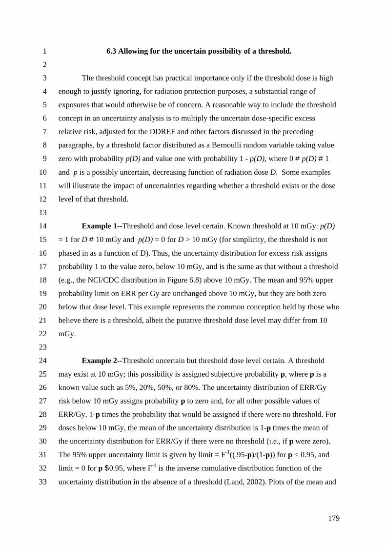
6.3 Allowing for the uncertain possibility of a threshold. 1
2
3
4
5
6
7
8
9
10
11
12
13
14
15
16
17
18
19
20
21
22
23
24
25
26
27
28
29
30
31
32
33
The threshold concept has practical importance only if the threshold dose is high
enough to justify ignoring, for radiation protection purposes, a substantial range of
exposures that would otherwise be of concern. A reasonable way to include the threshold
concept in an uncertainty analysis is to multiply the uncertain dose-specific excess
relative risk, adjusted for the DDREF and other factors discussed in the preceding
paragraphs, by a threshold factor distributed as a Bernoulli random variable taking value
zero with probability p(D) and value one with probability 1 - p(D), where 0 # p(D) # 1
and p is a possibly uncertain, decreasing function of radiation dose D. Some examples
will illustrate the impact of uncertainties regarding whether a threshold exists or the dose
level of that threshold.
Example 1--Threshold and dose level certain. Known threshold at 10 mGy: p(D)
= 1 for D # 10 mGy and p(D) = 0 for D > 10 mGy (for simplicity, the threshold is not
phased in as a function of D). Thus, the uncertainty distribution for excess risk assigns
probability 1 to the value zero, below 10 mGy, and is the same as that without a threshold
(e.g., the NCI/CDC distribution in Figure 6.8) above 10 mGy. The mean and 95% upper
probability limit on ERR per Gy are unchanged above 10 mGy, but they are both zero
below that dose level. This example represents the common conception held by those who
believe there is a threshold, albeit the putative threshold dose level may differ from 10
mGy.
Example 2--Threshold uncertain but threshold dose level certain. A threshold
may exist at 10 mGy; this possibility is assigned subjective probability p, where p is a
known value such as 5%, 20%, 50%, or 80%. The uncertainty distribution of ERR/Gy
risk below 10 mGy assigns probability p to zero and, for all other possible values of
ERR/Gy, 1-p times the probability that would be assigned if there were no threshold. For
doses below 10 mGy, the mean of the uncertainty distribution is 1-p times the mean of
the uncertainty distribution for ERR/Gy if there were no threshold (i.e., if p were zero).
The 95% upper uncertainty limit is given by limit = F-1((.95-p)/(1-p)) for p < 0.95, and
limit = 0 for p $0.95, where F-1 is the inverse cumulative distribution function of the
uncertainty distribution in the absence of a threshold (Land, 2002). Plots of the mean and
179

upper 95% limit, as functions of p, are shown in Figure 6.9 for the approximate
lognormal uncertainty distribution for ERR/Gy according to the NCI/CDC model as
represented in Figure 6.8.
1
2
3
4
5
6
7
8
9
10
11
12
13
14
15
16
17
18
19
20
21
22
23
24
25
26
27
28
29
30
31
32
33
34
This example shows that, when the probability of a dose threshold is uncertain,
the central estimate of the ERR/Gy for low doses decreases linearly with an increasing
probability that there is a threshold -- but the 95% upper limit remains quite high until the
probability of a threshold reaches 80-90%, after which it falls sharply. This indicates that
unless there is consensus agreement that a threshold is very likely, the potential for an
appreciable low-dose risk cannot be ruled out.
Example 3--Threshold certain but its dose level uncertain. A threshold is known
to exist somewhere between 5 and 25 mGy, but otherwise is completely uncertain:
p(D;D0) = 1 for D # D0, and = 0 for D >D0, where D0 is an uncertain (random) quantity
uniformly distributed between 5 and 25 mGy. Estimated ERR/Gy is zero below 5 mGy,
but the probability assigned to non-zero values by the uncertainty distribution for risk at
dose D increases linearly from zero at D= 5 mGy to one (or to the value assigned in the
absence of a threshold) at D = 25 mGy. The uncertainty distribution for ERR/Gy assigns
probability 1 to zero for D below 5 mGy, 100% to the non-threshold distribution for doses
above 25 mGy, and probability (25-D) /20 to zero and probability (D-5)/20 to the non-
threshold uncertainty distribution, for 5<D<25. The mean and upper 95% uncertainty
limit at dose D are as given in Example 2, and shown in Figure 6.9, for p = (25-D)/20.
The third example illustrates an important point: even when one is certain there is
a dose threshold but is still uncertain as to the dose level at which it occurs, the low-dose
ERR/Gy behaves very similarly to the result for Example 2 (which had a fixed threshold
dose but uncertainty as to whether there was a threshold). Specifically, there still is some
probability that the low-dose ERR/Gy is appreciable.
Example 3a—There is a threshold for each individual in a population, but the
dose level varies by individual. Thus, for a randomly chosen individual from the
population, there is a threshold, but its dose level is uncertain. Mathematically, this
example is essentially the same as example 3.
Example 4--Threshold probability very uncertain but its dose level, conditional
on existence of a threshold, is certain. A threshold may exist at 10 mGy, with uncertain
180

1
2
3
4
5
6
7
8
9
10
11
12
13
14
15
16
17
18
19
20
21
22
23
24
25
26
27
28
29
30
31
32
33
34
probability. Enough is known (or there is a consensus among experts, which may be a
compromise) to characterize the subjective uncertainty distribution of p(D), for D < 10
mGy; for example, as
i) uniform between 0 and 1: U(0, 1),
ii) triangular between 0 and 1 with peak at 0: Tr(0, 0, 1),
iii) Tr(0, 0.25 ,1) (peak at p = 0.25),
iv) Tr(0, 0.5 ,1),
v) Tr(0, 0.75 ,1),
vi) Tr(0, 1, 1).
In example 4, the proportion of the uncertainty distribution for ERR/Gy assigned
to zero is randomly distributed over the unit interval, and the mean and upper 95% limit
of the resulting distribution depends on the assumed distribution of p. Figure 6.10 shows
Monte Carlo estimates of the resulting uncertainty distributions for ERR/Gy for the six
cases, again using the NCI/CDC non-threshold distribution from Figure 6.8, and the
corresponding means and upper 95% uncertainty limits.
The probability distributions in Figure 6.10 show, not unexpectedly, that if the
consensus uncertainty distribution of p gives a high weight to the likelihood of a threshold
(e.g., subjective distribution vi), then the distribution of the low-dose ERR/Gy is weighted
toward small values, whereas the opposite is true when the probability of a threshold is
less likely (e.g., subjective distributions ii or iii). Nevertheless, even for distribution vi
the mean expected low-dose ERR/Gy of 5.7% is about a third as great as under the LNT
hypothesis (ERR/Gy = 17%), and it is between 40% and 70% as the LNT value for
distributions i – v.
Example 5 – Dose-dependent uncertain probability of a threshold. Suppose that
the uncertainty distribution for a threshold at 10 mGy corresponds to Example 4,
distribution ii, i.e., Tr(0,0,1), and that the uncertainty distributions for thresholds at 1
mGy and at 0.1 mGy correspond to Example 4 distributions iv (Tr(0, 0.5, 1)) and vi (Tr(0,
1, 1)), respectively. Then the subjective means and upper uncertainty limits for ERR/Gy
would be 11.5% and 27%, respectively, at 10 mGy, 8.6% and 21% at 1 mGy, and 5.7%
and 17% at 0.1 mGy. The corresponding values of ERR would be 0.115% and 0.27% at
10 mGy, 0.0086% and 0.021% at 1 mGy, and 0.00057% and 0.0017% at 0.1 mGy. The
mean and 95% upper limit for ERR at 0.1 mGy can be compared with the mean 0.0017%
and upper limit 0.0036% according to the LNT hypothesis.
181

1 2
3
4
5
6
7
8
9
10
11
12
13
14
15
16
17
18
19
20
21
22
23
24
25
26
27
28
29
30
31
32
33
34
Of the five examples above, Example 5 probably best reflects our present state of
knowledge about low dose risk, namely, that we are uncertain about the likelihood of a
dose threshold, and that in addition, if there should be a dose threshold, we are uncertain
about at what dose level it would be. However, as a counter to an agnostic viewpoint, it
should be noted that the mechanistic and experimental data discussed in this monograph
tend to give weight to a nonthreshold model, as do the solid tumor data in the Japanese
atomic bomb study. (In addition to apparent linearity of dose response down to doses
below 100 mGy, an analysis by Pierce and Preston (2000) found that a threshold above 60
mGy would be statistically inconsistent with LSS dose-response data for all solid cancers
combined.)
6.4 Conclusions
Information on radiation-related cancer risk is needed (1) as guidance for radiation
protection efforts, (2) as a basis for informed consent by persons who may be asked to
accept a certain level of exposure in the interests of medical research, economic progress,
or some other social good, (3) for adjudication of claims and disputes concerning cases of
disease possibly related to past radiation exposure, and (4) for risk-benefit analyses of
public policy initiatives related to radiation. As mentioned previously in this report, these
issues are essentially political, in the sense that different people have different interests
and points of view which must be taken into consideration when policies are developed.
Moreover, implementation of such policies inevitably involves accommodation and
consensus, and it is important that the policies are seen to be derived fairly and openly, on
the basis of facts and assumptions that are wholly accessible to those affected by
implementation.
Information useful for these purposes includes central estimates of dose-specific
risk, but also, lower and (especially) upper probability bounds on risk. Probability bounds
can reflect both statistical uncertainty, estimated by fitting a mathematical model to
observational data, and subjective uncertainty that may take into account model
assumptions that are necessary to calculate estimates but are themselves uncertain.
Probability bounds provide a level of transparency substantially beyond that provided by
a point estimate, such as the expected (mean) value of the uncertainty distribution for
estimated excess risk. A lower probability bound (e.g., a 95% lower confidence limit or
182

1
2
3
4
5
6
7
8
9
10
11
12
13
14
15
16
17
18
19
20
21
22
23
24
25
26
27
28
29
30
31
32
33
uncertainty limit) greater than zero is evidence that there really is an excess risk;
however, the carcinogenicity of ionizing radiation exposure is already well established. A
lower bound corresponding to a risk that is intolerably high would, of course, be evidence
in support of diversion of financial resources for exposure reduction, even from the
viewpoint of those who would bear the expense.
From the viewpoint of those who would bear the risk, if any, associated with
exposure, and of those responsible for their protection, the questions of interest concern
(1) the extent to which risks associated with a given level of exposure are low enough to
be tolerated in view of competing risk and loss of benefits associated with avoidance of
that exposure, and (2) whether we can conclude that there is no risk at all associated with
a given exposure. An upper probability bound, if less than some “tolerable” level of risk,
can be used to help justify a favorable risk-benefit assessment for a particular exposure,
and can provide a margin of safety in decisions regarding risk protection or informed
consent related to possible hazards of radiation exposure. An upper probability bound of
zero or less would be evidence in favor of a threshold or, more likely, a beneficial effect
of low-dose radiation.
The implications of a possible, but uncertain, low-dose threshold for radiation
protection are summarized by the dependence of the mean value and the upper 95%
probability limit on the presumed threshold probability value (Figure 6.9) or on the
uncertainty distribution for that probability (Figure 6.10). The mean value of estimated
ERR/Gy is proportional to 1-p for known threshold probability p and proportional to 1-
E(p) for an uncertain threshold probability p with expected value E(p). Thus, the effect
on the mean value is the same as that of an assumed constant DDREF equal to 1/p or
1/E(p). The effect on the upper 95% probability limit is less drastic, unless the assumed
probability of a threshold is high. As shown in Figure 6.9, the upper limit decreases with
increasing p, but the not nearly as steeply as for the mean until p approaches the
probability level of the upper limit, e.g., about 0.85 in the case of a 95% limit.
Obviously, the lower 95% limit (the 5th percentile of the distribution) is zero for p ≥ 0.05.
As mentioned earlier in this chapter, an established, universal or near-universal,
low-dose threshold for radiation-related cancer risk would obviate concern about risks
from exposures at doses lower than the threshold value. Our present information,
summarized in NCRP Report 136 (2001) and the present report, offers little support for
the existence of a universal low-dose threshold, but it cannot be ruled out as an uncertain
183

1
2
3
4
5
6
7
8
9
10
11
12
possibility. However, the implications of the uncertain possibility of a threshold are
qualitatively not much different from those of an uncertain DDREF: central values and
upper uncertainty limits are reduced somewhat, but they do not become zero. Moreover,
the argument that radiation protection standards should be relaxed “because it is possible
that there may not be any risk at low doses” is unlikely to be persuasive to persons who
are concerned about the possibility that risk associated with very low doses may be
unacceptably high, and it may undermine the more realistic argument that the risk, which
is understood rather well compared to that associated with other common carcinogens, is
almost certainly less than some stated value which may be considered tolerable, for
various reasons such as economic benefits or consideration of risks associated with
alternative strategies involving less exposure.
184

6.6 References 1
2
3
4
5
6
7
8
9
10
11
12
13
14
15
16
17
18
19
20
21
22
23
24
25
26
27
28
29
30
31
32
33
Boice JD Jr, Engholm G, Kleinerman RA, Blettner M, Stovall M, Lisco H, Moloney WC,
Austin DF, Bosch A, Cookfair DL, et al. Radiation dose and second cancer risk in
patients treated for cancer of the cervix. Radiat Res. 1988 Oct;116(1):3-55.
Carr ZA, Kleinerman RA, Weinstock R, Stovall M, Greim ML, Land CE. Malignant
neoplasms after radiation therapy for peptic ulcer. Radiation Research 2002;
157:668-677.
CIRRPC (1988), Committee on Interagency Radiation Research and Policy Coordination,
Science Panel Report No. 6. Use of probability of causation by the Veterans
Administration in the Adjudication of Claims of injury due to exposure to ionizing
radiation. Committee on Interagency Radiation Research and Policy Coordination,
Office of Science and Technology Policy, Executive Office of the President,
Washington, D.C., August 1988 (ORAU 88/F-4).
EPA (1999) Environmental Protection Agency. Estimating Radiogenic Cancer Risks.
EPA Report 402-R-00-003, Washington, D.C., May 1999.
Gilbert RO, Mart EI, Denham DH, Strenge DL, Miley TB. Uncertainty of historical
measurements of 131I in Hanford-area vegetation. Health Phys. 1996
Feb;70(2):160-70.
Grogan HA, Sinclair WK, Voilleque PG. Assessing Risks from Exposure to Plutonium.
Final Report. Part of Task 3: Independent Analysis of Exposure, Dose and
Health Risk to Offsite Individuals. Radiological Assessment Corporation (RAC)
Report no. 5, Revision 2, February, 2000.
IAEA (1989) International Atomic Energy Agency, Safety Series 100. Evaluating the
Reliability of Predictions Made Using Environmental Transfer Models. IAEA,
Vienna, 1989, 106 pp.
185

1
2
3
4
5
6
7
8
9
10
11
12
13
14
15
16
17
18
19
20
21
22
23
24
25
26
27
28
29
30
31
32
33
34
ICRP (1991) International Commission on Radiological Protection. ICRP Publication 60.
1990 Recommendations of the International Commission on Radiological
Protection. Annals of the ICRP 1991; 21, No. 1-3:201 pp.
Inskip PD, Monson RR, Wagoner JK, Stovall M, Davis FG, Kleinerman RA, Boice JD Jr.
Cancer mortality following radium treatment for uterine bleeding. Radiat Res.
1990; 123(3):331-44.
Land CE, Boice JD Jr, Shore RE, Norman JE, Tokunaga M. Breast cancer risk from low-
dose exposures to ionizing radiation: results of parallel analysis of three exposed
populations of women. J Natl Cancer Inst 1980 Aug;65(2):353-76
Land C. Uncertainty, low-dose extrapolation and the threshold hypothesis. J. Radiol. Prot.
2002; 22:A129-A135.
Little MP, Boice JD Jr. Comparison of breast cancer incidence in the Massachusetts
tuberculosis fluoroscopy cohort and in the Japanese atomic bomb survivors.
Radiat Res 1999 Feb;151(2):218-24
NCI/CDC 2003. Land CE, Gilbert E, Smith J, Hoffman FO, Apostoiae I, Thomas B,
Kocher DM. Report of the NCI-CDC Working Group to revise the 1985 NIH
Radioepidemiological Tables. U.S. Dept. of Health and Human Services, National
Institutes of Health, National Cancer Institute, Bethesda, Maryland. NIH
Publication No. 03-5387, 118 pp.
NCRP (1996) National Council on Radiation Protection and Measurements. A guide for
uncertainty analysis in dose and risk assessments related to environmental
contamination. NCRP Commentary No. 14. National Council on Radiation
Protection and Measurements, Bethesda, MD, 1996.
NCRP (1997) National Council on Radiation Protection and Measurements. Uncertainties
in fatal cancer risk estimates used in radiation protection. NCRP Report No. 126.
National Council on Radiation Protection and Measurements, Bethesda, MD,
1997.
186
1
2
3
4
5
6
7
8
9
10
11
12
13
14
15
16
17
18
19
20
21
22
23
24
25
NCRP (2001a) National Council on Radiation Protection and Measurements. Evaluation
of the linear-nonthreshold dose-response model for ionizing radiation. NCRP
Report No. 136. National Council on Radiation Protection and Measurements,
Bethesda, MD, 2001.
NIH (1985) National Institutes of Health. Rall JE, Beebe GW, Hoel DG, Jablon S, Land
CE, Nygaard OF, Upton AC, Yalow RS, Zeve VH. Report of the National
Institutes of Health Ad Hoc Working Group to Develop Radioepidemiological
Tables. National Institutes of Health, Bethesda, Maryland, 1985;355 pp.
Parkin DM, Whelan SL, Ferlay J, Teppo L, Thomas DB, eds (2002) Cancer Incidence in
Five Continents, Vol VIII (IARC Scientific Publications No. 155), Lyon, IARC.
Pierce DA, Preston DL, Stram DO, Vaeth M. Allowing for dose-estimation errors for the
A-bomb survivor data. J Radiat Res (Tokyo). 1991 Mar;32 Suppl:108-21.
Pisani P, Parkin DM, Ferlay J. Estimates of worldwide mortality from twenty-five major
cancers in 1990. Int J Cancer 1999 Sep 24;83(1):18-29. Erratum in: Int J Cancer
1999 Dec 10;83(6):870-3.
Preston DL, Mattsson A, Holmberg E, Shore R, Hildreth NG, Boice JD. Radiation effects
on breast cancer risk: a pooled analysis of eight cohorts. Radiat Res 2002
Aug;158(2):220-35.
SEER web site: http://seer.cancer.gov/faststats/html/dev_all.html26
27
28
29
30
31
32
33
Sinclair WK. Science, Radiation Protection and the NCRP: Lauriston Taylor Lecture No.
17. In: LA Sagan (ed.), Proceedings of the 29th Annual Meeting of the National
Council on Radiation Protection and Measurements, Proceedings No. 15,
Radiation Science and Societal Decision Making. NCRP, Bethesda, Md. pp201-
246, 1994.
187

1
2
3
4
5
6
7
8
9
10
11
12
13
14
15
16
17
18
19
20
21
22
23
24
25
Sposto R, Preston DL, Shimizu Y, Mabuchi K. The effect of diagnostic misclassification
on non-cancer and cancer mortality dose response in A-bomb survivors.
Biometrics. 1992 Jun;48(2):605-17.
Thompson DE, Mabuchi K, Ron E, Soda M, Tokunaga M, Ochikubo S, Sugimoto S,
Ikeda T, Terasaki M, Izumi S, Preston DL. Cancer incidence in atomic bomb
survivors. Part II: Solid tumors, 1958-1987. Radiat Res. 1994; 137:S17-S67.
UNSCEAR 1993. United Nations Scientific Committee on the Effects of Atomic
Radiation. Sources, Effects and Risks of Ionizing Radiation, No. E.94.IX.2,
United Nations, New York, 1993.
U.S. Nuclear Regulatory Commission. Reactor Safety Study, An Assessment of the
Accident Risks in U.S. Commercial Nuclear Power Plants, WASH-1400
(NUREG-75/014), Washington, DC, 1975
U.S. Nuclear Regulatory Commission. Severe Accident Risks: An Assessment for Five
U.S. Nuclear Power Plants. (NUREG-1150). Division of Systems Research, U.S.
Nuclear Regulatory Commission, Washington, DC, 1990
Warren-Hicks WJ, Moore DRJ. (1998). (eds) Uncertainty analysis in ecological risk
assessment. Society of Environmental Toxicology and Chemistry (SETAC),
Pensacola, Florida. 314 pp.
188

7. CONCLUSIONS 1
2
3
4
5
6
7
8
9
10
11
12
13
14
15
16
17
18
19
20
21
22
23
24
25
26
27
28
29
30
31
32
33
34
1. Epidemiological studies of cancer risk following radiation exposure provide the
primary basis for estimation of radiation-related risk in human populations. These
studies demonstrate the existence of dose response and its modification by other
factors, and show some variation by cancer site and by histological subtypes
within sites. At low and very low radiation doses, statistical and other variation in
baseline risk tends to be the dominant source of error in both epidemiological and
experimental carcinogenesis studies, and estimates of radiation-related risk tend to
be highly uncertain both because of a weak signal-to-noise ratio and because it is
difficult to recognize or to control for subtle confounding factors. Thus,
extrapolation of risk estimates based on observations at moderate to high doses
continues to be the primary basis for estimation of radiation-related risk at low
doses and dose rates.
2. There is no direct evidence, from either epidemiological or experimental
carcinogenesis studies, that radiation exposure at doses on the order of 1 mGy or
less is carcinogenic, nor would any be expected because of the considerations
outlined in Conclusion 1. There is, however, epidemiological evidence, unlikely
on the wholel to be an artifact of random variation, linking increased cancer risk
to exposures at doses on the order of 10 mGy. This evidence includes several
case-control studies of leukemia and solid cancers among different populations of
children exposed in utero to x-ray pelvimetry, cohort studies of breast cancer
among women given multiple fluoroscopy examinations during treatment for
tuberculosis or scoliosis, with average breast doses on the order of 10 mGy per
examination, and the observation that risk of mortality and morbidity among
atomic bomb survivors from all solid cancers combined is linear in radiation dose
down to about 100 mGy.
3. Overall, relevant animal tumor data from experimental carcinogenesis studies tend
to support a dose response that, at low doses, is linear with no threshold. This
inference does not conflict with experimental evidence for reductions in excess
risk per unit dose at low doses or with fractionation and/or protraction of dose.
Recent cytogenetic and molecular studies provide direct support for the view that
189

the critical radiation-associated events in the tumorigenic process are
predominantly early events involving DNA losses targeting specific genomic
regions harboring critical genes. The predominant importance of DNA DSB
induction and post-irradiation error-prone NHEJ repair for the induction of
aberrations, and the apparently critical role for radiation-induced aberrations in the
pathogenesis of cancer in these experimental models, argue against the
proposition of a low dose threshold in the dose response.
1
2
3
4
5
6
7
8
9
10
11
12
13
14
15
16
17
18
19
20
21
22
23
24
25
26
27
28
29
30
31
32
33
34
4. There is evidence from both epidemiological and experimental studies that
specific tissues and cancer sites may for various reasons vary from the general
rule articulated in Conclusions 1-3, in the sense that radiation carcinogenesis is
markedly and disproportionately less likely to occur at low doses than at high, and
may even suggest a threshold. Examples are the small intestine, bone, and skin.
However, these appear to be the exception rather than the general rule;
experimental studies of radiation-related life shortening, which represent the
integrated dose responses for a variety of tumor types, suggest linear dose
responses over a wide range of doses.
5. Ionizing radiation is able to produce a unique type of damage in which multiple
lesions are encountered within close spatial proximity. Even a single track through
a cell is likely to induce these unique clustered damages. This type of damage may
not be generated frequently endogenously or by other exogenous agents, and thus,
there may not have been a strong selective pressure driving efficient repair.
Although cells have a vast array of damage response mechanism that facilitate the
repair of DNA damage and the removal of damaged cells, these mechanisms are
not fool proof. Moreover, clustered radiation–induced lesions pose a particular
problem and current emerging evidence suggests that closely spaced lesions can
compromise the repair machinery. On this basis, there is not any strong evidence
for a radiation dose below which all radiation-induced damage can be repaired
with fidelity.
6. Although many of the cells containing such radiation-induced damage may be
eliminated by damage response pathways involving cell cycle checkpoint control
and apoptotic pathways, it is clear from analysis of cytogenetics and mutagenesis
190

that damaged or altered cells are capable of escaping these pathways and
propagating. This further argues against the likely possibility of a threshold for
radiation-induced cellular effects.
1
2
3
4
5
6
7
8
9
10
11
12
13
14
15
16
17
18
19
20
21
22
23
24
25
26
27
28
29
30
31
32
33
34
7. The processing and misrepair of radiation-induced DSBs, particularly complex
forms, are responsible for chromosome/gene alterations that manifest as
chromosome aberrations and mutations. Current understanding of mechanisms
and quantitative data on dose and time-dose relationships support a linear dose
response at low doses with no compelling evidence for the existence of a
threshold dose below which there would be no effect.
8. When considered as a whole, the emerging results with regard to radiation-related
adaptive response, genomic instability, and bystander effects suggest that the risk
of low level exposure to ionizing radiation is uncertain, and a simple extrapolation
from high dose effects may not be wholly justified in all instances. However, a
better understanding of the mechanisms for these phenomena, the extent to which
they are active in vivo, and how they are interrelated is needed before they can be
evaluated as factors to be included in the estimation of potential risk to the human
population of exposure to low levels of ionizing radiation.
9. Probability limits on risk provide additional information relevant to radiation
protection. In particular, a high lower limit attests to the reality of danger
associated with a given exposure, and a low upper limit provides assurance as to
the relative safety, and presumably the acceptability, of the exposure when seen in
the context of other hazards of daily life. The information reviewed in this report,
from epidemiology and from experimental studies of animal, cellular, and
molecular models, is consistent with proportionality between radiation-related
cancer risk at low doses and at low dose rates, including the dose delivered by a
single photon. It is also consistent, given uncertainties about the roles played by
repair and apoptosis at very low doses, with the existence of a dose threshold at a
dose level so low that radiation-related risk under the linear, no-threshold (LNT)
hypothesis would be statistically indistinguishable from random variation in
baseline risk. However, the uncertain possibility of a threshold does not drastically
reduce either central estimates or upper probability limits for low-dose risk
191

compared to those obtained using the LNT hypothesis, unless that possibility is
assumed to be very likely. To the contrary, the evidence reviewed in this report
suggests that a universal low-dose threshold above (say) 1 mGy of low-LET
radiation is an unlikely possibility. Thus, the LNT hypothesis remains the most
prudent risk model for guidance of radiation protection.
1
2
3
4
5
6
7
8
9
192

TABLES 1 2 3 4 5
Table 2.1 Some sources and amounts of ionizing radiation exposure (unless noted, from Mettler and Upton (1995)) Exposure Effective dose, in mSv Natural background (world population) Normal background areas High background areas Cosmic rays 0.38 / year 2.0 / year Terrestrial ( rays 0.46 / year 4.3 / year Radionuclides in tissue 0.25 / year 0.6 / year Inhaled radionuclides 2.5 / year Medical diagnostic (U.S. population) Per exam Skull 0.22 Cervical spine 0.20 Chest 0.08 Cholioangiogram 1.89 Lumbar Spine 1.27 Upper gastrointestinal series 2.44 Abdomen (KUB) 0.56 Barium enema 4.06 Intravenous pyelogram 1.58 Pelvis 0.44 Hip 0.83 Extremities 0.01 CT scan, head or body 1.11 Pediatric CT scan, abdomen1 25 (stomach dose) Single screening mammogram1 3 (breast dose) Astronaut, 3-day space shuttle mission2 2 – 3 Astronaut, 60-day space station mission2 50 Average cumulative occupational dose in monitored radiation workers3
Cumulative reported badge dose 20
Average neutron-weighted colon dose for LSS population with doses between 0.005 and 4 Gy 4
Colon dose 200
1 Brenner et al, 2003; 2 NCRP Report 138, 2001; 3 Gilbert, 2001; 4 Preston, 2003; computed using data set downloaded from RERF web site (Radiation Effects Research Foundation, 2003)
193

Table 2.2. Parameter estimates corresponding to the general dose-response model, ERR(D) = "D × (1 + $D) × exp(-(D),
where D is neutron-weighted (weight = 10), reconstructed radiation dose to the colon from the atomic bombings and ERR(D) is the dose-related excess relative risk of solid cancer morbidity, 1958-87, among members of the Radiation Effects Research Foundation’s Life Span Study cohort of survivors of the bombings.
Parameter Estimate 90% CI p-value
α β γ
0.52 0.94 0.84
0.16, 0.83 0*, 6.8 0*, 0.68
.02
.28
.07
α β γ
0.71 0§
0.11
0.56, 0.87 ----
0*, 0.24
<.001 ---- .07
α β γ
0.57 0* 0§
0.48, 0.68 ---- ----
<.001 ---- ----
Analysis restricted to survivors with estimated doses 2 Sv and less
α β γ
0.40 0.92 0.53
0*, 0.85 0*, 3.0 0*, 1.3
.24 >.5 >.5
α β γ
0.61 0.045
0§
0.35, 0.76 0*, 0.68
----
<.001 >.5
α β γ
0.64 0§
0*, 0§
0.54, 0.74 ---- ----
<.001
* Estimate constrained to be $0. §Estimate set = 0.
194

1 2 3 4 5
Table 2.3 Modification of radiation-related risk by individual and lifestyle factors, and by other exposures. Organ site / cancer
Population Factor Main factor effect on risk
Interaction with radiation exposure
References
Female breast
LSS cohort Young age at 1st full-term pregnancy
Decreased Multiplicative1
Land et al, 1994
“ “ Multiple births Decreased Multiplicative1 “ “ “ Lengthy lactation history Decreased Multiplicative1 “ “ NY mastitis series Assoc with 1st delivery Increased Not tested Shore,
1980 “ Massachusetts TB
fluoroscopy series Exposed yr of 1st delivery
Increased (NS)
Not tested Boice & Stone, 1978
Lung& bronchus
LSS cohort Smoking history Increased Additive2 Pierce, 2003
“ U.S. Uranium miners
“ “ NS closer to mult. than to additive
Lubin & Steindorf
Basal cell skin ca.
LSS cohort Sun-exposed cf. covered areas of skin
Additive2 Ron, 1998
“ NY tinea capitis series
White cf. Black patients Higher in whites
Multiplicative1 Shore, 2002
Liver LSS cohort Hepatitis C infection Increased Strongly synergistic
Sharp, 2002
Female breast
LSS cf. Euro/American pops
Population rates Japan rate 4-fold < US
Additive2 Preston, 2002
Stomach LSS cf. US peptic ulcer patients
Population rates Japan rate 12-fold > US rate
NS, closer to multiplicative than to additive
Carr, 2002
1 Additive interaction model rejected (statistically inconsistent with data); 2 Multiplicative interaction model rejected
6 7
195

1 2 3 4 5
Table 2.4. Statistical power calculations for a hypothetical study in which baseline cancer risk is known to be 10%, and the unknown radiation-related excess risk is 10% at 1 Gy and proportional to dose between 0 and 1 Gy.
Radiation dose
Excess risk
Total risk
Standard deviation of the estimated excess risk under the null and alternative hypotheses
Population size N needed for 80% power to detect the excess risk at the 5% significance level
1 Gy 10% 20% 0.316 / N1/2 0.447 / N1/2 124
100 mGy 1% 11% 0.316 / N1/2 0.332 / N1/2 6,800
10 mGy 0.1% 10.1% 0.316 / N1/2 0.318 / N1/2 624,000
1 mGy 0.01% 10.01% 0.316 / N1/2 0.316 / N1/2 61.9 million6 7 8 9
10 11
Table 2.5. Distribution of subjects, solid cancers, and estimated radiation-associated, excess solid cancers among 79,901 exposed members of the Life Span Study cohort of Hiroshima-Nagasaki atomic bomb survivors (Pearce and Preston, 2000). Estimated colon dose
Number of subjects
Number of solid cancers
Estimated number of radiation-associated excess cancers*
Exposed beyond 3000 m <5 mGy, exposed within 3000 m 5-100 mGy 100-200 mGy 200-500 mGy 0.5-1 Gy 1-2 Gy >2 Gy
23,49310,15930,5244,7755,8623,0481,570
470
3,2301,3014,119
739982582376126
01
7760
16417716580
12 13 14 15 16 17 18 19 20 21 22 23
* Fitted values, linear dose response
196

FIGURES
0 1 2 3 4 5-0.5
0.0
0.5
1.0
1.5
2.0
2.5
3.0
3.5
4.0
4.5
5.0
5.5
Neutron-Weighted Radiation Dose (Gy)
Exce
ss R
elat
ive
Ris
k (9
0% C
onfid
ence
Lim
its)
Fitted modelsfor trend tests:
Loglinear
Linear
1 2 3 4 5 6 7 8
Figure 2.1. Dose-specific excess relative risk of solid cancer among atomic bomb survivors, 1958-87, by interval of neutron-weighted, estimated radiation dose to the colon. Fitted dose-response functions correspond to statistical tests of increasing trend according to the linear (RR = 1 + αD) and log-linear (RR = exp(βD)) dose-response models. The baseline risk is adjusted for city of exposure (Hiroshima or Nagasaki), sex, and 5-year intervals of exposure age and age at observation for risk, using a saturated model.
197

1111111111
0 1 2 3 4 5
0.0
0.5
1.0
1.5
2.0
2.5
3.0
Exce
ss R
elat
ive
Ris
k
Neutron-Weighted Radiation Dose (Gy)
Linear fit o
ver 0-2 Gy
General model fit over 0-5 Gy
Figure 2.2. General dose-response model, ERR(D) = αD × (1 + βD) × exp(-γD - δD2), fit to the dose-response data of Figure 1, and linear dose-response model, ERR(D) = αD, fit to the data subset restricted to radiation doses between zero and 2 Sv. Details of the parameter estimates are given in Table 2.2.
1 2 3 4 5 6 7 8 9 0 1 2 3 4 5 6 7 8 9
198

0.0 0.5 1.0 1.5 2.0
1.0
1.5
2.0
2.5
0.0 0.1 0.2 0.3 0.4 0.5
1.0
1.1
1.2
1.3
1.4
1.5
Gamma Dose Equivalent
Figure 2.3. Estimated low-dose relative risks. Dose-specific cancer rates over the 1958-94 Follow-up period relative to those for an otherwise similar exposed person, averaged over the follow-up and over sex, and for age 30 at exposure. The dashed curves represent 1 standard error limits for the smoothed curve. The straight line is the estimated linear dose response for 0-2 Sv (see inset). The unity baseline corresponds to zero-dose survivors exposed within 3 km of the bombs. The horizontal dotted line represents the alternative baseline if survivors exposed beyond 3 km had been included. (Pierce and Preston, Radiation Res. 2000, 154:178-186.)
1 2 3 4 5 6 7 8
199

0.0 0.5 1.0 1.5 2.
-0.5
0.0
0.5
1.0
1.5
2.0
0
ER
R p
er S
v
Linear Regression Range to this Dose, in Gy
1 2 3 4 5 6 7 8 9
10 11 12 13 14 15 16 17 18 19 20 21 22 23 24 25 26
Figure 2.4. Linear regression estimates of the ERR per Gy (points and connecting line, with error bounds of ± one SE) for solid cancer incidence, based on Poisson regression over dose intervals of differing ranges from zero to the horizontal coordinate of the plotted point. The analysis is limited to proximal survivors exposed at distances under 3 km.
200

1
0.0 0.5 1.0 1.5 2.0
-0.5
0.0
0.5
1.0
1.5
2.0E
RR
per
Sv
Linear Regression Range to this Dose, in Gy
2 3 4 5 6 7 8
Figure 2.5. Linear regression estimates of the ERR per Gy (points and connecting line, with error bounds of ± one SE) for solid cancer incidence, based on Poisson regression over dose intervals of differing ranges from zero to the horizontal coordinate of the plotted point. The analysis is based on all exposed survivors with estimated radiation doses less than 2 Gy.
201

0 1 2 3 4 5
-1.0
-0.5
0.0
0.5
1.0
1.5Es
timat
ed E
RR
per
Gy
(90%
Con
fiden
ce L
imits
)
Mean of Highest Dose Interval (Gy)
1 2 3 4 5 6 7 8 9
10 11 12 13 14 15 16 17 18 19 20 21 22 23 24
Figure 2.6. All-age linear regression estimates of ERR per Gy for female breast cancer assuming a 12-year minimum latent period, with dose-specific data trimmed from the right. Horizontal placement corresponds to the mean breast tissue dose for the highest neutron-weighted kerma interval included in the regression. Thus, the right-most point corresponds to the full dose range, the next point to the left to doses under 4 Gy, the next to doses under 3 Gy, and so on.
202

203
1 2 3
FIGURE 3.1
XRCC4
Ku70Ku80
Ku70Ku80
DNA-PKcsArt
2) DNA-PKcs is recruited and the kinaseactivity activated. Autophosphorylation and phosphorylation of artemis likely ensues, potentially leading to release of DNA-PKcsArtemis nuclease activity may enhance processing of ends.
1) binding of Ku to the double stranded DNA end. The crystal structure of Ku shows that the DNA passes through a cavity in the structure with Ku encircling the DNA (a single DNA end is shown for simplicity)
Ku70Ku80
Ligase IV 3) Ku enhances the recruitment of DNA ligaseIV/XRCC4 complex and Ku translocatesinwards to allow ligase IV/XRCC4 access to the DNA ends.
Fig. 3.1. Model for DNA NHEJ.
Proposed steps involved in the process
Note that only a single DNA end is shown for simplicity - one function of DNA-PKcs may be to enhance synapsis of the DN A ends.

Figure 3.2. Depiction of homologous recombination. 1 2 3 4 5 6 7
3’
3’
non-crossing over crossing over
a) Resection possibly carried out by the MRN complex.b) binding of RPA ( ) to 3’ ssregions
c) single strand invasion catalysed by Rad51. Rad51 ( ) is delivered to the single stranded regions via a process likely involving BRCA2. Rad51 replaces RPA on the 3’ ss DNA overhands and promotes strand invasion. This is facilitated by Rad51 paralogues and Rad54.
204

1 2 3 4 5 6 7 8 9
10 11 12 13 14 15 16
17
19 20
21 22 23 24 25 26 27 28
Mean = 32.99
Figure 6.1. Lognormal distribution with 90% confidence limits 18%-43%, representing statistical uncertainty about percent cancer excess relative risk per Gy.
18
11.87 29.55 47.22 64.89 82.56
A1
Mean = 0.85
0.56 0.70 0.85 0.99 1.13
A2Frequency Chart
.000
.004
.008
.012
.016
0
387.7
775.5
1551
7.43 19.37 31.31 43.24 55.18
100,000 Trials 1,481 Outliers
Forecast: a1*a2
Figure 6.2. Normal uncertainty distribution for dosimetry bias correction factor, with mean 0.84 and 90% uncertainty limits 0.69-1.00.
Figure 6.3. Approximately lognormal uncertainty distribution for ERR per Gycorrected for dosimetry bias, with mean 0.26 and uncertainty limits 0.15 – 0.46.
205
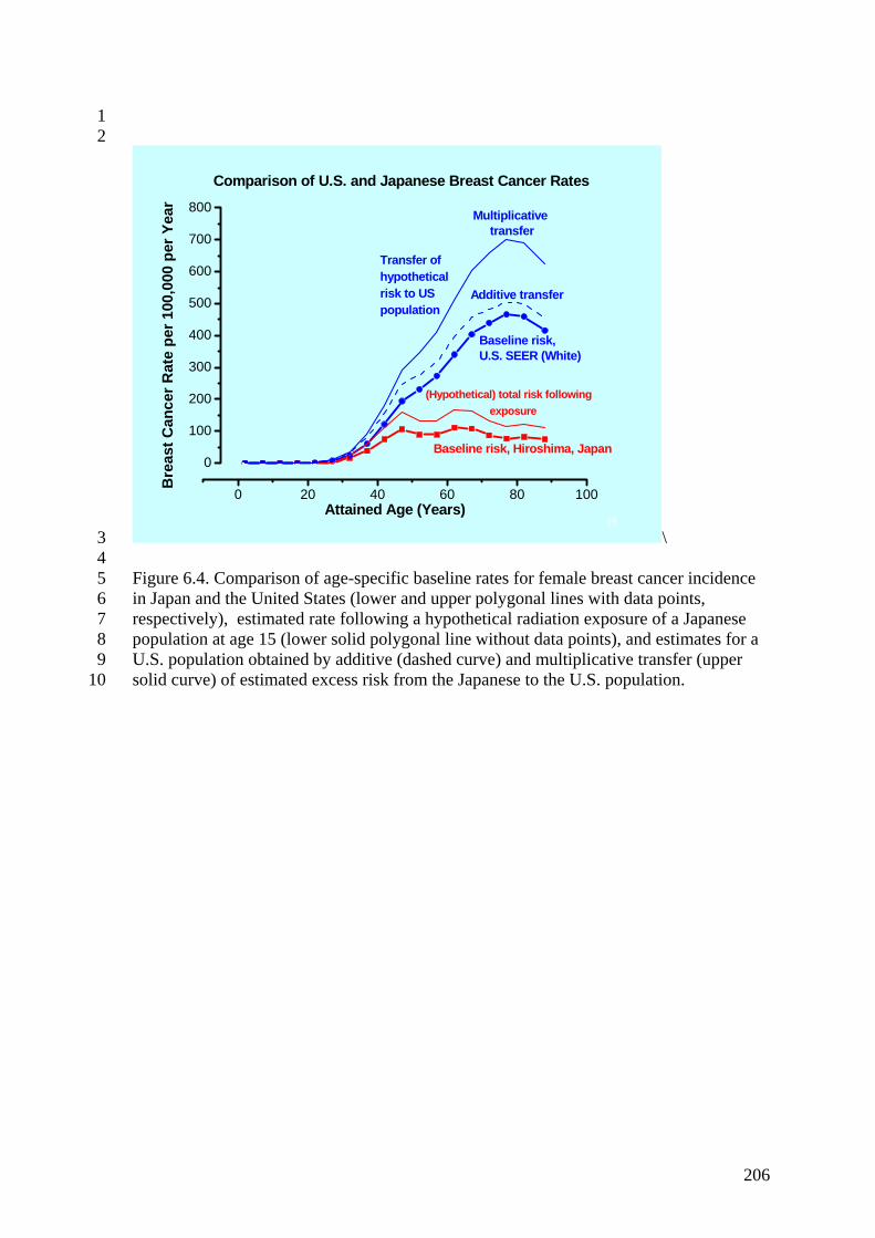
1 2
19
0 20 40 60 80 100
0
100
200
300
400
500
600
700
800
Additive transfer
Multiplicative transfer
Comparison of U.S. and Japanese Breast Cancer Rates
Baseline risk, Hiroshima, Japan
(Hypothetical) total risk following exposure
Baseline risk, U.S. SEER (White)
Bre
ast C
ance
r Rat
e pe
r 100
,000
per
Yea
r
Attained Age (Years)
Transfer of hypothetical risk to US population
3 4 5 6 7 8 9
10
\ Figure 6.4. Comparison of age-specific baseline rates for female breast cancer incidence in Japan and the United States (lower and upper polygonal lines with data points, respectively), estimated rate following a hypothetical radiation exposure of a Japanese population at age 15 (lower solid polygonal line without data points), and estimates for a U.S. population obtained by additive (dashed curve) and multiplicative transfer (upper solid curve) of estimated excess risk from the Japanese to the U.S. population.
206

1 2 3 4 5 6 7 8 9
10 11 12 13 14 15 16 17 18 19 20 21
Frequency Chart
.000
.004
.008
.012
.016
0
395.2
790.5
1581
4.83 15.84 26.84 37.85 48.86
100,000 Trials 1,627 Outliers
Forecast: c2*(1-.2308*a3)
Figure 6.5. Monte Carlo simulation of the uncertainty distribution for cancer ERR (in percent) at 1 Gy, after transfer to a U.S. population: the simulated distribution is approximately lognormal with mean 0.25 and 90% probability limits 0.13-0.41.
DDREF for solid tumors other than breast and thyroid
0.010.04
0.20
0.30 0.30
0.10
0.040.01
0.00
0.05
0.10
0.15
0.20
0.25
0.30
0.35
0.40
0.5 0.7 1.0 1.5 2.0 3.0 4.0 5.0
DDREF
prob
abili
ty
0.00
0.05
0.10
0.15
0.20
0.25
0.30
0.35
0.40
22 23 24 25 26 27 28 29 30 31 32 33 34
Figure 6.6. Continuous subjective uncertainty Figure 6.7. Discrete uncertainty distributions for DDREF used in recent analyses. Distribution for DDREF used in
NCI/CDC (2003) analysis.
207

1 2 3 4 5 6 7 8 9
10 11 12 13 14 15 16 17 18 19 20 21 22 23 24 25 26 27 28
Figure 6.8. Influence of DDREF assumptions on uncertainty for ERR/Gy (in percent). DDREF model Uncertainty. for
Frequency Chart
.000
.008
.017
.025
.033
0
832
3328
0.00 12.50 25.00 37.50 50.00
100,000 Trials 4 Outliers
Forecast: a1/(a2+a3)*2
(Source) ERR/Gy: Mean 95% limit
EPA (1999) 12% 20% Frequency Chart
.000
.008
.015
.023
.031
0
770.2
3081
0.00 12.50 25.00 37.50 50.00
100,000 Trials 37 Outliers
Forecast: a1/a4
NCRP (1997) 11% 27% Grogan et al (2000) 12% 28% 0
Frequency Chart
.000
.007
.014
.022
.029
0
719.5
2878
0.00 12.50 25.00 37.50 50.00
100,000 Trials 939 Outliers
Forecast: a1/a5
29 30 31 32 33 34 35 36 37 38 39 40 41 42 43
Frequency Chart
.000
.005
.010
.015
.020
0
500.7
2003
0.00 12.50 25.00 37.50 50.00
100,000 Trials 1,207 Outliers
Forecast: a1/a6
NCI/CDC (2003) 17% 36% ERR/Gy at low doses
208

1
0.0 0.2 0.4 0.6 0.8 1.00
5
10
15
20
25
30
35
40Lo
w-d
ose
ERR
per
Sv,
in p
erce
nt
Assumed low-dose threshold probability p
Upper 95% probability limit
Mean value
Figure 6.9. Mean and upper 95% probability limit for ERR/Gy as functions of threshold probability p, given (in the absence of a threshold) a lognormal uncertainty distribution with mean 0.17 and upper 95% limit 0.36.
209

Figure 6.10. Effect of uncertain threshold probability on the uncertain distribution for low-dose ERR/Gy.
1 2 3 4 5
6 7 8
9 10 11 12 13
15 16 17
Assumed uncertainty distribution for p Resulting distribution for ERR/Gy
Frequency Chart
.000
.006
.012
.018
.024
0
590.2
2361
0.00 7.28 14.56 21.84 29.12
100,000 Trials 2,460 Outliers
Forecast: c4*(1-a6)
Mean = 0.50
0.00 0.25 0.50 0.75 1.00
i) For p ~ U(0,1), the mean ERR/Gy is 8.6% and the 95% upper limit is 23%.
ii) For p ~ T(0,0,1), the mean ERR/Gy is 11.5% and the 95% upper limit is 27%.
14 iii) For p ~ T(0,0.25,1), the mean ERR/Gy is 10.4% and the 95% upper limit is 24%.
Mean = 0.33
0.00 0.25 0.50 0.75 1.00
Frequency Chart
.000
.006
.012
.018
.023
0
585.7
2343
0.51 8.69 16.87 25.06 33.24
100,000 Trials 2,660 Outliers
Forecast: c4*(1-a7)
Frequency Chart
.000
.006
.011
.017
.023
0
572.2
2289
0.33 7.31 14.30 21.28 28.26
100,000 Trials 2,974 Outliers
Forecast: c4*(1-a8)
Mean = 0.42
0.00 0.25 0.50 0.75 1.00
210

1 2 3 4 5 6 7
8 9
10
11 12 13 14
15 16
Figure 6.10 (continued). Effect of uncertain threshold probability on the uncertain distribution for low-dose ERR/Gy. Assumed uncertainty distribution for p Resulting distribution for ERR/Gy
iv) For p ~ T(0,0.5,1), the mean ERR/Gy is 8.6% and the 95% upper limit is 21%.
v) For p ~ T(0,0.75,1), the mean ERR/Gy is 7.2% and the 95% upper limit is 19%.
vi) For p ~ T(0,1,1), the mean ERR/Gy is 5.7% and the 95% upper limit is 17%.
Mean = 0.50
0.00 0.25 0.50 0.75 1.00
Frequency Chart
.000
.006
.012
.018
.024
0
592.2
2369
0.21 6.46 12.71 18.97 25.22
100,000 Trials 2,724 Outliers
Forecast: c4*(1-a9)
Frequency Chart
.000
.007
.014
.020
.027
0
680.5
2722
0.15 6.00 11.85 17.69 23.54
100,000 Trials 2,444 Outliers
Forecast: c4*(1-a10)
Mean = 0.58
0.00 0.25 0.50 0.75 1.00
Mean = 0.67
0.00 0.25 0.50 0.75 1.00
Frequency Chart
.000
.008
.016
.025
.033
0
821
3284
0.00 5.31 10.61 15.92 21.23
100,000 Trials 2,488 Outliers
Forecast: c4*(1-a11)
211

1
212